Unraveling the Night: How Deep Sleep Disruptions and Alzheimer’s Proteins Explain Age-Related Memory Loss

We have all experienced that fleeting moment of cognitive friction: walking out of a grocery store, scanning a crowded asphalt expanse, and realizing the exact location of our parked vehicle has temporarily vanished from our consciousness. While such minor lapses are frequently dismissed as the typical inconveniences of a busy day, they underscore a fundamental human faculty: episodic memory. This cognitive system allows individuals to encode, store, and consciously recall specific personal experiences, anchoring everything from monumental childhood milestones to the mundane logistics of daily life.
However, the neural machinery responsible for preserving these experiences undergoes a progressive decline as human beings age. The vivid sights, distinct sounds, and intricate contextual details of past events become increasingly difficult to retrieve. Although some degree of cognitive slowing is an accepted part of the natural aging trajectory, these memory alterations can become starkly more pronounced—and eventually pathological—in individuals developing neurodegenerative conditions such as Alzheimer’s disease. For decades, the precise physiological mechanisms driving this transition from normal age-related forgetting to clinical pathology remained elusive.
Now, a groundbreaking study published in the prestigious journal Nature Neuroscience by researchers at the University of California, Berkeley, provides critical new insight into the microstructural changes occurring within the aging brain. Led by neuroscientist Omer Sharon under the direction of former Berkeley sleep researcher Matthew Walker, the research team has established a concrete tripartite link: the accumulation of the pathological protein tau in the frontal cortex, the structural breakdown of coordinated brain waves during deep sleep, and a subsequent, measurable deterioration in overnight memory consolidation.
The Anatomy of Memory Consolidation During Deep Sleep
To understand the gravity of these findings, neuroscientists must look closely at the architecture of human sleep. Sleep is not a uniform state of unconsciousness; rather, it is a dynamic cycle composed of distinct stages characterized by divergent patterns of neural activity. Rapid eye movement (REM) sleep, famously associated with vivid dreaming and ocular movement, is vital for emotional processing. However, non-REM (NREM) sleep—particularly its deepest stages—serves as the primary biological workshop for cognitive maintenance and memory archiving.
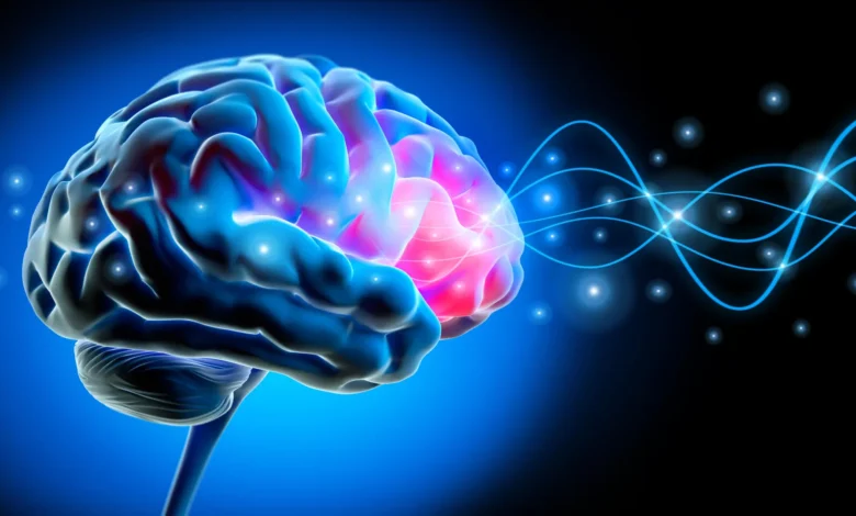
During deep NREM sleep, external sensory input fades, and large populations of neurons engage in a synchronized biochemical dance. Millions of brain cells briefly become electrically inactive before firing back to life in a precisely orchestrated cascade. This synchronized collective shutdown and reactivation generates large, rolling electrical oscillations known as slow waves. Originating predominantly within the frontal cortex—the brain’s seat of executive function, planning, and high-level reasoning—these slow waves ripple across vast expanses of the cerebral cortex, effectively transferring short-term memory traces from temporary holding sites in the hippocampus to permanent, distributed networks in the neocortex.
"Each slow wave reflects an enormous population of neurons switching off and back on together," explained Sharon, a postdoctoral researcher at UC Berkeley’s Center for Human Sleep Science and lead author of the study. "Our study shows that for memory, it’s critical that these events cascade in sequence across large parts of the brain. This coordinated shutdown has been observed only during deep sleep, and may be one reason why we sleep."
The Methodology: Tracking Brain Waves and Protein Accumulation
To investigate how this delicate nocturnal machinery falters with age and pathology, the UC Berkeley research team deployed a multi-layered methodological approach, combining high-density neuroimaging, nocturnal electrophysiology, and cognitive performance metrics.
The scientists began by monitoring the sleep architecture of two distinct demographic cohorts: cognitively healthy young adults in their early 20s and older participants ranging in age from their mid-60s to mid-70s. Using electroencephalograms (EEGs) to record electrical activity across the scalp during sleep, the researchers observed striking divergences in wave behavior between the two groups.
In younger adults, groups of slow waves traveled in harmonious lockstep across broad cerebral distances, roughly equivalent to the width of an adult hand on the scalp. In stark contrast, the electrical waves produced by older adults frequently faltered. Rather than moving in synchronized, sweeping fronts, the waves traveled shorter distances, appeared sporadically, and operated independently.
To determine the underlying catalyst for this breakdown, the research team partnered with Dr. William Jagust, a UC Berkeley professor emeritus of public health and neuroscience. Utilizing positron emission tomography (PET) scans—a sophisticated imaging technique that introduces radioactive tracers into the bloodstream to map biological activity and molecular distribution—the team measured the cerebral accumulation of tau, a microtubule-associated protein whose abnormal aggregation is a hallmark pathology of Alzheimer’s disease.
The imaging data revealed a direct correlation: greater concentrations of tau within the frontal cortex were intimately associated with the breakdown of long-distance, synchronized slow waves during deep sleep. Crucially, these physiological abnormalities were documented in older adults who exhibited no clinical diagnosis of Alzheimer’s disease.
"These people did not have Alzheimer’s," Sharon emphasized. "They had tau in their brains, but with subclinical impacts. Their memory decline was within the normal range for their age."
"Lonely Waves" and Overnight Forgetting
To assess the functional consequences of these fragmented sleep patterns, the researchers subjected participants to rigorous behavioral testing. In the evening hours prior to sleep, participants were tasked with memorizing novel pairs of associated words. The following day, they were tested on their ability to recall these pairings, providing a precise metric of overnight memory consolidation efficacy.
The behavioral results mirrored the neurophysiological data with startling clarity. Participants whose slow brain waves traveled shorter distances and displayed isolated, uncoordinated firing patterns—phenomena the research team colloquially termed "lonely waves"—consistently remembered significantly fewer word pairs the following day.
To expand the scope and validate these findings across broader populations, the UC Berkeley researchers established a cross-institutional collaboration with neurologist Yo-El Ju at Washington University in St. Louis. Because PET scans are resource-intensive and require specialized medical infrastructure, the Washington University team analyzed cerebrospinal fluid (CSF) extracted from a completely independent cohort of older adults.
Although CSF assays cannot map protein distribution down to specific anatomical regions like the frontal cortex, the biochemical measurements corroborated the imaging results. Individuals whose sleeping brains generated higher proportions of isolated slow waves exhibited elevated ratios of tau to amyloid-beta—another canonical protein associated with Alzheimer’s neuropathology—within their spinal fluid. The convergence of independent cohorts, electrophysiological recordings, PET imaging, and biomarker assays fundamentally reinforced the validity of the discovery.
Implications for the Future of Neurodegenerative Research
The publication of these findings in Nature Neuroscience marks a pivotal inflection point in the study of neuroaging. For decades, cognitive decline in the elderly was viewed through a generalized lens of biological wear and tear. This research demonstrates that subclinical neurodegenerative pathology—specifically the nascent accumulation of tau protein—begins disrupting fundamental homeostatic processes, such as deep sleep coordination, long before outward clinical symptoms of dementia manifest.
However, researchers exercise rigorous caution when interpreting causality. While the study firmly establishes a longitudinal correlation between rising tau levels, increasingly fragmented slow waves, and deteriorating episodic memory, it does not definitively prove the precise direction of causality. It remains an open scientific question whether accumulating tau directly dismantles the neural networks responsible for generating synchronized sleep waves, whether chronic disruptions in sleep architecture accelerate the deposition of tau, or whether a reciprocal, self-reinforcing feedback loop drives both phenomena.
"We can see tau and lonely waves rising together over time," Sharon noted. "What we cannot yet say is which leads—and that question will shape how we design interventions."
As the scientific community looks toward the horizon, the implications of this research extend far beyond academic curiosity. By identifying deep sleep wave degradation as a sensitive, early-stage biomarker of tau-mediated pathology, clinicians may eventually gain powerful new tools for early risk stratification and cognitive screening. Furthermore, targeting sleep architecture—whether through pharmacological interventions, non-invasive brain stimulation, or behavioral therapies designed to enhance slow-wave sleep—presents a promising therapeutic frontier. Protecting the restorative power of deep sleep may ultimately serve as a vital shield for the aging human mind, safeguarding the memories that define personal identity long before they have a chance to fade.