Revolutionary Microfluidic Brain Implant Opens New Frontiers in Neurological Research and Treatment

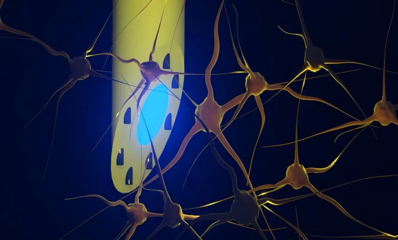
An international consortium of neuroscientists and engineers has unveiled a groundbreaking neural interface that promises to fundamentally transform how researchers investigate complex neurological processes and lay the technical groundwork for next-generation treatments for debilitating conditions such as epilepsy. Published in the peer-reviewed journal Advanced Science, the innovation—termed the microfluidic Axialtrode, or mAxialtrode—represents a significant departure from decades-old conventions in neuroengineering. Developed through a collaborative effort involving the Technical University of Denmark (DTU), the University of Copenhagen, and University College London (UCL), among other research institutions, the needle-thin polymer implant combines optical stimulation, electrical recording, and precise fluid delivery into a single, highly flexible thread measuring less than half a millimeter in diameter.
The introduction of the mAxialtrode addresses a long-standing bottleneck in neuroscience. For years, researchers probing the intricate architecture of the mammalian brain have been constrained by the limitations of traditional hardware. Conventional brain electrodes and optical fibers are typically rigid, manufactured from brittle silicon or hard glass, and functionally isolated to a single point of interaction at their terminal tip. By consolidating multiple modalities—light delivery, electrical monitoring, and targeted microfluidic drug infusion—along various discrete points across a single strand of soft polymer, the newly developed device allows scientists to interact with multiple layers of brain tissue simultaneously. While currently deployed strictly as an advanced investigative tool in laboratory settings, the technology holds profound clinical promise, potentially paving the way for closed-loop therapies that can dynamically monitor electrical anomalies, deliver localized anti-seizure medications, and apply targeted neuromodulation in real time.
The Evolution of Neurotechnology: Overcoming Rigid Constraints
To fully appreciate the significance of the mAxialtrode, one must examine the evolutionary trajectory of neural interface technology over the past half-century. Since the early days of electrophysiology, when researchers first inserted metal microelectrodes into neural tissue to record the firing of individual neurons, the field has grappled with an inherent biological incompatibility. The human brain is a soft, gelatinous organ that shifts and pulsates continuously within the skull, driven by vascular rhythms and cerebrospinal fluid dynamics. Inserting rigid, silicon-based probes into this dynamic environment inevitably causes chronic mechanical mismatch.
Over extended periods, the constant micro-motion of a rigid implant against surrounding soft tissue induces localized tissue trauma, leading to the activation of glial cells, neuroinflammation, and the formation of an insulating scar tissue capsule. This foreign-body response degrades the quality of electrical recordings over weeks or months, severely limiting the lifespan and efficacy of chronic brain implants. Furthermore, traditional optical fibers used in optogenetics—a revolutionary technique that allows researchers to control genetically modified neurons using pulses of light—have historically been restricted to surface-level or single-depth interactions. Because light exits only from the polished flat end or "nose" of a conventional fiber, studying deep-brain circuits or multi-layered structures like the cerebral cortex and the hippocampus required the implantation of multiple, bulky devices, compounding tissue damage and complicating experimental design.
The mAxialtrode project was conceived to dismantle these physical and functional barriers. Postdoc Kunyang Sui and Associate Professor Christos Markos spearheaded the conceptualization of the device at DTU, focusing on the convergence of microfluidics, polymer science, and neuro-instrumentation. By replacing hard silicon and glass with flexible, medical-grade polymer materials, the research team engineered an implant that mimics the mechanical compliance of neural tissue itself. This softness dramatically reduces the shear stress exerted on the brain during movement, thereby mitigating the neuroinflammatory responses that have plagued chronic neural implant applications for decades.
Engineering Precision: The Anatomy of the mAxialtrode
The manufacturing process of the mAxialtrode relies on advanced thermal drawing techniques adapted from fiber-optic manufacturing. The journey of each implant begins as a macroscopic polymer preform—a meticulously assembled rod featuring precisely engineered internal geometries. Researchers heat this polymer assembly under controlled tension, drawing it down into an extremely fine, continuous fiber with microscopic internal architecture intact, akin to an ultra-precise glassblowing process scaled down to the micron level.
At the core of the finished mAxialtrode lies a high-performance, light-conducting channel designed to transmit optical wavelengths deep into subcortical structures. Surrounding this central optical core are eight microscopic fluidic channels. These minuscule capillaries are capable of transporting precise volumes of liquid pharmaceuticals, neurotransmitters, or chemical tracers directly to targeted cellular populations. Crucially, these same microfluidic channels can also house ultra-thin, flexible metal microwires, transforming the fluid conduits into simultaneous electrical recording sites.
The entire composite fiber measures less than 0.5 millimeters across—roughly the thickness of a fine sewing thread. Despite its microscopic scale, the implant provides multiple functional nodes spaced millimeters apart along its longitudinal axis. This spatial distribution allows researchers to record electrical potentials from shallow cortical layers down to deep limbic structures like the hippocampus, all while injecting specific pharmacological agents at distinct, predetermined depths.
Rigorous Validation In Vivo: Testing in Living Subjects
Following initial benchtop characterization, the research team transitioned to rigorous in vivo testing to validate the mAxialtrode’s performance in living biological systems. These experiments were conducted in close partnership with neurophysiology experts Associate Professor Rune W. Berg of the University of Copenhagen and Associate Professor Rob C. Wykes of University College London, both of whom brought extensive expertise in neural circuit analysis and translational epilepsy models.
The electrodes were surgically implanted into the brains of living mice, where they were coupled to external diode laser systems, multichannel recording amplifiers, and ultra-precise micro-infusion pumps. The results of these trials exceeded initial expectations. The mAxialtrode successfully demonstrated the capacity to stimulate targeted populations of nerve cells using both blue and red light wavelengths, proving its utility for complex optogenetic protocols. Simultaneously, the integrated micro-wires recorded high-fidelity electrophysiological signals spanning both superficial neocortical layers and deeper subcortical structures.
Perhaps most impressively, the research team utilized the device’s microfluidic channels to inject distinct chemical agents at precise depths separated by nearly three millimeters along a single insertion tract. Throughout these multi-modal experiments, the subject mice exhibited no outward signs of physical discomfort or behavioral impairment, carrying the lightweight, flexible tethered assembly with remarkable ease. This successful integration confirmed that a single, minimally invasive fiber could replace the cluttered arrays of multiple electrodes, fluid cannulas, and optical fibers traditionally required for such comprehensive neuroscientific investigations.
Implications for Epilepsy Research and Clinical Horizons
The successful development and validation of the mAxialtrode arrive at a critical juncture for clinical neurology and psychiatric research. Neurological disorders, particularly drug-resistant focal epilepsy, present notoriously complex electrophysiological profiles characterized by aberrant electrical synchronization originating in deep brain structures and propagating across distinct cortical networks. Current therapeutic interventions—ranging from systemic anti-seizure medications that cause widespread side effects to deep brain stimulation (DBS) systems that apply generalized electrical pulses—often lack the spatial and temporal precision required to halt seizure activity at its precise anatomical source without disrupting healthy surrounding tissue.
The confluence of optogenetics, localized fluid delivery, and high-density electrical recording housed within the mAxialtrode points toward a sophisticated future for neuromodulation. In a hypothetical clinical scenario, a smart, closed-loop neural prosthesis based on this technology could continuously monitor local field potentials, detect the earliest electrographic signatures of an impending epileptic seizure, instantaneously deliver a precise micro-dose of an inhibitory pharmacological agent directly to the hyper-excitable focus, and apply corrective light or electrical stimulation—all within milliseconds and entirely confined to the affected neural circuit.
Despite the profound transformative potential of these findings, the research team maintains a rigorous and pragmatic perspective regarding the timeline for clinical translation. Kunyang Sui and his colleagues emphasize that the mAxialtrode is currently an advanced research instrument rather than a ready-to-use medical device. Moving from successful animal trials to human clinical applications requires navigating a complex regulatory landscape, conducting extensive biocompatibility and long-term degradation studies, and refining the peripheral hardware required to drive fluidics and optics in a fully implantable, hermetically sealed package.
Next Steps: Patents and Commercialization Pathways
As the academic community digests the findings published in Advanced Science, the multi-institutional consortium is actively laying the groundwork for the next phase of development. The immediate priority for the research group is securing comprehensive patent protection for the core architecture and manufacturing methodology of the mAxialtrode. Intellectual property security is widely regarded by technology transfer offices at DTU, the University of Copenhagen, and UCL as an essential prerequisite for attracting venture capital investment or establishing industrial partnerships with established medical device manufacturers.
Concurrently, the team is initiating preliminary safety evaluations and design modifications aimed at miniaturizing the peripheral fluidic and optical interfaces, scaling up manufacturing consistency, and designing fully implantable iterations suitable for long-term preclinical validation in larger mammalian models. While a realistic timeline for human clinical trials remains years away, the mAxialtrode stands as a monumental testament to the power of interdisciplinary collaboration, bridging polymer chemistry, micro-fluid engineering, and systems neuroscience to illuminate the darkest corners of the human brain.