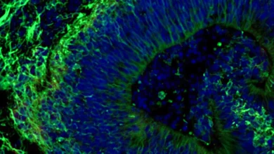
When Cell Death Machinery Promotes Survival: Weizmann Institute Scientists Uncover the Dual-Edged Sword of Cellular Regeneration and Cancer Recurrence

In the intricate microenvironment of living tissue, the boundary between destruction and rebirth is governed by mechanisms that science is only beginning to fully comprehend. For half a century, biologists have marveled at a phenomenon known as compensatory proliferation—the remarkable ability of epithelial layers and skin to orchestrate massive structural regrowth following extensive, catastrophic damage. While the macro-level results of this process have been documented extensively across various multicellular organisms, the micro-level choreography of how cells manage to trigger and sustain such dramatic recovery has remained shrouded in mystery.
Now, a groundbreaking study conducted by researchers at the Weizmann Institute of Science in Israel and published in the journal Nature Communications sheds unprecedented light on this biological enigma. The research team has successfully identified a sophisticated molecular mechanism that explains how tissues regenerate after severe trauma. More critically, the findings reveal a startling paradox: the very cellular machinery designed to execute damaged cells can be hijacked to confer extreme survival resistance, potentially explaining why certain aggressive cancers manage to survive therapeutic eradication and return with a vengeance.
A Historical Perspective on Compensatory Proliferation
To understand the magnitude of the recent discovery, one must look back to the origins of regenerative biology in the mid-20th century. The phenomenon of compensatory proliferation was first systematically documented in the 1970s. During this era, pioneering researchers exposed fruit fly (Drosophila melanogaster) larvae to high doses of ionizing radiation. Despite sustaining catastrophic damage to their epithelial tissues—the cellular sheets that cover and line internal organs and external body surfaces—the larvae demonstrated an astonishing capacity to bounce back, ultimately developing fully functional, anatomically correct wings.
In the decades that followed, similar compensatory responses were observed across a broad spectrum of species, ranging from simple invertebrates to complex mammals, including humans. Medical science recognized that the body possessed a built-in emergency response system capable of ramping up cell division to replace massive losses of cellular architecture. However, the exact biochemical triggers that incited healthy, surviving cells to divide furiously in response to neighboring cell death eluded researchers. Traditional dogma held a rigid line: cells either lived to perform their physiological functions, or they activated self-destruct programs to protect the organism from mutation and disease. The Weizmann Institute study challenges this rigid dichotomy, proving that the gray area between life and death is where true regenerative power lies.
Unraveling the Paradox of Caspases
At the heart of the new discovery is a class of enzymes long studied for their destructive capabilities: caspases. Traditionally, caspases have been categorized as the executioners of apoptosis, a meticulously regulated form of programmed cell death often described as cellular suicide. When a cell ages, incurs irreparable DNA damage, or receives external molecular cues indicating its utility has expired, it initiates an apoptotic cascade. This pathway typically begins with an initiator caspase, which subsequently activates downstream effector caspases. These executioner enzymes systematically dismantle cellular proteins, chop up internal structures, and neatly package the cell’s remnants for safe disposal by neighboring scavenger cells.
However, over the past two decades, a growing body of scientific literature—including seminal work from the laboratory of Professor Eli Arama in Weizmann’s Molecular Genetics Department—began to reveal that apoptotic caspases are not exclusively lethal. These versatile enzymes also participate in critical, nonlethal biological processes essential for normal development and tissue homeostasis.
Building upon this foundation, Professor Arama suspected that nonlethal caspase signaling might hold the key to unlocking the mystery of compensatory proliferation. To test this hypothesis, a research team led by Dr. Tslil Braun in Arama’s laboratory set out to recreate the classic 1970s irradiation experiments using contemporary, high-resolution genetic tools.
The Discovery of DARE and NARE Cells
By exposing fruit fly larvae to targeted doses of ionizing radiation and deploying advanced delayed genetic sensors, Dr. Braun and his colleagues were able to track epithelial tissue regeneration in real time with unprecedented precision. The sensor was specifically designed to flag cells in which the initiator caspase had been successfully activated, yet the cells stubbornly refused to die.
"We set out to identify cells that push the self-destruct button but survive anyway," Dr. Braun explained when discussing the methodology. This rigorous tracking led to the identification of a distinct, previously unknown population of cells dubbed DARE (Death-Associated and Radiation-Resistant) cells.
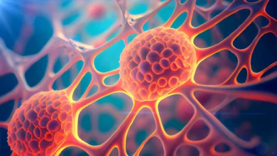
The behavioral profile of DARE cells proved extraordinary. Not only did these cells successfully withstand radiation levels that proved lethal to their neighbors, but they also initiated a rapid proliferative burst. Within a remarkably short window of 48 hours, DARE cells multiplied, repaired the structural deficits of the damaged epithelium, and single-handedly replenished nearly half of the lost tissue.
This discovery immediately introduced a biological accounting problem: if DARE cells accounted for approximately 50 percent of the regenerated tissue, where did the remaining half originate? Further investigation unveiled a second distinct cohort of death-resistant cells, which the researchers designated as NARE (Non-Death-Associated and Radiation-Resistant) cells.
While NARE cells ultimately played an equally vital role in tissue repair, they differed fundamentally from DARE cells in one crucial aspect: their initiator caspases had never been activated during the initial trauma. "Although NARE cells ultimately contribute to tissue regeneration, they cannot do it alone," Dr. Braun noted. When the research team experimentally removed DARE cells from the biological system, compensatory proliferation vanished entirely. This proved that DARE cells act as the master conductors of the repair orchestra, stimulated into action by distress signals emitted by their actively dying neighbors.
The Molecular Brake That Thwarts Death
Intrigued by how DARE cells managed to escape an apparent death sentence, the Weizmann team peered deeper into the intracellular machinery. They observed that the apoptotic sequence in DARE cells began precisely as expected: the initiator caspase switched on, signaling that the cell was entering the self-destruct protocol. However, at a critical juncture just before the executioner caspases could be summoned to complete the structural demolition, the pathway abruptly stalled.
The researchers traced this protective stall to a specific intracellular protein functioning as a molecular motor. This protein acts as a physical tether, anchoring the initiator caspase to the cell membrane and effectively preventing it from cascading down to activate the executioner enzymes. To confirm this hypothesis, the team genetically silenced the motor protein. The results were immediate and dramatic: DARE cells resumed their apoptotic trajectory, died off, and overall tissue regeneration was severely impaired.
Crucially, this molecular brake mechanism is not unique to simple tissue repair. Overactivation of the very same motor protein has previously been linked to tumorigenesis and unchecked cancerous growth. This intersection provides a compelling clue as to how cancer cells exploit native survival architecture to evade destruction.
Inherited Resistance: The Dark Side of Cellular Survival
The implications of the study extend far beyond acute injury repair, offering a sobering perspective on oncology and the persistent challenge of cancer recurrence. Standard oncological treatments, such as radiation therapy, rely heavily on inflicting enough genomic damage to tumor cells to forcefully trigger their apoptotic self-destruct mechanisms. Yet, clinicians have long observed that tumors which manage to recur following radiation therapy are frequently more aggressive, highly drug-resistant, and harder to eradicate than the primary tumor.
To determine whether the survival advantage of DARE cells could be passed down generations, Professor Arama’s team conducted follow-up tracking experiments. They wanted to understand if resistance to cell death is an inherited trait passed on to the descendants of death-resistant survivors.
The findings confirmed their worst suspicions. When the regenerated tissue was subjected to a second round of ionizing radiation, the number of cells dying during the initial hours was cut in half compared to the first exposure. Furthermore, the vast majority of those dying cells belonged to the baseline NARE population, while the descendants of the original DARE cells remained largely unscathed. Quantitatively, the progeny of DARE cells proved to be roughly seven times more resistant to cell death than standard cells in pristine, uninjured tissue.
This inherited resilience offers a compelling mechanistic explanation for clinical observations in cancer patients: surviving an initial therapeutic assault leaves a lasting biological legacy that hardens subsequent cellular generations against future interventions.
Balancing Growth: The Negative-Feedback Loop
While rampant cellular proliferation is necessary to close gaping wounds, unchecked growth is the foundational definition of cancer. The human body must maintain tight regulatory control over repair mechanisms to prevent a healing response from tipping over into tumorigenesis.
In the final phases of the study, the researchers uncovered a sophisticated signaling exchange between DARE and NARE cells that prevents runaway growth. DARE cells promote the proliferation of nearby NARE cells by secreting specific growth factor signals. In a classic homeostatic check-and-balance, NARE cells reciprocate by releasing molecular signals that inhibit the growth of DARE cells.
This dynamic interplay establishes a tightly regulated negative-feedback loop. The dual populations support the urgent metabolic and structural demands of tissue regeneration while simultaneously enforcing strict boundaries to halt proliferation once the tissue architecture is successfully restored.
Broader Implications for Regenerative Medicine and Oncology
Because these foundational experiments were conducted using Drosophila models, researchers emphasize that future studies will be required to validate how closely mirrored these mechanisms are in human physiology. Nevertheless, fruit fly models have historically served as remarkably reliable evolutionary blueprints for uncovering core biological pathways later confirmed to operate in humans.
The implications of this research are twofold. For regenerative medicine, understanding how DARE and NARE cells coordinate repair could pave the way for novel therapeutic interventions designed to accelerate the healing of severe wounds, chronic ulcers, and degenerative tissue damage. Clinicians might one day be able to safely stimulate DARE-like cellular pathways to jumpstart recovery in organs with limited regenerative capacities.
Conversely, in the realm of oncology, mapping the precise molecular brakes that allow cells to halt their own executioners offers a roadmap for defeating treatment resistance. By identifying the motor proteins and signaling circuits that cancer cells co-opt to evade radiation-induced apoptosis, drug developers can design targeted inhibitors to strip tumors of their acquired defenses.
As the scientific community digests these findings, the research highlights a profound biological reality: nature often employs the same conserved toolkits for both creation and destruction. By learning how to selectively promote the life-saving virtues of this survival machinery while blocking its oncological vices, modern medicine moves one step closer to mastering the delicate balance between tissue repair and disease eradication.