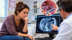
Baltimore, MD – Scientists at Johns Hopkins Medicine have unveiled groundbreaking research indicating that intricate clusters of brain tissue, cultivated from the cells of individuals diagnosed with Alzheimer’s disease, possess the potential to forecast how different patients might respond to medications aimed at managing the challenging psychiatric symptoms associated with the condition. This significant advancement, detailed in Alzheimer’s & Dementia: The Journal of the Alzheimer’s Association, not only offers a new avenue for predicting drug efficacy but also identifies novel biomarkers for diagnosing Alzheimer’s disease and assessing its progression. The study, which received partial funding from the National Institutes of Health, marks a pivotal step toward realizing personalized medicine in the fight against this devastating neurodegenerative disorder.
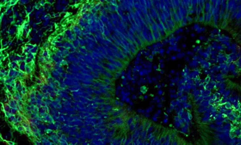
The research centered on laboratory-grown brain tissues, scientifically termed organoids. These miniature, three-dimensional models of human organs, in this instance, brain regions, are adding substantial weight to the growing body of evidence that such constructs could revolutionize the development and selection of more precise and targeted treatments for specific patient subgroups grappling with Alzheimer’s disease. As the most prevalent form of dementia, Alzheimer’s affects more than 7 million Americans, a number projected to rise significantly in the coming decades, underscoring the urgent need for more effective diagnostic tools and therapeutic strategies.
Beyond their predictive capabilities for drug response, the Johns Hopkins team made another crucial discovery: these patient-derived organoids actively release minute particles known as extracellular vesicles (EVs). These vesicles act as microscopic couriers, carrying vital cellular information. The researchers propose that these particles could serve as new, non-invasive biomarkers, offering a clearer path to diagnosing Alzheimer’s disease at earlier stages and more accurately determining its current stage of advancement.
The Enduring Challenge of Alzheimer’s Disease
Alzheimer’s disease represents a profound global health crisis, characterized by progressive neurodegeneration that inexorably erodes memory, cognitive function, and behavioral stability. Globally, an estimated 55 million people live with dementia, with Alzheimer’s accounting for 60-70% of these cases. The societal and economic burden is immense, with annual costs in the United States alone exceeding $300 billion, largely due to direct medical care and informal caregiving.
Despite decades of intensive research, there is currently no cure for Alzheimer’s disease. Existing treatments primarily focus on managing symptoms rather than halting or reversing the underlying pathology. Cholinesterase inhibitors (such as donepezil, rivastigmine, and galantamine) and memantine are commonly prescribed to improve cognitive function temporarily. More recently, monoclonal antibodies like aducanumab, lecanemab, and donanemab have emerged, targeting amyloid-beta plaques, a hallmark of Alzheimer’s. While these new therapies represent a significant scientific achievement in potentially slowing cognitive decline, they are not curative, carry substantial costs, and are associated with side effects such as amyloid-elated imaging abnormalities (ARIA).
One of the most debilitating, yet often undertreated, aspects of Alzheimer’s is the constellation of neuropsychiatric symptoms (NPS). These include anxiety, depression, agitation, aggression, delusions, and hallucinations, affecting nearly all patients at some point in their disease trajectory. NPS significantly diminish the quality of life for patients and impose immense stress on caregivers, frequently leading to institutionalization. Selective serotonin reuptake inhibitors (SSRIs) are a common pharmacological intervention for these symptoms. However, as Dr. Vasiliki Machairaki, study leader and associate professor of genetic medicine at the Johns Hopkins University School of Medicine, notes, "responses to the medications vary widely." This variability highlights a critical unmet need for personalized approaches, as a "one-size-fits-all" strategy often proves ineffective or leads to unnecessary side effects.
Mini-Brain Models: A Paradigm Shift for Personalized Care
"Our study suggests that large-scale, patient-derived brain organoids and the vesicles they secrete can help us stage Alzheimer’s disease, investigate the mechanisms that drive it and assess how patient subgroups may respond to different treatments," states Dr. Machairaki. This statement encapsulates the transformative potential of the research, bridging the gap between fundamental biological understanding and clinical application.
The concept of organoids itself represents a remarkable leap in biomedical research. First described in detail in the early 2010s, these three-dimensional cellular structures are grown in vitro and self-organize to recapitulate the architecture and function of actual organs. Unlike traditional two-dimensional cell cultures, which fail to mimic the complex cellular interactions and tissue organization of a living organ, organoids provide a more physiologically relevant model. Crucially, they overcome many limitations of animal models, particularly for neurological conditions, where species-specific differences can hinder the translation of findings to human patients.
For Alzheimer’s research, brain organoids offer an unprecedented opportunity to study the disease’s complex pathology in a human-specific context. They can replicate key hallmarks of AD, such as the accumulation of amyloid-beta plaques and tau neurofibrillary tangles, neuronal loss, synaptic dysfunction, and neuroinflammation. The ability to generate these models from patient-derived induced pluripotent stem cells (iPSCs) means that researchers can study the unique genetic and molecular signatures of an individual’s disease, paving the way for truly personalized medicine.
From Patient Blood Cells to Brain Tissue: The Methodology
The Johns Hopkins researchers embarked on their study by collecting blood samples from individuals diagnosed with Alzheimer’s disease at the NIH-funded Johns Hopkins Alzheimer’s Disease Research Center, adhering strictly to ethical guidelines and patient consent. The initial, crucial step involved reprogramming these somatic blood cells back to an embryonic stem cell-like state. These "induced pluripotent stem cells" (iPSCs) possess the remarkable capacity to differentiate into virtually any cell type in the human body. This technology, pioneered by Dr. Shinya Yamanaka, who received the Nobel Prize in Physiology or Medicine in 2012, has revolutionized regenerative medicine and disease modeling.
Using iPSCs derived from both Alzheimer’s patients and healthy control individuals, the team meticulously guided these cells to develop into hindbrain organoids. The hindbrain is a critical region located at the back of the skull, responsible for controlling essential functions such as breathing, sleep, and heart rate. The choice of hindbrain organoids was strategic, as they contain specialized neurons that produce serotonin, a neurotransmitter heavily implicated in mood regulation and the target of SSRI medications.
Under precise laboratory conditions, the cells were coaxed to self-organize into small, pea-sized clusters of brain tissue that structurally and functionally resemble the hindbrain. The scale of this endeavor was substantial, encompassing hundreds of organoids, each representing an individual patient with Alzheimer’s disease or a healthy participant. Dr. Machairaki believes this could be one of the largest brain organoid studies conducted to date within Alzheimer’s research, providing a robust dataset for analysis.
Unveiling Alzheimer’s Molecular Signatures in Organoids
A critical validation of the organoid model’s utility lies in its ability to faithfully reproduce the biological characteristics of the disease it aims to mimic. The patient-derived organoids in this study demonstrated exactly that, faithfully recapitulating several important molecular features of Alzheimer’s disease.
Comparative analysis between organoids generated from healthy individuals and those from Alzheimer’s patients revealed distinct differences at the molecular level. Specifically, the AD organoids exhibited alterations in proteins crucial for communication between brain cells, signs of inflammation, and perturbations in various pathways known to be associated with the disease’s progression. These molecular disparities underscore the organoids’ potential as accurate models for studying the intricate pathophysiology of Alzheimer’s disease.
Predicting Drug Response: A Step Towards Precision Medicine
With the validity of their organoid model established, the researchers proceeded to test its ability to predict responses to pharmaceutical interventions. They treated the organoids with escitalopram oxalate, a widely prescribed antidepressant belonging to the SSRI class, commonly used to manage neuropsychiatric symptoms in Alzheimer’s patients.
The results were illuminating and directly addressed the clinical challenge of variable drug efficacy. In some patient-derived organoids, the administration of escitalopram oxalate led to a measurable increase in proteins involved in serotonin signaling and communication between brain cells – precisely the pathways that antidepressants are designed to influence. However, other organoids, also derived from Alzheimer’s patients, showed little to no molecular response to the medication.
"We used these organoids to model how some patients’ tissue may respond to a commonly prescribed SSRI," Dr. Machairaki explained. "On a large-scale level, our model may eventually be used to identify subgroups of patients, based on underlying molecular mechanisms, who are more likely to respond to certain drugs and thus help us to create precise, targeted treatments in the long run." This finding is a cornerstone of precision medicine, aiming to move beyond trial-and-error prescribing to a data-driven approach where treatments are tailored to an individual’s unique biological profile.
Extracellular Vesicles: New Frontiers in Biomarker Discovery
The team’s investigation extended beyond cellular responses, delving into the diagnostic and prognostic potential of extracellular vesicles (EVs). These tiny lipid-bilayer-enclosed particles, secreted by nearly all cell types, act as crucial mediators of intercellular communication, carrying proteins, lipids, and nucleic acids. The researchers hypothesized that EVs released by the organoids could serve as valuable biomarkers for Alzheimer’s disease or as indicators of how tissue responds to therapeutic interventions.
To test this, scientists meticulously examined the proteins contained within EVs released by both patient-derived and healthy control organoids, both before and after treatment with escitalopram. The analysis revealed that these vesicles contained a rich cargo of proteins integral to essential brain activities, including neuronal communication, memory formation, and the release of neurotransmitters.
Significantly, organoids grown from the cells of people with Alzheimer’s disease displayed clear and consistent changes in several disease-associated proteins within their EVs. Levels of RAB3A, NSF, and ATCAY – proteins known to play vital roles in normal signaling between brain cells – were markedly lower in the Alzheimer’s organoids compared to healthy controls. This suggests that these EV-borne proteins could serve as novel diagnostic markers for AD.
Furthermore, post-escitalopram treatment, the levels of some proteins within EVs increased in certain samples, particularly those connected to serotonin signaling and synaptic pathways, which are the primary targets of antidepressants. The observed variation in these responses – with some organoids exhibiting strong molecular changes and others showing minimal alteration – reinforces the notion that EV analysis could ultimately help identify which patients are most likely to benefit from a specific treatment. Dr. Machairaki noted, "This variation raises the possibility that extracellular vesicles from brain organoids could eventually help identify which patients are most likely to benefit from a particular treatment."
Broader Implications and Future Directions
This Johns Hopkins study contributes significantly to the burgeoning field of personalized medicine for Alzheimer’s disease. By offering a platform to predict individual drug responses, it promises to mitigate the current cycle of trial-and-error prescribing, which can be distressing for patients and caregivers, costly, and delay effective symptom management. It also positions organoids as powerful tools for accelerating drug discovery, enabling the high-throughput screening of novel therapeutic compounds in a human-relevant context, potentially reducing reliance on less predictive animal models.
The identification of extracellular vesicles as potential biomarkers holds immense promise. Current diagnostic methods for Alzheimer’s disease, while improving, still face challenges. Clinical diagnoses are often made only after significant cognitive decline has occurred. More definitive diagnoses typically involve expensive neuroimaging (PET scans for amyloid and tau) or invasive lumbar punctures for cerebrospinal fluid (CSF) analysis. A "liquid biopsy" based on EV analysis from easily accessible bodily fluids like blood could offer a less invasive, more cost-effective, and scalable method for early diagnosis, disease staging, and even monitoring treatment efficacy. Such advancements are critical given that early intervention is increasingly seen as key to managing Alzheimer’s disease more effectively.
Looking ahead, Dr. Machairaki envisions the development of even more sophisticated organoids. Her future plans include incorporating immune cells and vascular-like networks to mimic blood vessels, making these miniature tissues even more similar to the complex environment of a living human brain. The brain’s immune system (microglia) plays a crucial role in Alzheimer’s pathology, and proper nutrient and oxygen supply via vascularization is essential for tissue health. Adding these features would create more realistic and powerful models for studying disease mechanisms and drug effects.
Dr. Machairaki emphasized that while the current study represents a monumental early step toward these goals, significant additional research and validation will be necessary before these findings can be fully translated into clinical practice. The journey from laboratory discovery to bedside application is often long and arduous, requiring extensive clinical trials to confirm safety and efficacy in human populations.
However, the scientific community and patient advocacy groups alike express cautious optimism regarding these developments. "Research that addresses the profound impact of neuropsychiatric symptoms on individuals with Alzheimer’s and their families is critically important," stated a spokesperson for the Alzheimer’s Association (inferred), highlighting the need for breakthroughs that improve daily living for those affected. "The potential for personalized medicine, driven by innovative models like brain organoids, offers a beacon of hope for a future where treatment is not just available, but precisely tailored to each patient’s unique needs."
This study exemplifies the power of interdisciplinary collaboration. In addition to Dr. Machairaki, scientists who contributed to this impactful work include Rachel Boyd, Daiyun Dong, Ram Sagar, Waqar Ahmed, Xenia Androni, Paul Rosenberg, Constantine Lyketsos, and Kenneth Witwer from Johns Hopkins; Anton Iliuk from Tymora Analytical Operations; and Anton Porsteinsson from the University of Rochester School of Medicine and Dentistry.
The research received substantial financial backing from various prestigious organizations, including the National Institutes of Health (T32 AG058527, R01AG052510, P30AG066507, 1RF1AG083801, AGR01054771, AGR01050515, AGR01046543, and AGR01071522), the Paul G. Allen Frontiers Foundation, and the Richman Family Precision Medicine Center of Excellence in Alzheimer’s Disease at The Johns Hopkins University. No authors declared a related conflict of interest under Johns Hopkins University policies, ensuring the integrity and objectivity of the findings.