The Silent Epidemic: Why Two-Thirds of Spinal Fractures in the US Go Undiagnosed

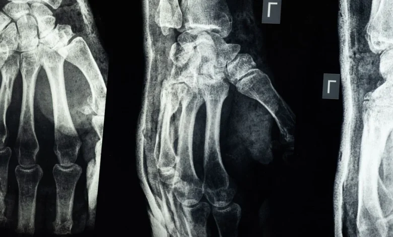
Approximately two-thirds of spinal fractures in the United States are missed annually, not due to their hidden nature, but because they often manifest without any discernible pain. This diagnostic void represents a significant failure in modern preventive medicine, where fractures of the forearm, upper arm, and pelvis are frequently treated as isolated injuries, neglecting the underlying bone disease – osteoporosis – that fuels them. This oversight leaves individuals vulnerable to subsequent, often more severe, fractures, creating a cycle of preventable harm.
Osteoporosis, a condition characterized by diminished bone density and compromised bone microarchitecture, renders bones brittle and susceptible to fractures from stresses that would be inconsequential for healthy bone. While it disproportionately affects postmenopausal women, its insidious nature means that crucial warning signs such as broken bones, height loss, and postural changes are frequently overlooked, misread, or simply missed, leading to compounding consequences over time.
The Pervasive Scale of Missed Diagnoses
The diagnosis of osteoporosis relies on established metrics: low bone mineral density, specific fragility fractures, and elevated fracture risk. Despite the existence of accurate diagnostic tools and effective treatments, a vast chasm exists between the number of individuals with the disease and those receiving care. Bone density scanning, a readily available diagnostic method, often fails to prompt treatment initiation. Many patients do not receive appropriate pharmacological intervention due to concerns about long-term efficacy, potential side effects, or simply because their physicians do not initiate treatment discussions following a fracture event.
Fragility fractures, frequently a hallmark of undiagnosed osteoporosis, constitute a substantial public health challenge. The escalating incidence of osteoporosis places an immense burden on healthcare systems when it is not proactively managed. In the United States, the statistics are stark: an estimated 10 million Americans aged 50 and older live with osteoporosis, with women comprising the vast majority of this group (8 million). Yet, the majority of these cases remain undetected. U.S. osteoporosis statistics reveal that a staggering 69.12% of all osteoporosis cases go undiagnosed, with men facing an even more alarming rate of 86.88% undiagnosed cases. This pervasive diagnostic gap is attributed to a confluence of factors: the asymptomatic nature of the disease prior to fracture, insufficient screening programs, and persistent misconceptions about who is truly at risk.
According to American Family Physician, four in 10 white women over the age of 50 are projected to experience a hip, spine, or vertebral fracture in their lifetime. This statistic alone should underscore the critical importance of aggressive fracture warning sign recognition. However, the current clinical paradigm too often waits for a second or even a third fracture event before connecting the dots of underlying bone fragility.
When a Fracture Becomes a Critical Warning Sign
A significant proportion of fragility fractures occur in individuals whose bone mineral density scores fall above the clinical threshold for osteoporosis. This observation highlights a critical nuance: these fractures serve as definitive indicators of skeletal fragility, even when bone density scans appear to be within normal ranges. This distinction is crucial, as it implies that an individual can sustain a fracture from a relatively minor fall, receive a bone scan that appears normal, and still possess severely compromised bone structure. In such instances, the fracture itself acts as the primary signal of underlying skeletal weakness.
Among the most frequently misconstrued fractures are spinal compression fractures. Because the majority of low-trauma vertebral fractures do not present with overt pain, dedicated vertebral imaging and careful review of past imaging studies are vital for identifying these fractures in at-risk patients. A 2022 National Institutes of Health (NIH) review underscored that most patients with vertebral fractures remain entirely unaware of their condition, with only about one-third ever receiving a clinical diagnosis. These are not rare occurrences; vertebral compression fractures, where a vertebra partially collapses under the body’s weight, affect over 700,000 Americans annually and represent the most common complication of osteoporosis.
Hip fractures, while often perceived as more dramatic injuries, can also present diagnostic challenges. Research published in Mayo Clinic Proceedings indicates that osteoporotic hip fractures may be clinically silent or manifest with non-specific symptoms, leading to significant diagnostic delays. A patient presenting with knee pain following a fall might be misdirected, their underlying bone fragility overlooked.
Wrist fractures complete a trio of commonly missed fracture types. A fragility fracture of the wrist, occurring from a mild trip or a minor fall while catching oneself, should invariably trigger a bone density assessment. The American Family Physician review emphasizes that fractures of this nature, resulting from low-energy mechanisms, should prompt an evaluation for underlying skeletal fragility. Unfortunately, these are too often treated with a splint and discharged without further investigation into their root cause.
The Subtle Physical Manifestations Preceding Diagnosis
Certain fracture warning signs can manifest physically long before a bone density scan is even considered. A measurable decline in standing height, exceeding 1.5 inches, can be indicative of cumulative vertebral compression fractures silently occurring in the spine. This is distinct from the gradual, age-related height loss that most individuals accept. A loss of this magnitude, particularly over a few years, serves as a significant clinical red flag warranting thorough investigation.
Postural changes also offer valuable clues. The development of a forward-rounding of the upper back, known as kyphosis, can result from multiple vertebral fractures that may have gone unfelt as acute pain. The MSD Manuals note that osteoporotic vertebral fractures are asymptomatic or cause only height loss and kyphosis in approximately two-thirds of patients, precisely explaining why so many go unrecognized. As the spine progressively bends forward, each collapsed vertebra contributes incrementally, leading patients to attribute the change to posture or general aging.
Underlying risk factors for bone fragility also act as indirect fracture warning signs. Declining estrogen levels in women, advancing age, a family history of osteoporosis, low body weight, and insufficient intake of calcium and vitamin D all significantly elevate an individual’s risk. A comprehensive medical history should meticulously identify risk factors associated with secondary bone loss, including smoking habits, chronic alcohol consumption, a family history of osteoporosis, and any prior fractures, particularly those sustained from low-energy or ground-level falls, or any fracture after the age of 40.
Low magnesium levels represent another underappreciated contributor to bone fragility. Magnesium plays a direct role in bone mineral density, and its deficiency increases the risk of osteoporosis development—a connection that often goes unaddressed in standard fracture workups.
The Cascading Consequences of Unrecognized Fractures
When the significance of a fracture as a warning sign is missed, the downstream consequences are profound. Despite the widespread availability of reliable diagnostic instruments and cost-effective medications proven to reduce fracture risk, the majority of patients who could benefit from treatment do not receive it. The osteoporosis care gap is estimated to be approximately 70% in both Europe and North America, and this deficit is now widely recognized as a critical patient care crisis.
The treatment gap following a first fracture is particularly stark. In Canada, for instance, approximately 90% of patients receive antiplatelet therapy following acute coronary syndrome to prevent future cardiac events. In contrast, only 10% to 20% of patients receive pharmacotherapy following a fragility fracture. This comparison starkly illustrates the imbalance: a bone fracture, which is as predictive of future harm as a heart attack, receives a fraction of the clinical urgency.
A 2025 study published in a rheumatology journal found that fewer than 25% of patients who experience an osteoporotic fracture receive appropriate treatment. Similarly, a 2021 PMC study revealed that fewer than 20% receive therapies aimed at reducing the risk of a subsequent fracture within the year following the initial event, even when that fracture has been clearly identified. The first fracture, therefore, serves simultaneously as the clearest warning sign and the most frequently ignored opportunity for intervention.
Remarkably, patients found to have secondary, treatable causes of bone fragility may not require additional long-term therapy once the underlying condition is addressed. This offers a significant advantage for a disease often perceived as permanent and inevitable. A 2024 study in Mayo Clinic Proceedings found that a contributing factor for osteoporosis can be identified in 32% to 85% of previously undiagnosed women when they undergo proper evaluation after a fracture. Secondary causes—such as thyroid disorders, vitamin D deficiency, celiac disease, or long-term steroid use—are present in 57% of patients with recent fractures, irrespective of their bone mineral density levels. If these underlying conditions are identified and effectively treated, some degree of bone loss can be slowed or even reversed without the necessity of long-term bone medication.
A 2022 review in Endocrine Reviews, published by the Endocrine Society, recommends general screening for secondary causes in all patients diagnosed with osteoporosis, reserving more advanced investigations for specific subgroups. This standard of care, however, hinges on the initial recognition of a fracture as a fracture worthy of investigation.
The existing clinical infrastructure for earlier detection of bone disease is already in place. The U.S. Preventive Services Task Force endorses dual-energy X-ray absorptiometry (DXA) bone mineral density testing, with or without fracture risk assessment, as the standard screening method. DXA scans are quick, relatively inexpensive, and expose patients to minimal radiation, yet this accessible test remains significantly underutilized.
The FRAX tool stands as the most widely employed fracture risk assessment instrument globally, integrated into over 100 international guidelines. It utilizes parameters such as age, weight, height, bone density (if available), prior fracture history, parental hip fracture history, smoking status, steroid use, and alcohol intake to calculate a 10-year probability of major fracture. It can be completed in under two minutes and requires no specialist referral. The FRAX calculator’s widespread use is attributable to its accessibility and robust validation across diverse populations, incorporating factors like prior fracture, parental history of hip fracture, current smoking, and glucocorticoid use to estimate a patient’s 10-year probability of major osteoporotic and hip fractures.
The current deficiency, therefore, is not technological. A July 2026 study by the Complexity Science Hub and the Ludwig Boltzmann Institute for Osteology, as reported by EurekAlert, concluded through a nationwide analysis of 1.7 million patients that fractures frequently go unrecognized and undocumented as warning signs in routine clinical care. The tools, medications, and knowledge necessary for effective intervention are all readily available. The critical failure lies in recognition—specifically, in the failure to perceive fractures as diagnostic warning signs rather than isolated, incidental accidents.
Proactive Steps for Enhanced Bone Health
Any fracture sustained from a low-energy fall—such as tripping on a flat surface, catching oneself against a wall, or a minor bump—warrants a discussion about bone health with a healthcare provider. This is particularly pertinent for individuals over 50, those with a family history of osteoporosis or hip fracture, and anyone who has experienced noticeable height loss in the past five years. These are not abstract risk markers but concrete fracture warning signs upon which clinical guidelines are fundamentally built.
Patients are encouraged to inquire with their physicians about the appropriateness of a DXA scan. Furthermore, individuals who have already experienced a fracture and have not undergone bone density testing or laboratory tests for secondary causes should proactively request such evaluations. Underlying contributors like vitamin D deficiency, thyroid dysfunction, and celiac disease are treatable conditions, and their identification following a first fracture represents a critical intervention window to prevent a subsequent one.
Osteoporosis is a silent disease, but its consequences, the fractures, are not. Treating a broken bone as merely a broken bone perpetuates a cycle where millions of individuals transition from one preventable fracture to another, underscoring the urgent need for improved recognition and proactive management of bone fragility.
Disclaimer: This information is intended for educational purposes only and should not be considered a substitute for professional medical advice, diagnosis, or treatment. Always seek the advice of your physician or other qualified health provider with any questions you may have regarding a medical condition. Do not disregard professional medical advice or delay in seeking it because of something you have read here.
AI Disclaimer: This article was generated with the assistance of AI tools and subsequently reviewed and edited by a human.