Dietary Fructose Released by Chemotherapy-Surviving Ovarian Cancer Cells Acts as a Molecular Catalyst for Tumor Metastasis

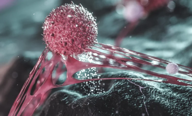
Researchers at The Wistar Institute have uncovered a surprising and complex mechanism by which aggressive ovarian cancer resists standard treatments and spreads throughout the body. According to a groundbreaking study published in the journal Nature Aging, cancer cells that manage to survive initial platinum-based chemotherapy regimens are far from inert. Although these persistent cells may no longer actively divide, they remain biologically active, secreting a common dietary sugar—fructose—into their immediate microenvironment. This secreted fructose acts as a potent biochemical signal, triggering neighboring tumor cells to break free from their structural anchors and initiate metastasis.
The findings illuminate a previously unrecognized pathway in cancer progression, challenging long-held assumptions regarding how tumors recur after seemingly successful interventions. Furthermore, the study raises urgent questions regarding the intersection of oncology, dietary habits, and widely prescribed cardiovascular medications, specifically statins. While the research opens thrilling new avenues for therapeutic intervention, scientists emphasize that the preclinical findings require extensive clinical validation before translating into actionable dietary or pharmacological guidelines for patients.
Understanding the Lethal Mechanics of Ovarian Cancer Metastasis
Ovarian cancer remains one of the most formidable and lethal malignancies affecting women worldwide. Standard protocol for the vast majority of diagnosed patients relies heavily on platinum-based chemotherapy. These pharmacological agents work by introducing platinum compounds that directly bind to and damage the DNA of rapidly proliferating cells, successfully halting their ability to replicate. Initially, this strategy yields favorable outcomes, with many patients achieving complete or partial clinical remission.
However, the clinical reality is stark. In a significant majority of cases, the tumor eventually returns. When ovarian cancer recurs, it frequently manifests as widespread metastasis throughout the abdominal cavity—a confined anatomical space housing critical organs including the stomach, intestines, liver, and spleen. This widespread dissemination, known medically as intraperitoneal metastasis, is responsible for approximately 90% of all ovarian cancer-related mortality.
For decades, the oncology community has sought to understand the precise cellular mechanisms that enable cancer cells to detach from the primary tumor site, survive transit through bodily fluids, and establish secondary colonies in distant tissues. Traditional hypotheses focused almost exclusively on actively proliferating cancer cells. The new research from The Wistar Institute redirects the scientific lens toward senescent-like, non-dividing persister cells—cells that survive the toxic onslaught of chemotherapy and covertly orchestrate the next phase of the disease.
The Experimental Breakthrough: Separating Cells from Secretions
To unravel the complex signaling networks operating within post-chemotherapy tumors, the research team—led by senior author Katherine Aird, Ph.D., professor and co-leader of the Molecular and Cellular Oncogenesis Program in the Ellen and Ronald Caplan Cancer Center at The Wistar Institute, and first author Aidan Cole, Ph.D., a postdoctoral fellow in her laboratory—designed a series of sophisticated preclinical experiments.
Historically, studying the tumor microenvironment has been complicated by the sheer density and heterogeneity of tumor tissue. To isolate the specific impact of factors released by surviving cells, Cole and his colleagues engineered experimental models that physically separated the chemotherapy-surviving cancer cells from the molecular secretions they produced.
The results were striking. The isolated conditioned media—devoid of the surviving cells themselves—was sufficient to significantly accelerate cancer cell migration and dissemination in preclinical models. "As far as we know, this is the first time anyone has shown, in a preclinical model rather than just a dish, that it’s the molecules these cells release—not the cells themselves—that drive the cancer’s spread," Cole noted. This revelation shifted the investigative focus toward identifying the precise chemical messengers driving this aggressive behavior.
Fructose as a Signaling Molecule, Not Just an Energy Source
Through rigorous biochemical analysis, the researchers identified the unexpected culprit driving the metastatic cascade: fructose.
Fructose is a simple monosaccharide naturally present in various fruits, vegetables, and honey. In modern diets, however, it is most commonly consumed via high-fructose corn syrup (HFCS) and refined table sugar (sucrose), which are ubiquitous in sweetened beverages, processed foods, and baked goods. In the United States, dietary intake of high-fructose corn syrup accounts for a substantial caloric percentage—ranging from roughly 8% to 20% of daily caloric intake among certain demographics.
In the context of the Wistar study, chemotherapy-surviving ovarian cancer cells demonstrated the capacity to synthesize and excrete fructose directly into the surrounding tumor microenvironment. Crucially, the sugar was not merely acting as a nutritional energy source to fuel cellular metabolism. Instead, it functioned as an active biochemical signaling molecule. When neighboring, non-surviving cancer cells encountered this fructose gradient, it fundamentally altered their internal signaling pathways, enhancing their locomotive capabilities and promoting metastatic readiness.
Worryingly, the team observed that high systemic levels of fructose—mimicking a diet rich in sugary beverages—could promote these pro-metastatic signals even in the absence of prior chemotherapy. This suggests that high dietary consumption of fructose could theoretically influence cancer progression and metastatic risk through a biochemical pathway previously unknown to science.
The Cellular Domino Effect: Fructose, Cholesterol, and Biological Glue
To decipher the exact intracellular mechanisms triggered by fructose, the research team employed advanced functional genomics, including a CRISPR-Cas9 knockout screen. This high-throughput screening technology allowed scientists to systematically deactivate thousands of genes to observe which specific genetic pathways were necessary for fructose to exert its metastasis-promoting effects.
The screening revealed a surprising intermediary in the metastatic cascade: cholesterol suppression.
While cholesterol is frequently discussed in popular culture in the context of cardiovascular disease and arterial plaque, it performs indispensable physiological functions at the cellular level. Cholesterol is a vital structural component of cell membranes, ensuring proper fluidity, integrity, and barrier function. Furthermore, cholesterol metabolism plays a critical role in maintaining intercellular adhesion—the biological "glue" that binds neighboring epithelial and tumor cells together into cohesive tissues.
When fructose floods the microenvironment of neighboring cancer cells, it suppresses their endogenous cholesterol production. As cellular cholesterol levels drop, the structural integrity of the cell-cell junctions begins to compromise. The biological glue holding the tumor mass together weakens. Deprived of sufficient structural cohesion, the cancer cells can easily detach, break free from the primary tumor architecture, migrate through the extracellular matrix, and invade surrounding tissues.
This mechanistic insight provides a coherent, scientifically rigorous explanation for how a common nutritional component can directly influence one of the most lethal characteristics of solid tumors.
The Statins Paradox: A Potential Intersection with Cardiovascular Medications
The discovery that fructose promotes metastasis by suppressing cholesterol production immediately prompted the research team to investigate a critical collateral question involving widely prescribed cardiovascular medications: statins.
Statins—including atorvastatin, rosuvastatin, and simvastatin—are among the most frequently prescribed classes of medications in the industrialized world. In the United States alone, approximately 39 million individuals take statins to lower circulating low-density lipoprotein (LDL) cholesterol and reduce the risk of myocardial infarctions and cerebrovascular accidents. Statins achieve this therapeutic effect by inhibiting HMG-CoA reductase, a key rate-limiting enzyme in the body’s mevalonate pathway, which is responsible for cholesterol biosynthesis.
Because the Wistar Institute study demonstrated that suppressed cholesterol production facilitates cancer cell detachment, the researchers sought to determine whether statins might induce a similar biological vulnerability.
Subsequent experimental models revealed that administering statins in vitro could indeed weaken intercellular connections among cancer cells, mirroring the effects observed with fructose exposure. This finding introduces a complex and provocative pharmacologic paradox. Given that ovarian cancer predominantly affects postmenopausal women—a demographic with a higher baseline prevalence of cardiovascular disease and statin prescriptions—understanding whether cholesterol-lowering drugs interact with chemotherapy or influence metastatic potential is of paramount clinical importance.
However, the researchers exercise extreme caution regarding these preliminary findings. "We haven’t tested this effect in patients yet, but it raises questions about combining cholesterol-lowering drugs with chemotherapy, especially since ovarian cancer is most common in postmenopausal women who are often already on statins," Dr. Aird emphasized. The scientific community stresses that these preclinical observations do not warrant any alteration in patient medication regimens. Patients currently prescribed statins for cardiovascular health are strongly advised not to discontinue their medication based on these preliminary laboratory discoveries.
Broader Implications Across Torso and Abdominal Cancers
Although the primary focus of The Wistar Institute study centered on epithelial ovarian cancer, the fundamental biological pathways identified may have profound implications for a broad spectrum of malignancies.
Cancers originating within the abdominal and thoracic cavities—such as pancreatic, colorectal, gastric, and hepatic cancers—share anatomical similarities with ovarian cancer. They grow within confined spaces and frequently metastasize by shedding cells directly into peritoneal or visceral fluids. The researchers hypothesize that the fructose-cholesterol signaling axis discovered in ovarian tumors may represent a more generalized mechanism utilized by multiple solid tumors to evade therapeutic containment.
"We think other cancers that spread within the torso—pancreatic, colon, liver—could behave similarly. We can’t call it universal yet, but we think the effects are not just limited to ovarian cancer," Dr. Aird stated. Building upon these initial conclusions, the Wistar research group has already initiated follow-up experimental models to test whether pancreatic, colorectal, and liver cancer cells exploit identical metabolic signaling pathways to drive metastasis.
Future Directions: Diet, Therapeutics, and Clinical Validation
The publication of this study in Nature Aging marks a significant milestone in cancer metabolism research, yet it also underscores the vast amount of work that remains.
Crucially, researchers have not yet conducted randomized clinical trials to test whether deliberate dietary interventions—such as strict dietary fructose restriction—improve clinical outcomes, reduce recurrence rates, or prolong survival in human ovarian cancer patients. Epidemiological correlations and mechanistic laboratory models provide a compelling theoretical framework, but they do not definitively establish that consuming dietary sugar directly causes cancer to spread in clinical settings.
Additional rigorous, peer-reviewed clinical investigations are urgently required before oncology specialists can formulate specific dietary recommendations based on this newly discovered mechanism. Nevertheless, the identification of fructose as an active signaling molecule—and the mapping of the downstream cholesterol-adhesion pathway—opens entirely new frontiers in oncology drug discovery.
By illuminating how surviving persister cells manipulate their environment long after chemotherapy has ceased, this research paves the way for novel therapeutic strategies. Future interventions might focus on blocking fructose signaling pathways, stabilizing cellular cholesterol dynamics, or neutralizing secreted molecular factors, ultimately offering new hope in the ongoing battle against metastatic cancer.