How the Aging Brain Forgets: New UC Berkeley Research Links Alzheimer’s Protein to Disrupted Deep Sleep Brain Waves

The frustrating, fleeting inability to recall where a vehicle was parked, a misplaced set of house keys, or the name of an acquaintance often
passes as a minor inconvenience of everyday life. For decades, cognitive decline and the fading of episodic memory—the mental faculty responsible for cataloging and retrieving personal experiences and specific life events—have been broadly accepted as an inevitable byproduct of the human aging process. However, groundbreaking new research emanating from the University of California, Berkeley, suggests that the physical deterioration of memory may be driven by microscopic neurological mechanisms far more specific than the simple passage of time.
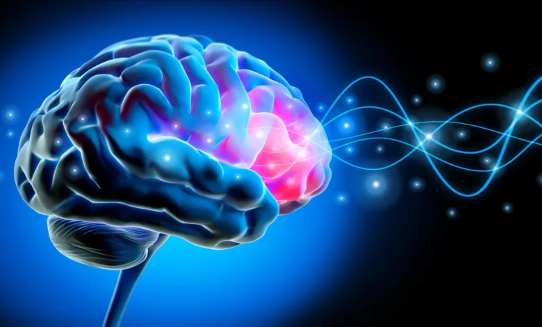
In a study published in the prestigious journal Nature Neuroscience, a team of neuroscientists has unveiled a critical missing link connecting the accumulation of tau, a hallmark protein of Alzheimer’s disease, with the fragmentation of electrical brain activity during deep sleep. Led by postdoctoral researcher Omer Sharon and directed by former Berkeley sleep researcher Matthew Walker, the investigation demonstrates that as tau builds up quietly within the frontal cortex of the brain, it disrupts the synchronized, traveling slow waves essential for consolidating memories overnight. This disruption transforms coordinated neurological symphonies into isolated, ineffective electrical bursts, ultimately starving the aging brain of its natural nightly filing system.
The Chronology of Discovery: From Sleep Science to Molecular Pathology
The roots of this study stretch across years of continuous inquiry within UC Berkeley’s Center for Human Sleep Science, building upon a growing body of scientific literature that identifies non-rapid eye movement (non-REM) sleep as a vital engine for cognitive maintenance. While REM sleep has historically captured public fascination due to its association with vivid dreaming and rapid eye movement, sleep scientists have increasingly focused on deep, non-REM sleep as the critical window during which the brain flushes metabolic waste and processes short-term memories into durable, long-term storage.
Historically, the connection between sleep fragmentation and cognitive decline was observed primarily at a macro level—older adults simply report sleeping more lightly and waking more frequently. To pierce deeper into the physiological mechanics, the UC Berkeley research team deployed a multi-tiered methodology combining electroencephalogram (EEG) sleep monitoring, positron emission tomography (PET) neuroimaging, and longitudinal cognitive tracking.
The investigative timeline moved from initial cross-sectional observations to rigorous clinical validation. First, the researchers compared cognitively healthy young adults in their early 20s with a cohort of older participants aged mid-60s to mid-70s. Utilizing high-density EEG caps fitted with numerous electrodes, the team recorded the electrical symphony of the sleeping brain. The generational contrast was immediate and stark. In young adults, large groups of neurons fired in unison, producing powerful slow electrical waves that cascaded across broad regions of the scalp—distances roughly equivalent to the width of a human hand. In contrast, the older participants exhibited fragmented, localized electrical activity.
To determine what was driving this geographical and electrical shrinkage within the sleeping brain, the team partnered with Dr. William Jagust, a UC Berkeley professor emeritus of public health and neuroscience. By administering PET scans using radioactive tracers injected into the bloodstream, the researchers mapped the precise biological distribution of tau proteins within the brains of the older participants. The imaging revealed a direct correlation: higher concentrations of tau accumulation in the frontal cortex directly predicted the breakdown of long-distance traveling slow waves.
Crucially, this pathological breakdown occurred in individuals who remained clinically healthy. "These people did not have Alzheimer’s," Sharon emphasized regarding the cohort. "They had tau in their brains, but with subclinical impacts. Their memory decline was within the normal range for their age."
Anatomy of a "Lonely Wave": How Tau Disrupts Memory Consolidation
To fully understand the gravity of these findings, neuroscientists examine the precise mechanics of non-REM deep sleep. During this restorative stage, dreaming subsides, and massive populations of neurons engage in a rhythmic, coordinated dance of switching off and back on together. These synchronized shifts generate sweeping electrical waves that originate predominantly in the frontal cortex before washing over the broader expanse of the brain.
"Each slow wave reflects an enormous population of neurons switching off and back on together," Sharon explained, detailing the delicate orchestration required for human cognition. "Our study shows that for memory, it’s critical that these events cascade in sequence across large parts of the brain. This coordinated shutdown has been observed only during deep sleep, and may be one reason why we sleep."
When tau proteins begin to aggregate within the frontal lobes, however, this collective synchronization fractures. Instead of sweeping across expansive neural networks, the electrical waves sputter, traveling shorter distances and firing independently. The research team dubbed these isolated electrical anomalies "lonely waves."
To test the functional consequences of these lonely waves, the researchers subjected participants to rigorous memory trials. In the evening, subjects were tasked with learning complex pairs of associated words. The following day, after a full night of monitored sleep, they were tested on their ability to recall those associations. The empirical results established a clear behavioral penalty: participants whose brains generated shorter, more isolated slow waves retained significantly fewer word associations. Furthermore, by tracking a subset of these participants over several years, the team observed that as tau levels in the frontal cortex progressively increased, slow waves became increasingly uncoordinated, and overnight memory retention declined in lockstep.
Cross-Cohort Validation: Spinal Fluid Analysis Strengthens the Hypothesis
Because advanced PET neuroimaging is resource-intensive, expensive, and constrained by specialized facility requirements, the research team sought to validate their imaging data through an independent biological marker. They established a collaborative partnership with neurologist Yo-El Ju at Washington University in St. Louis to analyze cerebrospinal fluid (CSF) samples obtained from a completely separate cohort of older adults.
While PET scans can isolate regional tau accumulation—specifically pinpointing the frontal cortex—spinal fluid analysis provides a systemic biochemical profile of Alzheimer’s-related pathology. The Washington University cohort tests yielded results that mirrored the Berkeley findings with remarkable consistency. Older adults whose sleeping brains produced a higher volume of isolated slow waves exhibited elevated ratios of tau to amyloid-beta, another hallmark protein associated with Alzheimer’s disease pathology, within their spinal fluid.
By replicating the core findings across distinct cohorts using entirely different analytical methods—brain imaging versus fluid biomarkers—the scientific team significantly bolstered the credibility of the association. "It’s collected in a totally different place and in a different cohort, so I think that adds to the strength of the evidence," Sharon noted, reflecting on the robustness of the cross-institutional data.
Broader Implications for Aging, Dementia, and Therapeutic Interventions
The implications of this study extend far beyond the academic understanding of sleep architecture, offering a new paradigm for evaluating preclinical neurodegeneration. For generations, memory loss in the elderly has been casually dismissed as general cognitive wear-and-tear. The UC Berkeley findings forcefully argue that much of this age-related memory deterioration is not merely a passive consequence of getting older, but rather the visible shadow of subclinical Alzheimer’s pathology actively disrupting fundamental neural maintenance systems.
"It’s not just about age," Sharon asserted, summarizing the paradigm shift. "It’s how much pathology you have in frontal areas where global waves originate."
Despite the clarity of the correlation between tau accumulation, wave fragmentation, and memory failure, the study’s authors maintain rigorous scientific caution regarding causation. The longitudinal data clearly demonstrates that tau and lonely waves increase in tandem over time, but the exact directionality remains an open scientific question. Does accumulating tau directly cause the breakdown of synchronized sleep waves, or does the chronic disruption of deep sleep accelerate the accumulation of pathological proteins by depriving the brain of its nightly cleaning cycle? Alternatively, could a third, underlying physiological process drive both phenomena simultaneously?
Answering this chicken-and-egg dilemma will dictate the future design of clinical interventions and therapeutic strategies. If disrupted sleep waves accelerate tau pathology, then targeted interventions designed to artificially boost deep sleep synchronization—whether through non-invasive electrical brain stimulation, acoustic stimulation during sleep, or novel pharmacological agents—could potentially slow the progression of preclinical Alzheimer’s disease and preserve cognitive function well into old age. Conversely, if tau is the primary driver, treatments must focus intensely on clearing neurotoxic proteins before they dismantle the brain’s delicate sleep circuitry.
As the global population ages and the socioeconomic burden of Alzheimer’s disease and related dementias mounts, identifying early, non-invasive markers of cognitive decline has never been more urgent. By turning an analytical lens toward the quiet hours of the night, UC Berkeley researchers have illuminated a promising new frontier in neuroscience—proving that the secret to preserving our waking memories may well lie in protecting the synchronized rhythm of our deepest sleep.
Funding for this pivotal research was provided by competitive grants awarded by the National Institute on Aging, a division of the National Institutes of Health (NIH). Additional institutional support for Omer Sharon was furnished through a Glenn Foundation for Medical Research Postdoctoral Fellowship and a Weill Neurohub Fellowship. The study’s co-authors include Xi Chen, James Westphal, Chelsea Brown, and Vyoma D. Shah of UC Berkeley, alongside Jason Dude of Washington University in St. Louis.