
The Surprising Science of Potatoes: How Chilling and Reheating Can Drastically Reduce Glycemic Impact and Influence Health Outcomes
A groundbreaking insight into the preparation of one of the world’s most ubiquitous staple foods reveals that consuming potatoes when they are cold, such as in potato salad, or after being chilled and subsequently reheated, can lead to a nearly 40% lower glycemic impact. This significant reduction in how quickly potatoes raise blood sugar levels presents a nuanced understanding of their role in a healthy diet, particularly for individuals managing conditions like type 2 diabetes or those aiming for improved metabolic health. The science behind this phenomenon lies in the formation of resistant starch, a dietary fiber that bypasses digestion in the small intestine, offering a range of health benefits beyond just glycemic control.
Potatoes in the Global Diet: A Nutritional Overview and Ongoing Debate
Potatoes (Solanum tuberosum) hold an undeniable place in global cuisine and agriculture. As the third most important food crop worldwide after rice and wheat, they serve as a primary energy source for billions and are economically vital for many regions. Beyond their caloric contribution, potatoes are a source of essential nutrients, including significant amounts of Vitamin C, potassium, Vitamin B6, and dietary fiber, especially when consumed with their skin. Historically, potatoes have been cultivated for millennia, originating in the Andes mountains of South America before spreading globally, adapting to diverse climates and becoming a cornerstone of diets across continents.

Despite their nutritional profile, potatoes have been subject to ongoing debate within dietary guidelines, particularly regarding their classification. While botanically a vegetable, some nutritional experts and public health bodies question whether they should be counted among the daily recommended servings of fruits and vegetables due to concerns about their high glycemic index and potential associations with chronic diseases. This distinction is crucial, as the emphasis on "non-starchy vegetables" often implicitly sidelines potatoes, prompting a closer look at how preparation methods might alter their health implications.
Understanding the Glycemic Index and Its Health Ramifications
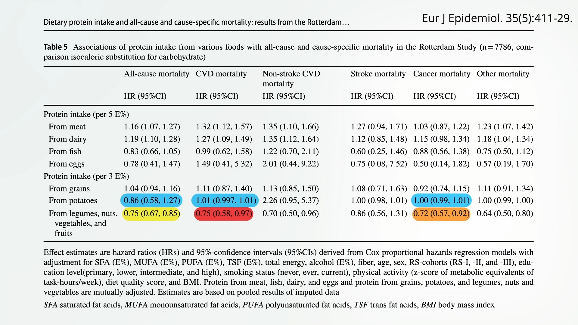
The glycemic index (GI) is a numerical scale used to rank carbohydrate-containing foods based on their effect on blood glucose levels over a two-hour period after eating. Foods with a high GI (typically above 70) cause a rapid and significant rise in blood sugar, while low GI foods (below 55) lead to a slower, more gradual increase. Pure glucose or sugar water is often used as a reference, standardized at 100. Foods like white bread and traditionally prepared white potatoes fall into the high-GI category, often scoring upwards of 70-80. In contrast, intact grains like barley groats, a super-low GI food, demonstrate a stark difference in metabolic response.
The implications of a diet consistently high in glycemic impact are substantial. Extensive research, including meta-analyses, has robustly associated high-GI diets with an increased risk of developing type 2 diabetes. Furthermore, current evidence strongly suggests that this relationship is not merely an association but a cause-and-effect link. Chronic spikes in blood sugar and insulin can lead to insulin resistance, pancreatic strain, and systemic inflammation, all precursors to metabolic dysfunction and various chronic ailments. Therefore, strategies to mitigate the glycemic impact of common foods, such as potatoes, hold considerable public health significance.
The Transformative Power of Temperature: Resistant Starch Explained
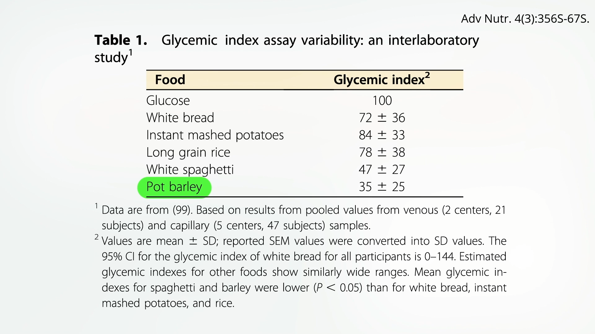
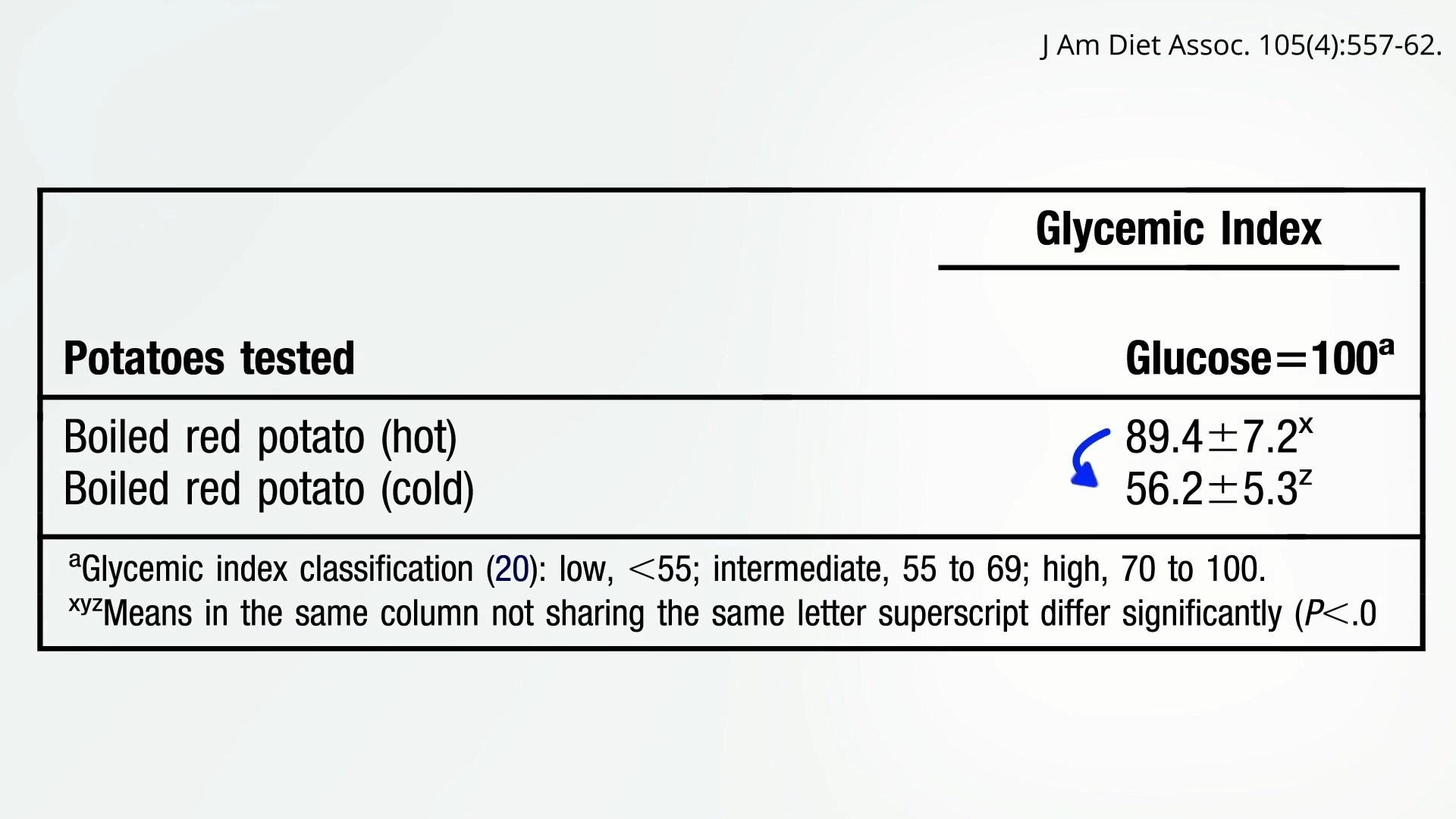
The revelation about the reduced glycemic impact of cold or reheated potatoes stems from the scientific principle of starch retrogradation. When potatoes are cooked, their starches gelatinize, making them easily digestible. However, upon cooling, some of these gelatinized starches undergo a structural change, crystallizing into a form known as resistant starch. This particular type of starch is aptly named because it resists enzymatic digestion in the small intestine, behaving more like soluble fiber than a rapidly absorbed carbohydrate.
When consumed, resistant starch travels largely intact to the large intestine, where it is fermented by beneficial gut bacteria. This fermentation process produces short-chain fatty acids, such as butyrate, which are crucial for colon health, immune function, and may even play a role in regulating appetite and metabolism. The amount of resistant starch formed, while previously considered relatively small, has been shown in direct human studies to translate into a dramatic drop in the glycemic index. Consuming potatoes as a cold potato salad, for example, can result in nearly a 40% lower glycemic impact compared to hot, freshly cooked potatoes. This chilling effect effectively slows the rate at which starch is broken down and absorbed into the bloodstream, leading to a more moderate glucose response. For individuals prioritizing metabolic health, this simple culinary trick represents a significant dietary advantage.
Potatoes and Chronic Disease Risk: Differentiating Preparation Methods
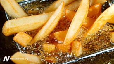
The relationship between potato consumption and chronic disease risk is complex and highly dependent on preparation methods. When systematically analyzing the best available studies, a clear association emerges for French fries with an increased risk of type 2 diabetes and hypertension. The deep-frying process adds unhealthy fats and often excessive sodium, while the high temperatures can create undesirable compounds.
However, for non-fried potatoes—boiled, baked, or mashed—the picture changes. These preparations were not found to be associated with an increased risk of high blood pressure. Yet, a "pesky link" with type 2 diabetes persisted, albeit potentially a small increase in risk for boiled potatoes. This continued association, even for healthier preparations, underscores the concern about their high glycemic index when hot. It also highlights the importance of dietary context; potatoes are often consumed as part of meals that may include other high-GI foods or less healthy components, potentially confounding results.
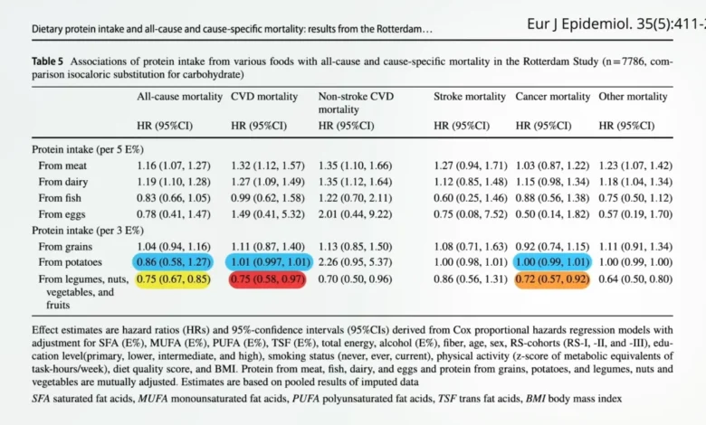
Regarding broader mortality, the research indicates that potatoes generally have a neutral impact. This contrasts sharply with other whole plant foods like nuts, fruits, vegetables, and legumes (beans, split peas, chickpeas, and lentils), which are consistently associated with living longer lives, including significantly less risk of dying from cancer, cardiovascular diseases, and a 25% lower chance of dying prematurely from all causes. The neutral effect of potatoes, therefore, can be viewed as an "opportunity cost"—every bite of a potato, particularly a hot, high-GI one, might be a missed opportunity to consume a food that actively promotes longevity. This is further distinguished from foods like processed meats, which may actively shorten life spans. The neutral impact of potatoes is thought to arise from a counterbalance: their beneficial fiber, vitamin C, and potassium content are potentially offset by the adverse effects of their high glycemic index.
The Satiety Factor: Maximizing Fullness for Weight Management
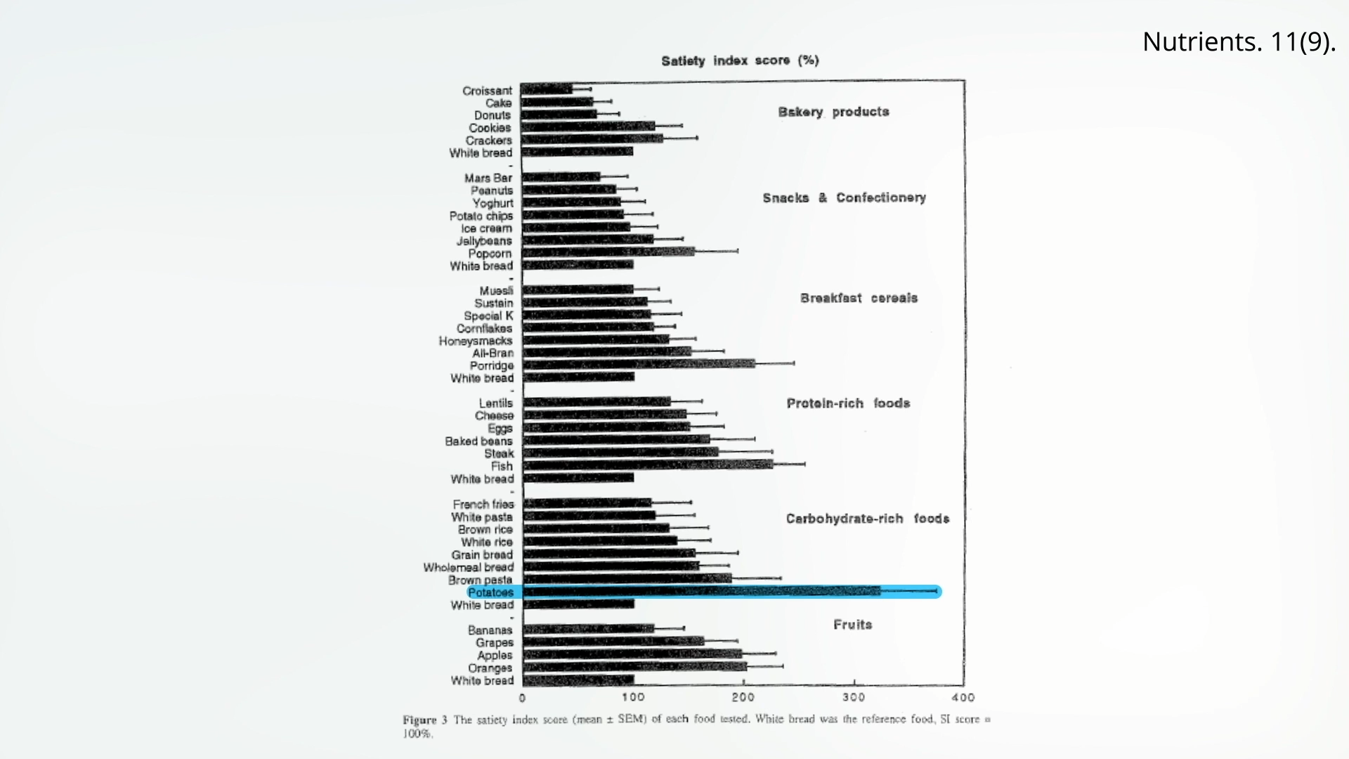
Beyond glycemic control, potatoes offer intriguing benefits related to satiety, the feeling of fullness that helps regulate food intake. A key factor in successful weight management, satiety can be significantly influenced by food choices and preparation methods. Potatoes naturally contain an appetite-suppressing protein called potato protease inhibitor II (PPI-II), which contributes to their satiating effect.
Research comparing the satiety levels of various foods has consistently shown that boiled and mashed potatoes are significantly more satiating than French fries. Interestingly, one landmark study identified boiled-then-cooled-then-reheated potatoes as the single most satiating food among dozens tested. This "best of both worlds" scenario allows individuals to benefit from both reduced glycemic impact (due to chilling) and enhanced satiety (due to reheating, which may improve palatability and sustained fullness). The satiety benefits are substantial: people experienced a marked drop in appetite after eating boiled mashed potatoes compared to white rice or white pasta, and crucially, compared to both fried and even baked French fries. This suggests that while baked fries might be perceived as a healthier alternative to fried, they offer little advantage in terms of satiety and thus may not be ideal for weight control. The "Doctor’s Note" from the original article clarifies that chilling is the crucial step for GI reduction, making cold potato salad a viable option, but for weight management, avoiding even baked fries might be advisable.
Navigating Industry Influence and Research Nuances
The landscape of nutritional research is not always free from external influence. The original article highlights an instance where a front group for the potato industry, the Alliance for Potato Research and Education, funded a study that concluded non-fried potato intake does not affect blood sugar markers. However, the critical context here is the comparison group: these potatoes were compared to "Wonder Bread," a refined product with a notoriously high glycemic index. This comparison sets a low bar and doesn’t genuinely assess the health impact of potatoes against truly optimal dietary choices, such as whole grains or legumes. Such industry-funded studies, while perhaps technically accurate within their narrow scope, underscore the importance of scrutinizing research methodology and funding sources to understand potential biases and derive truly actionable health advice. Independent, rigorous research remains paramount for advancing our understanding of food and health.
Broader Dietary Strategies and Expert Perspectives
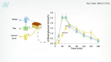
The journey to optimizing potato consumption extends beyond mere chilling and reheating. Several other dietary strategies can further blunt their glycemic impact. Adding vinegar, lemon juice, or even certain vegetables like broccoli to a potato meal can help. Vinegar, for instance, has been shown to slow gastric emptying and inhibit starch digestion, leading to a lower post-meal glucose response. Pairing potatoes with protein and healthy fats can also moderate blood sugar spikes by slowing digestion and absorption.
Nutritionists and public health experts, when inferring their perspectives on these findings, would likely emphasize a balanced approach. While acknowledging potatoes’ nutritional contributions (fiber, vitamins, minerals), they would advocate for mindful preparation. "Potatoes don’t have to be a dietary villain," one might infer a dietitian saying, "but how you prepare them makes all the difference. Incorporating them cold or reheated into a meal, especially alongside other low-GI foods, lean proteins, and plenty of non-starchy vegetables, transforms them into a more metabolically friendly option." Public health campaigns could leverage this research to offer practical, accessible advice to consumers, shifting the narrative around potatoes from a simple starch to a versatile food whose health benefits can be maximized through informed cooking. The food industry might also explore innovations in pre-prepared potato products that utilize chilling and reheating techniques to offer healthier convenience options.
Looking Ahead: The Evolving Understanding of Staple Foods
The ongoing scientific inquiry into potatoes reinforces a broader principle in nutrition: the health impact of a food is rarely absolute but rather a dynamic interplay of its inherent composition, preparation methods, and the overall dietary context. Potatoes are indeed a "double-edged sword"—a source of valuable nutrients but also a significant contributor to dietary glycemic load if consumed without consideration.
This evolving understanding encourages consumers to move beyond simplistic labels and embrace culinary creativity as a tool for health. The simple act of boiling potatoes, cooling them, and then perhaps reheating them, or enjoying them cold in a salad, represents an accessible and impactful dietary modification. As research continues to unravel the complexities of food science, our ability to transform everyday staples into powerhouses of health grows, offering promising avenues for managing chronic diseases and promoting overall well-being. The story of the potato, from a high-GI staple to a potential source of resistant starch, exemplifies the continuous learning curve in nutritional science and its profound implications for public health.


