Unlocking the Hepatic Gatekeeper: How Rejuvenating Aging Liver Cells Could Transform Alzheimer’s and Cardiovascular Disease Therapies

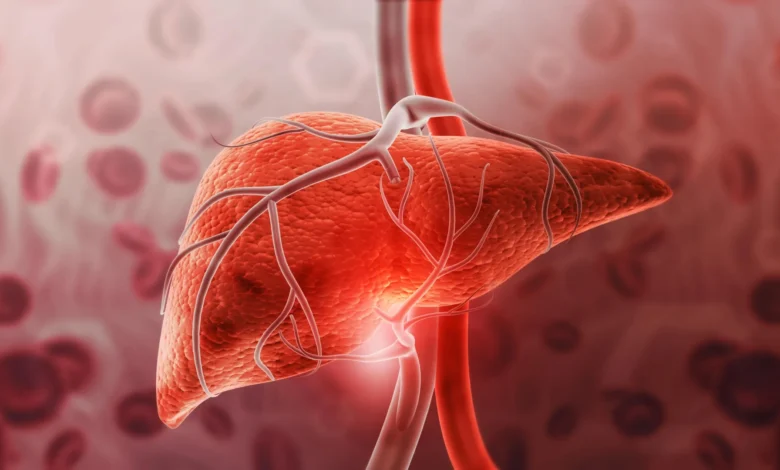
The medical community has long focused its efforts on treating Alzheimer’s disease and cardiovascular complications at the sites where they manifest most prominently: the brain and the circulatory system. However, groundbreaking new research from the Keck School of Medicine of the University of Southern California (USC) suggests that researchers may have been looking at the wrong organ altogether. A recent study reveals that the liver—traditionally viewed merely as the body’s primary filtration and metabolic hub—may hold a critical, overlooked key to mitigating some of the most devastating pathologies of aging. By restoring the biological function of aging liver cells, scientists have successfully reduced harmful circulating biomarkers associated with both neurodegeneration and heart disease in animal models, opening up an entirely novel therapeutic frontier in translational medicine.
The study, which was spearheaded by a multidisciplinary team at USC and published in the Journal of Biological Chemistry, centers on an experimental pharmaceutical compound known as cinaciguat. In a controlled laboratory setting involving older rats, administration of cinaciguat effectively reversed significant structural and functional hallmarks of aging within specialized microvascular cells in the liver. Within a remarkably brief treatment window of just two weeks, the researchers observed a fifty-percent reduction in circulating levels of amyloid beta—the neurotoxic protein fragment famously implicated in the pathogenesis of Alzheimer’s disease. Concurrently, levels of low-density lipoprotein (LDL), commonly recognized as "bad cholesterol" and a primary driver of atherosclerotic plaque buildup, plummeted back to baseline concentrations typically observed in young, healthy animals.
This unexpected dual efficacy has stunned researchers and challenged long-held dogmas regarding systemic clearance mechanisms. By illuminating a previously unrecognized cellular communication network within the hepatic architecture, the findings offer a compelling rationale for shifting therapeutic paradigms away from direct brain interventions and toward systemic metabolic rejuvenation.
The Chronology of a Breakthrough: From Observation to Cellular Discovery
The roots of this discovery trace back to a growing appreciation within the biomedical community for the interconnected nature of systemic aging. For decades, epidemiologists have noted strong correlations between hepatic dysfunction, advanced chronological age, and an elevated incidence of cognitive decline and vascular disorders. Yet, the precise mechanistic bridge connecting liver degradation to cerebral pathology remained elusive.
The USC research team chose to investigate a specialized, yet underappreciated, cellular population: liver sinusoidal endothelial cells, or LSECs. Unlike the continuous, tightly sealed endothelial linings found in most blood vessels throughout the body, LSECs form the specialized lining of the liver’s tiny blood vessels and are characterized by numerous microscopic pores, or fenestrations. These sieve-like openings are essential because they allow large macromolecules circulating in the blood to pass through and interact with neighboring hepatocytes—the primary metabolic workhorses of the liver responsible for breaking down and clearing waste products.
As biological organisms age, these critical fenestrations in LSECs progressively shrink, deform, or disappear entirely. This structural degradation creates a physical bottleneck, preventing large molecules like amyloid beta from reaching the hepatocytes necessary for their clearance. Consequently, these potentially toxic compounds accumulate in the bloodstream over time.
Hypothesizing that repairing these microscopic filters could restore systemic clearance, the investigators designed an experiment utilizing aged rodent models. They administered cinaciguat—a known activator of soluble guanylate cyclase—to a cohort of aged rats over a two-week period. Following the treatment regimen, the researchers compared the biological markers of the treated older rats against two distinct control groups: untreated aged rats and healthy young rats.
The outcomes exceeded initial expectations. Histological and biochemical analyses confirmed that cinaciguat successfully restored the structural integrity of the aging LSECs, reopening their fenestrations and reinstating their filtering capacity. More astonishingly, circulating amyloid beta concentrations dropped by roughly 50 percent, matching the youthful baseline, while the untreated older cohorts showed no such improvements.
Unraveling the Mystery of Cholesterol Clearance
While the reduction in amyloid beta aligned with the hypothesis that improving liver filtration would enhance waste clearance, the simultaneous drop in LDL cholesterol presented a profound scientific puzzle. Unlike large protein aggregates, LDL particles are physically small enough to exit the bloodstream independently of the specialized LSEC fenestrations. Initially, the research team had included LDL measurements merely as an internal baseline control, operating under the assumption that cinaciguat would exert little to no influence over lipid profiles.
When the data revealed that LDL concentrations in the treated older rats had plummeted to youthful levels, the investigators realized the drug was orchestrating a much more comprehensive biological restoration.
"We said, ‘Wait, why did that go down? Clearly, there’s more to this story,’" recalled first author Sam de Leve, a USC lab technician and researcher.
The unexpected finding prompted the team to look closer at the complex cellular crosstalk occurring within the liver tissue. Prior scientific literature had established that hepatocytes perform their metabolic duties far more efficiently when supported by healthy, functional LSECs. Building on this, the USC team discovered a novel biochemical signaling pathway that coordinates intercellular communication between these two cell types.
The investigation revealed that when cinaciguat activates soluble guanylate cyclase within LSECs, it triggers the release of a specific growth factor known as heparin-binding EGF-like growth factor (HB-EGF). This signaling molecule acts as a chemical messenger, dispatching instructions to neighboring hepatocytes. In response to the HB-EGF signal, the hepatocytes alter their cellular surfaces, upregulating their capacity to capture and internalize both LDL particles and amyloid beta from the bloodstream.
Thus, cinaciguat achieved its sweeping therapeutic effects not merely by physically reopening the liver’s cellular sieves, but by reactivating a dormant chemical signaling axis that had been silenced by the aging process.
Navigating the Obstacles of Modern Neuropharmacology
The implications of this discovery are particularly profound when viewed against the backdrop of historical clinical trial failures in Alzheimer’s research. For decades, the pharmaceutical industry has poured billions of dollars into developing therapeutic agents designed to target neurodegeneration directly within the central nervous system. These efforts, however, have been plagued by an estimated failure rate of approximately 98% in clinical trials.
A primary obstacle in neuropharmacology is the blood-brain barrier (BBB)—a highly selective, semipermeable cellular border that isolates the circulating blood from the extracellular fluid of the brain in the central nervous system. While the BBB is vital for protecting the delicate neural microenvironment from pathogens and toxins, it presents an almost insurmountable challenge for drug delivery. Most large-molecule therapeutics and targeted antibodies struggle to cross the barrier in sufficient quantities to exert a therapeutic effect, often requiring invasive delivery methods or chemical modifications that can introduce severe side effects.
By bypassing the central nervous system entirely and focusing on the hepatic clearance hypothesis, scientists could circumvent the blood-brain barrier altogether.
"If we’re right that the liver is impactful, what do we do about that?" noted Sam de Leve. "Rather than trying to get a drug through the blood-brain barrier, which is obnoxiously difficult, we could direct drugs to the liver, an organ where there might be more therapeutic impact and certainly an easier job of drug delivery."
By redirecting pharmaceutical design toward peripheral clearance organs, researchers could potentially harness the liver’s natural metabolic prowess to continuously "clean" the blood before circulating proteins have the opportunity to aggregate and cause downstream tissue damage.
Broader Implications, Limitations, and Future Directions
Despite the immense enthusiasm generated by the USC findings, the researchers and independent experts emphasize that the work remains in its preliminary stages. All current data have been derived exclusively from animal models; consequently, extensive translational research is required to determine whether identical cellular mechanisms and therapeutic benefits translate to human physiology.
Furthermore, scientists caution that lowering circulating levels of amyloid beta in the bloodstream does not automatically guarantee a reduction in pre-existing amyloid plaques within the human brain, nor does it definitively prove that Alzheimer’s disease can be prevented or reversed by this method alone. The complex, multifactorial etiology of neurodegenerative and cardiovascular diseases means that peripheral clearance is likely one piece of a much larger biological puzzle.
Nevertheless, the potential applications for this research are vast. Corresponding author Laurie DeLeve, MD, PhD, professor and vice chair for scientific affairs in the USC Department of Medicine, remains optimistic while maintaining rigorous scientific caution.
"We need to do further translational work to see whether our findings might point to new therapies," Dr. DeLeve stated. "We’re hopeful, though, because the findings line up with a lot of epidemiological results showing age as a major risk factor for Alzheimer’s and atherosclerosis, and liver damage as a risk factor for Alzheimer’s."
In light of the sheer volume of unexplored pathways opened up by this study, the research team has adopted an open-science philosophy, actively encouraging the broader scientific community—particularly researchers specializing in atherosclerosis, hepatology, and gerontology—to build upon their discoveries. Rather than hoarding the conceptual framework, the lab has signaled an eagerness for collaborative expansion.
"We hope that people who are in fields like atherosclerosis research read this, and want to grab the idea for themselves," Sam de Leve remarked. "There are six different ways someone could go with this, and we can’t do them all. So please, scoop us. We’d love it."
As funding from the National Institutes of Health (under grants R56AG071480 and 5R01DK100580) continues to support their ongoing investigations, the USC team plans to expand their studies in animal models to ascertain whether the cinaciguat-induced reductions in blood biomarkers translate into measurable, long-term neuroprotection and cardiovascular resilience. If subsequent translational phases prove successful, humanity’s approach to combating the diseases of aging may be permanently rewritten—not inside the microscopic convolutions of the human brain, but within the silent, industrious filters of the liver.