Groundbreaking Brain Imaging Study Reveals Strongest Evidence Yet Linking Long COVID to Dopamine Neuron Damage

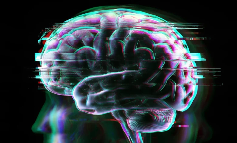
A landmark neuroimaging investigation conducted by researchers at the Centre for Addiction and Mental Health has delivered what scientific experts are calling the most compelling and robust evidence to date that long COVID is fundamentally linked to physical damage within the brain’s dopamine-releasing neural networks. Published in the peer-reviewed medical journal eBioMedicine, these findings shed critical light on the neurological underpinnings of some of the most debilitating and persistent symptoms associated with post-acute sequelae of SARS-CoV-2 infection, commonly known as long COVID.
For millions of individuals worldwide, the recovery path following an initial coronavirus infection remains blocked by enduring cognitive and physical impairments. These include profound fatigue-induced losses of motivation, significant motor slowing, and severe memory deficits. By isolating structural changes within the brain’s reward and movement centers, this new study moves the medical community a step closer to understanding the biological reality of these invisible wounds and establishes a foundation for targeted therapeutic interventions.
The Scope and Burden of Long COVID
Long COVID is estimated to impact approximately five percent of the global population, translating to roughly two million individuals in Canada alone and tens of millions more across the international landscape. The syndrome is clinically defined by a constellation of symptoms that persist, fluctuate, or emerge anew at least three months after the initial acute phase of a COVID-19 infection. While the multi-systemic nature of the condition can affect the cardiovascular, pulmonary, and gastrointestinal systems, the neurological and neuropsychiatric manifestations are among the most disruptive to daily functioning.
Patients frequently report chronic fatigue, debilitating brain fog, executive dysfunction, memory retrieval issues, sleep disturbances, and persistent low mood. Despite the staggering prevalence of these symptoms and the profound socioeconomic burden they impose on healthcare systems and workforces, evidence-based treatments have remained virtually non-existent. This therapeutic vacuum has persisted largely because the underlying pathophysiology within the central nervous system has remained elusive, leaving clinicians with little more than palliative symptom-management strategies.
Advanced PET Imaging Exposes Striatal Changes
To pierce the veil of uncertainty surrounding neurological long COVID, the research team at CAMH deployed positron emission tomography, an advanced molecular imaging technique capable of visualizing and quantifying biochemical processes and biological activity in the living human brain. The investigators focused their analytical lens on a well-validated radiotracer marker that serves as a reliable proxy for the health, functional integrity, and density of dopamine-releasing nerve terminals.
By comparing PET scans of patients suffering from long COVID against those of healthy, matched control participants, the researchers uncovered striking disparities. Individuals with long COVID exhibited substantially lower levels of the dopamine marker across all major anatomical subdivisions of the striatum. The striatum is a deeply embedded subcortical structure of the forebrain that plays a critical and multifaceted role in regulating voluntary movement, orchestrating goal-directed motivation, and facilitating complex cognitive thinking.
Crucially, the study mapped specific reductions in marker density to discrete clinical symptoms reported by the patients, establishing a direct correlation between biological injury and subjective experience. Lower marker availability within the ventral striatum was directly tied to a heightened loss of motivation, a state clinically analogous to severe apathy. Reductions observed within the dorsal putamen correlated with measurable motor slowing, while diminished levels in the caudate putamen tracked closely with impaired memory performance.
"Our findings provide compelling evidence that long COVID involves the loss of dopamine-releasing neurons," stated Dr. Jeffrey Meyer, Senior Scientist at the Brain Health Imaging Centre, Canada Research Chair, and senior author of the comprehensive study. "This kind of injury is well known to produce symptoms like lack of motivation and motor slowing, and may contribute to memory difficulties in other neurological conditions. Our results suggest a similar process is occurring in long COVID."
Tracing the Chronology of Neuroinflammation
To fully appreciate the weight of these new findings, it is necessary to examine the chronological progression of research carried out by Dr. Meyer’s laboratory over the course of the pandemic. As the global health crisis unfolded, early clinical observations quickly highlighted that viral respiratory pathogens could trigger significant neurological sequelae. Scientists began investigating whether the systemic immune response to SARS-CoV-2 could breach the blood-brain barrier and instigate neuroinflammation.
In earlier research published by the same CAMH team, investigators utilized neuroimaging to demonstrate that individuals experiencing long COVID harbored unusually elevated levels of translocator protein, a primary marker of glial activation and neuroinflammation, within the central nervous system. Crucially, this neuroinflammation was not distributed randomly throughout the brain; it was disproportionately concentrated in regions characterized by dense populations of dopamine-releasing neurons.
This chronological bridge connects the dots between two distinct pathological phenomena. While the initial phase of the research mapped the presence of persistent brain inflammation, the current eBioMedicine study provides the physical evidence that this inflammatory milieu corresponds directly with a reduction in the density of dopamine nerve terminals.
"We know that inflammation can injure dopamine neurons," Dr. Meyer explained. "While our earlier research showed high levels of inflammation in those regions, this study provides direct evidence that the dopamine neuron marker is reduced in the same regions—and that this loss correlates with patients’ symptoms."
When synthesized, these two landmark studies construct a cohesive working hypothesis: chronic, low-grade neuroinflammation triggered by the initial viral infection—or by persistent viral reservoirs and dysregulated immune responses—may lead to progressive cellular injury and loss within the dopaminergic pathways of the brain.
Paradigm Shift for Therapeutic Strategies
The identification of dopaminergic system compromise as a core driver of long COVID symptoms fundamentally shifts the trajectory of future biomedical research and clinical trial design. Historically, the vast majority of therapeutic investigations targeting neurological long COVID have focused broadly on mitigating systemic inflammation, modulating aberrant immune cell activity, or applying general antioxidant regimens. While these avenues remain important, clinical trials evaluating medications specifically tailored to restore or augment dopaminergic neurotransmission have been notably scarce.
By framing long COVID, at least in part, as a disorder of the brain’s dopamine system, researchers have unlocked a vast pharmacy of existing, FDA- and health-agency-approved medications that can be rapidly evaluated through repurposing clinical trials. Dr. Meyer notes that compounds designed to augment dopamine function—such as dopamine precursors, catechol-O-methyltransferase inhibitors, dopamine receptor agonists, and inhibitors of dopamine metabolism—represent highly promising candidates for upcoming intervention studies.
Such pharmacological agents have long histories of safe usage in the management of other medical conditions characterized by dopaminergic dysfunction, such as Parkinson’s disease and treatment-resistant depression. Translating these therapies to the post-viral population could offer a streamlined pathway toward alleviating refractory symptoms that have resisted all other forms of rehabilitative care.
The Human Toll and the Search for Validation
Beyond the biochemical data and neurological metrics, the human cost of long COVID remains staggering. For years, millions of patients have navigated a double burden: coping with disabling, multi-system physical symptoms while simultaneously facing skepticism from employers, insurers, and even segments of the medical community due to a lack of objective, measurable diagnostic markers.
Susan Deuville, a lived experience research advisor who worked alongside Dr. Meyer, articulated the profound emotional and psychological relief provided by these scientific insights. Contracting COVID-19 in 2021, Deuville endured a grueling multi-year odyssey in search of medical explanations for her abrupt cognitive and physical decline.
"For five years I have been seeking answers on what happened to me after I contracted COVID in 2021," Deuville reflected. "It was a crushing loss of the life I had and the person I was before. The research of Dr. Meyer brings hope. It also validates what long COVID sufferers have always known—long COVID is real and the effects are devastating."
By transforming invisible, subjective complaints into quantifiable neuroimaging data, studies of this caliber bridge the chasm between patient reality and clinical validation, empowering individuals to seek care armed with empirical proof of biological injury.
Upcoming Clinical Trials and Institutional Collaboration
Armed with the conclusive findings of the PET imaging study, the research consortium is not pausing at publication. Preparations are currently underway to launch a pioneering clinical trial within the next couple of months. This upcoming study will directly target dopamine function in human subjects suffering from long COVID.
The primary objective of the trial will be to evaluate whether targeted pharmacological interventions designed to modulate and support dopamine activity can successfully ameliorate core complaints such as chronic fatigue, executive dysfunction, and memory impairment. The scale and clinical rigor of the trial are bolstered by a strategic institutional partnership with the University Health Network. This collaboration represents a concerted effort to break down traditional silos between mental health and physical medicine, recognizing that post-viral syndromes demand an integrated, multidisciplinary approach to patient care.
Financial and institutional backing for the foundational imaging study was provided by the Canadian Institutes of Health Research, underscoring the federal commitment to unraveling the biological mysteries of the pandemic’s longest shadow. As the medical community prepares for the launch of these upcoming clinical trials, a renewed sense of momentum pervades the field. For the millions of individuals whose lives remain interrupted by long COVID, this research offers the first tangible blueprint toward recovery, illuminating a dark neurological pathway with the steady light of scientific discovery.
Related posts: