Unlocking the Blueprint of Movement: Researchers Discover Universal Tendon and Ligament Stem Cells and Potential New Treatments for Spinal Stenosis

In a significant breakthrough for musculoskeletal medicine, a collaborative team of researchers at Weill Cornell Medicine and the Hospital for Special Surgery (HSS) has successfully isolated and identified a previously elusive population of adult stem cells. These specialized cells serve as the primary biological engine responsible for generating the body’s tendons and ligaments—the vital connective tissues that seamlessly link skeletal muscles to bones and stabilize joints.
Published in the September issue of the peer-reviewed journal Cell, the discovery marks the culmination of years of meticulous investigation into skeletal biology. Beyond merely cataloging a new cell type, the research team uncovered a direct pathophysiological link between these stem cells and lumbar spinal stenosis, a debilitating and widespread degenerative condition. By pinpointing an overactive cellular mechanism responsible for abnormal tissue accumulation, the investigators have opened the door to entirely new, non-surgical therapeutic avenues, potentially involving existing classes of cardiovascular medications.
The implications of this discovery stretch far beyond the lower spine. Because these newly identified stem cells appear to act as a universal cellular reservoir throughout the entire human body, the findings could fundamentally reshape how clinicians approach chronic tendon injuries, joint reconstructions, and complex connective tissue disorders such as Marfan syndrome.
The Anatomy and Burden of Lumbar Spinal Stenosis
To understand the weight of the new discovery, one must examine the pervasive nature of the condition it aims to treat. Lumbar spinal stenosis is a degenerative disorder predominantly affecting older adults, though it can manifest earlier due to congenital factors or spinal trauma. Globally, the condition impacts an estimated 103 million people.
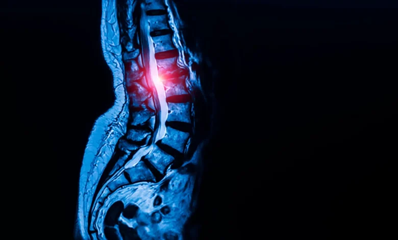
Spinal stenosis develops when the spaces within the spinal canal gradually narrow. This constriction is frequently caused by the pathological thickening and hypertrophy of the ligamentum flavum—a specialized ligament that runs along the inside of the spinal column. As this tissue expands, it encroaches upon the central spinal canal and intervertebral foramina, exerting severe mechanical pressure on the delicate bundle of spinal nerves housed within.
Patients suffering from lumbar spinal stenosis experience a spectrum of debilitating symptoms, including chronic lower back pain, radiating numbness and tingling down the legs, and a hallmark symptom known as neurogenic claudication—a cramping pain in the calves and thighs that worsens with walking and standing, forcing individuals to sit or bend forward to find relief. For millions, the condition severely compromises mobility and independence.
Historically, therapeutic options for severe spinal stenosis have been severely limited. While physical therapy, anti-inflammatory medications, and epidural steroid injections can temporarily manage symptoms, they do not halt or reverse the underlying structural progression of the disease. Once the compression becomes severe and conservative measures fail, patients have had virtually no recourse other than invasive decompressive surgery, such as a laminectomy, to physically carve away the overgrown ligament tissue and relieve pressure on the nerves.
A Chronology of Discovery: Tracking the Skeletal Blueprint
The identification of tendon and ligament stem cells did not happen overnight; it represents the latest milestone in a multi-year research trajectory led by Dr. Matthew Greenblatt and his laboratory at Weill Cornell Medicine.
The journey began in earnest around 2018, when Dr. Greenblatt’s research group successfully isolated the skeletal stem cell responsible for initiating fracture repair within the outer periosteum layer of bones. Galvanized by that success, the team expanded their search parameters, subsequently discovering specialized stem cell populations involved in the embryonic and postnatal formation of the skull and spine.
However, isolating the equivalent progenitor cells for tendons and ligaments proved to be a formidable scientific hurdle. Unlike bone, which possesses distinct structural compartments, tendons and ligaments are densely packed with fibroblast-like cells that bear a striking morphological resemblance to one another. Under a microscope, these mature cells look nearly identical, masking the rare, underlying stem cell population that continually gives rise to them.
To overcome this biological camouflage, the research team deployed advanced single-cell genomic technologies. By analyzing thousands of individual cells harvested from tissue samples and sorting them based on their genetic and molecular profiles, the investigators were able to isolate the specific population possessing the hallmark characteristics of stemness—namely, the capacity for indefinite self-renewal coupled with the ability to differentiate into the full complement of mature tendon and ligament cell types.
In murine models, the researchers located these rare cells residing within specialized anatomical niches inside tendons and ligaments, functioning as a localized biological reservoir dedicated to tissue maintenance and repair.
Transitioning from animal models to human tissue, the team collaborated with Dr. Sravisht Iyer, an orthopedic spine surgeon at HSS. Human ligament samples were procured—with explicit, informed patient consent—during routine spinal decompression surgeries. Postdoctoral fellow Dr. Lingling Hu, serving as the study’s first author, spearheaded the laboratory analyses that confirmed human cells isolated from these samples displayed the exact same dual capabilities: self-renewal and lineage-specific differentiation.
Crucially, when the researchers expanded their search to other anatomical regions, they made a startling realization. Stem cells matching this exact profile were recovered from the patellar ligament in the knee and the Achilles tendon in the heel.
"We looked in the kneecap ligament; we looked at the Achilles tendon; and everywhere we looked, we found this cell," Dr. Greenblatt stated. "So, we think this is the universal stem cell for tendons and ligaments throughout the body."
Mechanisms of Pathology: Unmasking the Culprit in Spinal Stenosis
Armed with the ability to identify and isolate these universal progenitor cells, the research team turned their attention to the pathogenesis of lumbar spinal stenosis. They sought to determine whether an overabundance or dysfunction of these stem cells might drive the abnormal tissue thickening characteristic of the disease.
The investigators conducted a comparative analysis, evaluating stem cells extracted from the spinal ligaments of patients undergoing surgery for severe spinal stenosis against control samples harvested from patients who had experienced herniated discs but showed no clinical signs of stenosis.
The results were striking. Ligaments retrieved from patients with spinal stenosis harbored a significantly higher concentration of the newly identified stem cells compared to the control group. Furthermore, when these stenosis-derived stem cells were transplanted into experimental mouse models, they exhibited a hyper-proliferative phenotype, generating a markedly higher volume of mature tendon cells than stem cells harvested from healthy individuals.
Delving deeper into the intracellular signaling pathways, the research team discovered that the stenosis-associated stem cells exhibited elevated levels of calcium signaling. In cellular biology, calcium ions act as crucial secondary messengers, orchestrating a vast array of intracellular processes ranging from metabolic activity to cellular proliferation and growth.
To test whether this heightened calcium signaling was merely a byproduct or a direct driver of pathology, the scientists utilized genetic techniques to artificially increase calcium signaling within healthy stem cells. This manipulation successfully replicated the disease state, triggering excessive, uncoordinated tissue growth. Conversely, when the researchers pharmacological or genetic interventions to reduce calcium signaling in a mouse model of lumbar spinal stenosis, the pathological overgrowth of tissue was effectively blocked.
Translational Horizons: From Bench Science to Clinical Care
The identification of calcium signaling as a primary driver of pathological ligament thickening offers an immediate, highly actionable therapeutic hypothesis. Because calcium channel blockers—a well-established class of pharmacological agents—are already widely prescribed across the globe to manage high blood pressure and cardiac arrhythmias, researchers have a pre-existing library of FDA-approved drugs with well-documented safety profiles.
While the prospect of repurposing common cardiovascular medications to treat a degenerative spine condition is theoretically promising, the medical community emphasizes caution. Rigorous, controlled clinical trials will be fundamentally required to evaluate whether administering calcium channel blockers locally or systemically can safely and effectively halt or reverse spinal stenosis in human patients.
Nevertheless, the paradigm shift represented by this study cannot be overstated.
"Identifying these specialized stem cells unlocks a new area of research that allows us to address this disease much more mechanistically, rather than just waiting until a patient’s condition worsens and requires surgery to relieve the nerve compression," noted Dr. Iyer. "The findings are exciting for their potential to change the way we deliver spinal care."
Dr. Iyer emphasized that this represents a monumental milestone in orthopedic research, marking what is likely the first time a concrete, druggable molecular target has been identified for one of the most prevalent spinal conditions in the world.
Broader Implications for Orthopedics and Connective Tissue Disorders
The scope of this discovery extends far beyond the confines of spinal clinics. Because the identified progenitor cells appear to serve as the foundational building blocks for every tendon and ligament in the human body, defects, mutations, or dysregulations within this single cell lineage are now viewed as the likely root cause of a wide array of chronic musculoskeletal disorders.
Looking ahead, Dr. Greenblatt and his colleagues plan to investigate the role these stem cells play in complex genetic and acquired connective tissue conditions. Among the primary targets of future research is Marfan syndrome, a genetic disorder caused by mutations in the fibrillin-1 gene that leads to systemic defects in connective tissues, often resulting in cardiovascular complications and skeletal overgrowth.
Furthermore, the insights gained from isolating these universal stem cells may finally illuminate why certain soft-tissue injuries—such as chronic rotator cuff tears, severe Achilles tendinopathy, and complex sports-related ligament injuries—possess such notoriously poor healing capacities. Tendons and ligaments are notoriously hypovascular, receiving limited blood supply, which hampers the delivery of endogenous repair cells following trauma. Understanding how to harness, stimulate, or protect these resident stem cells could ultimately revolutionize regenerative medicine, transforming how physicians treat sports injuries, facilitate post-surgical tendon healing, and manage degenerative joint diseases.
As funding bodies and independent foundations continue to support these investigations—with backing from institutions including the National Institutes of Health, the Marfan Foundation, the Arthritis National Research Foundation, and the Pershing Square Foundation—the medical community stands on the precipice of a new era in musculoskeletal care. What began as a difficult quest to find an elusive cell type has culminated in a universal blueprint of human movement, offering renewed hope to millions burdened by the quiet, creeping progression of degenerative connective tissue disease.