New Study Links Sleep Apnea and Low REM Oxygen Levels to Brain Degeneration and Memory Loss


Obstructive sleep apnea, a widespread and frequently underdiagnosed medical condition characterized by repeated disruptions in breathing during slumber, has long been associated with chronic fatigue, cardiovascular strain, and metabolic dysfunction. However, a compelling new peer-reviewed study published online on May 7, 2025, in Neurology, the premier medical journal of the American Academy of Neurology, sheds urgent new light on another alarming consequence of the disorder: the physical degeneration of critical brain regions dedicated to human memory.
According to the research team
, the cognitive toll of sleep apnea appears to be directly mediated by vascular damage to the brain’s delicate microvasculature. Specifically, the study revealed that significant drops in blood oxygen levels—hypoxia—during rapid eye movement (REM) sleep are strongly correlated with structural deterioration in brain areas responsible for memory consolidation. While the observational study establishes a robust statistical association rather than definitive causation, the findings offer vital clues into how sleep-disordered breathing may accelerate normal cognitive aging and pave the pathway toward devastating neurodegenerative conditions like Alzheimer’s disease.
Understanding the Mechanics of Obstructive Sleep Apnea
To fully comprehend the implications of the new research, it is essential to examine the physiological mechanics of obstructive sleep apnea (OSA). The condition occurs when the muscles located in the back of the throat relax excessively during sleep. This relaxation causes the soft tissue to collapse, physically blocking the upper airway. As oxygen levels in the bloodstream begin to plummet due to the cessation of airflow, the brain is forced to trigger micro-awakenings—brief, often imperceptible arousals that prompt the individual to resume breathing, usually accompanied by a gasp, snort, or sharp choking sound.
This destructive cycle of airway collapse and arousal can repeat dozens, or even hundreds, of times every single night. The resultant fragmentation prevents patients from achieving deep, restorative slow-wave sleep and disrupts the highly specialized architecture of REM sleep. REM sleep, famously characterized by rapid eye movements and vivid dreaming, is far from a passive state of rest. Instead, it is a metabolically active period of neurological functioning essential for emotional processing, synaptic plasticity, and, crucially, the consolidation of declarative and spatial memories. When breathing interruptions repeatedly puncture this critical sleep stage, the physiological consequences extend far beyond daytime somnolence, creating a systemic cascade of cellular stress, vascular inflammation, and intermittent hypoxia that batters the brain’s microvascular network.
Inside the Landmark University of California Irvine Study
Led by principal investigator Bryce A. Mander, PhD, a researcher at the University of California Irvine, the research team sought to untangle the complex relationship between sleep apnea, nocturnal oxygen deprivation, microvascular brain injury, and memory performance. The study cohort comprised 37 carefully screened older adults with a mean age of 73 who presented with no baseline clinical signs of cognitive impairment and were not currently taking sleep-modulating medications. Among this group, 24 individuals had been diagnosed with obstructive sleep apnea, while the remainder served as a control cohort.
The methodology combined rigorous physiological monitoring with advanced neuroimaging and cognitive evaluations. Participants underwent comprehensive overnight polysomnography—comprehensive sleep studies that meticulously measured their blood oxygen saturation levels continuously throughout all distinct stages of the sleep cycle, with a particular analytical focus on REM sleep. Following the sleep evaluations, the participants underwent high-resolution structural brain scans designed to map grey matter volume and white matter integrity. Furthermore, they completed standardized memory testing regimens both before and after a full night of sleep to quantitatively assess sleep-dependent memory consolidation capacities.
Microvascular Damage and White Matter Hyperintensities
The results of the neuroimaging analyses revealed a striking correlation between nocturnal hypoxia and structural brain pathology. Researchers discovered that lower minimum blood oxygen saturation levels—as well as a greater cumulative duration of time spent asleep with blood oxygen saturation levels dipping below the clinically concerning threshold of 90 percent—were predictive of a higher burden of white matter hyperintensities (WMHs) throughout the brain.
White matter hyperintensities appear as bright, abnormal patches on magnetic resonance imaging (MRI) scans. In clinical neurology, these hyperintensities are widely recognized as structural markers of cerebral small vessel disease. They reflect underlying damage, ischemia, and demyelination within the brain’s white matter tracts, which serve as the vital neural wiring connecting different regions of the central nervous system. When the delicate microvessels supplying the brain are repeatedly subjected to the hypoxic stress induced by sleep apnea, their structural integrity is compromised, leading to chronic leakage, micro-strokes, and localized tissue death.
Targeting Memory Centers: The Hippocampus and Entorhinal Cortex
The cascading damage did not stop at generalized white matter injury. The researchers extended their investigation to specific anatomical structures within the medial temporal lobe that are fundamentally anchored to human memory processing: the hippocampus and the entorhinal cortex.
Volumetric and morphometric analyses revealed that study participants exhibiting higher loads of white matter hyperintensities also displayed pronounced structural atrophy. Specifically, these individuals demonstrated a significantly decreased hippocampal volume and a reduced thickness in the entorhinal cortex. Both regions act as the gateway for memory formation, transforming short-term experiences into enduring long-term memories.
When the research team evaluated the cognitive performance data gathered from pre- and post-sleep memory assessments, the clinical relevance of these structural deficits became starkly apparent. Deficits in sleep-dependent memory consolidation—the brain’s ability to successfully store and stabilize memories overnight—were directly linked to the reduced thickness of the entorhinal cortex.
"Taken together, our findings may partially explain how obstructive sleep apnea contributes to cognitive decline associated with aging and Alzheimer’s disease through the degeneration of brain regions that support memory consolidation during sleep," noted Dr. Mander. The vulnerability of these precise anatomical networks underscores how systemic nocturnal events can manifest as focal, devastating cognitive impairments over time.
Chronology of Sleep Medicine and Cognitive Research
The publication of this study in Neurology represents the latest milestone in a rapidly evolving scientific timeline linking sleep architecture to neurological health. For decades, sleep medicine focused primarily on the immediate cardiopulmonary consequences of untreated sleep apnea, such as hypertension, stroke, and myocardial infarction.
- Early 2000s: Researchers began identifying epidemiological links between self-reported snoring, sleep-disordered breathing, and an increased risk of mild cognitive impairment (MCI) in older populations.
- 2010s: The advent of advanced neuroimaging allowed scientists to observe direct physical markers of neurodegeneration, such as amyloid-beta accumulation and structural atrophy, noting a high comorbidity between untreated sleep apnea and accelerated cognitive decline.
- Late 2010s to Early 2020s: Sleep researchers increasingly turned their attention to specific sleep stages, hypothesizing that disruptions during REM sleep and slow-wave sleep disproportionately impair the glymphatic system—the brain’s waste clearance mechanism—and memory consolidation pathways.
- May 2025: The current study bridges the gap between nocturnal hypoxia, small vessel ischemic injury (white matter hyperintensities), medial temporal lobe atrophy, and measurable memory deficits, providing a concrete mechanistic model for how sleep apnea damages the aging brain.
Broader Implications and Public Health Impact
The implications of these findings extend far beyond academic neurology, carrying profound public health ramifications. Obstructive sleep apnea is an exceptionally common condition, estimated to affect nearly one billion people globally, with prevalence rates climbing sharply with advancing age. Because a significant percentage of individuals suffering from OSA remain undiagnosed—often dismissing chronic fatigue, snoring, and morning headaches as normal parts of aging—millions of people may be unknowingly exposing their microvasculature to cumulative hypoxic injury.
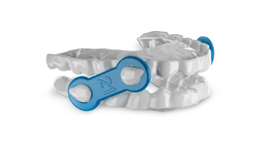
From a preventative medicine standpoint, the identification of a modifiable risk factor for cognitive decline offers a powerful therapeutic window. Unlike irreversible genetic predispositions to neurodegenerative diseases, obstructive sleep apnea is highly treatable. Standard clinical interventions, such as continuous positive airway pressure (CPAP) therapy, oral appliances, lifestyle modifications, and surgical interventions, can effectively restore normal airflow and maintain stable blood oxygen saturation levels throughout the night.
If future longitudinal and interventional clinical trials can definitively prove that treating sleep apnea halts or reverses microvascular brain damage and preserves memory function, screening for sleep-disordered breathing could become a cornerstone protocol in geriatric cognitive health management and Alzheimer’s disease prevention strategies.
Study Limitations and Future Research Directions
While the findings published in Neurology are robust and methodologically rigorous, the authors and independent experts have appropriately highlighted several important study limitations that must be addressed in subsequent research.
First, the sample size of 37 participants, while sufficient for detailed neuroimaging and polysomnographic correlations, is relatively small. Larger, population-scale cohorts will be required to replicate these findings across diverse demographic groups. Furthermore, the demographic composition of the current study population was primarily composed of white and Asian individuals, meaning the observed physiological responses may not be fully generalizable to other racial and ethnic populations.
Second, because the study design is cross-sectional and observational, it establishes a strong statistical association rather than definitive causation. While the biological plausibility of hypoxia-induced microvascular damage leading to white matter hyperintensities and hippocampal atrophy is high, longitudinal interventional trials are urgently needed. Researchers must now determine whether initiating effective sleep apnea treatment in older adults can successfully prevent the progression of white matter hyperintensities and preserve the structural integrity of the entorhinal cortex and hippocampus over time.
Financial support for the research was provided by prestigious institutions, including the National Institute on Aging and the American Academy of Sleep Medicine Foundation, signaling the scientific community’s commitment to unraveling the complex ties between sleep architecture, vascular health, and cognitive longevity. As the medical field continues to probe the intricate mechanisms connecting a good night’s rest to lifelong brain health, diagnosing and treating sleep disorders like obstructive sleep apnea may soon prove to be one of our most potent defenses against cognitive aging.