
UK COVID-19 Inquiry Report Reveals Vaccine Mandates Driven by Politics, Not Clinical Advice, Eroding Public Trust

A landmark report from the United Kingdom’s official COVID-19 inquiry has concluded that policies mandating vaccination for citizens and healthcare workers were primarily driven by political considerations rather than solely by clinical advice. Released on April 16, the comprehensive findings assert that this approach significantly undermined public trust in the National Health Service (NHS) and national health institutions, a sentiment that requires substantial effort to repair before any future public health crisis.
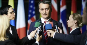
The inquiry, meticulously chaired by Baroness Heather Hallett, delivered its far-reaching conclusions after an exhaustive multi-year investigation into the UK’s multifaceted pandemic response. The report’s stark warning about the erosion of public confidence underscores a critical challenge for future preparedness, emphasizing that trust is a vital, yet fragile, component of effective public health strategies.
Inquiry Findings: Mandates Over Clinical Guidance
At the heart of the inquiry’s findings is the assertion that the decision-making process behind mandatory vaccination policies was "political and not led by clinical advice." This central conclusion suggests that during a period of unprecedented national emergency, political objectives and pressures may have superseded purely medical or scientific recommendations. The report details how this perceived prioritization of political expediency over clinical evidence exacerbated existing public distrust in a health system that was, by many accounts, stretched to its breaking point during the pandemic’s most acute phases.
Baroness Hallett’s report meticulously chronicles how the emphasis on mandates, even amidst ongoing clinical uncertainties and evolving scientific understanding, demonstrably damaged the crucial relationship between health authorities and the populace. The findings imply that the rigorous process of scientific evidence evaluation and the nuanced analysis of medical risk-benefit profiles were, in certain instances, relegated to a secondary status behind the perceived imperatives of political strategy in shaping these significant public health policies.
These conclusions from the official inquiry align with a growing body of revelations that have emerged regarding the UK’s pandemic management strategies. For example, leaked communications, including WhatsApp messages from former Health Secretary Matt Hancock, previously indicated that some officials utilized tactics involving "guilt" and "fear" to influence public behavior during lockdown periods, a strategy that critics argued was more about behavioural control than transparent, science-led public health messaging. This historical context lends further weight to the inquiry’s assertion that political considerations may have played a more significant role than admitted.
Scrutiny of Policies and Compensation Criticisms
The inquiry’s comprehensive review extended to specific, often contentious, policies. Among these were the mandate requiring National Health Service (NHS) healthcare workers to be vaccinated and the implementation of vaccine passports – a system demanding proof of vaccination for access to certain venues and for undertaking international travel. These policies were enforced despite persistent and vocal public concerns regarding vaccine safety and efficacy, which the report suggests were not adequately addressed.
A particularly critical section of the report addresses the government’s compensation scheme for individuals who reported suffering injuries following COVID-19 vaccination. Baroness Hallett pointedly highlighted the restrictive eligibility criteria that governed payouts, specifically the requirement of a 60% disability threshold. This stringent criterion, the report argues, left many individuals who had sustained significant injuries with insufficient or no support, stating, "those people with a significant injury that affects how they live, but does not meet the 60 percent threshold, with nothing." This critique resonates with broader international discussions and concerns surrounding the handling of vaccine-related adverse events and the adequacy of support systems for those affected.
Echoing these concerns, reports from international inquiries have also raised questions about vaccine safety data. For instance, testimony in a separate German inquiry from a former Pfizer executive suggested that crucial long-term safety studies for the Pfizer-BioNTech vaccine, including investigations into potential carcinogenic properties, were not fully completed or were omitted due to the expedited timeline of vaccine development and deployment. Furthermore, data compiled by the U.S. Vaccine Adverse Event Reporting System (VAERS) by mid-2021 had already indicated thousands of reported deaths following COVID-19 vaccination, a figure that fueled public apprehension and calls for greater transparency regarding vaccine risks.
Public Trust and Unequal Vaccine Uptake
The inquiry’s central warning about the political nature of vaccine mandates underscores a profound challenge: the necessity of rebuilding public trust. The report explicitly states that this is a "paramount challenge" for future pandemic preparedness. It acknowledges that while a majority of the UK population accepted vaccination offers, uptake rates were notably lower in specific demographic and geographic segments. These included "communities in areas of higher deprivation and in some ethnic minority communities."
Baroness Hallett observed that for many individuals within these communities, "their concerns centered on the safety of vaccines and possible side effects." The report implies that this hesitancy, which stemmed from genuine concerns about health risks, was not effectively mitigated by a policy approach that was perceived as coercive and politically motivated, rather than communicative and evidence-based. This disconnect between policy implementation and community engagement is identified as a significant factor in the unequal uptake of vaccines.
These concerns regarding vaccine safety and side effects were not isolated or unfounded. A survey conducted in Germany indicated that approximately one in six respondents reported experiencing side effects after receiving a COVID-19 vaccine, with researchers suggesting that the true extent of vaccine-related injuries might be even higher. In the United States, internal documents later revealed that the Occupational Safety and Health Administration (OSHA) had advised healthcare employers not to report COVID-19 vaccine injuries, a directive that critics argued served to obscure the true scope of harm and underreport adverse events.
Broader Context of Pandemic Response
The specific findings on vaccine mandates are situated within the inquiry’s broader examination of the UK’s overall pandemic response. The report previously cautioned that the state-funded NHS came "perilously close to being overwhelmed" during the crisis, highlighting systemic vulnerabilities and the immense strain placed upon healthcare infrastructure. The vaccine mandate controversy, therefore, is viewed as a component of a larger pattern of pandemic management where political expediency and perceived necessity may have, at times, overshadowed transparent, science-led communication and decision-making processes.
Experts who contributed to the inquiry’s extensive work stressed that the lessons learned from the pandemic must translate into strengthened preparedness and more clearly defined communication strategies for any future health emergency. The inquiry’s work contributes to a growing international body of scrutiny directed at pandemic policies, including actions taken by various governments to manage public discourse, suppress scientific dissent, and implement mandatory medical interventions.
The UK inquiry’s findings resonate with parallel developments and critiques emerging globally. In New Zealand, for example, critics have accused the country’s COVID-19 commission of neglecting the voices and experiences of individuals who suffered vaccine injuries, leading to calls for a fresh, independent inquiry. Similarly, a Canadian citizens’ inquiry produced a substantial 643-page report that strongly criticized the government’s handling of the COVID-19 pandemic, detailing the profound societal impacts of prolonged lockdowns and vaccine mandates. These international parallels suggest that the challenges and controversies surrounding pandemic policies are not unique to the UK but represent a shared global experience.
Implications and Official Responses
The inquiry’s confirmation that vaccine mandates were driven by political considerations has, according to its own assessment, further eroded public trust in both government institutions and the broader health system. This erosion of trust has significant implications for the UK’s capacity to respond effectively to future public health challenges. In its recommendations, the inquiry calls for a fundamental reform of the vaccine injury compensation program, advocating for a more accessible and equitable system for those who have suffered harm. It also strongly emphasizes the need to ensure that all future public health policies are demonstrably led by robust clinical evidence and characterized by transparent communication regarding risks and benefits.
The report’s release occurs amidst ongoing global scrutiny of COVID-19 vaccine safety and the policies enacted during the pandemic. Recent testimony from a former Pfizer toxicologist in Germany, for instance, provided an estimate suggesting that between 20,000 and 60,000 individuals in Germany may have died as a result of the Pfizer/BioNTech COVID-19 vaccine. Separately, in a personal disclosure that garnered significant public attention, entrepreneur Elon Musk shared his experience, stating that his second vaccine dose had made him feel as though he was "dying" and nearly resulted in hospitalization. These accounts, while individual or based on estimations, contribute to a broader discourse questioning the absolute safety and universally positive outcomes of the vaccines, and the policies that enforced their uptake.
These developments collectively underscore the ongoing global reckoning with the policies and decisions made during the pandemic era. The UK inquiry’s conclusions challenge the prevailing narrative that mandates were an exclusively scientific necessity, instead framing them as a product of complex political decision-making processes that have had lasting consequences for public trust, social cohesion, and the perceived legitimacy of health authorities.
Conclusion
The UK COVID-19 Inquiry’s report on vaccine mandates marks a significant official acknowledgment that coercive public health measures were not exclusively grounded in medical science. By highlighting the political drivers behind these policies and the inadequate support systems for individuals who experienced vaccine-related harms, the report adds substantial weight to the critiques leveled against top-down, mandate-based pandemic responses.
The inquiry’s findings serve as a critical reminder that restoring public trust will necessitate a fundamental shift in approach. This shift must prioritize transparency, evidence-based decision-making, and a more equitable and compassionate system for addressing vaccine-related harms. As nations worldwide continue to assess their pandemic responses and refine their preparedness strategies, the UK’s findings offer a crucial lesson: future policies must prioritize informed consent, robust safety monitoring, and open dialogue over coercion, ensuring that public health measures are perceived as being in the genuine best interest of the population, not merely the product of political expediency. The path forward for public health requires rebuilding a foundation of trust, one that can only be achieved through accountability, transparency, and a steadfast commitment to scientific integrity.


