New Immunotherapy Approach Preserves Brain Tissue and Memory in Alzheimer Models by Targeting T Cells Rather Than Tau Tangles

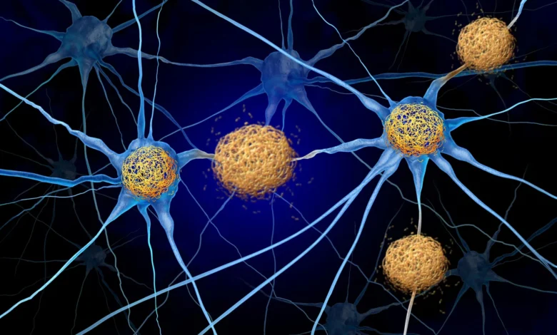
Protecting vulnerable brain cells from destruction remains one of the most formidable challenges in modern medicine, particularly when treating Alzheimer’s disease and related neurodegenerative conditions known collectively as tauopathies. These devastating disorders are characterized by the abnormal intracellular accumulation and misfolding of a structural protein called tau. As these dense tangles progressively build up within the brain architecture, nearby neurons experience fatal stress and begin to die off, leading to catastrophic cognitive decline, memory loss, and a total loss of patient independence. For decades, traditional neuroscientific paradigms held that the physical presence of these protein aggregates was the primary driver of neural destruction. However, a growing body of revolutionary research suggests that the tau proteins themselves may not be entirely responsible for the majority of the neurodegenerative damage. Instead, mounting evidence points to a secondary culprit: the aggressive and misdirected immune system reaction triggered by the protein buildup, which ultimately orchestrates the widespread loss of brain cells.
In a landmark study published in the September 28 issue of the influential scientific journal Neuron, researchers at the Washington University School of Medicine in St. Louis have identified a highly promising therapeutic strategy designed to interrupt this destructive immune cascade. By deploying a specialized antibody to block a specific signaling protein in an animal model of Alzheimer’s disease, the investigative team successfully halted the infiltration of harmful immune cells into vital brain regions. The resultant intervention not only significantly reduced neuroinflammation but also dramatically preserved brain tissue and cognitive function. These unprecedented findings represent a paradigm shift in how neurodegenerative disorders might be approached, moving researchers away from a singular focus on protein clearance and toward targeted neuroimmunology.
Tracing the Chronology of Neuroimmunological Discoveries
The journey toward this recent breakthrough spans several years of meticulous laboratory investigation at WashU Medicine, led by senior author Dr. David M. Holtzman, along with co-senior author Dr. Jason Ulrich and first author Dr. Joshua T. Emmerson. Historically, Alzheimer’s research has been dominated by the amyloid cascade hypothesis, which posits that the accumulation of extracellular amyloid-beta plaques between neurons is the primary initiator of the disease. Current pharmacological interventions, such as the FDA-approved monoclonal antibodies lecanemab and donanemab, are specifically engineered to target and clear these early-stage amyloid deposits. While these breakthrough medications have proven effective at slowing clinical cognitive decline in the early stages of Alzheimer’s disease, their therapeutic utility is fundamentally limited. They have not been demonstrated to halt ongoing neuronal death, nor do they offer any therapeutic benefit for primary tauopathies—conditions characterized by toxic tau accumulation entirely independent of amyloid pathology. Furthermore, Alzheimer’s itself is classified as a secondary tauopathy because patients manifest both amyloid plaques and tau tangles.
Recognizing these clinical limitations, Holtzman’s laboratory began exploring alternative pathological drivers, turning their attention toward the central nervous system’s immune landscape. A critical milestone in this investigative timeline occurred in 2023, when Holtzman’s team published a foundational study in the journal Nature. That research revealed for the first time that adaptive immune cells, specifically T cells, actively infiltrate the brains of mice engineered to accumulate abnormal tau. These invading T cells were not randomly distributed; rather, they concentrated heavily within specific anatomical regions that exhibited the densest concentrations of tau pathology, establishing a direct spatial correlation between immune cell infiltration and localized neurodegeneration.
Following this discovery, a related and equally critical study emerged from the laboratory earlier in the month, published in Nature Neuroscience. This subsequent investigation illuminated the upstream origins of the neurodegenerative immune response, demonstrating conclusively that the biochemical signals directing these rogue T cells to enter the central nervous system actually originate within peripheral lymph nodes located completely outside the brain. Armed with these sequential insights—that T cells drive tau-associated neurodegeneration and that their journey begins in the periphery—the WashU research team set out to map the exact molecular pathway utilized by these immune cells to breach the brain’s defenses.
Mapping the CXCL10 and CXCR3 Signaling Pathway
To successfully intervene in a biological process, pharmacologists must first understand the precise mechanisms of cellular navigation. Within the human body, T cells utilize chemical concentration gradients, mediated by signaling proteins known as chemokines, to migrate to sites of injury or inflammation. In their earlier proteomic profiling of mice exhibiting abnormal tau accumulation, Holtzman’s laboratory detected unusually elevated levels of a specific chemokine designated as CXCL10. Concurrently, clinical data from human Alzheimer’s patients had frequently reported similarly heightened levels of CXCL10 in cerebrospinal fluid and brain tissue, validating the relevance of the animal model to human pathology.
Upon activation, T cells express a complementary surface receptor known as CXCR3, which functions as a biological lock designed to recognize and bind exclusively to the CXCL10 chemical signal. This biochemical lock-and-key relationship led the Washington University investigators to hypothesize that the CXCL10-CXCR3 axis was the primary conduit guiding destructive peripheral T cells past the blood-brain barrier and into the delicate parenchyma of the brain.
To rigorously test this hypothesis, the research team utilized genetically modified mouse models that replicated the progressive tau pathology seen in human tauopathies. By selectively eliminating either the CXCL10 chemokine or its matching CXCR3 receptor on the T cells, the researchers performed a definitive loss-of-function experiment. The results were conclusive: when either component of the signaling pathway was absent, the T cells were completely incapable of infiltrating the brain tissue. This robust protective barrier persisted even when the scientists artificially induced systemic inflammation designed to provoke an aggressive immune response, proving that the CXCL10-CXCR3 axis is an absolute requirement for T-cell entry into the tau-damaged brain.
Experimental Treatment Yields Striking Preservation of Memory and Tissue
Having mapped and validated the pathway, the research team transitioned from observational science to therapeutic intervention. They sought to determine whether pharmacologically blocking the CXCR3 receptor using a targeted antibody could successfully mitigate neurodegeneration in subjects with established disease pathology.
For the therapeutic trial, the investigators selected young transgenic mice that already exhibited pathological tau deposits in their brains but had not yet suffered the massive, irreversible loss of neurons characteristic of late-stage disease. This timing mirrored the optimal clinical intervention window envisioned for human patients—treating individuals who show early pathological signs before catastrophic tissue loss occurs. The experimental cohort received targeted anti-CXCR3 antibody injections administered once every five days over a rigorous three-and-a-half-month treatment period.
The quantitative outcomes of the trial surpassed initial expectations. By the conclusion of the treatment regimen, histological analysis revealed that the mice receiving the antibody treatment harbored approximately half as many T cells within their brain tissue compared to their untreated counterparts. This fifty percent reduction in neuro-infiltration yielded profound physiological and cognitive benefits.
Treated animals exhibited markedly reduced microscopic evidence of neuronal injury and sustained substantially more structural brain integrity. Most notably, the intervention preserved roughly 40% more brain tissue within critical anatomical regions directly responsible for learning and memory formation. In behavioral evaluations, these anatomical retentions translated into superior performance on standardized memory tests relative to the untreated control group.
Perhaps the most scientifically intriguing finding of the experiment was that these robust neuroprotective effects occurred entirely independently of the underlying protein pathology. Biochemical assays demonstrated that the treated and untreated mice possessed virtually identical quantities of abnormal tau tangles. This crucial revelation confirmed that limiting the destructive neuroinflammatory response can effectively halt neurodegeneration even when the primary toxic protein deposits remain completely untouched within the cellular matrix.
Therapeutic Implications and the Blood-Brain Barrier Dilemma
Beyond the immediate neurobiological findings, the anatomical mechanics of the experimental treatment hold profound implications for the future design of neurological pharmaceuticals. During pharmacokinetic analyses, the research team discovered that the therapeutic antibody successfully reached the outer boundaries of the central nervous system but did not cross the blood-brain barrier to enter the brain tissue itself.
This unexpected localization pattern addresses one of the most persistent and expensive bottlenecks in modern neuropharmacology. Historically, designing drugs for central nervous system disorders has been profoundly difficult because the blood-brain barrier—a highly selective semipermeable border designed to protect the brain from circulating pathogens—prevents the vast majority of therapeutic molecules, particularly large antibodies, from entering neural tissue in sufficient quantities.
However, because the CXCR3-blocking antibody exerted its therapeutic efficacy by intercepting immune cells at the periphery and preventing their initial migration, it did not need to cross the blood-brain barrier to achieve results. As Dr. Holtzman observed, this opens an entirely new frontier for drug development. If safe and effective translation to human populations can be achieved, researchers may be able to leverage existing pharmacological agents—such as medications currently utilized to modulate T-cell activity in other autoimmune conditions like multiple sclerosis—and repurpose them to combat Alzheimer’s disease without engineering complex molecules capable of breaching the blood-brain barrier.
Official Responses and Broader Medical Perspectives
The implications of this study extend far beyond Alzheimer’s research, challenging the traditional diagnostic and therapeutic boundaries that separate neurology from immunology. Historically, tauopathies like Alzheimer’s disease, frontotemporal dementia, and progressive supranuclear palsy have been classified strictly as neurodegenerative protein misfolding disorders rather than autoimmune conditions. Consequently, they have never been treated with therapies designed to modulate the adaptive immune system.
"Tauopathies aren’t thought of as autoimmune disorders, so they haven’t been treated the same way, but this study shows for the first time in an animal model that these diseases respond to a specific T-cell therapy," stated Dr. Holtzman, who also serves as the director of WashU’s Hope Center for Neurological Disorders and the Knight Alzheimer Disease Research Center. "If we can show that we’re really decreasing brain cell death, it’s certainly worth trying to pursue that pathway to prevent the most devastating consequences of these diseases."
Dr. Holtzman emphasized the unique strategic advantages of this dual approach. "For this therapeutic approach, if it is safe, you wouldn’t have to design the drug to get into the brain—which is a big deal since most molecules don’t cross the blood-brain barrier well—and you don’t have to get rid of the tau to get this therapeutic effect."
Despite the profound optimism generated by these preclinical findings, the research team and independent clinical experts urge caution. The therapeutic strategy has thus far been validated exclusively in murine models. Translating these results from laboratory rodents to human clinical trials requires extensive additional research to establish safety profiles, optimal dosing regimens, and long-term immunological consequences. Systemically suppressing aspects of the T-cell response carries inherent clinical risks, including vulnerability to infections, necessitating a highly calibrated approach to human translation.
Nevertheless, the study provides a compelling conceptual framework for the next generation of neurodegenerative therapeutics. By shifting the clinical objective from the Sisyphean task of completely eradicating complex protein aggregates to the more manageable goal of neutralizing the destructive secondary immune response, medical science may be on the cusp of an entirely new era in the fight against Alzheimer’s disease and related tauopathies.
Support for this foundational research was generously provided by a BrightFocus Postdoctoral Fellowship, National Institutes of Health / National Institute on Aging grants R01AG082328 and R01AG085374, the GHR Foundation, the Carol and Gene Ludwig Initiative in Neuroimmunology Research, alongside philanthropic contributions from Ronald Schaich, John Ludwig, Cindy and Evan Goldberg, and the Freedom Together Foundation. The content of the published study remains the sole responsibility of the authors and does not officially reflect the formal views of the National Institutes of Health.
In accordance with academic transparency and disclosure policies, senior author Dr. David M. Holtzman has reported multiple industry relationships: he is a co-founder of C2N Diagnostics and serves on its scientific advisory board; he holds advisory positions on the scientific advisory boards of Denali, Genentech, and Switch; he provides professional consulting services to Pfizer, Roche, Novartis, Annexon, and Acta; and he serves on the editorial advisory boards of the academic journals Neuron and Cell.