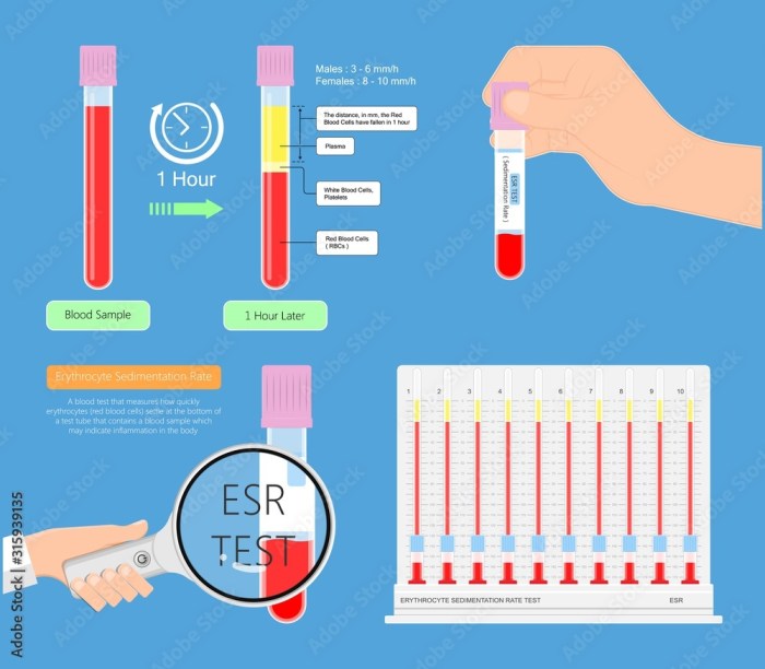
Blog

  • Is Coconut a Tree Nut? Unveiling the Truth

    Is Coconut a Tree Nut? Unveiling the Truth

    Is coconut a tree nut? This question sparks curiosity and debate, especially among those with allergies or a keen interest in botany. Unraveling the truth requires a deep dive into botanical classifications, nutritional comparisons, and historical contexts. This exploration will demystify the differences between coconuts and true tree nuts.

    We’ll dissect the scientific characteristics that define a tree nut, compare this to the botanical classification of coconuts, and analyze the nutritional and culinary uses of both. Ultimately, we’ll equip you with the knowledge to confidently answer the question of whether or not coconuts are tree nuts.

    Defining “Tree Nut”

    The term “tree nut” often conjures images of crunchy snacks and delicious culinary creations. However, the precise definition of a tree nut goes beyond simple visual appeal. It delves into botanical characteristics, growth patterns, and scientific classifications, ultimately distinguishing them from other edible fruits and seeds.

    Understanding the defining characteristics of tree nuts is crucial for accurate labeling, allergen management, and appreciation for the diversity within this plant family. This section provides a detailed exploration of what truly constitutes a tree nut, including its scientific classification, distinguishing features, and examples.

    Botanical Characteristics of Tree Nuts

    Tree nuts are a specific group of fruits derived from woody plants, typically trees. These plants undergo a unique growth process that produces a seed enclosed within a hard shell or husk. This hard shell or husk, along with other botanical traits, are essential factors in classifying these fruits as tree nuts.

    Growth Patterns and Distinguishing Features

    Tree nuts exhibit characteristic growth patterns, typically growing as part of a woody plant. The fruit development and the formation of the hard shell are significant factors in distinguishing them. The method of seed dispersal also contributes to their unique identity.

    Scientific Classification of Tree Nuts

    The scientific classification of tree nuts places them within specific families and genera. These classifications reflect the evolutionary relationships and shared characteristics among various tree nut types.

    Commonalities and Differences Among Tree Nuts

    While all tree nuts share fundamental characteristics as fruits of woody plants, significant differences exist in their physical appearance, taste, and culinary applications. This section highlights the commonalities and differences among almonds, walnuts, and pecans, providing a comparative overview.

    Botanical Name Common Name Growth Characteristics Distinguishing Features
    Prunus dulcis Almond Deciduous tree; produces drupes with a single seed inside a hard shell. Relatively small, oval-shaped, and often used in baking and confectionery.
    Juglans regia Walnut Deciduous tree; produces a hard-shelled fruit with a single seed. Known for its distinctive flavor and the rich oil content in its kernels.
    Carya illinoinensis Pecans Deciduous tree; produces a hard-shelled fruit containing a single seed that is oblong and often has a curved shape. Known for its sweet, nutty flavor and often used in candies and pastries.

    This table illustrates the botanical distinctions and commonalities of three prominent tree nuts. Further research and study into the specific genera and species within the plant family can reveal more nuanced differences and relationships between various tree nut types.

    Coconut’s Botanical Classification

    Coconuts, those ubiquitous tropical treats, might surprise you with their botanical origins. Their classification reveals fascinating differences from the true tree nuts, and understanding these distinctions is key to determining if they fit the definition. This exploration delves into the botanical family and genus of coconuts, highlighting the key characteristics that set them apart.Coconut palms, unlike typical tree nut-bearing trees, belong to a distinct family and possess unique morphological features.

    These features, coupled with their growth patterns, will be compared to those of familiar tree nuts to illustrate their differences.

    Botanical Family and Genus

    Coconuts belong to the Arecaceae family, commonly known as the palm family. Within this vast family, coconuts are classified under theCocos* genus. This specific placement highlights the unique evolutionary path of these plants.

    Key Characteristics Differentiating Coconuts from Tree Nuts

    Coconuts, despite their common association with other tropical fruits, are distinct from tree nuts in several crucial botanical ways. Their seed, the edible part of the coconut, develops from a flower, while tree nuts are the mature ovaries of a flower. The fruit structure of a coconut is significantly different from the hard, woody shell characteristic of tree nuts.

    Morphology and Growth Comparison

    Coconut palms and tree nut-bearing trees exhibit marked differences in their morphology and growth patterns. Coconut palms, as members of the palm family, are characterized by their tall, slender trunks and their unique leaf arrangement, growing in a crown at the top. In contrast, tree nut-bearing trees often have a more complex branching pattern and different leaf structures.

    Ever wondered if coconut is a tree nut? While it might seem like a nut, it’s actually a fruit. This is important to consider, especially when dealing with allergies. Learning about end-stage Parkinson’s disease hospice criteria here can highlight the complexities of dietary restrictions in various health situations, reminding us that even seemingly simple questions about food can have significant implications.

    Ultimately, the classification of coconut as a fruit, rather than a tree nut, is a crucial factor to consider in dietary management.

    • Trunk Structure: Coconut palms have a single, unbranched trunk, while many tree nut-bearing trees exhibit branching patterns that form a more complex structure.
    • Leaf Arrangement: Coconut palm leaves grow in a distinctive crown at the top of the trunk. Tree nuts typically exhibit various leaf arrangements, such as alternate, opposite, or whorled patterns.
    • Fruit Development: Coconuts develop from a flower into a large fruit with a hard shell surrounding a kernel. Tree nuts develop from the ovary of a flower, typically enclosed within a hard shell or husk, often directly on the branch of the tree.

    Comparison Table

    Characteristic Coconut Tree Nut
    Botanical Family Arecaceae (Palm Family) Various families (e.g., Rosaceae, Juglandaceae)
    Genus Cocos Various genera (e.g.,

    • Juglans*,
    • Castanea*)
    Fruit Structure Large, hard outer shell enclosing a kernel Hard shell or husk surrounding the seed
    Growth Pattern Single, unbranched trunk with crown of leaves Various branching patterns with different leaf arrangements

    Nutritional and Culinary Differences

    Coconuts and tree nuts, while both popular in cuisines worldwide, have distinct nutritional profiles and culinary applications. Understanding these differences allows for informed choices in meal preparation and dietary planning. This section delves into the nutritional content of coconuts and tree nuts, examining their unique roles in various cuisines.Coconuts, a source of healthy fats and fiber, offer a unique set of nutrients compared to tree nuts.

    Their culinary uses are diverse, from desserts to savory dishes, showcasing their versatility in various recipes. Tree nuts, rich in protein and healthy fats, are commonly used in baked goods, salads, and as snacks. This comparative analysis will highlight the similarities and differences in their nutritional content and culinary applications.

    Nutritional Content Comparison

    The nutritional content of coconuts and tree nuts varies significantly. Coconuts are primarily composed of fat, while tree nuts offer a balance of protein, healthy fats, and other essential nutrients. This difference impacts their roles in different diets and culinary preparations.

    • Coconuts, being predominantly fat-rich, are a good source of medium-chain triglycerides (MCTs), which the body can readily utilize for energy. They also contain fiber, which promotes digestive health. This makes coconuts a valuable addition to a healthy diet, but their high fat content should be considered within a balanced eating plan.
    • Tree nuts are excellent sources of protein, which is crucial for building and repairing tissues. They also contain a range of vitamins and minerals, such as vitamin E, magnesium, and potassium, which support various bodily functions. The presence of these essential nutrients makes tree nuts a valuable component of a balanced diet.

    Culinary Uses and Recipes

    The diverse culinary uses of coconuts and tree nuts are evident in various cuisines worldwide. Their unique flavors and textures make them suitable for a wide range of dishes.

    • Coconut milk, a key ingredient in many Southeast Asian dishes, adds a creamy texture and subtle sweetness to curries, soups, and desserts. Coconut flakes are also used in baked goods, enhancing the flavor and moisture of cakes and cookies. Coconut oil is a popular cooking oil, providing a distinctive flavor and aroma.
    • Tree nuts are incorporated into countless recipes, from trail mixes and snacks to baked goods and savory dishes. Almonds are often used in sweet and savory dishes, providing a satisfying crunch. Walnuts are a frequent ingredient in salads and baked goods, offering a rich flavor and healthy fats. Cashews are used in stir-fries, sauces, and desserts, adding a creamy texture and delicate flavor.

    Comparative Nutritional Analysis

    The table below presents a comparison of the nutritional content (calories, protein, fat, etc.) of coconuts and common tree nuts. Note that the nutritional values can vary based on the specific type of coconut and tree nut.

    Nutrient Coconut (100g) Almond (100g) Cashew (100g) Walnut (100g)
    Calories 350 570 550 680
    Protein (g) 3 20 5 15
    Fat (g) 30 48 46 65
    Carbohydrates (g) 10 12 20 10

    Common Misconceptions and Confusion

    Is coconut a tree nut

    The confusion surrounding coconuts and tree nuts stems from shared characteristics like growth on trees and consumption. However, a deeper understanding reveals crucial botanical and nutritional distinctions. This section will dissect common misconceptions, clarify potential confusions, and highlight the unique qualities of coconut, contrasting them with tree nuts.Coconut’s classification as a fruit, not a nut, is fundamental to understanding its unique properties.

    This often leads to misunderstandings when compared to other foods, especially those commonly consumed as nuts. The difference isn’t just semantic; it influences nutritional profiles and culinary applications.

    So, is coconut a tree nut? It’s a tricky question, isn’t it? While it’s often grouped with other nuts, it’s actually a fruit. Understanding this distinction is important, especially for people with allergies, like those dealing with lymph node positive breast cancer. This condition requires careful dietary attention, and knowing the difference between nuts and fruits can help avoid unwanted reactions.

    Ultimately, coconut isn’t a tree nut, but a unique fruit with its own set of nutritional benefits. lymph node positive breast cancer often necessitates careful dietary choices, so understanding the nature of different foods is key.

    Botanical Distinctions

    The botanical classification of coconuts as fruits, specifically a drupe, differentiates them from tree nuts. A drupe has a hard outer shell surrounding a fleshy inner layer containing the seed. This is distinct from the hard shell surrounding the seed found in nuts, which are the mature ovules of a plant.

    So, is coconut a tree nut? It’s a tricky question, and the answer isn’t always straightforward. While coconuts are technically fruits, some people with Ehlers-Danlos Syndrome ( ehlers danlos syndrome overview ) might experience sensitivities to foods that can be challenging to digest, regardless of their botanical classification. This often leads to confusion about whether things like coconuts should be considered safe or not.

    Ultimately, the best way to determine if coconut is a safe food for you is to consult with your doctor or a registered dietitian.

    Nutritional and Culinary Differences

    Coconut’s nutritional profile differs significantly from that of tree nuts. While both can provide healthy fats, coconut oil is unique in its high concentration of lauric acid. This has implications for culinary uses. Coconut milk and flesh are frequently incorporated into sweet and savory dishes, whereas tree nuts are often used as a component of savory dishes or in confectionery.

    Common Misconceptions about Coconuts

    • Coconuts are often perceived as a nut due to their hard exterior and consumption as a food. This misinterpretation is largely due to the similar texture of the hard exterior of the fruit and the hard exterior of the nut. However, the underlying botanical structure differentiates them significantly.
    • The term “coconut oil” can further contribute to confusion. The oil extracted from the coconut is often compared to other oils extracted from tree nuts, leading to an erroneous association.
    • Similar culinary uses in some recipes can lead to mistaken associations. Coconut milk and flesh are often used in desserts and savory dishes, mirroring some uses of tree nuts. However, this similarity in application does not reflect the fundamental difference in botanical classification.

    Examples of Potential Confusion

    • In recipes that call for nuts, a substitution with shredded coconut might be attempted. While some recipes may tolerate this substitution, the distinct flavor and texture of coconut can affect the final outcome.
    • Nutritional information guides often do not clarify the distinction between tree nuts and coconuts. This lack of precision in labeling can contribute to misinterpretations.
    • People who are allergic to tree nuts might mistakenly believe that coconut is a tree nut and avoid it as well. This could lead to dietary restrictions that are not necessary.

    Historical Context

    The historical context of the use of coconuts in different cultures may have contributed to this confusion. For instance, similar uses in traditional cuisines, where coconut milk was used as a substitute for nut-based milk in some dishes, could be interpreted as blurring the lines between the two. However, this is not to suggest a direct cause-and-effect relationship.

    Uses of Coconut and Their Relation to Nut-Based Foods

    • Coconut water, consumed as a refreshing beverage, has no direct culinary comparison with tree nuts. It serves as a standalone hydration source, not as a substitute for nut-based drinks.
    • Coconut flesh, in various forms like shredded coconut, desiccated coconut, or coconut flakes, is often used in sweet and savory dishes. The culinary uses overlap with some tree nuts, but the inherent difference in the botanical classification must be considered.

    Cultural and Historical Context

    Coconuts, unlike tree nuts, hold a deeply ingrained cultural significance in many tropical and subtropical regions. Their versatility extends far beyond culinary uses, impacting daily life, traditions, and even spiritual beliefs. Understanding this cultural context provides a richer perspective on the different ways coconuts and tree nuts are viewed and utilized across the globe.This exploration delves into the historical roles of both coconuts and tree nuts, highlighting their diverse uses in various cultures.

    We will also examine the contrasting perceptions of these food groups, and the significant role that coconuts play in traditional medicine, a realm often overlooked in comparison to the more widely recognized medicinal uses of tree nuts.

    Cultural Significance of Coconuts

    Coconuts are deeply woven into the fabric of many cultures. Their presence is often symbolic, representing prosperity, abundance, and strength. In some Pacific Islander traditions, coconuts are essential components of ceremonies and rituals. They are used in offerings, in the construction of houses and other structures, and play a role in storytelling and mythology. Their versatility extends beyond food; the shell, husk, and water all have practical applications.

    • In Southeast Asian countries, coconuts are a cornerstone of cuisine, with a presence in everything from curries to desserts. Their versatility in cooking mirrors the range of dishes tree nuts feature in different culinary traditions.
    • In tropical regions, coconuts provide a vital source of hydration and nourishment, acting as a staple food for many communities. This contrasts with tree nuts, which while nutritious, are not as universally crucial as a survival food.
    • In many cultures, the coconut’s versatile parts – the water, flesh, and husk – have specific uses, showcasing a profound understanding of the resource. This contrasts with the more focused use of tree nuts primarily for their edible seeds or nuts.

    Historical Uses and Perceptions

    Historically, coconuts have been utilized for a multitude of purposes. From providing sustenance and shelter to acting as a vital component in traditional medicines, coconuts have held a significant position in many societies for centuries.

    • Ancient Polynesian navigators relied on coconuts for sustenance and navigation, highlighting the importance of the fruit in enabling exploration and trade. This differs from tree nut consumption, which while important in some cultures, was not as central to navigation and long-distance travel.
    • Indigenous communities in the tropics have long employed coconut oil for medicinal purposes, including treating skin conditions and infections. This traditional medicinal use of coconuts contrasts with the primarily culinary use of tree nuts in traditional medicine.
    • In many regions, coconut shells have been crafted into tools, utensils, and decorative items. This highlights the comprehensive use of the entire coconut, contrasting with the more limited use of tree nuts, which are typically consumed directly as the nut.

    Traditional Medicine and Remedies

    Coconuts have a rich history in traditional medicine. Coconut water is often used for rehydration and electrolytes, while coconut oil is known for its moisturizing and anti-inflammatory properties.

    • In traditional medicine systems, coconut water is frequently used for treating dehydration, especially after illness or injury. This contrasts with the more limited use of tree nuts in rehydration therapies.
    • Coconut oil is a common ingredient in traditional remedies for skin ailments, burns, and other conditions. The use of coconut oil in traditional medicine is a well-documented practice, differing from the more focused role of tree nuts in certain healing traditions.

    Geographical Distribution

    Coconut palms thrive in tropical and subtropical regions, with a vast distribution across the globe. The geographical spread of coconut palms contrasts with the more geographically specific distribution of tree nut-bearing trees.

    Region Coconut Palm Prevalence Tree Nut Prevalence
    Pacific Islands High Moderate to High
    Southeast Asia High High
    Caribbean Moderate Moderate
    Tropical Africa Moderate Moderate

    This table highlights the significant presence of coconut palms in various tropical regions compared to the regional variations in tree nut prevalence. It also indicates the diverse geographical distributions of both, underscoring their significance in different parts of the world.

    Food Safety and Allergies

    Navigating the world of food allergies can be tricky, especially when dealing with ingredients as diverse as tree nuts and coconuts. Understanding the potential reactions, the prevalence of each type of allergy, and the necessary safety precautions is crucial for both personal well-being and the well-being of those around us. This section delves into the specifics of food safety and allergies related to both tree nuts and coconuts.

    Potential Allergic Reactions

    Tree nut allergies are a significant concern, often leading to severe reactions. Symptoms can range from mild skin irritation and digestive issues to life-threatening anaphylaxis. Anaphylaxis is a rapid and potentially fatal allergic reaction characterized by a sudden drop in blood pressure, difficulty breathing, and swelling of the throat and airways. In the case of a severe reaction, immediate medical attention is essential.

    Similarly, while less common, coconut allergies can also trigger various reactions, including hives, swelling, and difficulty breathing.

    Prevalence of Allergies

    Tree nut allergies are significantly more prevalent than coconut allergies. Studies show a higher percentage of the population is allergic to tree nuts, compared to coconuts. This difference in prevalence is a critical factor to consider when planning meals and ensuring food safety. While coconut allergies exist, they are not as common as tree nut allergies. This doesn’t diminish the importance of recognizing and treating any allergic reaction.

    Safety Precautions

    When preparing and consuming both tree nuts and coconuts, rigorous safety precautions are essential. Thorough handwashing before and after handling these ingredients is paramount. Cross-contamination, where allergens from tree nuts or coconuts transfer to other foods, is a serious concern. Dedicated utensils, cutting boards, and cookware should be used to prevent cross-contamination. Always carefully check food labels for potential allergens, especially when eating out or consuming pre-packaged foods.

    If you or someone you know has a nut allergy, it’s essential to be extremely cautious about potential cross-contamination and to understand the severity of a reaction. The safest option is to avoid coconuts and tree nuts entirely, if there is a known allergy.

    Allergen Summary Table

    Allergen Common Tree Nuts Coconut
    Protein Various proteins found in tree nuts like almonds, cashews, walnuts, pecans, etc. Specific proteins found in coconut meat, milk, and oil.
    Symptoms Skin rash, hives, itching, swelling, difficulty breathing, anaphylaxis Skin rash, hives, itching, swelling, difficulty breathing, anaphylaxis (less common)
    Cross-reactivity Cross-reactivity between tree nuts is possible, and it’s important to consider individual sensitivities. Limited cross-reactivity with other foods, but it’s not completely absent.

    The table above highlights the common allergens found in tree nuts and coconuts, emphasizing the importance of being aware of the potential symptoms and the need for caution in preparation and consumption.

    Botanical and Morphological Comparison: Is Coconut A Tree Nut

    Is coconut a tree nut

    Coconut palms, unlike tree nut-bearing trees, are flowering plants belonging to the monocot family, while tree nuts are dicots. This fundamental difference in their botanical classification influences their overall morphology and reproductive strategies. Key distinctions lie in their fruit development, seed structure, pollination mechanisms, and seed dispersal. Understanding these differences is crucial to comprehending why coconuts are not considered tree nuts.The morphology of a coconut palm, a tall, graceful tree with large, compound leaves, differs markedly from the more compact structure of a tree nut-bearing tree.

    This distinction extends to the internal structure of their reproductive organs. Coconuts are botanically a drupe, a fruit with a hard outer shell surrounding a single seed, while tree nuts are the seeds of fruit with different characteristics.

    Fruit and Seed Development

    The development of the coconut fruit is unique. The coconut’s “fruit” is actually a single seed encased in a thick, fibrous husk. This outer layer is a protective covering that develops from the ovary wall, unlike the hard shell of a typical nut. A diagram of coconut development would show a single fertilized ovule growing into a large seed enclosed in the fleshy, protective layer of the ovary wall, forming the characteristic coconut fruit.

    In contrast, tree nuts develop from the ovary of a flower. The seed, which is the nut itself, grows within the fruit. A diagram of tree nut development would show the flower’s ovary developing into the fruit that surrounds the nut seed.

    Structural Distinctions

    Coconut palms have a single, large seed enclosed in a fibrous husk and a hard outer shell. Tree nuts, in contrast, are seeds encased in a shell or husk derived from the fruit. The coconut’s fibrous husk and hard shell serve as a protective covering for the seed. Tree nuts have a shell or husk that differs in texture and thickness, serving a similar protective function.

    These structural differences stem from the fundamental botanical differences between monocots and dicots.

    Pollination and Seed Dispersal

    Coconut palms are wind-pollinated and the seeds are dispersed by water. The flowers are typically small and not visually striking. Their pollen is light and easily carried by the wind. The mature coconut floats on water, enabling dispersal over long distances. Tree nuts, on the other hand, are usually pollinated by insects, birds, or other animals.

    The fruit often contains mechanisms for attracting these pollinators, like bright colors or sweet scents. The nuts are often dispersed by animals, which consume the fruit and then excrete the seed, often far from the parent tree.

    Stages of Fruit Development, Is coconut a tree nut

    Stage Coconut Tree Nut Description
    Flowering Small, inconspicuous flowers clustered on the spadix More conspicuous flowers, varying in size and shape depending on the species Coconut flowers are typically clustered, while tree nut flowers can vary in appearance.
    Fruit Formation Thick, fibrous husk develops from the ovary wall around the seed. Fruit develops around the seed, which then matures into the nut. The coconut’s fruit forms a protective layer around the seed.
    Maturity The coconut has a hard outer shell, a fibrous husk, and a seed inside. The nut has a shell or husk, and a seed within. Coconut and nut maturity can be determined by the hardness and texture of the outer layers.

    Image Description for Coconut Table Row

    * A diagram showing a cross-section of a coconut palm flower, highlighting the small, inconspicuous flowers clustered on the spadix.

    Image Description for Tree Nut Table Row

    * A diagram showing a cross-section of a flowering tree nut fruit, showcasing the conspicuous flower with colorful petals or other visual cues.

    Conclusion

    In conclusion, while both coconuts and tree nuts are enjoyed in various cuisines and offer unique nutritional profiles, their botanical origins and structures differ significantly. Understanding these distinctions, particularly for those with allergies, is crucial. Coconuts, despite their resemblance to nuts in some contexts, are not classified as tree nuts. This comprehensive overview provides a clear answer and highlights the importance of accurate botanical knowledge.

  • Ehlers-Danlos Syndrome Overview Understanding the Condition

    Ehlers-Danlos Syndrome Overview Understanding the Condition

    Ehlers danlos syndrome overview – Ehlers-Danlos syndrome overview explores this complex group of connective tissue disorders. These conditions, often inherited, affect the body’s structure and function, impacting everything from skin elasticity to joint stability. This overview delves into the different types of EDS, their unique symptoms, and the challenges faced by those living with the condition. We’ll also explore the current understanding of diagnosis, treatment, and ongoing research.

    The diversity of EDS types and symptoms is a key component. From hypermobile EDS to vascular EDS, each presents a unique set of challenges. Understanding the genetic basis and comparing symptoms across types provides crucial insight for individuals and healthcare professionals.

    Introduction to Ehlers-Danlos Syndrome

    Ehlers danlos syndrome overview

    Ehlers-Danlos syndrome (EDS) is a group of inherited connective tissue disorders characterized by hypermobility of joints, skin fragility, and often, a range of other symptoms. These disorders affect the body’s connective tissues, which provide support and structure to various parts of the body, impacting everything from skin elasticity to joint stability. Understanding the diverse types of EDS is crucial for proper diagnosis and management of the condition.The genetic basis of EDS lies in mutations within genes responsible for producing proteins crucial for connective tissue function.

    These proteins, like collagen and elastin, are vital components of tissues that provide strength, flexibility, and resilience. Variations in these genes can lead to a spectrum of symptoms, ranging from mild to severe, highlighting the complex interplay between genetics and phenotype.

    Types of Ehlers-Danlos Syndrome

    EDS encompasses several distinct types, each with its own set of characteristics and genetic underpinnings. Understanding these variations is vital for personalized treatment plans.

    Genetic Basis of EDS

    The genetic foundation of EDS involves mutations in various genes that encode proteins essential for connective tissue formation. These proteins play critical roles in maintaining the integrity and functionality of different tissues throughout the body. The specific genes implicated in each type of EDS can vary significantly.

    Ehlers-Danlos syndrome (EDS) is a fascinating genetic disorder affecting connective tissues. It can cause a wide range of symptoms, and one often overlooked aspect is how it affects skeletal development. While some people wonder when their feet stop growing, for those with EDS, the answer isn’t always straightforward, and it can impact growth patterns in unexpected ways. Knowing more about typical foot growth patterns can help you understand the intricacies of EDS, and how individual cases may vary.

    More information on when feet typically stop growing can be found here: when do feet stop growing. Ultimately, a deeper understanding of EDS requires a holistic approach, considering the range of potential impacts on physical development.

    Comparison of EDS Types

    Type Symptoms Genetic Basis Common Characteristics
    Classical EDS Hypermobile joints, skin hyperextensibility, easy bruising, and tissue fragility. Some individuals may also experience joint dislocations, scoliosis, and dental problems. Mutations in the COL5A1 and COL5A2 genes, which encode proteins crucial for collagen type V synthesis. Characterized by stretchy, fragile skin, easy bruising, and significant joint hypermobility. The severity of symptoms can vary greatly.
    Hypermobile EDS (hEDS) Joint hypermobility, often presenting with a range of symptoms, including chronic pain, fatigue, and autonomic dysfunction. Individuals with hEDS may experience frequent dislocations, headaches, and gastrointestinal issues. Mutations in genes responsible for collagen production or related connective tissue components are often not identified, indicating a complex genetic basis that is still being researched. This lack of a specific genetic marker underscores the difficulty in diagnosing this particular type. While joint hypermobility is a hallmark, the broader spectrum of symptoms, including chronic pain, fatigue, and autonomic dysfunction, distinguishes this type.
    Vascular EDS Fragile blood vessels, easy bruising, skin fragility, and potential for life-threatening complications like aneurysms and arterial tears. Individuals may experience unusual bleeding or bruising. Mutations in the COL3A1 gene, which is responsible for collagen type III production. Characterized by a heightened risk of blood vessel abnormalities, requiring careful monitoring and management to prevent life-threatening complications.
    Kyphoscoliosis type Kyphoscoliosis (abnormal curvature of the spine), joint hypermobility, and skin fragility. This type is often associated with other connective tissue disorders. Mutations in genes involved in collagen synthesis or related processes. A precise genetic basis may not be identified in all cases. Kyphoscoliosis is a defining feature, along with the common symptoms of joint hypermobility and skin fragility.

    Symptoms and Diagnosis

    Understanding Ehlers-Danlos Syndrome (EDS) involves recognizing the wide range of symptoms and the often-complex diagnostic process. This isn’t a one-size-fits-all condition; the presentation varies significantly between individuals and even within families. Accurate diagnosis hinges on careful consideration of symptoms, a thorough medical history, and often, a multidisciplinary approach.

    Common Symptoms

    EDS manifests in a variety of ways, affecting multiple body systems. Recognizing these diverse symptoms is crucial for early diagnosis and appropriate management. The symptoms can range from mild to severe, impacting daily life in varying degrees.

    • Musculoskeletal System: Joint hypermobility, characterized by excessive movement and range of motion in the joints, is a hallmark of many EDS types. This can lead to frequent dislocations, subluxations, and pain. Muscle weakness and fatigue are also common, sometimes contributing to difficulty with physical activities. Individuals may experience chronic pain, often described as aching or throbbing, and sometimes associated with trigger points or myofascial restrictions.

    • Skin: Skin fragility is another defining feature of EDS. The skin may be unusually stretchy, and easily bruised or damaged. This can range from a mild tendency towards bruising to significant skin fragility, making even minor trauma lead to significant injuries. Individuals may also experience skin laxity, with loose or redundant skin folds.
    • Gastrointestinal System: Digestive issues are frequently reported in individuals with EDS. These may include problems with motility (movement of food through the digestive tract), leading to bloating, constipation, or diarrhea. Chronic pain and inflammation within the gastrointestinal tract can also contribute to discomfort and digestive challenges.
    • Cardiovascular System: Some types of EDS can affect the cardiovascular system, leading to conditions like aortic aneurysms or other vascular issues. The severity of these complications varies widely. Individuals with EDS may require ongoing monitoring of cardiovascular health.
    • Neurological System: Neurological symptoms, such as headaches, dizziness, and balance problems, can be present in some individuals with EDS. These can stem from the interplay of joint hypermobility, pain, and potential autonomic nervous system dysfunction.

    Diagnostic Criteria and Procedures

    Diagnosing EDS requires a careful evaluation of the individual’s medical history, physical examination, and sometimes, genetic testing. There’s no single test for EDS, making a thorough assessment critical.

    • Medical History: A comprehensive medical history, including details about family history of connective tissue disorders, personal symptoms, and past injuries, is essential. The history should include details about the onset and progression of symptoms, including the severity and frequency.
    • Physical Examination: A physical examination focuses on assessing joint hypermobility, skin characteristics, and other relevant features. The examination may include range-of-motion assessments and evaluation of skin elasticity and fragility.
    • Genetic Testing: In some cases, genetic testing can confirm a diagnosis of a specific type of EDS. This is especially useful when the clinical presentation is suggestive of a particular subtype or when family history strongly suggests a genetic link. Genetic testing is not always necessary and may not always be conclusive. The information from genetic testing should be interpreted within the context of the individual’s medical history and physical examination findings.

    Importance of a Multidisciplinary Approach

    Given the multifaceted nature of EDS, a multidisciplinary approach is often necessary for optimal diagnosis and management. This involves collaboration among various healthcare professionals.

    • Collaboration with specialists: Collaboration between rheumatologists, geneticists, pain management specialists, physical therapists, and other healthcare professionals can provide a more comprehensive understanding of the individual’s needs and provide the most effective treatment plan. The expertise of multiple specialists is crucial in navigating the complexities of EDS.

    Symptom Severity Table

    Symptom Body System Description Severity Level
    Joint Hypermobility Musculoskeletal Excessive range of motion in joints Mild, Moderate, Severe
    Skin Bruising Skin Easy bruising, tendency to bleed easily Mild, Moderate, Severe
    Gastrointestinal Issues Gastrointestinal Constipation, diarrhea, bloating Mild, Moderate, Severe
    Fatigue Musculoskeletal Chronic tiredness, lack of energy Mild, Moderate, Severe

    Misdiagnosis Scenarios and Early Diagnosis

    Misdiagnosis is unfortunately a concern in EDS cases, often due to the overlap of symptoms with other conditions. Early diagnosis is crucial for timely interventions and improved quality of life.

    • Common Misdiagnoses: Conditions such as fibromyalgia, chronic fatigue syndrome, or other musculoskeletal disorders can be misdiagnosed in individuals with EDS. The presence of overlapping symptoms can make accurate diagnosis challenging.
    • Importance of Early Diagnosis: Early diagnosis allows for prompt implementation of appropriate management strategies, including physical therapy, pain management, and potentially, genetic counseling. This leads to better outcomes in terms of long-term health and quality of life.

    Impact on Daily Life: Ehlers Danlos Syndrome Overview

    Living with Ehlers-Danlos Syndrome (EDS) significantly impacts daily life, requiring adjustments and accommodations across various aspects. The unpredictable nature of symptoms, from joint pain and hypermobility to fatigue and digestive issues, can create substantial challenges in managing everyday tasks and maintaining a fulfilling lifestyle. This section explores the multifaceted impact of EDS on daily activities, personal relationships, and overall well-being.

    Challenges in Daily Activities

    The chronic pain, fatigue, and physical limitations associated with EDS can make seemingly simple tasks feel overwhelming. Individuals may experience difficulty with basic movements like walking, standing, or even dressing themselves. This often leads to a reduction in physical activity, impacting overall health and potentially contributing to further complications. Maintaining personal hygiene, preparing meals, and performing household chores can also be demanding and require significant planning and support.

    The unpredictable nature of symptoms necessitates flexibility and adaptability in daily routines, and often necessitates frequent breaks and rest periods.

    Educational and Employment Challenges

    EDS can present significant obstacles in both education and employment settings. Students may experience difficulties attending classes regularly, concentrating in lectures, and completing assignments due to pain, fatigue, and the need for frequent medical appointments. Similarly, individuals in the workforce may face challenges maintaining consistent employment due to unpredictable absences, reduced productivity, and the need for flexible work arrangements.

    Finding accommodations that balance the demands of work or study with the needs of EDS is crucial for maintaining a sense of normalcy and independence. Examples of accommodations include flexible work hours, modified work tasks, and accessible work environments.

    Social Interactions and Relationships

    Maintaining social connections and relationships can be challenging for individuals with EDS. Fatigue, pain, and the need for frequent rest can limit social activities and make it difficult to participate in social gatherings or maintain close friendships. Understanding and acceptance from friends and family are crucial for managing the emotional and social aspects of living with EDS. Building a support system of understanding individuals and creating a safe and supportive social environment is vital.

    Psychological and Emotional Impact

    Living with a chronic illness like EDS can have a profound psychological and emotional toll. The unpredictability of symptoms, the limitations on physical activity, and the challenges in managing daily tasks can lead to feelings of frustration, anxiety, and depression. Experiencing pain, fatigue, and reduced physical ability can impact self-esteem and body image. The emotional burden of managing the condition can create stress and strain on personal relationships.

    Seeking professional support from therapists or counselors can provide valuable coping mechanisms and strategies to manage emotional well-being.

    Coping Strategies and Support Systems

    Developing effective coping strategies is essential for managing the challenges of EDS. These strategies may include time management techniques, stress reduction methods, and strategies for pain management. Creating a supportive network of family, friends, and healthcare professionals is crucial for navigating the complexities of the condition. Utilizing support groups and online communities provides valuable avenues for connection and shared experiences.

    Seeking professional guidance from therapists and counselors can help individuals develop coping mechanisms and strategies for managing emotional well-being.

    Examples of Accommodations and Support Systems

    Numerous accommodations and support systems are available to individuals with EDS. These may include assistive devices, modified work environments, and flexible educational plans. Support groups, online forums, and advocacy organizations can provide valuable resources and a sense of community. Financial assistance programs and disability benefits can provide financial support to individuals and families managing EDS. Understanding and implementing these supports can significantly improve the quality of life for those living with EDS.

    Treatment and Management

    Ehlers danlos syndrome overview

    Managing Ehlers-Danlos Syndrome (EDS) is a multifaceted process focusing on symptom relief and improving quality of life. A personalized approach is crucial, as EDS presents differently in each individual. Treatment often involves a combination of therapies, medications, and lifestyle adjustments. Holistic care acknowledges the interconnectedness of physical, emotional, and social well-being, creating a comprehensive strategy for managing the challenges associated with EDS.Effective management of EDS requires a strong partnership between the patient, healthcare providers, and support networks.

    Understanding the unique needs of each individual with EDS and tailoring interventions to address those needs is paramount. A proactive and ongoing approach to treatment and management is vital to maximizing functional capacity and overall well-being.

    Current Treatment Options

    Current EDS treatments focus on managing symptoms rather than curing the underlying condition. This includes a wide range of strategies, such as physical therapy, occupational therapy, and medication. Regular medical checkups and monitoring are essential for early detection and management of potential complications.

    Physical Therapy

    Physical therapy plays a vital role in managing EDS. It helps improve joint stability, muscle strength, and flexibility, thereby reducing pain and improving mobility. Specific exercises and techniques, tailored to the individual’s needs, can enhance functional capacity and prevent further joint damage. A physical therapist can develop a personalized exercise program that addresses specific needs, such as strengthening weakened muscles or improving range of motion.

    Occupational Therapy, Ehlers danlos syndrome overview

    Occupational therapy focuses on adapting daily activities and environments to accommodate the limitations imposed by EDS. This may involve modifications to workspaces, home environments, or daily routines. Occupational therapists can also provide strategies for managing pain and fatigue, and teach coping mechanisms for everyday tasks. This can include assistive devices, ergonomic adjustments, and adaptive techniques for various activities.

    Medications and Supplements

    Several medications and supplements may help manage specific EDS symptoms. Pain relievers, such as nonsteroidal anti-inflammatory drugs (NSAIDs) and certain types of analgesics, can provide temporary relief from pain. Medications for anxiety or depression may be helpful if these conditions are present. While supplements may offer potential benefits, consulting with a doctor before taking any supplements is essential to avoid interactions with other medications or potential adverse effects.

    Importance of Regular Medical Checkups and Monitoring

    Regular medical checkups and monitoring are critical for individuals with EDS. This allows for early detection and management of potential complications, such as joint dislocations, cardiovascular issues, or digestive problems. Early intervention can significantly improve outcomes and prevent more severe conditions. Monitoring progress and adjusting treatment plans as needed is vital for managing the fluctuating nature of EDS symptoms.

    Step-by-Step Guide for Managing Pain and Discomfort

    Managing pain and discomfort related to EDS requires a structured approach. This involves recognizing triggers, developing coping mechanisms, and seeking support.

    Understanding Ehlers-Danlos Syndrome (EDS) involves recognizing the diverse range of symptoms, from hypermobile joints to stretchy skin. Proper crutch positioning and sizing is crucial for managing EDS-related mobility limitations. Learning about the correct techniques for crutch usage can significantly improve daily function and prevent further injury. For detailed guidance on crutch positioning and sizing, check out this helpful resource: crutch positioning and sizing.

    Ultimately, a holistic approach to managing EDS requires considering various support strategies, like correctly fitting crutches, for optimal well-being.

    • Identify Pain Triggers: Keep a pain journal to note activities, foods, environmental factors, or other potential triggers that exacerbate pain. This helps to identify patterns and develop strategies to avoid or mitigate these triggers.
    • Develop Coping Mechanisms: Learn relaxation techniques such as deep breathing exercises, mindfulness, or progressive muscle relaxation. These can help manage pain and stress. Consider incorporating stress-reducing activities, such as yoga, meditation, or spending time in nature. This is vital in controlling pain.
    • Seek Support: Connect with support groups or online communities for individuals with EDS. Sharing experiences and coping strategies with others facing similar challenges can provide invaluable emotional support. Support networks are important for both emotional and practical support.
    • Communicate with Healthcare Providers: Regularly communicate with healthcare providers about pain levels, medication effectiveness, and any changes in symptoms. This ensures that the treatment plan remains effective and addresses evolving needs.

    Research and Future Directions

    Unraveling the complexities of Ehlers-Danlos Syndrome (EDS) requires ongoing research to understand its diverse presentations, develop effective treatments, and ultimately improve the lives of those affected. Current research efforts are multifaceted, exploring genetic factors, physiological mechanisms, and potential therapeutic avenues. This exploration into the future of EDS management promises innovative solutions and a better understanding of this challenging condition.The quest for breakthroughs in EDS treatment is fueled by the dedication of researchers, clinicians, and patient advocates.

    Ehlers-Danlos syndrome (EDS) is a fascinating but complex group of inherited connective tissue disorders. It often impacts multiple body systems, which can make managing symptoms challenging. Learning about new treatments like new high blood pressure drug tryvio for other conditions, like hypertension, is crucial for understanding how different medical advancements might affect overall well-being, even when addressing a seemingly unrelated health issue.

    Ultimately, a holistic approach to managing EDS, incorporating the latest research and treatment options, is key.

    A deep dive into the existing research landscape and potential future directions is crucial to inform and guide the pursuit of effective therapies. This knowledge can also facilitate personalized approaches to managing the symptoms and improving the overall quality of life for individuals with EDS.

    Current Research Efforts

    Research into EDS encompasses a broad range of approaches, including genetic studies to identify specific gene mutations associated with different EDS subtypes, and physiological investigations into the underlying mechanisms of connective tissue dysfunction. These efforts aim to enhance our understanding of the specific pathways affected in EDS and how these dysfunctions manifest as diverse symptoms. Further studies are examining the impact of environmental factors and lifestyle choices on symptom presentation and disease progression.

    Potential New Treatments and Therapies

    Several promising avenues for new treatments and therapies are being explored. These include targeted therapies that aim to address specific genetic defects or physiological imbalances, potentially ameliorating symptoms and improving overall health. For example, research is focusing on potential therapies that could mitigate joint pain, promote tissue repair, or improve autonomic nervous system regulation. Personalized medicine approaches tailored to individual genetic profiles and symptom presentations hold significant promise.

    Clinicians are also exploring the use of non-pharmacological therapies like physical therapy and occupational therapy, tailored to each patient’s specific needs.

    Areas Where More Research Is Needed

    Despite significant progress, critical areas require further investigation. Developing standardized diagnostic criteria for various EDS subtypes is still a priority. Improving diagnostic tools, including genetic testing and non-invasive imaging techniques, is essential for timely and accurate diagnoses. Understanding the long-term effects of EDS on different organ systems is another crucial area. This includes better understanding of cardiovascular and neurological implications.

    Additionally, research into the impact of psychological factors on the experience of EDS is needed, enabling comprehensive management strategies.

    Importance of Patient Advocacy and Participation in Research

    Patient advocacy plays a critical role in advancing research efforts and ensuring that research aligns with the needs of the EDS community. Direct input from individuals with EDS is essential in shaping research priorities, methodologies, and outcome measures. Participation in clinical trials and research studies is vital to gathering data, understanding disease progression, and evaluating the efficacy of new treatments.

    This active engagement of the patient community is critical to accelerate the translation of research findings into tangible improvements in care and outcomes.

    Potential Breakthroughs in Understanding and Treating EDS

    Future breakthroughs in understanding and treating EDS could include the development of disease-modifying therapies. These therapies, if successful, could potentially slow or halt the progression of the disease, minimizing the long-term impact on affected individuals. The development of new diagnostic tools could lead to earlier and more accurate diagnoses, enabling timely interventions and better management strategies. The combination of these advancements, supported by continued research and patient engagement, holds the potential to reshape the future of EDS care and create a more hopeful outlook for individuals living with this complex condition.

    Living with Ehlers-Danlos Syndrome

    Navigating life with Ehlers-Danlos Syndrome (EDS) is a unique journey, filled with both challenges and triumphs. Living with EDS requires a multifaceted approach to managing symptoms, maintaining a positive outlook, and fostering a strong support system. Understanding the complexities of the condition and developing effective coping strategies are key to achieving a high quality of life.Individuals with EDS face a range of physical and emotional challenges.

    These can include chronic pain, fatigue, joint instability, and limited mobility. Furthermore, the unpredictable nature of EDS symptoms can lead to anxiety and frustration. However, with the right resources and support, individuals with EDS can lead fulfilling lives.

    Building a Supportive Network

    A strong support network is crucial for individuals living with EDS. It provides emotional comfort, practical assistance, and shared experiences that can significantly impact well-being. The journey with EDS is often better navigated when surrounded by understanding individuals who can provide encouragement and practical support.

    Role of Family, Friends, and Support Groups

    Family and friends play a vital role in supporting individuals with EDS. Their understanding, patience, and willingness to learn about the condition are essential. Support groups offer a unique platform for connecting with others who understand the daily struggles and triumphs of living with EDS. Sharing experiences, finding solutions, and receiving encouragement from peers who understand the unique challenges is a vital part of the process.

    This can be particularly helpful in addressing the emotional impact of the condition.

    Communicating with Healthcare Professionals

    Effective communication with healthcare professionals is paramount. Individuals with EDS should clearly articulate their symptoms, concerns, and treatment goals. Maintaining detailed records of symptoms, treatments, and responses is beneficial. This detailed information aids in diagnosis, treatment planning, and adjustments to therapies. Open communication facilitates a collaborative relationship between the patient and their healthcare team, ensuring optimal management of the condition.

    Clear and concise communication helps to avoid misunderstandings and ensure that healthcare professionals fully understand the individual’s needs.

    Self-Advocacy for Individuals with EDS

    Self-advocacy is essential for individuals with EDS. This involves actively participating in their healthcare decisions, understanding their condition, and expressing their needs and preferences. Learning about EDS and related therapies empowers individuals to make informed choices about their care. Educating others about EDS is a vital part of self-advocacy. It helps to foster understanding and create a more supportive environment.

    Maintaining a Positive Outlook and Quality of Life

    Despite the challenges, maintaining a positive outlook is essential for individuals with EDS. This can involve setting realistic goals, celebrating small victories, and focusing on personal strengths. Finding healthy coping mechanisms, such as engaging in hobbies, connecting with nature, or pursuing creative outlets, can be beneficial. Establishing routines that support physical and emotional well-being can significantly enhance quality of life.

    Prioritizing self-care, including adequate rest, nutrition, and social connections, can help to manage symptoms and maintain a positive perspective.

    Wrap-Up

    In conclusion, Ehlers-Danlos syndrome overview reveals a complex tapestry of challenges and adaptations. While diagnosis can be challenging, early intervention and supportive care are crucial for improving quality of life. The ongoing research into EDS offers hope for future advancements in treatment and understanding, making it vital to stay informed about this condition. Ultimately, fostering a supportive environment for individuals with EDS is paramount to their well-being.

  • Sore Throat and Cough Understanding Causes and Relief

    Sore Throat and Cough Understanding Causes and Relief

    Sore throat and cough – a common ailment affecting people of all ages. This comprehensive guide delves into the intricacies of these symptoms, exploring everything from their various characteristics to potential causes and effective treatment options. We’ll examine how symptoms differ between adults and children, uncover common triggers, and compare them to similar conditions like allergies and the flu.

    Understanding the nuances of sore throats and coughs is crucial for effective self-care and prompt medical intervention when necessary. This article aims to equip you with the knowledge to navigate these often-disruptive symptoms with confidence.

    Understanding the Symptoms

    Sore throats and coughs are common ailments that can significantly impact daily life. Understanding the various characteristics, potential causes, and differences in symptoms between adults and children is crucial for effective self-care and seeking appropriate medical attention. This section delves into the specifics of these symptoms, allowing you to better recognize and manage them.Sore throats can manifest in several ways, from a mild scratchiness to a sharp, burning pain.

    The discomfort can vary in intensity and location, sometimes radiating to the ears or jaw. The feeling can be described as a constant irritation or a throbbing sensation, exacerbated by swallowing or talking.

    Sore Throat Characteristics

    Sore throats often accompany a range of sensations, including scratchiness, pain, and a burning feeling. The severity of these symptoms can fluctuate, and they can be localized to a specific area of the throat or more widespread. Factors such as the specific infection or irritation involved influence the exact characteristics experienced.

    Cough Types and Causes

    Coughs vary significantly in their presentation and cause. A dry cough, often characterized by a persistent, hacking sound, may not produce phlegm. Conversely, a productive cough produces mucus, which can vary in color and consistency. Barking coughs, often associated with croup in children, produce a distinctive, harsh sound.

    Differences in Symptoms Between Adults and Children

    The presentation of symptoms can differ between adults and children. Children may exhibit a higher fever, irritability, and difficulty swallowing compared to adults. The symptoms of coughs and sore throats can also manifest differently, impacting the overall well-being and daily activities of the child.

    Triggers and Associated Illnesses

    Sore throats and coughs can be triggered by various factors, including environmental irritants, such as dry air or smoke, and infections. Common illnesses associated with these symptoms include the common cold, influenza (flu), and strep throat. Other triggers can include allergies, exposure to pollutants, or even certain foods.

    Ugh, a sore throat and cough are never fun. Sometimes, those pesky symptoms can be a sign of something more serious, like an abscess. Understanding abscess causes and treatment is crucial if you’re experiencing persistent discomfort. Fortunately, there are effective ways to manage the condition, and a proper diagnosis is key. abscess causes and treatment can help you understand the various factors involved.

    No matter what the cause, it’s essential to seek medical attention for a sore throat and cough that doesn’t improve quickly.

    Comparison with Similar Conditions

    Sore throats and coughs can overlap with symptoms of other conditions, such as allergies or the flu. Distinguishing between these conditions can be challenging, but observing other symptoms, such as sneezing, runny nose, or body aches, can help in differentiating them. For instance, allergic reactions typically present with nasal congestion and itchy eyes alongside the cough and sore throat, while the flu often involves fever, body aches, and fatigue.

    A persistent sore throat and cough can sometimes be a symptom of various things, including a simple cold or flu. However, it’s crucial to consider more serious possibilities, like the potential connection to lymph node positive breast cancer. While less common, these symptoms can sometimes indicate a more significant underlying health issue. Even if you’re not experiencing other breast cancer symptoms, it’s best to consult a doctor if your sore throat and cough persist for an extended period.

    Table: Sore Throats and Coughs

    Symptom Type Possible Causes Treatment
    Dry Cough Viral infections, irritants (e.g., smoke, dry air), postnasal drip Rest, hydration, humidifiers, cough suppressants (consult a doctor)
    Productive Cough Bacterial infections, bronchitis, pneumonia Rest, hydration, expectorants (consult a doctor)
    Sore Throat (mild) Viral infections, dry air, allergies Rest, fluids, throat lozenges
    Sore Throat (severe) Strep throat, tonsillitis, other bacterial infections Antibiotics (prescribed by a doctor)

    Potential Causes

    Sore throat and cough

    Sore throats and coughs are common ailments, often signaling an underlying issue. Understanding the potential causes can help you determine the best course of action and when professional medical advice is necessary. This section delves into the various factors contributing to these symptoms, from viral infections to environmental irritants and lifestyle choices.Viral infections are a frequent culprit behind sore throats and coughs.

    Bacterial infections, while less common, can also lead to these symptoms. Other factors, such as environmental irritants and allergies, can also contribute to discomfort. Finally, lifestyle choices can influence the likelihood of developing these conditions. A thorough understanding of these causes is key to effective self-care and prompt medical intervention when appropriate.

    Viral Infections

    Viral infections are a leading cause of sore throats and coughs, often associated with the common cold and influenza. These infections are spread through respiratory droplets and can affect individuals of all ages. The body’s immune response to these infections often manifests as a sore throat, cough, and other symptoms.

    • Common Cold: Viruses like rhinoviruses, coronaviruses, and others are responsible for the common cold. Symptoms typically include a runny nose, sneezing, and a mild cough, alongside a scratchy or sore throat. The duration of a common cold is usually around 7-10 days, though some symptoms may linger for a few weeks.
    • Influenza (Flu): Influenza viruses cause the flu. Characterized by a sudden onset of symptoms, including fever, body aches, a severe cough, and a sore throat. Flu symptoms tend to be more severe and debilitating than a common cold, often lasting 1-2 weeks. Severe cases can lead to complications requiring medical attention.
    • Other Respiratory Viruses: Various other viruses can cause respiratory illnesses, leading to sore throats and coughs. Examples include parainfluenza virus, RSV (respiratory syncytial virus), and adenoviruses. Symptoms and duration can vary depending on the specific virus.

    Bacterial Infections

    Bacterial infections, while less frequent than viral infections, can also result in sore throats and coughs. Bacterial infections are often associated with more severe symptoms and a greater risk of complications. Prompt medical attention is crucial when suspecting a bacterial infection.

    Ugh, a sore throat and cough are seriously bumming me out lately. I’ve been trying various remedies, but nothing seems to be working perfectly. I even looked into using Questran Cholestyramine bile acid resin, which is sometimes used to help with other digestive issues. Questran cholestyramine bile acid resin is apparently a pretty common treatment for those kinds of issues, but it seems a bit of a stretch for a sore throat and cough.

    Hopefully, something will finally work soon to get me back on my feet!

    • Strep Throat (Streptococcal Pharyngitis): Streptococcal bacteria cause strep throat. This infection is characterized by a sudden onset of a sore throat, often accompanied by fever, headache, and body aches. The throat may appear red and inflamed. Prompt treatment is essential to prevent potential complications.
    • Whooping Cough (Pertussis): Pertussis bacteria cause whooping cough, a highly contagious respiratory illness. Early symptoms can resemble a common cold, but later stages are characterized by severe coughing fits, often followed by a whooping sound. Vaccination is crucial for prevention.
    • Other Bacterial Infections: Other bacteria, such as Haemophilus influenzae, can also cause respiratory infections with sore throats and coughs. These infections can vary in severity.

    Other Potential Causes

    Environmental irritants and allergies can trigger sore throats and coughs. Exposure to pollutants, smoke, or dry air can irritate the respiratory system, leading to these symptoms. Allergies to pollen, dust mites, or other substances can also cause inflammation and irritation. Lifestyle factors, such as smoking, excessive alcohol consumption, and poor sleep hygiene, can also increase the risk of developing these conditions.

    • Environmental Irritants: Exposure to smoke, dust, pollutants, or dry air can irritate the throat and airways, causing a sore throat and cough. The severity of symptoms depends on the duration and intensity of exposure.
    • Allergies: Seasonal allergies or allergies to indoor triggers can lead to a sore throat, cough, and runny nose. Symptoms can be similar to a cold or the flu.
    • Lifestyle Factors: Smoking, excessive alcohol consumption, and poor sleep hygiene can weaken the immune system and increase susceptibility to respiratory illnesses. These factors can also irritate the throat and airways, leading to coughs and sore throats.

    Potential Causes Table, Sore throat and cough

    Potential Cause Symptoms Typical Duration
    Common Cold Runny nose, sneezing, mild cough, sore throat 7-10 days
    Influenza (Flu) Fever, body aches, severe cough, sore throat 1-2 weeks
    Strep Throat Sudden sore throat, fever, headache, body aches 7-10 days with treatment
    Whooping Cough Initial cold-like symptoms, progressing to severe coughing fits 1-6 weeks
    Environmental Irritants Sore throat, cough, dry throat Variable, depending on exposure
    Allergies Sore throat, runny nose, cough, itchy eyes Variable, depending on allergen exposure

    Distinguishing Viral and Bacterial Infections

    Distinguishing between viral and bacterial infections is crucial for appropriate treatment. Viral infections typically resolve on their own within a few weeks, while bacterial infections may require antibiotics. A healthcare professional can help determine the cause and recommend the most effective treatment plan.

    Common Cold vs. Flu

    Differentiating between a common cold and the flu can be challenging based on symptoms alone. The flu generally presents with more severe symptoms, including fever, body aches, and a more pronounced cough. The duration of flu symptoms is also typically longer than a common cold. Consulting a healthcare professional can confirm the diagnosis and guide appropriate treatment.

    Diagnosis and Assessment

    Navigating a sore throat and cough can be unsettling. Understanding the diagnostic process empowers you to seek appropriate care and manage your symptoms effectively. This section details the steps involved in determining the cause of your discomfort and how healthcare professionals assess the severity of your condition.

    Diagnostic Steps for Sore Throat and Cough

    A thorough evaluation involves a systematic approach to pinpoint the underlying cause. This process is crucial for determining the best course of treatment.

    Step Description
    Patient History Gathering information about the patient’s medical history, including previous illnesses, allergies, medications, and lifestyle factors.
    Physical Examination A physical assessment of the throat, including observation for redness, swelling, or discharge, and listening to the lungs for signs of infection.
    Symptom Evaluation Detailed questioning about the duration, severity, and characteristics of the sore throat and cough.
    Diagnostic Tests Ordering tests like rapid strep tests, cultures, or imaging studies to confirm or rule out specific diagnoses.
    Differential Diagnosis Considering various potential causes of the symptoms based on the collected information and tests.
    Treatment Plan Developing a personalized treatment plan based on the confirmed diagnosis.

    Questions for Assessing Severity and Duration

    Healthcare professionals use specific questioning techniques to gain a comprehensive understanding of the patient’s condition. These questions help gauge the severity and duration of symptoms, crucial for determining the appropriate course of action. Examples include:

    • When did the symptoms begin? This helps establish the timeline and potential triggers.
    • How severe is the pain on a scale of 1 to 10? This quantifies the discomfort and helps prioritize treatment.
    • Are there any accompanying symptoms, such as fever, body aches, or difficulty swallowing? These additional symptoms can provide clues about the potential cause.
    • Have you had similar episodes in the past? Recurring patterns can indicate chronic conditions or specific triggers.
    • What have you done to alleviate the symptoms? Self-care measures provide insight into the patient’s approach to managing the problem.

    Diagnostic Tests

    Various tests may be conducted to confirm or rule out specific diagnoses. The choice of test depends on the suspected cause.

    • Rapid Strep Test: A quick test that detects the presence of Streptococcus bacteria, a common cause of strep throat. A positive result suggests the need for antibiotics.
    • Throat Cultures: A more definitive method for identifying bacterial or viral infections. A swab of the throat is sent to a laboratory for analysis.
    • Blood Tests: These may be conducted to assess for specific infections or underlying conditions, such as mononucleosis.
    • Imaging Tests (e.g., X-rays): These tests are used to evaluate the lungs and other areas for signs of infection or complications, particularly when the cough is persistent or severe.

    Importance of Medical History

    A thorough medical history is essential in the diagnostic process. Past illnesses, allergies, and current medications provide context for understanding the current symptoms.

    • Previous Infections: Knowing if the patient has had similar infections in the past can help determine if the current episode is a recurrence.
    • Allergies: Allergies to certain medications or substances can influence the choice of treatment.
    • Medications: Understanding any medications the patient is currently taking is crucial for avoiding drug interactions and determining the most appropriate course of action.

    Determining Severity

    The severity of a sore throat and cough is assessed based on the intensity of symptoms, duration, and presence of other complications.

    • Mild Symptoms: A sore throat with a mild cough, lasting a few days, may be managed with over-the-counter remedies.
    • Moderate Symptoms: More intense pain, a persistent cough, and accompanying symptoms like fever may warrant a visit to a healthcare professional.
    • Severe Symptoms: High fever, difficulty swallowing, or breathing difficulties require immediate medical attention.

    Self-Assessment Flowchart

    This flowchart guides individuals through a self-assessment process for sore throat and cough.[Flowchart Image Description: A simple flowchart would visually guide a user through questions regarding the duration, severity, and presence of other symptoms. Branches would lead to recommendations for self-care, over-the-counter medication, or seeking professional medical attention based on the answers to these questions. The flowchart would be designed with clear decision points and arrows connecting them, culminating in appropriate action.]

    Treatment and Management: Sore Throat And Cough

    Sore throat and cough

    Dealing with a sore throat and cough can be uncomfortable and disruptive. Fortunately, many effective remedies can help alleviate symptoms and promote recovery. This section details common treatments, emphasizing the importance of self-care and when to seek professional medical attention.Effective management of sore throats and coughs involves a multifaceted approach, combining over-the-counter medications, lifestyle adjustments, and potentially, home remedies.

    A crucial aspect is understanding when to seek medical intervention, as some cases may require specific treatments or further evaluation.

    Over-the-Counter Medications

    Common over-the-counter medications offer relief from sore throat and cough symptoms. Analgesics like ibuprofen or acetaminophen can help reduce pain and fever. Lozenges or sprays containing local anesthetics can numb the throat, providing temporary relief from discomfort. Cough suppressants can help manage persistent coughing, while expectorants can aid in clearing mucus. Always follow the recommended dosage instructions on the product label.

    Rest, Hydration, and Nutrition

    Rest is paramount in allowing your body to fight off infection. Adequate hydration is equally crucial for maintaining overall health and promoting the thinning and removal of mucus. Drinking plenty of fluids, such as water, herbal tea, or clear broths, is essential. A balanced diet rich in fruits, vegetables, and protein supports your immune system’s function.

    Home Remedies

    Several home remedies can offer soothing relief for sore throats and coughs. Warm salt water gargles can help soothe a sore throat by reducing inflammation. Honey, a natural cough suppressant, can be consumed directly or added to warm drinks. Steam inhalation, using hot water and essential oils (like eucalyptus), can help loosen congestion and soothe respiratory irritation.

    However, be mindful of potential allergies or sensitivities to certain ingredients.

    Home Care Measures

    Implementing certain home care measures can significantly contribute to symptom management. Maintaining a humid environment through a humidifier can help soothe dry throats and coughs. Avoiding irritants like smoke or strong odors can further reduce discomfort. Keeping your environment clean and well-ventilated can help prevent further spread of infections. Elevating your head while sleeping can help reduce nighttime coughing.

    When to Seek Medical Attention

    While many sore throats and coughs resolve with home remedies, certain situations warrant medical intervention. Persistent or severe pain, high fever, difficulty swallowing or breathing, or a persistent cough that lasts longer than a week should prompt a consultation with a healthcare professional. Also, if you have underlying health conditions, or are pregnant or breastfeeding, it’s always prudent to seek advice from a doctor before starting any treatment.

    Treatment Options Table

    Treatment Option Effectiveness Potential Side Effects
    Over-the-counter pain relievers (e.g., ibuprofen, acetaminophen) Generally effective in reducing pain and fever May cause stomach upset, allergic reactions in some individuals
    Throat lozenges/sprays Provide temporary numbing and soothing May cause mild local irritation
    Cough suppressants Can reduce coughing frequency May cause drowsiness, or other side effects depending on the medicine
    Expectorants Aid in mucus removal May cause nausea or vomiting in some individuals
    Warm salt water gargles Soothes sore throat Generally safe
    Honey Natural cough suppressant Not recommended for infants
    Steam inhalation Can help loosen congestion May cause discomfort if the steam is too hot

    Preventative Measures

    “Maintaining good hygiene practices, such as frequent handwashing, avoiding close contact with sick individuals, and getting enough rest, significantly reduces the risk of developing sore throats and coughs.”

    Prevention

    Sore throats and coughs, while often frustrating, are frequently preventable. Understanding the factors contributing to their development allows us to implement proactive measures to minimize the risk of infection and discomfort. By focusing on hand hygiene, vaccinations, immune system health, environmental controls, and preventative measures against spreading infection, we can significantly reduce the likelihood of experiencing these ailments.Effective prevention relies on a multi-faceted approach, incorporating both personal habits and environmental considerations.

    This section delves into strategies for mitigating risk and promoting overall respiratory health.

    Importance of Hand Hygiene

    Proper handwashing is a cornerstone of infection prevention. Washing hands frequently and thoroughly with soap and water, especially after coughing, sneezing, or touching shared surfaces, significantly reduces the transmission of viruses and bacteria. Germs can easily spread through direct contact, highlighting the crucial role of hand hygiene in preventing the spread of respiratory infections.

    Role of Vaccinations

    Vaccinations play a critical role in preventing certain infections that lead to sore throats and coughs. Specific vaccines may target influenza viruses, which are a common cause of these symptoms. Receiving recommended vaccinations can dramatically lower the risk of contracting these infections, thereby mitigating the need for treatment and recovery time. The benefits of vaccination extend beyond individual protection; they contribute to community immunity, safeguarding those who cannot be vaccinated.

    Strategies for Maintaining a Healthy Immune System

    A robust immune system is the body’s first line of defense against infections. A balanced diet rich in fruits, vegetables, and whole grains provides essential nutrients to support immune function. Adequate sleep, stress management techniques, and regular exercise further contribute to a strong immune response. Prioritizing these lifestyle factors strengthens the body’s natural defenses, making it less susceptible to infections that cause sore throats and coughs.

    Avoiding Environmental Irritants

    Environmental factors can trigger or exacerbate sore throats and coughs. Exposure to smoke, pollutants, and allergens can irritate the respiratory tract, increasing the risk of infection. Minimizing exposure to these irritants by staying indoors during periods of high pollution, using air purifiers, and avoiding known allergens can reduce discomfort and the likelihood of developing these conditions.

    Preventative Measures Against Spreading Infection

    Coughing and sneezing etiquette is essential in preventing the spread of infections. Covering the mouth and nose with a tissue or the elbow when coughing or sneezing helps contain the spread of respiratory droplets. Proper disposal of used tissues and frequent handwashing are vital to minimizing the risk of contagion. Individuals with infections should consider isolating themselves to reduce the risk of transmission.

    Improving Air Quality for Respiratory Health

    Maintaining good air quality is vital for respiratory health. Indoor air quality can be significantly improved by using air purifiers that filter out dust, pollen, and other irritants. Ensuring adequate ventilation by opening windows regularly can also help reduce the concentration of pollutants. These measures can contribute to a healthier environment, reducing the likelihood of respiratory issues, such as sore throats and coughs.

    Final Wrap-Up

    In conclusion, recognizing the symptoms, potential causes, and appropriate treatment strategies for sore throat and cough is vital for managing these conditions effectively. By understanding the differences between viral and bacterial infections, and when to seek medical attention, you can navigate these common ailments with greater ease and potentially prevent further complications. Remember, proactive self-assessment and appropriate medical guidance are key to a swift recovery.

  • What are Food Deserts? A Deep Dive

    What are Food Deserts? A Deep Dive

    What are food deserts? They’re areas where access to affordable, healthy food is limited, often due to a lack of grocery stores or supermarkets. This lack of access has profound effects on the health and well-being of communities, and understanding the complexities of food deserts is crucial to finding solutions.

    This exploration delves into the definition, impact, causes, solutions, case studies, and future trends of food deserts. We’ll examine how these areas are defined, the consequences for residents, the historical and socioeconomic factors contributing to their existence, and the strategies employed to address the issue. We’ll also see how different communities are tackling the challenge, and what the future might hold for these vulnerable populations.

    Defining Food Deserts

    What are food deserts

    Food deserts are a critical issue affecting communities across the globe. They represent more than just a lack of grocery stores; they signify a systemic inequality in access to healthy, affordable food, often impacting marginalized populations and contributing to health disparities. Understanding the complexities of food deserts is crucial to developing effective solutions and fostering equitable food systems.Food deserts are characterized by limited access to affordable and nutritious food options.

    This lack of access isn’t just about distance; it encompasses the absence of supermarkets, farmers’ markets, and other healthy food providers. This scarcity often forces residents to rely on less nutritious, and often more expensive, options like fast food restaurants or convenience stores, ultimately impacting their health and well-being.

    Defining Food Deserts: A Comprehensive Overview

    Food deserts are geographic areas where residents lack convenient access to affordable, fresh, and healthy food options. This is typically measured by distance to grocery stores, the density of healthy food retailers, and the availability of affordable produce and other essential nutrients. Importantly, this definition acknowledges the socio-economic factors contributing to the problem. Limited access to transportation, low income levels, and lack of transportation infrastructure can all exacerbate the challenges faced by residents in food deserts.

    Key Characteristics Distinguishing Food Deserts

    Food deserts are distinct from other related concepts like food swamps. While both involve access to food, food swamps are characterized by an abundance of unhealthy food options, particularly fast-food restaurants and convenience stores. Food deserts, in contrast, are marked by a scarcity of healthy food options, not necessarily an overabundance of unhealthy ones. The difference is crucial to understanding the distinct challenges and solutions needed for each situation.

    Food deserts are areas where fresh, healthy food options are limited, often due to a lack of grocery stores or farmers markets. This can significantly impact the health and wellness of residents, which is why a good health and wellness writer, like health and wellness writer , often highlights the importance of access to nutritious food. Ultimately, food deserts create significant challenges for maintaining a healthy diet and lifestyle in these communities.

    Measurement and Categorization of Food Deserts

    Food deserts are typically measured using a combination of factors, including distance to grocery stores, the density of supermarkets and other healthy food retailers, and the proportion of residents who live more than a certain distance from these retailers. Different organizations and researchers may use different thresholds for defining a food desert, leading to variations in the results and the extent of the problem in different areas.

    • Distance-based measures are frequently used, evaluating the proximity of a residence to supermarkets, farmers markets, and other grocery outlets. The precise distance threshold varies by location and study, but it often accounts for considerations such as travel time and ease of access, particularly in urban and rural environments.
    • Access to stores is another key factor. The density of supermarkets and healthy food retailers within a given area plays a critical role in defining a food desert. This measurement considers the overall availability of these stores in relation to the population density.
    • Economic factors are also considered. The proportion of residents with low incomes or limited transportation options often plays a significant role in assessing the severity of food insecurity in a given area. These socioeconomic factors influence the ability of residents to access healthy food.

    Historical Context and Evolution

    The concept of food deserts has evolved over time, gaining increased recognition in recent decades. Initially, the focus was on the physical distance to grocery stores. However, a more comprehensive understanding now encompasses the socioeconomic factors, environmental constraints, and the impact of food deserts on public health.

    Comparing Food Deserts and Food Swamps

    Characteristic Food Desert Food Swamp
    Definition Geographic area with limited access to affordable and nutritious food options. Geographic area with an abundance of unhealthy food options, such as fast-food restaurants and convenience stores.
    Focus Scarcity of healthy food options. Overabundance of unhealthy food options.
    Impact Increased risk of diet-related diseases, limited access to healthy foods. Increased risk of diet-related diseases due to high intake of unhealthy foods.
    Solutions Increased availability of healthy food options, improved transportation, community gardens. Public awareness campaigns, improved food choices, and urban planning that restricts the density of unhealthy food options.

    Impact on Communities

    Food deserts, areas with limited access to affordable and nutritious food, cast a long shadow on the communities they affect. The consequences extend far beyond mere inconvenience, impacting social well-being, economic stability, and public health. These areas often face a vicious cycle of limited resources, perpetuating a pattern of hardship and inequality.The lack of readily available fresh produce and healthy options contributes to a range of negative outcomes, affecting individuals and families across all demographics.

    Understanding the multifaceted impact of food deserts is crucial for developing effective solutions and promoting healthier communities.

    Social Consequences

    Limited access to fresh produce and healthy foods often leads to increased rates of diet-related diseases, affecting the social fabric of communities. Communities lacking grocery stores and farmers markets experience higher rates of social isolation and decreased participation in community activities. The stress and strain associated with limited food choices can contribute to increased interpersonal conflict and family tension.

    Economic Consequences

    Food deserts create a significant economic burden on households. The necessity of traveling longer distances or relying on less healthy, more expensive options forces families to allocate a larger portion of their income to food. This financial strain can limit their ability to invest in other necessities like education or healthcare, hindering their long-term economic prospects. Businesses in food deserts often suffer as well, facing lower sales and a diminished customer base due to a lack of access to affordable, healthy food.

    Health Consequences

    Food deserts directly contribute to health disparities. Limited access to fresh produce and healthy foods results in higher rates of diet-related diseases such as obesity, diabetes, and heart disease. These conditions can lead to significant healthcare costs for individuals and communities, further exacerbating the economic burden. Children growing up in food deserts are particularly vulnerable, as their diets may lack essential nutrients crucial for healthy development and growth.

    Impact on Children and Vulnerable Populations

    Children in food deserts are at a significant disadvantage when it comes to healthy development. Limited access to nutritious foods can hinder their physical and cognitive growth, affecting their academic performance and overall well-being. Vulnerable populations, including low-income families and the elderly, are disproportionately affected by food deserts. Their fixed incomes and mobility limitations often make it challenging to obtain healthy food, putting them at greater risk for health problems.

    Economic Strain on Households

    The economic strain on households in food deserts is substantial. Families spend a larger portion of their income on food due to the limited availability of affordable and nutritious options. This reduces the funds available for other essential expenses, creating a cycle of poverty and limiting opportunities for economic advancement. The cost of transportation to reach food retailers in neighboring areas can further exacerbate this financial burden, especially for families with limited transportation options.

    Correlation between Food Deserts and Obesity Rates

    A strong correlation exists between food deserts and obesity rates. The lack of access to fresh produce and healthy foods leads to increased consumption of processed, high-calorie foods. This dietary pattern, coupled with limited physical activity opportunities in some food deserts, often contributes to higher rates of obesity, particularly among children and low-income families. The combined effect of limited access to healthy foods and limited opportunities for physical activity is a contributing factor to the obesity epidemic in certain communities.

    Causes and Contributing Factors

    Food deserts aren’t simply a matter of convenience; they’re a complex issue rooted in a web of historical, economic, and political factors. Understanding these underlying causes is crucial to developing effective solutions and fostering equitable access to healthy food. These factors often intertwine, creating a challenging environment for communities to overcome.The formation of food deserts isn’t a sudden event but rather a consequence of systemic issues that have evolved over time.

    These issues are not isolated but rather interconnected, highlighting the need for a multifaceted approach to address this critical problem.

    Historical Factors

    Historical patterns of land use and discriminatory housing policies have played a significant role in the creation of food deserts. Redlining, a discriminatory practice that denied services and loans to residents of certain neighborhoods based on race and ethnicity, significantly limited investment in these areas. This resulted in the neglect of infrastructure, including grocery stores, and hindered the development of commercial centers that would have offered access to healthy food.

    The legacy of these practices continues to affect current conditions.

    Economic Policies

    Government policies have also contributed to the problem. Policies that favor large-scale retailers, such as tax incentives for supermarkets located in certain areas, often leave smaller stores and community-based grocery options at a disadvantage. Moreover, lack of support for local food initiatives and farmers markets in food desert areas further exacerbate the problem. The absence of support for locally sourced food and community-based agricultural projects hinders the development of alternative food systems in these areas.

    Urban Planning Decisions

    Urban planning decisions have a significant impact on food access. The location of grocery stores is often influenced by factors like zoning regulations, which can restrict the development of new stores in certain areas. The lack of consideration for the needs of low-income communities in these decisions leads to a disproportionate lack of access to affordable and healthy food options.

    For example, supermarkets may be located further away from low-income neighborhoods due to perceived higher crime rates or lower purchasing power in the area.

    Transportation Infrastructure

    Transportation limitations frequently contribute to food deserts. Limited access to reliable and affordable transportation options makes it difficult for residents to reach grocery stores, especially those located further from residential areas. This can be particularly challenging for seniors, those with disabilities, and individuals without personal vehicles. The lack of efficient public transportation can also significantly restrict access to healthy food.

    Local Zoning Regulations and Supermarket Siting Policies

    Local zoning regulations and supermarket siting policies play a crucial role in determining the availability of grocery stores. These policies can restrict the development of new stores in specific areas, effectively limiting the options for residents in food desert neighborhoods. Supermarket siting policies often prioritize profitability over community needs, leading to a concentration of stores in areas with higher incomes.

    This is a key aspect of the problem as it is driven by economic considerations rather than community requirements.

    Poverty and Income Inequality

    Poverty and income inequality are inextricably linked to food desert formation. Lower incomes often limit access to healthy food options, as more affordable, processed, and less nutritious foods are frequently the only accessible choices. This cycle of poverty and lack of access to healthy food contributes to poor health outcomes and perpetuates a cycle of disadvantage. For instance, lower-income communities may have a disproportionate number of residents struggling to afford fresh produce and other healthy options.

    Political and Economic Factors

    Political and economic factors are fundamental to the existence of food deserts. Political decisions regarding land use, zoning, and funding for community development programs play a crucial role in shaping food access. The influence of powerful economic interests, such as large supermarket chains, can also shape the availability of grocery stores. Economic disparities are a primary cause as they lead to uneven distribution of resources and services, with marginalized communities bearing the brunt of the lack of access.

    For example, the influence of large retailers on zoning regulations can limit the options for smaller, community-based grocery stores, effectively limiting the availability of fresh food in the area.

    Food deserts are areas lacking access to affordable, healthy food options. This often leads to dietary deficiencies and increased risk of chronic diseases, including heart disease. Wondering when prescription drugs might be necessary to manage heart health? Check out this expert advice on the subject ask an expert when should prescription drugs be used to heart disease.

    Ultimately, addressing food deserts is key to improving overall public health and reducing reliance on medication for preventable conditions.

    Addressing the Issue

    What are food deserts

    Food deserts aren’t just a geographical problem; they’re a societal challenge that impacts the health and well-being of entire communities. Finding solutions requires a multifaceted approach, considering the interplay of policy, community engagement, and individual action. Effective interventions address the root causes while fostering sustainable change.Tackling food deserts necessitates a comprehensive strategy that considers the specific needs of each community.

    This includes understanding the local economic conditions, cultural preferences, and existing infrastructure. Only then can tailored solutions be developed that are both impactful and sustainable. This involves creating pathways for healthier food options to become readily available and accessible.

    Policy Recommendations

    Government policies play a crucial role in mitigating the effects of food deserts. These policies can incentivize the development of grocery stores and farmers’ markets in underserved areas. Subsidies for local farmers and businesses involved in food production and distribution can make healthy options more affordable. Regulations that limit the proliferation of fast-food chains in these communities could also contribute to a healthier environment.

    Community Initiatives

    Community-based initiatives are equally vital in addressing food deserts. Local farmers’ markets provide a direct link between producers and consumers, offering fresh produce at affordable prices. Community gardens can transform vacant lots into productive spaces, fostering self-sufficiency and providing opportunities for education. Mobile food pantries can offer critical support to those with limited access to traditional grocery stores.

    Improving Access to Fresh Produce and Healthy Food Options

    Expanding access to fresh produce and healthy food options requires targeted interventions. One strategy is to partner with local farms to establish community gardens, thereby reducing the transportation costs of produce. Community supported agriculture (CSA) programs can connect consumers directly with local farmers, promoting sustainable food systems. Initiatives that educate communities on the importance of healthy eating and cooking can empower residents to make informed choices.

    Food deserts are areas lacking access to fresh, affordable produce. This can be a serious issue for anyone, but it’s particularly concerning when considering the link between nutrition and health conditions like prostate cancer. Understanding the different types of prostate cancer, like types of prostate cancer , is vital, but ultimately, improving access to healthy food options is key to combating the issue of food deserts.

    Role of Government Subsidies, Grants, and Community Programs

    Government subsidies, grants, and community programs can significantly contribute to overcoming food deserts. Financial incentives for grocery stores and farmers’ markets can incentivize their presence in underserved areas. Grants for community gardens and mobile food pantries can provide crucial start-up capital. Educational programs on nutrition and healthy cooking can empower residents to make informed choices.

    Involvement of Local Farmers’ Markets, Community Gardens, and Mobile Food Pantries

    Local farmers’ markets, community gardens, and mobile food pantries are essential components of a comprehensive strategy. Farmers’ markets offer a direct connection between producers and consumers, reducing the cost of fresh produce and offering access to local goods. Community gardens create self-sufficiency, provide opportunities for education, and transform unused land into productive spaces. Mobile food pantries address the critical need for immediate access to food in communities lacking traditional grocery stores.

    Effectiveness of Interventions

    Intervention Type Description Potential Effectiveness Considerations
    Government Subsidies Financial incentives for grocery stores, farmers’ markets High potential for increasing access to affordable healthy food Requires careful design to avoid unintended consequences and ensure equitable distribution
    Community Gardens Transforming unused land into productive spaces High potential for promoting self-sufficiency, education, and community building Requires ongoing support and maintenance
    Mobile Food Pantries Providing food access to underserved areas High immediate impact on addressing food insecurity Requires sustainable funding and logistical support
    Local Farmers’ Markets Direct connection between producers and consumers High potential for promoting local economies and providing access to fresh produce Requires marketing and outreach to reach target audiences

    Case Studies and Examples: What Are Food Deserts

    Food deserts aren’t a uniform problem; their manifestations and impacts vary significantly across different communities and countries. Understanding the diverse experiences in different regions provides crucial insights into effective interventions and policy development. This section explores case studies, highlighting the specific challenges and successful initiatives in addressing these complex issues.

    Case Studies from Various Locations

    Different locations have experienced varying degrees of food desert issues, with each presenting unique challenges. Examining these specific cases provides a deeper understanding of the diverse impacts and potential solutions. The severity and nature of the problem often depend on factors like population density, economic conditions, and access to transportation.

    • United States: The United States has numerous documented cases of food deserts, particularly in urban and rural areas. Studies have demonstrated the correlation between limited access to fresh, affordable food and health disparities. For example, communities with high proportions of low-income residents and limited grocery stores often experience higher rates of diet-related diseases like obesity and diabetes.

      These communities often rely on convenience stores and fast-food restaurants, which typically offer less nutritious options.

    • South Africa: Similar to the US, South Africa faces challenges related to food deserts, particularly in townships and rural areas. The high cost of transportation and limited availability of affordable fresh produce are significant contributing factors. Studies have shown the impact of food deserts on the nutritional status of children and the prevalence of malnutrition in these communities.

    • Brazil: In Brazil, the uneven distribution of supermarkets and agricultural markets in certain regions creates food deserts. These issues are often intertwined with poverty and lack of access to transportation, resulting in increased reliance on processed and less nutritious food options.

    Successful Community Initiatives, What are food deserts

    Numerous community-led initiatives have demonstrated success in addressing food desert issues. These initiatives highlight the importance of local engagement and the development of sustainable solutions tailored to the specific needs of the community.

    • Community Gardens and Urban Farms: These initiatives empower communities to grow their own fresh produce, reducing reliance on external sources and providing access to healthier options. The creation of community gardens and urban farms fosters a sense of community ownership and promotes healthy eating habits.
    • Mobile Food Pantries and Farmers’ Markets: Mobile food pantries bring essential resources to underserved areas, while farmers’ markets provide access to fresh, locally sourced produce. These initiatives are often effective in addressing immediate food needs and promoting healthier food choices.
    • Partnerships with Grocery Stores and Local Businesses: Collaborations between local businesses and community organizations can help increase the availability of affordable, healthy food options in underserved areas. These collaborations can ensure that a broader range of healthy foods are available and accessible.

    Challenges and Successes of Intervention Strategies

    Various factors contribute to the success or failure of food desert intervention strategies. Successful interventions are often characterized by strong community involvement, sustained funding, and adaptation to the specific needs of the community. Challenges can arise from logistical difficulties, funding limitations, and the need for long-term sustainability.

    • Funding and Sustainability: Ensuring long-term funding for initiatives is crucial for sustainability. Identifying reliable funding sources, including government grants, philanthropic donations, and community fundraising, is essential for sustained impact.
    • Community Engagement and Ownership: Involving the community in the planning and implementation of initiatives fosters ownership and ensures that solutions are tailored to their specific needs. Local knowledge and insights are crucial for successful interventions.
    • Logistical Challenges: Transportation, storage, and distribution can pose significant logistical challenges. Overcoming these obstacles requires innovative solutions, such as establishing mobile food delivery systems or partnerships with existing transportation networks.

    Comparison of Case Studies

    Country Case Study Intervention Strategy Challenges Outcomes
    United States Detroit Food Desert Community gardens, mobile food pantries Limited funding, transportation issues Increased access to fresh produce, improved community health
    South Africa Cape Town Township Mobile food distribution, partnerships with local vendors High cost of transportation, limited infrastructure Improved food security, reduced malnutrition
    Brazil Northeastern Region Farmers’ markets, urban farms Lack of transportation, inconsistent food supply Increased access to fresh produce, improved nutrition

    Future Trends and Projections

    The fight against food deserts is a long-term endeavor, requiring a forward-looking perspective. Understanding future trends in demographics, technology, and climate change is crucial for effectively addressing the evolving challenges of food access. Projections allow us to anticipate emerging needs and tailor interventions for maximum impact.The persistence of food deserts is a complex issue with interwoven factors, demanding multifaceted solutions.

    Recognizing these future trends will allow us to better prepare for and address the evolving challenges of food insecurity.

    Potential Demographic Shifts

    Changing demographics, including population growth, migration patterns, and aging populations, can significantly impact the distribution of food deserts. Urbanization and suburban sprawl will influence the location and characteristics of underserved communities. Understanding these shifts is vital for adapting food access initiatives. For example, increased elderly populations in certain areas might necessitate specialized delivery services or mobile food pantries to cater to their specific needs.

    Technological Advancements

    Technological advancements hold immense potential for improving food access in underserved communities. Mobile food delivery platforms, online ordering systems for farmers’ markets, and improved transportation infrastructure can help connect residents with healthy food options. For instance, drone delivery systems could revolutionize food delivery in remote areas, addressing the issue of last-mile connectivity.

    Impact of Climate Change

    Climate change is expected to exacerbate existing food insecurity issues in food deserts. Extreme weather events, changing agricultural yields, and disruptions in supply chains will further complicate access to affordable and nutritious food. The impact will be particularly severe in regions already vulnerable to drought, floods, or other climate-related disasters. For instance, prolonged droughts could reduce agricultural output in certain regions, leading to price hikes and scarcity of fresh produce, thereby deepening the impact on food deserts.

    Projected Growth of Food Deserts

    The growth of food deserts is expected to vary across different regions based on factors such as population growth, urbanization patterns, and economic development. Regions experiencing rapid urbanization or significant population shifts are likely to see an increase in the number of food deserts. For example, rapid expansion in suburban areas without sufficient infrastructure for grocery stores could lead to new food deserts.

    Future Technological Advancements and their Impact

    Future advancements in vertical farming, precision agriculture, and food preservation technologies could revolutionize food production and distribution, potentially mitigating the impact of food deserts. For example, vertical farms could provide access to fresh produce in urban areas, regardless of the availability of traditional farmland.

    Potential Future Scenarios and Challenges

    Scenario Challenges
    Increased reliance on online grocery delivery Potential digital divide, high delivery costs in remote areas
    Rise of urban vertical farms High initial investment costs, potential for food deserts in rural areas
    Climate change-induced food shortages Increased food prices, displacement of populations
    Disruptions in global food supply chains Dependence on international trade, potential for food shortages

    Final Review

    In conclusion, food deserts are complex issues with far-reaching consequences. Understanding the multifaceted nature of these areas, from their historical roots to their present-day impact on communities, is critical to developing effective solutions. Addressing these challenges requires a multi-pronged approach involving policy changes, community initiatives, and a commitment to ensuring equitable access to healthy food for all. Ultimately, the future of food deserts hinges on our collective ability to build healthier, more equitable communities.

  • Pagets Disease of the Breast A Comprehensive Guide

    Pagets Disease of the Breast A Comprehensive Guide

    Pagets disease of the breast – Paget’s disease of the breast is a rare skin condition that often presents as a rash or eczema-like lesion on the nipple or areola. Understanding this condition, from its characteristics to treatment options, is crucial for early diagnosis and effective management. This comprehensive guide delves into the intricacies of Paget’s disease, offering insights into its causes, diagnosis, and potential treatment strategies.

    This condition can sometimes be confused with other skin conditions, making early diagnosis challenging. The symptoms often present subtly, and a thorough evaluation by a healthcare professional is essential for proper assessment and treatment planning. We’ll explore the risk factors, potential complications, and the importance of ongoing monitoring in managing this disease.

    Introduction to Paget’s Disease of the Breast

    Paget’s disease of the breast is a rare form of breast cancer that typically presents as a skin lesion on the nipple or areola. It’s characterized by changes in the skin of the nipple and areola, often mimicking eczema or other skin conditions. Understanding its characteristics, risk factors, and presentation is crucial for early detection and appropriate management.Paget’s disease of the breast is a distinct type of breast cancer that primarily affects the skin of the nipple and areola.

    It’s important to note that this condition is not a separate entity but rather a manifestation of underlying invasive or non-invasive breast cancer. The cancerous cells infiltrate the skin, causing the characteristic symptoms.

    Key Characteristics and Features

    Paget’s disease of the breast is primarily recognized by its cutaneous presentation. The cancerous cells migrate along the ducts of the breast, ultimately reaching the nipple and areola, where they cause skin changes. These cells can also invade the underlying breast tissue. Key features include erythema (redness), scaling, crusting, itching, and pain. The lesions are often asymmetrical and can be mistaken for other skin conditions.

    Risk Factors

    Several factors can increase the likelihood of developing Paget’s disease of the breast. While a definitive cause isn’t always established, factors such as a family history of breast cancer, a personal history of breast cancer, and genetic predispositions like BRCA mutations are known risk factors. Age also plays a role, with the disease most commonly affecting women in their 50s and 60s.

    Paget’s disease of the breast is a rare and aggressive form of breast cancer. While there’s no proven connection to a fatty liver, maintaining a healthy liver is crucial for overall well-being, and exploring natural remedies for fatty liver prevention might offer some supportive strategies. For example, incorporating a diet rich in fruits, vegetables, and lean protein, as well as regular exercise, can play a vital role in promoting liver health and potentially influencing other aspects of your overall health.

    These preventative measures might indirectly benefit individuals facing Paget’s disease of the breast by supporting their general health and well-being. natural remedies for fatty liver prevention

    A history of benign breast conditions may also be a contributing factor.

    Typical Presentation and Symptoms

    The typical presentation of Paget’s disease of the breast often begins with subtle changes in the nipple and areola. Initial symptoms may include itching, scaling, or crusting, which may be mistaken for eczema or other skin conditions. The skin may also appear red, inflamed, or thickened. As the disease progresses, the symptoms become more pronounced, potentially leading to pain, bleeding, or discharge from the nipple.

    It’s crucial to remember that these symptoms can vary significantly from person to person.

    Stages of Paget’s Disease

    Staging Paget’s disease of the breast often depends on the extent of the underlying breast cancer. While not explicitly categorized like other cancers, it’s often assessed within the context of the more common staging systems for breast cancer. The stage reflects the size and location of the primary tumor, the presence of lymph node involvement, and the presence of distant metastases.

    This assessment is typically performed by a medical professional, and treatment strategies are tailored to the specific stage.

    Comparison with Other Breast Conditions

    Condition Key Characteristics Symptoms
    Paget’s Disease of the Breast Skin changes on the nipple/areola, often mimicking eczema; associated with underlying breast cancer. Itching, scaling, crusting, redness, pain, bleeding, or discharge from the nipple.
    Breast Cancer (Invasive Ductal Carcinoma) Cancerous cells invade the milk ducts. May include a lump, thickening, dimpling of the skin, changes in nipple shape, or discharge.
    Fibrocystic Breast Disease Benign condition characterized by fibrous and cystic tissue changes. Lumpiness, tenderness, and cyclical pain, often linked to hormonal changes.

    Pathophysiology and Etiology

    Paget’s disease of the breast, a perplexing condition, remains a subject of ongoing investigation. Understanding the underlying mechanisms, or pathophysiology, is crucial for developing effective treatments and improving patient outcomes. The interplay of cellular and molecular processes, along with potential causes and risk factors, shapes our understanding of this disease. This section delves into the current knowledge surrounding Paget’s disease of the breast, focusing on its complex etiology and relationship with underlying breast cancer.The exact mechanisms driving Paget’s disease of the breast are not fully elucidated.

    However, prevailing theories point to a complex interplay of genetic predisposition, hormonal influences, and environmental factors. It’s important to acknowledge the intricate dance between cellular events and the potential for this disease to be associated with an underlying invasive carcinoma.

    Cellular and Molecular Processes

    Paget’s cells, the hallmark of this disease, are atypical epithelial cells found in the epidermis of the nipple and areola. These cells exhibit unique morphological characteristics, including increased size, and demonstrate specific molecular alterations. One critical area of investigation focuses on the dysregulation of signaling pathways, such as the Wnt pathway and the epidermal growth factor receptor (EGFR) signaling pathway, which may contribute to the development and progression of Paget’s cells.

    Mutations in these pathways can disrupt normal cellular function, leading to uncontrolled cell growth and proliferation.

    Potential Causes and Risk Factors

    While a definitive cause for Paget’s disease of the breast remains elusive, various risk factors are associated with its development. A family history of breast cancer is a significant risk factor, suggesting a potential genetic predisposition. Moreover, hormonal influences, including estrogen levels and hormonal imbalances, are thought to play a role in the disease’s etiology. Environmental factors, such as exposure to carcinogens or certain lifestyle choices, could also contribute.

    However, it’s crucial to remember that the precise interplay between these factors remains under investigation.

    Paget’s disease of the breast, a rare condition, can sometimes be a tricky one to diagnose. It often presents with skin changes on the nipple and areola, but underlying issues like atypical ductal hyperplasia of the breast can sometimes be a contributing factor. It’s important to remember that Paget’s disease is often a sign of a more serious issue, so early detection and appropriate medical care are key.

    Comparison of Pathogenesis Theories

    Current theories regarding the pathogenesis of Paget’s disease of the breast often center around the idea that it arises from an underlying, often occult, invasive ductal carcinoma. However, some researchers suggest that Paget’s disease may sometimes be an independent process. This suggests a potential difference in cellular origins and mechanisms for the disease, prompting further research into the possible existence of distinct subtypes of Paget’s disease.

    A crucial area of future research is the development of biomarkers to distinguish between these possibilities.

    Relationship Between Paget’s Disease and Underlying Breast Cancer

    In the majority of cases, Paget’s disease of the breast is linked to an underlying invasive ductal carcinoma. This association highlights the importance of early detection and thorough diagnostic evaluations to identify and address any associated breast cancer. The presence of Paget’s disease often signifies a more advanced stage of breast cancer compared to other forms of presentation.

    Early intervention and aggressive treatment strategies are crucial for improved outcomes.

    Types of Paget’s Disease of the Breast

    Different subtypes of Paget’s disease of the breast may exist, with varied clinical presentations and prognoses. The heterogeneity of the disease warrants a detailed classification system to better understand and manage it.

    Type Description Prevalence
    Type A Characterized by the presence of an underlying invasive ductal carcinoma. Usually presents as an early stage breast cancer, often with a higher survival rate. More common
    Type B This type may represent an independent process without an underlying invasive ductal carcinoma. Its prevalence is lower compared to Type A, and often presents with a less favorable prognosis. Less common

    Diagnosis and Assessment

    Unveiling Paget’s disease of the breast requires a multi-faceted approach, combining meticulous clinical evaluation with sophisticated diagnostic tools. A thorough understanding of the disease’s presentation, coupled with appropriate investigations, is crucial for accurate diagnosis and subsequent treatment planning. Early and precise diagnosis allows for timely intervention, potentially impacting patient outcomes positively.

    Diagnostic Procedures for Paget’s Disease

    The diagnostic journey for Paget’s disease of the breast begins with a comprehensive evaluation of the patient’s symptoms and medical history. This is followed by a physical examination, which focuses on identifying the characteristic skin changes, such as eczema-like lesions, often localized to the nipple and areola. These findings, along with the patient’s history, provide initial clues about the possibility of Paget’s disease.

    Imaging Techniques

    Imaging plays a vital role in assessing the extent of the disease and identifying any associated abnormalities. Mammography is frequently employed to evaluate the breast tissue for any underlying masses or architectural distortions. Ultrasound can provide further details about the nature of any suspicious lesions detected on mammography. In certain cases, magnetic resonance imaging (MRI) might be employed to gain a more comprehensive view of the breast anatomy and potential involvement of surrounding tissues.

    These imaging techniques help to delineate the extent of the disease and guide subsequent biopsies.

    Histopathological Examination

    Histopathological examination is paramount in confirming the diagnosis of Paget’s disease. A tissue sample, often obtained through a biopsy, is examined under a microscope. Microscopic analysis reveals characteristic features of Paget’s cells, including their location within the epidermis, their cytological characteristics, and their atypical appearance. The presence of these specific features confirms the diagnosis of Paget’s disease.

    The histopathological evaluation provides crucial information for staging the disease and guiding therapeutic decisions.

    Role of Diagnostic Markers

    While not routinely used as primary diagnostic tools, certain biomarkers can potentially aid in the diagnostic process. For example, immunohistochemical stains can highlight the presence of specific proteins, such as cytokeratin 7 and 20, in the Paget cells. The presence of these markers can help confirm the diagnosis and differentiate Paget’s disease from other skin conditions.

    Step-by-Step Procedure for Diagnosis

    A systematic approach to diagnosis is essential. A detailed patient history, including symptoms, medical history, and family history, is initially gathered. Physical examination of the affected area is performed, noting any skin changes or lesions. Imaging studies, including mammography and ultrasound, are used to assess the breast tissue and identify any suspicious lesions. A biopsy is then performed to obtain tissue samples for histopathological examination.

    Immunohistochemical analysis can further aid in confirming the diagnosis. Finally, a comprehensive evaluation of the results and clinical findings leads to the definitive diagnosis.

    Diagnostic Flowchart

     Patient Presentation  -->   History & Physical Exam
                                      |
                                      V
                         Suspicion of Paget's Disease -->   Imaging Studies (Mammography, Ultrasound, MRI)
                                                                        |
                                                                        V
                                                    Suspicious Lesion Detected -->   Biopsy
                                                                                                     |
                                                                                                     V
                                                                               Histopathological Examination
                                                                                                     |
                                                                                                     V
                                                                       Confirmation of Paget's Disease or  Other Diagnosis
     

    Treatment Strategies

    Pagets disease of the breast

    Paget’s disease of the breast, while often a localized condition, requires a multifaceted approach to treatment.

    Successful management hinges on a tailored strategy that considers the extent of the disease, the patient’s overall health, and their preferences. The goal is not just to eradicate the disease but also to minimize potential side effects and preserve quality of life.

    Surgical Approaches, Pagets disease of the breast

    Surgical intervention is frequently a cornerstone of treatment for Paget’s disease. The specific surgical approach depends on several factors, including the extent of the Paget’s disease, the presence of underlying breast cancer, and the patient’s overall health. Lumpectomy, a procedure that removes only the affected area, is a viable option if the disease is confined to a small region and if there’s no evidence of invasive cancer.

    However, if the disease has spread beyond the immediate area or if invasive cancer is suspected, a mastectomy, the removal of the entire breast, may be necessary. In some cases, sentinel lymph node biopsy may be performed to determine if the cancer has spread to the lymph nodes. The choice between lumpectomy and mastectomy is often made in consultation with a surgeon and oncologist.

    Adjuvant Therapies

    Adjuvant therapies are often employed alongside surgery to reduce the risk of recurrence and improve overall outcomes. These therapies aim to eliminate any remaining cancer cells that may not have been detected during surgery. Radiation therapy is a common adjuvant treatment, delivering targeted radiation to the affected area to kill any remaining cancer cells. Systemic therapies, including chemotherapy and hormonal therapy, are used if the cancer has spread or if there’s a high risk of recurrence.

    The type and duration of adjuvant therapy depend on the individual characteristics of the disease and the patient.

    Potential Side Effects

    Treatment for Paget’s disease, like any medical intervention, carries potential side effects. Surgical procedures, such as mastectomies, can result in scarring, pain, and temporary changes in body image. Radiation therapy can cause skin irritation, fatigue, and a risk of long-term complications. Chemotherapy can cause nausea, hair loss, and other systemic side effects. Hormonal therapies may lead to menopausal symptoms or other hormonal imbalances.

    It’s crucial for patients to discuss potential side effects with their healthcare team and explore strategies to mitigate them.

    Summary of Treatment Modalities

    Treatment for Paget’s disease of the breast typically involves a combination of surgery, potentially including sentinel lymph node biopsy, followed by adjuvant therapies like radiation and, in some cases, chemotherapy or hormonal therapy. The specific treatment plan is individualized based on the extent of the disease, the presence of underlying invasive breast cancer, and the patient’s overall health. Careful consideration of potential side effects and a collaborative approach between the patient and their healthcare team are paramount to ensure optimal outcomes.

    Comparison of Treatment Options

    Choosing the right treatment for Paget’s disease of the breast is a personalized decision. A patient with early-stage Paget’s disease without underlying invasive cancer might opt for lumpectomy followed by radiation. Conversely, a patient with extensive Paget’s disease or suspected invasive cancer would likely undergo mastectomy and possibly additional adjuvant therapies. Patient factors, such as age, overall health, and personal preferences, play a significant role in the decision-making process.

    Treatment Options Table

    Treatment Description Effectiveness Side Effects
    Surgery (lumpectomy/mastectomy) Removal of the affected breast tissue. Highly effective in localized disease. Success rate varies depending on the stage and presence of invasive cancer. Scarring, pain, temporary body image changes. Risk of complications from surgery.
    Radiation Therapy Targeted radiation to the affected area. Effective in reducing recurrence rates. Skin irritation, fatigue, risk of long-term complications.
    Chemotherapy Systemic drug treatment to kill cancer cells. Effective for advanced or high-risk disease. Nausea, hair loss, fatigue, other systemic side effects.
    Hormonal Therapy Medication to block or reduce hormone activity. Effective for hormone receptor-positive cancers. Menopausal symptoms, hormonal imbalances.

    Prognosis and Follow-up: Pagets Disease Of The Breast

    Pagets disease of the breast

    Paget’s disease of the breast, while often treatable, necessitates a long-term approach to ensure optimal outcomes. Understanding the prognosis, the factors influencing it, and the crucial role of follow-up care is paramount for both patients and healthcare providers. This section delves into the specifics of managing Paget’s disease, emphasizing the importance of consistent monitoring and early intervention.

    The prognosis for Paget’s disease of the breast is generally good, especially when detected and treated early. However, the presence of underlying invasive breast cancer significantly impacts the outlook. The prognosis is closely tied to the extent of the disease, including the presence of invasive carcinoma and the extent of any associated lymph node involvement. Early detection and prompt treatment significantly improve the chances of a positive outcome.

    Real-life examples include patients who, upon early diagnosis and aggressive treatment, experience complete remission and long-term survival.

    Factors Influencing Prognosis

    Several factors influence the prognosis of Paget’s disease. The presence of underlying invasive breast cancer is a key determinant, as it signifies a more aggressive disease course. The extent of the Paget’s disease itself, including the size and location of the affected area, also plays a significant role. Other factors such as the patient’s overall health, age, and response to treatment can also influence the prognosis.

    For example, a younger patient with a robust immune system and quick response to therapy is more likely to experience a favorable outcome compared to an older patient with pre-existing health conditions.

    Paget’s disease of the breast, while often overlooked, can sometimes manifest in unusual ways. One of these is the experience of waking up with numb hands, a symptom that could be a sign of other health issues. It’s crucial to remember that this symptom alone isn’t definitive of Paget’s disease, and a proper medical evaluation is necessary. Seeking advice from a healthcare professional is always recommended when experiencing unusual symptoms like this.

    Further research into Paget’s disease of the breast can provide more information about potential symptoms and appropriate next steps, such as consulting a doctor. Waking up with numb hands can have a variety of causes, so a medical evaluation is vital to properly diagnose and address any potential underlying conditions.

    Importance of Long-Term Follow-Up Care

    Long-term follow-up care is crucial for patients diagnosed with Paget’s disease. Regular monitoring allows for early detection of any recurrence or development of other related conditions. This proactive approach is vital in maintaining the patient’s well-being and optimizing treatment efficacy. Without consistent monitoring, the risk of recurrence or the development of complications increases significantly.

    Potential Complications

    Potential complications associated with Paget’s disease of the breast include, but are not limited to, the development of invasive breast cancer, recurrence of Paget’s disease, and complications from the treatment itself. Treatment side effects may include pain, skin irritation, or scarring.

    Importance of Regular Monitoring for Recurrence

    Regular monitoring is essential to detect any recurrence of Paget’s disease or the development of other related conditions. Early detection of recurrence allows for timely intervention, potentially improving treatment outcomes. Examples of this include patients who, following initial treatment, have regular follow-up appointments that allow for the early detection of any recurrence or progression of the disease. This allows for prompt and appropriate intervention, thus improving patient outcomes.

    Typical Follow-Up Schedule

    The frequency of follow-up visits and procedures will vary based on individual patient needs and the specific circumstances of the case. However, a general guideline is presented below:

    Time Period Procedures Frequency
    First Year Mammogram, clinical breast exam, and blood tests. Follow-up consultation with the oncologist Quarterly
    Second Year Mammogram, clinical breast exam, and blood tests. Follow-up consultation with the oncologist Biannually

    Patient Experience and Support

    Living with Paget’s disease of the breast is a complex journey, impacting not only physical well-being but also emotional and psychological aspects of a patient’s life. Understanding the challenges patients face and providing appropriate support is crucial for their overall health and quality of life. The disease’s often subtle initial symptoms, coupled with the potential for complex treatment plans, can lead to significant emotional distress.

    Navigating the diagnosis, treatment, and long-term management of Paget’s disease requires resilience and a strong support network. Patients need not only medical care but also emotional and practical assistance to cope with the physical, emotional, and social repercussions of the condition.

    Impact on Quality of Life

    Paget’s disease can significantly affect a patient’s quality of life. The itching, burning, and pain associated with the condition can disrupt daily activities and routines. Changes in skin appearance and the potential for ulceration can impact body image and self-esteem. The uncertainty surrounding the progression of the disease and the need for ongoing monitoring and potential treatments can contribute to anxiety and stress.

    Emotional and Psychological Challenges

    Patients with Paget’s disease often experience a range of emotional and psychological challenges. Fear of cancer recurrence, the impact of treatment on body image, and the possibility of long-term side effects can all contribute to anxiety and depression. The diagnosis itself can be a significant emotional shock, and the adjustment period can be challenging. Social isolation can also be a factor for some patients.

    Importance of Patient Support Groups and Resources

    Support groups and resources play a critical role in helping patients cope with the emotional and practical challenges of Paget’s disease. Sharing experiences and receiving support from others who understand can provide a sense of community and reduce feelings of isolation. Access to reliable information about the disease, treatment options, and potential side effects is also essential.

    Strategies to Address Psychosocial Needs

    Addressing the psychosocial needs of patients with Paget’s disease requires a multi-faceted approach. Open communication between patients, healthcare providers, and support systems is paramount. Healthcare providers should actively assess patients’ emotional well-being and offer referrals to mental health professionals if necessary. Support groups and online forums can provide a valuable outlet for emotional expression and peer support.

    Role of Communication

    Effective communication is crucial in managing the patient experience. Open communication between patients and healthcare providers ensures that patients feel heard and understood. Healthcare providers should clearly explain treatment options, potential side effects, and the importance of ongoing monitoring. Patients should feel comfortable asking questions and expressing their concerns. Encouraging open communication between patients and their support systems is equally important.

    Support Organizations

    A strong support system is crucial in navigating the challenges of Paget’s disease. Here are some organizations that provide support for patients:

    • National Breast Cancer Foundation: Provides information and resources for breast cancer, which includes Paget’s disease. They offer support groups and educational materials.
    • The American Cancer Society: A comprehensive resource for cancer information, including Paget’s disease. They provide support services and information on treatment options.
    • Look Good Feel Better: Provides resources for women undergoing cancer treatment. While focused on general breast cancer, this resource can be helpful for patients with Paget’s disease, especially related to appearance concerns.
    • The Susan G. Komen Foundation: A prominent organization dedicated to breast cancer research and support. They offer resources and support services, though specific information about Paget’s disease might need to be sought out.

    Closing Summary

    In conclusion, Paget’s disease of the breast, while rare, requires careful attention to its presentation, diagnosis, and treatment. A multidisciplinary approach, combining surgical expertise, medical knowledge, and patient support, is key to successful management. The prognosis is often favorable with early detection and appropriate interventions. This guide aims to provide a comprehensive overview, empowering individuals and healthcare providers with the knowledge needed to navigate this condition effectively.

  • Cardiac Effects of Obesity Medications A Deep Dive

    Cardiac Effects of Obesity Medications A Deep Dive

    Cardiac effects of obesity medications are a crucial area of study, as these drugs can potentially impact the cardiovascular system. This blog post explores the different classes of obesity medications currently available, examining their mechanisms of action and potential physiological pathways through which they might affect the heart. We’ll delve into the potential cardiac risks associated with each class, analyzing the evidence supporting these risks and the underlying mechanisms.

    Factors influencing these effects, such as patient demographics and pre-existing conditions, will also be considered. The information presented here aims to provide a comprehensive understanding of this complex relationship.

    This exploration includes a detailed table comparing the various classes of obesity medications, highlighting their mechanisms of action, potential cardiac effects, and areas needing further research. A second table summarizes potential cardiac risks, their severity, and the supporting evidence. A third table explores the factors that influence the cardiac effects, analyzing their impact on different medication classes. Understanding these complexities is crucial for both patients and healthcare professionals to make informed decisions regarding obesity management.

    Introduction to Obesity Medications and Cardiac Effects: Cardiac Effects Of Obesity Medications

    Obesity is a significant health concern, contributing to various cardiovascular complications. Consequently, pharmaceutical interventions aimed at weight loss have become increasingly important. These medications, however, come with the potential for cardiac side effects, necessitating careful consideration and ongoing research. This discussion delves into the diverse classes of obesity medications, their mechanisms of action, and their potential impact on the cardiovascular system.Understanding the interplay between these medications and cardiac health is crucial for both patient safety and effective treatment strategies.

    Recent studies on the cardiac effects of obesity medications have sparked a lot of discussion. Understanding these potential side effects is crucial for anyone considering these treatments. Ultimately, a healthy lifestyle, combined with a strong sense of self-worth, which is directly related to what is self esteem , is often the most effective approach to managing weight and overall well-being.

    This approach can ultimately contribute to healthier hearts and better long-term outcomes in relation to the cardiac effects of these medications. The focus on self-care and confidence is often overlooked when discussing medical interventions.

    Clinicians and patients alike need a clear understanding of the potential risks and benefits associated with these drugs.

    Overview of Obesity Medication Classes

    Currently available obesity medications span various mechanisms of action. Some medications target appetite regulation, while others focus on influencing fat absorption or metabolism. This diversity of approaches highlights the complexity of obesity and the need for multiple therapeutic strategies.

    Mechanisms of Action of Obesity Medications

    The mechanisms of action vary widely among different obesity medications. Some drugs, for example, act on the central nervous system to suppress appetite. Others impact the gastrointestinal tract to alter nutrient absorption. A third group influences metabolism, potentially impacting energy expenditure.

    Potential Physiological Pathways for Cardiac Effects, Cardiac effects of obesity medications

    These medications can impact the cardiovascular system through several physiological pathways. For instance, changes in blood pressure, heart rate, or lipid profiles are potential consequences of altering appetite, metabolism, or gut function. Further, some medications may indirectly influence inflammation and clotting, potentially increasing cardiovascular risk.

    Comparison of Obesity Medication Classes

    Medication Class Mechanism of Action Potential Cardiac Effects Further Research Needed
    GLP-1 Receptor Agonists Mimic the effects of glucagon-like peptide-1, a hormone that regulates blood sugar and appetite. Often associated with improved insulin sensitivity and reduced appetite. Potential for increased heart rate and blood pressure in some individuals, though generally well-tolerated. Studies suggest a positive impact on cardiovascular risk factors in some cases. Long-term cardiovascular outcomes, particularly in individuals with pre-existing cardiovascular conditions. Specific impact on different cardiovascular risk factors.
    Semaglutide (a GLP-1 RA) A specific GLP-1 receptor agonist, known for its potent appetite-suppressing and blood sugar-regulating effects. Reported cases of increased heart rate and blood pressure, although these are often mild and transient. Studies are ongoing to fully evaluate the long-term cardiovascular safety profile. Specific effects on different heart conditions, and interactions with other medications.
    Lipase Inhibitors Reduce the absorption of dietary fats in the gut, leading to decreased caloric intake. Potential for mild gastrointestinal side effects like diarrhea and abdominal cramps, but cardiovascular effects are generally considered minimal. Long-term effects on lipid profiles and cardiovascular health in different patient populations. Potential for interactions with other medications.
    Amphetamine-like Medications Increase central nervous system activity, reducing appetite and increasing energy expenditure. Potential for increased heart rate and blood pressure, and possible cardiac arrhythmias. Associated with a higher risk of cardiovascular adverse events compared to other classes. Long-term effects on cardiovascular structure and function. Optimal patient selection criteria.

    Potential Cardiac Risks Associated with Obesity Medications

    Cardiac effects of obesity medications

    Navigating the world of obesity medications is a delicate balancing act. While these drugs can be incredibly helpful in managing weight, potential cardiac side effects are a significant concern. Understanding the potential risks associated with each class of medication is crucial for both patients and healthcare providers. This exploration dives into the potential cardiovascular complications, examining the evidence and underlying mechanisms.

    Potential Adverse Cardiovascular Effects by Medication Class

    Obesity medications target different pathways to achieve weight loss, and these differing mechanisms can lead to distinct cardiac risks. It’s essential to acknowledge that not all individuals will experience these side effects, and careful monitoring is key.

    Recent research into the cardiac effects of obesity medications is fascinating, but it’s crucial to consider the bigger picture. For example, studies like the cellular aging cancer study highlight the complex interplay between cellular aging and disease. Ultimately, understanding how these medications impact the heart requires a holistic view, encompassing not just immediate cardiac effects, but also the long-term consequences on cellular health.

    GLP-1 Receptor Agonists

    These medications, often prescribed for type 2 diabetes as well, stimulate the release of insulin and suppress glucagon. While generally considered safe, potential cardiac risks include palpitations, tachycardia, and a slight increase in blood pressure. Evidence suggests that these effects are usually mild and transient, but careful monitoring is essential. Some studies suggest a potential link between prolonged use and a slight increase in heart rate variability, though the clinical significance of this remains under investigation.

    Semaglutide (Wegovy)

    A specific GLP-1 receptor agonist, semaglutide, has garnered significant attention. While generally well-tolerated, reported cases of atrial fibrillation (irregular heartbeat) have emerged. The exact relationship between semaglutide and atrial fibrillation is still being studied, and further research is necessary to definitively establish a causal link.

    Amphetamines and Similar Medications

    These medications can increase heart rate and blood pressure. The evidence supports this effect, and the potential for exacerbating existing cardiovascular conditions is significant. The underlying mechanism involves the sympathomimetic effects of these drugs, stimulating the sympathetic nervous system. Caution is crucial for patients with pre-existing heart conditions.

    Other Medication Classes

    Other classes of obesity medications, such as those targeting appetite suppressants or lipase inhibitors, also present potential cardiac risks. These risks are often less well-documented compared to GLP-1 receptor agonists, and further research is needed to fully understand the spectrum of potential effects.

    While researching the cardiac effects of obesity medications, I stumbled upon some interesting connections. These medications can sometimes have surprising side effects, and understanding how they impact the heart is crucial. For instance, some individuals taking these medications have reported experiencing symptoms that mirror those of stroke, including numbness or weakness, particularly in women. Knowing the potential symptoms of stroke, especially in women, is essential.

    For more detailed information on recognizing stroke symptoms in women, check out this helpful resource: stroke symptoms in women. Ultimately, ongoing research into the cardiac effects of these medications is vital to ensure patient safety.

    Summary Table of Potential Cardiac Risks

    Medication Class Potential Cardiac Risk Severity of Risk Evidence Level
    GLP-1 Receptor Agonists Palpitations, tachycardia, slight blood pressure increase, potential increase in heart rate variability Generally mild and transient Moderate
    Semaglutide (Wegovy) Atrial fibrillation Potential for serious complications Emerging
    Amphetamines and Similar Increased heart rate and blood pressure Potentially significant, particularly in individuals with existing heart conditions Strong
    Other Classes (e.g., Appetite Suppressants, Lipase Inhibitors) Less well-documented cardiac risks; further research needed Unknown Limited

    Factors Influencing Cardiac Effects of Obesity Medications

    Cardiac effects of obesity medications

    Obesity medications, while potentially beneficial for weight management, can pose cardiac risks. Understanding the factors that influence these effects is crucial for tailoring treatment strategies and identifying high-risk patients. Different mechanisms of action in various medication classes contribute to the diverse range of potential cardiac impacts.Several factors significantly influence the cardiac effects of obesity medications. Patient characteristics, pre-existing conditions, and medication-specific properties all play a role in determining individual responses.

    Patient Characteristics and Pre-existing Conditions

    Patient age, gender, and overall health status are important factors. Older adults and individuals with pre-existing cardiovascular conditions, such as hypertension or coronary artery disease, may exhibit heightened sensitivity to cardiac side effects. Furthermore, co-morbidities like diabetes and sleep apnea can interact with obesity medications, potentially amplifying cardiovascular risks.

    Medication-Specific Properties

    The specific mechanism of action of an obesity medication significantly impacts its potential cardiac effects. For instance, medications that affect appetite regulation may influence heart rate and blood pressure in different ways compared to those that promote fat metabolism. The dosage, duration of use, and concurrent medications further influence the potential for adverse cardiac events. Pharmacokinetic factors, such as absorption, distribution, metabolism, and excretion, also contribute to the individual response to the medication.

    Drug Interactions

    Interactions with other medications can significantly influence the cardiac effects of obesity medications. Some medications can increase the risk of cardiac events if taken concomitantly with obesity medications. For example, certain medications for hypertension or other cardiovascular conditions may potentiate the negative effects of obesity medications.

    Lifestyle Factors

    Patient lifestyle factors, such as diet, exercise, and smoking habits, influence the overall cardiovascular health and response to medications. A healthy lifestyle, including a balanced diet and regular exercise, can mitigate the potential cardiac risks associated with obesity medications. Conversely, unhealthy lifestyle choices can exacerbate the effects, increasing the risk of adverse cardiovascular outcomes.

    Table: Factors Influencing Cardiac Effects of Obesity Medications

    Factor Medication Class Affected Mechanism of Influence Risk Level
    Patient Age Most classes Increased vulnerability in older adults due to decreased physiological reserve Moderate
    Pre-existing Cardiovascular Disease Most classes Underlying conditions can exacerbate medication-induced cardiac risks High
    Co-morbidities (e.g., Diabetes, Sleep Apnea) Most classes These conditions can interact with medication mechanisms, potentially increasing cardiac risk Moderate to High
    Medication Class Specific to each class Different mechanisms of action lead to varying cardiac effects Variable
    Dosage and Duration of Use Most classes Higher doses and longer durations can increase the potential for adverse events Moderate to High
    Drug Interactions Most classes Concurrent medications can alter the effects of obesity medications on the heart Variable
    Lifestyle Factors Most classes Healthy lifestyle choices can mitigate risks, while unhealthy choices can exacerbate them Moderate

    Conclusive Thoughts

    In conclusion, the cardiac effects of obesity medications are a complex issue requiring careful consideration. While these medications can be effective in managing obesity, potential cardiac risks exist, and their severity varies depending on the medication class and individual factors. Further research is crucial to fully understand the nuances of these interactions. This comprehensive overview provides valuable insights into the potential impact of obesity medications on cardiovascular health, empowering readers to make informed decisions in consultation with healthcare professionals.

  • How Myasthenia Gravis Is Diagnosed A Comprehensive Guide

    How Myasthenia Gravis Is Diagnosed A Comprehensive Guide

    How myasthenia gravis is diagnosed is a crucial process, often involving a multifaceted approach. This journey begins with recognizing the subtle and sometimes overlooked initial symptoms, progressing through neurological examinations, and culminating in a series of diagnostic tests. Understanding the various methods used in the diagnostic process can help patients and their families navigate this complex health challenge with greater awareness and confidence.

    This comprehensive guide explores the different steps involved in diagnosing myasthenia gravis, from initial symptom assessment to advanced laboratory tests and imaging techniques. We’ll delve into the nuances of muscle weakness, neurological evaluations, and the crucial role of diagnostic tests such as the Edrophonium test and repetitive nerve stimulation. A detailed understanding of these processes can help those affected better navigate the diagnostic pathway.

    Initial Symptoms and Presenting Complaints

    How myasthenia gravis is diagnosed

    Myasthenia gravis (MG) is a chronic autoimmune neuromuscular disease characterized by fluctuating muscle weakness. Understanding the initial symptoms and presenting complaints is crucial for early diagnosis and effective management. Recognizing the diverse ways MG manifests is essential to avoid delayed diagnosis, which can lead to a poorer prognosis. The symptoms vary widely, depending on the specific muscles affected.

    Common Initial Symptoms

    Initial symptoms of myasthenia gravis often begin subtly, with gradual worsening of muscle weakness over time. The hallmark feature is fluctuating muscle weakness, meaning the strength of the affected muscles can change throughout the day, and sometimes even within hours. This fluctuation can make it challenging for patients to accurately describe their symptoms. Common initial complaints often involve muscles used for repetitive or sustained activity.

    Presenting Complaints by Muscle Group

    The presenting complaints in myasthenia gravis can vary greatly depending on the muscle groups affected. Patients might experience difficulty with various tasks due to the varying weakness.

    • Ocular Symptoms: Double vision (diplopia) is a frequent and often the first symptom. It typically affects one or both eyes, often worse at the end of the day or after prolonged use. Ptosis (drooping eyelids) is another common ocular symptom, often occurring in conjunction with diplopia.
    • Facial Symptoms: Difficulty smiling, chewing, or making facial expressions is common. Weakness of facial muscles can lead to a characteristic “mask-like” appearance. Difficulty with swallowing (dysphagia) can also present as an early symptom.
    • Bulbar Symptoms: Difficulty speaking, swallowing, or chewing (bulbar weakness) can indicate involvement of the cranial nerves controlling these functions. These symptoms often manifest as slurred speech, difficulty with mastication, and progressive dysphagia.
    • Limb Symptoms: Weakness in the arms and legs, often affecting both sides equally, can occur. This weakness can present as difficulty with tasks like lifting objects, climbing stairs, or walking.

    Typical Onset and Progression of Different MG Subtypes

    The onset and progression of myasthenia gravis can vary significantly, even within the same patient. This variability necessitates a comprehensive understanding of the disease.

    Subtype Typical Onset Progression Notes
    Ocular MG Often insidious, starting with ptosis or diplopia Slowly progressive, may not affect other muscle groups Frequently the first subtype to appear
    Generalized MG Can start in any muscle group Rapid progression affecting multiple muscle groups Symptoms often more severe and widespread
    Bulbar MG Often starts with dysphagia, dysarthria, or both Can lead to significant swallowing and speaking difficulties May progress to respiratory compromise

    Triggers and Exacerbating Factors

    Several factors can exacerbate symptoms of myasthenia gravis. These factors are important to consider when assessing a patient’s condition.

    • Fatigue: Physical or mental exertion can significantly worsen muscle weakness. This is often a major factor for patients, leading to reduced functional capacity.
    • Stress: Emotional stress and anxiety can trigger or worsen symptoms. This can be an important consideration for managing symptoms.
    • Infection: Infections, especially viral infections, can temporarily worsen the symptoms of myasthenia gravis.
    • Certain Medications: Some medications, including antibiotics, can temporarily exacerbate symptoms.

    Indicators of Myasthenia Gravis in General Weakness

    General weakness can be a symptom of many conditions, but several indicators can suggest myasthenia gravis.

    • Fluctuating Weakness: Weakness that comes and goes, improving with rest and worsening with activity, is a strong indicator. This is a key differentiating feature from other causes of weakness.
    • Presence of Ocular Symptoms: The presence of ptosis or diplopia, even in the absence of other symptoms, should raise suspicion for myasthenia gravis.
    • Family History: A family history of autoimmune disorders may increase the likelihood of myasthenia gravis.
    • Exacerbation with Repetitive Activity: If the weakness worsens with repeated muscle use, it might suggest a neuromuscular junction problem like myasthenia gravis.

    Neurological Examination Procedures

    Unraveling the complexities of Myasthenia Gravis often hinges on meticulous neurological examinations. These procedures provide crucial insights into the patient’s muscle strength, function, and the specific involvement of the neuromuscular junction. By identifying characteristic patterns of weakness and evaluating cranial nerve function, clinicians can narrow down the diagnosis and tailor treatment strategies.

    Muscle Strength and Function Assessment

    Neurological examinations for myasthenia gravis (MG) focus on assessing muscle strength and fatiguability. Clinicians utilize various techniques to evaluate the extent and pattern of weakness. These techniques are designed to identify the characteristic fatigability patterns, a key diagnostic feature of MG. Precise documentation of these findings is vital for monitoring disease progression and treatment efficacy.

    Neuromuscular Junction Evaluation

    Specific tests are employed to evaluate the neuromuscular junction (NMJ), the site where nerves communicate with muscles. These tests aim to assess the efficiency of nerve impulse transmission. The rationale behind each procedure is to pinpoint any disruptions in the NMJ, which may be indicative of MG.

    Key Neurological Findings in Myasthenia Gravis

    Neurological Finding Description Clinical Significance Example
    Muscle Weakness Varying degrees of muscle weakness, particularly in muscles controlling eye movements, facial expression, swallowing, and limb movements. Weakness often worsens with repetitive use or prolonged activity. A hallmark symptom of MG, indicating impaired neuromuscular transmission. Difficulty raising eyelids, drooping eyelids (ptosis), double vision (diplopia), facial weakness, difficulty chewing, or swallowing.
    Cranial Nerve Dysfunction Impairment in cranial nerves, most commonly affecting those controlling eye movement (III, IV, VI), facial expression (VII), and swallowing (IX, X). Suggests the presence of neuromuscular dysfunction affecting specific nerve pathways. Ocular palsies, dysarthria, dysphagia, and facial weakness.
    Fatiguability Progressive weakening of muscles with sustained or repetitive use. A crucial diagnostic sign in MG, reflecting the impaired ability of the NMJ to maintain the necessary neurotransmitter release. Weakness worsens after sustained activity or repetition, like sustained eye movements.
    Response to Ice Packs Temporary improvement in muscle strength after applying ice packs to affected muscles. Can indicate the presence of a myasthenic crisis. Temporary improvement in eye movement after ice application.

    Detecting Characteristic Patterns of Muscle Weakness

    Identifying the characteristic patterns of muscle weakness in MG involves careful observation and testing. Clinicians evaluate the extent and location of weakness, noting whether it affects specific muscle groups or cranial nerves. They observe how weakness changes with rest and activity, specifically if the weakness increases with repeated use or activity. Furthermore, the presence of fatiguability during specific tasks like sustained eye movements is crucial for diagnosis.

    Importance of Cranial Nerve Function Evaluation

    Evaluating cranial nerve function is essential in a patient suspected of having myasthenia gravis. Cranial nerves innervate vital muscles involved in vision, facial expression, chewing, swallowing, and speech. Any impairment in these functions can significantly impact a patient’s quality of life. The presence of specific cranial nerve palsies, particularly those affecting ocular muscles, strongly suggests the possibility of MG.

    Thorough testing and documentation of cranial nerve function provide vital clues for diagnosis and management.

    Diagnostic Tests

    Navigating the diagnostic journey for Myasthenia Gravis (MG) often involves a combination of clinical assessments and specific tests. A thorough understanding of these tests is crucial for accurate diagnosis and effective management of the condition. The goal is to pinpoint the underlying neuromuscular dysfunction that characterizes MG, enabling the right treatment plan to be implemented.

    Diagnosing myasthenia gravis often involves a combination of physical exams, neurological assessments, and blood tests. Doctors look for specific symptoms, like muscle weakness that worships throughout the day, and often, try to rule out other conditions. Switching to healthier alternatives to full-fat dairy products, like healthy alternatives to full fat dairy products , can be a beneficial part of a comprehensive treatment plan, though it’s crucial to remember that this is a complex disease and dietary changes alone won’t diagnose or cure it.

    Ultimately, a team of specialists and thorough testing are key to accurate diagnosis.

    Edrophonium (Tensilon) Test, How myasthenia gravis is diagnosed

    This test is a crucial diagnostic tool in evaluating suspected myasthenia gravis. It involves administering edrophonium, a cholinesterase inhibitor, intravenously. The rationale behind this test is that cholinesterase inhibitors increase the amount of acetylcholine available at the neuromuscular junction. This, in turn, can temporarily improve muscle strength in individuals with MG.

    The mechanism of action is straightforward: edrophonium inhibits acetylcholinesterase, the enzyme that breaks down acetylcholine. This leads to an increased concentration of acetylcholine in the synaptic cleft, stimulating muscle contraction more effectively.

    During the test, muscle strength is meticulously observed before and after the injection. A positive response, characterized by a marked improvement in muscle strength within minutes of the injection, strongly suggests MG. Conversely, a lack of improvement or a minimal response typically indicates that MG is not the primary cause of the symptoms. Important considerations include potential side effects such as bradycardia, nausea, or muscle fasciculations.

    The test should only be performed under strict medical supervision.

    Diagnosing myasthenia gravis often involves a combination of physical exams, neurological tests, and sometimes even blood tests. It’s a complex process, much like figuring out how to manage a stuffy nose at night, which can often be a real struggle. Fortunately, there are some simple strategies to try, like using a saline nasal spray, which can help clear up congestion and make breathing easier.

    This process of elimination and targeted approaches, similar to figuring out the right combination of strategies for a stuffy nose, is important in diagnosing myasthenia gravis accurately. Ultimately, a doctor will need to consider various factors to make a precise diagnosis. how to reduce a stuffy nose at night can help find relief.

    Repetitive Nerve Stimulation (RNS) Testing

    RNS testing assesses the neuromuscular junction’s response to repeated nerve stimulation. The rationale is that in MG, repeated nerve stimulation leads to a quicker decline in muscle strength compared to healthy individuals. This decline is a reflection of the fatigued neuromuscular junction’s reduced ability to produce acetylcholine effectively.

    The procedure involves electrically stimulating a nerve repeatedly, measuring the strength of the resulting muscle contractions. The test identifies the pattern of muscle weakness as a result of repeated stimulation. A progressive decrease in the amplitude of muscle action potentials is a significant finding suggestive of MG.

    Electromyography (EMG)

    EMG is a diagnostic procedure that evaluates the electrical activity of muscles. It plays a crucial role in assessing neuromuscular transmission, the process by which nerve impulses trigger muscle contractions. The rationale for using EMG is that it can reveal subtle abnormalities in the electrical signals produced by muscles and nerves, helping to distinguish between different neuromuscular disorders.

    Diagnosing myasthenia gravis often involves a combination of physical exams, neurological tests, and sometimes even blood tests. It’s a tricky diagnosis, and sometimes, symptoms can overlap with other conditions like, for example, what is tmj disorder , making accurate identification crucial. Ultimately, a thorough evaluation by a neurologist is key to pinpointing the cause and receiving the right treatment for myasthenia gravis.

    During EMG, electrodes are inserted into specific muscles to record their electrical activity. Normal muscle activity is characterized by specific patterns of electrical signals. EMG can reveal characteristic abnormalities in MG, such as reduced amplitude of muscle action potentials and prolonged relaxation times. It’s important to note that EMG findings alone are not always definitive for MG and may require correlation with other clinical and test results.

    Comparison of Diagnostic Tests

    Test Strengths Limitations Typical Findings in MG
    Edrophonium (Tensilon) Test Quick, relatively simple, and can provide immediate diagnostic clues. Can cause adverse reactions, may not be conclusive in all cases, and is not suitable for everyone. Marked improvement in muscle strength after edrophonium administration.
    Repetitive Nerve Stimulation (RNS) Objective measure of neuromuscular junction fatigue, highly sensitive to early signs of MG. Requires specialized equipment and interpretation by trained personnel. Progressive decrease in muscle action potential amplitude with repeated stimulation.
    Electromyography (EMG) Provides detailed information about muscle and nerve function, helpful in ruling out other conditions. Can be invasive, requires specialized interpretation, and may not always directly pinpoint MG. Reduced amplitude of muscle action potentials and prolonged relaxation times.

    Laboratory Tests: How Myasthenia Gravis Is Diagnosed

    Beyond the crucial neurological examination, laboratory tests play a vital role in confirming and characterizing myasthenia gravis (MG). These tests help identify specific immune system responses and rule out other conditions with similar symptoms. They provide critical supporting evidence to aid in the diagnosis and management of MG.Laboratory tests are invaluable tools in the diagnostic process for myasthenia gravis, supplementing clinical findings and neurological examination results.

    These tests help differentiate MG from other neuromuscular disorders and provide insights into the underlying immune mechanisms.

    Serum Acetylcholine Receptor (AChR) Antibody Testing

    A crucial laboratory test in MG diagnosis is the serum AChR antibody test. AChR antibodies are autoantibodies that target the acetylcholine receptors (AChRs) at the neuromuscular junction. The presence of these antibodies strongly suggests MG.The procedure involves drawing a blood sample and analyzing it for the presence and level of AChR antibodies. Various methods exist for detecting these antibodies, including enzyme-linked immunosorbent assays (ELISAs).

    The interpretation of the results is critical. A positive result, indicating the presence of AChR antibodies, significantly strengthens the suspicion of MG, especially when coupled with clinical symptoms and a neurological examination. The level of antibodies can also provide information about the severity of the disease.

    Muscle-Specific Kinase (MuSK) Antibody Testing

    MuSK antibodies are another type of autoantibody implicated in MG. These antibodies target the muscle-specific kinase (MuSK) protein, also crucial at the neuromuscular junction. MuSK antibodies are present in a subset of MG patients, often those who do not have detectable AChR antibodies. This finding highlights the complexity of MG and the diverse mechanisms driving the disease.The procedure for MuSK antibody testing is similar to AChR antibody testing, involving blood draws and specialized laboratory analysis.

    Interpretation of MuSK antibody test results is essential. A positive result, indicating the presence of MuSK antibodies, suggests a form of MG, particularly when clinical symptoms and neurological examination support the diagnosis.

    Other Relevant Laboratory Tests

    Thyroid function tests are routinely included in the laboratory workup for MG, even though they are not directly related to the disease. This is because thyroid disorders can sometimes mimic MG symptoms. Other laboratory tests, such as complete blood counts (CBCs), electrolytes, and creatine kinase (CK) levels, might be ordered to rule out other conditions or assess overall health.

    These tests are vital for a comprehensive evaluation.

    Summary of Laboratory Test Results in Myasthenia Gravis

    Test Typical Finding in MG Deviation from Normal Clinical Significance
    Serum AChR Antibodies Positive Elevated levels Strong indication of MG; level may correlate with disease severity
    Serum MuSK Antibodies Positive (in some cases) Elevated levels Suggests a subtype of MG; often seen in patients negative for AChR antibodies
    Thyroid Function Tests Normal Abnormal thyroid hormone levels (hypothyroidism or hyperthyroidism) May mimic MG symptoms; necessitates further investigation
    Complete Blood Count (CBC) Normal Anemia, lymphopenia, or other abnormalities Could suggest other conditions or complications
    Electrolytes Normal Imbalances May indicate underlying issues or complications
    Creatine Kinase (CK) Normal Elevated levels Suggests muscle damage; may indicate a complication or other underlying conditions

    Imaging Techniques

    How myasthenia gravis is diagnosed

    Imaging techniques play a limited role in the diagnosis of myasthenia gravis (MG). While they can be helpful in excluding other conditions with similar symptoms, they don’t typically provide definitive evidence of MG. Their utility is most often in ruling out structural abnormalities in the neuromuscular junction or other neurological conditions that might mimic MG.Imaging studies are typically not the first-line diagnostic tools for MG.

    A comprehensive neurological examination, along with a detailed medical history and laboratory tests, are usually more critical in reaching a diagnosis. Imaging is often employed only when other tests are inconclusive or when there’s a strong suspicion of an underlying structural problem that might be contributing to the symptoms.

    Role of Imaging in MG Diagnosis

    Imaging plays a supportive role in the diagnostic process. It can help rule out alternative diagnoses or reveal structural abnormalities that might be affecting neuromuscular transmission, potentially mimicking the symptoms of MG. However, imaging alone is insufficient to establish a definitive diagnosis of MG.

    Specific Imaging Modalities

    Several imaging techniques may be employed in the evaluation of patients suspected of having MG. The choice depends on the specific clinical questions and the suspected location of any potential abnormality.

    Neuroimaging

    Neuroimaging techniques, including magnetic resonance imaging (MRI) and computed tomography (CT), are commonly used to evaluate the brain, spinal cord, and other neural structures. These studies can reveal the presence of masses, lesions, or other abnormalities that might be affecting the nervous system. While these techniques are valuable for ruling out other neurological conditions, they rarely show specific findings directly related to MG.

    Muscle Imaging

    Muscle imaging, using techniques like magnetic resonance imaging (MRI), can be utilized in certain situations. This is typically done when there’s a strong suspicion of muscle involvement that isn’t easily explained by other tests. Muscle MRI can potentially show evidence of muscle atrophy or other structural changes. However, these findings are often not pathognomonic for MG.

    Table of Imaging Modalities in MG

    Imaging Modality Usefulness in Identifying Associated Abnormalities Relevant Image Findings (Possible but not Specific) Limitations
    MRI of Brain and Spinal Cord Useful for ruling out other neurological conditions (e.g., tumors, multiple sclerosis). May show lesions or masses in the brain or spinal cord. Does not directly visualize neuromuscular junction.
    MRI of Muscles May reveal muscle atrophy or structural changes in cases of suspected myopathic involvement. Potential for showing atrophy or inflammation of specific muscle groups. Less sensitive for detecting the subtle neuromuscular changes of MG.
    CT Scan Less frequently used than MRI for evaluating the nervous system. May be helpful for detecting certain types of masses or bony abnormalities. May show bony abnormalities or lesions. Lower resolution compared to MRI for soft tissue structures.

    Differential Diagnosis

    Myasthenia gravis (MG) can sometimes masquerade as other neurological disorders, making accurate diagnosis crucial. A thorough differential diagnosis is essential to distinguish MG from conditions with overlapping symptoms, ensuring the correct treatment plan is implemented. This process involves ruling out various possibilities that might cause similar symptoms, such as muscle weakness or fatigue. Failure to identify and exclude these conditions can lead to inappropriate therapies and potentially delay the appropriate treatment for MG.Careful consideration of the patient’s medical history, physical examination findings, and results from various diagnostic tests are vital to differentiate MG from other neuromuscular conditions.

    Precise identification of the underlying cause of the symptoms is essential for appropriate management and to avoid potential misdiagnosis.

    Conditions Mimicking Myasthenia Gravis

    Many neurological and other medical conditions can present with symptoms similar to those of myasthenia gravis, including muscle weakness and fatigue. A comprehensive differential diagnosis considers these possibilities to ensure an accurate assessment of the patient’s condition.

    • Other Neuromuscular Disorders: Conditions such as Lambert-Eaton myasthenic syndrome (LEMS), botulism, and other neuromuscular junction disorders share overlapping symptoms with MG. The distinct characteristics of each condition, such as the presence or absence of specific antibodies or responses to different medications, are crucial for differentiation. For instance, LEMS often presents with muscle weakness that improves with repeated use, while MG typically worsens with repeated use.

    • Autoimmune Diseases: Systemic lupus erythematosus (SLE), rheumatoid arthritis, and other autoimmune disorders can sometimes cause muscle weakness as a secondary effect. The presence of characteristic symptoms and associated laboratory findings helps in distinguishing these conditions from MG.
    • Infectious Diseases: Infections like Lyme disease, poliomyelitis, and others can manifest with muscle weakness. Careful evaluation of the patient’s medical history and laboratory tests, such as serological testing, aid in distinguishing these conditions from MG.
    • Neurodegenerative Diseases: Amyotrophic lateral sclerosis (ALS) and other neurodegenerative diseases can cause progressive muscle weakness. The progressive nature and specific neurological findings associated with these diseases help differentiate them from the fluctuating weakness often seen in MG.
    • Endocrine Disorders: Hypothyroidism, hyperthyroidism, and other endocrine disorders can cause muscle weakness. Appropriate laboratory tests for thyroid function and other relevant endocrine parameters can aid in distinguishing these conditions.
    • Medication Side Effects: Certain medications can cause muscle weakness as a side effect. Carefully reviewing the patient’s medication list and considering the timing of symptom onset in relation to medication use is essential.
    • Structural Neuromuscular Diseases: Conditions like muscular dystrophy, myopathies, and peripheral neuropathies can mimic MG. Detailed neurological examination and specific muscle biopsy results help differentiate these from MG.

    Distinguishing Features

    Accurate diagnosis relies on a thorough understanding of the key differentiating characteristics between MG and other neuromuscular conditions. Precise identification of the distinctive features of each condition helps clinicians arrive at the correct diagnosis.

    Characteristic Myasthenia Gravis Lambert-Eaton Myasthenic Syndrome (LEMS) Amyotrophic Lateral Sclerosis (ALS)
    Muscle Weakness Fluctuating weakness, typically worse with repeated use; improved with rest. Often affecting ocular, bulbar, and limb muscles. Weakness improves with repeated use; characteristically affects proximal muscles first. Progressive, symmetrical muscle weakness affecting both upper and lower extremities.
    Response to Edrophonium (Tensilon) Test Improved muscle strength following edrophonium administration. Muscle strength worsens following edrophonium administration. No significant change in muscle strength with edrophonium.
    EMG Findings May show decremental response to repetitive nerve stimulation. May show incremental response to repetitive nerve stimulation. May show denervation and fibrillation potentials.
    Autoantibodies Presence of acetylcholine receptor antibodies (AChR-Abs) or muscle-specific kinase antibodies (MuSK-Abs). Presence of presynaptic calcium channel antibodies. Absence of specific autoantibodies.

    Closing Summary

    In conclusion, diagnosing myasthenia gravis requires a careful evaluation of symptoms, neurological findings, and a battery of diagnostic tests. This multifaceted approach ensures a more accurate and timely diagnosis, paving the way for appropriate treatment and management strategies. The key is early detection and intervention, allowing individuals to live more fulfilling lives despite the challenges presented by this condition.

  • VLDL Understanding Very Low Density Lipoprotein

    VLDL Understanding Very Low Density Lipoprotein

    Very low density lipoprotein vldl – Very low density lipoprotein (VLDL) plays a crucial role in transporting lipids throughout the body. This essential component of lipid metabolism is synthesized in the liver and carries triglycerides to various tissues. Understanding its structure, metabolism, and potential implications for health is vital for anyone interested in maintaining a healthy lifestyle.

    This comprehensive exploration delves into the intricacies of VLDL, from its chemical composition and metabolic pathways to its connection with various health conditions and the role of diet and medication in managing its levels. We’ll also touch upon current research and the importance of consulting a healthcare professional for personalized advice.

    Introduction to VLDL: Very Low Density Lipoprotein Vldl

    Very low-density lipoprotein (VLDL) is a crucial component of the body’s lipid transport system. It plays a vital role in delivering triglycerides, a type of fat, from the liver to various tissues throughout the body. Understanding VLDL is essential for comprehending how the body manages its fat stores and utilizes energy.VLDL particles are synthesized in the liver and are released into the bloodstream.

    They are essentially packages that transport triglycerides to cells for energy production or storage. These particles are dynamic, constantly undergoing changes in composition as they circulate, reflecting their changing cargo.

    Definition of VLDL

    VLDL is a type of lipoprotein, a complex molecule consisting of lipids (fats) and proteins. It’s categorized as a low-density lipoprotein because it has a relatively lower protein-to-lipid ratio compared to other lipoproteins, such as high-density lipoprotein (HDL). This classification reflects its primary function of transporting triglycerides.

    Role of VLDL in Lipid Transport

    VLDL is central to the body’s lipid transport system. It acts as a vehicle, transporting triglycerides produced in the liver to various tissues, such as muscle and adipose (fat) tissue. These triglycerides are then utilized for energy production or stored for later use. This process ensures a continuous supply of energy for the body’s metabolic needs.

    Chemical Composition of VLDL

    VLDL particles are composed primarily of triglycerides, with smaller amounts of cholesterol esters and phospholipids. The protein component, known as apolipoproteins, is crucial for the structure and function of VLDL. Apolipoproteins also facilitate the interaction of VLDL with enzymes and receptors involved in lipid metabolism.

    Diagram of VLDL Structure

    Imagine a spherical particle, somewhat like a tiny ball. This represents the VLDL structure. The core of this sphere is primarily composed of triglycerides and cholesterol esters. Surrounding this core is a shell of phospholipids and cholesterol molecules. Embedded within this shell are proteins, called apolipoproteins, which are crucial for the VLDL’s stability and interactions with other components of the lipid transport system.

    These proteins are like the handles and labels on the package, directing it to its destination.

    Key Functions of VLDL in the Body

    • Transport of Triglycerides: VLDL transports triglycerides from the liver to peripheral tissues, ensuring a steady supply of energy. This process is critical for maintaining energy balance in the body.
    • Delivery of Cholesterol: While not its primary function, VLDL also delivers cholesterol to tissues. This contribution, though smaller, is still an essential aspect of cholesterol transport in the body.
    • Metabolic Regulation: VLDL interacts with various enzymes and receptors during its journey, influencing lipid metabolism and energy balance. These interactions ensure that triglycerides are delivered and utilized efficiently.

    VLDL Metabolism

    Very low density lipoprotein vldl

    VLDL, or very-low-density lipoprotein, plays a crucial role in transporting triglycerides from the liver to peripheral tissues. Understanding its synthesis, secretion, and metabolism is essential for comprehending lipid homeostasis and the development of cardiovascular diseases. This process involves a complex interplay of enzymes and cellular mechanisms, and a detailed examination will help appreciate its intricate nature.The liver is the primary site for VLDL synthesis.

    This process involves the assembly of triglycerides, cholesterol esters, and apolipoproteins into nascent VLDL particles. Subsequent modifications refine these particles for efficient delivery of lipids to peripheral tissues.

    Very low-density lipoprotein (VLDL) is a type of fat in your blood. Understanding its role in your overall health is crucial, and a good health and wellness writer like this one can help you navigate the complexities of managing VLDL levels. Knowing how to keep your VLDL levels in check is key to a healthier lifestyle.

    VLDL Synthesis in the Liver

    The liver synthesizes triglycerides from excess dietary carbohydrates and fatty acids. These triglycerides are packaged into nascent VLDL particles within the endoplasmic reticulum. This process includes the incorporation of apolipoproteins, particularly apoB-100, which is crucial for VLDL’s structural integrity and interaction with lipoprotein lipase. The assembly also involves the incorporation of cholesterol esters and phospholipids.

    VLDL Secretion from the Liver

    Nascent VLDL particles are secreted into the bloodstream. This secretion is facilitated by the interaction of apoB-100 with specific receptors on the hepatocyte surface. The process ensures that VLDL particles are released in a controlled manner, contributing to efficient lipid transport. Secretion is a tightly regulated process to maintain a balanced lipid profile.

    Enzymes Involved in VLDL Metabolism

    Several enzymes play pivotal roles in the metabolic processing of VLDL. One key enzyme is lecithin-cholesterol acyltransferase (LCAT), which esterifies cholesterol in VLDL, making it more readily available for use in peripheral tissues. Another essential enzyme is hepatic lipase, which hydrolyzes triglycerides in VLDL, reducing its size and density. This sequential action of these enzymes is important to regulate the concentration of VLDL in the bloodstream.

    Role of Lipoprotein Lipase in VLDL Breakdown

    Lipoprotein lipase (LPL) is a crucial enzyme for the breakdown of VLDL. It hydrolyzes triglycerides in circulating VLDL particles, releasing free fatty acids (FFAs) into the bloodstream. These FFAs are then taken up by peripheral tissues, such as muscle and adipose tissue, for energy production or storage. The activity of LPL is regulated by various factors, including hormonal signals.

    Comparison of VLDL Metabolism with Other Lipoproteins

    Characteristic VLDL HDL LDL
    Source Liver Liver and intestine Liver
    Primary Lipid Triglycerides Cholesterol Cholesterol
    Apolipoprotein ApoB-100 ApoA-I ApoB-100
    Role Transport triglycerides from liver to tissues Reverse cholesterol transport Transport cholesterol to tissues
    Lipoprotein Lipase Target for LPL, broken down Not a major target Not a major target

    This table summarizes the key differences in the metabolism of VLDL, HDL, and LDL, highlighting their distinct functions and interactions with enzymes like lipoprotein lipase.

    VLDL and Lipid Disorders

    VLDL, or very-low-density lipoprotein, plays a crucial role in transporting triglycerides throughout the body. However, elevated levels of VLDL are strongly linked to a constellation of lipid disorders, often contributing to a higher risk of cardiovascular disease. Understanding the factors influencing VLDL levels is essential for preventative healthcare and effective management of these disorders.Elevated VLDL levels are a key component of hyperlipidemia, a condition characterized by abnormal amounts of lipids (fats) in the blood.

    This imbalance can lead to the buildup of plaque in the arteries, a process known as atherosclerosis. The accumulation of plaque narrows the arteries, hindering blood flow and increasing the risk of heart attack and stroke.

    Relationship Between Elevated VLDL and Hyperlipidemia

    Elevated VLDL levels are a significant contributor to hyperlipidemia. High levels of VLDL, carrying excess triglycerides, directly contribute to the overall lipid profile. This elevated triglyceride level, combined with other lipid abnormalities, often characterizes hyperlipidemia. The accumulation of triglycerides in the blood can result in the formation of fatty deposits on arterial walls, thus accelerating atherosclerosis.

    Risk Factors Associated with High VLDL Levels

    Several factors contribute to elevated VLDL levels, impacting individual susceptibility to lipid disorders. A diet high in saturated and trans fats is a primary driver, as these fats stimulate VLDL production. Inactivity, or a sedentary lifestyle, also plays a significant role in raising VLDL levels. Obesity and diabetes are also key risk factors; these conditions often accompany elevated VLDL and other lipid abnormalities.

    Genetic predispositions further influence individual responses to dietary and lifestyle choices. Certain genetic variations can increase VLDL production, making some individuals more vulnerable to lipid disorders.

    Clinical Significance of VLDL in Assessing Cardiovascular Risk

    VLDL levels are a crucial component in assessing cardiovascular risk. Elevated VLDL levels, alongside other lipid parameters, such as LDL cholesterol and HDL cholesterol, provide a comprehensive view of an individual’s risk profile. Clinicians use these measurements to identify individuals at risk for developing cardiovascular diseases, enabling proactive interventions and lifestyle modifications. For example, a patient with persistently elevated VLDL, along with high LDL and low HDL, would be classified as having a significantly higher risk of heart disease compared to a patient with normal lipid levels.

    Effects of Different Dietary Factors on VLDL Levels

    Dietary choices significantly impact VLDL levels. A diet rich in saturated and trans fats promotes increased VLDL production, while diets emphasizing unsaturated fats, particularly polyunsaturated fats like omega-3 fatty acids, tend to lower VLDL levels. The consumption of refined carbohydrates can also elevate VLDL levels, while fiber-rich foods generally help maintain healthy lipid profiles. For example, a diet high in processed foods, fast food, and red meat is associated with elevated VLDL, while a Mediterranean-style diet rich in fruits, vegetables, and healthy fats is associated with lower VLDL levels.

    Impact of Genetic Predispositions on VLDL Metabolism

    Genetic factors play a crucial role in how the body metabolizes VLDL. Variations in genes involved in lipoprotein metabolism can influence VLDL production, clearance, and overall lipid levels. Individuals with specific genetic predispositions may have a higher tendency towards elevated VLDL, necessitating personalized dietary and lifestyle interventions. Genetic testing can help identify individuals with such predispositions, allowing for early detection and proactive management of potential lipid disorders.

    For instance, familial hypercholesterolemia is a genetic condition associated with elevated LDL cholesterol, but it can also present with elevated VLDL levels, requiring targeted medical management.

    Clinical Assessment of VLDL

    VLDL, or very low-density lipoprotein, plays a crucial role in transporting triglycerides from the liver to peripheral tissues. Understanding its levels and characteristics is vital for assessing cardiovascular risk and managing lipid disorders. Clinical assessment of VLDL involves a multifaceted approach, examining not just the concentration but also the particle size and its relation to other lipid parameters.Assessing VLDL levels is an important part of evaluating a patient’s lipid profile.

    This allows clinicians to identify potential risks and tailor treatment strategies to manage lipid disorders. The methods employed, combined with the interpretation of VLDL particle size and concentration, provide valuable insights into the patient’s metabolic health.

    Methods for Measuring VLDL Cholesterol

    Direct measurement of VLDL cholesterol is often not performed. Instead, the concentration of VLDL is indirectly estimated from the total cholesterol, triglycerides, and HDL cholesterol levels. A calculation using the Friedewald equation is commonly employed, though it has limitations when triglycerides are elevated.

    Significance of VLDL Particle Size and Concentration

    VLDL particle size and concentration are significant factors to consider. Smaller, denser VLDL particles are often associated with a higher risk of cardiovascular disease compared to larger, less dense ones. This is because smaller particles are more easily oxidized, leading to inflammation and plaque buildup in the arteries. Furthermore, elevated VLDL concentrations, independent of particle size, are linked to increased risk of atherosclerosis.

    The interplay between particle size and concentration provides a more nuanced understanding of the risk posed by VLDL than simply relying on total VLDL cholesterol.

    Interpretation of VLDL Results in a Clinical Context

    Interpreting VLDL results requires considering the patient’s overall lipid profile, including total cholesterol, HDL cholesterol, and triglycerides. Elevated VLDL cholesterol, often in conjunction with high triglycerides, is a marker for dyslipidemia, a condition characterized by abnormal blood lipid levels. This often indicates a need for lifestyle modifications and potentially medication to manage the lipid abnormalities.

    Very low-density lipoprotein (VLDL) plays a role in transporting triglycerides throughout the body. High levels of VLDL can contribute to the development of fatty deposits in the arteries, which can form lesions. Understanding what a lesion is, particularly in the context of cardiovascular health, is crucial for comprehending the impact of VLDL. what is a lesion These deposits, if left unchecked, can eventually lead to serious issues, highlighting the importance of managing VLDL levels for overall well-being.

    Types of VLDL Abnormalities

    Several VLDL abnormalities can occur. These include:

    • Increased VLDL Cholesterol: Elevated VLDL cholesterol, often associated with high triglycerides, signifies an increased risk of cardiovascular disease. This may be due to genetic factors, metabolic syndrome, or dietary habits.
    • Decreased VLDL Cholesterol: Lower-than-normal VLDL cholesterol levels can sometimes be seen in conditions like malnutrition or certain medications. However, this is less common and usually part of a broader lipid profile evaluation.
    • Abnormal VLDL Particle Size: As mentioned earlier, smaller, denser VLDL particles are associated with a higher risk. This is a crucial aspect that should be considered alongside the concentration when evaluating VLDL’s role in cardiovascular health.

    Normal Ranges for VLDL Cholesterol

    Normal ranges for VLDL cholesterol can vary slightly between laboratories. They are usually expressed as a calculated value, not a direct measurement.

    Category Calculated VLDL-C (mg/dL)
    Desirable < 30
    Borderline High 30-40
    High > 40

    Note: These values are approximate and should be interpreted in conjunction with other lipid parameters and the patient’s overall clinical picture. Always consult with a healthcare professional for accurate interpretation of results.

    VLDL and Diet

    Dietary habits play a crucial role in managing VLDL levels. A diet rich in certain types of fats and carbohydrates can significantly impact VLDL production and, consequently, the risk of developing lipid disorders. Understanding the interplay between diet and VLDL metabolism is essential for effective preventive strategies and personalized dietary interventions.Dietary fat intake exerts a profound influence on VLDL levels.

    The types of fats consumed, whether saturated or unsaturated, directly affect the rate of VLDL synthesis in the liver. This impact on VLDL synthesis and secretion, in turn, affects the levels of cholesterol and triglycerides circulating in the blood.

    Impact of Dietary Fat on VLDL Levels

    Dietary fat is a key contributor to VLDL production. Saturated fats, often found in animal products like red meat and dairy, tend to elevate VLDL levels more significantly compared to unsaturated fats. Unsaturated fats, including monounsaturated and polyunsaturated fats found in olive oil, avocados, and nuts, can have a more neutral or even beneficial effect on VLDL levels. The specific effects depend on the type of unsaturated fat and the overall dietary pattern.

    Role of Different Types of Fats on VLDL Production, Very low density lipoprotein vldl

    Saturated fats are typically associated with an increase in VLDL production, leading to higher blood triglyceride levels. Conversely, unsaturated fats, particularly monounsaturated fats, may have a more neutral or potentially beneficial effect on VLDL levels. Polyunsaturated fats, while generally considered healthy, can also affect VLDL levels, depending on the specific fatty acid type. For example, omega-3 fatty acids, found in fatty fish, may have a modest lowering effect on VLDL.

    The balance between different types of fats is crucial in managing VLDL levels.

    Effects of Carbohydrates and Sugars on VLDL Levels

    High intakes of refined carbohydrates and sugars can significantly elevate VLDL levels. These simple carbohydrates are rapidly digested, leading to a surge in blood glucose and subsequently triggering the liver to produce more VLDL. The excess carbohydrates not used for energy are often converted into triglycerides and incorporated into VLDL particles. Therefore, controlling carbohydrate intake is important for managing VLDL levels.

    Dietary fiber, found in fruits, vegetables, and whole grains, can help regulate blood sugar levels and, consequently, VLDL production.

    Specific Dietary Interventions to Modify VLDL Levels

    Dietary interventions can be tailored to reduce VLDL levels. A diet low in saturated and trans fats, rich in fruits, vegetables, and whole grains, and moderate in healthy unsaturated fats can help achieve this goal. Replacing saturated fats with unsaturated fats and incorporating foods rich in soluble fiber can further contribute to reducing VLDL levels. Maintaining a balanced and consistent dietary pattern, rather than extreme fluctuations, is key to sustainable improvements in VLDL levels.

    Dietary Recommendations for Managing VLDL Levels

    The following table provides a summary of dietary recommendations for managing VLDL levels. It’s crucial to consult with a healthcare professional or registered dietitian for personalized dietary advice, especially if you have a history of lipid disorders.

    Dietary Component Recommendation
    Saturated Fats Limit intake from animal products like red meat, processed foods, and full-fat dairy products.
    Unsaturated Fats Prioritize sources like olive oil, avocados, nuts, and fatty fish.
    Carbohydrates Choose complex carbohydrates like whole grains, fruits, and vegetables over refined carbohydrates and sugars.
    Fiber Increase intake from fruits, vegetables, and whole grains to help regulate blood sugar levels.
    Protein Incorporate lean protein sources like poultry, fish, beans, and lentils.
    Portion Control Be mindful of portion sizes to manage calorie intake and prevent excess fat storage.

    VLDL and Medications

    VLDL, or very low-density lipoprotein, plays a crucial role in transporting triglycerides throughout the body. Elevated VLDL levels, often a symptom of underlying conditions, can contribute to an increased risk of cardiovascular disease. Managing these levels through medication can be an important part of a comprehensive treatment plan.Medications targeting VLDL levels are often used in conjunction with lifestyle modifications, such as dietary changes and exercise, to achieve optimal lipid profiles.

    The selection and dosage of these medications depend on individual factors, including the severity of the elevated VLDL, overall health, and the presence of other conditions.

    Pharmacological Approaches to VLDL Management

    Several medications can impact VLDL levels, primarily through their effects on lipoprotein metabolism. These medications aim to reduce the production of VLDL or increase its clearance from the bloodstream. Understanding the mechanisms of action and potential side effects is critical for safe and effective management.

    Examples of Medications Affecting VLDL

    • Statins: Statins are a common class of medications used to lower cholesterol. They primarily reduce the production of cholesterol and, as a secondary effect, often lower VLDL levels. The mechanism of action involves inhibiting an enzyme called HMG-CoA reductase, which is crucial for cholesterol synthesis. Statins are often the first-line treatment for hyperlipidemia due to their efficacy and generally favorable safety profile.

    • Fibrates: Fibrates, like fenofibrate and gemfibrozil, are another class of medications specifically targeting triglycerides and VLDL. Their mechanism involves activating peroxisome proliferator-activated receptor alpha (PPAR-α), which increases the activity of enzymes involved in lipid metabolism, promoting the breakdown and clearance of VLDL. This leads to a reduction in triglycerides and VLDL levels.
    • PCSK9 Inhibitors: These newer medications, such as evolocumab and alirocumab, target a protein called proprotein convertase subtilisin/kexin type 9 (PCSK9). Elevated PCSK9 levels can lead to increased LDL cholesterol, but the inhibition of this protein has a secondary effect on VLDL levels, as the pathway often affects both lipoproteins.

    Potential Side Effects of VLDL-Lowering Medications

    It is crucial to be aware of potential side effects when considering medication for VLDL management. While generally safe, these medications can cause adverse reactions.

    • Statins: Common side effects include muscle pain (myalgia) and liver enzyme abnormalities. Rarely, more severe muscle damage (rhabdomyolysis) can occur. The risk of these side effects can be influenced by factors like the specific statin, dosage, and concomitant medications.
    • Fibrates: Possible side effects include gallbladder problems, muscle pain, and gastrointestinal issues. Interactions with other medications are also possible.
    • PCSK9 Inhibitors: Potential side effects include injection-site reactions, nasopharyngitis, and allergic reactions. More severe reactions are rare.

    Importance of Professional Consultation

    “Never start or stop any medication without first consulting with a healthcare professional.”

    The decision to use medication for managing elevated VLDL levels should be made in consultation with a doctor. Individual factors, including medical history, current medications, and potential interactions, must be considered. A healthcare provider can assess the risks and benefits of different treatments and tailor a plan that is appropriate for each patient. They can also monitor for potential side effects and adjust the treatment as needed.

    VLDL and Diseases

    Elevated levels of very low-density lipoprotein (VLDL) are a significant risk factor for various diseases, primarily those related to lipid metabolism and cardiovascular health. VLDL, a crucial component of lipid transport in the body, plays a role in delivering triglycerides to tissues. However, excessive VLDL can contribute to the buildup of fats in blood vessels, leading to a cascade of health problems.

    VLDL, or very low-density lipoprotein, plays a role in transporting fats throughout the body. Maintaining a healthy lifestyle, including a balanced diet and regular exercise, is crucial for managing VLDL levels. Learning how to properly change an ostomy appliance, for example, is a vital skill for managing your health, and can improve your overall well-being, which in turn can affect VLDL levels.

    Knowing how to change an ostomy appliance here is key to living comfortably, which is an important factor in maintaining healthy VLDL levels. Ultimately, taking care of your health in all its aspects is important for managing VLDL.

    Association with Specific Diseases

    VLDL, primarily carrying triglycerides, is directly linked to the development of atherosclerosis. High VLDL levels contribute to the formation of atherosclerotic plaques within the arteries. These plaques, composed of cholesterol, triglycerides, and other substances, can harden and narrow the arteries, reducing blood flow and increasing the risk of heart attack or stroke. Elevated VLDL is also associated with other metabolic disorders, often co-occurring with conditions like type 2 diabetes and obesity.

    Impact on Atherosclerosis

    VLDL particles, when present in excess, release triglycerides into the artery walls. These triglycerides are then processed into fatty acids, which contribute to the formation of foam cells. Foam cells, a key component of atherosclerotic plaques, accumulate in the arterial walls, progressively narrowing the lumen. This process is further exacerbated by the oxidation of LDL cholesterol, which is often linked to elevated VLDL levels.

    The resulting plaque buildup significantly impairs blood flow, potentially leading to cardiovascular events.

    Connection with Other Metabolic Disorders

    VLDL abnormalities are frequently observed in conjunction with other metabolic disorders. For example, individuals with insulin resistance or type 2 diabetes often exhibit elevated VLDL levels, as insulin dysfunction can impact lipid metabolism. Furthermore, individuals with metabolic syndrome, a cluster of conditions that increases the risk of heart disease, often have elevated VLDL levels. This interplay underscores the importance of addressing VLDL levels as part of a comprehensive approach to managing metabolic health.

    Role in Pancreatitis

    Elevated VLDL levels, particularly when combined with hypertriglyceridemia (high triglyceride levels), can increase the risk of pancreatitis. The excessive triglycerides carried by VLDL can accumulate in the pancreas, leading to inflammation and potential damage. Acute pancreatitis, a severe inflammation of the pancreas, can have potentially life-threatening consequences. Managing both VLDL and triglyceride levels is crucial in mitigating this risk.

    Summary Table of Diseases Linked to VLDL Abnormalities

    Disease Mechanism/Association with VLDL
    Atherosclerosis Elevated VLDL contributes to the formation of atherosclerotic plaques by releasing triglycerides into the artery walls, leading to the buildup of foam cells and narrowing of the arteries.
    Type 2 Diabetes Insulin resistance and dysfunction often lead to elevated VLDL levels, highlighting the interplay between metabolic disorders and lipid profiles.
    Metabolic Syndrome Elevated VLDL levels are frequently observed in individuals with metabolic syndrome, a cluster of conditions increasing the risk of heart disease.
    Pancreatitis High VLDL levels, especially when accompanied by hypertriglyceridemia, can lead to triglyceride accumulation in the pancreas, causing inflammation and potential damage.

    VLDL and Research

    VLDL, or very low-density lipoprotein, plays a crucial role in lipid transport and metabolism. Understanding its intricate functions and potential dysregulation is vital for addressing cardiovascular diseases. Current research efforts are focusing on unveiling the complexities of VLDL’s actions and its implications for human health.Ongoing research is shedding light on the intricate mechanisms governing VLDL synthesis, secretion, and clearance.

    Scientists are actively exploring how these processes are influenced by genetic predispositions, dietary factors, and lifestyle choices. This knowledge is essential for developing targeted interventions to mitigate the negative impacts of elevated VLDL levels.

    Current Research Directions in VLDL Metabolism

    Research into VLDL metabolism is multifaceted, encompassing various aspects of its lifecycle. Scientists are investigating the specific genes and proteins involved in VLDL production, focusing on identifying key regulators of the synthesis pathway. They are also studying the mechanisms behind VLDL particle assembly and the factors influencing its secretion from the liver. Furthermore, research is actively investigating the enzymatic processes involved in VLDL clearance from the bloodstream.

    Examples of Ongoing Studies Investigating VLDL’s Role in Disease

    Numerous studies are investigating VLDL’s role in various cardiovascular diseases. One example is the investigation of VLDL’s contribution to atherosclerosis. Researchers are exploring the interactions between VLDL and vascular cells, focusing on how VLDL particles can promote the formation of plaque within the arteries. Another example involves examining the relationship between VLDL and metabolic syndrome, aiming to understand the complex interplay between lipid metabolism, insulin resistance, and inflammation.

    Advanced Technologies in VLDL Research

    Advanced technologies are revolutionizing VLDL research. Mass spectrometry is employed to precisely identify and quantify various VLDL components, enabling detailed analysis of their composition and modifications. Furthermore, lipidomics, a field analyzing the complete set of lipids in biological samples, is providing insights into the complex lipid profile associated with VLDL and its potential involvement in disease. These technologies are providing more comprehensive information than previously available, helping researchers understand the intricacies of VLDL.

    Potential Future Research Areas Related to VLDL

    Future research directions could focus on personalized medicine approaches to VLDL management. Researchers are exploring the possibility of developing genetic tests to identify individuals predisposed to elevated VLDL levels. Another promising area is investigating the potential of novel therapeutic targets to modulate VLDL metabolism. This could include exploring small molecule inhibitors of enzymes involved in VLDL production or developing methods to enhance VLDL clearance.

    Importance of Scientific Studies in Understanding VLDL

    Scientific studies play a crucial role in understanding VLDL’s complex role in human health. Well-designed research projects, utilizing robust methodologies and large sample sizes, are essential to establish definitive relationships between VLDL levels and various health outcomes. The findings from these studies will inform the development of effective preventative and therapeutic strategies for cardiovascular diseases. The accumulated data will contribute to more accurate risk assessment and personalized treatment plans for patients with elevated VLDL.

    Last Word

    Very low density lipoprotein vldl

    In conclusion, very low density lipoprotein (VLDL) is a complex molecule with significant roles in lipid transport and overall health. Its intricate metabolism, potential impact on various diseases, and response to dietary and medicinal interventions highlight the importance of understanding this lipoprotein. While this article provides a comprehensive overview, remember that individual experiences and needs may vary. Always consult with a healthcare professional for personalized guidance and accurate diagnosis.

  • Fertility When Are Your Most Fertile Days?

    Fertility When Are Your Most Fertile Days?

    Fertility when are your most fertile days – Fertility: When are your most fertile days? Understanding your menstrual cycle and fertile window is key to optimizing your chances of conception. This comprehensive guide explores everything from the intricacies of your monthly cycle to the various methods for pinpointing your fertile days, including the calendar method, basal body temperature tracking, and cervical mucus observation. We’ll also delve into factors affecting fertility, from lifestyle choices to underlying health conditions, and how to effectively track your cycle using apps and resources.

    We’ll examine the male side of fertility, discussing male reproductive health and factors influencing sperm quality. Furthermore, we’ll explore strategies for optimizing fertility for couples, including communication, support, and addressing potential obstacles. Finally, we’ll equip you with practical tips for improving your overall fertility health, from dietary recommendations to stress management techniques. Let’s uncover the secrets to maximizing your chances of conception!

    Understanding the Menstrual Cycle

    The menstrual cycle is a complex biological process that governs a woman’s reproductive health. Understanding its phases, hormonal fluctuations, and potential influences is crucial for optimizing fertility and overall well-being. This intricate cycle is a testament to the remarkable coordination within the female body.The menstrual cycle is a recurring cycle of hormonal changes that prepare the uterus for potential pregnancy.

    It’s a fundamental aspect of female reproductive health, impacting not only fertility but also general well-being. The length and regularity of the cycle can vary significantly between individuals, and understanding these variations is key to comprehending individual fertility patterns.

    Phases of the Menstrual Cycle

    The menstrual cycle is typically divided into four distinct phases: menstruation, the follicular phase, ovulation, and the luteal phase. Each phase is characterized by specific hormonal changes that contribute to the overall process.

    Hormonal Fluctuations During the Cycle

    Hormones play a pivotal role in regulating the menstrual cycle. The interplay between follicle-stimulating hormone (FSH), luteinizing hormone (LH), estrogen, and progesterone orchestrates the events of each phase. These hormones are responsible for the cyclical changes in the uterine lining and the maturation of an egg.

    Phase Duration (average) Key Hormonal Changes Uterine Lining Changes
    Menstruation 3-7 days Estrogen and progesterone levels are low. The uterine lining sheds, resulting in bleeding.
    Follicular Phase 7-10 days FSH stimulates the growth of follicles in the ovaries, which produce estrogen. The uterine lining begins to thicken.
    Ovulation 1-2 days LH surge triggers the release of the mature egg. The egg is released from the ovary.
    Luteal Phase 10-14 days Progesterone levels rise, preparing the uterus for potential implantation. The uterine lining continues to thicken.

    Role of Ovulation in Fertility

    Ovulation is the pivotal event in the menstrual cycle for fertility. It’s the release of a mature egg from the ovary, which is ready for fertilization. The timeframe for ovulation typically occurs mid-cycle, approximately 14 days before the next expected period. This precise timing is critical for successful conception.

    Indicators of Ovulation

    Various indicators can signal the approaching ovulation. Changes in cervical mucus, basal body temperature, and ovulation tests can help identify the fertile window.

    • Cervical Mucus Changes: Cervical mucus becomes clear, stretchy, and abundant, resembling egg white. This change facilitates sperm movement. A significant increase in the amount and consistency of cervical mucus often occurs a few days before ovulation.
    • Basal Body Temperature (BBT): BBT charts track slight temperature fluctuations throughout the cycle. A rise in BBT typically occurs after ovulation, signaling the beginning of the luteal phase. This can be a helpful indicator of when ovulation has occurred.
    • Ovulation Predictor Kits (OPKs): These kits detect the surge in LH hormone, which signals the impending release of the egg. Positive results on the OPK typically occur within 24-48 hours of ovulation.

    Factors Influencing Menstrual Cycle Regularity

    Numerous factors can impact the regularity of the menstrual cycle, including stress, diet, exercise, and underlying medical conditions. Significant changes in these areas can disrupt the hormonal balance, affecting cycle length and regularity.

    • Stress: Significant stress can disrupt the hormonal balance, leading to irregular cycles. Managing stress through relaxation techniques can help maintain cycle regularity.
    • Diet and Nutrition: A balanced diet rich in essential nutrients is crucial for maintaining hormonal balance and supporting a regular menstrual cycle. Nutrient deficiencies or extreme dietary changes can affect the cycle.
    • Exercise: Excessive or intense exercise can disrupt the hormonal balance, leading to irregular periods. A moderate exercise routine is recommended for optimal reproductive health.
    • Underlying Medical Conditions: Certain medical conditions can affect the menstrual cycle, such as polycystic ovary syndrome (PCOS). Consulting a healthcare professional is essential for accurate diagnosis and management.

    Identifying Fertile Days

    Pinpointing your fertile window is crucial for optimizing your chances of conception. Understanding the rhythm of your menstrual cycle is a first step, but identifying the precise days when you’re most likely to conceive requires further investigation. This section dives into various methods for calculating fertile days, highlighting their strengths and weaknesses.

    Different Methods for Calculating Fertile Days

    Various methods exist to help you identify your fertile window, each with its own strengths and limitations. The accuracy of these methods can vary, so it’s often helpful to combine them for a more comprehensive understanding of your cycle.

    • The Calendar Method: This method relies on tracking the length of your menstrual cycles over several months to predict future fertile periods. Understanding your typical cycle length is key, as it helps to identify the approximate time of ovulation. For example, if your average cycle is 28 days, ovulation would typically occur around day 14. Knowing this baseline allows you to anticipate your fertile window.

    • Basal Body Temperature (BBT) Tracking: BBT tracking involves recording your body temperature first thing in the morning, before any activity. Ovulation causes a slight increase in body temperature, which you can monitor. This method is particularly useful in conjunction with cervical mucus observation, as the temperature shift typically follows ovulation. For example, a woman with a 28-day cycle might notice a rise in BBT around day 14-15, signaling the fertile window.

    • Cervical Mucus Observation: This method focuses on changes in cervical mucus, which becomes clearer, wetter, and more stretchy around ovulation. This “egg white” consistency is a key indicator of ovulation. Tracking cervical mucus alongside other methods can significantly enhance accuracy. For example, a woman might notice a noticeable increase in cervical mucus around day 12-14 of a 28-day cycle, signifying her fertile window.

    • Ovulation Predictor Kits (OPKs): OPKs are urine tests that detect luteinizing hormone (LH) surges, which typically occur just before ovulation. These kits provide a convenient way to identify the peak fertile period. Results are usually positive 24-48 hours before ovulation, allowing for targeted timing of intercourse. For example, a positive OPK result would indicate a very high probability of ovulation in the next 24-48 hours, suggesting a fertile period.

      Figuring out when you’re most fertile can be tricky, but understanding your cycle is key. Knowing your body’s rhythm is important for many reasons, including timing those precious moments. While we’re on the topic of biological rhythms, did you know that ear mites can sometimes affect human health? It’s a fascinating area of study, and you can learn more about ear mites in humans here.

      Ultimately, understanding your fertile window is crucial for those hoping to conceive, so let’s dive back into the specifics of those fertile days!

    Accuracy and Reliability of Methods

    The accuracy of each method varies. The calendar method, while simple, can be less accurate for irregular cycles. BBT tracking requires diligence and attention to detail, but it can be quite precise. Cervical mucus observation is highly sensitive but can be subjective. OPKs provide a relatively objective measure, but they don’t indicate the quality of the egg or the health of the reproductive system.

    Step-by-Step Guide: Calendar Method

    • Track the length of your menstrual cycles for at least three months. Record the first day of your period each month.
    • Calculate the approximate length of your average cycle by averaging the lengths of the cycles tracked.
    • Determine your estimated ovulation date by subtracting 14 days from the length of your average cycle. For example, if your average cycle is 28 days, ovulation is expected around day 14.
    • Your fertile window typically extends for a few days around the estimated ovulation date. This typically includes the day before and the day of ovulation, plus a day or two after. For example, if ovulation is expected on day 14, the fertile window might be days 12, 13, 14, 15, and 16.
    • Repeat this process each month, using your average cycle length as a prediction tool.

    Potential Limitations and Inaccuracies

    Factors such as stress, illness, medications, and lifestyle changes can affect cycle regularity. The calendar method is less reliable for women with irregular cycles. BBT tracking can be affected by factors like sleep disturbances. Cervical mucus observation requires practice and awareness. OPKs can produce false positives or negatives due to variations in hormone levels.

    Comparison Table: Pros and Cons of Methods

    Method Pros Cons
    Calendar Method Simple, inexpensive, and easy to use. Less accurate for irregular cycles, doesn’t account for individual variations.
    BBT Tracking Highly accurate, tracks ovulation, and helps understand overall cycle health. Requires diligence, can be affected by factors like illness or sleep disturbances.
    Cervical Mucus Observation Relatively inexpensive, highly sensitive, and allows for detailed cycle understanding. Requires practice and can be subjective, can be affected by vaginal infections or medications.
    OPKs Convenient, objective measure of LH surge, easy to use. Can be expensive, doesn’t account for individual hormonal variations, doesn’t show egg quality.

    Factors Affecting Fertility

    Fertility when are your most fertile days

    Understanding your fertile window is crucial, but it’s not the sole determinant of your ability to conceive. Many other factors, both internal and external, can impact your fertility. This section will explore the diverse influences on reproductive health, including lifestyle choices, age-related changes, underlying health conditions, and the effects of certain medications.

    Lifestyle Factors and Fertility

    Lifestyle choices play a significant role in overall health, and fertility is no exception. A balanced approach to diet, exercise, and stress management can positively impact your reproductive potential. Conversely, unhealthy habits can hinder your ability to conceive.

    • Diet: A nutritious diet rich in fruits, vegetables, whole grains, and lean protein supports overall health, including reproductive function. Conversely, a diet high in processed foods, saturated fats, and sugary drinks can negatively affect hormone balance and ovulation, potentially reducing fertility.
    • Exercise: Regular physical activity is essential for maintaining a healthy weight and promoting hormonal balance. However, extreme or excessive exercise can disrupt menstrual cycles and ovulation, impacting fertility. Finding a balance between physical activity and rest is key.
    • Stress Levels: Chronic stress can negatively impact hormone production and disrupt the delicate balance needed for ovulation and fertilization. Effective stress management techniques, such as mindfulness, yoga, or meditation, can help mitigate these effects.

    Age and Fertility

    Age is a significant factor in fertility for both men and women. As we age, our reproductive systems undergo natural changes that can affect our ability to conceive.

    • Women: Female fertility naturally declines with age. Ovarian reserve, the number of eggs available for fertilization, decreases significantly after the age of 35. This decrease in egg quality and quantity leads to a higher risk of infertility as women age. For example, a 35-year-old woman might experience a longer time to conceive compared to a younger woman, and the chances of having a healthy pregnancy might be slightly reduced.

      Figuring out when you’re most fertile can be tricky, but understanding your cycle is key. Knowing your body’s rhythm can help you pinpoint those precious few days. While focusing on your fertility, it’s also important to be aware of conditions that can impact reproductive health. For example, familial dysautonomia, a rare genetic disorder, can have significant effects on the body’s systems, potentially affecting fertility.

      Learning more about this condition, including its causes and symptoms, can be invaluable. an overview of familial dysautonomia offers valuable insight. Ultimately, understanding your own body and seeking appropriate medical advice are essential for optimal reproductive health and fertility awareness.

    • Men: Male fertility also declines with age. Sperm quality and quantity can decrease, and the ability to produce healthy sperm can diminish. While male fertility tends to decline more gradually than female fertility, age is still a factor to consider. For example, a 40-year-old man may require more time to conceive compared to a younger man.

    Underlying Health Conditions and Medical Treatments

    Certain health conditions and medical treatments can impact fertility. These conditions can affect hormone production, ovulation, or sperm production.

    • Examples of conditions: Conditions like Polycystic Ovary Syndrome (PCOS), thyroid disorders, endometriosis, and certain autoimmune diseases can significantly affect fertility. For example, PCOS can disrupt ovulation, making it difficult to conceive. Medical interventions such as surgery, radiation therapy, or chemotherapy can also negatively impact fertility.
    • Role of medical treatments: Specific medications, such as certain chemotherapy drugs or some hormonal treatments, can negatively affect reproductive health. For example, some medications can reduce sperm count or disrupt ovulation. It is important to discuss potential fertility implications with a healthcare provider before starting any new medication.

    Medications and Fertility

    Certain medications can influence fertility, either positively or negatively. Understanding these effects is crucial for informed decision-making.

    • Potential effects: Some medications can interfere with hormone production or ovulation, potentially impacting fertility. Conversely, some medications may support fertility, but this requires careful monitoring and medical advice.

    Table: Lifestyle Factors and Fertility

    Lifestyle Factor Potential Impact on Fertility
    Unhealthy Diet Reduced hormone balance, irregular ovulation, lower sperm quality
    Lack of Exercise Weight gain, hormonal imbalances, irregular periods
    Chronic Stress Disrupted hormone production, irregular ovulation, reduced sperm count
    Excessive Exercise Disrupted menstrual cycles, hormonal imbalances, decreased ovulation

    Tracking Fertility

    Knowing your fertile window is crucial for optimizing your chances of conception. This involves diligently tracking your menstrual cycle and identifying the specific days when you are most likely to get pregnant. Accurate tracking allows you to plan sexual activity strategically and increase your chances of success.

    Tools and Resources for Cycle Tracking

    Effective cycle tracking relies on utilizing various tools and resources. These tools help you understand your body’s rhythms and predict your fertile window with greater precision. This section details different options available for cycle monitoring.

    • Calendar Method: A basic approach involves manually noting the start and end dates of your periods on a calendar. This method can help you identify general patterns in your cycle length. However, it’s less precise than other methods, as it doesn’t consider the variability that can occur in cycle length from month to month. It also doesn’t factor in the fluctuations in your fertile window.

    • Fertility Tracking Apps: Mobile apps dedicated to fertility tracking offer comprehensive features for monitoring your cycle and identifying fertile days. These apps often incorporate advanced algorithms to predict ovulation based on your cycle history and other factors. They can track multiple cycles, provide visual representations of your cycle data, and provide insights into your individual patterns. This approach is more accurate and detailed than the calendar method, offering more information and support in planning for conception.

    • Fertility Monitors: These devices measure various physiological parameters, such as basal body temperature (BBT), cervical mucus consistency, and hormone levels. Based on these measurements, they predict your fertile window. They often provide more data than apps alone and are increasingly popular due to their ability to capture subtle changes in your body.

    Examples of Cycle Tracking Apps

    A range of apps are available to assist in cycle tracking. Each app offers varying features and levels of sophistication. Some popular options include:

    • Clue: Known for its user-friendly interface and comprehensive data tracking, Clue offers insights into your cycle and possible ovulation dates. It also incorporates features for managing your health data.
    • Flo: A popular choice, Flo provides a broad range of information and tools related to women’s health, including tracking cycles, managing medications, and providing information about fertility and pregnancy.
    • Ovia: A well-regarded app for pregnancy planning, Ovia also offers robust cycle tracking capabilities. It provides educational resources and insights into potential pregnancy outcomes.
    • Kindara: Focused specifically on fertility, Kindara provides highly detailed cycle tracking and personalized insights into your fertile window. It also provides information about pregnancy and preconception health.

    Using a Fertility Tracking App

    Using a fertility tracking app effectively involves several steps:

    1. Download and Install: Download the chosen app from your device’s app store.
    2. Input Data: Enter the start date of your period and any relevant details, such as cycle length, symptoms, or previous pregnancies.
    3. Monitor Data: Observe the app’s predictions and insights into your fertile window.
    4. Record Observations: Add any other relevant observations to the app’s data entry sections.
    5. Review Patterns: Regularly review the data to understand your cycle patterns and identify trends.

    Interpreting Tracking Tool Data

    Interpreting the data from these tools involves analyzing the trends and patterns in your cycle data. The app should highlight your fertile window, which typically encompasses a few days around ovulation. Look for consistent patterns in your cycle length and ovulation timing to predict future cycles. By closely monitoring these patterns, you can anticipate your fertile days with greater accuracy.

    Figuring out when you’re most fertile can be tricky, but knowing your body’s cycle is key. While factors like stress and environmental toxins can affect your overall health, including potential impacts on fertility, understanding your ovulation window is crucial. Did you know that the EPA recently set new limits on PFAS in drinking water? epa pfas limits drinking water can significantly impact reproductive health.

    This means clean water is more important than ever, especially when trying to conceive. Knowing your fertile window and maintaining a healthy lifestyle are still vital for optimizing your chances of getting pregnant.

    Comparison of Fertility Tracking Apps

    App Features Cost User Reviews
    Clue User-friendly interface, comprehensive data tracking, health insights Free with in-app purchases Generally positive, praised for its ease of use
    Flo Wide range of women’s health information, cycle tracking Free with in-app purchases Positive feedback on its breadth of information, mixed on the quality of fertility prediction
    Ovia Pregnancy planning, cycle tracking, educational resources Free with in-app purchases Positive user reviews, noted for comprehensive pregnancy information
    Kindara Focused on fertility, detailed cycle tracking, personalized insights Subscription based Generally positive, praised for its accuracy in fertility prediction

    Optimizing Fertility

    Embarking on a journey to conceive requires a holistic approach, recognizing that fertility is a multifaceted process influenced by numerous factors. Beyond understanding your cycle and identifying fertile windows, optimizing your overall health plays a pivotal role in enhancing your chances of conception. This involves mindful choices regarding diet, exercise, and stress management. A comprehensive strategy addresses the interplay of these factors to create a supportive environment for conception.Optimizing fertility extends beyond just the fertile window; it encompasses a lifestyle that promotes overall well-being.

    A balanced approach to nutrition, regular physical activity, and effective stress management are crucial components for achieving optimal reproductive health.

    Dietary Recommendations for Fertility

    A healthy diet is fundamental for both male and female fertility. Proper nutrition provides the building blocks for healthy reproductive organs and ensures optimal hormone function. Nutrient deficiencies can significantly impact fertility.

    • Focus on a balanced diet rich in fruits, vegetables, whole grains, and lean protein. This ensures adequate intake of essential vitamins, minerals, and antioxidants crucial for reproductive health.
    • Prioritize foods rich in folate, vitamin D, and iron. These nutrients are vital for the development of a healthy embryo and the prevention of certain birth defects. For example, leafy greens, citrus fruits, and fortified cereals are excellent sources of folate. Fatty fish, fortified foods, and sunlight exposure are key sources of vitamin D. Red meat, beans, and leafy greens are good sources of iron.

    • Limit processed foods, sugary drinks, and excessive saturated and trans fats. These dietary choices can negatively impact hormone balance and overall health, which directly affects fertility.

    Sample Meal Plan for Fertility Support

    This sample meal plan is designed to provide a framework for incorporating fertility-boosting foods into your daily diet. Adjust portions and specific foods to fit your individual needs and preferences.

    Meal Description
    Breakfast Oatmeal with berries and nuts, or a protein smoothie with spinach and banana.
    Lunch Salad with grilled chicken or fish, quinoa, and a variety of vegetables.
    Dinner Baked salmon with roasted vegetables (broccoli, carrots, sweet potatoes) and brown rice.
    Snacks Fruits (apples, bananas, oranges), vegetables with hummus, or a handful of almonds.

    Exercise for Fertility

    Regular physical activity is vital for overall health and fertility. Moderate-intensity exercise promotes hormone balance, reduces stress, and supports healthy weight management.

    • Aim for at least 30 minutes of moderate-intensity aerobic exercise most days of the week. Examples include brisk walking, swimming, cycling, or dancing.
    • Incorporate strength training exercises 2-3 times a week. These exercises help build muscle mass and support overall health.
    • Avoid overly intense or strenuous exercise that can negatively impact hormone balance and menstrual cycles. Listen to your body and adjust your routine as needed.

    Stress Management Techniques for Fertility

    Chronic stress can negatively impact hormone production and menstrual regularity, potentially affecting fertility. Effective stress management techniques can mitigate these negative impacts.

    • Practice relaxation techniques such as deep breathing exercises, meditation, or yoga. These practices can help reduce stress levels and promote relaxation.
    • Prioritize sufficient sleep. Aim for 7-9 hours of quality sleep per night to support hormonal balance and overall well-being.
    • Engage in activities you enjoy. Making time for hobbies and interests can provide a healthy outlet for stress and promote emotional well-being.

    Understanding Male Fertility

    The journey to parenthood often involves a shared responsibility, and understanding both male and female fertility is crucial for a successful conception. This section delves into the intricacies of male fertility, exploring the male reproductive system, factors that impact it, and common issues. A healthy understanding of male fertility empowers couples to make informed decisions and potentially address any concerns proactively.The male reproductive system is responsible for producing and delivering sperm, the male gametes, essential for fertilization.

    This complex process involves a delicate interplay of organs, hormones, and physiological functions. Understanding this system and the factors that influence it can significantly contribute to a deeper understanding of male fertility.

    The Male Reproductive System

    The male reproductive system encompasses various organs that work in concert to produce, store, and transport sperm. The testes are the primary organs, responsible for sperm production. Spermatogenesis, the process of sperm development, takes place within the seminiferous tubules of the testes. The testes also produce testosterone, a crucial hormone for male sexual development and function. Ejaculation, the release of semen, involves the coordinated activity of various ducts and glands, including the epididymis, vas deferens, seminal vesicles, prostate, and bulbourethral glands.

    These glands produce fluids that nourish and protect the sperm, creating semen.

    Factors Affecting Male Fertility

    Numerous factors can influence male fertility. Lifestyle choices play a significant role. Smoking, excessive alcohol consumption, and drug use can negatively impact sperm production and quality. Obesity and a lack of physical activity can also affect fertility. Age is another critical factor.

    As men age, their sperm count and motility may decline. Certain health conditions, including hormonal imbalances, infections, and genetic disorders, can also contribute to infertility. Varicocele, a condition involving enlarged veins in the scrotum, is a common cause of infertility.

    Common Male Infertility Issues

    Several factors can lead to male infertility. Low sperm count, poor sperm motility (the ability of sperm to move), and abnormal sperm morphology (shape) are common causes. Varicocele, as previously mentioned, can hinder sperm production and maturation. Other conditions, such as testicular cancer, undescended testicles, and infections, can also affect fertility. The impact of lifestyle factors, like smoking and obesity, can also be significant in contributing to infertility.

    Sperm Health and Motility

    Sperm health and motility are essential for fertilization. Sperm count, motility, and morphology are crucial indicators of sperm quality. Sperm motility refers to the ability of sperm to move towards the egg. Abnormal sperm morphology, where the shape of the sperm is abnormal, can hinder the sperm’s ability to penetrate the egg. Sperm count is another key indicator, as a low sperm count can make fertilization less likely.

    A complete semen analysis can provide detailed information about sperm parameters.

    Maintaining a Healthy Lifestyle for Male Fertility

    Maintaining a healthy weight and avoiding excessive heat exposure are crucial for male fertility. Maintaining a healthy weight through a balanced diet and regular exercise is vital. Excessive body fat can disrupt hormonal balance, potentially impacting sperm production. Similarly, prolonged exposure to high temperatures, such as from saunas or hot tubs, can damage sperm. Minimizing these exposures can significantly contribute to healthy sperm production and quality.

    Conclusion

    Understanding the male reproductive system and the factors that affect male fertility is essential for proactive health management. By adopting healthy lifestyle choices and seeking medical attention when necessary, men can take steps to optimize their fertility and improve their chances of conceiving.

    Couple’s Fertility: Fertility When Are Your Most Fertile Days

    Fertility when are your most fertile days

    Conception is a journey often shared by two people. Understanding that fertility is a collaborative process, where both partners’ health and well-being play crucial roles, is essential for optimizing chances of a successful pregnancy. This shared responsibility extends beyond the physical aspects, encompassing emotional support, open communication, and a willingness to address potential obstacles together.

    The Importance of Partner Health

    Optimal fertility hinges on the health of both partners. Conditions such as PCOS (Polycystic Ovary Syndrome) in women and varicocele in men can significantly impact reproductive health. Regular check-ups with healthcare providers are vital for early detection and management of any underlying conditions that may hinder fertility.

    Strategies for Couples to Improve Their Chances of Conception

    Working together is key. A collaborative approach involves shared responsibility for lifestyle choices that affect fertility. This encompasses a healthy diet, regular exercise, stress management, and avoiding harmful substances like tobacco and excessive alcohol.

    Communication and Support within a Couple Regarding Fertility Concerns, Fertility when are your most fertile days

    Open communication is paramount. Discussing concerns, anxieties, and expectations regarding the conception process is crucial. Creating a supportive environment where both partners feel comfortable expressing their needs and fears can foster a healthy relationship and navigate the challenges that may arise.

    Potential Obstacles Encountered During the Conception Process

    Obstacles can range from lifestyle factors to underlying health issues. Stress, anxiety, and relationship conflicts can all affect hormone levels and overall well-being, making conception more challenging. Additionally, age-related decline in fertility, both in men and women, can be a significant factor. Sometimes, couples may require professional guidance and support from fertility specialists. In some cases, unexplained infertility might require further investigation and potential interventions.

    Communication Strategies for Couples Facing Fertility Challenges

    Communication Strategy Description
    Active Listening Paying close attention to what the other person is saying, asking clarifying questions, and reflecting back their feelings to ensure understanding.
    Empathy and Validation Understanding and acknowledging the emotions and feelings of the other person, even if they are difficult to comprehend.
    Honest and Open Dialogue Expressing concerns and anxieties openly and honestly, without judgment or blame.
    Seeking Professional Help Together Consulting with fertility specialists or therapists to address challenges and develop strategies together.
    Setting Realistic Expectations Acknowledging that the conception process can be unpredictable and may take time.
    Celebrating Small Victories Recognizing and appreciating progress made, no matter how small, and maintaining hope.

    Last Word

    In conclusion, understanding fertility and your most fertile days involves a deep dive into your menstrual cycle, the different methods for identifying fertile windows, and the various factors that influence both male and female fertility. We’ve explored methods for tracking cycles, lifestyle considerations, and strategies for optimizing your fertility journey. Remember, a holistic approach encompassing both physical and emotional well-being is key.

    This guide is meant to empower you with the knowledge to navigate this journey and make informed decisions about your reproductive health.

  • How to Get Rid of Peeling Skin A Comprehensive Guide

    How to Get Rid of Peeling Skin A Comprehensive Guide

    How to get rid of peeling skin? This comprehensive guide delves into the various causes, home remedies, medical treatments, and preventive measures for this common skin issue. From understanding the different types of peeling to exploring dietary considerations and tailored skincare routines, we’ll cover everything you need to know to restore healthy, smooth skin.

    We’ll explore the reasons behind peeling skin, from environmental factors to underlying medical conditions. We’ll also examine age-related differences in causes and discuss the specifics of various skin conditions that can lead to peeling. This will provide a solid foundation for understanding your particular situation.

    Causes of Peeling Skin

    Peeling skin, a common complaint, can stem from a multitude of factors. It’s often a sign that something is disrupting the skin’s natural barrier function, leading to dryness and flaking. Understanding the root causes can help pinpoint the appropriate treatment and preventative measures.Underlying medical conditions, environmental factors, and lifestyle choices all play a role in skin peeling. The reasons behind this issue can vary significantly depending on the individual’s age and overall health.

    Factors Contributing to Peeling Skin

    Various factors contribute to skin peeling. These range from seemingly innocuous environmental exposures to more serious underlying medical conditions. A thorough understanding of these factors is crucial in determining the appropriate course of action.

    • Underlying Medical Conditions: Certain medical conditions can trigger skin peeling as a symptom. These conditions can range from relatively benign issues to more serious underlying health concerns. Examples include eczema, psoriasis, and allergic reactions. A dermatologist can help identify these conditions through proper diagnosis and treatment.
    • Environmental Factors: Exposure to harsh weather conditions, like extreme cold or sun, can disrupt the skin’s moisture balance, leading to dryness and peeling. Similarly, excessive use of harsh soaps or detergents can strip the skin of its natural oils, resulting in dryness and peeling. Long-term exposure to dry indoor environments, such as central heating in winter, also contributes to skin peeling.

    • Lifestyle Choices: Dietary deficiencies, inadequate hydration, and insufficient sleep can impact skin health. These lifestyle choices can negatively affect the skin’s natural ability to retain moisture, contributing to dryness and peeling. Excessive alcohol consumption, smoking, and stress can also contribute to skin problems, including peeling.

    Age-Related Differences in Peeling Skin Causes

    The causes of peeling skin can differ significantly across various age groups. Understanding these differences can help in targeted interventions.

    • Children: In children, allergic reactions to certain foods, environmental allergens, or even irritants can cause peeling skin. Skin conditions like atopic dermatitis (eczema) are also prevalent in childhood, often presenting with dry, itchy, and peeling skin. Furthermore, contact dermatitis from substances like soaps or lotions can cause peeling skin.
    • Adults: Adults experience a wider range of potential causes. These include chronic skin conditions such as eczema and psoriasis, which can worsen with stress or environmental changes. Furthermore, the effects of aging, including decreased skin elasticity and reduced moisture production, can contribute to peeling skin. In adults, allergic reactions to skincare products or environmental allergens can cause peeling skin.

    • Seniors: In seniors, peeling skin may be linked to decreased blood flow to the skin, which can affect its ability to repair and maintain moisture. Age-related changes in skin elasticity and the reduced production of natural oils can also lead to dryness and peeling. Furthermore, certain medications taken by seniors can also contribute to skin dryness and peeling.

    Examples of Skin Conditions Causing Peeling Skin

    Several skin conditions manifest with peeling skin as a prominent symptom. Diagnosing the underlying cause is crucial for effective treatment.

    Skin Condition Common Symptoms (including peeling) Potential Causes
    Eczema (Atopic Dermatitis) Dry, itchy, inflamed skin; red patches; weeping or oozing; scaling and flaking; cracking skin Genetic predisposition; environmental triggers (allergens, irritants); immune system dysfunction
    Psoriasis Red, inflamed plaques covered with silvery scales; itching; cracking; bleeding; painful joints Genetic predisposition; immune system dysfunction; environmental triggers
    Allergic Reactions Hives; itching; redness; swelling; scaling and flaking; blistering Exposure to allergens (foods, medications, environmental factors); irritant contact dermatitis
    Seborrheic Dermatitis Red, scaly patches, often on the scalp, face, or body; greasy appearance; itching; peeling Yeast overgrowth; genetic predisposition; stress

    Home Remedies for Peeling Skin

    Dealing with peeling skin can be frustrating, but many effective and affordable solutions can be found at home. These remedies often target the underlying causes of dryness and irritation, providing a gentler approach than harsh chemical treatments. Understanding the mechanisms of these remedies allows you to choose the most suitable approach for your specific needs.Applying home remedies consistently is crucial for achieving noticeable results.

    Be patient and persistent, as improvement may take several days or even weeks depending on the severity of the peeling and the individual’s skin type.

    Moisturizers for Peeling Skin

    Moisturizers are essential for replenishing the skin’s moisture barrier. They trap water, preventing further dehydration and promoting healthy skin regeneration. Choosing a moisturizer tailored to your skin type is crucial. For example, a light, oil-free moisturizer might be better for oily skin, while a thicker, creamier moisturizer could be more suitable for dry or sensitive skin. Look for ingredients like ceramides, hyaluronic acid, and glycerin, which are known for their moisturizing properties.

    The mechanism of action involves increasing the skin’s hydration level, reducing inflammation, and promoting skin repair. Side effects are generally mild and can include temporary irritation if the moisturizer is not suitable for the skin type.

    Oils for Peeling Skin

    Oils can effectively hydrate and nourish the skin, preventing dryness and promoting healthy cell turnover. Carrier oils like almond oil, jojoba oil, and coconut oil are rich in vitamins and fatty acids, providing essential nutrients to nourish the skin. These oils penetrate deeply into the skin, replenishing lost moisture and promoting healing. The mechanism involves softening the skin’s surface, replenishing lost lipids, and promoting a healthy skin barrier.

    Possible side effects include allergic reactions in sensitive individuals.

    Natural Ingredients for Peeling Skin

    Many natural ingredients offer soothing and moisturizing benefits for peeling skin. For instance, aloe vera gel is well-known for its healing properties. It can soothe irritated skin, reduce inflammation, and promote faster healing. Similarly, honey has moisturizing and antibacterial properties, which can help to reduce inflammation and prevent infection. The mechanism of action for these ingredients often involves their anti-inflammatory and moisturizing effects.

    While generally safe, some individuals may experience allergic reactions.

    Comparison of Home Remedies

    Remedy Ingredients Cost Ease of Application Effectiveness Potential Side Effects
    Moisturizers Ceramides, hyaluronic acid, glycerin Variable High High Mild irritation
    Oils Almond oil, jojoba oil, coconut oil Variable Medium Moderate Allergic reactions
    Aloe Vera Aloe vera gel Low High Moderate Mild allergic reactions
    Honey Honey Low High Moderate Allergic reactions

    Ingredients to Avoid

    Avoid ingredients known to irritate or exacerbate peeling skin. Harsh chemicals, fragrances, and alcohol can disrupt the skin’s natural barrier, leading to further dryness and inflammation. Certain fragrances, preservatives, and strong acids should be avoided. Examples include harsh soaps, strong exfoliants, and overly perfumed products.

    Medical Treatments for Peeling Skin

    Peeling skin, while often manageable with home remedies, can sometimes necessitate medical intervention. This section delves into the various medical treatments available, considering their effectiveness, potential risks, and tailored approaches for different underlying conditions. Understanding these treatments can empower individuals to make informed decisions regarding their skin health.Medical treatments for peeling skin address the underlying cause and often involve topical applications, oral medications, or other therapies.

    Dealing with peeling skin can be a real drag, right? Moisturizing regularly is key, but sometimes you need a little extra help. Interestingly, while I was researching how to get rid of peeling skin, I stumbled across an interesting article about a tooth cracked in half, but without any pain ( tooth cracked in half no pain ).

    It got me thinking about how different issues can have similar underlying causes, and how sometimes, a little extra care and research can reveal hidden connections. Regardless, I’m back on track to finding the best solutions for peeling skin!

    The choice of treatment depends on the specific skin condition, its severity, and the individual’s overall health.

    Topical Creams

    Topical creams are frequently the first line of defense in managing peeling skin. These creams typically contain ingredients designed to moisturize, soothe, and promote healing. For instance, corticosteroids can reduce inflammation, while emollients like ceramides replenish the skin’s protective barrier.

    • Corticosteroids are potent anti-inflammatory agents, often used for conditions like eczema and psoriasis. They can effectively reduce inflammation and associated peeling. However, prolonged use can lead to skin thinning and other side effects.
    • Moisturizers and emollients are crucial for maintaining skin hydration. These products help to restore the skin’s protective barrier, preventing further dryness and peeling. Examples include creams, ointments, and oils rich in lipids.
    • Retinoids, while not directly addressing peeling, can promote cell turnover and improve skin texture, which can indirectly help with peeling conditions. However, they can cause irritation and dryness in some individuals.

    Oral Medications

    Oral medications are sometimes necessary when topical treatments alone are insufficient. These medications can target the root cause of the peeling skin condition, such as an autoimmune disorder or deficiency.

    • Oral corticosteroids are used to suppress the immune system and reduce inflammation in conditions like eczema and psoriasis. This can significantly improve peeling. However, long-term use carries the risk of side effects, including weight gain and bone thinning.
    • Immunomodulators, such as methotrexate or cyclosporine, are used to regulate the immune response. They are typically reserved for severe cases of inflammatory skin conditions that don’t respond to other treatments. These medications can be quite effective but also come with a range of potential side effects.
    • Vitamin supplements, especially vitamin D and zinc, may be beneficial for certain cases of peeling skin associated with deficiencies. Consult a healthcare professional to determine if a deficiency is present and if supplementation is appropriate.

    Other Therapies

    Beyond topical creams and oral medications, other therapies can be employed to address peeling skin.

    • Light therapy, such as phototherapy, uses ultraviolet light to target inflammation and reduce peeling in conditions like psoriasis. The effectiveness and safety of light therapy depend on the type of light and the individual’s skin sensitivity.
    • Systemic treatments, including immunosuppressants, are sometimes necessary for severe or widespread conditions that don’t respond to other therapies. These treatments require careful monitoring and management due to potential serious side effects.

    Comparison of Medical Treatments

    Type of Treatment Conditions Addressed Typical Duration of Treatment
    Topical Corticosteroids Eczema, Psoriasis, Contact Dermatitis Variable, often weeks to months
    Oral Corticosteroids Severe Eczema, Psoriasis, Inflammatory skin conditions Variable, often weeks to months
    Immunomodulators Severe Psoriasis, Eczema, Inflammatory skin conditions Months to years
    Phototherapy Psoriasis, Vitiligo Weeks to months, variable

    Prevention of Peeling Skin: How To Get Rid Of Peeling Skin

    Preventing peeling skin involves a multifaceted approach that addresses both lifestyle factors and skincare routines. Consistent habits and environmental awareness are crucial in maintaining healthy skin and minimizing the risk of peeling. Understanding the root causes of peeling, whether due to dryness, irritation, or underlying conditions, is key to developing a tailored prevention strategy.Maintaining a healthy skin barrier is essential in preventing peeling.

    This involves protecting the skin from external aggressors and promoting its natural ability to retain moisture. A proactive approach, incorporating consistent skincare practices and lifestyle adjustments, significantly reduces the likelihood of future skin peeling episodes.

    Lifestyle Changes for Healthy Skin

    Adopting a healthy lifestyle can dramatically reduce the risk of skin peeling. A balanced diet rich in essential vitamins and minerals supports overall skin health. Hydration is paramount, as sufficient water intake helps maintain skin elasticity and moisture. Adequate sleep allows the body to repair and rejuvenate, contributing to healthier skin overall. Reducing stress through relaxation techniques can also positively impact skin health.

    • Dietary Considerations: A diet rich in antioxidants, vitamins (like A, C, and E), and healthy fats supports skin barrier function and overall health. Include foods like fruits, vegetables, and lean proteins in your daily intake. Limit processed foods, sugary drinks, and excessive caffeine intake, as these can negatively impact skin hydration.
    • Hydration: Drinking plenty of water is crucial for skin hydration. Aim for at least eight glasses of water a day. Consider hydrating foods like fruits and vegetables as part of your daily intake.
    • Stress Management: Chronic stress can negatively impact skin health. Incorporate stress-reducing activities like yoga, meditation, or spending time in nature to help manage stress levels.

    Skincare Routines for Peeling Prevention

    Developing a consistent skincare routine tailored to your skin type is vital for preventing peeling. This routine should incorporate gentle cleansing, appropriate moisturizing, and sun protection. Consider the specific needs of your skin type when choosing products. For example, individuals with dry skin should focus on moisturizers with high oil content.

    • Gentle Cleansing: Use a mild, fragrance-free cleanser to avoid stripping away natural oils. Avoid harsh scrubbing, which can irritate the skin and worsen peeling.
    • Moisturization: Regular moisturizing is crucial for maintaining skin hydration and preventing dryness. Choose a moisturizer suitable for your skin type, and apply it regularly, especially after cleansing.
    • Sun Protection: UV radiation damages the skin barrier, leading to dryness and peeling. Apply a broad-spectrum sunscreen with an SPF of 30 or higher every day, even on cloudy days.

    Environmental Factors and Mitigation

    Environmental factors significantly impact skin health and can trigger peeling. Extreme temperatures, especially cold weather, can dry out the skin. Harsh weather conditions, including wind and dry air, can also contribute to peeling. Exposure to excessive sun or wind can cause sunburn or irritation, leading to peeling. Indoor environments with low humidity can also contribute to skin dryness.

    • Climate Control: In dry climates, use humidifiers to increase indoor humidity. In cold weather, wear protective clothing and apply moisturizers regularly to protect against wind and cold temperatures.
    • Sun Protection: Protect your skin from the sun’s harmful UV rays by using sunscreen, seeking shade, and wearing protective clothing, especially during peak sun hours.
    • Indoor Humidity: Maintain appropriate humidity levels indoors. If the air is too dry, using a humidifier can help keep your skin hydrated.

    Comparison of Skincare Routines

    Different skincare routines can address various skin types and concerns. A routine for oily skin might focus on oil-control products, while a routine for dry skin emphasizes moisturizing ingredients. Choosing a routine that addresses your specific skin type and concerns is crucial for preventing peeling.

    Skin Type Routine Focus Key Products
    Dry Skin Intensive hydration and moisture retention Creamy moisturizers, hydrating serums, occlusives
    Oily Skin Oil control and balancing Light moisturizers, mattifying products, salicylic acid
    Combination Skin Balancing hydration and oil control Light moisturizers, spot treatments, exfoliants

    Understanding Different Types of Peeling Skin

    Peeling skin, while often a temporary discomfort, can manifest in various ways. Understanding the different types of peeling can help you determine the appropriate course of action and whether medical intervention is necessary. This section delves into the characteristics of superficial, deep, and localized peeling, offering insights into the conditions associated with each type.Peeling skin can range from a mild, cosmetic concern to a more serious health issue.

    Identifying the type of peeling, its depth, and duration, along with any accompanying symptoms, are key factors in determining the underlying cause and necessary treatment approach.

    Types of Peeling Skin

    Different types of peeling skin vary in their depth, duration, and associated symptoms. This categorization helps in understanding the underlying cause and appropriate course of action.

    • Superficial Peeling: This type of peeling affects the outermost layer of the skin, the epidermis. It typically presents as flaking or mild scaling. The peeling is relatively shallow, and the underlying skin is usually intact. The duration of superficial peeling can vary from a few days to a few weeks, depending on the cause. Common symptoms include dryness, itchiness, and mild discomfort.

      Dealing with peeling skin can be frustrating, but thankfully, there are some simple solutions. Sometimes, underlying anxieties can contribute to skin issues, and understanding different types of anxiety disorders, like generalized anxiety disorder or panic disorder, types of anxiety disorders , can be helpful in managing them. Focusing on hydration, using gentle cleansers, and a healthy diet can often help soothe and heal the affected areas, leading to smoother, healthier skin.

      Examples include mild sunburn, certain allergic reactions, and some types of eczema.

    • Deep Peeling: Deep peeling involves the dermis, the layer beneath the epidermis. This deeper peeling often results in more significant discomfort, and the skin may appear red, raw, or even blistered. The duration of deep peeling can be longer, sometimes lasting several weeks or months, depending on the cause. Severe peeling may expose sensitive underlying tissues, increasing the risk of infection.

      Examples include severe burns, certain inflammatory skin conditions like Stevens-Johnson syndrome, and some chemical reactions.

    • Localized Peeling: This type of peeling is confined to a specific area of the skin. The affected area may be small or large, depending on the cause. The depth of peeling can vary, from superficial to deep, depending on the underlying condition. Localized peeling can be associated with various conditions, such as contact dermatitis, localized fungal infections, and certain skin reactions to medication.

    Characteristics of Each Type

    The table below summarizes the key characteristics of each type of peeling skin, including the depth of peeling, duration, and associated symptoms.

    Type of Peeling Depth of Peeling Duration Associated Symptoms Examples of Conditions
    Superficial Peeling Epidermis only Few days to few weeks Dryness, itchiness, mild discomfort Mild sunburn, certain allergic reactions, some types of eczema
    Deep Peeling Dermis involved Several weeks to months Significant discomfort, redness, raw skin, potential blistering Severe burns, Stevens-Johnson syndrome, some chemical reactions
    Localized Peeling Can be superficial or deep, localized to a specific area Variable, depending on the cause Symptoms vary depending on the cause Contact dermatitis, localized fungal infections, certain skin reactions to medication

    Importance of Seeking Medical Attention

    Persistent or severe peeling should always prompt a visit to a dermatologist or other qualified healthcare professional. They can accurately diagnose the underlying cause and recommend appropriate treatment. Ignoring persistent peeling can lead to complications, such as infection, scarring, or worsening of the underlying condition. Early intervention is crucial for effective management and optimal outcomes.

    Dietary Considerations for Peeling Skin

    How to get rid of peeling skin

    A healthy diet plays a vital role in maintaining overall skin health, including preventing and addressing peeling skin. Nourishing your body with the right nutrients supports skin cell regeneration and strengthens the skin’s barrier function, reducing the likelihood of dryness and subsequent peeling. Conversely, certain foods and beverages can exacerbate skin issues. Understanding these dietary connections is key to managing peeling skin effectively.Proper nutrition is fundamental for healthy skin, influencing its elasticity, hydration, and resilience to environmental stressors.

    The foods we consume directly impact the building blocks of skin cells, contributing to a smoother, healthier complexion. This section explores the crucial nutrients for skin health, how to incorporate them into your diet, and what foods to avoid for optimal skin outcomes.

    Crucial Nutrients for Skin Health

    A diet rich in essential nutrients is crucial for maintaining healthy skin and combating peeling. These nutrients are vital components in skin cell regeneration, collagen production, and overall skin hydration.

    • Vitamin A: This vitamin is crucial for cell growth and differentiation, supporting healthy skin renewal. Sources include sweet potatoes, carrots, and spinach. Sufficient vitamin A intake promotes healthy skin cell turnover, reducing the likelihood of peeling.
    • Vitamin C: A powerful antioxidant, vitamin C protects the skin from free radical damage, promoting collagen production and skin elasticity. Citrus fruits, berries, and bell peppers are excellent sources of this vital nutrient.
    • Vitamin E: This antioxidant helps protect the skin from damage caused by sun exposure and environmental stressors. Nuts, seeds, and leafy green vegetables are good sources of vitamin E. This nutrient is essential for maintaining skin moisture and preventing dryness.
    • Omega-3 Fatty Acids: These essential fatty acids are vital for maintaining skin hydration and preventing dryness. Fatty fish like salmon, flaxseeds, and walnuts are excellent sources. These acids support the skin’s barrier function, minimizing peeling.
    • Zinc: Zinc plays a role in wound healing and skin cell regeneration. Oysters, beef, and pumpkin seeds are good sources. Adequate zinc intake can accelerate skin repair and reduce the severity of peeling.

    Incorporating Nutrients into Your Diet

    Developing a balanced diet rich in these nutrients can be achieved through various strategies.

    • Prioritize whole foods: Choose fresh fruits, vegetables, and lean proteins over processed foods. This ensures a wider range of essential nutrients for optimal skin health.
    • Increase fruit and vegetable intake: Aim for a variety of colorful fruits and vegetables, as they are rich in vitamins, minerals, and antioxidants crucial for skin health. A diet rich in these items can significantly improve skin condition.
    • Include healthy fats: Incorporate sources of healthy fats like avocados, nuts, and seeds into your meals. These fats contribute to skin hydration and overall skin health.
    • Hydration is Key: Drink plenty of water throughout the day. Water is essential for transporting nutrients to the skin and maintaining hydration, which is vital for preventing peeling.

    Foods and Beverages to Avoid

    Certain foods and beverages can exacerbate peeling skin, impacting the skin’s moisture balance and health.

    • Processed Foods: These often contain high levels of sugar, salt, and unhealthy fats, which can dehydrate the skin and worsen peeling.
    • Sugary Drinks: Excessive sugar intake can lead to inflammation and dryness, making peeling worse.
    • Alcohol: Alcohol can dehydrate the body, including the skin, potentially contributing to peeling.

    Examples of Skin-Healthy Diets

    Various dietary approaches can promote healthy skin and reduce peeling.

    • Mediterranean Diet: This diet emphasizes fruits, vegetables, whole grains, healthy fats, and lean proteins, providing a rich source of nutrients for healthy skin.
    • Anti-inflammatory Diet: This diet focuses on reducing inflammation in the body, which can help improve skin health and reduce peeling. It emphasizes foods with anti-inflammatory properties.
    • Vegan Diet: A well-planned vegan diet can provide adequate nutrients for healthy skin, including vitamins, minerals, and essential fatty acids, promoting skin health and reducing peeling.

    Skincare Routine for Peeling Skin

    Dealing with peeling skin can be frustrating, but with the right approach, you can soothe the affected area and promote healthy skin regeneration. A gentle, hydrating skincare routine is crucial during this time. This routine focuses on minimizing irritation and maximizing moisture retention.A consistent skincare routine tailored to peeling skin is vital for restoring its natural barrier function.

    By using gentle products and avoiding harsh ingredients, you can effectively manage the peeling and encourage healthy skin renewal. This approach promotes healing and prevents further irritation.

    Gentle Cleansing

    Effective cleansing is the cornerstone of any skincare routine, especially when dealing with peeling skin. Harsh soaps and scrubs can further irritate and exacerbate the peeling, leading to discomfort and prolonged healing time. Opt for a mild, hydrating cleanser specifically formulated for sensitive skin. Look for cleansers containing ingredients like ceramides, hyaluronic acid, or glycerin, which help retain moisture and soothe the skin.

    Hydrating Moisturizer

    Hydration is paramount for peeling skin. The skin’s natural barrier function is compromised during the peeling process, making it more susceptible to dryness and irritation. A rich, hydrating moisturizer is essential to replenish lost moisture and support skin recovery. Choose a moisturizer with humectants, like hyaluronic acid or glycerin, and occlusives, like ceramides or shea butter, to lock in moisture.

    Targeted Treatment, How to get rid of peeling skin

    Specific treatments can support the skin’s healing process and reduce the severity of peeling. Look for products containing ingredients like ceramides, which help repair the skin’s barrier function, or soothing extracts like aloe vera, which can calm irritation.

    Sample Skincare Routine

    Step Product Frequency
    Cleansing Gentle, hydrating cleanser (e.g., ceramide-based) Twice daily (morning and evening)
    Moisturizing Rich, hydrating moisturizer (e.g., with hyaluronic acid and ceramides) Twice daily (morning and evening)
    Targeted Treatment (Optional) Moisturizing mask (e.g., containing aloe vera or ceramides) Once or twice a week

    Importance of Hydration

    Maintaining adequate hydration is crucial for peeling skin. When the skin’s moisture barrier is compromised, it loses its ability to retain water. This leads to dryness, which exacerbates peeling and can prolong the healing process. Drinking plenty of water and using hydrating skincare products can effectively replenish lost moisture and support skin recovery. A properly hydrated skin is less likely to peel.

    Avoiding Harsh Products

    Harsh soaps, exfoliants, and chemical treatments should be avoided when dealing with peeling skin. These products can further irritate the already compromised skin barrier, increasing peeling and delaying healing. Focus on gentle, hydrating products that support skin repair and minimize irritation. This approach helps promote a quicker recovery.

    Dealing with flaky, peeling skin can be frustrating, but luckily, there are some simple solutions. Moisturizing regularly is key, and finding a good lotion or cream can really make a difference. However, if you’re experiencing persistent skin issues, it might be worth exploring different treatment options. For example, learning about non-operative treatments for achilles tendon rupture might help you understand the importance of proper healing and recovery processes, which, surprisingly, can be linked to skin care routines.

    Achilles tendon rupture non operative treatment can reveal insights into the body’s response to injury and how to promote healthy tissue regeneration. Ultimately, a balanced approach to overall health can significantly impact how your skin looks and feels.

    Illustrative Examples of Peeling Skin

    How to get rid of peeling skin

    Peeling skin, while often a temporary discomfort, can manifest in various ways, depending on the underlying cause. Understanding the visual characteristics of peeling skin can be helpful in identifying potential problems and seeking appropriate care. This section provides illustrative examples and visual aids to help you recognize different types of peeling skin.Recognizing the pattern, extent, and texture of the peeling is crucial in determining the possible cause.

    Mild peeling might be subtle, while severe peeling can be more noticeable and uncomfortable. The location of the peeling can also offer clues. For example, peeling on the face often has different characteristics than peeling on the hands or feet.

    Visual Characteristics of Different Peeling Skin Types

    Different skin conditions and causes can lead to diverse peeling patterns. A key element in distinguishing these is the size and shape of the affected area. Consider the flakes themselves – are they small and dry, or large and moist? The texture of the skin, from smooth to rough, also contributes to the overall appearance.

    Examples of Peeling Skin by Cause

    Cause/Condition Visual Characteristics Description
    Dry Skin Small, flaky, dry patches Often appears in areas like the elbows, knees, and forearms. The flakes are usually small, white or light-colored, and easily removed. The skin may feel tight and itchy.
    Eczema Red, inflamed, itchy skin with flaky patches Can manifest with red, irritated skin that peels in patches. The peeling may be accompanied by inflammation, blisters, and intense itching. The affected area may be localized or widespread.
    Psoriasis Thick, silvery-white, flaky plaques Psoriasis typically presents as thick, raised patches of skin covered in silvery-white scales. The scales are often thick and tightly adhered to the skin. The affected area can be red and inflamed underneath the scales.
    Chemical Burns Moist, blistered peeling Burns from harsh chemicals can cause extensive peeling. The skin may appear wet, blistered, and the peeling can be significant, often revealing raw skin underneath.
    Allergic Reactions Localized, red, inflamed peeling with possible hives An allergic reaction might cause localized peeling, often accompanied by redness, swelling, and hives. The peeling is typically in response to a specific allergen.

    Mild vs. Severe Peeling

    The severity of peeling skin is directly related to the extent of skin damage and the thickness of the affected layer. Mild peeling presents as small, easily removable flakes, often without significant discomfort. Severe peeling, on the other hand, involves significant skin loss, revealing raw, sensitive skin underneath. The affected area might be extensive, and the peeling might be painful or accompanied by other symptoms.

    Illustrative Images (Descriptions Only)

    Image 1: A close-up view of dry skin. The skin appears very rough with small, white flakes dispersed across the surface. The skin is a light beige color.

    Image 2: A close-up of eczema. The skin shows patches of inflamed, red skin with areas of noticeable peeling. The affected skin is visibly dry and inflamed. The peeling skin is accompanied by small blisters.

    Image 3: A person with psoriasis. Thick, raised, silvery-white plaques are visible, especially on the elbows and knees. The scales appear firmly attached to the skin.

    Image 4: A close-up of a chemical burn. The skin is intensely red, blistered, and large, wet areas of peeling are present. The raw skin underneath is clearly visible.

    Image 5: An allergic reaction. A localized area of red, inflamed skin with visible peeling and hives. The affected area is surrounded by normal-looking skin.

    Concluding Remarks

    In conclusion, dealing with peeling skin requires a multifaceted approach. By understanding the underlying causes, exploring effective home remedies, considering medical treatments, and implementing preventative strategies, you can significantly improve your skin health. Remember that a tailored approach, incorporating dietary considerations and personalized skincare routines, is key to achieving lasting results. Seeking professional medical advice for persistent or severe peeling is also crucial.