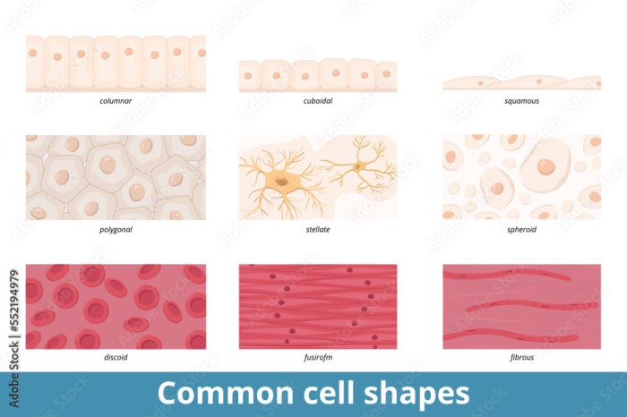
What are squamous cells? These flat, scale-like cells play crucial roles throughout the body, from lining our respiratory tracts to protecting our skin. Understanding their diverse functions, locations, and potential abnormalities is key to grasping their significance in health and disease.
This in-depth look at squamous cells will cover their defining characteristics, highlighting their structural features and contrasting them with other cell types. We’ll explore their diverse locations in the body, examining their specific roles in various tissues and organs. Furthermore, we’ll delve into their formation, development, and significance in both health and disease. This includes an exploration of squamous cell abnormalities, associated diseases, and the crucial role they play in clinical diagnostics.
Definition and Characteristics
Squamous cells, characterized by their flattened, scale-like appearance, are a fundamental component of various tissues throughout the body. Understanding their diverse forms and functions is crucial for comprehending human physiology and disease processes. These cells play critical roles in protection, secretion, and absorption, depending on their location and specialization.The shape and structure of squamous cells directly relate to their function.
Their thin, flat morphology allows for efficient diffusion of substances across their surfaces. This is particularly important in areas where gas exchange or absorption is paramount. This adaptability in structure contributes to their widespread distribution in the body.
Types of Squamous Cells
Squamous cells exhibit variations in their structure and function, categorized into keratinized and non-keratinized types. These differences stem from the presence or absence of keratin, a tough protein that provides a protective barrier. Keratinization is a crucial process in areas subjected to high wear and tear.
- Keratinized Squamous Cells: These cells are found in areas exposed to the external environment, such as the epidermis of the skin. The presence of keratin provides a robust barrier against pathogens and mechanical stress. This layer of keratinized cells acts as a shield, protecting the underlying tissues from dehydration, abrasion, and infection. Examples include the skin of the palms and soles.
- Non-keratinized Squamous Cells: These cells line internal surfaces, such as the lining of the mouth, esophagus, and vagina. The absence of keratin contributes to a moist, flexible surface that facilitates various functions, including protection from friction and allowing for ease of movement within the internal environment. These cells maintain a moist environment crucial for various physiological processes.
Structural Features
The defining characteristic of squamous cells is their flattened, scale-like shape. This structure is optimized for various functions, particularly in areas where diffusion is critical. The thinness allows for rapid movement of molecules across the cell layer.
- Shape and Size: Squamous cells are typically thin and flat, resembling irregular, polygonal scales. Their size varies depending on their location and function.
- Nucleus: The nucleus is typically centrally located and flattened in accordance with the shape of the cell.
- Cytoplasm: The cytoplasm is thin and sparse, with a minimal amount of organelles. This streamlined structure allows for efficient movement of molecules.
Comparison with Other Cell Types
Squamous cells differ significantly from other cell types in terms of shape, structure, and function. Their flattened morphology and thin cytoplasm are distinct characteristics that set them apart from cuboidal and columnar cells. This adaptability allows them to perform a range of functions in different parts of the body.
| Cell Type | Description | Location | Function |
|---|---|---|---|
| Squamous Epithelial Cells (Keratinized) | Flattened, scale-like cells with keratin | Epidermis of skin (palms, soles) | Protection against dehydration, abrasion, and infection |
| Squamous Epithelial Cells (Non-keratinized) | Flattened, scale-like cells without keratin | Lining of mouth, esophagus, vagina | Protection from friction, maintaining a moist environment |
| Cuboidal Epithelial Cells | Cube-shaped cells | Glands, kidney tubules | Secretion and absorption |
| Columnar Epithelial Cells | Column-shaped cells | Intestinal lining | Absorption and secretion |
Location and Function

Squamous cells, those flat, scale-like cells, are a crucial part of many bodily systems. Their thin, flattened shape allows for efficient diffusion of substances across their surface. This feature is vital for various functions, from gas exchange in the lungs to absorption in the intestines. Understanding their location and specific roles within different tissues provides a deeper appreciation for their importance in maintaining overall health.
Locations of Squamous Cells
Squamous cells are not found in isolation but rather form the lining of various surfaces throughout the body. Their prevalence in these locations highlights their vital role in covering and protecting underlying structures.
- Respiratory System: In the lungs, squamous cells, also known as type I alveolar cells, form the walls of the alveoli, the tiny air sacs where gas exchange occurs. This location is critical for oxygen uptake and carbon dioxide release, essential for respiration. The thinness of these cells facilitates the rapid diffusion of gases across their surface.
- Integumentary System: The epidermis, the outermost layer of skin, is primarily composed of squamous cells. This layer acts as a protective barrier against pathogens, dehydration, and physical trauma. The constant turnover of these cells is crucial for maintaining skin integrity.
- Cardiovascular System: Endothelium, the lining of blood vessels, is composed of squamous cells. These cells regulate blood flow, prevent blood clotting, and facilitate the exchange of nutrients and waste products between blood and surrounding tissues. The smooth surface of the endothelium minimizes friction, promoting efficient blood circulation.
- Digestive System: Parts of the digestive tract, including the mouth, esophagus, and vagina, contain squamous cells. These cells provide a protective barrier against the harsh conditions within the digestive system, preventing damage and facilitating absorption of nutrients.
Functions of Squamous Cells
The varied functions of squamous cells directly correlate with their location. Their primary function is to facilitate the diffusion of substances, whether gases, nutrients, or waste products.
- Gas Exchange: In the alveoli of the lungs, squamous cells enable the rapid diffusion of oxygen from the air into the bloodstream and the diffusion of carbon dioxide from the bloodstream into the air. This efficient exchange is critical for respiration.
- Protection: The squamous cells of the epidermis form a protective barrier against pathogens, preventing infection and protecting underlying tissues from environmental damage. This is essential for overall health and well-being.
- Absorption and Secretion: In the digestive system, squamous cells in the lining of the mouth and esophagus protect these delicate tissues from the harsh environment and facilitate the absorption of nutrients. In some locations, squamous cells also play a role in secretion of mucus.
- Blood Vessel Regulation: Squamous cells lining blood vessels (endothelium) regulate blood flow, prevent blood clotting, and facilitate the exchange of essential nutrients and waste products. These functions are vital for maintaining circulatory health.
Importance of Squamous Cell Health
Maintaining the health of squamous cells is crucial for optimal bodily function. Any disruption to these cells, such as damage from infection or trauma, can negatively impact the delicate balance of these processes. This can lead to serious consequences for health, ranging from respiratory problems to skin infections to cardiovascular issues. Healthy squamous cells are essential for overall well-being.
Squamous cells are a type of flat, scale-like cell that line various parts of your body, including your mouth. Knowing what these cells look like is important, as they often play a crucial role in healing. If you happen to get a cut inside your mouth, proper care is essential. Following the advice in this guide on how to treat a cut inside your mouth can speed up the healing process and help prevent complications.
Ultimately, understanding squamous cells is key to maintaining overall oral health.
| Location | Function |
|---|---|
| Alveoli (Lungs) | Gas exchange (oxygen and carbon dioxide) |
| Epidermis (Skin) | Protection against pathogens and environmental factors |
| Endothelium (Blood Vessels) | Blood flow regulation, nutrient/waste exchange |
| Digestive Tract (Mouth, Esophagus, Vagina) | Protection, absorption (in some areas) |
Formation and Development: What Are Squamous Cells

Squamous cells, those flat, scale-like cells, don’t just appear fully formed. Their development is a fascinating process, influenced by various factors and progressing through distinct stages. Understanding this process is crucial for comprehending their role in tissue health and disease. This journey from precursor cells to mature squamous cells is key to maintaining the integrity of surfaces throughout the body.The formation and development of squamous cells are tightly regulated.
This intricate process ensures that the cells are appropriately differentiated and functional. Factors like genetic instructions, local environment, and external signals all play a part in guiding the cells through their developmental path. A precise sequence of events and cellular changes determines the final form and function of the squamous cells.
Stages of Squamous Cell Differentiation
The transformation of precursor cells into mature squamous cells is a multi-step process. Each stage involves specific changes in cell shape, structure, and function. These changes reflect the increasing specialization of the cells.
- Proliferation: In this initial stage, precursor cells undergo rapid division and multiplication. This increase in cell number is essential to replenish existing cells and support tissue growth. The environment plays a significant role in stimulating this rapid cell growth, ensuring proper cell replenishment.
- Differentiation: As the cells progress, they begin to acquire specialized characteristics. This involves changes in cell morphology, with the cells becoming increasingly flattened and acquiring their characteristic squamous shape. Specific genes are activated and deactivated, guiding the cells down a particular developmental pathway.
- Maturation: During this stage, the cells reach their full functional capacity. Their structural components, like keratin filaments, become more prominent, contributing to the cell’s protective properties. The production of specific proteins, vital for cell function and interactions with surrounding tissues, increases.
Factors Influencing Squamous Cell Growth and Maturation
Several factors influence the growth and maturation of squamous cells. These include both intrinsic factors, inherent to the cells themselves, and extrinsic factors, originating from the surrounding environment.
- Genetic factors: The cell’s genetic makeup dictates the sequence of events and the characteristics of the developing squamous cells. Variations in the genetic code can affect the timing and efficiency of these developmental stages, potentially leading to abnormalities.
- Growth factors: These signaling molecules stimulate cell proliferation and differentiation. The presence and concentration of growth factors in the microenvironment significantly impact the rate and direction of squamous cell development. For example, epidermal growth factor (EGF) is known to promote squamous cell growth in the skin.
- Hormones: Hormonal influences can also play a significant role in squamous cell development. For instance, hormones can affect the rate of cell division and the production of specific proteins in the cells.
Visual Representation of Squamous Cell Development
| Stage | Description |
|---|---|
| Proliferation | Precursor cells rapidly divide and multiply, increasing cell numbers. |
| Differentiation | Cells acquire specialized characteristics, becoming flattened and adopting the squamous shape. Cellular organelles adapt to the new function. |
| Maturation | Cells achieve their full functional potential. Keratin production increases, enhancing protective qualities. |
Significance in Health and Disease
Squamous cells, crucial components of our skin and lining of various organs, play a vital role in maintaining the integrity and function of these tissues. Their flat, scale-like structure provides a protective barrier against external insults and pathogens. However, abnormalities in squamous cells can lead to serious health concerns. Understanding these abnormalities and the associated diseases is critical for effective diagnosis and treatment.Squamous cell abnormalities represent a significant area of concern in medicine.
These deviations from the normal structure and function of squamous cells can manifest as precancerous lesions or, in more advanced stages, as invasive cancers. The causes and risk factors behind these abnormalities are varied, but often include genetic predisposition, environmental exposures, and chronic irritations. Recognizing these factors is essential for prevention and early intervention.
Squamous Cell Abnormalities
Squamous cell abnormalities encompass a spectrum of conditions, ranging from benign lesions to cancerous growths. Identifying the specific type of abnormality is crucial for appropriate treatment and prognosis. These abnormalities can be characterized by changes in cell shape, size, and arrangement, often visible under a microscope.
Causes and Risk Factors
Numerous factors contribute to the development of squamous cell disorders. Genetic predispositions, such as certain inherited syndromes, can increase the risk. Environmental exposures, including prolonged sun exposure, certain chemical agents, and infections, also play a significant role. Chronic irritations, such as those caused by smoking, chewing tobacco, or chronic skin conditions, can further increase the risk. In some cases, the exact cause remains unknown.
Examples of Squamous Cell Diseases
Squamous cell disorders range from relatively benign conditions to life-threatening cancers. Understanding these conditions is critical for early detection and treatment.
Table of Squamous Cell Diseases, What are squamous cells
| Disease | Symptoms | Causes | Treatment Options |
|---|---|---|---|
| Actinic Keratosis | Rough, scaly patches on sun-exposed skin; may be slightly raised or discolored. | Prolonged sun exposure, cumulative UV radiation, aging. | Cryotherapy, topical medications (5-fluorouracil, imiquimod), surgical excision. |
| Squamous Cell Carcinoma (SCC) | Scaly, red, or flesh-colored lesions that may ulcerate, bleed, or crust over; may be firm or hard. | Prolonged sun exposure, smoking, chewing tobacco, chronic skin irritation, certain genetic syndromes. | Surgical excision, radiation therapy, chemotherapy (in advanced cases), targeted therapies. |
| Bowen’s Disease | Red, scaly patch that appears on skin; may be itchy or painful. | Exact cause unknown, but possibly related to chronic irritation or viral infections. | Topical medications, phototherapy, surgical excision. |
Microscopy and Imaging
Peeking into the microscopic world of squamous cells reveals a wealth of information crucial for understanding their structure and behavior, both in health and disease. Different microscopy techniques offer unique perspectives, enabling us to visualize cellular details that are otherwise invisible to the naked eye. This detailed exploration will illuminate how these techniques provide invaluable insights into squamous cell morphology and their role in various clinical scenarios.Advanced imaging techniques play a vital role in the diagnostic and monitoring processes for squamous cell conditions.
Analyzing images obtained through microscopy allows clinicians to identify specific features, detect abnormalities, and assess the progression of the disease. These techniques provide a precise picture of cellular structure, facilitating early diagnosis and targeted treatment strategies.
Appearance of Squamous Cells Under Different Microscopy Techniques
Visualizing squamous cells under various microscopy techniques provides a comprehensive understanding of their morphology. Light microscopy, with its relatively simple setup and affordability, is a fundamental tool for initial examination. High-quality light microscopy images can clearly reveal the flattened, scale-like appearance of squamous cells, along with their characteristic nuclei. Electron microscopy, including transmission electron microscopy (TEM) and scanning electron microscopy (SEM), offers significantly higher resolution.
TEM images allow visualization of internal cellular structures like the nucleus, cytoplasm, and organelles, revealing details about cell function. SEM provides a three-dimensional perspective of the cell surface, showcasing surface features and textures.
Use of Imaging Techniques in Diagnosis and Monitoring
Imaging techniques are indispensable tools in the diagnosis and monitoring of squamous cell conditions. Microscopy images, particularly those obtained from biopsies or cytological samples, are crucial for identifying squamous cell carcinoma (SCC). The presence of abnormal cell shapes, sizes, and nuclear features can be indicative of malignant transformation. Furthermore, monitoring the progression of the disease, response to treatment, and recurrence can be assessed through serial microscopy images.
This allows for adjustments in treatment plans and facilitates proactive intervention.
Key Features Observed in Microscopy Images of Squamous Cells
Microscopy images of squamous cells showcase a range of key features. The characteristic flattened shape and scale-like appearance are readily apparent. The presence and size of the nucleus, often centrally located, are also important indicators. Variations in these features, such as increased nuclear size, abnormal nuclear shape, or loss of cellular organization, can be indicative of squamous cell abnormalities.
The cytoplasm surrounding the nucleus may exhibit different staining characteristics or cellular structures. In malignant squamous cells, these features are often significantly altered.
Microscopy Techniques and Their Application in Studying Squamous Cells
The following table summarizes various microscopy techniques and their applications in studying squamous cells:
| Microscopy Technique | Application in Studying Squamous Cells |
|---|---|
| Light Microscopy | Initial examination; identification of general morphology, cell size, and shape; screening for abnormalities. |
| Transmission Electron Microscopy (TEM) | Detailed visualization of internal cellular structures, including the nucleus, cytoplasm, and organelles; identification of structural alterations associated with disease. |
| Scanning Electron Microscopy (SEM) | Three-dimensional visualization of the cell surface; examination of surface features, textures, and the presence of abnormalities or markers of infection. |
| Fluorescence Microscopy | Visualization of specific cellular components or markers using fluorescent dyes; identification of specific proteins, DNA, or other molecules relevant to the study of squamous cells. |
| Confocal Microscopy | Three-dimensional imaging of cells; acquisition of high-resolution images of thick specimens. |
Clinical Relevance
Squamous cells, a ubiquitous component of various tissues, play a crucial role in clinical diagnostics. Their presence, morphology, and abnormalities can provide valuable insights into the health status of an individual and the potential presence of disease. Understanding the clinical relevance of squamous cells extends beyond basic identification to encompass the intricate relationship between their characteristics and a wide spectrum of pathological conditions.Detailed analysis of squamous cells is often integral to diagnostic pathways.
Techniques such as cytology and histology, which examine the cellular components and tissue structures, rely heavily on the characteristics of squamous cells. The ability to identify and interpret squamous cell variations is essential for accurately assessing potential health concerns.
Importance in Diagnostic Procedures
The analysis of squamous cells is instrumental in various medical procedures. From routine pap smears to specialized biopsies, squamous cell evaluation provides critical information for early detection and management of diseases. This is particularly crucial in the context of cancers, where squamous cell abnormalities can be early indicators of malignant transformation.
Squamous cells are flat, scale-like cells found throughout the body, playing a crucial role in lining various organs. Understanding these cells is vital in various medical contexts, including medical underwriting processes, which assess an applicant’s health risk before insurance coverage. This process, explained further in this insightful guide on what is medical underwriting , often looks at factors related to squamous cell health, like any potential abnormalities or conditions.
In short, knowing what squamous cells are is fundamental to understanding medical underwriting and overall health assessment.
Role in Disease Diagnosis
Squamous cells play a pivotal role in the diagnosis of various diseases. Their presence, distribution, and cellular features can provide valuable clues about the underlying pathology. For instance, the presence of abnormal squamous cells in respiratory samples can suggest infections or malignancies. Similarly, the identification of atypical squamous cells in cervical smears is a significant indicator for the early detection of cervical cancer.
Examples of Squamous Cell Abnormalities and Associated Diseases
Several squamous cell abnormalities are strongly linked to specific diseases. One prominent example is the presence of dysplastic squamous cells in cervical tissue, which is a significant precursor to cervical cancer. Similarly, the identification of squamous cell carcinoma in situ (SCCIS) in the skin or oral cavity can indicate a high-risk of developing invasive squamous cell carcinoma. Other diseases that may involve squamous cell abnormalities include lung cancer, oral cancer, and various skin conditions.
Clinical Applications of Squamous Cell Analysis
| Clinical Application | Testing Method | Results Interpretation |
|---|---|---|
| Cervical Cancer Screening | Pap smear | Identification of abnormal squamous cells (e.g., dyskaryosis) suggests the possibility of precancerous or cancerous changes, requiring further investigation. |
| Lung Cancer Diagnosis | Bronchoscopy with cytology | Presence of squamous cells with atypical features in sputum or bronchoalveolar lavage fluid can suggest lung cancer or other respiratory diseases. |
| Oral Cancer Screening | Oral cytology | Identification of abnormal squamous cells in oral mucosa biopsies can indicate precancerous lesions or oral cancer. |
| Skin Cancer Diagnosis | Skin biopsy | Presence of atypical squamous cells in skin biopsies may suggest skin cancer, requiring further evaluation and potentially biopsy. |
| Vaginal/Vulvar Cancer Screening | Cytology or biopsy | Presence of abnormal squamous cells in vaginal or vulvar samples may suggest precancerous lesions or cancer. |
Related Cell Types
Squamous cells, with their flattened, scale-like shapes, are a common cell type found throughout the body. Understanding their relationship to other cell types provides crucial insight into their function and role in maintaining health. This exploration delves into the similarities and differences between squamous cells and related cell types, highlighting the cellular mechanisms that distinguish them and the functional implications of these distinctions.The comparison of squamous cells with other cell types emphasizes the intricate interplay of cellular structures and functions within the body.
The differences in morphology and function directly impact the roles these cells play in various tissues and organs, contributing to the overall homeostasis of the organism.
Squamous cells are a type of flat, scale-like cell found throughout the body, including the cervix. Sometimes, changes in these cells can lead to unusual discharge, like brown discharge before your period. Understanding these changes can help you better interpret your body’s signals, and if you’re concerned, consulting a healthcare professional like the ones at brown discharge before period is always a good idea.
Learning more about these cells and their role in reproductive health can be incredibly helpful.
Comparison of Squamous Cells with Related Cell Types
This comparison Artikels the key characteristics distinguishing squamous cells from their related counterparts, emphasizing the importance of understanding these differences for accurate diagnosis and treatment.
| Feature | Squamous Cells | Cuboidal Cells | Columnar Cells | Transitional Cells |
|---|---|---|---|---|
| Shape | Flattened, scale-like | Cube-shaped | Column-shaped | Variable, depending on the degree of stretching |
| Nucleus | Flattened, centrally located | Round, centrally located | Oval, located basally | Variable, depending on the degree of stretching |
| Location | Epithelial linings, alveoli, endothelium | Glandular tissue, kidney tubules | Gastrointestinal tract, respiratory tract | Urinary bladder, ureters |
| Function | Protection, diffusion, filtration | Secretion, absorption | Secretion, absorption, protection | Distensibility, protection |
Cellular Mechanisms Distinguishing Squamous Cells
The unique characteristics of squamous cells arise from specific cellular mechanisms. Their thin, flattened shape maximizes surface area for diffusion and filtration. This is critical in areas like the lungs where oxygen uptake and carbon dioxide release are paramount. The structural simplicity also facilitates rapid cell turnover, crucial for tissue repair.
Functional Implications of Differences
The differences in shape, location, and function between squamous cells and other cell types have significant implications. For instance, the flattened structure of squamous cells in the alveoli of the lungs is essential for efficient gas exchange. In contrast, the columnar cells lining the intestines have a larger surface area and specialized structures (microvilli) for maximizing nutrient absorption.
Understanding these differences is fundamental to comprehending the intricate workings of various organ systems.
Illustrations and Visualizations
Squamous cells, crucial components of various tissues, are best understood through visual representations. Illustrations and microscopic images provide critical insights into their structure, shape, and arrangement within different contexts. These visualizations bridge the gap between abstract descriptions and concrete understanding, enhancing comprehension of their roles in health and disease.
Illustration of a Squamous Cell
A squamous cell is typically depicted as a thin, flattened cell, resembling a fried egg. The cell’s nucleus is centrally located and oval or slightly irregular in shape. The cytoplasm surrounding the nucleus is thin and translucent, exhibiting minimal cellular organelles. A simple drawing of this flattened structure can be easily envisioned, with the nucleus prominently featured within the cell’s flattened body.
Microscopic Structure of a Squamous Cell
Under a microscope, a squamous cell displays a characteristically thin, flattened morphology. The cell’s nucleus, usually centrally located, is a prominent feature. Its chromatin appears finely dispersed within the nucleus, reflecting active cellular processes. The cytoplasm, being extremely thin, may not be easily distinguished from the surrounding extracellular matrix. The absence of prominent organelles, apart from the nucleus, further accentuates the cell’s flattened shape.
Appearance of Squamous Cells in Various Tissues
The appearance of squamous cells varies depending on the tissue in which they reside. In the epidermis (outer layer of skin), they form a continuous layer, protecting underlying tissues. In the lining of the respiratory tract, squamous cells are often found in the transitional zones between different tissue types, displaying slight variations in their shape and size depending on the specific location within the tract.
In the lining of the esophagus, squamous cells form a protective barrier against harsh environmental conditions. This varied appearance reflects the diverse functions of squamous cells in different tissues.
Table of Squamous Cell Illustrations and Descriptions
| Tissue Context | Illustration | Description |
|---|---|---|
| Epidermis (Skin) | A flat, mosaic-like arrangement of cells forming a continuous sheet | Tightly packed, thin cells forming a protective barrier. The nucleus is relatively flat and centrally positioned, and the cytoplasm is extremely thin. |
| Lining of the Respiratory Tract | Cells varying in shape and size, transitioning between stratified and simple squamous epithelia | Cells transitioning between cuboidal and squamous shapes, reflecting the dynamic nature of respiratory tissue. Nuclei vary in size and shape, and cytoplasm is visible but less extensive. |
| Lining of the Esophagus | Layered squamous cells | A stratified squamous epithelium, with multiple layers of cells. The cells near the surface are flat, while those deeper are more cuboidal. This layered arrangement provides protection against the harsh environment. |
| Inner lining of blood vessels (Endothelium) | A single layer of thin cells lining the blood vessel lumen | A single layer of squamous cells forming a smooth surface that facilitates blood flow. The nucleus is centrally located and the cytoplasm is very thin. |
Wrap-Up
In conclusion, squamous cells are essential components of our bodies, performing a multitude of vital functions. From their fundamental structure to their complex roles in health and disease, their significance is undeniable. This exploration has provided a comprehensive overview of these cells, from their formation and development to their clinical relevance in diagnostics and disease understanding. Hopefully, this has given you a deeper appreciation for the intricate role squamous cells play in the overall health and function of the human body.

