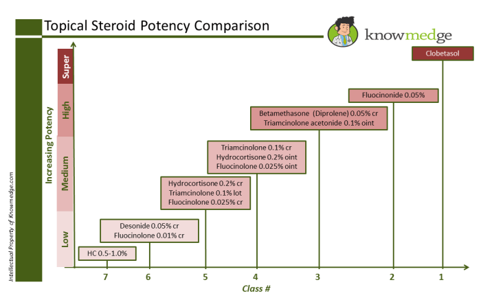
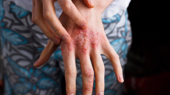
Tag: skin conditions

  • Four Skin Conditions That Make Your Feet Itch

    Four Skin Conditions That Make Your Feet Itch

    Four skin conditions that make your feet itch can be incredibly frustrating. Understanding the underlying causes and symptoms is key to finding relief. This guide explores athlete’s foot, eczema, psoriasis, and other fungal infections, providing detailed information about each condition, including symptoms, causes, treatments, and preventative measures. We’ll also discuss home remedies and when to seek professional help.

    From the common to the less familiar, we’ll unpack the various factors contributing to itchy feet. We’ll look at not only the skin conditions themselves but also other potential causes like allergies or insect bites. Prepare to delve into the world of foot health and discover the solutions that can bring you relief.

    Introduction to Foot Itch

    Itchy feet are a common complaint, often disrupting daily life and causing discomfort. While sometimes a minor annoyance, persistent foot itching can signal an underlying skin condition requiring medical attention. Understanding the potential causes and associated symptoms is crucial for proper diagnosis and treatment. This section delves into the various reasons behind itchy feet, emphasizing the importance of identifying the root cause to achieve effective relief.Identifying the specific skin condition causing the itch is vital for targeted treatment.

    A misdiagnosis can lead to ineffective therapies and prolonged discomfort. Symptoms vary depending on the underlying condition, providing valuable clues for proper medical evaluation. A comprehensive understanding of the symptoms associated with different skin conditions affecting the feet can aid in self-assessment and facilitate early diagnosis.

    Common Causes of Itchy Feet

    Various factors can contribute to itchy feet, ranging from simple irritants to more serious medical conditions. These include: contact dermatitis from irritants like harsh soaps or footwear materials; fungal infections, such as athlete’s foot; allergic reactions to certain substances; and skin conditions like eczema or psoriasis. Proper identification of the cause is essential to implement the appropriate treatment strategy.

    Symptoms of Foot Itch

    The symptoms associated with itchy feet vary depending on the underlying cause. Some common symptoms include: persistent itching, redness, dryness, flaking, cracking, blisters, and in some cases, burning or stinging sensations. Careful observation of these symptoms can assist in pinpointing the potential cause of the itching.

    Skin Conditions Causing Itchy Feet

    It is important to note that several skin conditions can lead to itchy feet. A proper diagnosis requires a comprehensive evaluation by a healthcare professional.

    Skin Condition Symptoms Causes Treatment
    Athlete’s Foot (Tinea pedis) Itching, burning, redness, scaling, blisters, cracking skin between toes. Fungal infection, often in damp or enclosed shoes. Antifungal creams or oral medications prescribed by a doctor. Maintaining good foot hygiene and avoiding sharing footwear are crucial preventative measures.
    Eczema (Atopic Dermatitis) Dry, itchy, red, inflamed skin; possible small bumps or blisters. Genetic predisposition, environmental triggers (allergens, irritants). Moisturizing creams, topical corticosteroids (as prescribed), avoidance of triggers.
    Psoriasis Red, scaly patches on the skin, often with silvery-white scales; itching is common. Autoimmune disorder, genetic predisposition. Topical medications, phototherapy, and sometimes oral medications.
    Contact Dermatitis Itching, redness, rash, blisters, or burning at the site of contact with an irritant. Reaction to specific substances (chemicals, cosmetics, plants). Identifying and avoiding the irritant; topical corticosteroids, antihistamines, and wet compresses.

    Athlete’s Foot

    Athlete’s foot, a common fungal infection, affects the skin of the feet. It’s characterized by itching, redness, and often, cracking or peeling skin. Understanding the cause, symptoms, transmission, and prevention is key to managing this often-annoying condition.

    Fungal Infection

    Athlete’s foot is caused by a group of fungi, primarily dermatophytes. These fungi thrive in warm, moist environments, making areas like locker rooms, public showers, and poorly ventilated shoes ideal breeding grounds. The fungi penetrate the skin’s surface, leading to inflammation and the characteristic symptoms of the infection. Fungal spores are ubiquitous, meaning they are present almost everywhere, but the fungi only grow and cause infection under specific conditions.

    Symptoms

    The symptoms of athlete’s foot often appear gradually. Common symptoms include itching, redness, and scaling between the toes, particularly the spaces between the fourth and fifth toes. The skin may become cracked, inflamed, and sometimes blister. In severe cases, the infection can spread to the soles of the feet. The intensity of the symptoms can vary greatly, ranging from mild discomfort to severe pain.

    Transmission

    Athlete’s foot is highly contagious. Transmission occurs through direct contact with infected skin, contaminated surfaces, or shared personal items. Common sources of transmission include: using public pools or locker rooms, walking barefoot on surfaces in public places, and sharing towels or shoes with someone infected. Maintaining good hygiene practices is essential to prevent its spread.

    Comparison with Other Foot Conditions

    Symptom Athlete’s Foot Other Conditions (e.g., Psoriasis, Eczema)
    Itching Often intense, particularly between toes Variable intensity, can be present in other areas
    Redness Localized, often between toes, may spread Can be widespread, or localized, may be associated with inflammation
    Scaling/Peeling Dry, flaky skin, often white or yellow Can present with scaling or shedding, but appearance may differ
    Cracking/Blisters Can occur in severe cases, especially in thick skin areas Can develop cracking or blisters, depending on the condition
    Odor May be present in advanced stages Typically not associated with strong odor

    Note: This table provides a general comparison. A healthcare professional should diagnose the specific condition.

    Prevention

    Prevention is crucial for avoiding athlete’s foot. Here are some key strategies:

    • Maintaining good foot hygiene. Regularly wash and dry your feet, paying close attention to the spaces between your toes. Use a clean towel each time.
    • Wearing appropriate footwear. Choose breathable shoes that allow air circulation to prevent moisture buildup. Change socks daily.
    • Avoiding walking barefoot in public areas. Always use flip-flops or other protective footwear in public showers or locker rooms.
    • Keeping your feet dry. Use a foot powder or antifungal spray to absorb moisture.
    • Using clean towels and personal items. Do not share towels, shoes, or other personal items that may come in contact with your feet.

    These simple preventative measures can significantly reduce the risk of contracting athlete’s foot.

    Eczema (Dermatitis) of the Feet

    Foot eczema, a common skin condition, presents as inflamed, itchy patches on the feet. It’s characterized by dry, cracked skin, often accompanied by blisters or weeping sores. Understanding the various types, triggers, and treatments can significantly improve management and alleviate discomfort.

    Types of Foot Eczema

    Foot eczema isn’t a single entity; rather, it encompasses several types, each with its own characteristics. Atopic dermatitis, a chronic condition, frequently affects the feet, manifesting as dry, itchy, and inflamed skin. Contact dermatitis, triggered by specific allergens or irritants, can also cause foot eczema. Lastly, dyshidrotic eczema, marked by tiny blisters on the soles of the feet and between toes, is another form that requires specific management.

    Triggers and Risk Factors

    Several factors contribute to the development of foot eczema. Common triggers include exposure to harsh soaps, detergents, or chemicals. Allergic reactions to certain materials, such as wool or latex, can also cause the condition. Individuals with a family history of eczema, asthma, or hay fever are at increased risk. Sweating, particularly in enclosed footwear, can exacerbate the condition.

    Wearing tight or ill-fitting shoes can also contribute to friction and irritation, leading to eczema.

    Symptoms of Foot Eczema

    Symptoms of foot eczema vary depending on the type and severity. Common signs include redness, itching, and dryness. The skin may appear cracked or rough, with blisters or small sores forming. In severe cases, the skin may become thickened or scaly. The affected areas often feel intensely itchy, making scratching and further irritation a significant concern.

    The symptoms can fluctuate in intensity, depending on factors such as the time of year, stress levels, and environmental conditions.

    Treatments for Foot Eczema

    Effective management of foot eczema involves a multifaceted approach. Moisturizing the affected areas regularly with a fragrance-free cream or ointment is crucial. Avoiding triggers is essential to prevent flare-ups. Prescription medications, such as topical corticosteroids, may be necessary in severe cases. In some situations, oral antihistamines can help alleviate itching.

    Maintaining good foot hygiene, including using gentle soaps and thoroughly drying the feet after bathing, can also aid in managing the condition. If symptoms persist or worsen, consulting a dermatologist is recommended.

    Differences Between Types of Foot Eczema

    Type of Eczema Symptoms Treatment
    Atopic Dermatitis Dry, itchy, inflamed skin; can involve weeping or scaling. Often associated with other allergic conditions. Moisturizing, avoidance of triggers, topical corticosteroids (as prescribed).
    Contact Dermatitis Inflammation and itching at the site of contact with an allergen or irritant; often localized. Identifying and avoiding the trigger; topical corticosteroids (as prescribed); antihistamines.
    Dyshidrotic Eczema Small blisters, often on the soles of the feet and between toes; intensely itchy. Moisturizing; avoiding triggers; topical corticosteroids (as prescribed); in severe cases, oral medications.

    Psoriasis: Four Skin Conditions That Make Your Feet Itch

    Psoriasis is a chronic autoimmune skin condition that causes skin cells to multiply rapidly. This rapid cell turnover leads to the formation of thick, scaly patches on the skin. While it can manifest anywhere on the body, including the feet, it often presents unique characteristics when affecting this area.

    Characteristics of Foot Psoriasis

    Psoriasis on the feet often displays inflamed, red patches that can be covered with silvery-white scales. These patches can vary in size and shape, and may be accompanied by cracking and fissures in the skin. The affected skin may also be itchy and painful, particularly when the scales are thick or inflamed. The presence of these characteristics can help differentiate foot psoriasis from other skin conditions.

    Ever wondered why your feet are constantly itching? Four common skin conditions can be culprits, like athlete’s foot, eczema, psoriasis, and fungal infections. While dealing with these foot issues, have you considered the amazing benefits of aloe vera? It’s a natural remedy that can help soothe skin irritations and even treat acne and acne scars, just like aloe vera for acne and acne scars demonstrates.

    Knowing how to use aloe vera topically can be a great way to ease the itchiness associated with these skin conditions on your feet.

    Potential Triggers for Foot Psoriasis

    Several factors can trigger or worsen psoriasis on the feet. These include infections, injuries, stress, and certain medications. Exposure to extreme temperatures or harsh chemicals can also play a role in exacerbating the condition. For example, a fungal infection could lead to psoriasis flare-ups. Similarly, trauma from ill-fitting shoes or repeated friction can initiate a flare-up.

    Also, periods of significant stress or emotional distress may also trigger or worsen psoriasis.

    Symptoms and Visual Presentation of Foot Psoriasis

    The symptoms of foot psoriasis can vary depending on the severity of the condition. Common symptoms include red, inflamed skin patches, often with a silvery-white buildup of scales. These patches can range from small and localized to large and widespread, affecting the entire foot. In some cases, the scales may be so thick that they form cracks or fissures in the skin, leading to pain and bleeding.

    The affected skin may also be itchy, dry, and sore. Visual presentation can range from mild to severe, with the severity depending on the individual and the specific triggers.

    Treatment Options for Foot Psoriasis

    Treatment for foot psoriasis aims to reduce inflammation, control scaling, and alleviate symptoms. Several approaches are available, including topical corticosteroids, vitamin D analogs, and coal tar preparations. These treatments can help manage the symptoms and slow the progression of the condition. Systemic medications, such as methotrexate or cyclosporine, may be necessary in severe cases. Phototherapy, using ultraviolet light, can also be effective in reducing inflammation and scaling.

    Comparison Table: Foot Psoriasis vs. Other Itchy Foot Conditions

    Feature Psoriasis Athlete’s Foot Eczema
    Appearance Red, inflamed patches with silvery-white scales, often with cracking and fissures. Red, itchy rash between toes, often with scaling and/or blisters. Red, itchy, and inflamed skin; may have weeping or oozing; can vary in appearance depending on the type.
    Cause Autoimmune response Fungal infection Allergic reaction or irritation
    Symptoms Inflammation, scaling, itching, pain, cracking, fissures. Itching, burning, scaling, blisters, peeling. Itching, dryness, redness, inflammation, sometimes oozing or crusting.
    Treatment Topical creams, phototherapy, systemic medications Antifungal creams or oral medications Moisturizers, topical corticosteroids, anti-inflammatory medications

    Fungal Infections (Beyond Athlete’s Foot)

    Four skin conditions that make your feet itch

    Itchy feet aren’t always limited to the common culprit, athlete’s foot. A variety of fungal infections can affect the skin on your feet, each with unique characteristics and treatment approaches. Understanding these variations is crucial for proper diagnosis and effective management.Beyond the typical symptoms of athlete’s foot, other fungal infections present with distinct features, impacting different parts of the foot and requiring tailored treatments.

    This discussion will explore various types of fungal infections, detailing their symptoms, causes, and treatment options to provide a comprehensive understanding.

    Types of Fungal Foot Infections

    Various fungal species can affect the feet, leading to different symptoms and requiring tailored treatment approaches. Recognizing these distinctions is key to effective management.

    • Tinea manuum (Hand Tinea): This fungal infection, similar to athlete’s foot, affects the skin on the hands. It often presents with itchy, red, scaly patches, sometimes with blisters. It’s typically spread through contact with contaminated surfaces or objects. Treatment involves topical antifungal creams or oral medications, depending on the severity.
    • Tinea cruris (Jock Itch): This fungal infection primarily affects the groin area, inner thighs, and buttocks. It’s characterized by itchy, reddish-brown, and often ring-shaped rash. Warm, humid environments and close-fitting clothing can contribute to its development. Topical antifungal creams are usually effective in treating this type of fungal infection.
    • Tinea pedis (Athlete’s Foot): While already discussed, a brief overview is included. This common fungal infection affects the skin between the toes and soles of the feet. It’s characterized by itching, burning, scaling, and cracking of the skin. Warm, humid environments and tight-fitting shoes can increase the risk. Topical antifungals are usually the first line of treatment.

    • Fungal Nail Infections (Onychomycosis): These infections affect the toenails or fingernails. They typically manifest as thickened, discolored, brittle, or crumbling nails. Fungal infections can spread from the skin to the nails. Treatment often involves oral antifungal medications or topical treatments, depending on the severity and location of the infection. In some cases, surgical removal of the infected nail might be necessary.

    Comparing Fungal Foot Infections

    Understanding the distinctions between different types of fungal infections is vital for effective treatment.

    Fungal Infection Symptoms Causes Treatment
    Tinea manuum Itchy, red, scaly patches, sometimes with blisters on the hands Contact with contaminated surfaces or objects, sharing towels, or poorly ventilated environments. Topical antifungal creams or oral medications (severe cases).
    Tinea cruris Itchy, reddish-brown, ring-shaped rash in the groin, inner thighs, and buttocks. Warm, humid environments, close-fitting clothing, and sharing of personal items. Topical antifungal creams.
    Tinea pedis (Athlete’s Foot) Itching, burning, scaling, and cracking of the skin between toes and soles of the feet. Warm, humid environments, tight-fitting shoes, and sharing of personal items. Topical antifungal creams.
    Fungal Nail Infections (Onychomycosis) Thickened, discolored, brittle, or crumbling nails. Fungal infection spreading from the skin to the nails, sharing of personal items, or weakened immune systems. Oral antifungal medications or topical treatments, possibly surgical nail removal.

    Other Potential Causes of Itchy Feet

    Itchy feet aren’t always a sign of a skin condition. A range of underlying factors, from allergies to nerve issues, can contribute to this discomfort. Understanding these possibilities is crucial for accurate diagnosis and effective treatment. Proper identification of the root cause is essential for appropriate management and alleviating the frustrating itch.Beyond the skin, several other factors can trigger foot itching.

    These can include allergic reactions, insect bites, or even underlying medical conditions. Pinpointing the precise cause often requires a thorough evaluation, considering both the symptoms and medical history.

    Allergic Reactions

    Allergic reactions to certain substances, such as detergents, certain fabrics, or even certain foods, can manifest as itchy feet. Symptoms often include localized redness, swelling, and a burning sensation alongside the itch. Direct contact with the allergen is not always necessary; some individuals might experience a reaction after consuming the allergen. Food allergies, for example, can sometimes manifest in unexpected ways, including itchy skin.

    The reaction’s severity can vary depending on the individual and the allergen’s potency. A detailed medical history and careful observation of potential triggers are important in identifying the specific allergen.

    Insect Bites and Stings

    Insect bites or stings can cause localized itching, redness, and swelling around the affected area. The intensity of the reaction can vary significantly. Some individuals might experience only a mild irritation, while others may develop a more pronounced allergic response. Identifying the specific insect responsible for the bite or sting can be challenging, but understanding the type of insect present and its usual biting pattern can help with diagnosis.

    A thorough examination of the bite site, along with consideration of the individual’s medical history, is critical in this diagnosis.

    Nerve Issues

    Certain nerve conditions, such as neuropathy, can cause itching, often accompanied by tingling or numbness in the feet. These sensations are often described as unusual, making them stand out from typical skin-related itches. The itching may not be the primary symptom; it can be associated with pain, weakness, or other neurological problems. A neurological examination and potential nerve conduction studies are essential diagnostic tools.

    Ever wondered why your feet are itching? It could be anything from athlete’s foot to eczema, psoriasis, or even a fungal infection. Dealing with these skin conditions can be frustrating, but imagine the challenges of caring for a loved one with ALS; caring for a loved one with ALS requires immense patience and understanding, just like learning to manage these annoying foot conditions.

    Ultimately, recognizing and treating these issues is key to alleviating discomfort and maintaining healthy feet.

    Symptoms can vary depending on the specific nerve condition.

    Other Medical Conditions

    Some underlying medical conditions can sometimes lead to itchy feet. For instance, kidney or liver diseases, diabetes, or even thyroid disorders can contribute to this discomfort. These conditions may exhibit symptoms beyond just itching. It’s essential to remember that itching can be a non-specific symptom, and it is crucial to consider a wider range of possibilities when assessing the issue.

    A thorough medical evaluation, including blood tests and other diagnostic procedures, is often required to rule out underlying medical conditions.

    Differentiating Itchy Feet Causes

    Potential Cause Symptoms Differentiation from Skin Conditions
    Allergic Reactions Localized redness, swelling, burning, itchiness; possible connection to specific triggers (e.g., detergents, fabrics). Allergic reactions often involve a wider area of inflammation and may have a distinct pattern, unlike skin conditions that typically involve discrete lesions.
    Insect Bites/Stings Localized itching, redness, swelling, sometimes a visible puncture mark. Insect bites are typically localized, while skin conditions can spread. Look for the presence of a bite mark or a pattern of bites.
    Nerve Issues Itching accompanied by tingling, numbness, pain, weakness; possible history of neurological disorders. Nerve-related itching is often associated with other neurological symptoms, unlike skin conditions which are usually isolated to the skin.
    Other Medical Conditions Itching accompanied by other symptoms related to the underlying condition (e.g., kidney problems, fatigue). Underlying medical conditions typically present with a broader range of symptoms beyond just itching. A thorough medical evaluation is essential to assess the condition’s impact on the body.

    Home Remedies and Self-Care

    Managing itchy feet often involves a multifaceted approach combining home remedies, proper hygiene, and a healthy lifestyle. Addressing the underlying cause is crucial, but many individuals find relief through simple, at-home strategies. These methods can provide temporary relief while working towards a long-term solution with a doctor.Many causes of itchy feet respond well to self-care measures. These steps can help manage symptoms and prevent future issues.

    Understanding the root cause and adopting appropriate home remedies is essential for effective management.

    Ever wondered why your feet are itching? Four common skin conditions can cause this irritating sensation. Dry skin, athlete’s foot, eczema, and psoriasis can all lead to itchy feet. While dealing with these issues, it’s important to remember that seasonal changes can affect our mood too. For instance, winter blues can impact your well-being, and finding ways to combat seasonal affective disorder (SAD) is key.

    Check out these 4 ways to tackle seasonal affective disorder to help you feel better: 4 ways to tackle seasonal affective disorder. Fortunately, understanding the triggers for these skin conditions can help you manage them better, leading to happier, healthier feet.

    Foot Hygiene Practices

    Maintaining proper foot hygiene is fundamental in preventing and managing itchy feet. Regular washing with mild soap and lukewarm water is crucial. Avoid harsh soaps or excessive scrubbing, which can irritate the skin. Thorough drying, especially between toes, is essential to prevent moisture buildup, a breeding ground for fungal infections. Properly fitting footwear that allows for airflow and ventilation is also vital to keep feet dry and comfortable.

    Balanced Diet and Hydration

    A balanced diet rich in vitamins and minerals, particularly vitamin C and zinc, contributes to overall skin health, including the health of your feet. Sufficient hydration keeps the skin supple and prevents dryness, which can exacerbate itching. Drinking plenty of water helps maintain the body’s overall fluid balance, which is beneficial for healthy skin function.

    Preventative Measures for Healthy Feet

    Implementing preventative measures for healthy feet is key to avoiding the discomfort of itchy feet. Wearing clean, breathable socks, especially during activities that lead to sweating, can prevent fungal infections. Regular foot checks for any signs of irritation, redness, or unusual growth are important. Using moisturizing creams or lotions regularly can help maintain healthy skin hydration. Proper footwear selection, avoiding tight or constricting shoes, and using foot powder can contribute to overall foot health.

    Home Remedy Comparison Chart

    Condition Home Remedy Effectiveness
    Athlete’s Foot Applying antifungal creams or powders Generally effective, especially when used consistently and as directed.
    Eczema (Dermatitis) of the Feet Applying hydrocortisone cream or other topical corticosteroids (use only as directed by a doctor) May provide temporary relief, but the underlying cause needs addressing.
    Psoriasis Applying moisturizing creams, emollients, or topical treatments as prescribed Can help soothe the affected skin and reduce inflammation, but long-term management is essential.
    Fungal Infections (Beyond Athlete’s Foot) Using antifungal foot soak solutions May provide relief from itching and dryness, but needs to be combined with other measures.
    Other Potential Causes Applying cool compresses to reduce inflammation and itching Can offer temporary relief from itching and swelling.

    When to Seek Professional Help

    Dealing with itchy feet can be frustrating, and sometimes, it’s a sign that something more serious is going on. Knowing when to seek professional help is crucial for getting the right diagnosis and treatment, preventing complications, and ensuring your comfort. Don’t hesitate to reach out to a healthcare provider if you experience persistent or worsening symptoms.Seeking medical attention for itchy feet is essential because self-treating can delay proper diagnosis and potentially worsen the condition.

    A healthcare professional can identify the underlying cause, differentiate between various conditions, and prescribe the most effective treatment plan tailored to your specific needs.

    Signs Warranting Immediate Medical Attention

    Persistent itching, especially if accompanied by other symptoms, requires prompt medical evaluation. The following situations demand immediate attention:

    • Sudden onset of intense itching, particularly if accompanied by swelling, redness, blistering, or open sores on the feet.
    • Itching that spreads rapidly across the foot or to other parts of the body.
    • Itching that’s associated with a fever, chills, or other systemic symptoms.
    • Signs of infection, such as pus, oozing, or a foul odor emanating from the affected area.
    • Presence of skin discoloration, thickening, or scaling beyond the typical appearance of common skin conditions.
    • Numbness or tingling in the affected area, alongside the itching.
    • Itching that significantly interferes with daily activities or sleep due to discomfort.

    Reasons to Consult a Doctor, Four skin conditions that make your feet itch

    Prompt medical intervention is vital for several reasons. A doctor can accurately diagnose the cause of your itchy feet, which is crucial for effective treatment. Furthermore, they can identify potential underlying health conditions. Early intervention is essential for preventing complications and maintaining overall well-being.

    • To rule out more serious conditions like skin infections or allergic reactions that require immediate medical intervention.
    • To determine the precise cause of the itching, distinguishing between various skin conditions such as fungal infections, eczema, or psoriasis.
    • To receive personalized treatment recommendations tailored to your specific needs, avoiding potential adverse effects from incorrect self-treatment.
    • To monitor the progression of the condition and adjust treatment strategies as needed, preventing potential complications.
    • To prevent the spread of infections to other parts of the body or to other individuals.
    • To ensure that the itching doesn’t mask a more serious underlying medical condition.

    Important Considerations

    Accurate diagnosis is paramount when dealing with itchy feet. A healthcare provider can distinguish between various conditions and develop a suitable treatment strategy. They can rule out underlying health issues and provide necessary care.

    • Delayed diagnosis and treatment of skin conditions can lead to more severe complications.
    • The presence of systemic symptoms, like fever, should always prompt immediate medical attention.
    • The severity of itching can vary considerably depending on the underlying cause, ranging from mild discomfort to intense pain.

    Final Review

    Four skin conditions that make your feet itch

    So, what’s the takeaway? Itchy feet can stem from a variety of skin conditions and other factors. Understanding the specific symptoms, causes, and treatments for each condition is crucial for effective management. This guide provides a comprehensive overview to help you navigate this frustrating issue. Remember, proper foot hygiene, a balanced diet, and staying hydrated are essential for maintaining healthy feet.

    Don’t hesitate to consult a doctor if you’re unsure about the cause or if symptoms worsen. You’ve got this!

  • Sebaceous Hyperplasia Causes, Symptoms, and Treatments

    Sebaceous Hyperplasia Causes, Symptoms, and Treatments

    Sebaceous hyperplasia causes symptoms and treatments are explored in detail. This comprehensive guide dives into the causes, symptoms, and various treatment options for sebaceous hyperplasia, a common skin condition. We’ll examine the underlying reasons for its development, the visible signs it presents, and the different approaches to managing it. Understanding this condition empowers individuals to make informed decisions about their health and well-being.

    Sebaceous hyperplasia, often appearing as small, yellowish bumps on the face, neck, or upper chest, is frequently misunderstood. This condition, often benign and not requiring immediate medical attention, can be easily confused with other skin issues. However, a thorough understanding of its characteristics, causes, and potential treatments is crucial for appropriate management.

    Introduction to Sebaceous Hyperplasia

    Sebaceous hyperplasia is a common, benign skin condition characterized by the overgrowth of sebaceous glands. These glands, responsible for producing sebum (oil), become enlarged and form small, often flesh-colored or yellowish bumps on the skin. Understanding this condition is crucial for differentiating it from other skin issues with similar appearances and for managing patient concerns.

    Definition of Sebaceous Hyperplasia

    Sebaceous hyperplasia is a benign (non-cancerous) proliferation of sebaceous glands, typically occurring in middle-aged and older adults. It’s a common finding, often appearing as small, raised bumps on the skin.

    Locations of Sebaceous Hyperplasia

    Sebaceous hyperplasia commonly appears on areas of the body with a high concentration of sebaceous glands. These locations include the face (especially the nose, cheeks, and forehead), chest, back, and shoulders. The distribution often mirrors areas with higher sebum production.

    Appearance of Sebaceous Hyperplasia

    Sebaceous hyperplasia typically presents as small, smooth, flesh-colored or yellowish papules (small, solid bumps). They are usually less than 5 millimeters in diameter and may appear slightly raised above the skin surface. The bumps often have a pearly or waxy texture. Their appearance can vary from subtle to more noticeable, depending on the individual and the affected area.

    Comparison to Other Skin Conditions

    Condition Typical Appearance Location Key Differences
    Sebaceous Hyperplasia Small, smooth, flesh-colored or yellowish papules, less than 5 mm in diameter Face, chest, back, shoulders Benign, often multiple, typically asymptomatic.
    Basal Cell Carcinoma Small, pearly, translucent, or reddish papules or nodules; may have a slightly crusted or ulcerated surface Face, ears, neck, scalp Can be cancerous; often has a non-healing or bleeding characteristic; may have a rolled or pearly border
    Moles (Melanocytic Nevi) Various colors, shapes, and sizes; typically dark brown or black Anywhere on the skin May have irregular borders; can be raised or flat; a change in size, shape, or color can be a warning sign.
    Acne Red, inflamed papules, pustules, or nodules; often accompanied by blackheads or whiteheads Face, back, chest Often accompanied by inflammation and pain; commonly seen in adolescents and young adults

    Risk Factors for Sebaceous Hyperplasia

    Risk Factor Description Potential Impact Supporting Evidence
    Age Increased incidence with advancing age Higher probability of developing sebaceous hyperplasia as one ages. Numerous studies show a correlation between age and prevalence.
    Genetics Family history of sebaceous hyperplasia Genetic predisposition may increase the risk of developing the condition. Family history studies suggest a potential genetic component.
    Hormonal changes Hormonal fluctuations (e.g., during puberty or pregnancy) Possible influence on sebaceous gland activity. Limited direct evidence, but hormonal fluctuations are known to affect skin conditions.
    Sun exposure Prolonged or intense exposure to sunlight No direct evidence linking sun exposure to the development of sebaceous hyperplasia. Other skin conditions can be affected by sun exposure. Data is inconclusive. The condition itself is not directly sun-related.

    Causes of Sebaceous Hyperplasia: Sebaceous Hyperplasia Causes Symptoms And Treatments

    Sebaceous hyperplasia causes symptoms and treatments

    Sebaceous hyperplasia, benign skin growths, often appear as small, yellowish bumps, particularly on the face, chest, and back. While their exact cause remains somewhat elusive, researchers are uncovering crucial insights into the factors that contribute to their development. Understanding these factors is vital for both diagnosis and management strategies.The underlying causes of sebaceous hyperplasia are complex and not fully understood.

    Current research suggests a combination of genetic predispositions and environmental influences. This interplay is likely the driving force behind the formation of these common skin lesions.

    Genetic Predisposition

    Genetic factors play a significant role in the development of sebaceous hyperplasia. Family history often reveals a pattern of these lesions. Inherited variations in genes related to skin cell growth and function might increase an individual’s susceptibility to developing these bumps. Studies on large families with a high prevalence of sebaceous hyperplasia have identified potential candidate genes, although definitive links are still being investigated.

    The exact mechanisms through which these genetic variations influence sebaceous hyperplasia remain a subject of ongoing research.

    Environmental Factors

    Environmental factors may also contribute to the development or exacerbation of sebaceous hyperplasia. Exposure to certain irritants or chronic inflammation could potentially trigger the growth of these benign lesions. Hormonal fluctuations, especially in individuals with hormonal imbalances, could also influence the proliferation of sebaceous glands. For instance, individuals experiencing significant hormonal shifts during puberty or pregnancy may see an increase in sebaceous hyperplasia lesions.

    Comparison of Theories

    Different theories exist regarding the precise cause of sebaceous hyperplasia. One prominent theory emphasizes the role of genetic predisposition, suggesting that individuals with a specific genetic makeup are more prone to developing these lesions, irrespective of environmental factors. Another theory highlights the importance of environmental factors, suggesting that prolonged exposure to specific irritants or chronic inflammation could trigger or exacerbate sebaceous hyperplasia in genetically susceptible individuals.

    These theories aren’t mutually exclusive, and a more accurate explanation likely involves an interplay between genetic predisposition and environmental triggers.

    Triggers for Exacerbation

    While the exact causes of sebaceous hyperplasia are not fully understood, several factors could potentially exacerbate the growth or appearance of existing lesions. For example, irritation from frequent rubbing or pressure on the affected area might lead to inflammation and a visible increase in lesion size. Likewise, individuals with sensitive skin might experience flare-ups when exposed to harsh skincare products.

    The effects of prolonged sun exposure on sebaceous hyperplasia lesions are also a potential factor, although further research is needed to determine the extent of this effect.

    Hypotheses on Triggers

    Hypothesis Description Supporting Evidence Limitations
    Genetic Predisposition Individuals inherit a predisposition to develop sebaceous hyperplasia due to specific gene variations. Family history studies show clustering of cases in families. Candidate genes have been identified, although definitive links remain to be established. Doesn’t explain all cases; environmental factors likely play a role. The specific genes and their interactions are still under investigation.
    Chronic Inflammation Chronic skin irritation or inflammation may trigger or exacerbate the development of sebaceous hyperplasia. Some studies suggest a correlation between chronic inflammation and sebaceous hyperplasia. Irritants like certain cosmetics or friction could contribute. Difficult to isolate inflammation as the sole cause; genetic predisposition likely plays a significant role. Studies on the specific inflammatory pathways are limited.
    Hormonal Fluctuations Changes in hormone levels, especially in puberty or pregnancy, could influence sebaceous gland activity and hyperplasia. Observations of increased lesions during hormonal shifts in some individuals suggest a potential link. Correlation does not equal causation; other factors may contribute. More rigorous studies are needed to assess the precise hormonal impact.
    Environmental Irritants Prolonged exposure to specific irritants or chemicals could contribute to the development or exacerbation of sebaceous hyperplasia. Certain cosmetics or products may trigger inflammation. Friction or pressure on the affected area could also exacerbate the condition. Identifying specific triggers and quantifying their impact on sebaceous hyperplasia remains challenging. Individual responses to irritants vary significantly.

    Symptoms of Sebaceous Hyperplasia

    Sebaceous hyperplasia, a common benign skin condition, often presents with subtle changes that can be easily overlooked. Understanding the typical symptoms is crucial for distinguishing it from other skin issues and for appropriate management. While generally harmless, it’s important to recognize any unusual developments.

    Common Symptoms

    Sebaceous hyperplasia is typically characterized by small, raised, and yellowish or flesh-colored bumps. These lesions are usually smooth and round, and they vary in size, often ranging from a few millimeters to a few millimeters in diameter. They are frequently found on the face, particularly on the cheeks, nose, and forehead, but can also appear on the chest, back, and shoulders.

    These lesions are typically painless and do not cause any discomfort. Their appearance can be subtle, with the bumps sometimes being so small they may be barely noticeable.

    Distinguishing Features

    Several key features help differentiate sebaceous hyperplasia from other skin conditions. Unlike skin cancers, sebaceous hyperplasia lesions are usually well-defined and do not have irregular borders. They tend to appear in clusters, rather than scattered individually. They are often associated with areas of increased sebum production, a factor that can be noted in some cases. It is important to note that sebaceous hyperplasia typically does not cause itching, bleeding, or pain.

    Potential Complications

    While sebaceous hyperplasia is generally benign, potential complications are rare. The following table Artikels some possibilities and their associated symptoms.

    Complication Description Symptoms Severity
    Inflammation Localized redness, swelling, and tenderness around the lesion. Pain, warmth, redness, potential swelling, discomfort. Mild to moderate. Often resolves with time.
    Secondary Infection Bacterial or fungal infection of the lesion. Pus-filled sores, increased redness, pain, fever. Moderate to severe. Requires medical intervention.
    Cosmetic Concerns Patient dissatisfaction with the appearance of lesions. Psychological distress, desire for treatment, concerns about social interaction. Mild to moderate. Management varies based on patient preference.
    Accidental Trauma Injury or damage to the lesion from external factors. Bleeding, broken skin, pain. Mild to severe depending on the extent of injury.

    Variations in Symptoms

    The appearance and extent of symptoms can vary depending on the location and extent of the sebaceous hyperplasia. Lesions on sun-exposed areas may be slightly darker in color. Larger clusters of lesions may cause a noticeable thickening of the skin in the affected area. In cases where the lesions are numerous and widespread, the appearance may be more noticeable.

    Individual variations in skin tone and texture can also affect the overall visual impression of the lesions.

    Sebaceous hyperplasia, a common skin condition, often presents with small, yellowish bumps, typically on the face and chest. While usually harmless, understanding its causes, symptoms, and treatments is important. Interestingly, some research suggests a potential link between certain medications, like NSAIDs (non-steroidal anti-inflammatory drugs), and inflammatory bowel disease (IBD), which can sometimes complicate the picture. For a deeper dive into the relationship between NSAIDs and IBD, check out this informative article: nsaids non steroidal anti inflammatories and ibd.

    Fortunately, various treatment options exist for sebaceous hyperplasia, ranging from topical creams to surgical removal, depending on the severity and location of the lesions.

    Treatments for Sebaceous Hyperplasia

    Sebaceous hyperplasia, while typically harmless, can be cosmetically bothersome for some individuals. Fortunately, various treatment options are available to address the lesions and improve appearance. The best approach depends on factors such as the size, number, and location of the lesions, as well as individual preferences and potential risks.Effective treatment strategies aim to reduce the size, flatten the surface, or remove the sebaceous hyperplasia lesions, restoring a more even skin tone.

    The decision of whether or not to pursue treatment is a personal one and should be made in consultation with a dermatologist.

    Treatment Options

    Several treatment options exist for sebaceous hyperplasia, each with varying degrees of effectiveness and potential side effects. The choice of treatment often depends on the severity and location of the lesions, as well as the patient’s individual preferences.

    Surgical Removal

    Surgical removal is a definitive treatment option for sebaceous hyperplasia. This procedure involves excising the lesions under local anesthesia. Surgical removal is generally effective in eliminating the lesions and can provide a very satisfactory aesthetic outcome. However, it carries a risk of scarring, which can be more noticeable on certain areas of the body. The extent of scarring depends on the size of the lesion, the surgical technique, and the individual’s skin characteristics.

    This treatment is often considered for larger or more numerous lesions, particularly those on areas of the body where scarring might be less noticeable, such as the back or chest.

    Cryotherapy

    Cryotherapy involves freezing the sebaceous hyperplasia lesion using liquid nitrogen. This method is generally effective in reducing the size and appearance of the lesions. It is often a less invasive approach compared to surgical removal, with a lower risk of scarring. However, it may not be as effective for larger lesions and may take multiple treatments for optimal results.

    Side effects include temporary skin discoloration and discomfort, and, in rare cases, blistering. This treatment is well-suited for smaller lesions on less sensitive skin areas.

    Laser Therapy

    Laser therapy uses focused light beams to target and destroy the sebaceous hyperplasia lesions. Different types of lasers can be used, each with varying effectiveness and potential side effects. Laser therapy can be highly effective in treating sebaceous hyperplasia, leading to a significant reduction in lesion size and improved skin texture. Potential side effects include redness, swelling, and temporary skin discoloration.

    The choice of laser therapy will depend on the type of laser and the individual patient’s skin characteristics. It is often a viable option for treating multiple lesions, especially those on visible areas, and can offer a more precise approach compared to cryotherapy.

    Chemical Peels

    Chemical peels can be used to treat sebaceous hyperplasia, although their effectiveness may vary depending on the concentration and type of chemical used. The treatment involves applying a chemical solution to the skin to remove the top layers, potentially causing the sebaceous hyperplasia lesions to become less noticeable. Chemical peels can be effective in treating sebaceous hyperplasia, particularly when used in conjunction with other treatments.

    Side effects can include temporary redness, swelling, and skin sensitivity. This treatment is often considered for less severe cases or in combination with other methods.

    Comparison Table

    Treatment Description Effectiveness Potential Side Effects
    Surgical Removal Excision of lesions under local anesthesia Generally very effective Scarring possible, depending on location and technique
    Cryotherapy Freezing lesions with liquid nitrogen Effective for smaller lesions, multiple treatments may be needed Temporary discoloration, discomfort, possible blistering
    Laser Therapy Using focused light beams to target lesions Highly effective, often precise Redness, swelling, temporary discoloration
    Chemical Peels Applying chemical solution to remove skin layers Variable effectiveness, often used in combination Temporary redness, swelling, skin sensitivity

    Spontaneous Resolution

    In some cases, sebaceous hyperplasia lesions may spontaneously resolve without any treatment. However, this is not a reliable outcome and the time it takes for resolution is unpredictable. The likelihood of spontaneous resolution varies depending on factors such as the size, location, and individual characteristics of the lesions.

    Diagnosis of Sebaceous Hyperplasia

    Sebaceous hyperplasia causes symptoms and treatments

    Sebaceous hyperplasia, while often benign and easily recognizable, requires a proper diagnosis to rule out similar skin conditions. Accurate identification is crucial for appropriate management and to ensure patients receive the right treatment. This process typically involves a combination of visual assessment and, sometimes, additional diagnostic tools.Proper diagnosis of sebaceous hyperplasia is vital to differentiate it from other skin lesions that may mimic its appearance.

    This distinction helps guide treatment and avoid unnecessary interventions. The process often begins with a detailed physical examination, focusing on the characteristics of the lesions.

    Sebaceous hyperplasia, those little bumps on your skin, are usually harmless and often caused by aging or hormones. While some remedies suggest turmeric for potential benefits, it’s important to understand how much turmeric is too much, as with any supplement. This page will give you more details on the safe dosage of turmeric. Ultimately, proper medical advice from a dermatologist remains the best approach for addressing sebaceous hyperplasia causes, symptoms, and effective treatments.

    Physical Examination, Sebaceous hyperplasia causes symptoms and treatments

    A thorough physical examination plays a key role in diagnosing sebaceous hyperplasia. Clinicians visually inspect the skin lesions, paying close attention to their size, shape, color, and distribution. The presence of multiple, well-defined, yellowish or flesh-colored papules or plaques, often clustered together, is a strong indicator. The location of the lesions, typically on the face, neck, chest, or back, can also provide valuable clues.

    Careful observation of the surrounding skin and the absence of any alarming symptoms like inflammation or ulceration further support the diagnosis.

    Differential Diagnosis

    Differentiating sebaceous hyperplasia from other skin conditions is crucial. Similar-appearing lesions, such as basal cell carcinoma, seborrheic keratosis, or other benign growths, can require further investigation. The clinician must carefully assess the characteristics of the lesion to rule out these potential alternatives. Features like irregular borders, ulceration, rapid growth, or bleeding strongly suggest the need for additional tests.

    Diagnostic Criteria

    Careful evaluation of diagnostic criteria is critical to confirm a diagnosis of sebaceous hyperplasia. This systematic approach helps clinicians determine if the lesion meets the criteria for sebaceous hyperplasia.

    Criterion Description Diagnostic Method Significance
    Appearance Well-defined, yellowish or flesh-colored papules or plaques, often clustered together. Visual inspection during physical examination. Suggests the presence of sebaceous hyperplasia.
    Location Typically found on the face, neck, chest, or back. Physical examination. Provides additional context and supports the diagnosis.
    Size Usually small, ranging from a few millimeters to a few centimeters in diameter. Physical examination. Helps differentiate from larger, potentially concerning lesions.
    Consistency Soft and slightly raised; often with a smooth surface. Palpation during physical examination. Assists in distinguishing from other lesions with different consistencies.

    Imaging Techniques

    While not always necessary, imaging techniques can aid in the diagnosis, particularly when differential diagnoses are suspected. Dermoscopy, a non-invasive technique that uses a specialized microscope to examine the skin’s surface, can provide detailed images of the lesion’s structure and characteristics. This can be useful in differentiating sebaceous hyperplasia from other skin conditions with similar appearances. In cases of doubt, a biopsy may be performed to obtain a tissue sample for microscopic examination.

    This is the definitive diagnostic method, confirming the presence and nature of the lesion.

    Sebaceous hyperplasia, those little bumps on your skin, often have simple causes like genetics or aging. Understanding the different types of carbs, like good vs bad carbs , might not directly impact the condition, but a balanced diet can certainly contribute to overall health and potentially influence skin health. Treatments range from topical creams to laser procedures, depending on the severity and location of the hyperplasia.

    Prevention of Sebaceous Hyperplasia

    Sebaceous hyperplasia, while typically harmless, can be bothersome due to its appearance. Unfortunately, there are no definitive preventive measures currently available to stop the development of these benign skin growths. Understanding the factors that might increase the risk, however, can help in managing potential triggers.Currently, the primary focus in managing sebaceous hyperplasia is on symptom management and treatment when necessary, rather than on prevention.

    This is because the exact causes of the condition are not fully understood. Research continues, but for now, there’s no proven method to prevent their formation.

    Lifestyle Factors Influencing Development

    Several lifestyle factors could potentially influence the likelihood of developing sebaceous hyperplasia. These factors, however, are not definitive causes, and further research is needed to establish clear links. It’s important to note that this is not an exhaustive list and more research is necessary to determine the complete picture.

    • Genetics: A family history of sebaceous hyperplasia might increase a person’s predisposition to developing the condition. However, this doesn’t guarantee development, and many individuals with a family history do not experience it.
    • Age: Sebaceous hyperplasia typically appears in middle-aged and older adults. This suggests an association with aging, but it doesn’t mean younger individuals are immune. The exact mechanisms behind this correlation are still under investigation.
    • Hormonal Changes: While not fully understood, hormonal fluctuations might play a role. However, there’s no conclusive evidence to support this theory, and further studies are needed to establish a definite link.

    Managing Potential Risk Factors

    While definitive prevention is not yet possible, managing potential risk factors can help in reducing the likelihood of developing sebaceous hyperplasia. These approaches focus on general health and well-being rather than on targeting the specific causes.

    • Healthy Diet: A balanced diet rich in fruits, vegetables, and whole grains may contribute to overall health and well-being. This may indirectly influence the body’s response to factors that might contribute to sebaceous hyperplasia.
    • Regular Exercise: Physical activity can contribute to a healthier lifestyle, which might help in managing potential risk factors. The exact impact on sebaceous hyperplasia development is not yet fully understood.
    • Stress Management: Managing stress effectively through techniques like yoga, meditation, or spending time in nature can have positive effects on overall health. There is some evidence linking stress to skin conditions, but further research is needed to explore its relationship with sebaceous hyperplasia.

    Potential Effectiveness of Preventive Measures

    The effectiveness of these preventive measures is difficult to assess due to the lack of definitive research on the causes of sebaceous hyperplasia. While adopting a healthy lifestyle can improve overall health, there’s no guarantee it will prevent the development of sebaceous hyperplasia. The current understanding is that these strategies contribute to general well-being, which might indirectly influence the development of various skin conditions.

    However, further research is crucial to determine a definitive link between lifestyle choices and the prevention of sebaceous hyperplasia.

    Illustrations and Visual Aids

    Visual aids are crucial for understanding and recognizing sebaceous hyperplasia. They allow for a clear comparison with other skin conditions and facilitate the identification of various stages and locations of the lesions. This section provides detailed visual representations of sebaceous hyperplasia in different contexts.Visual representations are a valuable tool for learning about the nuances of sebaceous hyperplasia. By visualizing the different stages of development, locations, microscopic structures, and comparisons with other skin conditions, one can gain a deeper understanding of this benign skin lesion.

    Detailed Visual Representation of Sebaceous Hyperplasia in Various Stages

    Sebaceous hyperplasia typically presents as small, yellowish or flesh-colored bumps. Early-stage lesions appear as slightly raised, smooth papules. As the lesions mature, they can become more prominent, slightly larger, and potentially develop a slightly rough or slightly pearly texture. The color might vary slightly, but generally remains in the yellowish or flesh-colored range. Advanced stages may show slightly more noticeable enlargement and a slight increase in the number of lesions.

    These visual representations help differentiate between benign and malignant lesions, particularly important for medical professionals.

    Graphic Illustration of Common Locations

    Sebaceous hyperplasia frequently appears on the face, particularly on the cheeks, nose, forehead, and chin. It can also appear on the back, chest, and shoulders. A visual representation of these common locations would show the distribution patterns on different areas of the body. The illustrations would highlight the clustering tendency of these lesions in certain areas, providing context for potential misdiagnosis.

    For example, a cluster of lesions on the nose might be mistaken for another skin condition.

    Microscopic Structure of Sebaceous Hyperplasia

    A microscopic view of sebaceous hyperplasia reveals an accumulation of sebaceous glands. These glands are often enlarged and appear as clusters of cells, with a slightly irregular arrangement. The cells within the glands might show some degree of atypia but generally maintain a benign appearance. The surrounding skin tissue usually appears normal, lacking significant inflammation or other cellular abnormalities.

    A clear visual comparison with a normal sebaceous gland would be valuable for educational purposes.

    Visual Comparison with Other Skin Conditions

    Differentiating sebaceous hyperplasia from other skin conditions, like basal cell carcinoma or acne, can be crucial. A visual comparison chart, including images of each condition, would highlight the key differences. Sebaceous hyperplasia often presents as a more rounded, slightly raised bump, while basal cell carcinoma might have a slightly ulcerated or pearly appearance. Acne lesions, in contrast, might be inflamed and have a pustular or papular appearance.

    Accurate visual representations help medical professionals make informed diagnoses.

    Recognizing Sebaceous Hyperplasia: A Detailed Illustration

    A detailed illustration should guide the viewer through the process of identifying sebaceous hyperplasia. The illustration could begin with a general overview of the appearance of the lesion, progressing to close-up views showing the characteristic features. Specific details, such as the size, shape, color, and consistency of the lesions, should be highlighted. This visual representation could be combined with a key identifying the key characteristics to aid in diagnosis.

    Concluding Remarks

    In conclusion, sebaceous hyperplasia, while generally harmless, warrants careful consideration. Understanding its causes, symptoms, and treatment options is key to managing this condition effectively. This discussion highlights the importance of accurate diagnosis and the various approaches available to address sebaceous hyperplasia, from watchful waiting to more interventionist treatments. Remember, consulting a dermatologist is crucial for personalized guidance and effective management.

  • Steroids Topical Steroid Strengths A Deep Dive

    Steroids Topical Steroid Strengths A Deep Dive

    Steroids topical steroid strengths are crucial for treating various skin conditions. This guide delves into the different potencies, their applications, and potential side effects. Understanding the strengths and appropriate usage is vital for effective treatment and minimizing adverse reactions. We’ll explore the diverse range of skin conditions treatable with topical steroids, examining the factors determining the right strength for each case.

    From mild inflammation to severe skin disorders, the right topical steroid strength can significantly impact treatment outcomes. This comprehensive overview will guide you through the potency levels, application methods, and potential side effects associated with various topical steroid strengths. Learn about the importance of following a healthcare professional’s instructions and understanding the unique considerations for specific skin conditions.

    Introduction to Topical Steroids

    Topical steroids, also known as topical corticosteroids, are a crucial part of treating various skin conditions. They are potent anti-inflammatory medications applied directly to the affected skin area. Their effectiveness stems from their ability to reduce inflammation and itching, often providing significant relief from discomfort and improving the appearance of the skin. Understanding how these medications work is essential for both patients and healthcare providers.Topical corticosteroids exert their effects by decreasing the production of inflammatory mediators within the skin.

    This anti-inflammatory action results in a reduction of redness, swelling, and itching, which are common symptoms of many skin conditions. Their mechanisms of action typically involve suppressing the release of substances like histamine and prostaglandins that trigger the inflammatory response. These drugs work by binding to specific receptors in the skin, thus altering cellular processes and reducing inflammation.

    Common Skin Conditions Treated with Topical Steroids

    Topical steroids are frequently prescribed to treat a variety of skin conditions. These medications effectively address inflammatory skin conditions, including eczema, psoriasis, contact dermatitis, and various forms of allergic reactions. They also play a role in managing conditions like seborrheic dermatitis and some types of acne. The selection of the specific topical steroid and its strength will depend on the severity and type of the skin condition.

    Classes of Topical Steroids

    The potency of topical steroids varies significantly. This variation is crucial for treating different skin conditions effectively and minimizing potential side effects. The potency is often categorized into different classes.

    Class Strength Examples Conditions Typically Treated
    Very High 0.05% clobetasol propionate Clobex, Temovate Severe, recalcitrant eczema; psoriasis plaques
    High 0.1% triamcinolone acetonide Kenalog, Aristocort Moderate eczema, psoriasis plaques, contact dermatitis
    Medium 0.05% betamethasone valerate Betnovate, Uticort Mild eczema, seborrheic dermatitis
    Low 0.1% hydrocortisone Hydrocortisone cream, lotion Mild skin irritations, insect bites, rashes

    The table above highlights the different classes of topical steroids, their typical strengths, examples of available medications, and the types of skin conditions they are often used to treat. The choice of steroid depends on the specific condition and its severity. A healthcare provider will determine the most appropriate strength and type of topical steroid based on the individual patient’s needs.

    Topical Steroid Strengths

    Steroids topical steroid strengths

    Choosing the right strength of topical steroid is crucial for effective treatment without causing unwanted side effects. The potency of a topical steroid directly impacts its effectiveness and the potential for adverse reactions. Understanding the different strengths and their appropriate uses is essential for optimal skin health.

    Comparing Topical Steroid Strengths

    Different strengths of topical steroids are available to address various skin conditions with varying degrees of severity. This section presents a table outlining common strengths and their corresponding brand names, offering a practical overview.

    Understanding topical steroid strengths is key for managing skin conditions, but did you know that your health insurance subsidy eligibility might depend on your income? Knowing how to calculate your MAGI, or Modified Adjusted Gross Income, for health insurance subsidy eligibility, is crucial for navigating healthcare costs. This is especially relevant when considering the varying strengths of topical steroids, and how different strengths affect treatment duration and potential side effects.

    For a detailed guide on MAGI calculation, check out this helpful resource: magi calculation for health insurance subsidy eligibility. Ultimately, choosing the right topical steroid strength involves careful consideration of your individual needs and budget, alongside the correct MAGI calculation.

    Strength Common Brand Names Potency
    0.05% Hydrocortisone Low
    0.1% Hydrocortisone Low
    0.5% Triamcinolone acetonide Medium
    1% Betamethasone valerate Medium
    2.5% Fluocinolone acetonide High
    0.05% clobetasol propionate Temovate Very High

    Factors Determining Appropriate Strength

    Several factors influence the selection of the optimal topical steroid strength. The severity of the skin condition, the location of the affected area, and the patient’s overall health are all critical considerations. A dermatologist can assess these factors and tailor the treatment to the individual’s needs. For example, a mild rash might only require a low-potency steroid, whereas a severe inflammatory condition might necessitate a higher potency option.

    Importance of Correct Strength

    Using the correct strength of topical steroid is paramount to achieving optimal results while minimizing the risk of adverse effects. Overuse of high-potency steroids can lead to skin thinning, stretch marks, and potentially even fungal infections. Furthermore, inappropriate use can delay healing and increase the likelihood of rebound inflammation.

    Steroid Potency and Skin Condition Severity

    The potency of the topical steroid should align with the severity of the skin condition. Mild conditions often respond well to low-potency steroids, whereas moderate to severe conditions may require higher potency options. The goal is to use the least potent steroid that effectively controls the inflammation and symptoms.

    Strengths and Potential Side Effects

    The table below contrasts different potency levels of topical steroids with their potential side effects. It’s crucial to remember that these are not exhaustive lists, and individual reactions can vary. Consulting a healthcare professional is always recommended for personalized advice.

    Potency Level Potential Side Effects
    Low Minimal side effects; possible delayed response in severe cases
    Medium Potential for skin thinning and atrophy; use for short periods
    High Increased risk of skin thinning, striae (stretch marks), and fungal infections; use cautiously and for a limited duration
    Very High Significant risk of skin atrophy, striae, and rebound inflammation; use only under strict medical supervision for short periods

    Understanding Potency Levels

    Topical steroids come in varying strengths, and understanding these potency levels is crucial for effective and safe treatment. Choosing the right strength is vital, as using a stronger steroid than necessary can lead to unwanted side effects, while a weaker steroid may not provide sufficient relief. This section delves into the different potency classifications and how they affect clinical practice.

    Potency Classification Explained

    Topical steroids are categorized by their potency, reflecting their ability to reduce inflammation. These categories range from very mild to very strong, allowing physicians to tailor treatment to individual needs. The potency of a topical steroid is determined by its ability to suppress the inflammatory response at the site of application. This is a key factor in choosing the appropriate strength for a particular skin condition.

    Different Potency Levels

    Topical steroids are categorized into various potency classes, each with a unique strength. These potency levels directly influence the effectiveness and potential side effects of the treatment. The choice of potency depends on the severity of the skin condition and the patient’s individual needs. Factors such as the area of application and the duration of treatment are also considered.

    Understanding topical steroid strengths is key, especially if you’re experiencing chest pain when coughing. A common issue is dealing with inflamed areas, and the right strength of topical steroid can make a real difference. If you’re struggling with a persistent cough and chest pain, you might want to explore the potential causes further, such as chest hurts when i cough.

    Different strengths target different levels of inflammation, so finding the right one for your specific needs is important. Ultimately, the best course of action is to consult with a healthcare professional to determine the appropriate topical steroid strength for your situation.

    Examples of Topical Steroids in Each Potency Class

    Different topical steroids exhibit varying potencies. The selection of a specific topical steroid is tailored to the specific skin condition and the severity of the inflammatory response. Understanding the potency of different steroids is essential for appropriate clinical management.

    Typical Potency Scale and Examples

    Potency Level Examples Description
    Very Low Hydrocortisone 1% Suitable for mild skin conditions and widespread applications. Often used for initial treatment or as a maintenance therapy.
    Low Hydrocortisone valerate 0.2% Slightly stronger than very low potency, suitable for mild to moderate skin conditions. May be used for limited areas or more persistent conditions.
    Medium Triamcinolone acetonide 0.1% Effective for moderate skin conditions, particularly when used for shorter periods. May be used in limited areas to prevent the development of skin atrophy.
    High Fluocinolone acetonide 0.025% Reserved for moderate to severe skin conditions that haven’t responded to lower potency steroids. Requires careful monitoring due to potential side effects.
    Very High Clobetasol propionate 0.05% Strongest topical steroids. Used for severe, recalcitrant conditions where other treatments have failed. Must be used judiciously to prevent adverse effects like skin thinning.

    Clinical Implications of Varying Potency Levels

    The choice of topical steroid potency directly influences the clinical outcomes and potential side effects. Using a stronger steroid than necessary may lead to skin thinning, stretch marks, or other adverse reactions. Conversely, using a weaker steroid may not effectively address the underlying inflammation. Therefore, clinicians must carefully evaluate the patient’s condition and select the appropriate potency level for optimal results.

    Application and Usage Guidelines

    Proper application of topical steroids is crucial for maximizing effectiveness and minimizing potential side effects. Incorrect application can lead to inconsistent results and increased risk of complications. Adherence to the prescribed guidelines is paramount for achieving optimal outcomes.Careful attention to application techniques, frequency, and duration is essential for managing skin conditions effectively. Overuse can lead to unwanted side effects, while insufficient application may not provide adequate relief.

    This section will detail the proper methods for applying topical steroids, highlighting the importance of avoiding overuse and carefully monitoring treated areas.

    Proper Application Techniques

    Applying topical steroids correctly is essential for their effectiveness. Using clean hands and applying the medication to the affected area as directed by your healthcare professional is crucial. Using a thin layer of medication is typically sufficient. Rubbing vigorously can be counterproductive, and excessive pressure should be avoided.

    Frequency and Duration of Application

    The frequency and duration of topical steroid application are determined by the specific condition and the prescribed strength of the medication. A healthcare professional will tailor a treatment plan to individual needs. The duration of treatment should be determined by the healthcare provider, as continued use beyond the prescribed period can lead to potential complications.

    Avoiding Overuse and Potential Side Effects

    Overuse of topical steroids can lead to a range of side effects, including skin thinning, redness, irritation, and even the development of fungal infections. These adverse reactions can often be avoided by adhering strictly to the prescribed application guidelines. It’s crucial to follow the dosage and frequency recommended by your healthcare provider.

    Importance of Monitoring Treated Areas

    Careful monitoring of treated areas is essential for detecting any adverse reactions or unexpected side effects. Regular observation of the treated area can help identify issues early on. Changes in skin appearance, such as increased redness, swelling, or unusual discharge, should be promptly reported to your healthcare provider.

    Adherence to Prescribed Guidelines

    Following the guidelines provided by your healthcare professional is essential for the successful and safe management of your skin condition. This includes understanding the prescribed strength, frequency, and duration of application. It’s critical to adhere to the instructions provided by your doctor to prevent unwanted side effects and achieve the best possible results. A healthcare provider can assess your individual needs and tailor a treatment plan that best suits your condition.

    It’s crucial to contact your doctor with any concerns or questions.

    Example Application Schedule (Illustrative – Consult your doctor):

    Day Application Time Frequency Duration (Weeks)
    Day 1-7 Morning and Evening Twice daily 1
    Day 8-14 Morning and Evening Twice daily 2
    Day 15-21 Morning Once daily 3

    This is a sample schedule and should not be used as a substitute for professional medical advice. Your doctor will provide a tailored treatment plan.

    Potential Side Effects and Precautions

    Topical steroids, while effective for many skin conditions, can have side effects. Understanding these potential issues and the precautions to take is crucial for safe and effective use. Proper application and adherence to dosage instructions are vital in minimizing the risk of adverse reactions.Topical steroid use, despite its localized application, can sometimes lead to systemic effects or reactions in susceptible individuals.

    The severity and type of side effects depend heavily on the potency of the steroid and the duration of treatment. Knowing the potential side effects and recognizing the signs of problems is essential for prompt intervention and avoiding complications.

    Potential Side Effects

    Topical steroids, while generally safe when used correctly, can lead to various side effects. These range from mild and temporary to more severe and persistent, and understanding these is crucial for patient management. It’s important to be aware that the risk of side effects increases with higher potency and prolonged use.

    • Skin Irritation and Irritation Worsening: Burning, itching, redness, and dryness are common initial reactions. These can sometimes worsen the original skin condition, particularly if the steroid is not applied correctly or if the skin is already compromised.
    • Thinning of the Skin (Atrophy): Prolonged use of potent topical steroids can lead to thinning of the skin, making it more fragile and prone to bruising. This is particularly true for prolonged or frequent use of higher potency topical steroids. This effect can be minimized by using the lowest effective potency for the shortest duration possible.
    • Striae (Stretch Marks): In some cases, prolonged use of topical steroids, especially in areas prone to stretching, can lead to the formation of stretch marks.
    • Perioral Dermatitis: This skin condition can occur around the mouth, appearing as redness, scaling, and sometimes small bumps. This is more likely with potent topical steroids.
    • Folliculitis: Inflammation of hair follicles can be caused by topical steroids, especially in individuals with pre-existing skin conditions.

    Risk Factors for Adverse Reactions

    Several factors can increase the likelihood of developing adverse reactions to topical steroids. Understanding these risk factors can help healthcare providers tailor treatment plans and monitor patients more closely.

    • Pre-existing Skin Conditions: Individuals with sensitive skin or pre-existing skin conditions, such as eczema or psoriasis, may be more susceptible to side effects.
    • Dosage and Duration of Treatment: Higher potency steroids used for longer durations increase the risk of side effects.
    • Areas of Application: Applying topical steroids to large areas or delicate areas, such as the face or genitals, may increase the risk of certain side effects.
    • Individual Patient Factors: Genetic predispositions or other underlying health conditions may influence an individual’s response to topical steroids.

    Importance of Discontinuing Use

    If any adverse reactions occur during topical steroid treatment, it’s essential to discontinue use immediately and seek medical advice. This is crucial to prevent worsening of the skin condition and potential long-term complications.

    Frequency of Side Effects by Potency Level

    The frequency of side effects varies depending on the potency of the topical steroid. Lower potency steroids generally cause fewer side effects compared to higher potency ones.

    Potency Level Likelihood of Side Effects Examples
    Low Generally Low Hydrocortisone 1%
    Medium Moderate Triamcinolone acetonide 0.1%
    High Higher Fluocinolone acetonide 0.025%

    Precautions

    Adhering to prescribed guidelines and taking precautions can significantly reduce the risk of side effects.

    • Proper Application Technique: Applying the medication precisely as prescribed and avoiding overuse is critical.
    • Duration of Treatment: Limiting the duration of treatment to the minimum effective period is essential to minimize the risk of side effects.
    • Monitoring for Side Effects: Regularly monitoring the treated area for any signs of irritation or unusual reactions is crucial.
    • Seeking Medical Advice: Consulting a healthcare professional if any side effects arise is essential.

    Interactions with Other Medications

    Topical steroids, while generally safe, can interact with other medications you might be taking. Understanding these interactions is crucial for preventing unexpected outcomes and ensuring the optimal effectiveness of your treatment. This section will detail potential interactions, emphasizing the importance of open communication with your healthcare provider.

    Importance of Informing Healthcare Professionals

    Thorough communication about all medications, including over-the-counter drugs, supplements, and herbal remedies, is essential when using topical steroids. Your doctor or pharmacist needs this complete picture to assess potential interactions and adjust your treatment plan if necessary. Failing to disclose all medications could lead to unforeseen reactions or reduced effectiveness of the topical steroid. This comprehensive approach ensures personalized care and avoids potential complications.

    Topical steroid strengths can be surprisingly helpful for various skin conditions, but it’s important to understand their potential side effects. One crucial aspect of overall health, often overlooked, is preventing osteoporosis, which can be greatly influenced by lifestyle choices. Learning how to prevent osteoporosis, like incorporating weight-bearing exercises and a balanced diet rich in calcium and vitamin D, can significantly improve your bone health.

    Ultimately, choosing the right topical steroid strength requires careful consideration, weighing the benefits against potential risks, just as a proactive approach to bone health, like exploring how to prevent osteoporosis , is essential for a healthier life.

    Interactions with Other Topical Treatments

    Topical steroids can interact with other topical medications applied to the same area. For example, applying a topical steroid directly over a topical antibiotic cream could potentially alter the absorption rate of either medication. This can affect the effectiveness of both treatments. Careful consideration of the timing and sequence of topical applications is crucial. It is important to consult your healthcare provider about any potential interactions between topical treatments.

    How Medications Might Alter Topical Steroid Effectiveness

    Certain medications can influence the effectiveness of topical steroids. For instance, some medications may increase or decrease the absorption of the topical steroid, affecting its potency. Similarly, some medications may interfere with the steroid’s ability to reach its target cells, reducing its effectiveness. Therefore, it’s vital to discuss all medications you’re taking with your doctor. This information will help them tailor the topical steroid regimen for optimal results.

    Table of Potential Interactions, Steroids topical steroid strengths

    Topical Steroid Interacting Medication Potential Interaction
    Hydrocortisone Topical antifungal agents Possible alteration in the effectiveness of either medication.
    Betamethasone Topical retinoids May increase the risk of skin irritation or dryness.
    Triamcinolone Topical antibiotics Possible interaction that could affect the absorption or effectiveness of both medications.
    Fluticasone propionate Oral corticosteroids Potential for increased side effects or reduced effectiveness of either medication.
    Any topical steroid Sunscreen (containing certain ingredients) Possible interaction that may affect the absorption of the steroid or the effectiveness of the sunscreen.

    Note: This table is not exhaustive and should not be used as a definitive guide. Always consult with a healthcare professional for personalized advice.

    Special Considerations for Specific Skin Conditions: Steroids Topical Steroid Strengths

    Topical steroids are a valuable treatment option for a wide range of skin conditions, but the appropriate strength and application method can vary significantly depending on the specific issue. Choosing the right steroid for the job is crucial to maximize efficacy and minimize potential side effects. This section delves into the nuanced considerations for different skin conditions, including variations in treatment for children and the elderly.Understanding the unique characteristics of each skin condition is paramount to selecting the optimal topical steroid.

    Factors like the severity, location, and type of inflammation need careful consideration. Furthermore, potential interactions with other medications and pre-existing medical conditions must be taken into account.

    Varying Strengths Based on Skin Condition

    Different skin conditions exhibit varying degrees of inflammation and require different levels of steroid potency. A mild skin condition might respond well to a weaker steroid, while a more severe condition may necessitate a stronger preparation. The selection process should always be guided by a healthcare professional.

    • Contact Dermatitis: This common condition, often triggered by allergens or irritants, usually responds well to lower-strength topical steroids. Mild to moderate cases often see improvement with hydrocortisone 1% or 2.5%. More severe cases may require stronger steroids, like triamcinolone acetonide 0.1%, under medical supervision.
    • Eczema (Atopic Dermatitis): Eczema can manifest in various forms, ranging from mild dryness to severe inflammation. The selection of topical steroid strength depends on the severity of the eczema flare-up. Mild cases might benefit from low-potency hydrocortisone, while moderate to severe cases might need higher-potency steroids, but always under the guidance of a dermatologist.
    • Psoriasis: Psoriasis is characterized by red, scaly patches. Topical steroids can help manage the inflammation and scaling associated with psoriasis. However, the choice of strength often depends on the severity and location of the affected areas. Mild to moderate psoriasis might respond well to lower-potency steroids, while more extensive or severe cases may require higher-potency options, but should be carefully monitored for potential side effects.

    Topical Steroid Use in Children and the Elderly

    Special considerations are needed when prescribing topical steroids to children and the elderly. Children’s skin is generally thinner and more sensitive, and the elderly may have decreased skin barrier function. Therefore, lower-potency steroids are often preferred for children and individuals with sensitive skin. The elderly may also require a shorter duration of treatment.

    • Children: The skin of children is more delicate and prone to thinning. Consequently, lower-potency topical steroids are typically used to minimize potential side effects like skin atrophy or striae (stretch marks). The treatment duration should be as short as possible, and the application area limited.
    • Elderly: Elderly individuals may have decreased skin barrier function, making them more susceptible to side effects from topical steroids. Low-potency steroids and shorter treatment durations are often preferred to mitigate these risks. Regular monitoring of the treated area is critical to ensure the safety and efficacy of the treatment.

    Specific Precautions for Certain Skin Conditions

    Certain skin conditions may require specific precautions when using topical steroids. For example, conditions with a history of infection or those that are located in areas with potential for increased absorption need extra attention. Strict adherence to the prescribed application guidelines is crucial in these situations.

    • Skin Infections: Topical steroids should not be used as a primary treatment for skin infections. If a skin infection is suspected, the appropriate antibiotic or antifungal medication should be used in conjunction with or instead of topical steroids.
    • Facial Applications: Topical steroids applied to the face may be associated with a higher risk of skin thinning. Therefore, lower-potency steroids are generally preferred for facial applications.
    • Intertriginous Areas: Areas of skin folds (intertriginous areas) can trap moisture, which may increase the risk of skin infections. Higher-potency steroids are generally avoided in these areas.

    Examples of Skin Conditions Treated with Topical Steroids

    Topical steroids are commonly used to treat various skin conditions. These include but are not limited to:

    • Contact dermatitis
    • Eczema
    • Psoriasis
    • Dermatitis
    • Seborrheic dermatitis
    • Allergic reactions

    Table of Topical Steroid Strengths for Different Skin Conditions

    Skin Condition Typical Topical Steroid Strength Considerations
    Mild Contact Dermatitis Hydrocortisone 1% Avoid higher-potency steroids unless advised by a doctor.
    Moderate Eczema Triamcinolone acetonide 0.1% Consider other treatment options if symptoms persist.
    Severe Psoriasis Clobetasol propionate 0.05% Use only under the guidance of a dermatologist.
    Facial Dermatitis Hydrocortisone 1% or lower Use for a shorter duration to minimize skin thinning.

    Patient Education and Counseling

    Steroids topical steroid strengths

    Topical steroids are powerful medications that, when used correctly, can effectively treat various skin conditions. However, improper use can lead to adverse effects. Thorough patient education is crucial to ensure safe and effective treatment. A well-informed patient is more likely to adhere to the prescribed regimen and achieve optimal outcomes.Clear and concise communication about the medication’s benefits, risks, and proper application techniques is paramount.

    This empowers patients to actively participate in their care and fosters a collaborative relationship with their healthcare provider.

    Guidelines for Educating Patients

    Effective patient education should cover all aspects of topical steroid use, from application methods to potential side effects. A comprehensive approach that anticipates potential questions and concerns is key to successful patient management. Patients should be informed about the medication’s purpose, how to use it, and what to expect during treatment.

    • Understanding the Condition: Explain the specific skin condition being treated and how topical steroids work to alleviate symptoms. Highlight the importance of addressing the underlying cause, if possible, along with the topical steroid’s role in symptom management.
    • Proper Application Techniques: Demonstrate the correct method of applying the medication, emphasizing the importance of precise dosage and consistent application. This includes avoiding overuse, which can lead to thinning of the skin. Visual aids, such as diagrams or videos, can be highly effective in reinforcing instructions.
    • Frequency and Duration of Use: Clearly Artikel the recommended frequency and duration of treatment, emphasizing the importance of following the prescribed regimen and not exceeding the recommended duration. Patients should understand that discontinuation should be gradual under the supervision of a healthcare provider to avoid rebound effects.
    • Potential Side Effects: Discuss common and rare potential side effects, such as skin irritation, redness, or thinning. Explain how to recognize these side effects and when to contact the healthcare provider. Emphasize the importance of reporting any unusual symptoms immediately.
    • Avoiding Overuse and Areas to Avoid: Educate patients about the potential for skin thinning and other adverse effects from prolonged or excessive use. Explain which areas of the body should not be treated with topical steroids (e.g., eyes, mucous membranes) and why. Examples include the face, especially around the eyes, and the groin area.

    Importance of Clear Communication

    Providing clear and concise information is essential for patient comprehension and adherence. Patients should feel comfortable asking questions and expressing concerns without fear of judgment. Active listening and empathetic communication are crucial components of effective patient counseling. The goal is to foster a collaborative relationship where patients feel empowered to manage their skin condition effectively.

    • Active Listening and Questioning: Encourage patients to ask questions and actively listen to their concerns. Create a safe space for open dialogue and address any misconceptions or anxieties they may have.
    • Use of Plain Language: Avoid medical jargon and use simple, understandable language to explain complex information. Use analogies or examples to clarify concepts. Consider using visual aids to further enhance understanding.
    • Follow-up Appointments: Schedule regular follow-up appointments to monitor treatment progress and address any emerging concerns. This allows for adjustments to the treatment plan as needed, based on individual responses.

    Role of the Healthcare Provider

    The healthcare provider plays a vital role in guiding patients through the complexities of topical steroid use. They are responsible for providing comprehensive information, addressing concerns, and ensuring the safety and effectiveness of the treatment.

    Responsibility Action
    Patient Education Providing clear and concise instructions on application, frequency, and duration of use.
    Monitoring for Adverse Effects Encouraging patients to report any adverse reactions and adjusting treatment plans accordingly.
    Addressing Concerns Creating a supportive environment for open communication and addressing patient anxieties.
    Ensuring Adherence Providing clear instructions and reminders for medication adherence.

    Helping Patients Understand Risks and Benefits

    Presenting the potential risks and benefits of topical steroid use in a balanced way is crucial for informed decision-making. A frank discussion about potential side effects, along with their likelihood and severity, is essential. This allows patients to weigh the benefits against the risks and make choices that align with their individual needs and preferences.

    “A balanced discussion of risks and benefits empowers patients to make informed decisions and actively participate in their care.”

    End of Discussion

    In conclusion, choosing the correct topical steroid strength is paramount for effective and safe treatment. This detailed exploration of strengths, potencies, applications, and potential side effects equips you with the knowledge to navigate this complex area of dermatological care. Remember, consulting a healthcare professional is essential for personalized guidance and optimal results.

  • Treating Psoriasis on Hands and Feet A Comprehensive Guide

    Treating Psoriasis on Hands and Feet A Comprehensive Guide

    Treating psoriasis on your hands and feet can be a challenging journey, but understanding the condition and exploring various treatment options is crucial for managing symptoms effectively. This guide delves into the intricacies of hand and foot psoriasis, providing a comprehensive overview of its causes, symptoms, and a range of treatment approaches, from topical medications to lifestyle modifications and even home remedies.

    We’ll also explore the vital role of professional medical guidance and the emotional impact of living with this condition.

    From identifying the telltale signs of hand and foot psoriasis to understanding its potential triggers, this guide empowers you with the knowledge to take control of your well-being. We’ll compare various treatment options, offering a clear understanding of their pros and cons, while also emphasizing the significance of self-care and proper hygiene. Discover how lifestyle choices, dietary factors, and even home remedies can complement professional medical care.

    Overview of Psoriasis on Hands and Feet

    Psoriasis is a chronic autoimmune skin condition that causes the rapid growth of skin cells. While it can manifest anywhere on the body, its appearance on the hands and feet often presents unique challenges due to the frequent exposure to friction, injury, and environmental factors. Understanding the specific characteristics of hand and foot psoriasis is crucial for effective management and treatment.Psoriasis on the hands and feet typically involves red, inflamed patches of skin covered with silvery-white scales.

    These patches can appear as thick, rough plaques, sometimes with fissures or cracks, particularly on the palms and soles. The affected areas may be itchy and painful, and in severe cases, can become cracked, bleeding, and susceptible to infection. The inflammation can also lead to joint pain and stiffness, a condition known as psoriatic arthritis.

    Symptoms and Signs of Hand and Foot Psoriasis

    The symptoms of hand and foot psoriasis often resemble other skin conditions. Careful observation of the affected areas, including the presence of scales, inflammation, and any associated pain or discomfort, can help differentiate it from other possibilities. Red, inflamed patches covered in silvery-white scales are characteristic of psoriasis. These patches are often raised and thickened, appearing as plaques, particularly on the palms and soles.

    Fissures, or cracks, can develop in the skin, which can be painful and bleed easily. In severe cases, the skin may become excessively thick and painful, interfering with daily activities.

    Common Triggers and Risk Factors

    Several factors can trigger or exacerbate psoriasis on the hands and feet. These include injuries, infections, stress, and certain medications. Skin trauma, such as cuts, scrapes, or blisters, can initiate or worsen psoriasis plaques. Infections, both bacterial and viral, can also act as triggers. Emotional stress is often cited as a contributing factor, and certain medications, such as lithium and some antimalaria drugs, can potentially exacerbate the condition.

    Dealing with psoriasis on your hands and feet can be tough, but incorporating certain nutrients might help. One such nutrient is pantothenic acid, often referred to as vitamin B5, which plays a crucial role in various bodily functions. Understanding its benefits and nutritional sources, as detailed in this article about pantothenic acid benefits and nutrition , could potentially offer a supportive approach to managing psoriasis symptoms on these areas.

    Ultimately, a holistic approach, including diet and lifestyle changes, may be beneficial for treating psoriasis on your hands and feet.

    Genetic predisposition also plays a significant role. A family history of psoriasis increases the risk of developing the condition in these areas.

    Comparison with Other Skin Conditions

    Characteristic Psoriasis (Hands/Feet) Eczema (Hands/Feet) Dermatitis (Hands/Feet)
    Appearance Red, inflamed patches with silvery-white scales, often raised and thickened plaques; fissures/cracks possible. Red, itchy patches, often with weeping or oozing; can be dry and cracked, but less often with scales. Red, inflamed patches, may be dry, itchy, and cracked; sometimes with blisters or oozing. Can be caused by irritants.
    Symptoms Itching, pain, burning, cracking, bleeding. Intense itching, burning, and discomfort. Itching, burning, pain, and possible blistering.
    Triggers Injuries, infections, stress, medications. Irritants, allergens, stress, changes in weather. Irritants, allergens, soaps, detergents, and other environmental factors.

    This table provides a general comparison. A dermatologist’s assessment is essential for accurate diagnosis and personalized treatment plans.

    Treatment Approaches

    Psoriasis on the hands and feet can be incredibly frustrating, impacting both physical comfort and self-esteem. Effective treatment aims to reduce inflammation, slow skin cell growth, and alleviate symptoms like redness, scaling, and pain. A multifaceted approach, often combining different therapies, is usually the most successful strategy.A tailored treatment plan is crucial, considering individual factors like the severity of the condition, personal preferences, and potential side effects.

    Dealing with psoriasis on your hands and feet can be tough, but finding the right approach is key. While there’s no magic bullet, understanding the dosage of supplements like creatine, for example, how much creatine per day , might play a role in overall health, which in turn could indirectly influence your body’s ability to manage the condition.

    Focusing on a balanced diet, regular exercise, and appropriate topical treatments remains essential for effectively treating psoriasis on your hands and feet.

    Consulting with a dermatologist is essential to develop a personalized strategy.

    Topical Medications

    Topical medications are often the first line of defense for hand and foot psoriasis. These creams, ointments, and lotions are applied directly to the affected skin. They work by reducing inflammation and slowing the rapid skin cell turnover characteristic of psoriasis. Common topical treatments include corticosteroids, vitamin D analogs, and coal tar. Corticosteroids are potent anti-inflammatory agents, but prolonged use can lead to skin thinning and other side effects.

    Vitamin D analogs, such as calcipotriene, are often effective and well-tolerated. Coal tar, a natural substance, can help reduce inflammation and itching but may have a strong odor.

    Oral Medications

    Oral medications are often reserved for more severe cases of hand and foot psoriasis that don’t respond adequately to topical treatments. These medications can systemically target the immune response and reduce inflammation throughout the body. Examples include methotrexate, cyclosporine, and apremilast. Methotrexate is a potent immunosuppressant, potentially impacting other bodily functions. Cyclosporine can suppress the immune system, and apremilast targets specific inflammatory pathways.

    Careful monitoring is necessary with oral medications due to potential side effects.

    Light Therapy (Phototherapy)

    Light therapy, or phototherapy, utilizes specific types of ultraviolet (UV) light to target psoriatic plaques. Narrowband UVB phototherapy is a common form, precisely targeting the affected areas with minimal impact on surrounding healthy skin. This method can be highly effective, but patients may experience temporary skin reactions like redness or tenderness. It is important to follow the dermatologist’s instructions carefully and use proper sun protection.

    Dealing with psoriasis on your hands and feet can be tough, and sometimes simple lifestyle changes can make a big difference. For example, avoiding tight-fitting shoes, like those high heels that can put a lot of stress on your feet, is a great step. High heeled shoes bad for the body in many ways, and that stress can potentially exacerbate existing skin conditions.

    So, consider your footwear choices when managing your psoriasis, especially on your extremities.

    Lifestyle Modifications

    Lifestyle modifications can significantly impact the management of hand and foot psoriasis. These modifications often focus on stress reduction and improving overall health. Stress can exacerbate psoriasis symptoms. Regular exercise, a balanced diet, and sufficient sleep can help manage stress and support overall well-being. Quitting smoking and limiting alcohol consumption are also important lifestyle considerations.

    Dietary Factors

    Dietary factors can play a role in managing psoriasis. While a specific diet hasn’t been definitively proven to cure psoriasis, some individuals find that certain foods might influence their symptoms. Avoiding inflammatory foods like processed foods, refined sugars, and excessive saturated fats might be beneficial. A balanced diet rich in fruits, vegetables, and lean protein may support overall health and potentially contribute to symptom management.

    Treatment Options Comparison

    Treatment Option Pros Cons
    Topical Medications Localized treatment, often first-line, readily available May not be effective for severe cases, potential for skin irritation
    Oral Medications Systemic effect for severe cases, potential for significant improvement Potentially serious side effects, need for regular monitoring
    Light Therapy Effective for many, targeted treatment, less systemic impact Potential for skin reactions, requires specific equipment and procedures
    Lifestyle Modifications Improve overall health, potentially reduce symptoms May not be sufficient for severe cases, requires consistent effort
    Dietary Factors Potential to reduce inflammation, support overall well-being No definitive proof of a cure, requires careful monitoring and adjustment

    Home Remedies and Self-Care

    Managing hand and foot psoriasis can involve a combination of medical treatments and self-care strategies. While these remedies are not cures, they can often help alleviate symptoms and improve comfort. Remember, consulting a dermatologist is crucial for a personalized treatment plan and to rule out any underlying conditions.Effective management of hand and foot psoriasis frequently relies on a holistic approach that considers both topical treatments and lifestyle factors.

    Self-care plays a significant role in mitigating the discomfort and reducing the severity of flares.

    Natural Ingredients

    Natural ingredients, like aloe vera and tea tree oil, have shown potential in soothing irritated skin and reducing inflammation associated with psoriasis. Aloe vera’s cooling and moisturizing properties can help calm inflamed skin, while tea tree oil’s antiseptic qualities may help prevent infection.

    • Aloe vera gel, extracted from the aloe vera plant, is often used topically to soothe and moisturize affected areas. Its cooling effect can provide relief from itching and burning sensations.
    • Tea tree oil, known for its antimicrobial properties, is sometimes applied topically to help reduce inflammation and prevent bacterial infections. However, it’s important to dilute it with a carrier oil, like coconut oil, before application, as undiluted tea tree oil can irritate the skin.

    Proper Hand and Foot Hygiene

    Maintaining meticulous hand and foot hygiene is crucial for managing psoriasis. This involves gentle cleansing, thorough drying, and moisturizing. Avoid harsh soaps and excessive scrubbing, as these can irritate the skin further.

    • Use mild, fragrance-free soaps to wash hands and feet.
    • Pat the skin dry gently to avoid further irritation.
    • Apply a moisturizing cream or lotion immediately after washing to hydrate the skin and prevent dryness.

    Effectiveness Comparison of Home Remedies

    The effectiveness of home remedies for psoriasis can vary greatly depending on individual responses and the severity of the condition. A consistent approach and patience are key.

    Home Remedy Potential Benefits Potential Drawbacks Effectiveness Rating (1-5, 5 being highest)
    Aloe Vera Soothing, moisturizing, cooling May not be effective for all individuals 3
    Tea Tree Oil Antiseptic, potential anti-inflammatory Can be irritating if not diluted properly 2
    Oatmeal Baths Soothing, anti-inflammatory May not be suitable for all skin types 3
    Coconut Oil Moisturizing, potential anti-inflammatory May not be suitable for all skin types 3

    Soothing Foot Soak for Psoriasis

    A soothing foot soak can provide significant relief for individuals experiencing hand or foot psoriasis. This procedure can be tailored to individual needs and preferences.

    A soothing foot soak can help relieve inflammation and itching, promoting a sense of calm and well-being.

    1. Gather Supplies: Warm water, Epsom salts, a few drops of tea tree oil (diluted), a basin, and a clean towel.
    2. Prepare the Soak: Fill the basin with warm water. Add a generous amount of Epsom salts. If using, add a few drops of diluted tea tree oil.
    3. Soak Your Feet: Immerse your feet in the prepared soak for 15-20 minutes. Avoid soaking for extended periods to prevent skin irritation.
    4. Dry Your Feet: Gently pat your feet dry with a clean towel.
    5. Moisturize: Apply a moisturizer to your feet immediately after the soak to retain moisture and prevent dryness.

    Professional Medical Guidance

    Seeking professional medical advice is crucial for effectively managing hand and foot psoriasis. Ignoring the condition can lead to worsening symptoms, potentially causing discomfort, reduced mobility, and emotional distress. A dermatologist, a specialist in skin conditions, possesses the expertise to accurately diagnose psoriasis and develop a tailored treatment plan.Dermatologists are trained to distinguish psoriasis from other skin conditions that may present with similar symptoms.

    Their experience allows them to evaluate the severity of the condition, identify potential triggers, and recommend the most appropriate course of action. They can also assess for any complications associated with psoriasis, such as secondary skin infections or joint inflammation.

    Importance of Dermatologist Consultation

    A dermatologist’s expertise is vital in diagnosing and managing psoriasis on the hands and feet. They can accurately identify the type and severity of psoriasis, differentiate it from other skin conditions, and determine the best course of treatment. This specialized knowledge ensures appropriate management tailored to the individual’s needs.

    Questions to Ask a Dermatologist, Treating psoriasis on your hands and feet

    Thorough questioning of your dermatologist is essential for an effective treatment plan. This includes inquiring about the specific type of psoriasis, potential triggers, the effectiveness of various treatment options, and the long-term implications of the condition.

    • What are the potential triggers for my hand and foot psoriasis? Understanding triggers can help you avoid them, reducing flare-ups.
    • What are the various treatment options available, and which one is most suitable for my condition?
    • What are the potential side effects of each treatment, and how can they be managed?
    • What are the long-term implications of my psoriasis? Are there potential complications?
    • What lifestyle changes might help manage my psoriasis?

    Potential Complications of Untreated Psoriasis

    Untreated psoriasis can lead to a range of complications. These complications can affect the skin, joints, and overall well-being. Examples include skin infections, joint pain (psoriatic arthritis), and psychological distress due to the visible nature of the condition. Early intervention and appropriate management are crucial to minimize the risk of these complications.

    • Secondary skin infections can arise from the cracked, inflamed skin associated with severe psoriasis.
    • Psoriatic arthritis, a chronic inflammatory condition, can develop in some individuals with psoriasis. Early diagnosis and treatment are key to preventing joint damage.
    • The visible nature of psoriasis can lead to emotional distress and body image issues.

    Importance of Regular Follow-up Appointments

    Regular follow-up appointments with your dermatologist are crucial for monitoring the effectiveness of treatment and adjusting the plan as needed. This proactive approach helps to ensure that the condition is managed effectively and any potential complications are identified early. These appointments also allow for open communication between patient and doctor, enabling adjustments to treatment as needed.

    Steps to Seek Professional Medical Advice

    This table Artikels the steps involved in seeking professional medical advice for hand and foot psoriasis.

    Step Action
    1 Schedule an appointment with a dermatologist.
    2 Prepare a list of questions regarding your condition and treatment options.
    3 Provide your medical history and any relevant information to the dermatologist.
    4 Actively participate in the diagnostic process by answering questions and providing details about your symptoms.
    5 Follow the dermatologist’s recommendations for treatment and follow-up appointments.

    Understanding the Impact of Psoriasis: Treating Psoriasis On Your Hands And Feet

    Treating psoriasis on your hands and feet

    Psoriasis on the hands and feet, while often a physical concern, can significantly impact a person’s emotional and psychological well-being. The chronic nature of the condition, coupled with the visible nature of the lesions, can lead to a range of challenges beyond the discomfort and inconvenience. This section explores the multifaceted effects of psoriasis on individuals.Living with psoriasis can be emotionally taxing.

    The constant presence of the condition, the potential for pain and discomfort, and the variability in its severity can create a cycle of stress and anxiety. This can affect self-esteem and body image, leading to feelings of isolation and depression.

    Emotional and Psychological Impact

    Psoriasis can trigger a cascade of emotional responses. The visible nature of the skin lesions can lead to feelings of self-consciousness and embarrassment, potentially affecting social interactions and confidence. The chronic nature of the condition often results in feelings of frustration and helplessness, especially when treatments prove ineffective or have side effects. The impact can extend beyond the individual to affect family members and loved ones as well.

    This emotional toll can be profound and requires understanding and support.

    Social Challenges

    The physical manifestations of psoriasis on hands and feet can present social challenges. Simple tasks, like shaking hands or wearing certain types of clothing, can become difficult or even anxiety-provoking. Fear of judgment or discrimination can lead to social isolation and avoidance of social situations. This can negatively impact relationships and create feelings of inadequacy. Understanding these potential social challenges is crucial in developing appropriate support strategies.

    Stress and Anxiety Management Strategies

    Managing stress and anxiety associated with psoriasis requires a multifaceted approach. Stress reduction techniques, such as mindfulness exercises, yoga, and deep breathing, can be invaluable tools. Regular physical activity, even gentle walks, can help alleviate stress and improve mood. Establishing a healthy sleep routine and prioritizing healthy eating can contribute to overall well-being. Seeking professional help from a therapist or counselor can provide valuable support and coping mechanisms.

    Importance of Support Systems

    Strong support systems are essential for individuals living with psoriasis. Family, friends, and support groups can provide emotional comfort, practical assistance, and a sense of belonging. Connecting with others who understand the challenges of living with psoriasis can foster a sense of community and reduce feelings of isolation. This shared experience can provide valuable insights and strategies for coping with the condition.

    Professional support, such as counseling, can further bolster coping mechanisms and overall well-being.

    Testimonial

    “Living with psoriasis on my hands and feet has been incredibly challenging. The constant discomfort and the stares from strangers have made me feel isolated and self-conscious. However, joining a support group has been invaluable. Sharing experiences with others who understand has helped me feel less alone and more empowered to manage the condition. I’m grateful for the community and the support system I’ve found.”

    Sarah M.

    Illustrative Examples

    Psoriasis on the hands and feet can manifest in various ways, impacting individuals differently. Understanding the visual characteristics of different stages of the condition can be crucial for both self-assessment and communication with healthcare professionals. This section provides detailed descriptions of common presentations, along with a case study illustrating the experience of someone living with hand and foot psoriasis.

    Visual Stages of Hand and Foot Psoriasis

    The progression of psoriasis can vary significantly. Initial signs often include small, red, inflamed patches, which may or may not be accompanied by itching. Over time, these patches can become thicker, scaling, and more inflamed. The affected skin may also become cracked, painful, and bleed. The severity of the condition can differ from person to person.

    Detailed Descriptions of Psoriasis Stages

    • Early Stage: Small, red, inflamed patches appear on the skin. These patches may be slightly raised and have a subtle scaling. Itching is often present, but the overall discomfort may be mild. This early stage often goes unnoticed, or is mistaken for other skin conditions.
    • Moderate Stage: The patches enlarge and become more numerous. The redness intensifies, and the scaling becomes more pronounced. The skin may exhibit cracking and some pain. Symptoms such as itching, burning, and dryness are more prominent.
    • Severe Stage: Extensive areas of skin are affected. Patches are thick, inflamed, and covered with thick, silvery scales. Significant cracking, bleeding, and significant pain are evident. Joint pain and swelling may accompany the skin condition.

    Illustrative Image Descriptions

    Image Description
    Image 1 A small, red, slightly raised patch on the back of the hand. The patch appears inflamed and shows mild scaling. Itching is likely present in this early stage.
    Image 2 A larger, more inflamed patch on the sole of the foot. The redness is more intense, and the scaling is thicker. The skin appears cracked in a few areas. Moderate discomfort is expected.
    Image 3 Multiple, thick, inflamed patches on the palms of the hands. The patches are covered with thick, silvery scales. Significant cracking and bleeding are visible. Severe pain and discomfort are likely.

    Case Study: Sarah’s Experience

    Sarah, a 35-year-old woman, experienced hand and foot psoriasis for over a decade. Initially, the condition manifested as small, red patches on her hands. Over time, the psoriasis spread to her feet, becoming more severe, with extensive cracking and bleeding. This led to significant pain and discomfort, impacting her daily life. After consulting multiple dermatologists, she finally found a treatment regimen combining topical corticosteroids, phototherapy, and lifestyle adjustments.

    The treatment significantly reduced the inflammation and scaling, enabling her to manage the condition effectively and return to a more active lifestyle. Her experience highlights the importance of seeking professional medical guidance and exploring various treatment options for optimal management.

    Last Word

    Treating psoriasis on your hands and feet

    In conclusion, treating psoriasis on your hands and feet is a multifaceted process requiring a holistic approach. This guide has highlighted the importance of understanding the condition, exploring diverse treatment options, and prioritizing self-care. Remember that professional medical guidance is essential for accurate diagnosis and effective management. By equipping yourself with the knowledge and resources presented here, you can navigate this journey with greater confidence and find the path towards better symptom control and improved quality of life.

  • Common Foot Skin Problems A Deep Dive

    Common Foot Skin Problems A Deep Dive

    Common foot skin problems affect many people, from athletes to the elderly. This comprehensive guide explores the various types, causes, symptoms, and effective prevention and treatment strategies. Understanding these issues is key to maintaining healthy feet and preventing discomfort and potential complications.

    We’ll cover everything from identifying different types of foot skin conditions to the role of footwear and hygiene in preventing problems. Whether you’re experiencing foot pain, dryness, or other concerns, this resource aims to equip you with the knowledge and tools to take care of your feet.

    Types of Common Foot Skin Problems

    Our feet, often overlooked, endure a lot. From daily wear and tear to environmental factors, they’re susceptible to a variety of skin conditions. Understanding these conditions is crucial for proper diagnosis and treatment. Knowing the symptoms and potential causes can empower you to take proactive steps towards foot health.

    Common Foot Skin Conditions

    A range of skin conditions can affect the feet, each with its own set of characteristics. Recognizing these differences can aid in early diagnosis and appropriate treatment.

    Problem Name Description Common Symptoms Potential Causes
    Athlete’s Foot (Tinea Pedis) A fungal infection of the skin between the toes, often characterized by itchy, scaly skin. Itching, burning, redness, peeling, scaling, blisters, cracking skin between toes. Fungal spores, warm, humid environments, sharing of footwear.
    Corns and Calluses Thickened areas of skin caused by friction and pressure. Corns are usually more localized, while calluses are broader. Hard, thickened skin, pain, sometimes tenderness. Corns often have a small, hard center. Tight shoes, repetitive pressure on certain areas, improper footwear, bony prominences.
    Ingrown Toenails A toenail that grows into the surrounding skin. Pain, redness, swelling, inflammation, possible infection around the nail, pus, throbbing pain. Tight shoes, improper trimming of nails, heredity, trauma to the nail.
    Plantar Warts Viral infections that appear as rough, raised bumps on the soles of the feet. Painful, rough bumps, often with small black dots (blood vessels). Human papillomavirus (HPV), contact with infected surfaces.
    Fungal Nail Infections (Onychomycosis) Fungal infections that affect the toenails or fingernails. Thickening, discoloration (yellowing, browning, or blackening), crumbling or splitting of the nail, possible pain or discomfort. Fungal spores, warm, moist environments, weakened immune systems, poor hygiene.
    Eczema A chronic inflammatory skin condition that can manifest on the feet. Dry, itchy, red, inflamed skin, sometimes with small bumps or blisters. Genetic predisposition, allergies, irritants, stress, environmental factors.
    Psoriasis A chronic autoimmune disease that can cause skin inflammation, often leading to red, scaly patches on the feet. Red, raised patches of skin covered with silvery scales, often itchy, dry, and painful. Genetic predisposition, immune system dysfunction, stress, certain medications.

    Frequency and Prevalence

    Athlete’s foot is highly prevalent, especially in individuals with frequent exposure to warm, moist environments. Corns and calluses are common due to the pressure and friction of everyday activities. Ingrown toenails can affect individuals of any age group, but are frequently observed in those with improper toenail trimming habits or tight footwear. Plantar warts are also relatively common, often seen in children and young adults due to exposure and contact.

    Fungal nail infections are widespread, especially in elderly populations with other health conditions. Eczema and psoriasis, while less frequent as isolated foot conditions, can affect feet as part of a larger skin issue. Prevalence data can vary depending on the population studied and the specific conditions investigated.

    Foot Conditions Across Different Populations

    Foot skin problems exhibit varying frequencies among different populations. Athletes, for example, are at higher risk for athlete’s foot due to prolonged exposure to moist environments during activities like sports or gym sessions. The elderly are more susceptible to various conditions like fungal nail infections, often due to weakened immune systems and potential health issues. Proper foot care and hygiene practices are essential for all individuals to mitigate the risk of developing these conditions.

    Causes of Foot Skin Problems

    Our feet, the unsung heroes of our daily journeys, bear the brunt of constant wear and tear. Understanding the various factors that contribute to foot skin problems is crucial for preventing and addressing these issues effectively. From everyday friction to underlying medical conditions, a multitude of causes can lead to a range of foot ailments.

    External Factors

    External factors, such as friction, pressure, and improper footwear, are significant contributors to many foot skin problems. These factors often create an environment where skin irritation and damage are more likely to occur.

    • Friction: Excessive friction between the skin and another surface, or between different parts of the skin itself, can lead to blisters, calluses, and corns. For instance, ill-fitting shoes, especially those with seams that rub against the skin, can create friction and cause blisters and painful calluses. Repeated rubbing from sports activities or certain occupations also increases the risk of friction-related problems.

    • Pressure: Prolonged pressure on specific areas of the foot can lead to the development of plantar warts, calluses, and corns. This pressure is often associated with ill-fitting shoes or activities that put excessive strain on certain areas of the foot. For example, individuals who stand for extended periods may develop plantar warts in areas of high pressure, like the heel or ball of the foot.

    • Footwear: Poorly fitting shoes, especially those that are too tight or too loose, can contribute significantly to various foot skin problems. Tight shoes can cause friction and pressure, leading to blisters, corns, and calluses. Loose shoes, on the other hand, may lead to rubbing and abrasion against the foot, causing similar problems. High heels, while stylish, often put undue pressure on the ball of the foot and can lead to hammertoe deformities.

    Underlying Medical Conditions

    Certain medical conditions can also contribute to foot skin problems. These conditions can affect the skin’s ability to heal and repair itself, making it more vulnerable to irritation and damage.

    • Diabetes: Diabetes can affect blood flow to the feet, making them more susceptible to injury and slower to heal. This can result in skin infections, ulcers, and nerve damage. The decreased sensation in the feet due to diabetic neuropathy can lead to unnoticed injuries and infections, further compounding the issue.
    • Eczema: Individuals with eczema may experience dry, cracked, and inflamed skin on their feet. This can make them more susceptible to infection and irritation. The dryness and itchiness associated with eczema can lead to skin breakdown and discomfort.
    • Psoriasis: Psoriasis can manifest on the feet, causing red, scaly patches that can be itchy and painful. These patches can be easily irritated and prone to infection.

    Lifestyle Choices

    Certain lifestyle choices can also play a role in the development of foot skin problems. These choices may affect the health and resilience of the skin, making it more vulnerable to damage.

    • Obesity: Excess weight can put extra pressure on the feet, leading to the development of calluses, corns, and other skin problems. The increased pressure on weight-bearing joints and the added stress on the foot structures can worsen existing foot problems.
    • Lack of Hydration: Dehydration can contribute to dry skin, making the feet more prone to cracking and irritation. Dry skin is more vulnerable to infection and damage.
    • Inadequate Foot Care: Ignoring proper foot care, such as regular washing, moisturizing, and inspection, can increase the risk of developing various foot skin problems. Regular foot care helps maintain skin health and identifies potential problems early.

    Comparison of Causes and Effects

    Cause Description Effect on Foot Skin
    Friction Repeated rubbing or scraping of the skin Blisters, calluses, corns, abrasions
    Pressure Sustained compression on specific areas Plantar warts, calluses, corns, skin thickening
    Footwear Ill-fitting or inappropriate footwear Blisters, calluses, corns, ingrown toenails, bunions
    Diabetes Impaired blood flow and nerve function Slow-healing wounds, infections, ulcers, nerve damage
    Eczema Chronic skin inflammation Dry, cracked, inflamed skin, increased risk of infection
    Psoriasis Autoimmune skin condition Red, scaly patches, itching, pain, increased risk of infection
    Obesity Excess body weight Increased pressure on feet, calluses, corns, plantar fasciitis

    Symptoms and Diagnosis of Foot Skin Problems

    Understanding the symptoms of foot skin problems is crucial for accurate diagnosis and effective treatment. Ignoring or misinterpreting these signs can delay appropriate care and potentially worsen the condition. Recognizing the specific symptoms associated with various foot ailments allows for prompt action, preventing further discomfort and complications.Accurate symptom recognition is paramount in the diagnosis of foot skin issues.

    The subtle differences in presentation can indicate diverse underlying causes, necessitating careful observation and potentially specialized medical evaluation. This section delves into the key symptoms of common foot skin problems, emphasizing the importance of distinguishing between them and when to seek professional medical attention.

    Key Symptoms of Common Foot Skin Problems

    Recognizing the specific symptoms associated with different foot skin conditions is vital for appropriate self-assessment and prompt medical intervention when necessary. The presentation of symptoms can vary significantly depending on the underlying cause, making accurate identification crucial for effective treatment.

    • Athlete’s Foot (Tinea Pedis): Symptoms often include itchy, red, and scaly skin between the toes, accompanied by a burning or stinging sensation. Blisters and cracking skin may also occur. The affected area may be inflamed and have a characteristic macerated appearance.
    • Calluses and Corns: These are thickened areas of skin caused by friction or pressure. Calluses are typically flat and cover a larger area, while corns are more localized and often painful, especially when pressure is applied. The affected area may appear hard and dry.
    • Fungal Nail Infections (Onychomycosis): Symptoms can include discolored, thickened, brittle, and crumbly nails. The nails may also show a yellowish or whitish discoloration, or exhibit an unusual texture. Thickening of the nail plate and loosening of the nail from the nail bed are common.
    • Plantar Warts: These are caused by a viral infection. They often appear as small, hard, rough bumps on the soles of the feet, sometimes with small black dots (blood vessels). Pain can range from mild to severe, depending on the size and location of the wart.
    • Psoriasis: This chronic skin condition can manifest on the feet as red, scaly patches. The scales may be silvery or white, and the affected skin may be itchy and inflamed. Thickened, inflamed, and painful skin is characteristic.

    Importance of Accurate Symptom Recognition

    Distinguishing between various foot skin problems relies heavily on accurate symptom recognition. A clear understanding of the specific symptoms associated with each condition allows for early detection and potential prevention of complications.

    Dealing with common foot skin problems can be tricky, but did you know that incorporating herbs like sage into your diet might help? Using sage, a flavorful herb, can potentially lower cholesterol levels, which could indirectly improve overall foot health. Check out this article on add flavor and lower cholesterol with sage for more details. Of course, healthy feet still depend on proper foot care, including regular moisturizing and good hygiene.

    Distinguishing Between Different Types

    Careful observation of the affected area, its location, and the presence of accompanying symptoms is key. For example, itchy, red skin between the toes strongly suggests athlete’s foot, while a painful, hard bump on the sole could be a plantar wart. If unsure, seeking professional medical advice is always recommended.

    Self-Assessment and Seeking Professional Help

    Regular self-assessment of your feet for any unusual changes in skin texture, color, or sensation is crucial. Note the location, size, and any accompanying symptoms. If symptoms persist or worsen, or if you have concerns about the cause, consult a podiatrist or dermatologist. Early intervention can often prevent more serious problems.

    Table of Symptoms, Causes, and Actions

    Symptom Potential Cause(s) Recommended Action
    Itchy, red, scaly skin between toes Athlete’s Foot Consult a doctor for diagnosis and treatment.
    Thickened, painful skin on pressure points Calluses, Corns Proper footwear and padding. Consult a doctor for severe cases.
    Discolored, thickened, brittle nails Fungal Nail Infections Consult a podiatrist for diagnosis and treatment.
    Small, hard, rough bumps on soles Plantar Warts Consult a doctor for diagnosis and treatment.
    Red, scaly patches on feet Psoriasis Consult a dermatologist for diagnosis and treatment.

    Prevention and Treatment Strategies: Common Foot Skin Problems

    Taking care of your feet is crucial for overall well-being. Common foot skin problems, while often treatable, can become more serious if left unaddressed. Effective prevention and appropriate treatment are key to maintaining healthy feet and avoiding discomfort and potential complications. This section will Artikel practical preventative measures and detailed treatment options for various foot skin conditions.Addressing foot skin problems effectively involves understanding both prevention and treatment strategies.

    Dealing with dry, cracked heels or athlete’s foot? Common foot skin problems can be a real pain. While researching those pesky foot issues, I stumbled upon an interesting question: how long does implantation bleeding last? Knowing the answer to that question can be helpful for those trying to understand their bodies. Ultimately, whether you’re looking for solutions to foot discomfort or pregnancy-related concerns, it’s important to prioritize your health and well-being.

    The information available on how long does implantation bleeding last is a great resource if you’re curious about that particular topic. But back to feet – good foot care is key to avoiding those annoying problems.

    Prevention focuses on proactive measures to avoid the development of issues, while treatment aims to manage existing conditions and promote healing. By adopting good foot hygiene and seeking appropriate medical care when necessary, you can significantly reduce the risk of foot problems and maintain comfortable, healthy feet.

    Preventive Measures for Healthy Feet

    Maintaining good foot hygiene is paramount in preventing many foot skin problems. Regular washing, drying, and moisturizing of the feet are essential. This routine helps maintain a healthy skin barrier, preventing dryness, cracking, and infections. Proper footwear selection is also critical. Wearing shoes that fit well, offer adequate support, and allow for proper airflow is important to avoid pressure points and excessive sweating.

    Avoiding walking barefoot in public places helps to prevent infections and injuries. Regular foot inspections for any unusual changes in skin or nails are also vital for early detection and prompt treatment.

    Treatment Options for Foot Skin Problems

    Effective treatment for foot skin problems depends heavily on the specific condition. Home remedies can often be sufficient for mild cases, while more severe issues may require professional medical care.

    • Home Remedies: For minor conditions like athlete’s foot, simple remedies like antifungal powders or creams can often provide relief. Soaking feet in warm salt water can soothe inflammation and help to remove dead skin cells. Over-the-counter pain relievers can help with discomfort. Remember that home remedies are best suited for mild cases and should not replace professional medical care for more severe or persistent problems.

    • Professional Medical Care: For more serious conditions like fungal infections, diabetic foot ulcers, or skin cancers, professional medical care is essential. Doctors can diagnose the specific condition, prescribe appropriate medications (topical or oral), and recommend specialized treatments. They can also provide advice on wound care and foot hygiene to prevent further complications.

    Comparing Treatment Options

    The effectiveness of various treatments depends on the underlying cause and severity of the foot skin problem. A table below provides a comparison of different treatment options for common foot skin conditions.

    Foot Skin Problem Home Remedies Professional Medical Care
    Athlete’s Foot Antifungal powders/creams, warm salt soaks Prescription antifungal medications, topical treatments
    Corns and Calluses Specialized pads, foot soaks Surgical removal (in severe cases), cortisone injections
    Ingrown Toenails Soaking feet in warm water, over-the-counter pain relievers Surgical removal of the ingrown nail, or other treatments
    Diabetic Foot Ulcers Limited effectiveness; typically require medical intervention Specialized wound care, antibiotics, debridement, possibly surgery

    Importance of Proper Foot Hygiene

    Maintaining proper foot hygiene is a cornerstone of preventing foot skin problems. Regular washing, drying, and moisturizing are essential to maintain healthy skin and prevent infections. Proper footwear selection is equally important, as ill-fitting shoes can lead to pressure points, blisters, and calluses. Inspecting feet regularly for any unusual changes can help detect problems early, improving treatment outcomes and reducing discomfort.

    Illustrative Examples of Foot Skin Problems

    Common foot skin problems

    Understanding the diverse presentations of foot skin problems is crucial for accurate diagnosis and effective treatment. This section provides detailed case studies and descriptions of common conditions, highlighting the variability in symptoms and the importance of a thorough examination. Visual representations aid in recognizing these conditions.

    Case Study: Athlete’s Foot, Common foot skin problems

    A 28-year-old male presents with itchy, scaling patches between his toes, particularly between the fourth and fifth toes. The affected areas are red and slightly cracked. The patient reports increased itching after sweating during his daily workout routine. He denies any other symptoms. Athlete's Foot Image Description

    Ever wondered why your feet are acting up? Common foot skin problems like athlete’s foot or cracked heels can sometimes be linked to underlying health issues. For example, a total thyroxine test, used to assess thyroid function total thyroxine test uses , might be ordered if a doctor suspects a hormonal imbalance impacting skin health. Fortunately, most foot skin problems are easily treatable, and a visit to a podiatrist can help identify the root cause and get you back on your feet!

    Image Description: A picture of athlete’s foot would show scaling and redness on the foot, with a possible peeling or flaking appearance, especially between the toes. The affected skin may appear slightly raised and inflamed. The redness and scaling can also be present on the sole of the foot. The area might have a rough texture.

    This case exemplifies the common presentation of athlete’s foot. The itching and scaling between the toes are classic symptoms. The potential cause is likely fungal infection, aggravated by moisture and friction from shoes. The treatment would involve antifungal topical creams or ointments, along with maintaining good foot hygiene and wearing breathable footwear.

    Case Study: Plantar Warts

    A 12-year-old girl complains of a painful, hard bump on the sole of her right foot. She has been participating in a new intensive dance class. The bump has been present for about two months and is increasing in size. The patient reports discomfort during physical activity. Plantar Wart Image Description

    Image Description: A plantar wart image would show a rough, slightly raised, and thickened area on the sole of the foot. The wart often appears as a small, hard, grayish-white or flesh-colored bump. It may have small black dots (tiny blood vessels) visible on its surface. The area surrounding the wart may be calloused or thickened.

    This case points to plantar warts. The hard bump on the sole of the foot, the duration of the issue, and the increased activity are suggestive of a viral infection. Diagnosis would involve visual inspection and possibly a biopsy to confirm. Treatment options include salicylic acid treatments or cryotherapy (freezing).

    Case Study: Ingrown Toenail

    A 65-year-old female reports pain and redness on her big toe. The pain is worse when wearing shoes. The patient mentions a recent pedicure that she believes may have caused the problem. The affected area shows redness and swelling around the nail edge. Ingrown Toenail Image Description

    Image Description: An ingrown toenail image would depict redness, swelling, and possible pus or inflammation around the affected toenail edge. The affected area might be painful and tender to the touch. The nail itself may appear to be growing into the surrounding skin.

    This case is indicative of an ingrown toenail. The symptoms, including pain, redness, and swelling, are characteristic of this condition. The potential cause is likely improper trimming of the toenail or trauma during a pedicure. Treatment would likely involve soaking the foot, careful trimming of the nail, and possible antibiotic use if infection is present.

    Footwear and Foot Care Recommendations

    Common foot skin problems

    Proper footwear and foot care are crucial for preventing and managing various foot skin problems. Ignoring these aspects can lead to discomfort, infections, and even chronic conditions. Choosing the right shoes and practicing good hygiene are essential steps towards healthy feet.A proactive approach to foot care involves understanding the interplay between footwear, hygiene, and activity. This knowledge empowers individuals to make informed choices that minimize the risk of developing foot problems and allows for effective management of existing issues.

    Appropriate Footwear Types and Materials

    Selecting suitable footwear is vital for foot health. Different activities demand different footwear types, and the material plays a significant role in preventing problems. Shoes should offer proper support, cushioning, and breathability.

    • Closed-toe shoes provide better protection for the toes and are recommended for activities involving potential hazards.
    • Supportive shoes, such as athletic shoes or boots, are ideal for sports or prolonged walking, providing necessary arch support and cushioning.
    • Breathable materials like leather or mesh are preferable to prevent excessive sweating and moisture buildup, which can lead to fungal infections.
    • Properly fitting shoes are paramount. Shoes that are too tight or too loose can cause rubbing, blisters, and other issues.
    • Avoid high heels for extended periods, as they can put undue stress on the feet and increase the risk of conditions like bunions and hammertoe.

    Importance of Proper Foot Hygiene

    Regular foot hygiene is essential for maintaining healthy feet and preventing infections. Cleanliness and proper drying are key elements in this process.

    • Daily foot washing with mild soap and warm water is recommended to remove dirt and debris. Be thorough, paying attention to areas between toes.
    • Drying thoroughly, especially between toes, is crucial to prevent moisture buildup, a breeding ground for bacteria and fungi.
    • Using antifungal foot powder can help absorb moisture and prevent fungal infections. This is particularly helpful in high-risk scenarios, such as athletes who sweat extensively.
    • Regular nail care is also important, keeping nails trimmed and filed straight to prevent ingrown toenails.
    • Checking for cuts, blisters, and other skin abnormalities daily and promptly addressing them is critical.

    Choosing Appropriate Socks and Footwear for Different Activities

    The type of socks and footwear chosen should align with the activity. Consider the level of activity, the expected duration, and the environment.

    • Moisture-wicking socks are recommended for activities that involve sweating, such as sports or long walks, to prevent blisters and fungal infections.
    • Thick socks are useful for protecting feet from cold weather or rough surfaces.
    • Socks made of breathable materials, like cotton or wool, are better for everyday use to promote air circulation.
    • Proper footwear selection for specific activities is critical. Hiking boots are different from running shoes.
    • Rotating footwear allows shoes to dry completely, reducing the risk of odor and fungal growth.

    Maintaining Healthy Foot Skin and Preventing Infections

    Maintaining healthy foot skin and preventing infections requires a proactive approach to foot care. A holistic approach to foot care is essential.

    • Regular foot checks for any signs of infection, such as redness, swelling, or pus, are vital. Prompt attention is essential.
    • Avoiding walking barefoot in public areas, especially in moist environments, can significantly reduce the risk of fungal infections.
    • Using foot creams or lotions to keep skin hydrated and prevent dryness and cracking.
    • Maintaining good blood circulation in the feet through regular exercise and avoiding prolonged sitting or standing.
    • Seeking professional help for persistent or worsening foot problems is essential. Early intervention can often prevent complications.

    Last Recap

    In conclusion, common foot skin problems can stem from a variety of factors, ranging from external pressures and medical conditions to lifestyle choices. Proper diagnosis, early intervention, and preventative measures are crucial. By understanding the types, causes, and symptoms of these issues, you can proactively maintain healthy feet and seek appropriate care when needed. Remember, proactive care is key to long-term foot health.

  • Atopic Dermatitis vs Eczema A Deep Dive

    Atopic Dermatitis vs Eczema A Deep Dive

    Atopic dermatitis vs eczema – are they the same thing? This exploration delves into the similarities and differences between these skin conditions, highlighting their often-overlapping nature. We’ll uncover the historical understanding, examine the underlying causes, and explore the nuances of diagnosis and management. Understanding these distinctions is key to providing the best possible care for those affected.

    Atopic dermatitis and eczema often appear similar, making differentiation crucial. This detailed comparison will uncover the subtle but significant distinctions, paving the way for a clearer understanding of these common skin conditions.

    Understanding Atopic Dermatitis and Eczema

    Atopic dermatitis vs eczema

    Atopic dermatitis, often referred to as eczema, is a chronic inflammatory skin condition characterized by dry, itchy, and inflamed skin. These conditions are closely related, with eczema being the broader term encompassing various types of skin inflammation, while atopic dermatitis is a specific subtype often associated with other allergic conditions like asthma and hay fever. Understanding their relationship and historical context helps in effectively managing and treating these common skin issues.The historical understanding of atopic dermatitis and eczema has evolved significantly.

    Early descriptions focused on the symptoms, with treatments often relying on soothing topical remedies. Modern research has revealed the complex interplay of genetic, environmental, and immunological factors in the development and progression of these conditions. This deeper understanding allows for more targeted and effective treatment strategies.

    While atopic dermatitis and eczema are often used interchangeably, there are subtle differences. Understanding these nuances is key, but sometimes a different health concern like a sinus infection can throw things off. If you’re experiencing sinus pressure, it’s crucial to consult a doctor about the right treatment plan, like antibiotics for sinus infection , as inappropriate treatments can complicate skin conditions like atopic dermatitis.

    Ultimately, a proper diagnosis for atopic dermatitis vs. eczema hinges on careful evaluation by a healthcare professional.

    Distinguishing Atopic Dermatitis and Eczema

    The terms atopic dermatitis and eczema are often used interchangeably, but differentiating them is crucial for accurate diagnosis and treatment. Eczema is a general term for various skin conditions characterized by inflammation, while atopic dermatitis is a specific type of eczema associated with a genetic predisposition to allergies. This predisposition can manifest as other allergic conditions like asthma or hay fever, highlighting the interconnected nature of these diseases.

    Common Features of Atopic Dermatitis and Eczema

    A comprehensive understanding of these conditions involves recognizing their shared and distinct characteristics. The table below Artikels some common features.

    Symptom Description Affected Areas Typical Triggers
    Dry, itchy skin; redness; inflammation; possible weeping or crusting; scaling; skin thickening; and sometimes small bumps or blisters. Flexural areas (like the inner elbows and knees); face, especially in infants; scalp; hands; and feet. Symptoms can vary depending on the age and individual. Allergens (dust mites, pollen, pet dander); irritants (soaps, detergents, harsh chemicals); stress; temperature extremes; infections; and certain foods. Exposure to allergens can cause a severe inflammatory response in individuals with a genetic predisposition.

    The symptoms described in the table often vary in intensity and presentation, highlighting the complexity of these conditions. Individual experiences can differ greatly, emphasizing the importance of personalized approaches to diagnosis and management.

    Similarities: Atopic Dermatitis Vs Eczema

    Atopic dermatitis (AD) and eczema are often used interchangeably, reflecting their significant overlap. While distinct conditions can sometimes exist, the shared characteristics frequently blur the lines between them, making precise diagnosis and management challenging. This section will delve into the similarities between AD and eczema, focusing on their underlying causes, clinical presentations, and risk factors.Underlying causes and mechanisms of AD and eczema share a common thread: a complex interplay of genetic predisposition and environmental factors.

    Both conditions involve an aberrant immune response, leading to inflammation and skin barrier dysfunction. Genetic variations predisposing individuals to both AD and eczema often involve genes controlling the skin’s barrier function and the immune system’s response to allergens and irritants. This intricate interplay results in an increased susceptibility to both skin conditions.

    Shared Clinical Presentations and Diagnostic Challenges

    The clinical presentations of AD and eczema can be remarkably similar, often presenting as itchy, inflamed skin lesions. This similarity frequently poses diagnostic challenges, requiring a thorough assessment by a dermatologist to distinguish between the two. Different patterns of skin involvement, including the distribution of rashes and the presence of other symptoms, can provide clues. A detailed patient history and physical examination are essential for accurate diagnosis.

    Overlapping Symptoms and Manifestations

    Both atopic dermatitis and eczema often exhibit similar symptoms. Dry, itchy skin is a common feature, often accompanied by redness, scaling, and inflammation. The location of the lesions can vary, though some areas, such as the folds of the elbows and knees, are frequently affected. In children, facial involvement is also common. These overlapping symptoms highlight the difficulty in distinguishing between AD and eczema without a deeper analysis of the individual case.

    Shared Risk Factors and Predisposing Genetic Conditions

    A shared constellation of risk factors contributes to the development of both AD and eczema. A family history of allergic diseases, such as asthma or hay fever, is a significant risk factor for both conditions. Exposure to environmental allergens, irritants, and infections also plays a critical role in triggering or exacerbating both AD and eczema. Certain genetic predispositions, such as mutations in genes related to the skin barrier, increase the susceptibility to both conditions.

    It is important to note that these factors are not mutually exclusive, and their interplay can significantly influence the severity and presentation of both diseases.

    Table of Overlapping Diagnostic Criteria and Symptoms

    Characteristic Atopic Dermatitis Eczema Ambiguity
    Skin Lesions Erythematous, itchy, and often excoriated plaques, typically in flexural areas Variable, including erythema, papules, vesicles, and lichenification, possibly in flexural areas Both conditions can manifest similarly, making visual differentiation difficult
    Pruritus (Itching) Intense, often nocturnal itching Itching is a prominent feature, though intensity can vary Severity of itching doesn’t always provide a clear distinction
    Age of Onset Often begins in infancy or early childhood Can manifest at any age, though often diagnosed in infants or children Age alone is not a definitive diagnostic criterion
    Family History Strong association with family history of atopy (allergies, asthma) Family history of atopy is often a risk factor Shared genetic predisposition creates overlap
    Associated Symptoms Possible associated symptoms include asthma, allergic rhinitis, and food allergies May show similar associated symptoms The presence of associated symptoms can aid but not always guarantee distinction

    The table above highlights the overlapping diagnostic criteria and symptoms associated with AD and eczema. Note the areas of ambiguity, emphasizing the need for a comprehensive assessment by a healthcare professional for accurate diagnosis and appropriate management.

    Differences

    Atopic dermatitis vs eczema

    While atopic dermatitis and eczema are closely related, subtle yet significant differences exist. Understanding these distinctions is crucial for accurate diagnosis and effective treatment. These variations impact the severity, chronicity, and response to treatment, as well as the associated health complications.These differences stem from the complex interplay of genetic predisposition, environmental triggers, and immune system responses. While both conditions manifest as skin inflammation, the specific characteristics and underlying mechanisms can vary.

    Distinct Features

    Atopic dermatitis, a chronic inflammatory skin condition, is characterized by specific features like intense itching, dry skin, and recurring flares. Eczema, a broader term encompassing various skin conditions, may also exhibit these symptoms, but the underlying causes and characteristics can differ. For example, contact dermatitis, a type of eczema, arises from direct skin contact with allergens, while atopic dermatitis has a more complex, multi-factorial origin.

    A key difference lies in the presence of a strong familial tendency for atopic dermatitis, often linked to other allergic conditions like asthma and hay fever.

    Severity and Chronicity

    Atopic dermatitis typically presents with a more severe and chronic course compared to some forms of eczema. The intensity of itching and inflammation can fluctuate, leading to periods of remission and exacerbation. While eczema can be chronic in some cases, it might not always involve the same degree of severity or persistent itchiness. The severity and chronicity of both conditions can vary significantly from person to person, impacting their quality of life.

    Treatment Responses

    The response to treatment can also differ between atopic dermatitis and various types of eczema. Atopic dermatitis often requires a multifaceted approach, encompassing moisturizers, topical corticosteroids, and potentially immunomodulatory therapies, to manage symptoms effectively. The efficacy of these treatments can vary depending on the individual and the specific type of eczema. Some forms of eczema may respond well to topical treatments like emollients and avoidance of triggers, while others might necessitate more intensive interventions.

    Associated Comorbidities

    Atopic dermatitis is frequently associated with other allergic conditions, such as asthma and allergic rhinitis. These comorbidities often appear in a pattern, highlighting the interconnectedness of immune responses. While eczema can sometimes be linked to allergies, the specific comorbidities associated with different types of eczema can differ.

    Comparison Table

    Feature Atopic Dermatitis Eczema (Various Types)
    Severity Generally more severe, with frequent and intense flares. Severity varies depending on the specific type of eczema.
    Duration Chronic, often persistent, with periods of remission and exacerbation. Can be chronic or acute, depending on the specific type and trigger.
    Treatment Approaches Multifaceted, often involving moisturizers, topical corticosteroids, and potential immunomodulators. May vary depending on the type, with options ranging from topical emollients and avoidance of triggers to more intensive therapies.

    Diagnosis and Management

    Diagnosing atopic dermatitis (AD) and eczema often involves a combination of clinical evaluation and sometimes additional testing. A thorough medical history, including a family history of allergies or AD, is crucial. The physical examination plays a vital role in identifying characteristic skin lesions, such as erythema (redness), scaling, and pruritus (itching).The diagnostic process for both conditions typically begins with a detailed patient history and a comprehensive physical examination.

    A healthcare professional will assess the location, duration, and severity of the skin rash, and will look for other symptoms, such as dryness, cracking, and oozing. The clinician will also consider the patient’s age and family history to identify potential risk factors.

    Diagnostic Process

    The diagnostic process aims to differentiate AD from other skin conditions. This involves a careful evaluation of the patient’s symptoms, medical history, and physical examination findings. Skin biopsies and allergy testing may be necessary to confirm the diagnosis and rule out other potential causes.

    Skin Biopsies

    Skin biopsies are sometimes performed to provide a definitive diagnosis when the clinical presentation is unclear or atypical. A small skin sample is taken and examined under a microscope. This allows the dermatologist to assess the skin’s cellular structure and identify any inflammatory changes that may indicate AD or another skin condition. The results of a skin biopsy can help confirm the diagnosis and rule out other possible causes of the skin rash.

    Allergy Testing

    Allergy testing can be valuable in identifying potential allergens that may trigger or worsen AD symptoms. Different types of allergy tests, such as skin prick tests or blood tests, can identify specific allergens. Identifying these allergens is crucial for developing a personalized treatment plan and minimizing exposure to triggers. In some cases, patients may need to undergo both skin prick and blood tests to obtain a complete picture of their allergic profile.

    Treatment Approaches for Atopic Dermatitis

    Managing AD involves a multi-faceted approach that addresses both the acute and chronic aspects of the condition. Treatment focuses on reducing inflammation, controlling itching, and preventing flare-ups.

    Management Strategies for Eczema

    Managing eczema, like AD, requires a personalized approach that addresses the specific needs of each patient. A combination of topical treatments, medications, and lifestyle modifications is often used. Treatment aims to alleviate symptoms, reduce inflammation, and improve skin barrier function.

    Topical Treatments

    Topical treatments are often the first line of defense in managing AD and eczema. These treatments are applied directly to the affected skin and aim to reduce inflammation, relieve itching, and moisturize the skin.

    Personalized Treatment Plans

    Developing personalized treatment plans is essential for managing AD and eczema effectively. The treatment approach should be tailored to the individual patient’s specific needs, symptoms, and triggers. This may include different topical treatments, medications, or lifestyle modifications. Regular follow-up appointments are crucial to monitor the effectiveness of the treatment and adjust the plan as needed.

    Comparison of Topical Treatments

    Treatment Active Ingredients Application Method Potential Side Effects
    Hydrocortisone Hydrocortisone Apply thinly to affected areas, as directed Skin thinning, skin irritation, or acne
    Topical Calcineurin Inhibitors (e.g., tacrolimus, pimecrolimus) Tacrolimus or pimecrolimus Apply thinly to affected areas, as directed Skin irritation, burning, or rash
    Moisturizers (e.g., ceramides, emollients) Ceramides, emollients Apply liberally to damp skin after bathing or showering Generally well-tolerated, but some individuals may experience mild skin irritation

    Note: This table provides a general overview and is not exhaustive. Always consult with a dermatologist or healthcare professional for personalized recommendations and guidance on appropriate treatment options. The information provided here is not a substitute for professional medical advice.

    Impact on Quality of Life

    Atopic dermatitis and eczema, while often viewed as primarily skin conditions, significantly impact the overall quality of life for individuals and families. The chronic nature of these conditions, coupled with the potential for discomfort, social stigma, and emotional distress, can profoundly affect daily routines, relationships, and well-being. Understanding these multifaceted impacts is crucial for developing effective management strategies.The constant itch, inflammation, and potential for secondary infections associated with atopic dermatitis and eczema can disrupt sleep, concentration, and overall mood.

    This, in turn, affects various aspects of life, including academic performance, professional success, and social interactions. The challenges faced by individuals and families often extend beyond the physical symptoms, touching upon emotional and psychological well-being.

    Daily Life Disruptions

    The persistent itch and discomfort associated with atopic dermatitis and eczema can lead to significant disruptions in daily life. Sleep is frequently interrupted by the need to scratch or manage flare-ups. Concentration and focus during school or work can be impaired due to the physical discomfort and emotional distress. Simple tasks like dressing, bathing, or participating in physical activities can become challenging and frustrating.

    These challenges can cascade into broader issues, affecting participation in social activities and overall well-being.

    Social and Emotional Impacts

    The visible nature of atopic dermatitis and eczema can lead to social challenges. Children and adults may experience teasing, bullying, or feelings of isolation due to their skin condition. This can impact self-esteem and body image, leading to emotional distress. Social withdrawal, anxiety, and depression are potential consequences of the chronic nature of these conditions. The emotional toll on families is also substantial.

    Caregivers often face increased stress and responsibilities, while the emotional well-being of affected individuals can be profoundly impacted.

    Educational and Occupational Impacts

    Atopic dermatitis and eczema can significantly impact educational performance. Children may experience difficulty concentrating in class due to itchiness and discomfort. Missed school days due to flare-ups or secondary infections can lead to falling behind in their studies. Similarly, adults with atopic dermatitis or eczema may face challenges in maintaining focus and productivity at work. Missed workdays, impaired concentration, and the potential for social stigma can all contribute to career setbacks.

    Impact on Relationships

    The constant discomfort and emotional distress associated with atopic dermatitis and eczema can strain relationships. Individuals may become irritable or withdrawn, impacting their interactions with family members and friends. Understanding and empathy from those around them are vital for maintaining healthy relationships and fostering emotional well-being. The stress placed on families due to the ongoing management of the condition can also create tensions.

    Importance of Support Systems

    Strong support systems are essential for individuals and families managing atopic dermatitis and eczema. These systems can provide emotional support, practical assistance, and access to resources and information. Family members, friends, healthcare professionals, support groups, and online communities can all play crucial roles in mitigating the impact of these conditions. These systems provide a network of understanding and encouragement, promoting a more positive outlook and better management of the condition.

    Strategies for Coping

    Developing coping strategies is essential for managing the impact on quality of life. These strategies can include stress management techniques, such as relaxation exercises and mindfulness practices. Building a support network and actively seeking out resources and information are also crucial. Educating oneself and others about the condition is key to reducing stigma and fostering understanding.

    Illustrations

    A crucial aspect of understanding atopic dermatitis and eczema lies in recognizing the various skin manifestations. Visual cues are often invaluable in diagnosis and management, enabling both patients and healthcare providers to identify the condition and tailor appropriate treatment strategies. Visual identification of the lesions can be significantly helpful in distinguishing between these conditions.The appearance of skin lesions in atopic dermatitis and eczema, while often overlapping, can present with distinct features.

    While atopic dermatitis and eczema are often used interchangeably, there are subtle differences. Understanding these nuances can be key to effective management. Fortunately, exploring creative approaches to skin conditions like psoriasis can offer valuable insights. For instance, checking out ask an expert creative ways to manage psoriasis might reveal techniques applicable to atopic dermatitis and eczema, too.

    Ultimately, finding the right solutions for managing these skin conditions requires a personalized approach, considering individual needs and triggers.

    Understanding these differences is essential for accurate diagnosis and personalized treatment plans.

    Ever wondered about the difference between atopic dermatitis and eczema? While often used interchangeably, they’re essentially the same thing. Finding ways to manage the itch and discomfort is key, and incorporating activities like walking with a weighted vest walking with a weighted vest might be a helpful strategy. These activities can help build strength and potentially reduce inflammation, which can contribute to atopic dermatitis/eczema management.

    Typical Skin Lesions in Atopic Dermatitis

    Atopic dermatitis, often referred to as eczema, typically presents with intensely itchy, inflamed skin patches. The lesions are frequently erythematous, meaning they appear red or pink due to inflammation. The affected skin can also become dry, rough, and cracked, often with visible scaling. In some cases, small vesicles (blister-like bumps filled with fluid) or oozing might be observed, especially in acute phases.

    The affected areas are commonly located in skin folds, such as the inside of the elbows, behind the knees, and the neck. These areas are more prone to moisture loss and friction, contributing to the development and exacerbation of lesions.

    Characteristic Appearance of Eczema Lesions

    Eczema, a broad term encompassing several types of skin conditions, can present with a range of appearances. The most common form, atopic dermatitis, has been previously discussed. However, other types of eczema exist, each with unique characteristics. Contact dermatitis, for instance, manifests as localized reactions to specific allergens or irritants, often appearing as red, itchy rashes at the site of contact.

    Dyshidrotic eczema presents with small, fluid-filled blisters, typically on the palms of the hands and soles of the feet. Nummular eczema is characterized by coin-shaped, itchy lesions. Seborrheic dermatitis, often seen in infants, is typically characterized by yellow, greasy scales on the scalp, face, and body folds.

    Visible Differences between Atopic Dermatitis and Eczema, Atopic dermatitis vs eczema

    While atopic dermatitis is a type of eczema, the differences in appearance, particularly in chronic cases, can be significant. Atopic dermatitis is frequently associated with intense itching and a characteristic dry, rough texture. Other types of eczema might manifest with different patterns, such as localized rashes or blister formations.

    Lesions in Children and Adults

    The appearance of skin lesions in atopic dermatitis and eczema can vary based on age. In children, the lesions often appear as red, itchy, and scaly patches, frequently located on the face, scalp, and extensor surfaces of the limbs. As children grow into adulthood, the lesions may shift to different locations, such as the hands and feet, and the presentation might become less acute, characterized by dry, thickened skin.

    Comparison Table of Eczema and Atopic Dermatitis Types

    Type Affected Areas Symptoms Description
    Atopic Dermatitis (Eczema) Flexural areas (elbows, knees), face, neck, hands Itching, redness, dryness, scaling, possible oozing Chronic, relapsing inflammatory skin condition.
    Contact Dermatitis Area of contact with allergen or irritant Redness, itching, burning, blistering Localized reaction to allergens or irritants.
    Dyshidrotic Eczema Palms, soles Small blisters, intense itching Characterized by small, fluid-filled blisters.
    Nummular Eczema Various locations Coin-shaped lesions, intense itching Lesions resemble coins in shape.
    Seborrheic Dermatitis Scalp, face, body folds Yellow, greasy scales, redness Common in infants, often characterized by greasy scales.

    Prevention and Prognosis

    Atopic dermatitis and eczema, while often frustrating and impacting quality of life, are manageable conditions. Understanding the factors contributing to their development and progression is crucial for effective prevention and long-term well-being. This section delves into strategies for reducing the risk of these conditions, the importance of early intervention, and the potential long-term outlook.Early intervention and consistent management are key to minimizing the severity and frequency of flare-ups.

    This proactive approach significantly impacts the long-term prognosis, potentially reducing the risk of complications and improving the overall quality of life for those affected.

    Strategies to Reduce the Risk of Developing Atopic Dermatitis

    Several factors can increase the likelihood of developing atopic dermatitis. Identifying and mitigating these factors can significantly reduce the risk. Avoiding known triggers and maintaining healthy skin practices are essential preventive measures.

    • Genetic Predisposition: A family history of atopic conditions, such as asthma, hay fever, or eczema, increases the risk. While genetics are a significant factor, it’s not a guarantee of developing the condition. Environmental factors play a crucial role in determining whether or not a person with a genetic predisposition will develop atopic dermatitis.
    • Environmental Factors: Exposure to irritants, allergens, and environmental factors like dry air, can exacerbate existing conditions or trigger flare-ups. Understanding personal triggers is essential for preventative measures.
    • Early Introduction to Allergens: Delayed exposure to common allergens might reduce the risk of developing allergies and atopic dermatitis. However, this is not always the case, and proper medical guidance is essential for determining the best approach for each individual.

    Importance of Early Intervention and Management

    Prompt diagnosis and appropriate management are crucial for mitigating the severity of atopic dermatitis and eczema. Early intervention can prevent complications and improve long-term outcomes.

    • Preventing Skin Damage: Early intervention and consistent management reduce the risk of chronic skin damage and complications. Addressing flare-ups promptly can prevent long-term skin problems.
    • Improving Quality of Life: Early and effective management can significantly improve the quality of life for individuals with atopic dermatitis and eczema. This includes reduced discomfort, improved sleep, and enhanced self-esteem.
    • Minimizing Future Complications: Conditions such as bacterial skin infections, chronic inflammation, and psychological distress are more likely to develop if atopic dermatitis and eczema are left untreated or inadequately managed. Early intervention significantly minimizes these risks.

    Long-Term Prognosis and Potential Complications

    The long-term prognosis for atopic dermatitis and eczema varies greatly depending on factors such as the severity of the condition, the individual’s response to treatment, and the consistency of management strategies. However, consistent management strategies can often lead to a manageable condition.

    • Chronic Condition: Atopic dermatitis and eczema are often chronic conditions, meaning they may recur or worsen over time. Consistent management and preventative measures are crucial to maintain the condition under control.
    • Potential Complications: Complications can include skin infections, psychological distress, and reduced quality of life. These complications are more likely in individuals who do not receive early and consistent treatment or who experience frequent flare-ups.
    • Remission and Recurrence: Periods of remission, where symptoms are minimal or absent, are possible. However, the condition can recur. Identifying triggers and consistently managing the condition are crucial to maintain remission and prevent recurrence.

    Methods for Preventing Flare-Ups

    Identifying and avoiding triggers is crucial for preventing flare-ups in atopic dermatitis and eczema.

    • Identifying Triggers: Identifying specific triggers is essential for preventative measures. This could include certain foods, fabrics, or environmental factors. Keeping a detailed log can help pinpoint patterns.
    • Avoiding Irritants: Avoiding harsh soaps, detergents, and other irritants is crucial. Opting for gentle cleansers and moisturizers can help maintain skin barrier function.
    • Maintaining Skin Moisture: Maintaining skin hydration is a critical preventative measure. Regular moisturizing can help prevent dryness and cracking, which can lead to flare-ups.

    Preventative Measures for Atopic Dermatitis and Eczema

    A comprehensive approach to prevention involves a combination of strategies. Consistency is key to long-term management.

    Category Specific Measures
    Lifestyle Modifications Avoiding known triggers, stress management, maintaining a healthy diet, and adequate sleep
    Skin Care Routine Using gentle cleansers, regular moisturizing, avoiding harsh fabrics, and applying emollients
    Medical Management Following prescribed treatments, attending follow-up appointments, and managing co-occurring conditions

    Last Word

    In conclusion, while atopic dermatitis and eczema share some overlapping features, crucial distinctions exist in their presentation, severity, and management. This in-depth look has highlighted the complexities of these conditions, emphasizing the importance of accurate diagnosis and personalized treatment plans. By understanding the nuances of atopic dermatitis and eczema, we can work towards better support and improved quality of life for those affected.

  • Scalp Psoriasis vs Dandruff A Comprehensive Guide

    Scalp Psoriasis vs Dandruff A Comprehensive Guide

    Scalp psoriasis vs dandruff: Understanding these seemingly similar scalp conditions is crucial for effective treatment. This guide dives deep into the differences, symptoms, causes, diagnosis, and treatment options for both, allowing you to better manage your scalp health.

    From the initial symptoms to potential complications, we’ll explore everything you need to know to differentiate between psoriasis and dandruff, ensuring you get the right diagnosis and treatment.

    Introduction to Scalp Psoriasis and Dandruff

    Scalp conditions like psoriasis and dandruff can significantly impact a person’s self-esteem and quality of life. Understanding the differences between these two common conditions is crucial for proper diagnosis and effective management. This section provides a clear overview of both scalp psoriasis and dandruff, highlighting their distinct characteristics and symptoms.Scalp psoriasis and dandruff, while both affecting the scalp, are fundamentally different in their underlying causes and clinical presentations.

    Recognizing these distinctions is key to receiving appropriate medical advice and treatment.

    Definition of Scalp Psoriasis

    Scalp psoriasis is a chronic autoimmune skin condition characterized by red, inflamed patches of skin covered with silvery scales. The immune system mistakenly attacks healthy skin cells, leading to rapid skin cell production and the formation of these characteristic lesions. This rapid cell turnover is a key factor differentiating psoriasis from other scalp conditions.

    Definition of Dandruff

    Dandruff is a common scalp condition characterized by the excessive shedding of dead skin cells. This shedding is typically more noticeable than the normal process, resulting in flakes of white or yellowish skin. Dandruff is often triggered by factors like dryness, yeast overgrowth, or a weakened scalp barrier.

    Key Differences between Scalp Psoriasis and Dandruff

    The primary difference between scalp psoriasis and dandruff lies in their underlying causes and the resulting symptoms. Psoriasis is an autoimmune disorder, while dandruff is typically a reaction to environmental or scalp factors. The appearance and texture of the lesions also differ significantly. Understanding these differences is crucial for proper diagnosis and treatment.

    Common Symptoms of Scalp Psoriasis

    Scalp psoriasis presents with a variety of symptoms. Commonly, affected areas display red, inflamed skin patches. These patches are often covered with silvery-white scales that can be thick and easily visible. Itching is a frequent symptom, ranging from mild to intense. Dryness and cracking of the skin can also accompany the condition.

    Common Symptoms of Dandruff

    Dandruff is characterized by the excessive shedding of dead skin cells. The resulting flakes are typically white or yellowish and may be noticeable on the shoulders or clothing. Mild to moderate itching is common, but it is usually not as severe as in psoriasis. The scalp itself may feel dry or slightly irritated.

    Figuring out if your itchy scalp is psoriasis or just dandruff can be tricky. While both can cause discomfort, psoriasis often presents with more noticeable, flaky patches. Interestingly, understanding the complexities of conditions like metastatic castration resistant prostate cancer metastatic castration resistant prostate cancer can highlight the importance of recognizing the subtle differences between various skin issues.

    Ultimately, if you’re unsure, consulting a dermatologist is always the best course of action for accurate diagnosis and personalized treatment for scalp psoriasis vs dandruff.

    Comparison Table of Symptoms

    Feature Scalp Psoriasis Dandruff
    Appearance of Lesions Red, inflamed patches covered with silvery-white scales; plaques; sometimes thick White or yellowish flakes; often in small amounts; less visually noticeable as plaques
    Itching Moderate to severe, often persistent itching Mild to moderate itching, intermittent
    Scalp Texture Dry, sometimes cracked skin May feel dry, but not as severe as psoriasis
    Inflammation Significant inflammation, redness Mild or no inflammation
    Underlying Cause Autoimmune disorder Environmental factors, yeast overgrowth, or weakened scalp barrier

    Causes and Risk Factors

    Understanding the causes and risk factors for scalp psoriasis and dandruff is crucial for effective management and prevention. These conditions, while often similar in presentation, have distinct underlying mechanisms. Pinpointing the triggers can significantly impact treatment strategies and lifestyle choices.The precise causes of both conditions remain somewhat elusive, but a combination of genetic predisposition and environmental factors play significant roles.

    Research suggests that immune system dysregulation plays a key part in the development of psoriasis, while environmental factors, like stress and infections, are more closely associated with dandruff.

    Causes of Scalp Psoriasis

    Scalp psoriasis arises from an overactive immune response in the skin cells. This response leads to rapid cell growth, resulting in the characteristic red, scaly patches. While the exact trigger remains unknown, several factors are thought to contribute to this immune system malfunction.

    • Genetics: A family history of psoriasis significantly increases the risk of developing the condition. Inherited genes influence the immune system’s response, making individuals more susceptible to this inflammatory skin disorder.
    • Environmental Factors: Certain environmental factors, such as infections (e.g., strep throat), stress, and injuries (e.g., cuts or scrapes), can trigger or worsen psoriasis in susceptible individuals. These factors may act as a catalyst, activating the underlying genetic predisposition.
    • Medications: Certain medications, including lithium and some antimalarials, have been linked to the onset or exacerbation of psoriasis in some patients. The precise mechanism is not fully understood, but it is believed that these medications can influence the immune system’s response in a way that promotes inflammation.

    Causes of Dandruff

    Dandruff, characterized by flaky skin on the scalp, is a common condition primarily caused by an overgrowth of a fungus called Malassezia globosa. This fungus is naturally present on the scalp, but under certain conditions, it can multiply excessively, leading to inflammation and shedding of skin cells.

    • Malassezia Overgrowth: The primary culprit behind dandruff is an overgrowth of the fungus
      -Malassezia globosa*, which normally resides on the scalp. Factors influencing this overgrowth can include hormonal changes, stress, and skin conditions.
    • Sebum Production: The amount of sebum, the oily substance produced by the scalp, can influence the growth of
      -Malassezia globosa*. Increased sebum production can create an environment conducive to fungal growth.
    • Hygiene Practices: While not a direct cause, certain hygiene practices can exacerbate dandruff. Washing the scalp too infrequently may allow the fungus to flourish, while using harsh shampoos or excessively drying the scalp can also contribute to irritation and scaling.

    Risk Factors for Scalp Psoriasis and Dandruff

    The following table summarizes the potential risk factors for both scalp psoriasis and dandruff. It highlights the similarities and differences in their underlying causes.

    Risk Factor Scalp Psoriasis Dandruff
    Genetics High Moderate
    Stress Moderate High
    Certain Infections Moderate Low
    Medications Moderate Low
    Hormonal Changes Low Moderate
    Sebum Production Low High

    Diagnosis and Treatment

    Scalp psoriasis vs dandruff

    Understanding the differences in diagnosis and treatment between scalp psoriasis and dandruff is crucial for effective management. Both conditions can cause similar symptoms, leading to confusion. Accurate diagnosis is the first step in developing a personalized treatment plan.Precise identification of the underlying cause allows for tailored therapies and minimizes the risk of misdiagnosis and ineffective treatments. This section delves into the diagnostic processes and treatment options for both scalp psoriasis and dandruff, highlighting the key differences and providing practical guidance.

    Diagnostic Procedures for Scalp Psoriasis

    Diagnosing scalp psoriasis typically involves a thorough physical examination of the scalp. A dermatologist will visually inspect the affected area, looking for characteristic signs like red, inflamed patches, silvery scales, and plaques. The location and appearance of the lesions can be helpful clues. Sometimes, a skin biopsy may be necessary to confirm the diagnosis, especially if the condition is unclear or atypical.

    The biopsy helps in ruling out other skin conditions with similar symptoms.

    Diagnostic Procedures for Dandruff

    Diagnosing dandruff involves a physical examination of the scalp, looking for symptoms like flaking, itching, and redness. A dermatologist will assess the severity and pattern of the scaling, which can vary significantly. A visual assessment is usually sufficient for diagnosis. In rare cases, further testing might be required to rule out other conditions, particularly if the symptoms are unusually severe or persistent.

    Treatment Options for Scalp Psoriasis

    Scalp psoriasis treatment aims to reduce inflammation, control scaling, and alleviate symptoms. Topical medications are often the first line of defense. These can include corticosteroids, vitamin D analogs, anthralin, and coal tar. Systemic medications, like methotrexate or cyclosporine, may be necessary in severe cases or when topical treatments are insufficient. Phototherapy, such as UVB light therapy, is another effective treatment option.

    This approach targets the inflamed areas with controlled doses of ultraviolet light.

    Figuring out if your flaky scalp is dandruff or psoriasis can be tricky. While both cause those pesky white flakes, the underlying causes differ significantly. Just like managing blood sugar levels, finding the right approach to scalp health often involves exploring different treatment options. For those dealing with diabetes, understanding sugar replacements for diabetics can be crucial for overall well-being.

    Ultimately, proper diagnosis and tailored treatment plans are key to effectively addressing scalp psoriasis or dandruff.

    Treatment Options for Dandruff

    Dandruff treatment focuses on managing the flaking and itching. Topical antifungals, such as selenium sulfide or ketoconazole shampoos, are frequently prescribed. These shampoos help to control the yeast that can contribute to dandruff. Other options include anti-inflammatory shampoos containing salicylic acid or pyrithione zinc. These help to exfoliate the scalp and reduce inflammation.

    Lifestyle factors, such as stress management and proper hydration, can also play a role in managing dandruff.

    Common Treatment Approaches for Both Conditions

    Treatment Approach Scalp Psoriasis Dandruff
    Topical Medications Corticosteroids, vitamin D analogs, anthralin, coal tar Antifungals (selenium sulfide, ketoconazole), anti-inflammatory agents (salicylic acid, pyrithione zinc)
    Systemic Medications Methotrexate, cyclosporine (severe cases) Generally not necessary unless severe
    Phototherapy UVB light therapy Not typically used

    Managing Scalp Psoriasis Symptoms

    Effective symptom management for scalp psoriasis involves a combination of consistent treatment and lifestyle adjustments. Maintaining a regular treatment schedule, as prescribed by a dermatologist, is crucial. Avoiding triggers, such as stress and certain products, can help reduce flare-ups. Using gentle, fragrance-free products on the scalp is also recommended. Moisturizing the scalp regularly can help manage dryness and scaling.

    Managing Dandruff Symptoms

    Managing dandruff involves consistent use of prescribed shampoos and avoiding triggers. Regular scalp hygiene is essential. Choosing appropriate shampoos and avoiding harsh or irritating products can help reduce the frequency and severity of flaking. Stress management techniques, such as meditation or yoga, might also help in mitigating dandruff symptoms. A balanced diet and adequate hydration can also contribute to overall scalp health.

    Figuring out if you have scalp psoriasis or just dandruff can be tricky. Often, a good posture assessment can be helpful in understanding the underlying causes of scalp irritation. Improving posture, by practicing exercises and mindful body alignment, can actually reduce stress and inflammation throughout your body, potentially easing the symptoms of scalp psoriasis. While dandruff is usually a less severe condition, knowing the difference between the two can help you choose the best treatment approach.

    For more detailed information on how to improve posture, check out this helpful guide: how to improve posture. Ultimately, a correct diagnosis is key to finding the right solution for your specific scalp issue.

    Visual Representation: Scalp Psoriasis Vs Dandruff

    Recognizing the differences between scalp psoriasis and dandruff is crucial for accurate diagnosis and appropriate treatment. Visual cues, though not definitive, can be helpful in distinguishing between the two conditions. Careful observation of the affected area, including color, texture, and size of lesions, can provide valuable information to a healthcare professional.

    Scalp Psoriasis Lesions

    Scalp psoriasis lesions typically present as red, inflamed patches on the scalp. These patches can vary in size, from small, pinpoint areas to larger, plaque-like formations. The skin within these patches often appears thickened and rough, with a silvery-white buildup of scales. This scaling can be easily scraped off, revealing a moist, red surface beneath. The texture of the affected skin can feel like sandpaper or rough, dry leather.

    The color of the plaques can range from a pale, pinkish-red to a more vibrant, reddish hue, depending on the individual and the severity of the condition.

    Dandruff Flakes

    Dandruff is characterized by the presence of small, white or yellowish flakes that appear on the scalp. These flakes are typically dry and powdery, often appearing as fine particles that easily fall from the scalp. The texture is typically dry and easily detachable from the scalp, unlike the more adherent scales of psoriasis. The flakes are usually small and numerous, and may be associated with itching and/or a feeling of dryness.

    Comparison Table

    Feature Scalp Psoriasis Dandruff
    Color Red, pinkish-red, or reddish, often with silvery-white scales White or yellowish, appearing as fine flakes
    Texture Thickened, rough, like sandpaper, often with adherent silvery-white scales Dry, powdery, easily detachable flakes
    Size Variable, from small pinpoint areas to large, plaque-like formations Small, numerous flakes
    Distribution Often appears in distinct patches or plaques, sometimes spreading over large areas Typically distributed diffusely over the scalp, often appearing more noticeable at the hairline or around the ears
    Scaling Silvery-white scales that can be easily scraped off, revealing a moist, red surface beneath Dry, powdery flakes that easily fall from the scalp

    Prevention and Management Strategies

    Scalp psoriasis vs dandruff

    Taking proactive steps to manage scalp psoriasis and dandruff can significantly improve your quality of life. Understanding the triggers and employing effective strategies for both conditions can lead to noticeable improvements in scalp health and overall well-being. Consistent effort in prevention and management plays a crucial role in achieving long-term control and minimizing flare-ups.Effective management of scalp conditions requires a multi-faceted approach encompassing lifestyle adjustments, topical treatments, and consistent scalp care.

    This section delves into preventative measures, highlighting crucial lifestyle changes that can significantly impact the progression of both psoriasis and dandruff. The importance of regular scalp care and a tailored approach to managing these conditions is emphasized.

    Preventative Measures for Scalp Psoriasis

    Scalp psoriasis often responds well to preventative measures aimed at minimizing triggers and promoting overall scalp health. Avoiding known triggers can be crucial in preventing flare-ups.

    • Identify and Avoid Triggers: Certain factors can exacerbate psoriasis. These may include stress, certain medications, infections, and changes in weather. Recognizing and avoiding these triggers is essential for preventing flare-ups. For instance, if a particular food seems to worsen your psoriasis, limiting or eliminating it from your diet could be a helpful step. Similarly, stress management techniques, such as meditation or yoga, may help reduce the impact of stress-related psoriasis outbreaks.

    • Maintain a Healthy Diet: A balanced diet rich in fruits, vegetables, and whole grains can support overall health, which may indirectly contribute to better scalp health. Limiting processed foods, sugary drinks, and excessive alcohol consumption may also be beneficial. A nutritious diet can be particularly helpful in conjunction with other preventative measures.
    • Stress Management Techniques: Chronic stress can trigger or worsen psoriasis. Implementing stress-reducing strategies, such as exercise, meditation, or spending time in nature, can help manage stress and potentially reduce psoriasis flare-ups. Consistent practice of stress-reducing activities can be valuable in preventing outbreaks.

    Preventative Measures for Dandruff

    Dandruff, often caused by a combination of factors, can be managed effectively through proactive measures.

    • Regular Scalp Cleansing: Washing your hair with a gentle, dandruff-specific shampoo regularly is crucial. This helps remove excess oil and flakes. Consistency is key in managing dandruff; it is important to follow a regular shampooing routine, tailored to your specific needs.
    • Avoid Over-Washing: While regular cleansing is important, over-washing can disrupt the scalp’s natural oils, leading to dryness and potentially worsening dandruff. Finding a balance between sufficient cleansing and avoiding over-washing is important.
    • Use a Mild Shampoo: Harsh shampoos can irritate the scalp and worsen dandruff. Opting for a gentle, medicated shampoo, if needed, can be beneficial. Using a mild shampoo is a good preventative step against potential irritation.

    Lifestyle Changes for Managing Scalp Psoriasis, Scalp psoriasis vs dandruff

    Implementing lifestyle changes can complement topical treatments and significantly improve the management of scalp psoriasis.

    • Stress Management: Chronic stress can exacerbate psoriasis. Techniques like yoga, meditation, or spending time in nature can help manage stress levels and potentially reduce flare-ups. Incorporating these stress-relieving activities into a routine can contribute to overall well-being and help manage psoriasis.
    • Adequate Sleep: Getting enough sleep is essential for overall health. A lack of sleep can weaken the immune system, potentially impacting psoriasis management. Prioritizing adequate sleep is crucial for both physical and mental well-being.
    • Balanced Diet: A diet rich in fruits, vegetables, and whole grains can support overall health, which may indirectly benefit psoriasis management. This can include limiting processed foods, sugary drinks, and excessive alcohol consumption.

    Lifestyle Changes for Managing Dandruff

    Implementing lifestyle changes can complement topical treatments and significantly improve the management of dandruff.

    • Balanced Diet: A healthy diet may indirectly influence scalp health. A diet rich in fruits, vegetables, and whole grains may contribute to overall well-being, which could benefit dandruff management. This includes limiting processed foods and sugary drinks.
    • Hydration: Drinking plenty of water can support overall health and potentially contribute to a healthier scalp. Proper hydration can be a significant part of overall well-being.
    • Regular Exercise: Exercise can improve circulation and overall health, which might have a positive impact on dandruff management. Maintaining a healthy lifestyle through exercise is a positive step towards managing dandruff.

    Tips for Maintaining Scalp Health

    Maintaining healthy scalp habits is essential for both preventing and managing scalp conditions.

    • Gentle Scalp Scrubs (as needed): A gentle scrub can help remove dead skin cells and promote blood circulation. This is a useful step in maintaining a healthy scalp, but should be used sparingly to avoid irritation.
    • Use a Scalp Massager: A gentle scalp massage can promote blood circulation and reduce stress. This can be an effective way to help maintain scalp health and potentially manage related conditions.
    • Regular Hair Washing: Regular washing removes excess oil and dirt, which can contribute to dandruff and other scalp issues. A consistent hair-washing routine is beneficial for maintaining a healthy scalp.

    Importance of Regular Scalp Care

    Consistent scalp care is essential for preventing and managing both scalp psoriasis and dandruff. A tailored approach, incorporating proper cleansing, moisturizing, and addressing underlying triggers, can significantly improve scalp health and overall well-being.

    Complications and Associated Conditions

    Untreated scalp conditions like psoriasis and dandruff can lead to a range of complications, impacting both physical and mental well-being. Understanding these potential issues is crucial for proactive management and seeking appropriate medical attention. While both conditions can cause significant discomfort, the specific complications and associated risks differ.A thorough understanding of these complications allows individuals to make informed decisions about their health and seek timely medical intervention when necessary.

    This section will delve into the potential consequences of untreated scalp psoriasis and dandruff, and discuss associated conditions that may co-occur with these skin disorders.

    Potential Complications of Untreated Scalp Psoriasis

    Untreated scalp psoriasis can lead to a number of complications, some of which can be quite severe. These complications stem from the chronic inflammation and skin damage characteristic of the condition. Prolonged, untreated psoriasis can result in thickened, inflamed, and painful lesions, potentially leading to significant discomfort and impacting daily activities.

    • Secondary Infections: Cracked, bleeding skin lesions from psoriasis are susceptible to bacterial or fungal infections. These infections can worsen the inflammation and cause further discomfort. Treatment may require antibiotics or antifungals, alongside ongoing psoriasis management.
    • Joint Pain and Psoriatic Arthritis: A significant percentage of individuals with psoriasis develop psoriatic arthritis, a chronic inflammatory condition affecting the joints. Symptoms range from mild stiffness to severe pain and swelling, impacting mobility and quality of life. Early diagnosis and treatment are essential to mitigate the long-term effects of this condition.
    • Emotional Distress: The visible nature of scalp psoriasis can lead to significant emotional distress and impact self-esteem. Social isolation and anxiety are potential consequences of the condition, requiring psychological support alongside medical interventions.

    Potential Complications of Untreated Dandruff

    While dandruff itself isn’t typically a serious condition, untreated cases can lead to complications and potential secondary issues. These complications are generally less severe than those associated with psoriasis, but they can still significantly impact an individual’s quality of life.

    • Severe Scalp Irritation and Inflammation: Persistent dandruff can lead to chronic scalp inflammation, causing significant itching and discomfort. This can make daily activities difficult and frustrating.
    • Scalp Flaking: In severe cases, excessive flaking can lead to a buildup of dry, flaky skin on the scalp. This can result in an uncomfortable and unsightly appearance. While not life-threatening, it significantly impacts the individual’s appearance and self-confidence.
    • Hair Loss: While rare, some individuals experience hair loss associated with chronic, untreated dandruff. This can be attributed to the inflammation and irritation of the scalp, though this is less common than other conditions.

    Associated Conditions Linked to Scalp Psoriasis

    Several medical conditions are often associated with scalp psoriasis. The exact mechanisms are not fully understood, but the link between these conditions suggests a potential underlying inflammatory or immune system component.

    • Cardiovascular Disease: Studies have indicated a correlation between psoriasis and an increased risk of cardiovascular diseases like heart disease and stroke. This connection is attributed to the chronic inflammatory nature of psoriasis.
    • Metabolic Syndrome: Individuals with psoriasis may be more prone to developing metabolic syndrome, a cluster of conditions including high blood pressure, high blood sugar, and abnormal cholesterol levels. Again, the chronic inflammation linked to psoriasis may play a role.
    • Obesity: Some studies suggest a link between psoriasis and obesity, though the exact relationship is not fully established. The potential role of inflammation and immune responses needs further research.

    Associated Conditions Linked to Dandruff

    While dandruff is not typically associated with the same range of serious conditions as psoriasis, it can still be a significant factor in a person’s overall health.

    • Seborrheic Dermatitis: Dandruff is often a symptom of seborrheic dermatitis, a chronic skin condition that affects various areas of the body, including the scalp. This condition can result in more severe scalp inflammation and discomfort than typical dandruff.

    Potential Complications and Associated Conditions Table

    Condition Scalp Psoriasis Dandruff
    Secondary Infections Yes Potentially, if severe
    Joint Pain (Psoriatic Arthritis) Yes No
    Emotional Distress Yes Potentially, if severe
    Severe Scalp Irritation/Inflammation Yes Yes
    Hair Loss Rare Rare
    Cardiovascular Disease Potentially Increased Risk No
    Metabolic Syndrome Potentially Increased Risk No
    Obesity Potentially Increased Risk No
    Seborrheic Dermatitis No Yes

    Last Point

    In conclusion, while both scalp psoriasis and dandruff can cause discomfort, understanding their distinct characteristics is vital for proper diagnosis and treatment. This guide has provided a comprehensive overview, highlighting the key differences, potential causes, and available management strategies. Remember, consulting a dermatologist is essential for accurate diagnosis and personalized care.

  • Do You Have Rosacea, Psoriasis, or Eczema?

    Do you have rosacea psoriasis or eczema – Do you have rosacea, psoriasis, or eczema? This guide dives deep into the symptoms, differences, and diagnostic considerations for these common skin conditions. We’ll explore the unique characteristics of each, comparing their appearances, triggers, and typical patterns.

    Understanding these conditions is crucial for proper self-care and seeking appropriate medical help. This comprehensive resource aims to provide a clear overview of each, empowering you with the knowledge to better understand your skin.

    Identifying Skin Conditions

    Understanding the differences between rosacea, psoriasis, and eczema is crucial for proper diagnosis and treatment. These three conditions, while distinct, can share some overlapping symptoms, making accurate identification essential. This section delves into the specific characteristics of each condition, including their appearances, locations, and potential triggers.

    Wondering if you have rosacea, psoriasis, or eczema? Sometimes skin conditions can be linked to other health issues, like digestive problems. For example, a bout of contagious and infectious diarrhea can sometimes cause inflammation that mimics certain skin conditions. Ultimately, if you’re concerned about your skin, it’s always best to see a dermatologist for proper diagnosis and treatment.

    Symptoms of Rosacea

    Rosacea is characterized by redness and inflammation of the skin, often appearing on the face. The redness can vary in intensity, from mild flushing to persistent redness. Small, visible blood vessels might become more prominent, giving the skin a flushed or blotchy appearance. Some individuals experience bumps or pimples, similar to acne, and in more severe cases, swelling and thickening of the skin can occur.

    Commonly affected areas include the cheeks, nose, forehead, and chin.

    Symptoms of Psoriasis

    Psoriasis presents as red, scaly patches on the skin. These patches are typically covered in silvery-white scales, often accompanied by itching and dryness. The lesions can vary in size and shape, and their location can vary depending on the type of psoriasis. Commonly affected areas include the elbows, knees, scalp, and lower back, but can occur anywhere on the body.

    The appearance of psoriasis lesions can be described as thick, raised plaques, often with a well-defined border.

    Symptoms of Eczema

    Eczema, also known as atopic dermatitis, is characterized by dry, itchy skin. The skin can appear red, inflamed, and cracked, often with small blisters or oozing sores. The intensity of itching can range from mild to severe, making it a significant discomfort. Common locations include the folds of the skin, such as the elbows, knees, wrists, and ankles, and also the face, particularly in infants.

    The appearance of eczema lesions can vary, with some appearing as dry and flaky skin, while others exhibit oozing or weeping lesions.

    Location of Skin Lesions

    Condition Common Locations
    Rosacea Cheeks, nose, forehead, chin
    Psoriasis Elbows, knees, scalp, lower back, anywhere on the body
    Eczema Folds of skin (elbows, knees, wrists, ankles), face (especially infants)

    Appearance of Skin Lesions

    Condition Appearance
    Rosacea Redness, flushing, visible blood vessels, bumps/pimples, swelling
    Psoriasis Red, scaly patches, silvery-white scales, raised plaques, well-defined borders
    Eczema Dry, itchy skin, redness, inflammation, cracking, small blisters, oozing/weeping sores

    Triggers for Skin Conditions

    Condition Potential Triggers
    Rosacea Stress, spicy foods, hot drinks, alcohol, sun exposure, extreme temperatures
    Psoriasis Stress, infections, injuries, certain medications, weather changes, smoking
    Eczema Dry skin, allergens (pollen, dust mites), irritants (soaps, detergents), stress, infections

    Patterns of Skin Inflammation

    Condition Inflammation Pattern
    Rosacea Occasional flare-ups, redness, flushing, potential for persistent redness
    Psoriasis Patches of inflammation, scaling, and redness, periods of remission and exacerbation
    Eczema Chronic, intermittent itching, dry skin, inflammation that can vary in intensity

    Differentiating Features

    Understanding the unique characteristics of rosacea, psoriasis, and eczema is crucial for accurate diagnosis and effective treatment. Each condition presents with distinct features, impacting skin texture, inflammation, and symptoms. This section will delve into the key differences in these conditions, helping to clarify their individual presentations.Identifying the specific skin condition is a complex process that requires careful consideration of multiple factors.

    Skin biopsies, physical examinations, and a detailed patient history are frequently utilized to arrive at a diagnosis. The presence of specific symptoms, along with their duration and severity, plays a vital role in the diagnostic process.

    Skin Texture, Scaling, and Thickness

    The variations in skin texture, scaling, and thickness provide important clues in differentiating these conditions. Rosacea often presents with a flushed, reddened appearance, sometimes with visible blood vessels. Psoriasis, on the other hand, commonly manifests with raised, inflamed plaques covered in silvery-white scales. Eczema typically shows dry, inflamed skin, with varying degrees of redness and scaling, sometimes accompanied by weeping or crusting.

    The consistency and appearance of the skin changes can provide valuable insights.

    Inflammatory Responses

    Inflammation is a common feature in all three conditions. Rosacea involves chronic inflammation, often leading to persistent redness and dilated blood vessels. Psoriasis is characterized by acute and chronic inflammatory responses, manifesting as inflamed plaques and scaling. Eczema displays a variety of inflammatory responses, from mild redness and dryness to more severe inflammation with weeping and crusting. The severity and duration of inflammation are essential factors in the diagnostic process.

    Itching, Burning, or Pain

    Itching, burning, and pain are frequent symptoms that vary among these conditions. Rosacea can cause a burning or stinging sensation. Psoriasis can result in itching, which is often intense. Eczema is commonly associated with severe itching, sometimes accompanied by burning or pain. The character and intensity of these symptoms are helpful in differentiating the conditions.

    Duration and Severity of Flare-ups

    The duration and severity of flare-ups vary significantly among these conditions. Rosacea tends to have chronic, fluctuating flare-ups, with periods of remission and exacerbation. Psoriasis flare-ups can be sudden and severe, with periods of remission and recurrence. Eczema flare-ups can be triggered by various factors, resulting in varying degrees of severity and duration. The frequency and intensity of flare-ups are important indicators of the condition.

    Genetics and Environmental Factors

    Genetics and environmental factors play significant roles in the development of these conditions. Rosacea, psoriasis, and eczema are all influenced by genetic predispositions, but environmental factors, such as stress, allergens, and infections, can also trigger or exacerbate these conditions. For example, a person with a genetic predisposition to psoriasis might experience a flare-up after a viral infection.

    Diagnostic Considerations

    Accurately diagnosing skin conditions like rosacea, psoriasis, and eczema requires a systematic approach. A thorough understanding of the patient’s medical history, a detailed physical examination, and potentially, specialized testing are crucial steps in reaching a definitive diagnosis. Differentiating these conditions, often characterized by overlapping symptoms, necessitates careful consideration of various factors.The diagnostic process for these skin conditions often begins with a comprehensive history taking session.

    This includes questioning the patient about the onset, duration, and location of the skin changes, along with any associated symptoms, such as itching, burning, or pain. A detailed family history can also provide valuable insights, as certain genetic predispositions can increase the likelihood of developing these conditions.

    Medical History and Physical Examination

    Understanding the patient’s medical history is fundamental in the diagnostic process. Factors such as previous skin conditions, allergies, medications, and relevant family history are considered. The physical examination plays a vital role in assessing the characteristics of the skin lesions. This involves noting the distribution, morphology, and color of the affected areas. Observing any associated signs, such as scaling, inflammation, or pustules, is crucial in differentiating between the conditions.

    For example, a symmetrical, plaque-like rash with silvery scales strongly suggests psoriasis, while erythematous, papulopustular lesions, often concentrated on the central face, could point towards rosacea.

    Skin Biopsies

    A skin biopsy, a procedure involving the removal of a small tissue sample, is a valuable diagnostic tool when clinical presentations are ambiguous or when other conditions are suspected. The biopsy sample is examined under a microscope to evaluate the cellular structure and patterns, which can help in differentiating between various skin disorders. For instance, psoriasis often exhibits characteristic epidermal changes, such as acanthosis and parakeratosis, which are readily apparent on a microscopic level.

    Similarly, inflammatory cell infiltrates and specific epidermal changes can distinguish eczema from other conditions.

    Laboratory Tests

    In some cases, laboratory tests may be necessary to rule out other potential causes or to confirm a diagnosis. Blood tests can help assess underlying systemic conditions that might mimic skin conditions or indicate an autoimmune component. For example, elevated inflammatory markers could suggest a more severe inflammatory process in a patient with suspected psoriasis. Specialized blood tests can aid in excluding other skin diseases that may exhibit similar symptoms.

    Furthermore, allergy testing can assist in determining if an allergic reaction might be contributing to or causing the skin condition.

    Common Misdiagnosis Scenarios

    Misdiagnosis is a possibility in any medical field. Some common misdiagnosis scenarios include confusing rosacea with eczema due to their shared inflammatory features or misinterpreting psoriasis as a fungal infection. Accurate diagnosis requires a thorough understanding of the specific characteristics of each condition. For example, a patient with rosacea might be initially misdiagnosed with acne due to the presence of pustules.

    Similarly, a patient with psoriasis might be misdiagnosed with seborrheic dermatitis, given the overlapping clinical presentation.

    Differentiating Based on Patient History

    The patient’s history plays a crucial role in differentiating between these conditions. A patient reporting a history of repetitive skin flares, particularly during periods of stress, might be more likely to have psoriasis. Similarly, a patient with a history of allergies or known triggers for eczema, like specific foods or environmental factors, can be important diagnostic clues. Detailed questioning regarding the timeline of symptoms, triggers, and response to prior treatments can greatly assist in distinguishing these conditions.

    For example, a patient with a history of facial flushing and burning sensations accompanied by visible facial redness would strongly suggest rosacea.

    Presenting Information: Do You Have Rosacea Psoriasis Or Eczema

    Understanding the nuances of rosacea, psoriasis, and eczema requires careful examination of their distinct characteristics. Differentiating these conditions is crucial for appropriate treatment. This section presents a comparative analysis of their key features, treatments, medications, potential complications, and the vital role of professional medical advice.

    Key Differences Between Rosacea, Psoriasis, and Eczema

    Differentiating these conditions is essential for accurate diagnosis and targeted treatment. The following table highlights the key differences in appearance, location, and other characteristics.

    Characteristic Rosacea Psoriasis Eczema
    Appearance Redness, flushing, visible blood vessels, bumps, and pimples, often on the face. Thick, red, scaly patches, often on elbows, knees, scalp, and other areas. Dry, itchy, inflamed skin, often with a rash, appearing as red, cracked, or bumpy patches. Can be localized or widespread.
    Location Primarily on the face, but can extend to the chest and neck. Can occur anywhere on the body, but common on elbows, knees, scalp, and lower back. Can occur anywhere on the body, often in areas of friction or irritation, like folds of skin.
    Triggers Stress, heat, alcohol, spicy food, and sun exposure. Stress, injury, infection, and certain medications. Allergens, irritants, stress, and environmental factors.

    Treatment Options for Rosacea, Psoriasis, and Eczema

    Effective treatment strategies vary considerably. The table below Artikels common treatment approaches, highlighting potential side effects and effectiveness.

    Condition Treatment Options Potential Side Effects Effectiveness
    Rosacea Topical medications (antibiotics, azelaic acid), oral medications (antibiotics, anti-inflammatory drugs), laser therapy, lifestyle modifications. Possible skin irritation, allergic reactions, or gastrointestinal issues with oral medications. Often effective in managing symptoms, but long-term management may be necessary.
    Psoriasis Topical corticosteroids, vitamin D analogs, salicylic acid, phototherapy (UVB light), systemic medications. Skin irritation, thinning of the skin, or allergic reactions with topical treatments. Systemic medications may have more severe side effects. Varying degrees of effectiveness depending on the type and severity of psoriasis.
    Eczema Moisturizers, topical corticosteroids, topical calcineurin inhibitors, antihistamines, phototherapy. Skin irritation, allergic reactions, or thinning of the skin with corticosteroids. Often effective in managing symptoms and preventing flare-ups.

    Common Medications Used for Managing Rosacea, Psoriasis, and Eczema

    A variety of medications are employed in managing these conditions. The following table provides examples of common medications used for each condition.

    Condition Medication Examples
    Rosacea Metronidazole, azelaic acid, brimonidine, topical corticosteroids.
    Psoriasis Topical corticosteroids, calcipotriol, tazarotene, methotrexate, ciclosporin.
    Eczema Hydrocortisone, topical calcineurin inhibitors, antihistamines, emollients.

    Potential Complications and Management

    Uncontrolled conditions can lead to various complications. This table Artikels potential complications and management strategies.

    Condition Potential Complications Management
    Rosacea Eye involvement (dry eye, inflammation), skin thickening, psychological distress. Early diagnosis and treatment, managing triggers, and seeking professional ophthalmologic care if needed.
    Psoriasis Psoriatic arthritis, skin infections, depression, anxiety. Early diagnosis and treatment, managing triggers, and seeking support for mental health concerns.
    Eczema Secondary skin infections, psychological distress, sleep disruption. Maintaining skin hydration, avoiding triggers, and seeking support for mental health issues.

    Seeking Professional Medical Advice

    Accurate diagnosis and effective treatment are paramount. Consulting a dermatologist or other qualified healthcare professional is essential for appropriate management of these conditions. They can provide personalized treatment plans and monitor progress.

    Wondering if you have rosacea, psoriasis, or eczema? While I’m no doctor, I can tell you that a healthy diet can play a role in managing skin conditions. Learning how to eat chia seeds properly can be a great addition to a balanced diet. For example, checking out how to eat chia seeds might offer some ideas for incorporating them into your meals.

    Ultimately, if you’re still concerned about your skin, consulting a dermatologist is the best course of action to determine if you have rosacea, psoriasis, or eczema.

    Illustrative Examples

    Understanding the visual characteristics of rosacea, psoriasis, and eczema is crucial for accurate diagnosis. This section provides detailed descriptions of typical lesions, highlighting key features like appearance, texture, location, and variations across different demographics. These examples will help you visualize the range of presentations these conditions can take.

    Rosacea Lesions

    Rosacea lesions typically manifest as small, red bumps or papules, often clustered together. The skin may appear flushed or inflamed, and the texture can feel slightly rough or warm to the touch. Common locations include the central face, particularly the nose, cheeks, and forehead. In some cases, the redness can be diffuse and less defined, resembling a persistent flush.

    The appearance can vary depending on the individual and the stage of the condition.

    Psoriasis Plaques

    Psoriasis plaques are characterized by raised, well-defined patches of skin. The color of the plaques can range from silvery-white to a reddish-pink, and the surface often appears rough and scaly. These plaques can vary in size, from small to large, and their shape can be irregular. The skin texture is typically thickened, with a characteristic “pearl” or “sand” appearance.

    The location of psoriasis plaques can be anywhere on the body but is often found on the elbows, knees, scalp, and lower back.

    Eczema Patches

    Eczema patches can manifest in various ways, but a common presentation involves red, itchy patches of skin. The affected area may be dry, cracked, or oozing, and often shows signs of inflammation. Distribution can be widespread or localized, sometimes appearing in specific areas like the folds of the elbows or knees, the neck, or the face. The skin texture can vary from dry and flaky to swollen and moist, depending on the severity and type of eczema.

    Associated symptoms often include intense itching and sometimes burning sensations.

    Figuring out if you have rosacea, psoriasis, or eczema can be tricky, right? Sometimes skin conditions can be tricky to diagnose. Physical therapy exercises can be beneficial for various issues, like a knee meniscus tear. For example, pt exercises for knee meniscus tear can help strengthen surrounding muscles and improve mobility. Ultimately, though, it’s best to consult a dermatologist for a proper diagnosis if you’re concerned about these skin conditions.

    Rosacea, Psoriasis, and Eczema in Children

    Rosacea is less common in children and may present differently than in adults. In children, rosacea might appear as a persistent redness in the facial area, similar to adult presentations but potentially less prominent. Psoriasis in children can manifest with inflamed patches, scales, and sometimes nail changes. Eczema in children often presents as dry, itchy skin, which can be widespread or concentrated in specific areas.

    These conditions in children might also be associated with other symptoms like sleep disturbance or skin infections.

    Rosacea, Psoriasis, and Eczema in the Elderly

    In the elderly, rosacea may appear as persistent redness, often with telangiectasia (visible blood vessels). The condition might worsen due to factors like sun exposure or environmental triggers. Psoriasis in the elderly can present with thick, plaque-like lesions, often affecting areas prone to dryness, such as the hands, feet, and scalp. Eczema in the elderly can manifest with dry, itchy skin and potentially secondary infections.

    Factors like decreased skin barrier function and diminished immune response can contribute to increased severity.

    Rosacea, Psoriasis, and Eczema in Different Skin Tones

    The appearance of these conditions can vary in individuals with different skin tones. Rosacea can manifest as redness or inflammation that might appear more pronounced in individuals with darker skin tones. Psoriasis can appear as red or silvery-white plaques, which might be more subtle or difficult to discern in individuals with darker skin tones. Eczema can manifest with dry, itchy skin, and inflammation, which might vary in appearance depending on skin tone.

    Accurate diagnosis often requires a thorough evaluation by a dermatologist.

    Management and Prevention

    Taking control of your skin health involves understanding the triggers and implementing strategies to manage flare-ups and prevent future occurrences. This proactive approach empowers you to lead a more comfortable and confident life, regardless of the condition. Effective management often involves a multifaceted approach combining lifestyle modifications, stress reduction techniques, and a balanced diet.Managing flare-ups and preventing future episodes requires a proactive and individualized approach.

    Each person’s experience with rosacea, psoriasis, or eczema is unique, so tailored strategies are crucial for success. Understanding the specific triggers for your condition is vital for creating a plan that works best for you.

    Strategies for Managing Flare-ups

    Effective management of flare-ups involves a multifaceted approach. Immediate actions are critical for controlling the discomfort and preventing the spread of inflammation. These strategies may include cold compresses, topical treatments, or over-the-counter medications, as directed by a healthcare professional.

    Lifestyle Modifications for Prevention, Do you have rosacea psoriasis or eczema

    Implementing consistent lifestyle modifications plays a pivotal role in preventing flare-ups. A consistent routine can minimize the impact of triggers and promote overall skin health. Regularity in sleep patterns, stress management, and a balanced diet contribute significantly to the prevention of flare-ups.

    • Stress Management: Chronic stress can exacerbate skin conditions. Practicing stress-reducing techniques such as meditation, yoga, deep breathing exercises, or spending time in nature can significantly help in managing stress and its impact on the skin. Engaging in relaxing activities, like listening to music or spending time with loved ones, is also important.
    • Balanced Diet: A well-balanced diet rich in fruits, vegetables, and whole grains provides essential nutrients that support overall skin health. Limiting processed foods, sugary drinks, and excessive caffeine can help minimize potential triggers. Avoiding inflammatory foods, such as certain types of fatty foods or highly processed items, can be beneficial.
    • Hydration: Maintaining proper hydration is crucial for overall health and skin well-being. Drinking plenty of water supports the skin’s natural moisture barrier and helps flush out toxins. The amount of water required can vary based on individual needs and activity levels.
    • Sun Protection: Protecting the skin from excessive sun exposure is vital for preventing flare-ups, especially for rosacea and psoriasis. Use sunscreen with a high SPF, wear protective clothing, and seek shade during peak sun hours.

    Potential Triggers and Prevention Strategies

    Identifying and avoiding potential triggers is a cornerstone of effective prevention. Triggers vary from person to person, and understanding your individual sensitivities is key.

    Skin Condition Potential Triggers Prevention Strategies
    Rosacea Spicy foods, alcohol, extreme temperatures, sun exposure, stress, and certain medications. Avoid known triggers, use sun protection, manage stress, and consult with a dermatologist.
    Psoriasis Stress, infections, injuries, certain medications, and environmental factors like cold weather or dry air. Manage stress, maintain a healthy diet, use moisturizers, and consult with a dermatologist for tailored treatment plans.
    Eczema Irritants (soaps, detergents, perfumes), allergens (pollen, dust mites), stress, and environmental factors (dry air). Avoid irritants and allergens, use gentle cleansers, moisturize regularly, and consider allergy testing.

    Final Thoughts

    In conclusion, recognizing the distinct characteristics of rosacea, psoriasis, and eczema is vital for accurate diagnosis and effective management. By understanding the symptoms, triggers, and typical patterns of each condition, individuals can make informed decisions about their skin health and seek appropriate medical advice when needed. Remember, professional medical guidance is essential for proper diagnosis and treatment.

  • Itchy Hands and Feet Causes and Solutions

    Itchy Hands and Feet Causes and Solutions

    Itchy hands and feet can be a frustrating and uncomfortable experience. This comprehensive guide delves into the various potential causes, symptoms, diagnosis, treatment options, and preventive measures for this common ailment. Understanding the nuances of itchy hands and feet is key to finding relief and restoring comfort.

    From skin conditions to allergies and underlying health issues, we’ll explore a range of possibilities. We’ll also look at how different symptoms manifest and how severity varies. Furthermore, the guide will cover diagnostic procedures, treatment approaches, and lifestyle adjustments to manage and prevent future occurrences.

    Possible Causes

    Itchy hands and feet can be a frustrating and uncomfortable experience. While often a sign of a relatively harmless skin issue, persistent itching can sometimes indicate a more serious underlying health problem. Understanding the potential causes is crucial for proper diagnosis and treatment. This section delves into various factors that can trigger this symptom.The itchiness can arise from a range of causes, from simple skin irritations to more complex medical conditions.

    Identifying the root cause is key to effective management and relief.

    Medical Conditions, Itchy hands and feet

    Various medical conditions can manifest as itchy hands and feet. These conditions range from relatively common skin issues to more severe systemic problems. Some examples include:

    • Diabetes: High blood sugar levels can damage nerves and blood vessels, leading to itching, particularly in the extremities. This is often accompanied by other symptoms like frequent urination and increased thirst.
    • Thyroid disorders: An overactive or underactive thyroid can disrupt various bodily functions, sometimes causing itching in the hands and feet, along with other symptoms like weight changes, fatigue, or changes in mood.
    • Anemia: A deficiency in red blood cells can lead to various symptoms, including itching, particularly after a warm bath or shower. Other common symptoms may include fatigue, weakness, and pale skin.
    • Kidney disease: As kidney function declines, waste products can accumulate in the body, potentially causing itching, especially in the hands and feet. This symptom often accompanies other signs like swelling, fatigue, and changes in urine output.
    • Liver disease: Liver dysfunction can lead to a buildup of toxins in the body, resulting in itching, often affecting the entire body, including the hands and feet. Other symptoms can include jaundice (yellowing of the skin and eyes), abdominal swelling, and fatigue.

    Skin Conditions

    Several skin conditions can cause itchy hands and feet. These conditions often involve inflammation or irritation of the skin. Examples include:

    • Eczema: This chronic skin condition can cause dry, itchy, and inflamed skin. The affected areas may appear red, scaly, or cracked, and itching can be intense, particularly at night. Various triggers can exacerbate eczema, including stress, allergies, and environmental factors.
    • Psoriasis: This autoimmune disorder causes skin cells to build up rapidly, leading to red, scaly patches that can be itchy. The affected areas are often located on the elbows, knees, and scalp, but can also appear on the hands and feet.
    • Dermatitis: A general term for inflammation of the skin, dermatitis can have various causes, including contact with allergens or irritants. This can result in itchy, red, and inflamed skin on the hands and feet. The specific type of dermatitis (e.g., allergic contact dermatitis, irritant contact dermatitis) will determine the potential triggers and symptoms.

    Allergies

    Allergies can trigger itching in the hands and feet, particularly if the allergen comes into direct contact with the skin. Examples include:

    • Contact dermatitis: Direct contact with an allergen, such as certain plants (poison ivy, poison oak), metals (nickel), or chemicals (detergents), can lead to itchy, red, and inflamed skin on the hands and feet.
    • Food allergies: Certain foods can trigger a reaction, causing itching and other symptoms, although food allergies less commonly manifest as itching solely on the hands and feet. Other symptoms may include hives, swelling, and difficulty breathing.
    • Medication allergies: Some medications can cause allergic reactions, resulting in itchy hands and feet along with other symptoms like hives, swelling, and difficulty breathing.

    Environmental Factors

    Environmental factors can also play a role in causing itchy hands and feet. These include:

    • Dry air: Low humidity can lead to dry skin, which can become itchy, particularly in the hands and feet. This is more common during the winter months.
    • Exposure to harsh chemicals: Certain chemicals found in detergents, soaps, and cleaning products can irritate the skin, causing itching and other skin problems. Proper hand hygiene practices are essential to reduce exposure.
    • Heat and sweating: Excessive sweating can lead to irritation and itching, particularly in areas where friction or pressure occur. Maintaining proper hygiene and avoiding prolonged exposure to heat are important strategies.

    Medications

    Certain medications can cause itching as a side effect. This can be a temporary or persistent issue. Examples include:

    • Antibiotics: Some antibiotics can trigger allergic reactions, leading to itching, hives, and other symptoms.
    • Anti-inflammatory drugs: Nonsteroidal anti-inflammatory drugs (NSAIDs) can sometimes cause skin reactions, including itching.
    • Other medications: Other medications, including some types of chemotherapy, can also cause itching as a side effect. It is crucial to discuss any unusual symptoms with a healthcare provider.

    Underlying Health Issues

    Certain underlying health conditions can sometimes cause itchy hands and feet. These conditions are not exclusive causes and can present with other symptoms. Examples include:

    • Infections: Certain infections, such as fungal infections (e.g., athlete’s foot), can cause itching and discomfort in the hands and feet. Proper hygiene and treatment are important to alleviate the symptoms.
    • Autoimmune diseases: Autoimmune disorders can disrupt various bodily functions, sometimes leading to itching in the hands and feet. Conditions like lupus or other autoimmune disorders may cause a range of symptoms, including fatigue, joint pain, and skin rashes.

    Symptom Comparison Table

    Condition Symptoms Possible Treatments
    Eczema Dry, itchy, inflamed skin; red, scaly, or cracked skin; intense itching, especially at night Moisturizers, topical corticosteroids, anti-histamines, avoidance of triggers
    Psoriasis Red, scaly patches; itching; thick, silvery-white scales Topical corticosteroids, retinoids, phototherapy, biologics
    Allergic Contact Dermatitis Itching, redness, swelling, blistering at the site of contact with allergen Avoiding allergen, topical corticosteroids, antihistamines
    Diabetes Itching, particularly in extremities; frequent urination; increased thirst Blood sugar control, appropriate medications, managing other symptoms

    Symptoms and Severity

    Itchy hands and feet

    Itchy hands and feet can range from a mild annoyance to a debilitating condition, significantly impacting daily life. Understanding the different ways itching manifests, its intensity, and duration, is crucial for proper diagnosis and management. This section delves into the various symptoms and severity levels associated with this common discomfort.The sensation of itching, while seemingly simple, can vary considerably in its presentation.

    It’s not just about the presence or absence of an itch, but also about its character, location, and duration. Understanding these nuances is vital for a comprehensive evaluation of the underlying cause.

    Manifestations of Itching

    The itch can manifest in various ways on the hands and feet. Some experience a subtle tingling sensation, while others describe a more intense, burning, or stinging feeling. The location of the itch can also vary, sometimes concentrated in specific areas or spreading across the entire hand or foot. It may also involve other parts of the body in some cases.

    Ever get those annoying itchy hands and feet? It can be a real nuisance, and sometimes it’s a sign of an underlying issue. One possibility is a thyroid problem, and correctly taking your thyroid medication is crucial for managing symptoms like that. Learning how to take your thyroid medication properly can significantly improve your overall well-being, and potentially alleviate those itchy sensations.

    So, if you’re experiencing this, remember to prioritize good medication practices, and always consult your doctor.

    Intensity and Duration of Itching

    The intensity of the itch can fluctuate significantly. It might be a light tickle that’s easily ignored, or a severe, throbbing sensation that disrupts sleep and daily activities. The duration of the itch also varies considerably. Some episodes are brief, lasting only a few hours, while others can persist for days, weeks, or even months. Understanding these variations is essential for evaluating the potential causes.

    Itching Severity Levels

    The following table provides a general guideline for understanding the different levels of itching severity. It is crucial to remember that this is a general guideline and individual experiences can vary. Consult a healthcare professional for proper diagnosis and management.

    Severity Level Description Potential Causes
    Mild A light, barely noticeable tickle or tingling sensation. Easily ignored, and doesn’t significantly disrupt daily activities. Dry skin, minor irritants, or early stages of certain skin conditions.
    Moderate A noticeable itching sensation that occasionally interferes with sleep or concentration. May cause scratching, but the discomfort is manageable. More advanced stages of dry skin, contact dermatitis, or insect bites.
    Severe Intense, persistent itching that significantly disrupts sleep, daily activities, and overall well-being. May lead to significant scratching and skin damage. Severe allergic reactions, eczema flares, or infestations.
    Intense/Unbearable An overwhelming, almost unbearable itching sensation that significantly impacts quality of life. Often accompanied by restlessness and difficulty concentrating. Severe skin conditions, infections, or nerve-related disorders.

    Connection with Other Symptoms

    Itching on hands and feet can sometimes be accompanied by other symptoms. These associated symptoms can provide valuable clues for diagnosing the underlying cause. For example, if the itching is accompanied by redness, swelling, or blisters, it might indicate an allergic reaction or infection. If the itching is accompanied by fever, chills, or fatigue, it could point towards a more systemic condition.

    Comparison of Itching Severity Across Conditions

    The severity of itching can vary significantly depending on the underlying condition. For example, mild dry skin typically results in a mild itch, while atopic dermatitis (eczema) can lead to intense, persistent itching. Similarly, insect bites usually cause a localized, intense itch that subsides over time, while chronic conditions like psoriasis can cause widespread, persistent itching.

    Describing the Itch

    The way the itch is described can provide valuable information to healthcare professionals. For instance, describing the itch as “burning,” “stinging,” or “prickling” can help differentiate it from other sensations. Providing details like the location, duration, and intensity of the itch can aid in accurate diagnosis. Furthermore, noting any factors that seem to exacerbate or alleviate the itch can also be helpful.

    Diagnosis and Treatment: Itchy Hands And Feet

    Itchy hands and feet

    Itchy hands and feet can be a frustrating experience, impacting daily life. Understanding the diagnostic process and available treatments is crucial for effective management. This section details the steps involved in determining the cause and alleviating the discomfort.A comprehensive approach to itchy hands and feet involves identifying the underlying cause. This often requires a detailed medical history and physical examination.

    Different conditions may present with similar symptoms, making accurate diagnosis essential for appropriate treatment.

    Diagnostic Process

    The diagnostic process for itchy hands and feet begins with a thorough medical history. This includes questions about the duration, location, and characteristics of the itching, as well as any associated symptoms like rash, swelling, or pain. The doctor will also inquire about potential triggers, such as certain foods, medications, or environmental factors. A physical examination, including inspection of the affected areas, is essential.

    This allows the doctor to assess the skin’s condition, identify any lesions or rashes, and evaluate the overall health of the patient.

    Common Tests and Procedures

    Several tests may be conducted to pinpoint the cause of the itching. Skin biopsies are often employed to examine skin tissue under a microscope. This procedure involves removing a small sample of skin from the affected area for analysis. Allergy testing may be necessary if an allergic reaction is suspected. This involves exposing the patient to potential allergens to determine if a reaction occurs.

    Blood tests can also be used to check for underlying conditions like thyroid problems or infections. In some cases, cultures of skin scrapings are used to identify fungal infections or bacterial infestations.

    Treatment Options

    Treatment for itchy hands and feet is tailored to the underlying cause. If the cause is an allergic reaction, avoiding the trigger is crucial. Antihistamines can be prescribed to alleviate itching and inflammation. For fungal infections, antifungal medications are typically used. Bacterial infections might require antibiotics.

    In some cases, topical corticosteroids or other topical medications can help reduce inflammation and itching. The severity of the condition, the patient’s overall health, and the suspected cause will all influence the chosen treatment approach.

    Summary of Treatment Approaches

    Treatment Approach Description Potential Outcomes
    Topical Corticosteroids Creams or ointments applied directly to the affected skin to reduce inflammation and itching. Effective for mild to moderate itching, but may have side effects if used for extended periods.
    Antihistamines Oral medications that block the effects of histamine, a chemical released during allergic reactions. Helpful in reducing itching associated with allergies or skin conditions.
    Antifungal Medications Topical or oral medications to treat fungal infections. Effective in eliminating fungal infections and reducing itching.
    Allergen Avoidance Identifying and eliminating triggers of allergic reactions. Can significantly reduce itching and prevent future episodes.

    Home Remedies and Self-Care Strategies

    Simple home remedies can provide temporary relief from itching. Applying cool compresses or taking a lukewarm bath can soothe the skin. Using gentle cleansers and avoiding harsh soaps or detergents is important. Keeping the affected areas moisturized with a fragrance-free lotion can help maintain skin hydration. Avoiding scratching is essential to prevent further skin irritation and potential infection.

    Itchy hands and feet can be a frustrating symptom, and while often harmless, it’s important to consider potential underlying causes. Sometimes, these sensations can be linked to more serious conditions, like certain types of cancer. For example, some research suggests a correlation between itchy extremities and advanced lung cancer stages, such as stage 3 lung cancer overview.

    If the itching persists or worsens, it’s always a good idea to see a doctor for a proper diagnosis and treatment plan, especially if accompanied by other concerning symptoms. Regardless of the cause, persistent itchy hands and feet require attention.

    Diagnostic and Treatment Flowchart

    1. Patient presents with itchy hands and feet. A detailed medical history is taken, including duration, location, and associated symptoms.
    2. Physical examination is performed. The doctor assesses the skin condition and identifies any lesions or rashes.
    3. Diagnostic tests are ordered (if necessary). This may include skin biopsies, allergy testing, blood tests, or cultures.
    4. Underlying cause is identified. Possible causes such as allergies, infections, or other medical conditions are determined.
    5. Treatment plan is developed. The treatment plan is customized based on the identified cause and the severity of the condition.
    6. Treatment is implemented. Appropriate medications, lifestyle changes, or other interventions are followed.
    7. Follow-up appointments are scheduled. Regular check-ups ensure treatment effectiveness and monitor for potential complications.

    Prevention and Management

    Itchy hands and feet can be a frustrating and sometimes debilitating condition. While a definitive cure may not always be readily available, effective prevention and management strategies can significantly reduce discomfort and improve quality of life. Understanding the triggers and implementing proactive measures is key to controlling these symptoms.

    Preventive Measures

    Preventing itchy hands and feet involves a multi-faceted approach that considers environmental factors, lifestyle choices, and overall health. A combination of these strategies often yields the best results. Avoiding known triggers, like harsh soaps or certain fabrics, can dramatically reduce the frequency and intensity of outbreaks.

    Maintaining Good Hygiene

    Maintaining proper hygiene is fundamental in preventing many skin conditions. Regular handwashing with mild, fragrance-free soap and water is crucial, especially after contact with potential irritants. Gentle cleansing of the affected area with a mild, hypoallergenic cleanser is also recommended. Avoid harsh scrubbing, which can further irritate the skin. Proper drying is equally important to prevent moisture buildup, a breeding ground for fungal or bacterial infections.

    Lifestyle Modifications

    Lifestyle adjustments can play a significant role in managing itchiness. For instance, avoiding prolonged exposure to hot water, which can dry out the skin, is beneficial. Wearing breathable, natural fabrics, like cotton, can help regulate skin temperature and prevent moisture buildup. Adequate hydration by drinking plenty of water helps maintain skin health and can also aid in flushing out toxins.

    Ever get those annoying itchy hands and feet? It can be a real nuisance, and sometimes it’s a sign of something more serious, like a symptom of underlying health conditions. Learning about the differences between type 1 vs type 2 diabetes type 1 vs type 2 diabetes can be crucial in understanding the potential connection. While itchy hands and feet aren’t always a direct indicator, they could be a red flag worth discussing with your doctor.

    So, if you’re experiencing this, don’t hesitate to get it checked out.

    Stress Management

    Stress can exacerbate many health issues, including skin conditions. Stress-reducing techniques, such as meditation, deep breathing exercises, or engaging in relaxing activities, can help manage the itching associated with stress. Chronic stress can lead to a weakened immune system, making the body more susceptible to skin irritations.

    Comparing Preventative Strategies

    Different preventative strategies cater to various needs and situations. For instance, avoiding allergens like certain foods or environmental triggers can be effective for specific types of itchiness. Lifestyle modifications like maintaining a healthy diet and regular exercise are beneficial for overall well-being, which can indirectly reduce skin irritations. Ultimately, a personalized approach tailored to individual triggers and sensitivities is most effective.

    Do’s and Don’ts for Prevention

    Implementing a proactive approach to prevention is crucial. Here’s a simple guide to help you manage itchy hands and feet:

    • Do: Maintain good hygiene, including regular handwashing and gentle cleansing of the affected area with a mild, fragrance-free soap.
    • Do: Wear breathable clothing, such as cotton, to prevent moisture buildup and regulate skin temperature.
    • Don’t: Scratch the affected area, as this can worsen the itching and potentially lead to skin damage or infection.
    • Do: Identify and avoid known triggers, such as harsh soaps, certain fabrics, or allergens.
    • Do: Practice stress-reducing techniques to manage the impact of stress on itching.

    Illustrations and Visual Aids

    Visual aids play a crucial role in understanding and managing itchy hands and feet. Illustrations and diagrams can effectively convey complex medical information, making it easier for patients and healthcare providers to grasp the different aspects of this condition. These visuals can highlight the various causes, symptoms, diagnostic procedures, and treatment options, facilitating a clearer understanding of the issue.

    Visual Representation of Skin Conditions

    Visual aids are essential to differentiate between healthy and unhealthy skin conditions. A clear depiction of healthy skin will provide a valuable point of comparison for understanding various skin pathologies. Imagine a picture of a person’s hand or foot exhibiting healthy, smooth skin with even pigmentation, lacking any visible lesions, redness, or scaling. This healthy skin image will contrast effectively with images depicting different skin conditions.

    The visual comparison will aid in identifying abnormalities associated with itchy hands and feet.

    Illustrative Table of Symptoms and Potential Causes

    A table visually displaying the correlation between symptoms and potential causes can be very helpful in identifying possible underlying issues. The table should have columns for symptoms (e.g., redness, blisters, dryness, scaling), potential causes (e.g., allergies, fungal infections, eczema), and severity levels (e.g., mild, moderate, severe). Each row should contain a specific symptom and the associated possible causes, providing a concise and easily digestible overview.

    This organized table format will facilitate quick identification of probable factors contributing to the itching. For example, a row might show “Blistering” as the symptom, with potential causes listed as “allergic reactions” or “dermatitis,” and severity categorized as “moderate.”

    Illustrations of Treatment Approaches

    Different treatment approaches for itchy hands and feet can be depicted using illustrations. For instance, a picture showing topical steroid application could visually demonstrate the process of applying a prescribed cream or ointment to the affected area. Illustrations of oral medication administration, such as taking pills or capsules, should also be included. Images demonstrating physical therapies, such as ultrasound treatments, can also provide valuable visual information.

    Visual aids illustrating these approaches will provide clarity and confidence to patients.

    Visual Depiction of Diagnostic Tests

    Various diagnostic tests can be depicted in illustrations to show the process and provide context for patients. An image of a skin biopsy, showcasing the procedure’s steps, can be helpful. Pictures of a skin scraping test, highlighting the collection and examination of skin samples, can provide context. Images of allergy tests, like skin prick tests, can visually explain the methodology and result interpretation.

    These visual representations can help patients understand the diagnostic process and the rationale behind the tests.

    Visual Representation of Remedies and Preventive Measures

    Visual aids can also illustrate various remedies and preventive measures for itchy hands and feet. A picture showing the application of a moisturizing cream, highlighting the importance of hydration for maintaining healthy skin, would be beneficial. Images demonstrating protective measures, such as wearing gloves when handling harsh chemicals, can also be included. Illustrations of specific diets or lifestyle changes, such as avoiding triggers, can further explain preventive strategies.

    Visual aids help convey the importance of these practices in managing and preventing future occurrences.

    Final Thoughts

    In conclusion, itchy hands and feet can stem from a variety of factors, ranging from minor skin irritations to more serious underlying health problems. Proper diagnosis and treatment are crucial for alleviating discomfort and preventing complications. By understanding the potential causes, symptoms, and available treatments, individuals can take proactive steps toward managing this common issue effectively. This guide provides a solid foundation for understanding and addressing this often-overlooked problem.

  • Skin Lesions Types, Pictures, Causes & Treatment

    Skin Lesions Types, Pictures, Causes & Treatment

    Types of skin lesion pictures causes and treatment is a comprehensive guide to understanding skin abnormalities. From identifying various lesion types and their characteristics, to exploring potential causes and effective treatments, this article provides a thorough overview. We’ll delve into the specifics of primary and secondary lesions, offering visual aids and descriptions to aid in recognition. This will cover the importance of proper diagnosis and treatment options for different types of skin lesions.

    Learning about skin lesions is crucial for recognizing potential health concerns. Knowing the characteristics of different types of lesions, their potential causes, and appropriate treatment options empowers individuals to take proactive steps toward their well-being.

    Introduction to Skin Lesions

    Types of skin lesion pictures causes and treatment

    Skin lesions are any noticeable changes in the appearance or texture of the skin. These changes can range from minor imperfections to serious medical conditions. Understanding skin lesions is crucial for both individuals and healthcare professionals, as early detection and diagnosis can lead to prompt treatment and improved outcomes. Recognizing the different types of lesions is essential for determining potential causes and appropriate management strategies.Skin lesions are broadly classified into primary and secondary lesions.

    Primary lesions are the initial changes in the skin’s structure, while secondary lesions are the changes that occur as a result of the primary lesion or due to other factors like scratching or infection. Accurate identification of these lesions is fundamental to differentiating between benign and malignant conditions. This differentiation is vital for initiating appropriate treatment plans.

    Primary Skin Lesions

    Primary skin lesions represent the initial or intrinsic alterations in the skin’s structure. They are crucial for initial assessment and diagnosis. Understanding their characteristics helps in differentiating various conditions.

    • Macule: A flat, discolored area on the skin, varying in size. Examples include freckles, birthmarks, and flat moles. Macules are typically less than 1 cm in diameter.
    • Papule: A small, solid elevation on the skin, typically less than 1 cm in diameter. Examples include warts, skin tags, and some types of skin cancer.
    • Plaque: A flat-topped, raised area of skin, larger than a papule. Examples include psoriasis and eczema lesions.
    • Nodule: A solid, elevated lesion that extends deeper into the skin than a papule, typically larger than 1 cm in diameter. Examples include lipomas and some types of skin cancer.
    • Tumor: A large, solid mass that can extend deep into the skin. Examples include melanomas and other cancerous tumors.
    • Wheal: A raised, itchy area of skin, often appearing as a hive or welt. Examples include allergic reactions and insect bites.
    • Vesicle: A small, fluid-filled blister on the skin, typically less than 1 cm in diameter. Examples include herpes simplex and chickenpox.
    • Bulla: A large, fluid-filled blister on the skin, larger than 1 cm in diameter. Examples include severe burns and some allergic reactions.
    • Pustule: A pus-filled blister on the skin. Examples include acne and impetigo.
    • Cyst: A sac-like lesion containing fluid or semi-solid material. Examples include sebaceous cysts and epidermal cysts.

    Secondary Skin Lesions

    Secondary skin lesions are alterations to the primary lesion, or they develop as a result of other factors like scratching, infection, or healing. Analyzing secondary lesions can provide insights into the lesion’s evolution and the underlying condition.

    Type Description Examples
    Primary Initial changes in the skin’s structure. Macule, Papule, Plaque, Nodule, Tumor, Wheal, Vesicle, Bulla, Pustule, Cyst
    Secondary Changes resulting from the primary lesion or other factors. Erosion, Excoriation, Fissure, Ulcer, Scar, Crust, Scale, Atrophy
    • Erosion: A superficial loss of the epidermis, leaving a shallow depression in the skin. Examples include skin irritation from scratching.
    • Excoriation: A scratch mark or abrasion on the skin. Examples include intense itching or scratching.
    • Fissure: A linear crack or split in the skin. Examples include athlete’s foot or cracked lips.
    • Ulcer: A deep loss of skin tissue, extending into the dermis. Examples include pressure sores and some skin infections.
    • Scar: A fibrous tissue that forms after the healing of a wound. Examples include surgical scars or acne scars.
    • Crust: A dried, scab-like accumulation of blood, serum, or pus on the skin. Examples include impetigo and skin infections.
    • Scale: Flaky or dry skin, often associated with inflammatory conditions. Examples include psoriasis and eczema.
    • Atrophy: Thinning of the skin, resulting in a depressed appearance. Examples include prolonged sun exposure and some skin conditions.

    Types of Skin Lesions

    Types of skin lesion pictures causes and treatment

    Skin lesions are changes in the skin’s appearance, texture, or color that can be caused by various factors. Understanding different types of skin lesions is crucial for accurate diagnosis and appropriate treatment. This section delves into the characteristics of various skin lesions, providing a visual guide for identification.

    Classifying Skin Lesions by Morphology, Types of skin lesion pictures causes and treatment

    Skin lesions are categorized based on their visual characteristics, including size, shape, color, and elevation from the surrounding skin. These morphological features are key in differentiating between various conditions and aid in proper diagnosis.

    Lesion Type Description Size Range (mm) Color Elevation
    Macule A flat, discolored area on the skin. It does not protrude above or below the surface. Less than 10 Various (e.g., red, brown, black, white, pink) Flat
    Papule A small, solid, elevated lesion. It is typically less than 1 centimeter in diameter. 1-10 Various (e.g., red, brown, pink, dark-red) Elevated
    Plaque A flat-topped, elevated lesion, often larger than a papule. A plaque can be slightly raised or flat-topped, and is typically more than 1 centimeter in diameter. Greater than 10 Various Elevated, but not as prominent as a nodule
    Nodule A solid, elevated lesion that is deeper and firmer than a papule or plaque. It can extend into the dermis and may be palpable. Greater than 10 Various (e.g., red, brown, skin-colored) Elevated and palpable
    Vesicle A small, fluid-filled blister, less than 0.5 centimeters in diameter. Less than 5 Usually translucent or slightly pink Elevated and filled with fluid
    Bullae A large, fluid-filled blister, greater than 0.5 centimeters in diameter. Similar to vesicles, but significantly larger. Greater than 5 Usually translucent or slightly pink Elevated and filled with fluid
    Pustule A small, pus-filled blister. Less than 10 Yellowish or whitish Elevated and filled with pus
    Ulcer A break in the skin’s surface that extends into the dermis. It may be shallow or deep, and may be filled with exudate. Variable Red, yellow, or brown Depressed below the skin surface

    Lesion Shapes

    The shape of a skin lesion can also provide important clues about its potential cause. Different shapes may indicate different types of underlying conditions.

    Shape Description Example
    Round Circular or nearly circular. A mole that is perfectly round
    Oval Elliptical or egg-shaped. A birthmark that follows an oval pattern
    Irregular Not round or oval; often with uneven borders. A skin lesion with a complex, non-symmetrical shape
    Annular Ring-shaped. A ring-like rash
    Linear Long and narrow. A rash that follows a line on the skin

    Pictures of Skin Lesions

    A crucial aspect of understanding skin lesions is visual recognition. Detailed descriptions of skin lesions, highlighting key characteristics like size, shape, color, and texture, are essential for accurate diagnosis. These descriptions help healthcare professionals differentiate between benign and potentially serious conditions. Visual aids, although not directly presented here, can be invaluable tools in confirming the diagnosis.Visual recognition plays a vital role in identifying skin lesions.

    Detailed descriptions of characteristics, including size, shape, color, and texture, are critical for accurate diagnosis. By understanding these visual cues, we can distinguish between benign and potentially concerning conditions.

    Examples of Skin Lesions

    Accurate diagnosis often hinges on the detailed characteristics of a skin lesion. Careful observation of size, shape, color, and texture is crucial. These features contribute significantly to distinguishing between various types of skin lesions.

    Image Description Lesion Type Key Features
    (Imagine a small, round, red, slightly raised lesion, with a well-defined border.) A small, round, red papule, approximately 3-5 mm in diameter, with a smooth, slightly raised surface. The lesion appears erythematous (red) and has a well-defined border. Erythematous Papule Small size, round shape, red color, slightly raised, well-defined border.
    (Imagine a large, irregular, brown, flat lesion with a slightly scaly texture.) A large, irregular, brown macule, approximately 2-3 cm in diameter, with a slightly scaly texture. The lesion is flat and displays a varied, mottled brown coloration. Macule (possibly a lentigo) Large size, irregular shape, brown color, flat, slightly scaly texture.
    (Imagine a raised, firm, flesh-colored nodule with a slightly rough surface.) A raised, firm, flesh-colored nodule, approximately 1-2 cm in diameter, with a slightly rough, or slightly crusted, surface. The lesion appears firm to the touch and is well-circumscribed. Flesh-colored Nodule Raised, firm texture, flesh-colored, slightly rough or crusted surface, well-circumscribed.
    (Imagine a collection of small, fluid-filled blisters on a reddened base.) Multiple small, fluid-filled blisters (vesicles) approximately 2-5 mm in diameter, clustered together on an erythematous base. The blisters are clear or translucent. Vesicular Eruption Multiple small, fluid-filled blisters, clustered, erythematous base, clear or translucent fluid.

    Detailed Descriptions of Skin Lesions

    The following descriptions exemplify the variety of skin lesions, emphasizing their diverse characteristics:

    • Actinic Keratosis: A rough, scaly patch, often appearing reddish-brown, typically found on sun-exposed areas. The patch may be slightly raised and have a rough, sandpaper-like texture. It can vary in size from a few millimeters to a centimeter or more.
    • Seborrheic Keratosis: A well-defined, raised, often warty lesion that is typically brown, black, or tan. It has a somewhat greasy or stuck-on appearance and may be slightly rough to the touch. It is often found on the face, neck, or chest.
    • Psoriasis: A common skin condition characterized by red, inflamed plaques covered with silvery-white scales. The plaques can vary in size and often have well-defined borders. The scaling is a key characteristic of this condition.
    • Basal Cell Carcinoma: A common type of skin cancer, typically appearing as a pearly, flesh-colored, or waxy papule or nodule. It often has a slightly raised border and may have telangiectasias (small blood vessels) visible on the surface. It can slowly grow over time.

    Causes of Skin Lesions

    Skin lesions, ranging from seemingly harmless spots to severe conditions, can arise from a multitude of factors. Understanding the underlying causes is crucial for proper diagnosis and effective treatment. These causes often intertwine, making it essential to consider the interplay of various factors in a given individual. This section delves into the common causes, mechanisms, and associated risk factors.

    Infectious Causes

    Infections are a significant contributor to skin lesions. Microorganisms like bacteria, viruses, and fungi can invade the skin, triggering an inflammatory response that manifests as various lesions. The specific type of lesion often depends on the infecting agent and the individual’s immune response.

    Understanding different skin lesion pictures, their causes, and treatments is crucial for good health. While factors like genetics and sun exposure play a role, a diet high in sodium can also indirectly affect skin health. For example, excessive consumption of processed foods, often loaded with foods high in sodium , can lead to water retention, potentially exacerbating certain skin conditions.

    Ultimately, a balanced diet, coupled with proper skincare, is key to managing various skin lesion types effectively.

    • Bacterial infections, such as impetigo and folliculitis, are characterized by pustules, vesicles, or crusted lesions. These infections typically arise from skin trauma or compromised skin barriers, allowing bacteria to colonize and multiply. Examples include staphylococcal infections that can lead to boils or carbuncles.
    • Viral infections, including herpes simplex and varicella-zoster (chickenpox), typically present with blisters or grouped vesicles. These viruses can remain dormant in the body, reactivating under certain conditions to cause outbreaks. Herpes simplex virus, for instance, can cause cold sores or genital herpes, and varicella-zoster virus causes shingles.
    • Fungal infections, such as ringworm and tinea pedis (athlete’s foot), manifest as red, scaly, or ring-shaped lesions. These infections often occur in areas with moisture or in individuals with weakened immune systems. Tinea capitis is another example, causing hair follicle infections.

    Allergic Reactions

    Allergic reactions can lead to a variety of skin lesions, ranging from mild hives to severe contact dermatitis. These reactions occur when the immune system overreacts to an allergen, a substance that triggers an immune response. The intensity of the reaction depends on the individual’s sensitivity and the amount of allergen encountered.

    • Contact dermatitis, a common allergic reaction, arises from direct contact with an allergen, such as certain plants (poison ivy), chemicals (nickel), or cosmetics. The resulting skin lesions can be itchy, red, and inflamed, often with blisters or oozing.
    • Urticaria (hives) is characterized by raised, itchy wheals on the skin. These lesions are typically caused by allergic reactions to foods, medications, or insect bites. They can vary in size and duration.

    Autoimmune Diseases

    Autoimmune diseases are conditions where the body’s immune system mistakenly attacks healthy tissues. This can manifest in various skin lesions, reflecting the specific tissues affected. The lesions’ appearance can be diverse and vary depending on the particular autoimmune disease.

    • Lupus, a chronic autoimmune disease, can cause a wide range of skin lesions, including butterfly rash (a red rash across the cheeks and nose), discoid lupus (coin-shaped lesions), and other skin manifestations. Lupus is a systemic disease, impacting various organ systems.
    • Psoriasis is a chronic inflammatory condition causing red, scaly plaques on the skin. The exact cause of psoriasis remains unknown, but it’s believed to involve an interplay of genetic and environmental factors.

    Genetic Factors

    Genetic predisposition plays a role in the development of certain skin lesions. Inherited mutations or variations in genes can increase the risk of developing specific skin conditions. This often leads to conditions present from birth or manifesting early in life.

    • Certain inherited skin disorders, such as epidermolysis bullosa, result in fragile skin that blisters or tears easily due to defects in the skin’s structural proteins.
    • Some inherited conditions, like porphyria, can cause skin lesions that are sensitive to sunlight.

    Trauma

    Trauma to the skin, whether physical injury or repetitive friction, can lead to skin lesions. The type of lesion depends on the nature and extent of the trauma.

    • Burns, from various sources like heat, chemicals, or radiation, result in varying degrees of skin damage, leading to blisters, scars, and other lesions.
    • Pressure sores (decubitus ulcers) develop due to prolonged pressure on the skin, often in individuals confined to bed or with limited mobility. These wounds are prone to infections and can be difficult to heal.

    Treatment of Skin Lesions

    Treating skin lesions requires a multifaceted approach, considering the type, cause, and severity of the lesion. Effective treatment aims to eliminate or significantly reduce the lesion while minimizing potential side effects. The choice of treatment depends heavily on the specific diagnosis and individual patient factors. Proper identification and diagnosis by a dermatologist are crucial for selecting the most appropriate and effective course of action.Effective treatment strategies encompass a wide range of options, ranging from topical medications to surgical procedures.

    The selection process often involves weighing the benefits of each approach against potential risks and side effects. Understanding the rationale behind different treatments is essential for informed decision-making and for achieving the best possible outcomes.

    Common Topical Treatments

    Topical treatments are often the first line of defense for many skin lesions. They are frequently used for superficial lesions and offer a less invasive approach. These treatments typically involve applying medications directly to the affected area. The effectiveness and suitability of topical treatments depend on the specific type of lesion.

    Learning about different skin lesion pictures, their causes, and treatments is super helpful. But sometimes, concerning symptoms pop up elsewhere, like lower back pain. If you’re wondering if your lower back pain could be something more serious, like cancer, checking out this resource on is my lower back pain cancer might be a good idea.

    Ultimately, understanding skin lesions and their treatment options remains a crucial aspect of overall health awareness.

    • Corticosteroids: These potent anti-inflammatory medications are frequently used to reduce inflammation and swelling associated with various skin conditions, including eczema and psoriasis. They are particularly useful for inflammatory lesions. Expected outcomes include reduced inflammation and itching. Potential side effects include skin thinning, striae (stretch marks), and local skin atrophy, especially with prolonged use. The duration and strength of the corticosteroid treatment are carefully determined by the healthcare provider.

      Learning about different skin lesions and their pictures, causes, and treatments is fascinating. It’s crucial to understand these visual clues and the underlying reasons for their development. While exploring the causes of sleepwalking, for instance, why do people sleepwalk , can shed light on the complexities of the human body, ultimately leading to a better understanding of various health conditions.

      Ultimately, understanding skin lesion pictures, causes, and treatments is a vital aspect of self-care and proactive health management.

    • Antibiotics: Antibiotics are used to treat bacterial infections of the skin. They target the bacteria causing the lesion, aiming to clear the infection and prevent further spread. Expected outcomes include resolution of the infection and healing of the affected area. Potential side effects can include allergic reactions, antibiotic resistance, and disruption of the skin’s natural microbiome.

    • Antivirals: Used for viral infections, antivirals aim to suppress or eliminate the virus responsible for the lesion. Examples include treatments for herpes simplex virus (HSV) and warts. Expected outcomes include reduction or elimination of viral lesions. Potential side effects may include localized skin irritation, or rarely, systemic side effects. Treatment duration depends on the type and severity of the viral infection.

    Surgical Treatments

    Surgical interventions are often necessary for more complex or deep-seated lesions. These methods vary widely, and the specific procedure depends on the type of lesion and its location.

    • Excision: This involves surgically removing the lesion, typically with a scalpel. It is suitable for various types of skin lesions, including benign tumors, moles, and cysts. Expected outcomes include complete removal of the lesion. Potential complications include scarring, bleeding, and infection. The extent of scarring depends on the size and location of the lesion, and the surgical technique employed.

    • Cryotherapy: This method uses extreme cold (liquid nitrogen) to freeze and destroy the lesion. It is effective for certain skin lesions, such as warts and some precancerous lesions. Expected outcomes include the destruction of the targeted lesion. Potential complications include scarring, blistering, and pain. The choice of cryotherapy depends on the lesion’s size, depth, and location.

    • Laser therapy: Laser treatment utilizes focused light beams to target and destroy specific skin lesions. It is used for various conditions, including vascular lesions, pigmented lesions, and some skin cancers. Expected outcomes include removal or reduction of the lesion. Potential complications include scarring, hyperpigmentation, and pain. The specific type of laser used depends on the lesion type and the desired outcome.

    Treatment Options Table

    Lesion Type Treatment Options Expected Outcomes Potential Complications
    Actinic Keratosis Cryotherapy, topical chemotherapy, excision Resolution of the lesion, prevention of progression to skin cancer Scarring, blistering, pain, allergic reactions to topical agents
    Basal Cell Carcinoma Surgical excision, Mohs surgery, radiation therapy Complete removal of the cancer, preservation of surrounding tissue Scarring, nerve damage, infection, radiation side effects
    Melanoma Surgical excision, radiation therapy, chemotherapy, immunotherapy Complete removal of the cancer, treatment of metastasis Scarring, nerve damage, systemic side effects, fatigue
    Warts Cryotherapy, topical medications, laser therapy, surgical excision Removal or resolution of the lesion Scarring, pain, blistering, allergic reactions
    Skin Tags Surgical excision, ligation, electrocautery Removal of the skin tag Bleeding, infection, scarring

    Diagnosis and Evaluation

    Accurate diagnosis is crucial for effective skin lesion management. A proper diagnosis helps determine the nature of the lesion, its potential severity, and the appropriate course of treatment. This involves careful evaluation of the lesion’s appearance, location, and other relevant factors. Incorrect diagnoses can lead to delayed or inappropriate treatment, potentially impacting the patient’s health and well-being.Understanding the diagnostic process provides a clear framework for identifying and managing skin lesions.

    This includes employing various methods, ranging from a simple physical examination to sophisticated laboratory tests, all tailored to the specific characteristics of the lesion. This meticulous approach is vital to ensure the most accurate and timely diagnosis.

    Diagnostic Methods

    A comprehensive approach to skin lesion diagnosis typically starts with a thorough physical examination. This initial assessment helps to identify key features such as size, shape, color, border definition, and any associated symptoms. Observing the lesion’s location and any surrounding skin changes is also important. The clinician will carefully document these observations, creating a baseline for further investigation.

    Physical Examination

    A physical examination involves a detailed visual inspection of the skin lesion. This includes noting the lesion’s characteristics such as:

    • Size and Shape: Measuring the lesion’s dimensions and describing its shape (e.g., round, oval, irregular) are crucial for differentiating potential causes.
    • Color and Texture: Noting the lesion’s pigmentation (e.g., red, brown, black) and texture (e.g., smooth, rough, scaly) provides additional clues.
    • Location and Distribution: Observing where the lesion is located on the body and whether it appears in clusters or scattered patterns helps to narrow down possible diagnoses.
    • Associated Symptoms: Identifying any accompanying symptoms like itching, pain, bleeding, or tenderness is important for understanding the lesion’s potential nature and severity.

    Biopsy

    A biopsy is a procedure where a small tissue sample is removed from the lesion for microscopic examination. It is a crucial diagnostic tool for suspected cancerous or atypical lesions. This method allows pathologists to examine the cells under a microscope, aiding in the definitive diagnosis. There are different types of biopsies, including incisional and excisional biopsies, each with its specific application.

    Laboratory Tests

    Laboratory tests can be used to aid in the diagnosis of skin lesions. For example, blood tests may be conducted to check for inflammatory markers or specific antibodies. These tests can provide valuable information about the overall health status of the patient and help differentiate various skin conditions.

    Evaluation Process

    The evaluation process for skin lesions involves a systematic approach. This begins with a detailed history and physical examination. Further testing, such as a biopsy or lab tests, may be necessary to confirm the diagnosis. A final diagnosis is typically made based on the combination of all gathered information.

    Examples of Diagnostic Tests

    • Dermoscopy: This non-invasive technique uses a dermatoscope to examine the skin lesion’s surface in detail. It enhances the visualization of fine structures, assisting in differentiating benign and malignant lesions. Dermoscopy is particularly helpful in identifying subtle features of melanocytic lesions (moles). Dermoscopy is a powerful tool in early melanoma detection.
    • Skin Biopsy: A skin biopsy involves removing a small sample of skin tissue for examination under a microscope. It is crucial for diagnosing conditions such as skin cancer, inflammatory skin diseases, and infections. The type of biopsy performed depends on the size and location of the lesion, with options like punch biopsies, shave biopsies, and excisional biopsies.
    • Immunofluorescence Tests: These tests can help identify specific antibodies or antigens associated with certain skin conditions. This aids in diagnosing autoimmune skin diseases, such as bullous pemphigoid, by detecting the presence of particular antibodies.

    Final Conclusion: Types Of Skin Lesion Pictures Causes And Treatment

    In conclusion, understanding skin lesions involves recognizing diverse types, appreciating their varied causes, and selecting appropriate treatments. This comprehensive guide provides a framework for recognizing and managing these conditions. From initial identification to eventual treatment, the information presented will aid in understanding and addressing these often-complex issues. Remember, proper diagnosis and consultation with a healthcare professional are essential for any concerning skin lesion.