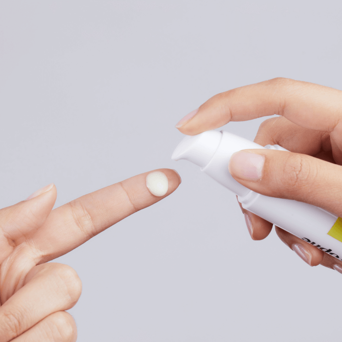
Tag: skin care

  • What are Skin Tag Patches? A Comprehensive Guide

    What are Skin Tag Patches? A Comprehensive Guide

    What are skin tag patches? They’re small, adhesive plasters designed to cover and potentially resolve skin tags. This comprehensive guide explores various types, application methods, ingredients, effectiveness, safety precautions, and comparisons to other removal methods. We’ll delve into the science behind these patches, offering a clear picture of their benefits and drawbacks. Learn everything you need to know about skin tag patches in one place.

    From understanding the different materials and application techniques to evaluating the potential risks and benefits, this guide will empower you to make informed decisions about skin tag patches. We’ll analyze the science behind their purported effectiveness and examine their comparative advantages and disadvantages relative to other treatment options. This detailed look at skin tag patches aims to provide a comprehensive overview for anyone considering this method for skin tag management.

    Defining Skin Tag Patches

    What are skin tag patches

    Skin tag patches are a popular and often effective non-invasive method for managing skin tags. These small, adhesive patches are designed to be applied directly to the skin tag, aiming to reduce its size and potentially promote its eventual resolution. While not a cure-all, they can be a helpful part of a comprehensive skin tag management strategy.Skin tag patches work by applying pressure and/or providing targeted treatments to the skin tag.

    The materials used in the patch and the specific treatments incorporated into the patch play a crucial role in their effectiveness. The various patch types available on the market demonstrate the range of approaches to managing skin tags, highlighting both advantages and drawbacks.

    Skin Tag Patch Types

    Different skin tag patches employ various approaches to addressing skin tags. This variety reflects the diversity of underlying causes and characteristics of skin tags. Understanding the different types of patches can help individuals choose the most appropriate option for their specific needs.

    Patch Materials

    A wide range of materials are used in creating skin tag patches. The choice of material significantly impacts the patch’s comfort, effectiveness, and longevity. Some common materials include:

    • Silicone-based materials: These materials are often preferred for their ability to create a moist environment around the skin tag, promoting its softening and eventual shedding. They are known for their comfort and generally good compatibility with skin.
    • Hydrocolloid materials: These materials are known for their ability to absorb moisture and create a protective barrier. This can help reduce irritation and promote healing.
    • Acrylic adhesives: These adhesives are used for their strong bonding properties, enabling the patch to remain firmly in place for extended periods. However, they can sometimes cause skin irritation for some individuals.
    • Natural fibers: Certain patches incorporate natural fibers like cotton for added comfort and breathability. These options can be more gentle on sensitive skin.

    Effectiveness Comparison

    The effectiveness of different skin tag patch types varies depending on several factors, including the individual’s skin type, the size and characteristics of the skin tag, and the specific components of the patch. A patch’s ability to absorb moisture, create a localized pressure, and deliver targeted treatments directly to the skin tag all contribute to its efficacy.

    Patch Type Characteristics

    The table below summarizes various skin tag patch types, their materials, and their associated benefits and drawbacks.

    Patch Type Material Benefits Drawbacks
    Silicone-based Silicone, often combined with other polymers Gentle on skin, promotes moisture retention, potentially accelerates resolution May not be as effective for large or stubborn skin tags, potential for skin reactions in sensitive individuals
    Hydrocolloid Hydrocolloid polymers Absorbs moisture, creates a protective barrier, reduces irritation Can be less comfortable than silicone patches, potential for adhesive residue
    Acrylic adhesive Acrylic polymers Strong adhesion, long-lasting Can cause skin irritation or allergic reactions in some individuals, may be less comfortable than silicone
    Natural fiber Cotton, other natural fibers Gentle on sensitive skin, breathable Adhesion may be weaker than other patch types, potentially less effective for treating larger skin tags

    Application and Use

    Applying skin tag patches correctly is crucial for their effectiveness and minimizing potential complications. Proper application ensures the patch adheres securely to the skin tag, promoting healing and preventing irritation. Following the manufacturer’s instructions is paramount for optimal results.Understanding the application process and the importance of proper care is essential for achieving the desired outcome. This includes knowing how to apply the patch, how often to change it, and how to remove it safely.

    Careful attention to detail throughout the application and removal process will help prevent discomfort and ensure the patch performs its intended function.

    Proper Application Method

    The application of a skin tag patch requires precision and care to ensure proper adhesion and prevent skin irritation. Cleanliness of the skin tag and the surrounding area is key. Use a gentle cleanser and pat the area dry before applying the patch. This step removes any dirt or oils that could hinder adhesion.

    Steps Involved in Applying a Skin Tag Patch, What are skin tag patches

    1. Thoroughly clean the skin tag and the surrounding skin with a mild cleanser.
    2. Pat the area completely dry using a soft cloth or paper towel.
    3. Carefully remove the backing of the patch, being mindful not to touch the adhesive side.
    4. Position the patch directly over the skin tag, ensuring proper alignment.
    5. Gently press the patch onto the skin tag to secure its adhesion.
    6. Avoid excessive pressure, as this could cause discomfort or damage.

    Frequency of Patch Changes

    The frequency of patch changes varies depending on the specific patch and the individual’s healing process. The manufacturer’s instructions usually specify the recommended change intervals. Adhering to the suggested schedule is important for optimal results. If you experience increased redness, pain, or unusual discharge around the patch, consult a dermatologist immediately. Changing the patch more frequently than recommended might be necessary if the area becomes irritated.

    Potential Complications

    Potential complications from using skin tag patches include skin irritation, allergic reactions, and infection. These complications are less common when following the instructions carefully. If you notice any signs of irritation, such as redness, swelling, or itching, discontinue use and consult a healthcare professional. Allergic reactions can manifest as hives, rashes, or difficulty breathing. In such cases, seek immediate medical attention.

    Infection can occur if the patch is not changed regularly or if the skin around the patch is not kept clean.

    Step-by-Step Guide for Applying a Skin Tag Patch

    1. Preparation: Cleanse the skin tag and surrounding area gently with a mild cleanser. Pat dry.
    2. Patch Placement: Carefully remove the backing from the patch, avoiding touching the adhesive side. Position the patch directly over the skin tag.
    3. Secure Adhesion: Gently press the patch onto the skin tag to ensure secure adhesion.
    4. Monitoring: Observe the area for any signs of irritation, such as redness, swelling, or itching. Change the patch as directed by the manufacturer.

    Importance of Following Instructions

    Following the manufacturer’s instructions is critical for effective treatment and minimizing risks. These instructions are designed to ensure the patch functions as intended and to protect the user. The patch’s composition, application method, and change schedule are all meticulously formulated for the best results. Failure to adhere to these instructions may compromise the effectiveness of the treatment and could lead to complications.

    How to Remove a Skin Tag Patch Safely

    Removing a skin tag patch safely involves careful handling to prevent skin damage. A gentle approach is essential to avoid pulling or tugging at the skin. The procedure for removal should be as follows:

    1. Gently loosen the edges of the patch, starting from one corner.
    2. Peel the patch away from the skin in a slow, smooth motion.
    3. Avoid abrupt movements, which could irritate the skin.
    4. If the patch is difficult to remove, gently soak the area with warm water for a few minutes to soften the adhesive.
    5. Use a mild adhesive remover, if necessary, to help loosen the adhesive. Always follow the instructions on the remover.
    6. Clean the area with a mild cleanser and pat dry.

    Ingredients and Composition

    Skin tag patches, a popular at-home treatment, often utilize a combination of ingredients to promote the detachment or reduction of skin tags. Understanding these ingredients and their potential effects can help you make informed decisions about using these products. This section delves into the common components of skin tag patches, their intended functions, and the possible side effects.The composition of skin tag patches varies among brands.

    While some share similar ingredients, others may use unique formulations. This variability can affect the efficacy and safety of the treatment. A crucial aspect of using these patches is awareness of the ingredients and their potential to cause reactions.

    Common Ingredients

    Various ingredients are incorporated into skin tag patches, each playing a specific role in the treatment process. The primary purpose of these ingredients is often to create an environment conducive to the skin tag’s detachment or to reduce its size.

    Active Ingredients

    Many patches utilize ingredients known for their adhesive and/or irritant properties. These properties are designed to draw moisture away from the skin tag and/or irritate the area surrounding the skin tag to stimulate detachment. Commonly used ingredients include salicylic acid, which has keratolytic properties, and certain plant extracts.

    Purpose of Ingredients

    Salicylic acid, a common active ingredient, is a keratolytic agent that helps exfoliate the skin. This exfoliation can lead to the gradual detachment of the skin tag. Other ingredients, such as certain plant extracts, are sometimes added to promote wound healing and reduce inflammation. The adhesive component is crucial for holding the patch in place and ensuring consistent contact with the skin tag.

    Skin tag patches are small, harmless growths that can appear on the skin. Understanding these little skin blemishes is important, but it’s also helpful to know the difference between receiving an immunization and getting a vaccination. For example, a better understanding of the nuances of the process can help you make informed decisions about your health. Ultimately, learning about skin tag patches, like researching the difference between immunization and vaccination , helps you become more proactive about your well-being, leading to better overall health choices.

    Just remember that skin tags are usually nothing to worry about, unless you have any concerns, it’s always best to see a dermatologist.

    Potential Side Effects

    While skin tag patches are generally considered safe, potential side effects can occur. Skin irritation, redness, and discomfort are possible reactions to the ingredients, especially in individuals with sensitive skin. Allergic reactions to specific components, such as certain plant extracts or adhesives, are also a possibility. Always perform a patch test before applying the patch to a larger area to assess individual sensitivity.

    Skin tag patches are small, harmless growths that sometimes appear on the skin. While they often don’t require treatment, some people turn to natural remedies like ashwagandha for potential benefits, but it’s important to research the potential side effects of ashwagandha before using it. For a comprehensive guide to ashwagandha’s benefits, side effects, and more, check out this resource: ashwagandha benefits side effects and more.

    Ultimately, if you’re concerned about skin tags, consulting a dermatologist is always a good idea.

    Comparison of Ingredients

    Different skin tag patches may use varying combinations of ingredients. Some brands might prioritize salicylic acid for its exfoliating properties, while others might focus on plant extracts for their potential anti-inflammatory benefits. A thorough review of the specific ingredients listed on the product label is essential before use.

    Ingredient Table

    Ingredient Purpose Potential Side Effects
    Salicylic Acid Keratolytic agent, promotes exfoliation. Skin irritation, redness, dryness, allergic reactions.
    Plant Extracts (e.g., Witch Hazel, Aloe Vera) Promote wound healing, reduce inflammation, and soothe skin. Allergic reactions, potential skin sensitivity.
    Adhesive Maintains patch contact with skin tag. Skin irritation, allergic reactions, potential residue.

    Effectiveness and Results: What Are Skin Tag Patches

    What are skin tag patches

    Skin tag patches offer a promising, non-invasive approach to addressing skin tags. While they aren’t a guaranteed cure for everyone, numerous users have reported positive experiences. Understanding the factors influencing their effectiveness, the typical timeframe for results, and the potential limitations is crucial for making informed decisions.Effective skin tag patches typically work by applying targeted pressure or a combination of ingredients to the skin tag, gradually causing it to dry out and fall off.

    The specific mechanisms vary depending on the type of patch. However, the fundamental principle remains the same: consistent application can promote natural detachment.

    Overall Effectiveness

    Skin tag patches demonstrate varying degrees of effectiveness depending on factors such as the individual’s skin type, the size and location of the skin tag, and the patch’s specific formulation. While some users experience complete resolution, others may only see partial or no improvement. It’s essential to approach patch use with realistic expectations.

    Factors Influencing Patch Effectiveness

    Several factors contribute to the success or failure of skin tag patches. The size and thickness of the skin tag can impact the patch’s ability to effectively target the area. Skin type, the presence of other skin conditions, and individual metabolic processes also play a role. Consistent application and adherence to the patch’s instructions are paramount. Patches designed for specific skin types or conditions may also influence the results.

    Success Rates of Different Patch Types

    Precise success rates for various skin tag patch types are difficult to quantify definitively due to the lack of large-scale, controlled clinical trials. Anecdotal evidence and user testimonials from online forums and reviews often point to varying levels of effectiveness. Different patch compositions with varied active ingredients might affect the results.

    Testimonials and Success Stories

    Numerous user testimonials suggest that skin tag patches can be an effective solution for many individuals. These accounts highlight positive experiences, such as gradual shrinking and eventual detachment of the skin tag. However, individual responses can differ significantly.

    Typical Timeframe for Seeing Results

    The typical timeframe for observing results varies considerably. Some users might see noticeable changes within a few weeks, while others might require several months. The process is often gradual, with a slow but consistent shrinking or detachment of the skin tag. The timeframe is also influenced by factors like skin tag size, location, and individual response.

    Visual Representation of Potential Results Timeline

    Week Possible Result
    1-4 Slight shrinkage, minimal change
    4-8 Moderate shrinkage, some detachment visible
    8-12 Significant shrinkage, noticeable detachment
    12+ Complete detachment, resolution

    This table provides a general guideline; individual results may vary. It is crucial to understand that the timeframe is not a strict rule, and results can differ based on the factors mentioned earlier.

    Comparison of Result Speed with Different Patch Types

    Comparing the speed of results across various patch types is challenging due to limited data and varying user experiences. Some users report faster results with certain patch compositions, while others may find similar efficacy across different brands. It is recommended to carefully review product descriptions and user reviews to understand the potential outcomes.

    Consulting a Doctor for Severe Cases

    For individuals with skin tags that are excessively large, painful, bleeding, or showing signs of infection, consulting a dermatologist or other healthcare professional is highly recommended. They can assess the skin tag, determine the appropriate course of action, and rule out any underlying medical conditions. This approach is especially crucial for individuals with specific medical conditions or concerns.

    Safety Precautions and Considerations

    Using skin tag patches can be a convenient way to address skin tags, but it’s crucial to understand the potential risks and precautions. Proper use and awareness of possible complications are essential for a safe and effective experience. Before applying any patch, thoroughly research the specific product and its potential side effects.Understanding the potential risks associated with skin tag patches, and knowing when they’re not the right choice, is paramount.

    This section Artikels crucial safety considerations to ensure a positive and safe experience.

    Potential Risks

    Skin tag patches, while generally considered safe, can sometimes cause adverse reactions. These reactions can range from mild irritation to more severe allergic responses. Possible side effects can include redness, itching, swelling, or a rash at the application site. Less common, but potentially more serious, are allergic reactions requiring immediate medical attention.

    Situations Where Patches Aren’t Recommended

    Certain situations make skin tag patches less suitable. These include individuals with known allergies to any of the patch’s ingredients. Pre-existing skin conditions, such as eczema or psoriasis, can also increase the risk of adverse reactions. If you have open wounds or broken skin near the skin tag, the patch may not be appropriate. Patches should not be used on skin that is already irritated or inflamed.

    Avoiding Adverse Reactions

    Several steps can help minimize the risk of adverse reactions. Always follow the manufacturer’s instructions carefully. Apply the patch to a clean, dry area of skin. Avoid applying the patch over existing skin irritations or wounds. If any irritation or allergic reaction occurs, immediately discontinue use and consult a healthcare professional.

    Importance of Professional Consultation

    Before using any skin tag patch, it’s essential to consult a dermatologist or other qualified healthcare professional. They can assess your specific situation, identify potential risks, and provide personalized advice. A healthcare professional can help determine if skin tag patches are appropriate for your skin type and health condition.

    Skin tag patches are small, harmless skin growths, often mistaken for other things. While they’re generally not a cause for concern, understanding their appearance and potential causes is important. Fortunately, resources like metastatic breast cancer resources offer valuable information on various skin conditions, and it’s helpful to differentiate them from potentially more serious concerns.

    Ultimately, if you’re unsure about a skin growth, consulting a dermatologist is always a good idea for proper diagnosis and treatment options for skin tag patches.

    Caution and Appropriate Use

    Skin tag patches should be used with caution and adherence to the recommended guidelines. Avoid applying the patch to areas with sensitive skin. Do not use the patch on children without consulting a pediatrician. Always carefully read and understand the instructions provided with the product.

    Dealing with Potential Complications

    If you experience any adverse reaction, such as severe itching, swelling, or difficulty breathing, immediately stop using the patch and seek immediate medical attention. Mild reactions, like redness or itching, may subside on their own. However, it’s always best to consult a healthcare professional for proper guidance and treatment.

    Safety Guidelines and Warnings

    “Always prioritize your health and safety. Consult a healthcare professional before using any skin tag patch, especially if you have pre-existing skin conditions or allergies.”

    • Avoid using patches on broken or irritated skin.
    • Do not use patches on children without consulting a pediatrician.
    • Follow the manufacturer’s instructions carefully.
    • Discontinue use and seek medical attention if any adverse reaction occurs.

    Comparison with Other Treatments

    Skin tag patches offer a convenient and often less invasive alternative to traditional skin tag removal methods. However, understanding the strengths and weaknesses of various approaches is crucial for making an informed decision. This comparison will help you weigh the pros and cons of different methods, ultimately enabling you to choose the best option for your specific needs and circumstances.Different methods for skin tag removal cater to diverse needs and preferences.

    Comparing the efficacy, cost, recovery time, and potential risks associated with each technique is essential for a well-informed decision.

    Surgical Removal

    Surgical removal, often performed by a dermatologist or surgeon, involves cutting the skin tag off. This method is generally effective, but it requires a small incision and often results in some degree of scarring. While the procedure is usually quick and relatively straightforward, it can be more costly and time-consuming compared to using patches. Potential risks include bleeding, infection, and uneven skin texture.

    Cryotherapy

    Cryotherapy utilizes extreme cold to freeze and destroy the skin tag. This method is less invasive than surgery and generally results in less scarring. However, cryotherapy can be painful, and the effectiveness may vary depending on the size and location of the skin tag. The recovery time can also be longer than that for patch treatment.

    Electrocautery

    Electrocautery uses an electric current to burn off the skin tag. It’s a quick and effective method, but it may result in more noticeable scarring than cryotherapy. Pain management might be necessary, and potential risks include burns or uneven skin texture. This method is often more expensive than using a patch.

    Laser Removal

    Laser removal employs concentrated beams of light to vaporize the skin tag. It’s a precise method that usually results in minimal scarring, making it a popular choice for cosmetic reasons. Laser removal is often more expensive than other methods, and the recovery time can be longer. Additionally, laser treatment is best suited for specific cases and may not be suitable for all skin tag types.

    Comparison Table

    Treatment Advantages Disadvantages
    Skin Tag Patches Convenient, non-invasive, relatively inexpensive, minimal downtime, often suitable for multiple skin tags. May not be effective for all skin tags, results may vary depending on individual factors, potential for skin irritation in some cases.
    Surgical Removal High effectiveness rate, quick procedure (in most cases). Invasive, potential for scarring, requires professional expertise, greater cost, potential for complications like infection or bleeding.
    Cryotherapy Less invasive than surgery, generally less scarring, relatively inexpensive. Potential for pain, varying effectiveness, longer recovery time, possible uneven skin texture.
    Electrocautery Quick procedure, effective. Potential for more noticeable scarring, pain, potential for burns or uneven skin texture.
    Laser Removal Precise, minimal scarring, good cosmetic outcome. Expensive, longer recovery time, not suitable for all skin tags, requires specialized equipment and expertise.

    Potential Benefits and Drawbacks

    Skin tag patches offer a convenient, at-home approach to addressing skin tags. However, like any treatment option, they come with potential benefits and drawbacks that users should be aware of before deciding if they’re the right choice. Understanding these nuances can help individuals make informed decisions about their skin tag removal journey.

    Potential Benefits

    Skin tag patches, when used as directed, can offer several advantages. These often include a reduced risk of infection and bleeding compared to some other removal methods. The non-invasive nature of the treatment often leads to minimal discomfort and a faster recovery time. Patch application is typically straightforward, allowing for home-based treatment and potentially saving on medical costs associated with clinic visits.

    • Reduced risk of infection and bleeding: The controlled environment and direct application of the patch can minimize the chances of infection or significant bleeding during the treatment process. This is particularly beneficial for individuals with pre-existing conditions or those who may be more susceptible to these complications.
    • Minimal discomfort and faster recovery: The non-invasive nature of skin tag patches usually results in minimal discomfort and a quicker recovery time compared to other, more invasive procedures. Patients often report less pain and quicker healing compared to surgical excision.
    • Convenience and cost-effectiveness: The ability to apply the patch at home offers significant convenience, avoiding the need for clinic visits or appointments. This can potentially lead to lower overall costs compared to professional removal services.
    • Potential for increased patient control: The home-based application allows patients to control the timing and frequency of treatment, making it potentially more suitable for individuals with busy schedules.

    Potential Drawbacks

    While skin tag patches offer numerous advantages, potential drawbacks exist. Not all skin tags respond uniformly to the patch treatment. In some cases, the patch may not be effective in completely removing the skin tag. Furthermore, the patch might cause skin irritation or allergic reactions in sensitive individuals. Consistency in application is critical, and missing applications can affect the effectiveness of the treatment.

    • Inconsistent results: Not all skin tags respond to patch treatment in the same way. Some skin tags may not fully detach or resolve, requiring additional or alternative methods. Factors such as the size, type, and location of the skin tag may influence its response.
    • Skin irritation or allergic reactions: Certain individuals may experience skin irritation, redness, or allergic reactions to the patch’s ingredients. This is particularly important for those with known sensitivities or allergies to specific substances.
    • Potential for incomplete removal: Some skin tags may not be completely removed by the patch treatment, requiring further interventions or alternative treatments.
    • Importance of consistent application: Regular and consistent application of the patch is essential for optimal results. Irregular application can hinder the patch’s effectiveness, potentially prolonging the treatment duration.
    • Limited suitability for certain skin types or conditions: Individuals with specific skin conditions or sensitivities might not be suitable candidates for patch treatment. Skin that is extremely sensitive, prone to irritation, or has existing skin conditions may not respond well to the patch treatment.

    Situations Where Patches May Not Be Suitable

    There are situations where skin tag patches might not be the ideal treatment option. For instance, if the skin tag is particularly large or has a complex structure, surgical removal might be more appropriate. Also, if the skin tag is causing significant discomfort or bleeding, seeking professional medical advice is crucial.

    • Large or complex skin tags: Skin tags that are significantly large or exhibit a complex structure may require more extensive removal methods, such as surgical excision, rather than a patch treatment. The patch may not be sufficient to fully address these types of skin tags.
    • Significant discomfort or bleeding: If the skin tag is causing persistent discomfort or significant bleeding, it is essential to consult a healthcare professional. In these situations, professional medical attention might be necessary to manage the condition effectively.
    • Suspected cancerous growth: If there is any suspicion that the skin tag might be a cancerous growth, immediate medical evaluation is critical. A professional diagnosis and treatment plan are necessary in such cases.

    Last Point

    In conclusion, skin tag patches offer a convenient and relatively non-invasive approach to addressing skin tags. While they may not be a perfect solution for everyone, they provide a viable alternative for those seeking a less aggressive removal method. This guide has highlighted the various factors to consider when deciding if skin tag patches are right for you, emphasizing the importance of careful consideration and consultation with a healthcare professional.

    Remember, always prioritize your well-being and consult a doctor before implementing any new treatment method.

  • Weeping Eczema Overview and More

    Weeping Eczema Overview and More

    Weeping eczema overview and more delves into the complexities of this skin condition, offering a comprehensive understanding of its causes, symptoms, and management. We’ll explore everything from the underlying triggers to effective treatments and preventive strategies. Prepare to gain valuable insights into this common yet often challenging skin issue.

    This comprehensive guide covers the full spectrum of weeping eczema, from its definition and diagnostic process to lifestyle adjustments and long-term effects. We’ll also delve into illustrative examples, prevention strategies, and valuable resources to support your understanding and journey toward effective management.

    Introduction to Weeping Eczema

    Weeping eczema, also known as exudative eczema, is a type of eczema characterized by the formation of blisters that leak fluid. This fluid leakage, often accompanied by crusting, creates a weeping appearance on the skin. Understanding its causes, symptoms, and distinctions from other eczema types is crucial for effective management and treatment.Underlying causes of weeping eczema can be multifaceted, encompassing both internal and external factors.

    A compromised immune system, certain allergies, and reactions to irritants can all contribute to the development of weeping eczema. Skin conditions like psoriasis, or atopic dermatitis, can also present with weeping characteristics. The precise cause often requires careful medical evaluation.Typical symptoms associated with weeping eczema include the formation of blisters filled with clear or yellowish fluid. These blisters can appear on various parts of the body, but often cluster in areas prone to friction or irritation, such as the folds of the skin or areas exposed to frequent scratching.

    The affected skin may appear red, inflamed, and swollen, accompanied by intense itching. The weeping aspect of the condition is crucial to recognizing and differentiating it from other types of eczema.

    Comparison with Other Eczema Types, Weeping eczema overview and more

    Understanding the differences between weeping eczema and other eczema forms aids in proper diagnosis and treatment. The following table highlights key distinctions:

    Characteristic Weeping Eczema Dry Eczema Contact Eczema
    Appearance Blisters, fluid leakage, crusting Dry, cracked, scaly skin Redness, itching, rash often localized to contact area
    Cause Compromised immune system, allergies, irritants Dry skin, reduced skin barrier function Direct contact with allergens or irritants
    Symptoms Itching, blisters, weeping fluid, inflammation Itching, dryness, cracking, flaking Itching, redness, rash at the point of contact
    Location Can appear anywhere, often folds of skin Often on hands, feet, and elbows Typically localized to the area of contact

    The table demonstrates the distinct visual, causative, and symptomatic differences between the various eczema types. This comparative analysis helps differentiate weeping eczema from its counterparts.

    Causes and Risk Factors: Weeping Eczema Overview And More

    Understanding the causes and risk factors of weeping eczema is crucial for effective management. While a definitive cause isn’t always pinpointed, various contributing elements play a significant role in the development and exacerbation of this skin condition. This deeper dive explores the factors influencing weeping eczema, from genetic predispositions to environmental triggers.Weeping eczema, a distressing skin condition, isn’t triggered by a single factor.

    Instead, it’s often a complex interplay of genetic susceptibility, environmental irritants, and sometimes, even emotional stress. Pinpointing the precise cause can be challenging, but recognizing the potential triggers allows for better preventative measures and improved management strategies.

    Underlying Genetic Predisposition

    A person’s genetic makeup significantly influences their susceptibility to eczema. A family history of atopic dermatitis, asthma, or hay fever often indicates an increased risk. This genetic predisposition, inherited from parents, plays a crucial role in the body’s immune response, making individuals more prone to skin inflammation. Studies show a correlation between specific genes and the development of eczema, although further research is needed to fully understand the intricate genetic mechanisms.

    Environmental Triggers

    Environmental factors can significantly exacerbate eczema symptoms. Various substances and situations can irritate the skin, leading to flare-ups. Recognizing these triggers is vital for managing the condition effectively.

    Potential Environmental Triggers

    Category Examples
    Irritants Soaps, detergents, perfumes, harsh chemicals, wool, certain fabrics (synthetic fibers)
    Allergens Dust mites, pollen, pet dander, certain foods (e.g., nuts, dairy, eggs), molds, and insect bites
    Temperature Extremes Extreme heat or cold, prolonged exposure to wind
    Stress Emotional stress, anxiety, and physical exertion can trigger or worsen eczema symptoms.
    Infections Bacterial or viral infections can sometimes trigger or worsen existing eczema.
    Humidity High humidity can cause increased moisture on the skin, which may worsen eczema

    These triggers can vary from person to person, emphasizing the personalized approach to managing weeping eczema.

    Prevalence Across Demographics

    The prevalence of weeping eczema can vary based on demographics. While exact figures can be difficult to obtain, studies suggest that eczema tends to affect children more frequently than adults. Furthermore, certain ethnic groups may have a higher or lower prevalence compared to others. This difference might be linked to factors like environmental exposures and cultural practices.

    It’s important to remember that these are general observations, and individual experiences may differ.

    Diagnosis and Assessment

    Diagnosing weeping eczema involves a careful evaluation of the patient’s symptoms, medical history, and physical examination. Accurate diagnosis is crucial for effective treatment and management. It’s essential to distinguish weeping eczema from other skin conditions, as treatment approaches vary significantly. A proper assessment of the severity is also critical to guide treatment decisions.A comprehensive approach to diagnosis necessitates a thorough understanding of the patient’s presentation, including the location, appearance, and duration of the rash.

    This helps in narrowing down the possibilities and identifying potential contributing factors. Furthermore, a detailed history of any triggers, previous treatments, and family history of skin conditions can significantly aid in the diagnostic process.

    Diagnostic Process

    The diagnostic process for weeping eczema begins with a detailed patient history, encompassing the onset, progression, and characteristics of the rash. This includes the location, appearance (e.g., weeping, oozing, crusting), and any associated symptoms (e.g., itching, burning). The physician also inquires about potential triggers, such as allergens, irritants, or stressors. A physical examination of the affected skin is performed, focusing on the distribution, morphology, and severity of the rash.

    Skin biopsies may be necessary in some cases to confirm the diagnosis or rule out other conditions.

    Methods of Differentiation

    Differentiating weeping eczema from other skin conditions requires careful consideration of several factors. The presence of weeping, oozing, and crusting lesions, often accompanied by intense itching, can be characteristic of eczema. However, other skin conditions, such as allergic contact dermatitis, psoriasis, or fungal infections, can present with similar symptoms. A thorough history, physical examination, and possibly diagnostic tests are crucial to distinguish weeping eczema from these conditions.

    For instance, allergic contact dermatitis typically presents with a clear relationship to exposure to specific allergens, whereas psoriasis may exhibit characteristic silvery scales. Fungal infections often display specific patterns and may respond differently to treatment.

    Weeping eczema, a frustrating skin condition, often involves intense itching and oozing. It can be really tough to manage, and understanding its various triggers is key. While researching ways to soothe this discomfort, I stumbled across some interesting insights into how to manage your heart rate after a night out, which might be surprising. For example, how to slow heart rate after drinking alcohol can be really helpful, especially when you’re trying to calm down after a night of socializing.

    Learning to manage your body’s responses in different situations, whether it’s related to eczema or alcohol consumption, is definitely valuable. Back to eczema, there are many more aspects to explore and understand about this skin condition.

    Patient History Questions

    A structured approach to gathering patient information is essential. The following questions can aid in assessing the patient’s condition:

    • When did the rash first appear?
    • What are the characteristics of the rash (e.g., weeping, oozing, scaling)?
    • Where is the rash located on the body?
    • Has the rash been present before? If so, when?
    • What factors seem to trigger or worsen the rash?
    • What other medical conditions does the patient have?
    • Are there any family members with a history of skin conditions?
    • What medications or topical treatments has the patient used in the past?
    • Has the patient experienced any recent changes in their lifestyle or environment?

    Assessment of Severity

    Assessing the severity of weeping eczema is vital for determining the appropriate treatment plan. The severity is typically evaluated based on several factors, including the extent of skin involvement, the intensity of symptoms (e.g., itching, pain), and the presence of complications (e.g., secondary infections). The following criteria can be used to gauge the severity of the condition:

    • Area of involvement: The percentage of body surface area affected by the rash.
    • Intensity of symptoms: The level of itching, pain, or discomfort.
    • Presence of complications: Evidence of secondary infections, such as bacterial or fungal infections.
    • Impact on daily life: The degree to which the rash interferes with daily activities and routines.

    A scoring system, such as the Eczema Area and Severity Index (EASI), may be used to quantify the severity of the condition. This standardized method allows for a more objective assessment and facilitates comparison across different patients. The assessment is also influenced by the patient’s overall health status and the presence of any other co-morbidities.

    Management and Treatment

    Managing weeping eczema requires a multifaceted approach, focusing on both immediate relief and long-term prevention of flare-ups. A key component is identifying and avoiding triggers, as well as implementing appropriate treatment strategies. Effective management involves a combination of topical medications, lifestyle adjustments, and possibly, in severe cases, oral medications.Understanding the underlying causes of the weeping eczema is crucial for developing a tailored management plan.

    Addressing the root causes, along with consistent treatment, is vital for achieving and maintaining remission.

    General Approach to Management

    A comprehensive management plan for weeping eczema involves a combination of strategies, aiming to control inflammation, reduce moisture loss, and prevent secondary infections. This often includes identifying and avoiding triggers, employing appropriate topical treatments, and maintaining a consistent skin care routine. Maintaining a balanced diet and adequate hydration can also contribute to overall skin health and reduce the risk of flare-ups.

    Common Treatments for Weeping Eczema

    Topical corticosteroids are frequently prescribed to reduce inflammation and relieve itching. They come in various strengths, with stronger formulations reserved for more severe cases. Moisturizers are essential for restoring the skin’s barrier function, preventing moisture loss, and maintaining hydration. Other topical treatments, such as calcineurin inhibitors, may be used for cases where corticosteroids are not effective or are contraindicated.

    In some instances, oral medications like antihistamines or immunosuppressants may be considered to manage severe cases or when topical treatments are insufficient.

    Methods for Preventing Weeping Eczema Flare-ups

    Identifying and avoiding triggers is paramount in preventing weeping eczema flare-ups. These triggers can include specific foods, allergens, irritants, and environmental factors. Keeping a detailed eczema diary can help identify patterns and pinpoint potential triggers. Maintaining a consistent skincare routine, including regular moisturizing, can also help maintain skin health and reduce the risk of flare-ups. Stress management techniques, such as exercise and relaxation, can also contribute to overall well-being and potentially reduce eczema triggers.

    Topical Treatments and Their Mechanisms of Action

    Effective topical treatments target various aspects of eczema inflammation. The following table Artikels some common treatments and their mechanisms of action:

    Treatment Mechanism of Action
    Topical Corticosteroids (e.g., hydrocortisone) Reduce inflammation by suppressing the immune response in the skin.
    Moisturizers (e.g., emollients) Restore the skin barrier function, preventing moisture loss and maintaining hydration.
    Topical Calcineurin Inhibitors (e.g., tacrolimus) Inhibit the activity of calcineurin, a protein involved in immune response, thus reducing inflammation.
    Antibacterial or Antifungal creams (e.g., for secondary infections) Target specific microorganisms if secondary infection is present.

    Lifestyle and Self-Care

    Managing weeping eczema effectively often involves a multifaceted approach that goes beyond just topical treatments. Lifestyle choices, dietary habits, and self-care practices all play a crucial role in reducing flare-ups and improving overall well-being. Understanding how these factors interact with the condition can significantly impact its management.Lifestyle factors, including stress levels, diet, and environmental triggers, can significantly influence the severity and frequency of weeping eczema outbreaks.

    By proactively addressing these areas, individuals can experience greater control over their symptoms.

    Understanding weeping eczema, and its various forms, is crucial for effective management. It’s a tricky condition, and building a consistent skincare routine is key. Think about how long it takes to form a habit – how long does it take to form a habit – and apply that same principle to your eczema care. This consistency, over time, will help you better control the symptoms and find relief.

    Dietary Considerations

    Dietary choices can influence the body’s inflammatory response, potentially exacerbating or alleviating eczema symptoms. While there’s no single “eczema diet,” certain foods may trigger allergic reactions or worsen inflammation in predisposed individuals.Identifying and avoiding potential triggers is key. Common culprits include dairy products, eggs, nuts, soy, wheat, and certain fruits and vegetables. Keeping a food diary can help pinpoint specific foods that provoke a reaction.

    Consulting a registered dietitian or allergist can provide personalized guidance and recommendations. Eliminating suspected triggers, or gradually reintroducing them, can help determine their impact. Consider incorporating foods rich in antioxidants and omega-3 fatty acids, which may have anti-inflammatory properties.

    Self-Care Practices

    Implementing consistent self-care routines can be instrumental in managing weeping eczema symptoms. These practices can help soothe the skin, reduce irritation, and prevent further damage.

    • Gentle Skin Care: Using fragrance-free, hypoallergenic soaps and cleansers is essential. Avoid harsh scrubbing or hot water, as these can further irritate the skin. Pat the skin dry gently instead of rubbing.
    • Moisturization: Maintaining skin hydration is crucial. Apply a fragrance-free moisturizer frequently, especially after bathing or showering. Look for moisturizers that contain ceramides or other skin-repairing ingredients.
    • Avoiding Irritants: Identify and avoid triggers such as harsh detergents, wool clothing, and certain fabrics. Wear loose-fitting, breathable clothing, and opt for natural fibers like cotton.
    • Protecting from the Environment: Weather conditions can also aggravate eczema. Protect the skin from extreme temperatures, sun exposure, and wind. Apply a broad-spectrum sunscreen with an SPF of 30 or higher.
    • Stress Management Techniques: Chronic stress can worsen eczema symptoms. Incorporating stress-reduction techniques such as meditation, yoga, deep breathing exercises, or spending time in nature can help manage stress levels effectively.

    Stress Management Techniques

    Stress is a significant factor in exacerbating various health conditions, including eczema. Understanding the connection between stress and eczema can lead to proactive strategies for managing stress and improving symptom control.Recognizing the impact of stress on eczema allows individuals to implement targeted interventions. Effective stress management techniques can include mindfulness exercises, deep breathing techniques, regular physical activity, sufficient sleep, and spending time in nature.

    These methods can significantly reduce stress levels and contribute to improved skin health.

    Complications and Long-Term Effects

    Weeping eczema overview and more

    Weeping eczema, while treatable, can lead to a range of complications if left unmanaged. Understanding these potential issues and long-term effects is crucial for proactive management and improved quality of life. This section delves into the complications, highlighting the importance of early intervention and consistent care.Untreated weeping eczema can lead to secondary infections, scarring, and psychological distress. These complications can significantly impact a person’s well-being and require careful attention to prevent further harm.

    The impact on quality of life is substantial, affecting daily activities and social interactions.

    Potential Complications of Weeping Eczema

    Weeping eczema, due to its nature of oozing and inflammation, creates a perfect environment for bacterial or fungal infections. These secondary infections can worsen the eczema, prolong treatment, and cause discomfort. The skin’s barrier function is compromised, making it vulnerable to these external agents.

    • Secondary Infections: Bacterial infections, like impetigo, or fungal infections, such as candidiasis, can easily colonize the damaged skin. These infections manifest as pus-filled sores, redness, and increased pain. Early diagnosis and prompt treatment of these infections are crucial to prevent further spread and complications.
    • Skin Scarring: Chronic scratching and inflammation associated with weeping eczema can lead to permanent skin discoloration and scarring. Repeated damage to the skin’s tissues can result in noticeable changes in skin texture and appearance. Preventing excessive scratching is paramount.
    • Skin thickening (lichenification): Persistent scratching can lead to thickened, hardened skin areas. This condition, known as lichenification, is characterized by a rough, leathery appearance and is often associated with intense itching and discomfort.

    Long-Term Effects of Untreated Weeping Eczema

    The long-term effects of untreated weeping eczema can extend beyond physical discomfort. The constant itching and inflammation can significantly impact a person’s mental well-being.

    • Psychological Impact: The persistent itching, discomfort, and social stigma associated with weeping eczema can contribute to anxiety, depression, and low self-esteem. The emotional toll of managing this chronic condition is often underestimated and needs specific attention.
    • Compromised Sleep: The intense itching and discomfort can disrupt sleep patterns, leading to fatigue and impaired daily functioning. This is a significant consequence of the chronic nature of the condition.
    • Reduced Quality of Life: Weeping eczema can affect various aspects of daily life, including school or work performance, social interactions, and overall enjoyment of activities. The condition can lead to feelings of isolation and social withdrawal, further impacting quality of life.

    Strategies to Mitigate Complications

    Early diagnosis and consistent treatment are crucial for mitigating potential complications. Lifestyle adjustments and appropriate skincare routines can also play a vital role.

    • Consistent Treatment: Adherence to prescribed medications and topical treatments, as directed by a dermatologist, is essential for controlling inflammation and preventing secondary infections. This includes regular application of prescribed creams and ointments.
    • Avoiding Irritants: Identifying and avoiding triggers like harsh soaps, detergents, and certain fabrics can minimize the frequency and severity of eczema flare-ups. This proactive approach is critical for long-term management.
    • Proper Skin Care: Maintaining a gentle skin care routine, using fragrance-free products, and moisturizing regularly can help maintain skin barrier function and prevent dryness. Moisturizing is key to preventing exacerbations.
    • Stress Management: Stress can exacerbate eczema. Practicing stress-reducing techniques like meditation, yoga, or deep breathing exercises can be beneficial. Strategies for stress reduction can contribute to better overall health.

    Illustrative Examples

    Understanding weeping eczema through real-life cases and visual representations helps solidify its characteristics and treatment pathways. This section provides detailed examples of patient experiences and the appearance of the condition, highlighting the variability and challenges associated with this skin disorder.

    A Case Study of a Patient with Weeping Eczema

    A 32-year-old woman presented with a history of intermittent, itchy skin rashes. Initially, the rash appeared as small, red bumps, primarily on her hands and forearms. Over time, the rash evolved into weeping, oozing lesions, especially following periods of stress or changes in weather. The patient reported that the weeping eczema worsened after contact with certain fabrics, detergents, and environmental triggers.

    The severity of the condition significantly impacted her daily life, affecting her work performance and social interactions due to the discomfort and visible nature of the rash.

    A Patient Journey Through Weeping Eczema Treatment

    This patient’s journey began with self-treatment using over-the-counter lotions and creams. While providing some temporary relief, the symptoms persisted and worsened. She then sought medical advice, receiving a diagnosis of weeping eczema and a prescription for topical corticosteroids. The medication provided significant relief, but the patient experienced some side effects like skin thinning. With the help of a dermatologist, she identified specific triggers, such as certain soaps and wool clothing, and learned to implement avoidance strategies.

    The patient’s routine included moisturizing daily and using protective clothing during periods of heightened sensitivity. Regular follow-up appointments helped monitor the condition and adjust the treatment plan as needed, leading to long-term symptom management and improved quality of life.

    Weeping eczema, a frustrating skin condition, often leaves sufferers with intense itchiness. This persistent itch can be particularly bothersome after a shower, when the skin’s natural moisture barrier is compromised. Understanding why this happens can be crucial in managing the condition. For example, figuring out if your shower routine is contributing to the itchiness is key to finding relief.

    Check out this helpful article on reasons why you itch after taking a shower for more insights. Ultimately, addressing the underlying causes of weeping eczema and its symptoms, like persistent itching, is vital for better management.

    Detailed Illustration of Weeping Eczema Lesions

    Weeping eczema lesions are characterized by areas of erythema (redness) and weeping or oozing fluid. The fluid may be clear, yellowish, or contain crusts. The skin often appears inflamed, with a moist and sometimes raw surface. The lesions can vary in size and shape, often appearing in clusters. The affected skin may feel hot to the touch, and itching is a common symptom.

    In severe cases, the lesions can be widespread, affecting large areas of the body. The weeping can lead to skin cracking and discomfort.

    Visual Representation of Different Stages of Weeping Eczema

    Visual representation of weeping eczema stages can illustrate the progressive nature of the condition. Early stages often manifest as small, red papules or plaques, with minimal oozing. As the condition progresses, the lesions enlarge and become more inflamed, exhibiting increased weeping and crusting. The lesions can vary in severity, with some exhibiting only minor weeping, while others have extensive and persistent weeping.

    Severe cases can show deep skin fissures and significant tissue damage. Understanding these stages is critical for appropriate diagnosis and tailored treatment.

    Prevention Strategies

    Weeping eczema, while frustrating, is manageable with proactive strategies. Prevention focuses on identifying and avoiding triggers, establishing a consistent routine, and understanding your body’s unique responses. This allows for better control over flare-ups and minimizes the discomfort associated with weeping eczema.Effective eczema prevention isn’t about a one-size-fits-all approach. It’s about tailoring a personalized strategy to your specific triggers and lifestyle.

    By understanding your eczema’s quirks, you can create a plan that works for you, not against you.

    Trigger Avoidance Strategies

    Identifying and eliminating triggers is paramount in preventing weeping eczema flare-ups. Common triggers include certain fabrics, harsh chemicals, and environmental factors. A detailed log of your eczema episodes can be invaluable in pinpointing patterns and specific irritants.

    • Environmental Triggers: Extreme temperatures (both hot and cold), humidity, and even certain weather conditions can aggravate eczema. Understanding your body’s reactions to these elements can help you adapt your environment to minimize flare-ups.
    • Allergens: Substances like pollen, dust mites, pet dander, and certain foods can trigger allergic reactions that manifest as eczema. Avoiding contact with these allergens is crucial.
    • Irritants: Harsh soaps, detergents, and certain fabrics (like wool) can irritate the skin and exacerbate eczema. Switching to gentler alternatives and choosing soft fabrics can make a significant difference.
    • Stress: While not a direct cause, stress can worsen eczema symptoms. Incorporating stress-reducing activities like yoga, meditation, or spending time in nature can be beneficial.

    Personalized Eczema Management Plan

    A personalized eczema management plan is crucial for long-term control. This plan should be adaptable and flexible to accommodate your daily life and specific needs. It’s not just about avoiding triggers, but about creating a skin-friendly routine.

    1. Skincare Routine: A gentle, fragrance-free skincare routine, including moisturizing creams and barrier repair products, is essential. Regular application of emollients, particularly after bathing, helps maintain skin hydration and prevents dryness, a major contributor to eczema flare-ups.
    2. Trigger Log: Maintain a detailed log of potential triggers, including foods, environmental factors, and activities. This helps identify patterns and allows you to make adjustments to your lifestyle accordingly.
    3. Professional Guidance: Consulting a dermatologist or healthcare professional provides valuable insights into your specific needs. They can offer personalized recommendations, including specific treatments and tailored avoidance strategies.
    4. Stress Management: Chronic stress can significantly worsen eczema symptoms. Incorporate stress-reducing activities, such as exercise, meditation, or spending time in nature, into your daily routine.

    Preventative Strategies for Daily Routines

    Implementing preventative strategies into your daily routines can significantly reduce the frequency and severity of weeping eczema flare-ups. These strategies should become an integral part of your daily life, not just a temporary fix.

    • Gentle Washing: Use lukewarm water and fragrance-free, hypoallergenic soaps or cleansers. Avoid harsh scrubbing or excessive washing.
    • Moisturizing: Apply a moisturizer, especially after showering or bathing, to maintain skin hydration. This creates a protective barrier that helps prevent moisture loss and irritation.
    • Avoiding Irritants: Be mindful of clothing materials, detergents, and other products that may irritate your skin. Opt for natural fabrics and hypoallergenic cleaning products.
    • Stress Reduction Techniques: Incorporate relaxation techniques like meditation, deep breathing exercises, or spending time in nature into your daily routine. Stress can significantly worsen eczema symptoms.

    Additional Resources

    Weeping eczema overview and more

    Staying informed and connected with the right resources is crucial for managing weeping eczema effectively. This section provides valuable avenues for further learning and support, empowering you to navigate your eczema journey with confidence. Understanding the available resources can help you access comprehensive information and expert advice, promoting better self-management and overall well-being.

    Reputable Websites and Organizations

    Numerous websites and organizations offer in-depth information on eczema, including weeping eczema. These platforms often provide evidence-based content, support groups, and educational materials to help individuals understand and manage their condition.

    • The National Eczema Association (NEA): A leading organization dedicated to eczema research, education, and support. They offer comprehensive resources, including articles, webinars, and information on various eczema types, including weeping eczema.
    • The American Academy of Dermatology (AAD): A reputable source of information on skin conditions, including eczema. Their website provides up-to-date information on diagnosis, treatment, and management strategies for various forms of eczema.
    • Mayo Clinic: Known for its extensive medical information, the Mayo Clinic website offers reliable information on eczema, including causes, symptoms, and treatment options. They also provide guidance on managing chronic skin conditions.
    • Cleveland Clinic: Another trusted source for health information, the Cleveland Clinic website provides detailed insights into eczema, including different types, potential triggers, and treatment approaches. They offer specific information on managing skin conditions effectively.

    Healthcare Professionals

    Consulting with healthcare professionals specializing in eczema management is essential for tailored care and effective treatment plans. Finding a dermatologist or allergist familiar with eczema can significantly improve your experience and outcomes.

    • Dermatologists: These physicians are specifically trained in diagnosing and treating skin conditions like eczema. They can provide accurate diagnoses, recommend appropriate treatments, and monitor your condition over time.
    • Allergists: Allergists can identify and manage potential triggers for eczema, helping you avoid specific environmental factors that exacerbate your condition. They can also provide guidance on desensitization therapies if needed.
    • Primary Care Physicians: Your primary care physician can offer initial guidance, make referrals to specialists, and manage your overall health, including your eczema. They are a valuable first point of contact for many individuals.

    Books, Articles, and Other Resources

    Numerous books, articles, and other resources offer practical advice and insights into managing weeping eczema. Seeking out these resources can enhance your understanding of the condition and its management.

    • The Eczema Solution by Dr. [Author Name]: This book might offer insights into dietary considerations, lifestyle modifications, and treatment approaches. Verify the author’s credentials to ensure reliability.
    • Eczema and Skin Conditions by [Author Name]: Articles and resources available online can offer a wide range of information and perspectives on various skin conditions, including eczema.
    • Support groups and online forums: Connecting with others who experience weeping eczema can provide valuable emotional support, shared experiences, and practical tips for managing the condition.

    Table of Reliable Eczema Information Sources

    This table provides quick access to reputable sources for further research and information.

    Source Website
    National Eczema Association [Link to NEA website]
    American Academy of Dermatology [Link to AAD website]
    Mayo Clinic [Link to Mayo Clinic website]
    Cleveland Clinic [Link to Cleveland Clinic website]

    Last Word

    In conclusion, weeping eczema, while often frustrating, is manageable with the right knowledge and approach. By understanding its causes, symptoms, and treatment options, individuals can take proactive steps toward controlling their symptoms and improving their overall well-being. This overview provides a roadmap for navigating this skin condition, empowering readers to take charge of their health and lead fulfilling lives.

  • Hypochlorous Acid Acne Body Odor A Deep Dive

    Hypochlorous Acid Acne Body Odor A Deep Dive

    Hypochlorous acid acne body odor is a topic gaining traction as a potential new treatment. This in-depth exploration examines the science behind HOCl, its potential benefits and risks for acne and body odor, and compares it to existing methods. We’ll delve into the chemical properties of HOCl, its mechanisms of action, and crucial safety considerations.

    The article will explore the scientific evidence surrounding HOCl’s effectiveness in combating acne-causing bacteria and its role in reducing body odor. We’ll examine the potential benefits, like targeted bacterial control, alongside the possible risks, such as skin irritation. This comprehensive overview also considers alternative treatments and highlights the importance of patient consultation before adopting any new treatment.

    Introduction to Hypochlorous Acid: Hypochlorous Acid Acne Body Odor

    Hypochlorous acid acne body odor

    Hypochlorous acid (HOCl) is a weak acid, a key component of the human immune system, and a versatile compound with diverse applications. It’s a naturally occurring molecule, and its properties make it interesting for both biological and industrial uses. Understanding its structure, occurrence, and potential applications can provide valuable insights into its unique characteristics.

    Dealing with hypochlorous acid for acne and body odor can be tricky, and sometimes it’s not just skin-deep. It’s easy to overlook underlying issues like gastroesophageal reflux disease GERD, which can significantly impact your overall health, including skin and body odor. If you’re struggling, consulting a doctor to rule out conditions like gastroesophageal reflux disease gerd is crucial before focusing solely on topical treatments for hypochlorous acid acne and body odor.

    Understanding these interconnected factors can help you find the most effective solutions.

    Chemical Properties and Structure

    Hypochlorous acid is a monoprotic acid, meaning it can donate one proton (H+). Its chemical structure involves a chlorine atom bonded to an oxygen atom and a hydrogen atom. The chlorine atom carries a partial positive charge, while the oxygen atom carries a partial negative charge. This polarity influences its interactions with other molecules and its reactivity.

    The structure of HOCl is crucial to its behavior in different contexts.

    Natural Occurrence and Sources

    HOCl is a naturally occurring molecule, particularly important in the human immune system. White blood cells, specifically neutrophils, produce HOCl as a potent antimicrobial agent. This natural production of HOCl plays a crucial role in combating bacterial and viral infections within the body. HOCl is also present in low concentrations in some environmental sources, like rainwater.

    Potential Uses in Various Applications

    Beyond its biological role, HOCl has shown promise in diverse applications. Its antimicrobial properties make it a potential disinfectant in water treatment and food preservation. Research suggests its efficacy in combating various pathogens, leading to its investigation as a topical treatment for skin conditions.

    Dealing with hypochlorous acid for acne and body odor can be tough, but finding ways to improve your overall health can help. Getting good quality sleep is crucial for your well-being, and if you’re taking Cymbalta, quality sleep while taking cymbalta can be a challenge. However, prioritizing rest can positively impact your body’s natural healing processes, potentially aiding in managing those pesky hypochlorous acid-related issues.

    So, remember to incorporate healthy sleep habits into your routine for better overall health, even if you’re on medication.

    Table of Hypochlorous Acid Properties and Applications

    Chemical Formula Molecular Weight (g/mol) pH range (in aqueous solution) Common Applications
    HOCl 52.46 ~7.5 (in water) Antimicrobial agent in the human immune system, disinfectant, food preservation, potential topical treatment for skin conditions.

    Hypochlorous Acid and Acne

    Hypochlorous acid acne body odor

    Hypochlorous acid (HOCl), a naturally occurring antimicrobial compound, has shown promise as a potential treatment for acne. Its ability to target bacteria and inflammation makes it an intriguing alternative to traditional acne therapies. This section delves into the potential mechanisms by which HOCl might affect acne-causing bacteria, existing research, and potential benefits and risks associated with its use.HOCl’s antimicrobial properties stem from its ability to disrupt bacterial cell membranes and interfere with essential cellular processes.

    This direct action on bacteria could offer a novel approach to controlling acne-causing bacteria, potentially reducing inflammation and promoting skin healing. However, the safety and efficacy of HOCl-based treatments for acne still require further investigation.

    Potential Mechanisms of HOCl Action on Acne Bacteria

    HOCl’s antimicrobial action against acne-causing bacteria, primarilyCutibacterium acnes*, is believed to involve several mechanisms. The most prominent include oxidative stress, cell membrane damage, and inhibition of bacterial enzyme function. HOCl’s reactive nature leads to the generation of highly reactive oxygen species (ROS) that can damage bacterial components and disrupt cellular processes. The disruption of bacterial cell walls is another key mechanism through which HOCl inhibits bacterial growth.

    Further, HOCl may directly inhibit the enzymes critical for bacterial metabolism and survival.

    Dealing with hypochlorous acid for acne and body odor can be tricky. A great way to support the treatment is to use a warm compress, like the ones described in this guide on how to make a warm compress. Applying a warm compress to the affected areas can help open up pores and potentially increase the absorption of the hypochlorous acid, which can be beneficial in fighting breakouts and odor.

    So, next time you’re tackling that acne or body odor, remember the power of a warm compress!

    Existing Research on HOCl and Acne

    Limited research exists on the direct application of HOCl for acne treatment. However, studies on HOCl’s antimicrobial properties against various bacteria, includingC. acnes*, suggest its potential effectiveness. These studies often focus on in vitro (laboratory) conditions, which provide initial insights but need to be validated in vivo (within a living organism) studies. Ongoing research is crucial to confirm the efficacy and safety of HOCl-based acne treatments.

    Comparison of HOCl Effects on Different Acne Bacteria

    Studies on the effect of HOCl on

    • C. acnes* are more prevalent than those on other acne-causing bacteria. While
    • C. acnes* appears to be sensitive to HOCl, the sensitivity of other bacteria, such as
    • Propionibacterium*, may vary. More research is needed to comprehensively understand HOCl’s effect across different types of acne bacteria.

    Potential Benefits and Risks of HOCl-Based Acne Products

    Potential benefits of HOCl-based acne products include their ability to directly target acne-causing bacteria without relying on complex mechanisms like antibiotics. This direct action could lead to faster and more targeted treatment outcomes. However, the potential risks associated with HOCl, such as skin irritation, allergic reactions, and the development of bacterial resistance, need careful consideration. Precise control over HOCl concentration and application methods is crucial to minimizing potential side effects.

    Comparison of HOCl to Traditional Acne Treatments

    Treatment Active Ingredient Mechanism of Action Potential Side Effects
    Hypochlorous Acid (HOCl) HOCl Disrupts bacterial cell membranes, generates ROS, inhibits enzyme function Skin irritation, allergic reactions, potential for bacterial resistance (long-term use)
    Benzoyl Peroxide Benzoyl Peroxide Oxidizes bacterial components, inhibits bacterial growth Skin dryness, irritation, redness, potential for photosensitivity
    Salicylic Acid Salicylic Acid Exfoliates skin, unclogs pores Skin dryness, irritation, redness, potential for allergic reactions
    Retinoids Retinoids Stimulates cell turnover, reduces inflammation, unclogs pores Skin dryness, irritation, redness, potential for increased sensitivity to sunlight

    This table provides a basic comparison of HOCl to common acne treatments. It highlights the differing mechanisms of action and potential side effects, emphasizing the need for further research on HOCl’s role in acne treatment.

    Hypochlorous Acid and Body Odor

    Body odor, a common discomfort, is primarily caused by the breakdown of sweat by bacteria. Understanding the bacterial role and the potential of hypochlorous acid (HOCl) in inhibiting their growth is key to exploring its use in odor reduction. This discussion delves into the mechanisms, potential benefits, risks, and side effects associated with HOCl-based products for managing body odor.

    Bacterial Role in Body Odor Production

    Bacteria residing on the skin, particularly those in the armpit and groin areas, metabolize sweat components. This metabolic process produces volatile organic compounds (VOCs), including sulfur-containing compounds, that contribute to the unpleasant odor. The types and concentrations of these VOCs vary significantly between individuals, impacting the intensity of body odor.

    Potential Mechanisms of HOCl in Inhibiting Bacterial Growth

    Hypochlorous acid, a naturally occurring antimicrobial compound in the human body, exhibits strong oxidizing properties. This characteristic is believed to disrupt bacterial cell structures and metabolic processes, thus inhibiting their growth. The high reactivity of HOCl may lead to the inactivation of enzymes and damage to bacterial DNA. The specific mechanisms through which HOCl inhibits odor-causing bacteria require further investigation, but its antimicrobial action suggests a potential role in odor reduction.

    Potential Benefits and Risks of HOCl-Based Products

    HOCl-based products may offer a potential solution for body odor management by targeting the bacteria responsible. These products could be effective in reducing bacterial populations, leading to a decrease in the production of VOCs associated with odor. However, the potential benefits must be weighed against the potential risks and side effects. Excessive or inappropriate use of HOCl-based products could potentially damage the skin’s natural protective barrier.

    Potential Side Effects of Topical HOCl Application

    Topical application of HOCl can potentially lead to a range of side effects. These can include skin irritation, redness, dryness, and allergic reactions. Individual sensitivity to HOCl varies significantly, and some individuals may experience more severe reactions. It is crucial to perform a patch test before widespread application to identify potential sensitivities.

    Comparison of HOCl to Other Antiperspirant/Deodorant Methods

    Method Ingredients Effectiveness Potential Side Effects
    HOCl-based products Hypochlorous acid Potentially effective in reducing bacterial growth and odor, but needs more research. Skin irritation, redness, dryness, allergic reactions, potential damage to skin barrier.
    Aluminum-based antiperspirants Aluminum salts Effective in reducing sweating, thus decreasing odor. Potential skin irritation, allergic reactions, and long-term health concerns.
    Antibacterial deodorants Antibacterial agents (e.g., triclosan) Effective in reducing bacterial populations, but can disrupt the skin’s microbiome. Skin irritation, allergic reactions, and potential environmental concerns.

    The table illustrates the differences in ingredients, effectiveness, and potential side effects among various body odor management methods. Each method has its own advantages and drawbacks, and the most suitable approach may vary depending on individual needs and sensitivities.

    Safety and Efficacy of HOCl

    Hypochlorous acid (HOCl), a naturally occurring antimicrobial compound, has shown promise in various topical applications, including acne and body odor treatment. However, its use necessitates careful consideration of safety and efficacy. While preliminary studies suggest potential benefits, further research and clinical trials are crucial to establish its long-term safety and optimal application protocols.HOCl’s antimicrobial properties stem from its ability to disrupt cellular processes in microorganisms.

    This characteristic can be beneficial in treating conditions like acne and body odor, which often involve microbial overgrowth. However, the same properties that make it effective against bacteria and fungi can also pose risks to human tissue if not carefully managed.

    Safety Concerns of Topical HOCl

    HOCl, while effective against pathogens, can potentially irritate human skin. The concentration and application method are critical factors in minimizing these risks. Skin sensitivity varies greatly among individuals, and some may experience adverse reactions even at low concentrations.

    Potential for Skin Irritation and Allergic Reactions

    Direct contact with HOCl can lead to skin irritation, ranging from mild redness and itching to more severe reactions like blistering and dermatitis. Allergic reactions, triggered by an immune response to HOCl, are also a possibility. The likelihood of such reactions depends on individual skin sensitivity and the concentration of HOCl used. Proper dilution and application techniques are paramount to mitigate these risks.

    Recommended Concentrations and Application Methods

    The recommended concentration and application methods for HOCl are still under development and not standardized. Current research suggests that low concentrations, typically in the range of 0.01-0.1%, are being explored for topical applications. These low concentrations aim to maximize efficacy while minimizing potential irritation. Localized application and avoiding prolonged exposure are crucial safety measures.

    Efficacy in Acne and Body Odor Treatment

    Preliminary research indicates that HOCl may have some efficacy in treating acne and body odor. Studies have shown promising results in reducing bacterial counts associated with these conditions. However, these studies are often limited in scope and sample size. More extensive, long-term clinical trials are needed to validate these findings and establish the true effectiveness of HOCl in these applications.

    A large-scale study would be needed to assess the effectiveness against different strains of bacteria and fungi.

    Potential Long-Term Effects

    The long-term effects of using HOCl products for topical applications are largely unknown. Limited data exist to assess potential cumulative effects on skin health, including the potential for skin thinning, damage to collagen, and the development of long-term inflammatory responses. Long-term use requires careful monitoring and evaluation to identify any adverse consequences that may not be immediately apparent.

    Further research is needed to establish any long-term risks.

    Alternative Treatments and Comparisons

    Exploring hypochlorous acid (HOCl) as a treatment for acne and body odor presents an interesting comparison with existing methods. While HOCl shows promise, understanding its limitations and potential interactions with other treatments is crucial for informed decision-making. This section delves into alternative approaches, highlighting the advantages and disadvantages of each, alongside potential interactions with HOCl-based products.HOCl’s antimicrobial properties are intriguing, but its efficacy and safety profile need rigorous comparison with existing treatments.

    A balanced assessment of various acne and body odor solutions, along with the identification of potential interactions, is essential for determining the most suitable approach for individual needs.

    Comparison to Other Acne Treatments

    Various topical and systemic treatments are available for acne management. Benzoyl peroxide, salicylic acid, and retinoids are common topical options, each with specific mechanisms of action. Systemic treatments, such as antibiotics and isotretinoin, are reserved for more severe cases.

    • Benzoyl peroxide acts as an antibacterial agent, reducing the bacterial population on the skin. Its effectiveness against P. acnes, the bacterium associated with acne, is well-established.
    • Salicylic acid has keratolytic properties, helping to exfoliate the skin and unclog pores, reducing comedones. It is often used in combination with other treatments for synergistic effects.
    • Retinoids promote cell turnover, improving skin texture and reducing inflammation, which are significant contributors to acne formation. However, retinoids can be irritating to the skin, and their use should be guided by a dermatologist.
    • Antibiotics, like topical clindamycin or erythromycin, are often prescribed to reduce bacterial counts, thereby minimizing inflammation. They can be effective but can lead to antibiotic resistance over time.
    • Isotretinoin, a powerful systemic retinoid, is typically reserved for severe, recalcitrant acne. It is highly effective but carries significant side effects and requires close monitoring by a physician.

    Comparison to Other Treatments for Body Odor, Hypochlorous acid acne body odor

    Managing body odor involves addressing the underlying causes, which often include bacterial overgrowth. Antiperspirants, which block sweat glands, and deodorants, which mask odor, are common approaches.

    • Antiperspirants utilize aluminum-based compounds to constrict sweat ducts, reducing sweat production. They are generally safe and effective for many individuals but may cause irritation in sensitive skin.
    • Deodorants employ fragrances and antibacterial agents to mask or neutralize body odor. While effective in masking odor, they do not address the underlying cause of body odor.
    • Topical antimicrobials, such as chlorhexidine or benzalkonium chloride, can reduce bacterial populations, potentially lessening body odor. However, these treatments may have drawbacks like skin irritation or dryness.

    Limitations and Advantages of HOCl-Based Products

    HOCl demonstrates antimicrobial activity, targeting both bacteria and fungi. However, its use in skincare applications is still under investigation.

    • Advantages: HOCl’s broad-spectrum antimicrobial activity is a potential advantage over many existing treatments. Its ability to target bacteria associated with acne and body odor is promising. The potential for minimal side effects compared to systemic treatments is an attractive feature.
    • Limitations: The safety and efficacy of HOCl-based products in long-term use require further study. Skin irritation or allergic reactions are potential concerns, necessitating careful monitoring and patch testing. The optimal concentration and delivery method of HOCl for topical use are still being explored.

    Potential Interactions

    The potential interaction of HOCl with other topical treatments requires careful consideration. Mixing HOCl with other antimicrobials might lead to synergistic or antagonistic effects. More research is necessary to define the exact interactions and to ensure safe application.

    Comparative Table of Antimicrobial Agents

    Agent Mechanism of Action Advantages Disadvantages
    Hypochlorous Acid (HOCl) Broad-spectrum antimicrobial Potentially effective against various microbes; minimal systemic absorption Safety and efficacy in long-term use need further evaluation; potential for skin irritation
    Benzoyl Peroxide Antibacterial, anti-inflammatory Effective against P. acnes; readily available Potential for skin irritation, discoloration
    Salicylic Acid Keratolytic, exfoliant Effective in unclogging pores; often used in combination Potential for skin irritation, especially with high concentrations

    Future Directions and Research

    Hypochlorous acid (HOCl) shows promise as a potential treatment for acne and body odor. However, further research is crucial to fully understand its efficacy, safety profile, and optimal application methods. This section explores potential avenues for future investigation, highlighting the need for rigorous clinical trials and regulatory oversight.

    Potential Areas for Future Research

    A critical aspect of advancing HOCl-based therapies involves exploring different delivery methods and formulations. Investigating topical solutions, gels, and other application techniques can optimize HOCl’s penetration and effectiveness, minimizing potential skin irritation. Research should also focus on developing personalized treatment strategies, tailoring HOCl concentrations and application frequencies based on individual patient characteristics.

    Need for Robust Clinical Trials

    Rigorous clinical trials are essential to evaluate HOCl’s safety and efficacy. These trials should include larger sample sizes and diverse patient populations to ensure that the results are generalizable and applicable to a wider range of individuals. Trials should meticulously monitor adverse effects, such as skin irritation, and assess the long-term impact on skin health. Data collection should also include detailed patient questionnaires to understand the subjective experiences and satisfaction levels with the treatment.

    Potential for Improved Management of Acne and Body Odor

    HOCl’s antimicrobial properties hold significant potential for improving acne management. By targeting the bacteria associated with acne formation, HOCl could help reduce inflammation, lesion formation, and the recurrence of acne. Furthermore, its antimicrobial activity may contribute to the reduction of body odor-causing bacteria, leading to more effective odor control. The possibility of a combined approach, incorporating HOCl with other existing acne treatments, deserves investigation.

    Importance of Regulatory Oversight

    Thorough regulatory oversight is crucial to ensure the safe and responsible development and marketing of HOCl products. This involves rigorous pre-clinical and clinical testing to establish safety parameters and efficacy benchmarks. Stringent quality control measures are necessary to guarantee consistent product composition and performance. Collaboration between researchers, regulatory bodies, and healthcare professionals is essential to develop guidelines and standards for the use of HOCl in dermatological and personal care applications.

    Patient Consultation and Informed Consent

    “Patient consultation and informed consent are paramount before any new treatment, especially those involving potentially reactive or unfamiliar compounds like HOCl, are initiated.”

    Thorough patient education about the benefits, potential risks, and alternative treatment options is critical. Discussions regarding the patient’s medical history, current medications, and any pre-existing skin conditions are essential. This process ensures that patients make informed decisions about their healthcare, prioritizing their well-being and safety.

    Epilogue

    In conclusion, hypochlorous acid shows promise as a potential treatment for acne and body odor. However, more robust research and clinical trials are needed to fully understand its efficacy and safety profile. The article underscores the importance of careful consideration of potential side effects, alongside a thorough comparison with existing methods. Ultimately, this exploration aims to provide a balanced perspective for informed decision-making.

  • Sebaceous Hyperplasia Causes, Symptoms, and Treatments

    Sebaceous Hyperplasia Causes, Symptoms, and Treatments

    Sebaceous hyperplasia causes symptoms and treatments are explored in detail. This comprehensive guide dives into the causes, symptoms, and various treatment options for sebaceous hyperplasia, a common skin condition. We’ll examine the underlying reasons for its development, the visible signs it presents, and the different approaches to managing it. Understanding this condition empowers individuals to make informed decisions about their health and well-being.

    Sebaceous hyperplasia, often appearing as small, yellowish bumps on the face, neck, or upper chest, is frequently misunderstood. This condition, often benign and not requiring immediate medical attention, can be easily confused with other skin issues. However, a thorough understanding of its characteristics, causes, and potential treatments is crucial for appropriate management.

    Introduction to Sebaceous Hyperplasia

    Sebaceous hyperplasia is a common, benign skin condition characterized by the overgrowth of sebaceous glands. These glands, responsible for producing sebum (oil), become enlarged and form small, often flesh-colored or yellowish bumps on the skin. Understanding this condition is crucial for differentiating it from other skin issues with similar appearances and for managing patient concerns.

    Definition of Sebaceous Hyperplasia

    Sebaceous hyperplasia is a benign (non-cancerous) proliferation of sebaceous glands, typically occurring in middle-aged and older adults. It’s a common finding, often appearing as small, raised bumps on the skin.

    Locations of Sebaceous Hyperplasia

    Sebaceous hyperplasia commonly appears on areas of the body with a high concentration of sebaceous glands. These locations include the face (especially the nose, cheeks, and forehead), chest, back, and shoulders. The distribution often mirrors areas with higher sebum production.

    Appearance of Sebaceous Hyperplasia

    Sebaceous hyperplasia typically presents as small, smooth, flesh-colored or yellowish papules (small, solid bumps). They are usually less than 5 millimeters in diameter and may appear slightly raised above the skin surface. The bumps often have a pearly or waxy texture. Their appearance can vary from subtle to more noticeable, depending on the individual and the affected area.

    Comparison to Other Skin Conditions

    Condition Typical Appearance Location Key Differences
    Sebaceous Hyperplasia Small, smooth, flesh-colored or yellowish papules, less than 5 mm in diameter Face, chest, back, shoulders Benign, often multiple, typically asymptomatic.
    Basal Cell Carcinoma Small, pearly, translucent, or reddish papules or nodules; may have a slightly crusted or ulcerated surface Face, ears, neck, scalp Can be cancerous; often has a non-healing or bleeding characteristic; may have a rolled or pearly border
    Moles (Melanocytic Nevi) Various colors, shapes, and sizes; typically dark brown or black Anywhere on the skin May have irregular borders; can be raised or flat; a change in size, shape, or color can be a warning sign.
    Acne Red, inflamed papules, pustules, or nodules; often accompanied by blackheads or whiteheads Face, back, chest Often accompanied by inflammation and pain; commonly seen in adolescents and young adults

    Risk Factors for Sebaceous Hyperplasia

    Risk Factor Description Potential Impact Supporting Evidence
    Age Increased incidence with advancing age Higher probability of developing sebaceous hyperplasia as one ages. Numerous studies show a correlation between age and prevalence.
    Genetics Family history of sebaceous hyperplasia Genetic predisposition may increase the risk of developing the condition. Family history studies suggest a potential genetic component.
    Hormonal changes Hormonal fluctuations (e.g., during puberty or pregnancy) Possible influence on sebaceous gland activity. Limited direct evidence, but hormonal fluctuations are known to affect skin conditions.
    Sun exposure Prolonged or intense exposure to sunlight No direct evidence linking sun exposure to the development of sebaceous hyperplasia. Other skin conditions can be affected by sun exposure. Data is inconclusive. The condition itself is not directly sun-related.

    Causes of Sebaceous Hyperplasia: Sebaceous Hyperplasia Causes Symptoms And Treatments

    Sebaceous hyperplasia causes symptoms and treatments

    Sebaceous hyperplasia, benign skin growths, often appear as small, yellowish bumps, particularly on the face, chest, and back. While their exact cause remains somewhat elusive, researchers are uncovering crucial insights into the factors that contribute to their development. Understanding these factors is vital for both diagnosis and management strategies.The underlying causes of sebaceous hyperplasia are complex and not fully understood.

    Current research suggests a combination of genetic predispositions and environmental influences. This interplay is likely the driving force behind the formation of these common skin lesions.

    Genetic Predisposition

    Genetic factors play a significant role in the development of sebaceous hyperplasia. Family history often reveals a pattern of these lesions. Inherited variations in genes related to skin cell growth and function might increase an individual’s susceptibility to developing these bumps. Studies on large families with a high prevalence of sebaceous hyperplasia have identified potential candidate genes, although definitive links are still being investigated.

    The exact mechanisms through which these genetic variations influence sebaceous hyperplasia remain a subject of ongoing research.

    Environmental Factors

    Environmental factors may also contribute to the development or exacerbation of sebaceous hyperplasia. Exposure to certain irritants or chronic inflammation could potentially trigger the growth of these benign lesions. Hormonal fluctuations, especially in individuals with hormonal imbalances, could also influence the proliferation of sebaceous glands. For instance, individuals experiencing significant hormonal shifts during puberty or pregnancy may see an increase in sebaceous hyperplasia lesions.

    Comparison of Theories

    Different theories exist regarding the precise cause of sebaceous hyperplasia. One prominent theory emphasizes the role of genetic predisposition, suggesting that individuals with a specific genetic makeup are more prone to developing these lesions, irrespective of environmental factors. Another theory highlights the importance of environmental factors, suggesting that prolonged exposure to specific irritants or chronic inflammation could trigger or exacerbate sebaceous hyperplasia in genetically susceptible individuals.

    These theories aren’t mutually exclusive, and a more accurate explanation likely involves an interplay between genetic predisposition and environmental triggers.

    Triggers for Exacerbation

    While the exact causes of sebaceous hyperplasia are not fully understood, several factors could potentially exacerbate the growth or appearance of existing lesions. For example, irritation from frequent rubbing or pressure on the affected area might lead to inflammation and a visible increase in lesion size. Likewise, individuals with sensitive skin might experience flare-ups when exposed to harsh skincare products.

    The effects of prolonged sun exposure on sebaceous hyperplasia lesions are also a potential factor, although further research is needed to determine the extent of this effect.

    Hypotheses on Triggers

    Hypothesis Description Supporting Evidence Limitations
    Genetic Predisposition Individuals inherit a predisposition to develop sebaceous hyperplasia due to specific gene variations. Family history studies show clustering of cases in families. Candidate genes have been identified, although definitive links remain to be established. Doesn’t explain all cases; environmental factors likely play a role. The specific genes and their interactions are still under investigation.
    Chronic Inflammation Chronic skin irritation or inflammation may trigger or exacerbate the development of sebaceous hyperplasia. Some studies suggest a correlation between chronic inflammation and sebaceous hyperplasia. Irritants like certain cosmetics or friction could contribute. Difficult to isolate inflammation as the sole cause; genetic predisposition likely plays a significant role. Studies on the specific inflammatory pathways are limited.
    Hormonal Fluctuations Changes in hormone levels, especially in puberty or pregnancy, could influence sebaceous gland activity and hyperplasia. Observations of increased lesions during hormonal shifts in some individuals suggest a potential link. Correlation does not equal causation; other factors may contribute. More rigorous studies are needed to assess the precise hormonal impact.
    Environmental Irritants Prolonged exposure to specific irritants or chemicals could contribute to the development or exacerbation of sebaceous hyperplasia. Certain cosmetics or products may trigger inflammation. Friction or pressure on the affected area could also exacerbate the condition. Identifying specific triggers and quantifying their impact on sebaceous hyperplasia remains challenging. Individual responses to irritants vary significantly.

    Symptoms of Sebaceous Hyperplasia

    Sebaceous hyperplasia, a common benign skin condition, often presents with subtle changes that can be easily overlooked. Understanding the typical symptoms is crucial for distinguishing it from other skin issues and for appropriate management. While generally harmless, it’s important to recognize any unusual developments.

    Common Symptoms

    Sebaceous hyperplasia is typically characterized by small, raised, and yellowish or flesh-colored bumps. These lesions are usually smooth and round, and they vary in size, often ranging from a few millimeters to a few millimeters in diameter. They are frequently found on the face, particularly on the cheeks, nose, and forehead, but can also appear on the chest, back, and shoulders.

    These lesions are typically painless and do not cause any discomfort. Their appearance can be subtle, with the bumps sometimes being so small they may be barely noticeable.

    Distinguishing Features

    Several key features help differentiate sebaceous hyperplasia from other skin conditions. Unlike skin cancers, sebaceous hyperplasia lesions are usually well-defined and do not have irregular borders. They tend to appear in clusters, rather than scattered individually. They are often associated with areas of increased sebum production, a factor that can be noted in some cases. It is important to note that sebaceous hyperplasia typically does not cause itching, bleeding, or pain.

    Potential Complications

    While sebaceous hyperplasia is generally benign, potential complications are rare. The following table Artikels some possibilities and their associated symptoms.

    Complication Description Symptoms Severity
    Inflammation Localized redness, swelling, and tenderness around the lesion. Pain, warmth, redness, potential swelling, discomfort. Mild to moderate. Often resolves with time.
    Secondary Infection Bacterial or fungal infection of the lesion. Pus-filled sores, increased redness, pain, fever. Moderate to severe. Requires medical intervention.
    Cosmetic Concerns Patient dissatisfaction with the appearance of lesions. Psychological distress, desire for treatment, concerns about social interaction. Mild to moderate. Management varies based on patient preference.
    Accidental Trauma Injury or damage to the lesion from external factors. Bleeding, broken skin, pain. Mild to severe depending on the extent of injury.

    Variations in Symptoms

    The appearance and extent of symptoms can vary depending on the location and extent of the sebaceous hyperplasia. Lesions on sun-exposed areas may be slightly darker in color. Larger clusters of lesions may cause a noticeable thickening of the skin in the affected area. In cases where the lesions are numerous and widespread, the appearance may be more noticeable.

    Individual variations in skin tone and texture can also affect the overall visual impression of the lesions.

    Sebaceous hyperplasia, a common skin condition, often presents with small, yellowish bumps, typically on the face and chest. While usually harmless, understanding its causes, symptoms, and treatments is important. Interestingly, some research suggests a potential link between certain medications, like NSAIDs (non-steroidal anti-inflammatory drugs), and inflammatory bowel disease (IBD), which can sometimes complicate the picture. For a deeper dive into the relationship between NSAIDs and IBD, check out this informative article: nsaids non steroidal anti inflammatories and ibd.

    Fortunately, various treatment options exist for sebaceous hyperplasia, ranging from topical creams to surgical removal, depending on the severity and location of the lesions.

    Treatments for Sebaceous Hyperplasia

    Sebaceous hyperplasia, while typically harmless, can be cosmetically bothersome for some individuals. Fortunately, various treatment options are available to address the lesions and improve appearance. The best approach depends on factors such as the size, number, and location of the lesions, as well as individual preferences and potential risks.Effective treatment strategies aim to reduce the size, flatten the surface, or remove the sebaceous hyperplasia lesions, restoring a more even skin tone.

    The decision of whether or not to pursue treatment is a personal one and should be made in consultation with a dermatologist.

    Treatment Options

    Several treatment options exist for sebaceous hyperplasia, each with varying degrees of effectiveness and potential side effects. The choice of treatment often depends on the severity and location of the lesions, as well as the patient’s individual preferences.

    Surgical Removal

    Surgical removal is a definitive treatment option for sebaceous hyperplasia. This procedure involves excising the lesions under local anesthesia. Surgical removal is generally effective in eliminating the lesions and can provide a very satisfactory aesthetic outcome. However, it carries a risk of scarring, which can be more noticeable on certain areas of the body. The extent of scarring depends on the size of the lesion, the surgical technique, and the individual’s skin characteristics.

    This treatment is often considered for larger or more numerous lesions, particularly those on areas of the body where scarring might be less noticeable, such as the back or chest.

    Cryotherapy

    Cryotherapy involves freezing the sebaceous hyperplasia lesion using liquid nitrogen. This method is generally effective in reducing the size and appearance of the lesions. It is often a less invasive approach compared to surgical removal, with a lower risk of scarring. However, it may not be as effective for larger lesions and may take multiple treatments for optimal results.

    Side effects include temporary skin discoloration and discomfort, and, in rare cases, blistering. This treatment is well-suited for smaller lesions on less sensitive skin areas.

    Laser Therapy

    Laser therapy uses focused light beams to target and destroy the sebaceous hyperplasia lesions. Different types of lasers can be used, each with varying effectiveness and potential side effects. Laser therapy can be highly effective in treating sebaceous hyperplasia, leading to a significant reduction in lesion size and improved skin texture. Potential side effects include redness, swelling, and temporary skin discoloration.

    The choice of laser therapy will depend on the type of laser and the individual patient’s skin characteristics. It is often a viable option for treating multiple lesions, especially those on visible areas, and can offer a more precise approach compared to cryotherapy.

    Chemical Peels

    Chemical peels can be used to treat sebaceous hyperplasia, although their effectiveness may vary depending on the concentration and type of chemical used. The treatment involves applying a chemical solution to the skin to remove the top layers, potentially causing the sebaceous hyperplasia lesions to become less noticeable. Chemical peels can be effective in treating sebaceous hyperplasia, particularly when used in conjunction with other treatments.

    Side effects can include temporary redness, swelling, and skin sensitivity. This treatment is often considered for less severe cases or in combination with other methods.

    Comparison Table

    Treatment Description Effectiveness Potential Side Effects
    Surgical Removal Excision of lesions under local anesthesia Generally very effective Scarring possible, depending on location and technique
    Cryotherapy Freezing lesions with liquid nitrogen Effective for smaller lesions, multiple treatments may be needed Temporary discoloration, discomfort, possible blistering
    Laser Therapy Using focused light beams to target lesions Highly effective, often precise Redness, swelling, temporary discoloration
    Chemical Peels Applying chemical solution to remove skin layers Variable effectiveness, often used in combination Temporary redness, swelling, skin sensitivity

    Spontaneous Resolution

    In some cases, sebaceous hyperplasia lesions may spontaneously resolve without any treatment. However, this is not a reliable outcome and the time it takes for resolution is unpredictable. The likelihood of spontaneous resolution varies depending on factors such as the size, location, and individual characteristics of the lesions.

    Diagnosis of Sebaceous Hyperplasia

    Sebaceous hyperplasia causes symptoms and treatments

    Sebaceous hyperplasia, while often benign and easily recognizable, requires a proper diagnosis to rule out similar skin conditions. Accurate identification is crucial for appropriate management and to ensure patients receive the right treatment. This process typically involves a combination of visual assessment and, sometimes, additional diagnostic tools.Proper diagnosis of sebaceous hyperplasia is vital to differentiate it from other skin lesions that may mimic its appearance.

    This distinction helps guide treatment and avoid unnecessary interventions. The process often begins with a detailed physical examination, focusing on the characteristics of the lesions.

    Sebaceous hyperplasia, those little bumps on your skin, are usually harmless and often caused by aging or hormones. While some remedies suggest turmeric for potential benefits, it’s important to understand how much turmeric is too much, as with any supplement. This page will give you more details on the safe dosage of turmeric. Ultimately, proper medical advice from a dermatologist remains the best approach for addressing sebaceous hyperplasia causes, symptoms, and effective treatments.

    Physical Examination, Sebaceous hyperplasia causes symptoms and treatments

    A thorough physical examination plays a key role in diagnosing sebaceous hyperplasia. Clinicians visually inspect the skin lesions, paying close attention to their size, shape, color, and distribution. The presence of multiple, well-defined, yellowish or flesh-colored papules or plaques, often clustered together, is a strong indicator. The location of the lesions, typically on the face, neck, chest, or back, can also provide valuable clues.

    Careful observation of the surrounding skin and the absence of any alarming symptoms like inflammation or ulceration further support the diagnosis.

    Differential Diagnosis

    Differentiating sebaceous hyperplasia from other skin conditions is crucial. Similar-appearing lesions, such as basal cell carcinoma, seborrheic keratosis, or other benign growths, can require further investigation. The clinician must carefully assess the characteristics of the lesion to rule out these potential alternatives. Features like irregular borders, ulceration, rapid growth, or bleeding strongly suggest the need for additional tests.

    Diagnostic Criteria

    Careful evaluation of diagnostic criteria is critical to confirm a diagnosis of sebaceous hyperplasia. This systematic approach helps clinicians determine if the lesion meets the criteria for sebaceous hyperplasia.

    Criterion Description Diagnostic Method Significance
    Appearance Well-defined, yellowish or flesh-colored papules or plaques, often clustered together. Visual inspection during physical examination. Suggests the presence of sebaceous hyperplasia.
    Location Typically found on the face, neck, chest, or back. Physical examination. Provides additional context and supports the diagnosis.
    Size Usually small, ranging from a few millimeters to a few centimeters in diameter. Physical examination. Helps differentiate from larger, potentially concerning lesions.
    Consistency Soft and slightly raised; often with a smooth surface. Palpation during physical examination. Assists in distinguishing from other lesions with different consistencies.

    Imaging Techniques

    While not always necessary, imaging techniques can aid in the diagnosis, particularly when differential diagnoses are suspected. Dermoscopy, a non-invasive technique that uses a specialized microscope to examine the skin’s surface, can provide detailed images of the lesion’s structure and characteristics. This can be useful in differentiating sebaceous hyperplasia from other skin conditions with similar appearances. In cases of doubt, a biopsy may be performed to obtain a tissue sample for microscopic examination.

    This is the definitive diagnostic method, confirming the presence and nature of the lesion.

    Sebaceous hyperplasia, those little bumps on your skin, often have simple causes like genetics or aging. Understanding the different types of carbs, like good vs bad carbs , might not directly impact the condition, but a balanced diet can certainly contribute to overall health and potentially influence skin health. Treatments range from topical creams to laser procedures, depending on the severity and location of the hyperplasia.

    Prevention of Sebaceous Hyperplasia

    Sebaceous hyperplasia, while typically harmless, can be bothersome due to its appearance. Unfortunately, there are no definitive preventive measures currently available to stop the development of these benign skin growths. Understanding the factors that might increase the risk, however, can help in managing potential triggers.Currently, the primary focus in managing sebaceous hyperplasia is on symptom management and treatment when necessary, rather than on prevention.

    This is because the exact causes of the condition are not fully understood. Research continues, but for now, there’s no proven method to prevent their formation.

    Lifestyle Factors Influencing Development

    Several lifestyle factors could potentially influence the likelihood of developing sebaceous hyperplasia. These factors, however, are not definitive causes, and further research is needed to establish clear links. It’s important to note that this is not an exhaustive list and more research is necessary to determine the complete picture.

    • Genetics: A family history of sebaceous hyperplasia might increase a person’s predisposition to developing the condition. However, this doesn’t guarantee development, and many individuals with a family history do not experience it.
    • Age: Sebaceous hyperplasia typically appears in middle-aged and older adults. This suggests an association with aging, but it doesn’t mean younger individuals are immune. The exact mechanisms behind this correlation are still under investigation.
    • Hormonal Changes: While not fully understood, hormonal fluctuations might play a role. However, there’s no conclusive evidence to support this theory, and further studies are needed to establish a definite link.

    Managing Potential Risk Factors

    While definitive prevention is not yet possible, managing potential risk factors can help in reducing the likelihood of developing sebaceous hyperplasia. These approaches focus on general health and well-being rather than on targeting the specific causes.

    • Healthy Diet: A balanced diet rich in fruits, vegetables, and whole grains may contribute to overall health and well-being. This may indirectly influence the body’s response to factors that might contribute to sebaceous hyperplasia.
    • Regular Exercise: Physical activity can contribute to a healthier lifestyle, which might help in managing potential risk factors. The exact impact on sebaceous hyperplasia development is not yet fully understood.
    • Stress Management: Managing stress effectively through techniques like yoga, meditation, or spending time in nature can have positive effects on overall health. There is some evidence linking stress to skin conditions, but further research is needed to explore its relationship with sebaceous hyperplasia.

    Potential Effectiveness of Preventive Measures

    The effectiveness of these preventive measures is difficult to assess due to the lack of definitive research on the causes of sebaceous hyperplasia. While adopting a healthy lifestyle can improve overall health, there’s no guarantee it will prevent the development of sebaceous hyperplasia. The current understanding is that these strategies contribute to general well-being, which might indirectly influence the development of various skin conditions.

    However, further research is crucial to determine a definitive link between lifestyle choices and the prevention of sebaceous hyperplasia.

    Illustrations and Visual Aids

    Visual aids are crucial for understanding and recognizing sebaceous hyperplasia. They allow for a clear comparison with other skin conditions and facilitate the identification of various stages and locations of the lesions. This section provides detailed visual representations of sebaceous hyperplasia in different contexts.Visual representations are a valuable tool for learning about the nuances of sebaceous hyperplasia. By visualizing the different stages of development, locations, microscopic structures, and comparisons with other skin conditions, one can gain a deeper understanding of this benign skin lesion.

    Detailed Visual Representation of Sebaceous Hyperplasia in Various Stages

    Sebaceous hyperplasia typically presents as small, yellowish or flesh-colored bumps. Early-stage lesions appear as slightly raised, smooth papules. As the lesions mature, they can become more prominent, slightly larger, and potentially develop a slightly rough or slightly pearly texture. The color might vary slightly, but generally remains in the yellowish or flesh-colored range. Advanced stages may show slightly more noticeable enlargement and a slight increase in the number of lesions.

    These visual representations help differentiate between benign and malignant lesions, particularly important for medical professionals.

    Graphic Illustration of Common Locations

    Sebaceous hyperplasia frequently appears on the face, particularly on the cheeks, nose, forehead, and chin. It can also appear on the back, chest, and shoulders. A visual representation of these common locations would show the distribution patterns on different areas of the body. The illustrations would highlight the clustering tendency of these lesions in certain areas, providing context for potential misdiagnosis.

    For example, a cluster of lesions on the nose might be mistaken for another skin condition.

    Microscopic Structure of Sebaceous Hyperplasia

    A microscopic view of sebaceous hyperplasia reveals an accumulation of sebaceous glands. These glands are often enlarged and appear as clusters of cells, with a slightly irregular arrangement. The cells within the glands might show some degree of atypia but generally maintain a benign appearance. The surrounding skin tissue usually appears normal, lacking significant inflammation or other cellular abnormalities.

    A clear visual comparison with a normal sebaceous gland would be valuable for educational purposes.

    Visual Comparison with Other Skin Conditions

    Differentiating sebaceous hyperplasia from other skin conditions, like basal cell carcinoma or acne, can be crucial. A visual comparison chart, including images of each condition, would highlight the key differences. Sebaceous hyperplasia often presents as a more rounded, slightly raised bump, while basal cell carcinoma might have a slightly ulcerated or pearly appearance. Acne lesions, in contrast, might be inflamed and have a pustular or papular appearance.

    Accurate visual representations help medical professionals make informed diagnoses.

    Recognizing Sebaceous Hyperplasia: A Detailed Illustration

    A detailed illustration should guide the viewer through the process of identifying sebaceous hyperplasia. The illustration could begin with a general overview of the appearance of the lesion, progressing to close-up views showing the characteristic features. Specific details, such as the size, shape, color, and consistency of the lesions, should be highlighted. This visual representation could be combined with a key identifying the key characteristics to aid in diagnosis.

    Concluding Remarks

    In conclusion, sebaceous hyperplasia, while generally harmless, warrants careful consideration. Understanding its causes, symptoms, and treatment options is key to managing this condition effectively. This discussion highlights the importance of accurate diagnosis and the various approaches available to address sebaceous hyperplasia, from watchful waiting to more interventionist treatments. Remember, consulting a dermatologist is crucial for personalized guidance and effective management.

  • Best Sunscreen for Tattoos A Comprehensive Guide

    Best Sunscreen for Tattoos A Comprehensive Guide

    What is the best sunscreen for tattoos? This in-depth guide explores the crucial considerations for protecting your ink from the sun’s harmful rays. We’ll delve into different sunscreen types, discuss crucial factors influencing your choice, and provide practical application techniques.

    From understanding the various ingredients in sunscreens to identifying potential issues and solutions, this guide aims to empower you with the knowledge to select the perfect sunscreen for your unique tattoo and skin type. We’ll also offer recommendations, reviews, and advice on long-term protection and maintenance.

    Sunscreen Types for Tattoos

    What is the best sunscreen for tattoos

    Choosing the right sunscreen for your tattooed skin is crucial for preserving its vibrant colors and preventing premature fading. Different sunscreen formulations react differently with ink, and some ingredients can cause irritation or discoloration. Understanding the various types of sunscreens and their potential effects on tattoos is key to making an informed decision.

    Sunscreen Types and Their Suitability for Tattoos

    Different sunscreen formulations use various approaches to protect skin from the sun’s harmful UV rays. This section explores the common types and their specific characteristics.

    Sunscreen Type Ingredients Pros Cons
    Mineral Sunscreen Zinc oxide and titanium dioxide are the primary active ingredients. These are considered physical or mineral sunscreens. Generally well-tolerated by sensitive skin, including tattooed skin. Less likely to cause allergic reactions. Known to be gentler on tattoos, minimizing the risk of discoloration. Can sometimes leave a white cast on the skin. May not be as effective as chemical sunscreens in high-UV conditions. Some formulations may feel heavy or greasy.
    Chemical Sunscreen Active ingredients include oxybenzone, avobenzone, octisalate, octocrylene, homosalate, and octinoxate. These ingredients absorb UV rays and convert them into heat. Often provides broader spectrum protection against both UVA and UVB rays. Can be lighter and less greasy than mineral sunscreens. Generally more effective in high-UV conditions. Some chemical sunscreen ingredients have been linked to potential environmental concerns and some studies have indicated potential skin irritation or allergic reactions in some individuals. The effectiveness of some ingredients can vary depending on the formulation.
    Hybrid Sunscreen Combines both mineral and chemical filters. Formulations often include zinc oxide and titanium dioxide along with chemical filters. Offers broader spectrum protection than mineral alone, often with less visible white cast. Can provide a good balance of effectiveness and gentleness on the skin. May have the same potential drawbacks as both mineral and chemical sunscreens, depending on the specific formulation and ingredients.

    Ingredients and Their Potential Effects on Tattoos

    The specific ingredients in a sunscreen can have a direct impact on the longevity and appearance of a tattoo. Understanding these interactions is crucial for maintaining tattoo integrity.

    Zinc oxide and titanium dioxide, common in mineral sunscreens, are generally considered safe for tattoos due to their physical blocking action. However, some formulations may still cause mild irritation or discoloration in certain individuals.

    Chemical sunscreen ingredients, while often more effective, can sometimes interact with tattoo ink. In rare cases, certain chemical filters can cause discoloration or fading of the tattoo over time. Always test a small, inconspicuous area of the tattoo with a new sunscreen before applying it to the entire area.

    Pros and Cons of Each Sunscreen Type in Terms of Tattoo Protection and Skin Health

    This section provides a comparative overview of the advantages and disadvantages of each sunscreen type.

    Mineral sunscreens are often a good choice for tattooed skin due to their generally low risk of causing discoloration or irritation. However, their potential for leaving a white cast and less broad spectrum protection should be considered.

    Chemical sunscreens, with their broader spectrum protection and often lighter feel, might be preferred in high-UV environments. However, the potential for allergic reactions or discoloration needs to be considered and assessed on a case-by-case basis.

    Hybrid sunscreens aim to strike a balance between the advantages of both mineral and chemical sunscreens. They offer a good combination of broad-spectrum protection with reduced white cast compared to mineral alone. However, the exact performance and safety profiles depend on the specific hybrid formula.

    Factors Affecting Sunscreen Choice for Tattoos

    Choosing the right sunscreen for your tattoos is crucial for maintaining their vibrant colors and preventing premature fading. Different factors play a significant role in this decision, impacting not only the longevity but also the overall health of the skin around the tattoo. Understanding these factors allows for a more informed approach to sun protection, ensuring your ink stays beautiful for years to come.The effectiveness of a sunscreen on a tattoo depends on several factors.

    These include the type of ink used, the individual’s skin sensitivity, and the environmental conditions. A sunscreen that works well for one person and one tattoo might not be ideal for another. Therefore, a personalized approach is essential for optimal results.

    Tattoo Ink Type

    Different tattoo inks react differently to sunlight. Some inks are more susceptible to fading or discoloration when exposed to UV rays. Certain pigments may require sunscreens with specific ingredients or filters to maintain their vibrancy. For instance, dark inks might benefit from sunscreens with a higher SPF to prevent fading, while lighter inks might need a milder approach.

    This necessitates a careful consideration of the specific ink composition when selecting a sunscreen.

    Skin Sensitivity, What is the best sunscreen for tattoos

    Individual skin sensitivity plays a critical role in choosing a sunscreen. Some people may experience allergic reactions or irritation from certain sunscreen ingredients, like certain preservatives or chemical filters. This is particularly important for tattoos, as the skin around the ink may be more sensitive than other areas. A sunscreen with hypoallergenic or fragrance-free formulations is preferable for individuals with sensitive skin.

    Environmental Conditions

    Environmental conditions, such as high altitudes, intense sunlight, or frequent swimming, significantly impact sunscreen effectiveness. For example, individuals who frequently swim or work outdoors may need a water-resistant sunscreen with a higher SPF to provide adequate protection. The intensity of UV radiation can also vary greatly depending on the time of day, season, and geographic location. This necessitates choosing a sunscreen that effectively blocks UV radiation under the specific environmental conditions.

    Finding the perfect sunscreen for tattoos can be tricky, but a broad-spectrum, mineral-based option is generally best. It’s important to avoid harsh chemicals that could irritate the skin, especially delicate tattooed areas. Sometimes, though, it’s not the sunscreen that’s the problem; it could be a child’s picky eating habits. If you’re concerned about your child not eating anything, checking out resources like what to do when your child is not eating anything might offer some helpful insights.

    Ultimately, the best sunscreen for tattoos is one that protects and doesn’t cause any issues, like irritation or fading.

    Water Resistance and SPF

    Water resistance is a crucial factor for those who frequently swim or sweat. A water-resistant sunscreen will maintain its protective effect even after exposure to water. The sun protection factor (SPF) indicates the sunscreen’s ability to protect the skin from UVB rays. A higher SPF generally provides better protection, but it’s important to note that SPF alone doesn’t protect against all forms of UV radiation.

    The use of broad-spectrum sunscreens, which protect against both UVA and UVB rays, is highly recommended.

    Skin Sensitivity and Recommended Sunscreen Types

    Skin Sensitivity Recommended Sunscreen Types
    Normal Skin Most sunscreens are suitable, including chemical and mineral-based options.
    Sensitive Skin Hypoallergenic, fragrance-free, and mineral-based sunscreens are recommended. Avoid chemical filters like oxybenzone and octinoxate.
    Dry Skin Cream-based or lotion-based sunscreens with moisturizing ingredients are preferable. Look for sunscreens that contain emollients or humectants.
    Oily Skin Oil-free or non-comedogenic sunscreens are best to prevent clogged pores.
    Acne-prone Skin Non-comedogenic, oil-free, and mineral-based sunscreens are recommended.

    Sunscreen with a high SPF and water resistance is generally recommended for tattoos.

    Sunscreen Application Techniques for Tattoos

    Applying sunscreen to a tattoo is crucial for protecting the ink and the skin underneath. Proper application techniques prevent damage from the sun’s harmful UV rays, which can fade the ink, cause premature aging, and increase the risk of skin cancer. This meticulous approach is especially important for those with tattoos, as the skin in the tattooed area may be more susceptible to certain sun-related issues.

    Finding the perfect sunscreen for your tattoos can be tricky, especially if you’re prone to skin sensitivities. A good sunscreen should be gentle and non-irritating, but what if you suspect you have a skin condition like atopic dermatitis? Before diving into sunscreen recommendations, it’s a good idea to check out resources like ask an expert self diagnose atopic dermatitis to rule out any underlying issues.

    Ultimately, a dermatologist or skincare professional can give you the best personalized advice on the best sunscreen for your specific needs and skin type, ensuring your tattoos stay vibrant and protected.

    Proper Application for Even Coverage

    Even coverage is paramount to ensure complete UV protection. A thin, even layer of sunscreen should be applied to the entire tattooed area. Avoid applying too much sunscreen at once, as this can lead to a thick, uneven layer that may not absorb properly. Using your fingertips or a soft-bristled brush can aid in the distribution of the sunscreen.

    Be mindful of areas where the skin is raised or recessed. Ensure that these areas are also thoroughly covered, which might require a slightly adjusted application technique.

    Complete Absorption

    To maximize the sunscreen’s effectiveness, ensure complete absorption. Allow the sunscreen to sit on the skin for a few minutes before getting dressed or engaging in activities. This allows the sunscreen to form a protective barrier against UV radiation. Avoid rubbing the sunscreen in vigorously, as this can disrupt the protective layer.

    Reapplication Frequency

    Reapplication frequency depends on activity level and sun exposure. For example, if you’re swimming or sweating, reapply sunscreen more frequently. This is crucial for maintaining adequate protection throughout the day. As a general guideline, reapply every two hours, or more often if you’re sweating or swimming. Always check the sunscreen’s label for specific instructions on reapplication.

    Step-by-Step Guide for Applying Sunscreen to a Tattoo

    This step-by-step guide will help you apply sunscreen to your tattoo effectively:

    1. Preparation: Cleanse the tattooed area gently with a mild cleanser. Pat dry thoroughly to avoid leaving any water droplets on the skin.
    2. Sunscreen Selection: Choose a broad-spectrum sunscreen with an SPF of 30 or higher. Look for sunscreens specifically formulated for sensitive skin or tattoos if needed.
    3. Application: Using your fingertips or a soft-bristled brush, apply a thin, even layer of sunscreen to the entire tattoo area. Don’t forget to cover any raised or recessed areas.
    4. Absorption: Allow the sunscreen to absorb into the skin for a few minutes before dressing or engaging in activities. Avoid rubbing the sunscreen in.
    5. Reapplication: Reapply sunscreen every two hours, or more frequently if swimming or sweating. Pay close attention to the label’s reapplication instructions.

    Potential Issues and Solutions

    Protecting your beautiful tattoo from the sun is crucial, but choosing the right sunscreen can be tricky. Certain sunscreens can cause irritation, discoloration, or even fading of the ink, impacting the longevity and appearance of your tattoo. Understanding potential problems and having solutions readily available is key to maintaining your tattoo’s vibrant look.Using the right sunscreen is essential for preventing damage to your tattoo.

    Many factors can contribute to problems, from the ingredients in the sunscreen to the application technique. This section will delve into common issues, their causes, and practical solutions to ensure your tattoo stays stunning.

    Identifying Potential Problems

    Protecting your tattoo from the sun is important for maintaining its vibrancy and preventing damage. However, some sunscreens can interact negatively with the ink, leading to irritation, discoloration, or even fading. This can manifest as redness, itching, or changes in the tattoo’s color, which can be quite disheartening. Understanding these potential problems is crucial for making informed decisions about sunscreen selection.

    Finding the perfect sunscreen for tattoos can be tricky, as harsh chemicals can fade or irritate the ink. A good rule of thumb is to look for broad-spectrum protection with mineral-based ingredients like zinc oxide or titanium dioxide. Knowing when the brain fully develops, as detailed in this article when is the brain fully developed , is fascinating, but ultimately, choosing a gentle, effective sunscreen for your tattoos is key.

    It’s all about finding a balance between protecting your skin and keeping your ink looking vibrant.

    Sunscreen Ingredients and Tattoo Compatibility

    Some sunscreen ingredients can cause irritation or discoloration. For example, certain preservatives or fragrances can trigger allergic reactions, leading to redness, itching, or even a rash around the tattoo. Active ingredients like oxybenzone or octinoxate can sometimes lead to discoloration or fading in some cases. Choosing a sunscreen specifically formulated for sensitive skin or tattoos is vital to minimize these risks.

    Look for products labeled as fragrance-free, hypoallergenic, or specifically designed for sensitive skin. These options often contain milder ingredients and are less likely to cause irritation.

    Patch Testing: A Crucial Step

    Before applying any new sunscreen to a tattoo, it’s absolutely essential to perform a patch test. Applying a small amount of the sunscreen to a hidden area of skin, like the inside of your wrist, and observing for any adverse reactions like redness, itching, or rash for 24-48 hours is crucial. This precautionary step helps you identify potential allergies or sensitivities to particular ingredients, allowing you to adjust your sunscreen choice if necessary.

    Table of Common Sunscreen Problems, Causes, and Solutions

    Problem Potential Cause Solution
    Irritation (redness, itching, rash) Harsh chemicals in sunscreen, sensitivity to preservatives, fragrances, or other ingredients Use fragrance-free, hypoallergenic, or tattoo-specific sunscreens. Perform a patch test before full application. Consider switching to mineral sunscreens (zinc oxide or titanium dioxide) as they are often less irritating.
    Discoloration (change in tattoo color) Certain sunscreen ingredients interacting with tattoo ink, prolonged exposure to harsh UV rays Use a tattoo-specific sunscreen, perform patch tests, and limit sun exposure. Opt for mineral sunscreens which are less likely to affect tattoo color.
    Fading (loss of tattoo vibrancy) Prolonged exposure to UV rays without adequate protection, interaction between sunscreen ingredients and tattoo ink Consistent use of a broad-spectrum sunscreen with an SPF of 30 or higher, limiting sun exposure, using a protective clothing covering the tattoo

    Brand Recommendations and Reviews: What Is The Best Sunscreen For Tattoos

    Finding the perfect sunscreen for tattooed skin can feel like searching for a needle in a haystack. Different brands offer varying levels of protection and ingredients, and what works for one person might not work for another. This section dives into some popular choices, their user experiences, and how they stack up against each other.Choosing a sunscreen for your tattoos isn’t just about finding one that works; it’s about finding one that respects your skin’s unique needs.

    Different brands have different formulations, and understanding these differences is crucial for ensuring your tattoo stays vibrant and your skin remains healthy.

    Sunscreen Brand Recommendations

    Various brands have gained recognition for their tattoo-friendly formulas. This list offers some of the top contenders, but remember that individual experiences can vary. Always perform a patch test before applying any new product to a large area.

    • La Roche-Posay Anthelios Melt-In Sunscreen Milk: Known for its broad-spectrum protection and lightweight formula, this milk is often praised for its ability to blend seamlessly into tattoos, preventing streaks and white cast. Users appreciate its non-greasy feel and fast absorption. A common concern, however, revolves around the potential for slight fragrance, which might be a sensitive point for some individuals.

    • EltaMD UV Clear Broad-Spectrum SPF 46: Formulated with ceramides and antioxidants, this sunscreen is highly regarded for its hydrating properties, which are beneficial for tattoo maintenance. Many users find its ability to prevent redness and irritation helpful, particularly after tattooing. Its slightly thicker texture may not be ideal for every skin type.
    • CeraVe AM Facial Moisturizing Lotion SPF 30: A popular choice due to its gentle formula, CeraVe is a good option for those with sensitive skin or those who have experienced irritation with other sunscreens. Users appreciate its ability to provide hydration without clogging pores. While it generally works well under makeup, its slightly thicker texture might not be ideal for those who prefer a lighter feel.

    • Badger Balm SPF 30: A natural and gentle option, Badger Balm is beloved by those who prefer chemical-free or minimal-chemical sunscreens. Its gentle formula is appreciated by those who want to minimize potential irritation around tattoos. The downside, for some, is that the balm might have a slightly heavier feel than some other sunscreens.

    Comparative Analysis of Sunscreen Brands

    The following table provides a concise overview of different sunscreen brands and their key features, alongside user feedback.

    Brand Key Features Customer Feedback Protection Level
    La Roche-Posay Anthelios Lightweight, broad-spectrum, blends well Good for tattoos, non-greasy, fast absorption, some report slight fragrance High
    EltaMD UV Clear Hydrating, broad-spectrum, good for sensitive skin Reduces redness, good for aftercare, slightly thicker texture Very High
    CeraVe AM Gentle, hydrating, suitable for sensitive skin Good under makeup, gentle formula, slightly thicker texture High
    Badger Balm Natural, chemical-free, gentle Good for sensitive skin, gentle, slightly heavier feel Medium-High

    Sunscreen Ingredients to Avoid

    Protecting your newly inked or treasured tattoos from the sun is crucial for their longevity and appearance. However, not all sunscreens are created equal when it comes to tattoo-friendly formulas. Certain ingredients can cause irritation, discoloration, or even permanent damage to the delicate skin surrounding the tattoo. Understanding which ingredients to avoid is key to maintaining your tattoo’s vibrant beauty.

    Identifying Problematic Sunscreen Ingredients

    Sunscreen formulations contain a wide array of chemical filters and other additives. Some of these components can interact with the ink in your tattoo, potentially leading to undesirable outcomes. Careful selection of sunscreen is vital for preserving your tattoo’s integrity and preventing long-term issues.

    Problematic Sunscreen Ingredients and Their Risks

    Certain chemical sunscreen filters can cause skin irritation, discoloration, or even permanent damage to tattooed skin. It’s important to be aware of these ingredients and choose products that avoid them.

    • Oxybenzone: This chemical UV filter is known to potentially cause allergic reactions and irritation in some individuals. It has been linked to coral reef damage, though its effects on tattoos are less extensively documented. Alternatives like mineral sunscreens can offer a safer solution.
    • Octinoxate: Similar to oxybenzone, octinoxate is another chemical UV filter that may cause allergic reactions or skin irritation, especially in sensitive individuals. Again, opting for mineral-based sunscreens can be a safer approach.
    • Avobenzone: While generally safe, avobenzone can sometimes cause skin discoloration or irritation in some individuals, particularly those with sensitive skin. Alternatives, such as titanium dioxide and zinc oxide, are typically well-tolerated.
    • Parabens: Parabens are preservatives frequently found in sunscreen formulations. Some individuals may experience allergic reactions to parabens. Choosing paraben-free sunscreens is a practical option for those concerned about such sensitivities.
    • Fragrances: Fragrances are often added to sunscreens for scent. These can trigger allergic reactions in individuals with sensitive skin, potentially causing redness or irritation around the tattoo area. Fragrance-free sunscreens are a safer alternative.

    Safe Alternatives for Tattoo-Friendly Sunscreen

    Mineral-based sunscreens are a great alternative for tattoo-protected skin. They utilize titanium dioxide and zinc oxide as UV filters, which are generally well-tolerated and pose a significantly lower risk of causing irritation or discoloration.

    • Titanium Dioxide: A mineral UV filter that is generally considered safe and effective for most skin types. It works by creating a physical barrier that reflects UV rays.
    • Zinc Oxide: Another mineral UV filter, zinc oxide also acts as a physical barrier, reflecting UV rays away from the skin. It is known for its broad-spectrum protection and generally low risk of irritation.

    Important Considerations

    Always conduct a patch test before applying any new sunscreen to a large area of tattooed skin. This precaution helps to identify any potential allergic reactions or irritations.
    Remember that even with safe ingredients, individual reactions can vary. If you experience any irritation, discontinue use and consult with a dermatologist.

    Sunscreen Testing Methods

    What is the best sunscreen for tattoos

    Testing sunscreen on tattooed skin requires careful consideration to avoid irritation or allergic reactions. A thorough approach involves various methods, starting with patch tests on small areas of the tattoo to identify potential sensitivities before widespread application. This process ensures safety and helps determine the best sunscreen for individual skin needs.

    Patch Test Methodology

    Patch testing is a crucial method for evaluating sunscreen compatibility with tattooed skin. It involves applying a small amount of sunscreen to a discreet, inconspicuous area of the tattoo and observing for any signs of reaction. This method allows for early detection of potential issues like redness, itching, or blistering.

    Safe and Effective Testing Procedures

    A safe and effective patch test protocol involves several key steps:

    • Select a small, inconspicuous area of the tattoo. Avoid areas with existing irritation or inflammation. The area should be easily visible and accessible for monitoring.
    • Apply a small, thin layer of the sunscreen to the selected test area. Ensure even coverage.
    • Monitor the test area for any signs of irritation or allergic reactions for at least 24-48 hours. Look for redness, swelling, itching, or blistering. Note the time of appearance and severity of any reaction.
    • If no reaction occurs within 48 hours, the sunscreen can be considered potentially safe for use on the tattoo. Continue monitoring for a few more days to account for delayed reactions.
    • If any reaction is observed, discontinue use of the sunscreen immediately. Consult a dermatologist for appropriate treatment.

    Detailed Sunscreen Patch Test Procedure

    To perform a detailed patch test, follow these steps:

    1. Preparation: Cleanse the selected area of the tattoo thoroughly with a gentle, fragrance-free cleanser. Pat dry with a soft towel.
    2. Application: Apply a pea-sized amount of the sunscreen to the chosen area. Use a cotton swab or your fingertip to ensure even distribution. Avoid rubbing the sunscreen into the skin.
    3. Observation: Cover the test area with a bandage to prevent it from rubbing against clothing or other materials. Monitor the test site for any signs of irritation or reaction. Record the time and any observed changes in a notebook or journal.
    4. Monitoring: Check the area for redness, swelling, itching, or blistering at least once a day, and preferably twice daily. Note the severity and location of any reaction. Continue monitoring for 48-72 hours to ensure no delayed reactions occur.
    5. Documentation: Maintain a record of the date, time, and any changes observed during the test. This detailed documentation will aid in identifying any patterns or triggers.

    Importance of Monitoring for Reactions

    Monitoring for any reaction is paramount. Delayed reactions can occur, so prolonged observation is crucial. Early detection of adverse effects allows for prompt intervention and prevents potential complications. A delayed reaction might manifest as a mild rash several hours or even a day after application, emphasizing the necessity of continued vigilance.

    Long-Term Protection and Maintenance

    Maintaining the health and vibrancy of your tattoos requires consistent care, especially when using sunscreen. This involves more than just applying a product; it’s about a proactive approach to long-term protection and ongoing maintenance. Understanding the interplay between sunscreen and your skin’s unique needs is crucial for preserving your tattoo’s appearance and preventing potential issues.Long-term sunscreen use is essential for preserving tattoo integrity and preventing premature fading or damage.

    The sun’s ultraviolet (UV) rays can significantly affect the color and longevity of your ink, while regular use of appropriate sunscreen minimizes this impact.

    Sunscreen Application Strategies for Lasting Protection

    Consistent application is key to maintaining the integrity of your tattoo. Apply sunscreen liberally and evenly to the tattooed area, ensuring complete coverage. Use a broad-spectrum sunscreen with an SPF of 30 or higher. Reapply sunscreen frequently, especially after swimming, sweating, or toweling off. This is crucial because these activities can cause the sunscreen to wear off, exposing your tattoo to harmful UV rays.

    Regular Checks for Irritation or Damage

    Regularly examining your tattooed skin for any signs of irritation, redness, or unusual changes is vital. This vigilance allows you to catch problems early and address them promptly. Early intervention can often prevent more significant issues from developing. Changes in the texture, color, or shape of your tattoo could signal a problem. Prompt attention to any anomalies can help prevent further complications.

    A Checklist for Long-Term Tattoo Sunscreen Protection

    • Consistent Application: Apply sunscreen liberally and evenly to the tattooed area before sun exposure, and reapply frequently, especially after swimming, sweating, or toweling off.
    • Broad-Spectrum Protection: Choose a broad-spectrum sunscreen with an SPF of 30 or higher. This ensures protection against both UVA and UVB rays.
    • Frequent Reapplication: Reapply sunscreen every two hours, or more often if you’re swimming or sweating.
    • Waterproof/Water-Resistant Formula: Select a water-resistant or waterproof sunscreen to maintain protection in water-based activities.
    • Gentle Cleaning: Use mild, fragrance-free cleansers and avoid harsh soaps or scrubbing when cleaning the area around your tattoo.
    • Regular Examination: Examine your tattoo regularly for any signs of irritation, changes in color, or unusual skin reactions. Document any changes and consult a dermatologist if you have concerns.

    End of Discussion

    Choosing the right sunscreen for your tattoos is a personalized journey, and this guide provides a roadmap to navigate the process effectively. Remember to consider your tattoo ink type, skin sensitivity, and environmental conditions. Prioritize ingredients that won’t harm your ink and your skin, and always practice proper application techniques. By following the tips and insights presented here, you can maintain the vibrant beauty of your tattoos while safeguarding them from the sun’s damaging effects for years to come.

  • Atopic Dermatitis vs Eczema A Deep Dive

    Atopic Dermatitis vs Eczema A Deep Dive

    Atopic dermatitis vs eczema – are they the same thing? This exploration delves into the similarities and differences between these skin conditions, highlighting their often-overlapping nature. We’ll uncover the historical understanding, examine the underlying causes, and explore the nuances of diagnosis and management. Understanding these distinctions is key to providing the best possible care for those affected.

    Atopic dermatitis and eczema often appear similar, making differentiation crucial. This detailed comparison will uncover the subtle but significant distinctions, paving the way for a clearer understanding of these common skin conditions.

    Understanding Atopic Dermatitis and Eczema

    Atopic dermatitis vs eczema

    Atopic dermatitis, often referred to as eczema, is a chronic inflammatory skin condition characterized by dry, itchy, and inflamed skin. These conditions are closely related, with eczema being the broader term encompassing various types of skin inflammation, while atopic dermatitis is a specific subtype often associated with other allergic conditions like asthma and hay fever. Understanding their relationship and historical context helps in effectively managing and treating these common skin issues.The historical understanding of atopic dermatitis and eczema has evolved significantly.

    Early descriptions focused on the symptoms, with treatments often relying on soothing topical remedies. Modern research has revealed the complex interplay of genetic, environmental, and immunological factors in the development and progression of these conditions. This deeper understanding allows for more targeted and effective treatment strategies.

    While atopic dermatitis and eczema are often used interchangeably, there are subtle differences. Understanding these nuances is key, but sometimes a different health concern like a sinus infection can throw things off. If you’re experiencing sinus pressure, it’s crucial to consult a doctor about the right treatment plan, like antibiotics for sinus infection , as inappropriate treatments can complicate skin conditions like atopic dermatitis.

    Ultimately, a proper diagnosis for atopic dermatitis vs. eczema hinges on careful evaluation by a healthcare professional.

    Distinguishing Atopic Dermatitis and Eczema

    The terms atopic dermatitis and eczema are often used interchangeably, but differentiating them is crucial for accurate diagnosis and treatment. Eczema is a general term for various skin conditions characterized by inflammation, while atopic dermatitis is a specific type of eczema associated with a genetic predisposition to allergies. This predisposition can manifest as other allergic conditions like asthma or hay fever, highlighting the interconnected nature of these diseases.

    Common Features of Atopic Dermatitis and Eczema

    A comprehensive understanding of these conditions involves recognizing their shared and distinct characteristics. The table below Artikels some common features.

    Symptom Description Affected Areas Typical Triggers
    Dry, itchy skin; redness; inflammation; possible weeping or crusting; scaling; skin thickening; and sometimes small bumps or blisters. Flexural areas (like the inner elbows and knees); face, especially in infants; scalp; hands; and feet. Symptoms can vary depending on the age and individual. Allergens (dust mites, pollen, pet dander); irritants (soaps, detergents, harsh chemicals); stress; temperature extremes; infections; and certain foods. Exposure to allergens can cause a severe inflammatory response in individuals with a genetic predisposition.

    The symptoms described in the table often vary in intensity and presentation, highlighting the complexity of these conditions. Individual experiences can differ greatly, emphasizing the importance of personalized approaches to diagnosis and management.

    Similarities: Atopic Dermatitis Vs Eczema

    Atopic dermatitis (AD) and eczema are often used interchangeably, reflecting their significant overlap. While distinct conditions can sometimes exist, the shared characteristics frequently blur the lines between them, making precise diagnosis and management challenging. This section will delve into the similarities between AD and eczema, focusing on their underlying causes, clinical presentations, and risk factors.Underlying causes and mechanisms of AD and eczema share a common thread: a complex interplay of genetic predisposition and environmental factors.

    Both conditions involve an aberrant immune response, leading to inflammation and skin barrier dysfunction. Genetic variations predisposing individuals to both AD and eczema often involve genes controlling the skin’s barrier function and the immune system’s response to allergens and irritants. This intricate interplay results in an increased susceptibility to both skin conditions.

    Shared Clinical Presentations and Diagnostic Challenges

    The clinical presentations of AD and eczema can be remarkably similar, often presenting as itchy, inflamed skin lesions. This similarity frequently poses diagnostic challenges, requiring a thorough assessment by a dermatologist to distinguish between the two. Different patterns of skin involvement, including the distribution of rashes and the presence of other symptoms, can provide clues. A detailed patient history and physical examination are essential for accurate diagnosis.

    Overlapping Symptoms and Manifestations

    Both atopic dermatitis and eczema often exhibit similar symptoms. Dry, itchy skin is a common feature, often accompanied by redness, scaling, and inflammation. The location of the lesions can vary, though some areas, such as the folds of the elbows and knees, are frequently affected. In children, facial involvement is also common. These overlapping symptoms highlight the difficulty in distinguishing between AD and eczema without a deeper analysis of the individual case.

    Shared Risk Factors and Predisposing Genetic Conditions

    A shared constellation of risk factors contributes to the development of both AD and eczema. A family history of allergic diseases, such as asthma or hay fever, is a significant risk factor for both conditions. Exposure to environmental allergens, irritants, and infections also plays a critical role in triggering or exacerbating both AD and eczema. Certain genetic predispositions, such as mutations in genes related to the skin barrier, increase the susceptibility to both conditions.

    It is important to note that these factors are not mutually exclusive, and their interplay can significantly influence the severity and presentation of both diseases.

    Table of Overlapping Diagnostic Criteria and Symptoms

    Characteristic Atopic Dermatitis Eczema Ambiguity
    Skin Lesions Erythematous, itchy, and often excoriated plaques, typically in flexural areas Variable, including erythema, papules, vesicles, and lichenification, possibly in flexural areas Both conditions can manifest similarly, making visual differentiation difficult
    Pruritus (Itching) Intense, often nocturnal itching Itching is a prominent feature, though intensity can vary Severity of itching doesn’t always provide a clear distinction
    Age of Onset Often begins in infancy or early childhood Can manifest at any age, though often diagnosed in infants or children Age alone is not a definitive diagnostic criterion
    Family History Strong association with family history of atopy (allergies, asthma) Family history of atopy is often a risk factor Shared genetic predisposition creates overlap
    Associated Symptoms Possible associated symptoms include asthma, allergic rhinitis, and food allergies May show similar associated symptoms The presence of associated symptoms can aid but not always guarantee distinction

    The table above highlights the overlapping diagnostic criteria and symptoms associated with AD and eczema. Note the areas of ambiguity, emphasizing the need for a comprehensive assessment by a healthcare professional for accurate diagnosis and appropriate management.

    Differences

    Atopic dermatitis vs eczema

    While atopic dermatitis and eczema are closely related, subtle yet significant differences exist. Understanding these distinctions is crucial for accurate diagnosis and effective treatment. These variations impact the severity, chronicity, and response to treatment, as well as the associated health complications.These differences stem from the complex interplay of genetic predisposition, environmental triggers, and immune system responses. While both conditions manifest as skin inflammation, the specific characteristics and underlying mechanisms can vary.

    Distinct Features

    Atopic dermatitis, a chronic inflammatory skin condition, is characterized by specific features like intense itching, dry skin, and recurring flares. Eczema, a broader term encompassing various skin conditions, may also exhibit these symptoms, but the underlying causes and characteristics can differ. For example, contact dermatitis, a type of eczema, arises from direct skin contact with allergens, while atopic dermatitis has a more complex, multi-factorial origin.

    A key difference lies in the presence of a strong familial tendency for atopic dermatitis, often linked to other allergic conditions like asthma and hay fever.

    Severity and Chronicity

    Atopic dermatitis typically presents with a more severe and chronic course compared to some forms of eczema. The intensity of itching and inflammation can fluctuate, leading to periods of remission and exacerbation. While eczema can be chronic in some cases, it might not always involve the same degree of severity or persistent itchiness. The severity and chronicity of both conditions can vary significantly from person to person, impacting their quality of life.

    Treatment Responses

    The response to treatment can also differ between atopic dermatitis and various types of eczema. Atopic dermatitis often requires a multifaceted approach, encompassing moisturizers, topical corticosteroids, and potentially immunomodulatory therapies, to manage symptoms effectively. The efficacy of these treatments can vary depending on the individual and the specific type of eczema. Some forms of eczema may respond well to topical treatments like emollients and avoidance of triggers, while others might necessitate more intensive interventions.

    Associated Comorbidities

    Atopic dermatitis is frequently associated with other allergic conditions, such as asthma and allergic rhinitis. These comorbidities often appear in a pattern, highlighting the interconnectedness of immune responses. While eczema can sometimes be linked to allergies, the specific comorbidities associated with different types of eczema can differ.

    Comparison Table

    Feature Atopic Dermatitis Eczema (Various Types)
    Severity Generally more severe, with frequent and intense flares. Severity varies depending on the specific type of eczema.
    Duration Chronic, often persistent, with periods of remission and exacerbation. Can be chronic or acute, depending on the specific type and trigger.
    Treatment Approaches Multifaceted, often involving moisturizers, topical corticosteroids, and potential immunomodulators. May vary depending on the type, with options ranging from topical emollients and avoidance of triggers to more intensive therapies.

    Diagnosis and Management

    Diagnosing atopic dermatitis (AD) and eczema often involves a combination of clinical evaluation and sometimes additional testing. A thorough medical history, including a family history of allergies or AD, is crucial. The physical examination plays a vital role in identifying characteristic skin lesions, such as erythema (redness), scaling, and pruritus (itching).The diagnostic process for both conditions typically begins with a detailed patient history and a comprehensive physical examination.

    A healthcare professional will assess the location, duration, and severity of the skin rash, and will look for other symptoms, such as dryness, cracking, and oozing. The clinician will also consider the patient’s age and family history to identify potential risk factors.

    Diagnostic Process

    The diagnostic process aims to differentiate AD from other skin conditions. This involves a careful evaluation of the patient’s symptoms, medical history, and physical examination findings. Skin biopsies and allergy testing may be necessary to confirm the diagnosis and rule out other potential causes.

    Skin Biopsies

    Skin biopsies are sometimes performed to provide a definitive diagnosis when the clinical presentation is unclear or atypical. A small skin sample is taken and examined under a microscope. This allows the dermatologist to assess the skin’s cellular structure and identify any inflammatory changes that may indicate AD or another skin condition. The results of a skin biopsy can help confirm the diagnosis and rule out other possible causes of the skin rash.

    Allergy Testing

    Allergy testing can be valuable in identifying potential allergens that may trigger or worsen AD symptoms. Different types of allergy tests, such as skin prick tests or blood tests, can identify specific allergens. Identifying these allergens is crucial for developing a personalized treatment plan and minimizing exposure to triggers. In some cases, patients may need to undergo both skin prick and blood tests to obtain a complete picture of their allergic profile.

    Treatment Approaches for Atopic Dermatitis

    Managing AD involves a multi-faceted approach that addresses both the acute and chronic aspects of the condition. Treatment focuses on reducing inflammation, controlling itching, and preventing flare-ups.

    Management Strategies for Eczema

    Managing eczema, like AD, requires a personalized approach that addresses the specific needs of each patient. A combination of topical treatments, medications, and lifestyle modifications is often used. Treatment aims to alleviate symptoms, reduce inflammation, and improve skin barrier function.

    Topical Treatments

    Topical treatments are often the first line of defense in managing AD and eczema. These treatments are applied directly to the affected skin and aim to reduce inflammation, relieve itching, and moisturize the skin.

    Personalized Treatment Plans

    Developing personalized treatment plans is essential for managing AD and eczema effectively. The treatment approach should be tailored to the individual patient’s specific needs, symptoms, and triggers. This may include different topical treatments, medications, or lifestyle modifications. Regular follow-up appointments are crucial to monitor the effectiveness of the treatment and adjust the plan as needed.

    Comparison of Topical Treatments

    Treatment Active Ingredients Application Method Potential Side Effects
    Hydrocortisone Hydrocortisone Apply thinly to affected areas, as directed Skin thinning, skin irritation, or acne
    Topical Calcineurin Inhibitors (e.g., tacrolimus, pimecrolimus) Tacrolimus or pimecrolimus Apply thinly to affected areas, as directed Skin irritation, burning, or rash
    Moisturizers (e.g., ceramides, emollients) Ceramides, emollients Apply liberally to damp skin after bathing or showering Generally well-tolerated, but some individuals may experience mild skin irritation

    Note: This table provides a general overview and is not exhaustive. Always consult with a dermatologist or healthcare professional for personalized recommendations and guidance on appropriate treatment options. The information provided here is not a substitute for professional medical advice.

    Impact on Quality of Life

    Atopic dermatitis and eczema, while often viewed as primarily skin conditions, significantly impact the overall quality of life for individuals and families. The chronic nature of these conditions, coupled with the potential for discomfort, social stigma, and emotional distress, can profoundly affect daily routines, relationships, and well-being. Understanding these multifaceted impacts is crucial for developing effective management strategies.The constant itch, inflammation, and potential for secondary infections associated with atopic dermatitis and eczema can disrupt sleep, concentration, and overall mood.

    This, in turn, affects various aspects of life, including academic performance, professional success, and social interactions. The challenges faced by individuals and families often extend beyond the physical symptoms, touching upon emotional and psychological well-being.

    Daily Life Disruptions

    The persistent itch and discomfort associated with atopic dermatitis and eczema can lead to significant disruptions in daily life. Sleep is frequently interrupted by the need to scratch or manage flare-ups. Concentration and focus during school or work can be impaired due to the physical discomfort and emotional distress. Simple tasks like dressing, bathing, or participating in physical activities can become challenging and frustrating.

    These challenges can cascade into broader issues, affecting participation in social activities and overall well-being.

    Social and Emotional Impacts

    The visible nature of atopic dermatitis and eczema can lead to social challenges. Children and adults may experience teasing, bullying, or feelings of isolation due to their skin condition. This can impact self-esteem and body image, leading to emotional distress. Social withdrawal, anxiety, and depression are potential consequences of the chronic nature of these conditions. The emotional toll on families is also substantial.

    Caregivers often face increased stress and responsibilities, while the emotional well-being of affected individuals can be profoundly impacted.

    Educational and Occupational Impacts

    Atopic dermatitis and eczema can significantly impact educational performance. Children may experience difficulty concentrating in class due to itchiness and discomfort. Missed school days due to flare-ups or secondary infections can lead to falling behind in their studies. Similarly, adults with atopic dermatitis or eczema may face challenges in maintaining focus and productivity at work. Missed workdays, impaired concentration, and the potential for social stigma can all contribute to career setbacks.

    Impact on Relationships

    The constant discomfort and emotional distress associated with atopic dermatitis and eczema can strain relationships. Individuals may become irritable or withdrawn, impacting their interactions with family members and friends. Understanding and empathy from those around them are vital for maintaining healthy relationships and fostering emotional well-being. The stress placed on families due to the ongoing management of the condition can also create tensions.

    Importance of Support Systems

    Strong support systems are essential for individuals and families managing atopic dermatitis and eczema. These systems can provide emotional support, practical assistance, and access to resources and information. Family members, friends, healthcare professionals, support groups, and online communities can all play crucial roles in mitigating the impact of these conditions. These systems provide a network of understanding and encouragement, promoting a more positive outlook and better management of the condition.

    Strategies for Coping

    Developing coping strategies is essential for managing the impact on quality of life. These strategies can include stress management techniques, such as relaxation exercises and mindfulness practices. Building a support network and actively seeking out resources and information are also crucial. Educating oneself and others about the condition is key to reducing stigma and fostering understanding.

    Illustrations

    A crucial aspect of understanding atopic dermatitis and eczema lies in recognizing the various skin manifestations. Visual cues are often invaluable in diagnosis and management, enabling both patients and healthcare providers to identify the condition and tailor appropriate treatment strategies. Visual identification of the lesions can be significantly helpful in distinguishing between these conditions.The appearance of skin lesions in atopic dermatitis and eczema, while often overlapping, can present with distinct features.

    While atopic dermatitis and eczema are often used interchangeably, there are subtle differences. Understanding these nuances can be key to effective management. Fortunately, exploring creative approaches to skin conditions like psoriasis can offer valuable insights. For instance, checking out ask an expert creative ways to manage psoriasis might reveal techniques applicable to atopic dermatitis and eczema, too.

    Ultimately, finding the right solutions for managing these skin conditions requires a personalized approach, considering individual needs and triggers.

    Understanding these differences is essential for accurate diagnosis and personalized treatment plans.

    Ever wondered about the difference between atopic dermatitis and eczema? While often used interchangeably, they’re essentially the same thing. Finding ways to manage the itch and discomfort is key, and incorporating activities like walking with a weighted vest walking with a weighted vest might be a helpful strategy. These activities can help build strength and potentially reduce inflammation, which can contribute to atopic dermatitis/eczema management.

    Typical Skin Lesions in Atopic Dermatitis

    Atopic dermatitis, often referred to as eczema, typically presents with intensely itchy, inflamed skin patches. The lesions are frequently erythematous, meaning they appear red or pink due to inflammation. The affected skin can also become dry, rough, and cracked, often with visible scaling. In some cases, small vesicles (blister-like bumps filled with fluid) or oozing might be observed, especially in acute phases.

    The affected areas are commonly located in skin folds, such as the inside of the elbows, behind the knees, and the neck. These areas are more prone to moisture loss and friction, contributing to the development and exacerbation of lesions.

    Characteristic Appearance of Eczema Lesions

    Eczema, a broad term encompassing several types of skin conditions, can present with a range of appearances. The most common form, atopic dermatitis, has been previously discussed. However, other types of eczema exist, each with unique characteristics. Contact dermatitis, for instance, manifests as localized reactions to specific allergens or irritants, often appearing as red, itchy rashes at the site of contact.

    Dyshidrotic eczema presents with small, fluid-filled blisters, typically on the palms of the hands and soles of the feet. Nummular eczema is characterized by coin-shaped, itchy lesions. Seborrheic dermatitis, often seen in infants, is typically characterized by yellow, greasy scales on the scalp, face, and body folds.

    Visible Differences between Atopic Dermatitis and Eczema, Atopic dermatitis vs eczema

    While atopic dermatitis is a type of eczema, the differences in appearance, particularly in chronic cases, can be significant. Atopic dermatitis is frequently associated with intense itching and a characteristic dry, rough texture. Other types of eczema might manifest with different patterns, such as localized rashes or blister formations.

    Lesions in Children and Adults

    The appearance of skin lesions in atopic dermatitis and eczema can vary based on age. In children, the lesions often appear as red, itchy, and scaly patches, frequently located on the face, scalp, and extensor surfaces of the limbs. As children grow into adulthood, the lesions may shift to different locations, such as the hands and feet, and the presentation might become less acute, characterized by dry, thickened skin.

    Comparison Table of Eczema and Atopic Dermatitis Types

    Type Affected Areas Symptoms Description
    Atopic Dermatitis (Eczema) Flexural areas (elbows, knees), face, neck, hands Itching, redness, dryness, scaling, possible oozing Chronic, relapsing inflammatory skin condition.
    Contact Dermatitis Area of contact with allergen or irritant Redness, itching, burning, blistering Localized reaction to allergens or irritants.
    Dyshidrotic Eczema Palms, soles Small blisters, intense itching Characterized by small, fluid-filled blisters.
    Nummular Eczema Various locations Coin-shaped lesions, intense itching Lesions resemble coins in shape.
    Seborrheic Dermatitis Scalp, face, body folds Yellow, greasy scales, redness Common in infants, often characterized by greasy scales.

    Prevention and Prognosis

    Atopic dermatitis and eczema, while often frustrating and impacting quality of life, are manageable conditions. Understanding the factors contributing to their development and progression is crucial for effective prevention and long-term well-being. This section delves into strategies for reducing the risk of these conditions, the importance of early intervention, and the potential long-term outlook.Early intervention and consistent management are key to minimizing the severity and frequency of flare-ups.

    This proactive approach significantly impacts the long-term prognosis, potentially reducing the risk of complications and improving the overall quality of life for those affected.

    Strategies to Reduce the Risk of Developing Atopic Dermatitis

    Several factors can increase the likelihood of developing atopic dermatitis. Identifying and mitigating these factors can significantly reduce the risk. Avoiding known triggers and maintaining healthy skin practices are essential preventive measures.

    • Genetic Predisposition: A family history of atopic conditions, such as asthma, hay fever, or eczema, increases the risk. While genetics are a significant factor, it’s not a guarantee of developing the condition. Environmental factors play a crucial role in determining whether or not a person with a genetic predisposition will develop atopic dermatitis.
    • Environmental Factors: Exposure to irritants, allergens, and environmental factors like dry air, can exacerbate existing conditions or trigger flare-ups. Understanding personal triggers is essential for preventative measures.
    • Early Introduction to Allergens: Delayed exposure to common allergens might reduce the risk of developing allergies and atopic dermatitis. However, this is not always the case, and proper medical guidance is essential for determining the best approach for each individual.

    Importance of Early Intervention and Management

    Prompt diagnosis and appropriate management are crucial for mitigating the severity of atopic dermatitis and eczema. Early intervention can prevent complications and improve long-term outcomes.

    • Preventing Skin Damage: Early intervention and consistent management reduce the risk of chronic skin damage and complications. Addressing flare-ups promptly can prevent long-term skin problems.
    • Improving Quality of Life: Early and effective management can significantly improve the quality of life for individuals with atopic dermatitis and eczema. This includes reduced discomfort, improved sleep, and enhanced self-esteem.
    • Minimizing Future Complications: Conditions such as bacterial skin infections, chronic inflammation, and psychological distress are more likely to develop if atopic dermatitis and eczema are left untreated or inadequately managed. Early intervention significantly minimizes these risks.

    Long-Term Prognosis and Potential Complications

    The long-term prognosis for atopic dermatitis and eczema varies greatly depending on factors such as the severity of the condition, the individual’s response to treatment, and the consistency of management strategies. However, consistent management strategies can often lead to a manageable condition.

    • Chronic Condition: Atopic dermatitis and eczema are often chronic conditions, meaning they may recur or worsen over time. Consistent management and preventative measures are crucial to maintain the condition under control.
    • Potential Complications: Complications can include skin infections, psychological distress, and reduced quality of life. These complications are more likely in individuals who do not receive early and consistent treatment or who experience frequent flare-ups.
    • Remission and Recurrence: Periods of remission, where symptoms are minimal or absent, are possible. However, the condition can recur. Identifying triggers and consistently managing the condition are crucial to maintain remission and prevent recurrence.

    Methods for Preventing Flare-Ups

    Identifying and avoiding triggers is crucial for preventing flare-ups in atopic dermatitis and eczema.

    • Identifying Triggers: Identifying specific triggers is essential for preventative measures. This could include certain foods, fabrics, or environmental factors. Keeping a detailed log can help pinpoint patterns.
    • Avoiding Irritants: Avoiding harsh soaps, detergents, and other irritants is crucial. Opting for gentle cleansers and moisturizers can help maintain skin barrier function.
    • Maintaining Skin Moisture: Maintaining skin hydration is a critical preventative measure. Regular moisturizing can help prevent dryness and cracking, which can lead to flare-ups.

    Preventative Measures for Atopic Dermatitis and Eczema

    A comprehensive approach to prevention involves a combination of strategies. Consistency is key to long-term management.

    Category Specific Measures
    Lifestyle Modifications Avoiding known triggers, stress management, maintaining a healthy diet, and adequate sleep
    Skin Care Routine Using gentle cleansers, regular moisturizing, avoiding harsh fabrics, and applying emollients
    Medical Management Following prescribed treatments, attending follow-up appointments, and managing co-occurring conditions

    Last Word

    In conclusion, while atopic dermatitis and eczema share some overlapping features, crucial distinctions exist in their presentation, severity, and management. This in-depth look has highlighted the complexities of these conditions, emphasizing the importance of accurate diagnosis and personalized treatment plans. By understanding the nuances of atopic dermatitis and eczema, we can work towards better support and improved quality of life for those affected.

  • Do You Have Rosacea, Psoriasis, or Eczema?

    Do you have rosacea psoriasis or eczema – Do you have rosacea, psoriasis, or eczema? This guide dives deep into the symptoms, differences, and diagnostic considerations for these common skin conditions. We’ll explore the unique characteristics of each, comparing their appearances, triggers, and typical patterns.

    Understanding these conditions is crucial for proper self-care and seeking appropriate medical help. This comprehensive resource aims to provide a clear overview of each, empowering you with the knowledge to better understand your skin.

    Identifying Skin Conditions

    Understanding the differences between rosacea, psoriasis, and eczema is crucial for proper diagnosis and treatment. These three conditions, while distinct, can share some overlapping symptoms, making accurate identification essential. This section delves into the specific characteristics of each condition, including their appearances, locations, and potential triggers.

    Wondering if you have rosacea, psoriasis, or eczema? Sometimes skin conditions can be linked to other health issues, like digestive problems. For example, a bout of contagious and infectious diarrhea can sometimes cause inflammation that mimics certain skin conditions. Ultimately, if you’re concerned about your skin, it’s always best to see a dermatologist for proper diagnosis and treatment.

    Symptoms of Rosacea

    Rosacea is characterized by redness and inflammation of the skin, often appearing on the face. The redness can vary in intensity, from mild flushing to persistent redness. Small, visible blood vessels might become more prominent, giving the skin a flushed or blotchy appearance. Some individuals experience bumps or pimples, similar to acne, and in more severe cases, swelling and thickening of the skin can occur.

    Commonly affected areas include the cheeks, nose, forehead, and chin.

    Symptoms of Psoriasis

    Psoriasis presents as red, scaly patches on the skin. These patches are typically covered in silvery-white scales, often accompanied by itching and dryness. The lesions can vary in size and shape, and their location can vary depending on the type of psoriasis. Commonly affected areas include the elbows, knees, scalp, and lower back, but can occur anywhere on the body.

    The appearance of psoriasis lesions can be described as thick, raised plaques, often with a well-defined border.

    Symptoms of Eczema

    Eczema, also known as atopic dermatitis, is characterized by dry, itchy skin. The skin can appear red, inflamed, and cracked, often with small blisters or oozing sores. The intensity of itching can range from mild to severe, making it a significant discomfort. Common locations include the folds of the skin, such as the elbows, knees, wrists, and ankles, and also the face, particularly in infants.

    The appearance of eczema lesions can vary, with some appearing as dry and flaky skin, while others exhibit oozing or weeping lesions.

    Location of Skin Lesions

    Condition Common Locations
    Rosacea Cheeks, nose, forehead, chin
    Psoriasis Elbows, knees, scalp, lower back, anywhere on the body
    Eczema Folds of skin (elbows, knees, wrists, ankles), face (especially infants)

    Appearance of Skin Lesions

    Condition Appearance
    Rosacea Redness, flushing, visible blood vessels, bumps/pimples, swelling
    Psoriasis Red, scaly patches, silvery-white scales, raised plaques, well-defined borders
    Eczema Dry, itchy skin, redness, inflammation, cracking, small blisters, oozing/weeping sores

    Triggers for Skin Conditions

    Condition Potential Triggers
    Rosacea Stress, spicy foods, hot drinks, alcohol, sun exposure, extreme temperatures
    Psoriasis Stress, infections, injuries, certain medications, weather changes, smoking
    Eczema Dry skin, allergens (pollen, dust mites), irritants (soaps, detergents), stress, infections

    Patterns of Skin Inflammation

    Condition Inflammation Pattern
    Rosacea Occasional flare-ups, redness, flushing, potential for persistent redness
    Psoriasis Patches of inflammation, scaling, and redness, periods of remission and exacerbation
    Eczema Chronic, intermittent itching, dry skin, inflammation that can vary in intensity

    Differentiating Features

    Understanding the unique characteristics of rosacea, psoriasis, and eczema is crucial for accurate diagnosis and effective treatment. Each condition presents with distinct features, impacting skin texture, inflammation, and symptoms. This section will delve into the key differences in these conditions, helping to clarify their individual presentations.Identifying the specific skin condition is a complex process that requires careful consideration of multiple factors.

    Skin biopsies, physical examinations, and a detailed patient history are frequently utilized to arrive at a diagnosis. The presence of specific symptoms, along with their duration and severity, plays a vital role in the diagnostic process.

    Skin Texture, Scaling, and Thickness

    The variations in skin texture, scaling, and thickness provide important clues in differentiating these conditions. Rosacea often presents with a flushed, reddened appearance, sometimes with visible blood vessels. Psoriasis, on the other hand, commonly manifests with raised, inflamed plaques covered in silvery-white scales. Eczema typically shows dry, inflamed skin, with varying degrees of redness and scaling, sometimes accompanied by weeping or crusting.

    The consistency and appearance of the skin changes can provide valuable insights.

    Inflammatory Responses

    Inflammation is a common feature in all three conditions. Rosacea involves chronic inflammation, often leading to persistent redness and dilated blood vessels. Psoriasis is characterized by acute and chronic inflammatory responses, manifesting as inflamed plaques and scaling. Eczema displays a variety of inflammatory responses, from mild redness and dryness to more severe inflammation with weeping and crusting. The severity and duration of inflammation are essential factors in the diagnostic process.

    Itching, Burning, or Pain

    Itching, burning, and pain are frequent symptoms that vary among these conditions. Rosacea can cause a burning or stinging sensation. Psoriasis can result in itching, which is often intense. Eczema is commonly associated with severe itching, sometimes accompanied by burning or pain. The character and intensity of these symptoms are helpful in differentiating the conditions.

    Duration and Severity of Flare-ups

    The duration and severity of flare-ups vary significantly among these conditions. Rosacea tends to have chronic, fluctuating flare-ups, with periods of remission and exacerbation. Psoriasis flare-ups can be sudden and severe, with periods of remission and recurrence. Eczema flare-ups can be triggered by various factors, resulting in varying degrees of severity and duration. The frequency and intensity of flare-ups are important indicators of the condition.

    Genetics and Environmental Factors

    Genetics and environmental factors play significant roles in the development of these conditions. Rosacea, psoriasis, and eczema are all influenced by genetic predispositions, but environmental factors, such as stress, allergens, and infections, can also trigger or exacerbate these conditions. For example, a person with a genetic predisposition to psoriasis might experience a flare-up after a viral infection.

    Diagnostic Considerations

    Accurately diagnosing skin conditions like rosacea, psoriasis, and eczema requires a systematic approach. A thorough understanding of the patient’s medical history, a detailed physical examination, and potentially, specialized testing are crucial steps in reaching a definitive diagnosis. Differentiating these conditions, often characterized by overlapping symptoms, necessitates careful consideration of various factors.The diagnostic process for these skin conditions often begins with a comprehensive history taking session.

    This includes questioning the patient about the onset, duration, and location of the skin changes, along with any associated symptoms, such as itching, burning, or pain. A detailed family history can also provide valuable insights, as certain genetic predispositions can increase the likelihood of developing these conditions.

    Medical History and Physical Examination

    Understanding the patient’s medical history is fundamental in the diagnostic process. Factors such as previous skin conditions, allergies, medications, and relevant family history are considered. The physical examination plays a vital role in assessing the characteristics of the skin lesions. This involves noting the distribution, morphology, and color of the affected areas. Observing any associated signs, such as scaling, inflammation, or pustules, is crucial in differentiating between the conditions.

    For example, a symmetrical, plaque-like rash with silvery scales strongly suggests psoriasis, while erythematous, papulopustular lesions, often concentrated on the central face, could point towards rosacea.

    Skin Biopsies

    A skin biopsy, a procedure involving the removal of a small tissue sample, is a valuable diagnostic tool when clinical presentations are ambiguous or when other conditions are suspected. The biopsy sample is examined under a microscope to evaluate the cellular structure and patterns, which can help in differentiating between various skin disorders. For instance, psoriasis often exhibits characteristic epidermal changes, such as acanthosis and parakeratosis, which are readily apparent on a microscopic level.

    Similarly, inflammatory cell infiltrates and specific epidermal changes can distinguish eczema from other conditions.

    Laboratory Tests

    In some cases, laboratory tests may be necessary to rule out other potential causes or to confirm a diagnosis. Blood tests can help assess underlying systemic conditions that might mimic skin conditions or indicate an autoimmune component. For example, elevated inflammatory markers could suggest a more severe inflammatory process in a patient with suspected psoriasis. Specialized blood tests can aid in excluding other skin diseases that may exhibit similar symptoms.

    Furthermore, allergy testing can assist in determining if an allergic reaction might be contributing to or causing the skin condition.

    Common Misdiagnosis Scenarios

    Misdiagnosis is a possibility in any medical field. Some common misdiagnosis scenarios include confusing rosacea with eczema due to their shared inflammatory features or misinterpreting psoriasis as a fungal infection. Accurate diagnosis requires a thorough understanding of the specific characteristics of each condition. For example, a patient with rosacea might be initially misdiagnosed with acne due to the presence of pustules.

    Similarly, a patient with psoriasis might be misdiagnosed with seborrheic dermatitis, given the overlapping clinical presentation.

    Differentiating Based on Patient History

    The patient’s history plays a crucial role in differentiating between these conditions. A patient reporting a history of repetitive skin flares, particularly during periods of stress, might be more likely to have psoriasis. Similarly, a patient with a history of allergies or known triggers for eczema, like specific foods or environmental factors, can be important diagnostic clues. Detailed questioning regarding the timeline of symptoms, triggers, and response to prior treatments can greatly assist in distinguishing these conditions.

    For example, a patient with a history of facial flushing and burning sensations accompanied by visible facial redness would strongly suggest rosacea.

    Presenting Information: Do You Have Rosacea Psoriasis Or Eczema

    Understanding the nuances of rosacea, psoriasis, and eczema requires careful examination of their distinct characteristics. Differentiating these conditions is crucial for appropriate treatment. This section presents a comparative analysis of their key features, treatments, medications, potential complications, and the vital role of professional medical advice.

    Key Differences Between Rosacea, Psoriasis, and Eczema

    Differentiating these conditions is essential for accurate diagnosis and targeted treatment. The following table highlights the key differences in appearance, location, and other characteristics.

    Characteristic Rosacea Psoriasis Eczema
    Appearance Redness, flushing, visible blood vessels, bumps, and pimples, often on the face. Thick, red, scaly patches, often on elbows, knees, scalp, and other areas. Dry, itchy, inflamed skin, often with a rash, appearing as red, cracked, or bumpy patches. Can be localized or widespread.
    Location Primarily on the face, but can extend to the chest and neck. Can occur anywhere on the body, but common on elbows, knees, scalp, and lower back. Can occur anywhere on the body, often in areas of friction or irritation, like folds of skin.
    Triggers Stress, heat, alcohol, spicy food, and sun exposure. Stress, injury, infection, and certain medications. Allergens, irritants, stress, and environmental factors.

    Treatment Options for Rosacea, Psoriasis, and Eczema

    Effective treatment strategies vary considerably. The table below Artikels common treatment approaches, highlighting potential side effects and effectiveness.

    Condition Treatment Options Potential Side Effects Effectiveness
    Rosacea Topical medications (antibiotics, azelaic acid), oral medications (antibiotics, anti-inflammatory drugs), laser therapy, lifestyle modifications. Possible skin irritation, allergic reactions, or gastrointestinal issues with oral medications. Often effective in managing symptoms, but long-term management may be necessary.
    Psoriasis Topical corticosteroids, vitamin D analogs, salicylic acid, phototherapy (UVB light), systemic medications. Skin irritation, thinning of the skin, or allergic reactions with topical treatments. Systemic medications may have more severe side effects. Varying degrees of effectiveness depending on the type and severity of psoriasis.
    Eczema Moisturizers, topical corticosteroids, topical calcineurin inhibitors, antihistamines, phototherapy. Skin irritation, allergic reactions, or thinning of the skin with corticosteroids. Often effective in managing symptoms and preventing flare-ups.

    Common Medications Used for Managing Rosacea, Psoriasis, and Eczema

    A variety of medications are employed in managing these conditions. The following table provides examples of common medications used for each condition.

    Condition Medication Examples
    Rosacea Metronidazole, azelaic acid, brimonidine, topical corticosteroids.
    Psoriasis Topical corticosteroids, calcipotriol, tazarotene, methotrexate, ciclosporin.
    Eczema Hydrocortisone, topical calcineurin inhibitors, antihistamines, emollients.

    Potential Complications and Management

    Uncontrolled conditions can lead to various complications. This table Artikels potential complications and management strategies.

    Condition Potential Complications Management
    Rosacea Eye involvement (dry eye, inflammation), skin thickening, psychological distress. Early diagnosis and treatment, managing triggers, and seeking professional ophthalmologic care if needed.
    Psoriasis Psoriatic arthritis, skin infections, depression, anxiety. Early diagnosis and treatment, managing triggers, and seeking support for mental health concerns.
    Eczema Secondary skin infections, psychological distress, sleep disruption. Maintaining skin hydration, avoiding triggers, and seeking support for mental health issues.

    Seeking Professional Medical Advice

    Accurate diagnosis and effective treatment are paramount. Consulting a dermatologist or other qualified healthcare professional is essential for appropriate management of these conditions. They can provide personalized treatment plans and monitor progress.

    Wondering if you have rosacea, psoriasis, or eczema? While I’m no doctor, I can tell you that a healthy diet can play a role in managing skin conditions. Learning how to eat chia seeds properly can be a great addition to a balanced diet. For example, checking out how to eat chia seeds might offer some ideas for incorporating them into your meals.

    Ultimately, if you’re still concerned about your skin, consulting a dermatologist is the best course of action to determine if you have rosacea, psoriasis, or eczema.

    Illustrative Examples

    Understanding the visual characteristics of rosacea, psoriasis, and eczema is crucial for accurate diagnosis. This section provides detailed descriptions of typical lesions, highlighting key features like appearance, texture, location, and variations across different demographics. These examples will help you visualize the range of presentations these conditions can take.

    Rosacea Lesions

    Rosacea lesions typically manifest as small, red bumps or papules, often clustered together. The skin may appear flushed or inflamed, and the texture can feel slightly rough or warm to the touch. Common locations include the central face, particularly the nose, cheeks, and forehead. In some cases, the redness can be diffuse and less defined, resembling a persistent flush.

    The appearance can vary depending on the individual and the stage of the condition.

    Psoriasis Plaques

    Psoriasis plaques are characterized by raised, well-defined patches of skin. The color of the plaques can range from silvery-white to a reddish-pink, and the surface often appears rough and scaly. These plaques can vary in size, from small to large, and their shape can be irregular. The skin texture is typically thickened, with a characteristic “pearl” or “sand” appearance.

    The location of psoriasis plaques can be anywhere on the body but is often found on the elbows, knees, scalp, and lower back.

    Eczema Patches

    Eczema patches can manifest in various ways, but a common presentation involves red, itchy patches of skin. The affected area may be dry, cracked, or oozing, and often shows signs of inflammation. Distribution can be widespread or localized, sometimes appearing in specific areas like the folds of the elbows or knees, the neck, or the face. The skin texture can vary from dry and flaky to swollen and moist, depending on the severity and type of eczema.

    Associated symptoms often include intense itching and sometimes burning sensations.

    Figuring out if you have rosacea, psoriasis, or eczema can be tricky, right? Sometimes skin conditions can be tricky to diagnose. Physical therapy exercises can be beneficial for various issues, like a knee meniscus tear. For example, pt exercises for knee meniscus tear can help strengthen surrounding muscles and improve mobility. Ultimately, though, it’s best to consult a dermatologist for a proper diagnosis if you’re concerned about these skin conditions.

    Rosacea, Psoriasis, and Eczema in Children

    Rosacea is less common in children and may present differently than in adults. In children, rosacea might appear as a persistent redness in the facial area, similar to adult presentations but potentially less prominent. Psoriasis in children can manifest with inflamed patches, scales, and sometimes nail changes. Eczema in children often presents as dry, itchy skin, which can be widespread or concentrated in specific areas.

    These conditions in children might also be associated with other symptoms like sleep disturbance or skin infections.

    Rosacea, Psoriasis, and Eczema in the Elderly

    In the elderly, rosacea may appear as persistent redness, often with telangiectasia (visible blood vessels). The condition might worsen due to factors like sun exposure or environmental triggers. Psoriasis in the elderly can present with thick, plaque-like lesions, often affecting areas prone to dryness, such as the hands, feet, and scalp. Eczema in the elderly can manifest with dry, itchy skin and potentially secondary infections.

    Factors like decreased skin barrier function and diminished immune response can contribute to increased severity.

    Rosacea, Psoriasis, and Eczema in Different Skin Tones

    The appearance of these conditions can vary in individuals with different skin tones. Rosacea can manifest as redness or inflammation that might appear more pronounced in individuals with darker skin tones. Psoriasis can appear as red or silvery-white plaques, which might be more subtle or difficult to discern in individuals with darker skin tones. Eczema can manifest with dry, itchy skin, and inflammation, which might vary in appearance depending on skin tone.

    Accurate diagnosis often requires a thorough evaluation by a dermatologist.

    Management and Prevention

    Taking control of your skin health involves understanding the triggers and implementing strategies to manage flare-ups and prevent future occurrences. This proactive approach empowers you to lead a more comfortable and confident life, regardless of the condition. Effective management often involves a multifaceted approach combining lifestyle modifications, stress reduction techniques, and a balanced diet.Managing flare-ups and preventing future episodes requires a proactive and individualized approach.

    Each person’s experience with rosacea, psoriasis, or eczema is unique, so tailored strategies are crucial for success. Understanding the specific triggers for your condition is vital for creating a plan that works best for you.

    Strategies for Managing Flare-ups

    Effective management of flare-ups involves a multifaceted approach. Immediate actions are critical for controlling the discomfort and preventing the spread of inflammation. These strategies may include cold compresses, topical treatments, or over-the-counter medications, as directed by a healthcare professional.

    Lifestyle Modifications for Prevention, Do you have rosacea psoriasis or eczema

    Implementing consistent lifestyle modifications plays a pivotal role in preventing flare-ups. A consistent routine can minimize the impact of triggers and promote overall skin health. Regularity in sleep patterns, stress management, and a balanced diet contribute significantly to the prevention of flare-ups.

    • Stress Management: Chronic stress can exacerbate skin conditions. Practicing stress-reducing techniques such as meditation, yoga, deep breathing exercises, or spending time in nature can significantly help in managing stress and its impact on the skin. Engaging in relaxing activities, like listening to music or spending time with loved ones, is also important.
    • Balanced Diet: A well-balanced diet rich in fruits, vegetables, and whole grains provides essential nutrients that support overall skin health. Limiting processed foods, sugary drinks, and excessive caffeine can help minimize potential triggers. Avoiding inflammatory foods, such as certain types of fatty foods or highly processed items, can be beneficial.
    • Hydration: Maintaining proper hydration is crucial for overall health and skin well-being. Drinking plenty of water supports the skin’s natural moisture barrier and helps flush out toxins. The amount of water required can vary based on individual needs and activity levels.
    • Sun Protection: Protecting the skin from excessive sun exposure is vital for preventing flare-ups, especially for rosacea and psoriasis. Use sunscreen with a high SPF, wear protective clothing, and seek shade during peak sun hours.

    Potential Triggers and Prevention Strategies

    Identifying and avoiding potential triggers is a cornerstone of effective prevention. Triggers vary from person to person, and understanding your individual sensitivities is key.

    Skin Condition Potential Triggers Prevention Strategies
    Rosacea Spicy foods, alcohol, extreme temperatures, sun exposure, stress, and certain medications. Avoid known triggers, use sun protection, manage stress, and consult with a dermatologist.
    Psoriasis Stress, infections, injuries, certain medications, and environmental factors like cold weather or dry air. Manage stress, maintain a healthy diet, use moisturizers, and consult with a dermatologist for tailored treatment plans.
    Eczema Irritants (soaps, detergents, perfumes), allergens (pollen, dust mites), stress, and environmental factors (dry air). Avoid irritants and allergens, use gentle cleansers, moisturize regularly, and consider allergy testing.

    Final Thoughts

    In conclusion, recognizing the distinct characteristics of rosacea, psoriasis, and eczema is vital for accurate diagnosis and effective management. By understanding the symptoms, triggers, and typical patterns of each condition, individuals can make informed decisions about their skin health and seek appropriate medical advice when needed. Remember, professional medical guidance is essential for proper diagnosis and treatment.

  • Skin Lesions Types, Pictures, Causes & Treatment

    Skin Lesions Types, Pictures, Causes & Treatment

    Types of skin lesion pictures causes and treatment is a comprehensive guide to understanding skin abnormalities. From identifying various lesion types and their characteristics, to exploring potential causes and effective treatments, this article provides a thorough overview. We’ll delve into the specifics of primary and secondary lesions, offering visual aids and descriptions to aid in recognition. This will cover the importance of proper diagnosis and treatment options for different types of skin lesions.

    Learning about skin lesions is crucial for recognizing potential health concerns. Knowing the characteristics of different types of lesions, their potential causes, and appropriate treatment options empowers individuals to take proactive steps toward their well-being.

    Introduction to Skin Lesions

    Types of skin lesion pictures causes and treatment

    Skin lesions are any noticeable changes in the appearance or texture of the skin. These changes can range from minor imperfections to serious medical conditions. Understanding skin lesions is crucial for both individuals and healthcare professionals, as early detection and diagnosis can lead to prompt treatment and improved outcomes. Recognizing the different types of lesions is essential for determining potential causes and appropriate management strategies.Skin lesions are broadly classified into primary and secondary lesions.

    Primary lesions are the initial changes in the skin’s structure, while secondary lesions are the changes that occur as a result of the primary lesion or due to other factors like scratching or infection. Accurate identification of these lesions is fundamental to differentiating between benign and malignant conditions. This differentiation is vital for initiating appropriate treatment plans.

    Primary Skin Lesions

    Primary skin lesions represent the initial or intrinsic alterations in the skin’s structure. They are crucial for initial assessment and diagnosis. Understanding their characteristics helps in differentiating various conditions.

    • Macule: A flat, discolored area on the skin, varying in size. Examples include freckles, birthmarks, and flat moles. Macules are typically less than 1 cm in diameter.
    • Papule: A small, solid elevation on the skin, typically less than 1 cm in diameter. Examples include warts, skin tags, and some types of skin cancer.
    • Plaque: A flat-topped, raised area of skin, larger than a papule. Examples include psoriasis and eczema lesions.
    • Nodule: A solid, elevated lesion that extends deeper into the skin than a papule, typically larger than 1 cm in diameter. Examples include lipomas and some types of skin cancer.
    • Tumor: A large, solid mass that can extend deep into the skin. Examples include melanomas and other cancerous tumors.
    • Wheal: A raised, itchy area of skin, often appearing as a hive or welt. Examples include allergic reactions and insect bites.
    • Vesicle: A small, fluid-filled blister on the skin, typically less than 1 cm in diameter. Examples include herpes simplex and chickenpox.
    • Bulla: A large, fluid-filled blister on the skin, larger than 1 cm in diameter. Examples include severe burns and some allergic reactions.
    • Pustule: A pus-filled blister on the skin. Examples include acne and impetigo.
    • Cyst: A sac-like lesion containing fluid or semi-solid material. Examples include sebaceous cysts and epidermal cysts.

    Secondary Skin Lesions

    Secondary skin lesions are alterations to the primary lesion, or they develop as a result of other factors like scratching, infection, or healing. Analyzing secondary lesions can provide insights into the lesion’s evolution and the underlying condition.

    Type Description Examples
    Primary Initial changes in the skin’s structure. Macule, Papule, Plaque, Nodule, Tumor, Wheal, Vesicle, Bulla, Pustule, Cyst
    Secondary Changes resulting from the primary lesion or other factors. Erosion, Excoriation, Fissure, Ulcer, Scar, Crust, Scale, Atrophy
    • Erosion: A superficial loss of the epidermis, leaving a shallow depression in the skin. Examples include skin irritation from scratching.
    • Excoriation: A scratch mark or abrasion on the skin. Examples include intense itching or scratching.
    • Fissure: A linear crack or split in the skin. Examples include athlete’s foot or cracked lips.
    • Ulcer: A deep loss of skin tissue, extending into the dermis. Examples include pressure sores and some skin infections.
    • Scar: A fibrous tissue that forms after the healing of a wound. Examples include surgical scars or acne scars.
    • Crust: A dried, scab-like accumulation of blood, serum, or pus on the skin. Examples include impetigo and skin infections.
    • Scale: Flaky or dry skin, often associated with inflammatory conditions. Examples include psoriasis and eczema.
    • Atrophy: Thinning of the skin, resulting in a depressed appearance. Examples include prolonged sun exposure and some skin conditions.

    Types of Skin Lesions

    Types of skin lesion pictures causes and treatment

    Skin lesions are changes in the skin’s appearance, texture, or color that can be caused by various factors. Understanding different types of skin lesions is crucial for accurate diagnosis and appropriate treatment. This section delves into the characteristics of various skin lesions, providing a visual guide for identification.

    Classifying Skin Lesions by Morphology, Types of skin lesion pictures causes and treatment

    Skin lesions are categorized based on their visual characteristics, including size, shape, color, and elevation from the surrounding skin. These morphological features are key in differentiating between various conditions and aid in proper diagnosis.

    Lesion Type Description Size Range (mm) Color Elevation
    Macule A flat, discolored area on the skin. It does not protrude above or below the surface. Less than 10 Various (e.g., red, brown, black, white, pink) Flat
    Papule A small, solid, elevated lesion. It is typically less than 1 centimeter in diameter. 1-10 Various (e.g., red, brown, pink, dark-red) Elevated
    Plaque A flat-topped, elevated lesion, often larger than a papule. A plaque can be slightly raised or flat-topped, and is typically more than 1 centimeter in diameter. Greater than 10 Various Elevated, but not as prominent as a nodule
    Nodule A solid, elevated lesion that is deeper and firmer than a papule or plaque. It can extend into the dermis and may be palpable. Greater than 10 Various (e.g., red, brown, skin-colored) Elevated and palpable
    Vesicle A small, fluid-filled blister, less than 0.5 centimeters in diameter. Less than 5 Usually translucent or slightly pink Elevated and filled with fluid
    Bullae A large, fluid-filled blister, greater than 0.5 centimeters in diameter. Similar to vesicles, but significantly larger. Greater than 5 Usually translucent or slightly pink Elevated and filled with fluid
    Pustule A small, pus-filled blister. Less than 10 Yellowish or whitish Elevated and filled with pus
    Ulcer A break in the skin’s surface that extends into the dermis. It may be shallow or deep, and may be filled with exudate. Variable Red, yellow, or brown Depressed below the skin surface

    Lesion Shapes

    The shape of a skin lesion can also provide important clues about its potential cause. Different shapes may indicate different types of underlying conditions.

    Shape Description Example
    Round Circular or nearly circular. A mole that is perfectly round
    Oval Elliptical or egg-shaped. A birthmark that follows an oval pattern
    Irregular Not round or oval; often with uneven borders. A skin lesion with a complex, non-symmetrical shape
    Annular Ring-shaped. A ring-like rash
    Linear Long and narrow. A rash that follows a line on the skin

    Pictures of Skin Lesions

    A crucial aspect of understanding skin lesions is visual recognition. Detailed descriptions of skin lesions, highlighting key characteristics like size, shape, color, and texture, are essential for accurate diagnosis. These descriptions help healthcare professionals differentiate between benign and potentially serious conditions. Visual aids, although not directly presented here, can be invaluable tools in confirming the diagnosis.Visual recognition plays a vital role in identifying skin lesions.

    Detailed descriptions of characteristics, including size, shape, color, and texture, are critical for accurate diagnosis. By understanding these visual cues, we can distinguish between benign and potentially concerning conditions.

    Examples of Skin Lesions

    Accurate diagnosis often hinges on the detailed characteristics of a skin lesion. Careful observation of size, shape, color, and texture is crucial. These features contribute significantly to distinguishing between various types of skin lesions.

    Image Description Lesion Type Key Features
    (Imagine a small, round, red, slightly raised lesion, with a well-defined border.) A small, round, red papule, approximately 3-5 mm in diameter, with a smooth, slightly raised surface. The lesion appears erythematous (red) and has a well-defined border. Erythematous Papule Small size, round shape, red color, slightly raised, well-defined border.
    (Imagine a large, irregular, brown, flat lesion with a slightly scaly texture.) A large, irregular, brown macule, approximately 2-3 cm in diameter, with a slightly scaly texture. The lesion is flat and displays a varied, mottled brown coloration. Macule (possibly a lentigo) Large size, irregular shape, brown color, flat, slightly scaly texture.
    (Imagine a raised, firm, flesh-colored nodule with a slightly rough surface.) A raised, firm, flesh-colored nodule, approximately 1-2 cm in diameter, with a slightly rough, or slightly crusted, surface. The lesion appears firm to the touch and is well-circumscribed. Flesh-colored Nodule Raised, firm texture, flesh-colored, slightly rough or crusted surface, well-circumscribed.
    (Imagine a collection of small, fluid-filled blisters on a reddened base.) Multiple small, fluid-filled blisters (vesicles) approximately 2-5 mm in diameter, clustered together on an erythematous base. The blisters are clear or translucent. Vesicular Eruption Multiple small, fluid-filled blisters, clustered, erythematous base, clear or translucent fluid.

    Detailed Descriptions of Skin Lesions

    The following descriptions exemplify the variety of skin lesions, emphasizing their diverse characteristics:

    • Actinic Keratosis: A rough, scaly patch, often appearing reddish-brown, typically found on sun-exposed areas. The patch may be slightly raised and have a rough, sandpaper-like texture. It can vary in size from a few millimeters to a centimeter or more.
    • Seborrheic Keratosis: A well-defined, raised, often warty lesion that is typically brown, black, or tan. It has a somewhat greasy or stuck-on appearance and may be slightly rough to the touch. It is often found on the face, neck, or chest.
    • Psoriasis: A common skin condition characterized by red, inflamed plaques covered with silvery-white scales. The plaques can vary in size and often have well-defined borders. The scaling is a key characteristic of this condition.
    • Basal Cell Carcinoma: A common type of skin cancer, typically appearing as a pearly, flesh-colored, or waxy papule or nodule. It often has a slightly raised border and may have telangiectasias (small blood vessels) visible on the surface. It can slowly grow over time.

    Causes of Skin Lesions

    Skin lesions, ranging from seemingly harmless spots to severe conditions, can arise from a multitude of factors. Understanding the underlying causes is crucial for proper diagnosis and effective treatment. These causes often intertwine, making it essential to consider the interplay of various factors in a given individual. This section delves into the common causes, mechanisms, and associated risk factors.

    Infectious Causes

    Infections are a significant contributor to skin lesions. Microorganisms like bacteria, viruses, and fungi can invade the skin, triggering an inflammatory response that manifests as various lesions. The specific type of lesion often depends on the infecting agent and the individual’s immune response.

    Understanding different skin lesion pictures, their causes, and treatments is crucial for good health. While factors like genetics and sun exposure play a role, a diet high in sodium can also indirectly affect skin health. For example, excessive consumption of processed foods, often loaded with foods high in sodium , can lead to water retention, potentially exacerbating certain skin conditions.

    Ultimately, a balanced diet, coupled with proper skincare, is key to managing various skin lesion types effectively.

    • Bacterial infections, such as impetigo and folliculitis, are characterized by pustules, vesicles, or crusted lesions. These infections typically arise from skin trauma or compromised skin barriers, allowing bacteria to colonize and multiply. Examples include staphylococcal infections that can lead to boils or carbuncles.
    • Viral infections, including herpes simplex and varicella-zoster (chickenpox), typically present with blisters or grouped vesicles. These viruses can remain dormant in the body, reactivating under certain conditions to cause outbreaks. Herpes simplex virus, for instance, can cause cold sores or genital herpes, and varicella-zoster virus causes shingles.
    • Fungal infections, such as ringworm and tinea pedis (athlete’s foot), manifest as red, scaly, or ring-shaped lesions. These infections often occur in areas with moisture or in individuals with weakened immune systems. Tinea capitis is another example, causing hair follicle infections.

    Allergic Reactions

    Allergic reactions can lead to a variety of skin lesions, ranging from mild hives to severe contact dermatitis. These reactions occur when the immune system overreacts to an allergen, a substance that triggers an immune response. The intensity of the reaction depends on the individual’s sensitivity and the amount of allergen encountered.

    • Contact dermatitis, a common allergic reaction, arises from direct contact with an allergen, such as certain plants (poison ivy), chemicals (nickel), or cosmetics. The resulting skin lesions can be itchy, red, and inflamed, often with blisters or oozing.
    • Urticaria (hives) is characterized by raised, itchy wheals on the skin. These lesions are typically caused by allergic reactions to foods, medications, or insect bites. They can vary in size and duration.

    Autoimmune Diseases

    Autoimmune diseases are conditions where the body’s immune system mistakenly attacks healthy tissues. This can manifest in various skin lesions, reflecting the specific tissues affected. The lesions’ appearance can be diverse and vary depending on the particular autoimmune disease.

    • Lupus, a chronic autoimmune disease, can cause a wide range of skin lesions, including butterfly rash (a red rash across the cheeks and nose), discoid lupus (coin-shaped lesions), and other skin manifestations. Lupus is a systemic disease, impacting various organ systems.
    • Psoriasis is a chronic inflammatory condition causing red, scaly plaques on the skin. The exact cause of psoriasis remains unknown, but it’s believed to involve an interplay of genetic and environmental factors.

    Genetic Factors

    Genetic predisposition plays a role in the development of certain skin lesions. Inherited mutations or variations in genes can increase the risk of developing specific skin conditions. This often leads to conditions present from birth or manifesting early in life.

    • Certain inherited skin disorders, such as epidermolysis bullosa, result in fragile skin that blisters or tears easily due to defects in the skin’s structural proteins.
    • Some inherited conditions, like porphyria, can cause skin lesions that are sensitive to sunlight.

    Trauma

    Trauma to the skin, whether physical injury or repetitive friction, can lead to skin lesions. The type of lesion depends on the nature and extent of the trauma.

    • Burns, from various sources like heat, chemicals, or radiation, result in varying degrees of skin damage, leading to blisters, scars, and other lesions.
    • Pressure sores (decubitus ulcers) develop due to prolonged pressure on the skin, often in individuals confined to bed or with limited mobility. These wounds are prone to infections and can be difficult to heal.

    Treatment of Skin Lesions

    Treating skin lesions requires a multifaceted approach, considering the type, cause, and severity of the lesion. Effective treatment aims to eliminate or significantly reduce the lesion while minimizing potential side effects. The choice of treatment depends heavily on the specific diagnosis and individual patient factors. Proper identification and diagnosis by a dermatologist are crucial for selecting the most appropriate and effective course of action.Effective treatment strategies encompass a wide range of options, ranging from topical medications to surgical procedures.

    The selection process often involves weighing the benefits of each approach against potential risks and side effects. Understanding the rationale behind different treatments is essential for informed decision-making and for achieving the best possible outcomes.

    Common Topical Treatments

    Topical treatments are often the first line of defense for many skin lesions. They are frequently used for superficial lesions and offer a less invasive approach. These treatments typically involve applying medications directly to the affected area. The effectiveness and suitability of topical treatments depend on the specific type of lesion.

    Learning about different skin lesion pictures, their causes, and treatments is super helpful. But sometimes, concerning symptoms pop up elsewhere, like lower back pain. If you’re wondering if your lower back pain could be something more serious, like cancer, checking out this resource on is my lower back pain cancer might be a good idea.

    Ultimately, understanding skin lesions and their treatment options remains a crucial aspect of overall health awareness.

    • Corticosteroids: These potent anti-inflammatory medications are frequently used to reduce inflammation and swelling associated with various skin conditions, including eczema and psoriasis. They are particularly useful for inflammatory lesions. Expected outcomes include reduced inflammation and itching. Potential side effects include skin thinning, striae (stretch marks), and local skin atrophy, especially with prolonged use. The duration and strength of the corticosteroid treatment are carefully determined by the healthcare provider.

      Learning about different skin lesions and their pictures, causes, and treatments is fascinating. It’s crucial to understand these visual clues and the underlying reasons for their development. While exploring the causes of sleepwalking, for instance, why do people sleepwalk , can shed light on the complexities of the human body, ultimately leading to a better understanding of various health conditions.

      Ultimately, understanding skin lesion pictures, causes, and treatments is a vital aspect of self-care and proactive health management.

    • Antibiotics: Antibiotics are used to treat bacterial infections of the skin. They target the bacteria causing the lesion, aiming to clear the infection and prevent further spread. Expected outcomes include resolution of the infection and healing of the affected area. Potential side effects can include allergic reactions, antibiotic resistance, and disruption of the skin’s natural microbiome.

    • Antivirals: Used for viral infections, antivirals aim to suppress or eliminate the virus responsible for the lesion. Examples include treatments for herpes simplex virus (HSV) and warts. Expected outcomes include reduction or elimination of viral lesions. Potential side effects may include localized skin irritation, or rarely, systemic side effects. Treatment duration depends on the type and severity of the viral infection.

    Surgical Treatments

    Surgical interventions are often necessary for more complex or deep-seated lesions. These methods vary widely, and the specific procedure depends on the type of lesion and its location.

    • Excision: This involves surgically removing the lesion, typically with a scalpel. It is suitable for various types of skin lesions, including benign tumors, moles, and cysts. Expected outcomes include complete removal of the lesion. Potential complications include scarring, bleeding, and infection. The extent of scarring depends on the size and location of the lesion, and the surgical technique employed.

    • Cryotherapy: This method uses extreme cold (liquid nitrogen) to freeze and destroy the lesion. It is effective for certain skin lesions, such as warts and some precancerous lesions. Expected outcomes include the destruction of the targeted lesion. Potential complications include scarring, blistering, and pain. The choice of cryotherapy depends on the lesion’s size, depth, and location.

    • Laser therapy: Laser treatment utilizes focused light beams to target and destroy specific skin lesions. It is used for various conditions, including vascular lesions, pigmented lesions, and some skin cancers. Expected outcomes include removal or reduction of the lesion. Potential complications include scarring, hyperpigmentation, and pain. The specific type of laser used depends on the lesion type and the desired outcome.

    Treatment Options Table

    Lesion Type Treatment Options Expected Outcomes Potential Complications
    Actinic Keratosis Cryotherapy, topical chemotherapy, excision Resolution of the lesion, prevention of progression to skin cancer Scarring, blistering, pain, allergic reactions to topical agents
    Basal Cell Carcinoma Surgical excision, Mohs surgery, radiation therapy Complete removal of the cancer, preservation of surrounding tissue Scarring, nerve damage, infection, radiation side effects
    Melanoma Surgical excision, radiation therapy, chemotherapy, immunotherapy Complete removal of the cancer, treatment of metastasis Scarring, nerve damage, systemic side effects, fatigue
    Warts Cryotherapy, topical medications, laser therapy, surgical excision Removal or resolution of the lesion Scarring, pain, blistering, allergic reactions
    Skin Tags Surgical excision, ligation, electrocautery Removal of the skin tag Bleeding, infection, scarring

    Diagnosis and Evaluation

    Accurate diagnosis is crucial for effective skin lesion management. A proper diagnosis helps determine the nature of the lesion, its potential severity, and the appropriate course of treatment. This involves careful evaluation of the lesion’s appearance, location, and other relevant factors. Incorrect diagnoses can lead to delayed or inappropriate treatment, potentially impacting the patient’s health and well-being.Understanding the diagnostic process provides a clear framework for identifying and managing skin lesions.

    This includes employing various methods, ranging from a simple physical examination to sophisticated laboratory tests, all tailored to the specific characteristics of the lesion. This meticulous approach is vital to ensure the most accurate and timely diagnosis.

    Diagnostic Methods

    A comprehensive approach to skin lesion diagnosis typically starts with a thorough physical examination. This initial assessment helps to identify key features such as size, shape, color, border definition, and any associated symptoms. Observing the lesion’s location and any surrounding skin changes is also important. The clinician will carefully document these observations, creating a baseline for further investigation.

    Physical Examination

    A physical examination involves a detailed visual inspection of the skin lesion. This includes noting the lesion’s characteristics such as:

    • Size and Shape: Measuring the lesion’s dimensions and describing its shape (e.g., round, oval, irregular) are crucial for differentiating potential causes.
    • Color and Texture: Noting the lesion’s pigmentation (e.g., red, brown, black) and texture (e.g., smooth, rough, scaly) provides additional clues.
    • Location and Distribution: Observing where the lesion is located on the body and whether it appears in clusters or scattered patterns helps to narrow down possible diagnoses.
    • Associated Symptoms: Identifying any accompanying symptoms like itching, pain, bleeding, or tenderness is important for understanding the lesion’s potential nature and severity.

    Biopsy

    A biopsy is a procedure where a small tissue sample is removed from the lesion for microscopic examination. It is a crucial diagnostic tool for suspected cancerous or atypical lesions. This method allows pathologists to examine the cells under a microscope, aiding in the definitive diagnosis. There are different types of biopsies, including incisional and excisional biopsies, each with its specific application.

    Laboratory Tests

    Laboratory tests can be used to aid in the diagnosis of skin lesions. For example, blood tests may be conducted to check for inflammatory markers or specific antibodies. These tests can provide valuable information about the overall health status of the patient and help differentiate various skin conditions.

    Evaluation Process

    The evaluation process for skin lesions involves a systematic approach. This begins with a detailed history and physical examination. Further testing, such as a biopsy or lab tests, may be necessary to confirm the diagnosis. A final diagnosis is typically made based on the combination of all gathered information.

    Examples of Diagnostic Tests

    • Dermoscopy: This non-invasive technique uses a dermatoscope to examine the skin lesion’s surface in detail. It enhances the visualization of fine structures, assisting in differentiating benign and malignant lesions. Dermoscopy is particularly helpful in identifying subtle features of melanocytic lesions (moles). Dermoscopy is a powerful tool in early melanoma detection.
    • Skin Biopsy: A skin biopsy involves removing a small sample of skin tissue for examination under a microscope. It is crucial for diagnosing conditions such as skin cancer, inflammatory skin diseases, and infections. The type of biopsy performed depends on the size and location of the lesion, with options like punch biopsies, shave biopsies, and excisional biopsies.
    • Immunofluorescence Tests: These tests can help identify specific antibodies or antigens associated with certain skin conditions. This aids in diagnosing autoimmune skin diseases, such as bullous pemphigoid, by detecting the presence of particular antibodies.

    Final Conclusion: Types Of Skin Lesion Pictures Causes And Treatment

    In conclusion, understanding skin lesions involves recognizing diverse types, appreciating their varied causes, and selecting appropriate treatments. This comprehensive guide provides a framework for recognizing and managing these conditions. From initial identification to eventual treatment, the information presented will aid in understanding and addressing these often-complex issues. Remember, proper diagnosis and consultation with a healthcare professional are essential for any concerning skin lesion.

  • Ways to Reduce Cellulite and Tighten Your Skin

    Ways to Reduce Cellulite and Tighten Your Skin

    Ways to reduce cellulite and tighten your skin is a topic many are searching for answers to. This comprehensive guide dives deep into understanding the causes, exploring various lifestyle changes, and examining effective topical and professional treatments. We’ll cover everything from dietary adjustments to targeted exercises, and even delve into the science behind skin tightening procedures. Prepare to uncover a wealth of knowledge to help you achieve your skin goals.

    This in-depth exploration of cellulite reduction and skin tightening methods will cover the science behind the problem, offering insights into the biological mechanisms involved. We’ll also compare different treatments, helping you make informed decisions based on your individual needs and preferences. Prepare to embark on a journey to understanding the best ways to address cellulite and achieve the skin tone you desire.

    Understanding Cellulite and Skin Tightening

    Cellulite, those dimpled areas of skin, affects many people, regardless of weight or body shape. It’s a common cosmetic concern, often leading to insecurity and the pursuit of solutions. Understanding the biological mechanisms behind cellulite and the science of skin tightening procedures can empower individuals to make informed decisions about their aesthetic goals.The underlying causes of cellulite are complex and multifactorial.

    It involves a combination of factors that affect the connective tissue beneath the skin’s surface. Fibrous bands, called septa, form a network beneath the skin. These septa surround pockets of fat, and when these bands become thick and irregular, they trap fat cells, leading to the characteristic dimpled appearance. Genetics, hormonal influences, and lifestyle factors like diet and exercise all play a role in the development and severity of cellulite.

    Fluctuations in estrogen levels can affect the connective tissue, while a lack of collagen production contributes to the thinning and laxity of the skin, which can worsen the appearance of cellulite.

    Causes of Cellulite

    Cellulite arises from a complex interplay of factors, primarily affecting the connective tissue beneath the skin. Genetics plays a significant role, with individuals predisposed to the development of cellulite. Hormonal changes, particularly fluctuating estrogen levels, can also influence the structure and firmness of the connective tissue, increasing the likelihood of cellulite formation. Lifestyle factors, including diet, exercise, and hydration, can either exacerbate or mitigate the appearance of cellulite.

    Science Behind Skin Tightening Procedures

    Skin tightening procedures aim to improve the appearance of cellulite and address skin laxity by stimulating collagen and elastin production. The science behind these procedures varies depending on the technique. Some methods use heat or radiofrequency energy to stimulate collagen production, while others use lasers or ultrasound to target the underlying fat and connective tissue. The effectiveness of these procedures depends on the individual’s response to treatment, the severity of the condition, and the expertise of the practitioner.

    Comparison of Skin Tightening Methods

    Different skin tightening methods offer varying approaches and results. A crucial aspect to consider is the level of invasiveness, the recovery time, and the potential side effects. Non-invasive procedures, like radiofrequency or ultrasound treatments, offer a less invasive approach but may require multiple sessions to achieve significant results. Surgical procedures, on the other hand, often offer more dramatic results but carry a higher risk of complications and recovery time.

    Focusing on ways to reduce cellulite and tighten skin involves a multifaceted approach, including diet and exercise. While researching effective strategies, it’s important to remember that unrelated health conditions like strep throat overview of strep throat can sometimes impact overall well-being. Ultimately, a healthy lifestyle remains key to achieving optimal skin tone and firmness.

    Effectiveness, Cost, and Recovery Time Comparison

    Treatment Effectiveness Cost Recovery Time
    Radiofrequency Moderate to high, depending on the individual and treatment protocol Moderate Minimal; usually no downtime
    Ultrasound Moderate to high, depending on the individual and treatment protocol Moderate Minimal; usually no downtime
    Laser Treatments Moderate to high, depending on the individual and treatment protocol High Minimal; usually no downtime
    Surgical Procedures (e.g., Liposuction) High, but with potential for uneven results High Moderate to long; weeks to months of recovery

    This table provides a general overview, and individual results may vary. It’s essential to consult with a qualified professional to determine the most suitable treatment option for your specific needs and goals.

    Lifestyle Changes for Cellulite Reduction

    Embarking on a journey to reduce cellulite and improve skin tone often involves a multifaceted approach. Beyond topical treatments and skincare routines, lifestyle modifications play a crucial role in achieving visible results. These changes address the underlying factors that contribute to cellulite formation, fostering a healthier and more vibrant complexion.Dietary choices, exercise routines, and strategies for improving circulation all impact the appearance and texture of the skin.

    By understanding the role each plays, you can create a personalized plan that supports your goals.

    Dietary Modifications for Healthy Skin

    A balanced diet rich in specific nutrients is essential for maintaining healthy skin. Certain foods nourish the skin from within, while others can exacerbate inflammation and hinder collagen production.

    • Foods to Include: Focus on fruits, vegetables, and lean proteins. Leafy greens, berries, and citrus fruits provide vitamins and antioxidants crucial for skin health. Lean proteins like fish and poultry support collagen production, a key component for skin elasticity. Healthy fats, found in avocados, nuts, and seeds, also contribute to skin hydration and overall well-being.
    • Foods to Exclude or Limit: Processed foods, sugary drinks, and excessive salt intake should be minimized. These items often lead to water retention, contributing to the appearance of cellulite. Excessive sugar intake can also damage collagen and elastin, impacting skin firmness. A diet high in saturated fats can also negatively impact skin health.

    Exercise for Cellulite Reduction and Skin Tightening

    Regular exercise is not just about weight loss; it significantly impacts circulation and lymphatic drainage, which directly affects the appearance of cellulite. Targeted exercises are particularly beneficial for strengthening and toning the muscles in problem areas.

    Looking for ways to reduce cellulite and tighten your skin? A healthy lifestyle plays a huge role, but did you know that your overall well-being, including how your body uses energy when you’re under the weather, can also influence your results? For instance, understanding if you burn more calories when sick might help you adjust your approach to skin tightening exercises and overall health.

    Learning more about this can help you create a more well-rounded plan for achieving your skin goals. do you burn more calories when sick This knowledge, combined with regular exercise and a balanced diet, will give you the best possible chance to achieve that desired skin tightening and reduce cellulite.

    • Targeted Exercises: Exercises like squats, lunges, and planks work the muscles in the thighs, glutes, and abdomen, improving muscle tone and definition. Cardiovascular activities like running, swimming, and cycling boost circulation and lymphatic drainage. Consider incorporating Pilates and yoga for their focus on core strength and flexibility, which can enhance overall body contour.
    • Important Considerations: Consistency is key. Aim for at least 150 minutes of moderate-intensity or 75 minutes of vigorous-intensity aerobic activity per week. Combine cardio with strength training exercises for optimal results. Remember to listen to your body and adjust the intensity and duration of workouts as needed.

    Improving Circulation and Lymphatic Drainage

    Optimal circulation and lymphatic drainage are essential for delivering nutrients to the skin and removing waste products. Various techniques can promote these processes.

    • Techniques for Improved Circulation: Regular massages, warm baths, and contrast showers can improve blood flow. These methods can help stimulate lymphatic drainage, assisting in the removal of excess fluid that contributes to cellulite.
    • Importance of Hydration: Drinking plenty of water helps maintain proper hydration, supporting healthy skin and overall bodily functions. Proper hydration also assists in flushing out toxins and supporting lymphatic drainage.

    Sample Weekly Exercise Plan

    Day Exercise Duration Description
    Monday Cardio (running/cycling) 30-45 minutes Moderate-intensity cardio for cardiovascular health.
    Tuesday Strength Training (Legs) 45-60 minutes Squats, lunges, leg press, hamstring curls.
    Wednesday Yoga/Pilates 60 minutes Focus on core strength and flexibility.
    Thursday Cardio (Swimming/elliptical) 30-45 minutes Vary cardio routine for different muscle groups.
    Friday Strength Training (Upper Body) 45-60 minutes Chest, back, shoulders, biceps, triceps.
    Saturday Rest/Active Recovery Rest or light activity (walk) Allow body to recover and rebuild muscle.
    Sunday Long Walk/Hike 60-90 minutes Enjoy a longer duration of cardio for full-body benefit.

    Topical Treatments and Home Remedies

    Ways to reduce cellulite and tighten your skin

    Many people turn to topical treatments and home remedies in their quest to reduce cellulite and tighten skin. While these methods often offer a sense of control and personalization, it’s crucial to understand that the effectiveness of these products varies greatly and is not a guaranteed solution. Some products may offer temporary improvements, while others may not deliver any noticeable results.

    It’s essential to approach these methods with realistic expectations and in conjunction with a holistic approach to skincare and overall health.Topical treatments and home remedies often target the appearance of cellulite by improving skin texture, promoting circulation, and stimulating collagen production. However, these methods usually address the symptoms rather than the underlying causes. Their effectiveness depends significantly on individual factors, including skin type, genetics, and the severity of cellulite.

    Always consult with a dermatologist before starting any new topical treatment regimen, especially if you have pre-existing skin conditions.

    Various Topical Treatments

    Numerous creams, lotions, and gels claim to reduce cellulite and tighten skin. Common ingredients include caffeine, retinol, hyaluronic acid, and various botanical extracts. Caffeine is purported to improve circulation, while retinol is known for its potential to stimulate collagen production and improve skin texture. Hyaluronic acid can help hydrate the skin, and some botanical extracts are believed to have anti-inflammatory properties.

    Comparing the purported mechanisms of action, caffeine focuses on circulation, retinol on collagen production, hyaluronic acid on hydration, and botanical extracts on a combination of factors.

    Home Remedies for Cellulite Reduction

    Several home remedies are popular for reducing cellulite and tightening skin, emphasizing natural ingredients.

    • Coffee scrubs: Coffee grounds are often incorporated into scrubs due to their purported ability to stimulate blood circulation and improve skin texture. The physical exfoliation aspect of coffee scrubs may also contribute to the perceived reduction in the appearance of cellulite.
    • Lemon juice: Lemon juice is frequently used as a natural toner and is thought to promote skin elasticity. However, it’s important to be cautious as lemon juice can be harsh and may cause skin sensitivity.
    • Aloe vera: Aloe vera is known for its moisturizing and soothing properties. Its purported ability to promote skin elasticity and reduce inflammation might contribute to a reduction in the appearance of cellulite.
    • Honey: Honey is frequently used in skincare due to its moisturizing and antioxidant properties. Its ability to potentially improve skin texture and elasticity may contribute to reducing the appearance of cellulite.
    • Green tea extract: Green tea extract is believed to have anti-inflammatory properties, which might contribute to reducing the appearance of cellulite and promoting skin elasticity.

    Effective Application Techniques

    Proper application is crucial for maximizing the potential benefits of topical treatments and home remedies. Always follow the manufacturer’s instructions for specific products.

    • Massage: Gently massaging the affected areas while applying topical treatments can potentially improve circulation and lymphatic drainage, promoting the absorption of ingredients and potentially reducing the appearance of cellulite.
    • Consistency: Consistency is key to achieving any noticeable results with topical treatments. Applying the product regularly, as directed by the manufacturer, is essential for optimal effectiveness.
    • Frequency: The frequency of application varies depending on the product and individual needs. Consult the product instructions or a dermatologist for recommendations.

    Professional Treatments for Cellulite and Skin Tightening

    Beyond lifestyle changes and topical remedies, professional treatments offer more targeted approaches to reducing cellulite and tightening skin. These procedures often combine advanced technologies with expert knowledge to address the underlying causes of the appearance of cellulite and improve skin elasticity. A personalized approach is crucial, as individual responses to different treatments can vary significantly.Professional treatments are often considered for individuals who have not seen satisfactory results from other methods or who want more dramatic and faster improvements.

    These interventions can provide visible improvements in skin texture and appearance, but they should be considered alongside the potential risks and benefits. It is essential to discuss these options with a qualified professional to ensure they align with your specific goals and health needs.

    Laser Treatments

    Laser treatments utilize light energy to stimulate collagen production and improve skin tone and texture. Different wavelengths target various skin layers, potentially reducing the appearance of cellulite and tightening the skin. These treatments are often used for various skin concerns beyond cellulite, including pigmentation issues, and can result in visible improvement over multiple sessions.

    Trying to reduce cellulite and tighten skin can be a real journey! There are many different approaches, from exercise and a healthy diet to topical creams and even some procedures. While exploring these methods, it’s important to remember that some medical treatments, like BCG treatment for bladder cancer, focus on different health concerns entirely. BCG treatment for bladder cancer is a specific type of immunotherapy, and isn’t directly related to reducing cellulite.

    Ultimately, focusing on a holistic approach to health and well-being is key when looking at ways to improve your skin’s appearance.

    Radiofrequency Treatments

    Radiofrequency treatments use heat to stimulate collagen production, leading to skin tightening and potentially reducing the appearance of cellulite. The heat penetrates the skin’s deeper layers, promoting collagen regeneration and improving skin elasticity. These treatments can be effective for various skin types and concerns, often showing noticeable results after a course of sessions.

    Micro-needling

    Micro-needling involves using tiny needles to create controlled micro-injuries in the skin. This stimulates collagen production and elastin regeneration, leading to improved skin texture, tone, and reduced appearance of cellulite. The procedure can be customized to address different areas and concerns. The results are often gradual and cumulative, requiring multiple sessions to achieve optimal results.

    Role of Professional Skin Care Experts

    Professional skin care experts, like dermatologists and estheticians, play a vital role in evaluating individual needs and recommending appropriate treatments. They assess skin type, concerns, and medical history to determine the most suitable approach. They also monitor the treatment progress and address any potential side effects. This personalized approach is crucial for achieving the best results and ensuring safety.

    Treatment Type Procedure Details Expected Results Potential Side Effects
    Laser Treatments Utilizing specific wavelengths of light to stimulate collagen production and improve skin texture. Reduction in cellulite appearance, improved skin tone and texture. Possible redness, swelling, and temporary skin sensitivity. Scarring is rare with appropriate technique.
    Radiofrequency Treatments Using heat to stimulate collagen production and tighten the skin. Skin tightening, reduced appearance of cellulite, improved skin elasticity. Redness, swelling, and mild discomfort are possible. Rarely, skin discoloration or blistering.
    Micro-needling Creating controlled micro-injuries in the skin with tiny needles to stimulate collagen and elastin production. Improved skin texture, tone, reduced appearance of cellulite, improved skin elasticity. Redness, swelling, and temporary discomfort. Bruising is possible. Infection risk is minimal with proper technique.

    Realistic Expectations and Results

    Embarking on a journey to reduce cellulite and tighten skin requires understanding that results are not uniform and vary greatly from person to person. The effectiveness of treatments is influenced by a complex interplay of factors, including your body type, genetics, lifestyle choices, and the specific treatment approach. Maintaining realistic expectations is key to a positive and sustainable experience.Understanding that cellulite and skin laxity are deeply rooted in a combination of factors, like genetics, hormonal fluctuations, and lifestyle, means that the timeframe for improvement varies significantly.

    Furthermore, the response to specific treatments may differ. Be prepared for a personalized journey, recognizing that consistent effort is essential for seeing meaningful changes.

    Factors Influencing Results

    The effectiveness of any cellulite reduction or skin tightening treatment is influenced by numerous factors. These include the individual’s genetic predisposition, overall health, current lifestyle choices, and the type of treatment employed. Diet, exercise, hydration, and stress levels all play a part. The duration of treatment and consistency are also vital.

    Potential Results and Before-and-After Examples

    Results from various treatments can vary greatly, with gradual improvement being the norm rather than an instantaneous transformation.A woman in her 30s, who opted for a combination of topical creams and regular exercise, experienced a noticeable reduction in the appearance of cellulite on her thighs and buttocks over a period of six months. Initially, the dimpling was moderately pronounced.

    After six months, the appearance of dimpling was significantly lessened, with the skin appearing smoother and firmer. The overall tone and texture of her skin also improved.Another example is a 45-year-old woman who underwent a series of professional treatments like radiofrequency and laser therapy. Initially, her skin on her abdomen showed noticeable cellulite and loose skin. After six sessions, there was a significant improvement in skin tone and texture.

    The cellulite dimpling was noticeably reduced, and the skin appeared more taut. However, maintenance treatments were recommended to sustain the results achieved.A 25-year-old man who focused on a healthy diet and regular exercise saw a positive shift in his skin’s overall appearance. Initially, he had some mild cellulite and loose skin in his lower body. Over a year, he experienced a visible reduction in the appearance of cellulite, along with an improvement in skin firmness and tone, resulting in a noticeable tightening effect.

    Individual Variability, Ways to reduce cellulite and tighten your skin

    The pace of improvement for cellulite reduction and skin tightening varies greatly from person to person. This is due to a complex interplay of individual factors. Genetic predisposition, hormonal balance, age, and overall health status all play a role in how the body responds to treatments. Lifestyle choices, such as diet, exercise, and stress levels, further influence the effectiveness of different approaches.

    Consistency is key.

    Maintaining Results and Long-Term Strategies: Ways To Reduce Cellulite And Tighten Your Skin

    Achieving smoother, firmer skin is a journey, not a destination. While treatments can provide noticeable improvements, maintaining those results requires a sustained commitment to a holistic approach. This involves understanding that the journey to lasting results is about building healthy habits that support your skin’s natural elasticity and firmness.Maintaining the results of cellulite reduction and skin tightening treatments is not about a magic bullet, but about consistent effort and understanding your body’s response to various strategies.

    This proactive approach will help you maintain the improvements you’ve achieved and prevent the return of cellulite.

    Consistent Lifestyle Approach for Long-Term Skin Health

    A holistic approach to skin health encompasses a range of lifestyle factors. Nutrition plays a critical role in overall health and skin elasticity. Adequate hydration, both internally and externally, is crucial. Regular exercise, particularly strength training, contributes to muscle tone, supporting skin firmness. Stress management techniques like yoga or meditation can also play a vital role in preventing collagen breakdown.

    Ultimately, a balanced approach encompassing these factors creates a supportive environment for your skin to thrive.

    Role of Consistent Skin Care Routines in Maintaining Results

    A diligent skin care routine, tailored to your skin type and needs, is paramount in maintaining the results of treatments. Using moisturizers formulated to support skin hydration and firmness is key. Sunscreen application is crucial for protecting skin from environmental damage and premature aging, which can lead to the breakdown of collagen and elastin. Regular exfoliation, when appropriate, can help maintain skin cell turnover and a healthy glow.

    Recommendations for Maintaining Results

    • Balanced Diet: Focus on a diet rich in protein, healthy fats, and fruits and vegetables. This fuels your body and supports healthy skin cell regeneration. Reduce processed foods, sugary drinks, and excessive salt intake, as these can negatively impact hydration and skin elasticity.
    • Hydration: Drink plenty of water throughout the day. Water supports overall bodily functions and contributes to skin hydration. Carry a water bottle and make a conscious effort to sip water regularly.
    • Regular Exercise: Incorporate both cardiovascular exercise and strength training into your routine. Strength training builds muscle mass, which can contribute to improved skin tone and firmness. Aim for at least 150 minutes of moderate-intensity cardio or 75 minutes of vigorous-intensity cardio per week, along with strength training exercises two or more days per week.
    • Stress Management: Incorporate stress-reducing activities into your daily routine, such as yoga, meditation, or spending time in nature. Chronic stress can negatively impact collagen production, contributing to skin laxity.
    • Skincare Routine: Establish a consistent skincare routine that includes cleansing, moisturizing, and applying sunscreen daily. Choose products that are suitable for your skin type and address your specific needs, such as anti-aging serums or products specifically designed to support skin firmness.
    • Sun Protection: Always use sunscreen with an SPF of 30 or higher, even on cloudy days. Sun exposure is a significant contributor to premature aging and skin damage. Apply sunscreen liberally 15-30 minutes before sun exposure and reapply every two hours.
    • Professional Follow-ups: Schedule follow-up appointments with your dermatologist or aesthetician to monitor your progress and adjust your treatment plan as needed. This ensures you are maintaining results and addressing any emerging concerns.

    Final Review

    Ways to reduce cellulite and tighten your skin

    In conclusion, reducing cellulite and tightening skin is a multifaceted process. While there’s no magic bullet, a combination of lifestyle adjustments, targeted treatments, and realistic expectations is key to achieving lasting results. Remember, consistency and patience are crucial in this journey. This guide provides a roadmap for you to navigate the various options and tailor a plan that works best for you.

    By understanding the science, implementing lifestyle changes, and considering professional treatments, you’ll be well on your way to achieving firmer, more toned skin.

  • Ask an Expert Creative Psoriasis Management

    Ask an Expert Creative Psoriasis Management

    Ask an expert creative ways to manage psoriasis: This comprehensive guide delves into a wide range of strategies, from understanding the nuances of psoriasis to exploring innovative lifestyle modifications and cutting-edge treatments. We’ll explore everything from the basics of psoriasis, its diverse types, and contributing factors, to the latest in topical therapies, phototherapy, and systemic treatments. We’ll also touch upon complementary therapies and the critical importance of seeking expert advice.

    From simple dietary changes and stress reduction techniques to advanced medical interventions, this in-depth exploration empowers you with the knowledge to take an active role in managing your psoriasis. We’ll analyze different approaches, considering their potential benefits and challenges, helping you make informed decisions about your care.

    Understanding Psoriasis

    Ask an expert creative ways to manage psoriasis

    Psoriasis is a chronic autoimmune skin condition that causes skin cells to multiply much faster than normal. This rapid cell turnover results in inflamed, red, and scaly patches on the skin. While it can affect anyone, it’s often first diagnosed in young adults and adolescents. Understanding the different types, causes, and progression of psoriasis is crucial for effective management and symptom control.Psoriasis isn’t contagious, and it’s important to remember that it’s not caused by poor hygiene or lifestyle choices.

    The condition arises from a complex interplay of genetic predisposition, environmental triggers, and the body’s immune response. This multifaceted nature makes managing psoriasis a challenge, but understanding its components is a significant step toward improved well-being.

    Types of Psoriasis

    Psoriasis manifests in various forms, each with its own characteristics. The most common types include plaque psoriasis, guttate psoriasis, inverse psoriasis, pustular psoriasis, and erythrodermic psoriasis. These distinctions are crucial for tailored treatment strategies.

    • Plaque Psoriasis: This is the most common type, characterized by raised, red, scaly patches called plaques. These plaques are often found on the elbows, knees, scalp, and lower back. The scales can be silvery-white or grayish-white, and they can be itchy or painful. Plaque psoriasis is the most frequent type, and it usually presents in stages with increasing severity and size of the affected skin areas.

    • Guttate Psoriasis: This type is characterized by small, red, drop-like spots on the skin, often appearing after an infection like strep throat. While often less severe than plaque psoriasis, it can still cause discomfort and affect a larger area over time. Guttate psoriasis usually occurs in children and young adults, and the affected skin areas are often concentrated on the torso and limbs.

    • Inverse Psoriasis: This type affects skin folds, such as the armpits, groin, and under the breasts. It appears as smooth, red patches that are often inflamed and may appear less scaly than other types. Inverse psoriasis is often triggered by friction and moisture in the skin folds, and its symptoms can be exacerbated by tight clothing.
    • Pustular Psoriasis: This type involves the formation of pus-filled blisters on the skin. These pustules can be localized or widespread, and they can be painful and accompanied by fever, chills, and fatigue. Pustular psoriasis is generally more severe and requires prompt medical attention.
    • Erythrodermic Psoriasis: This is the most severe form, involving widespread redness and scaling of the entire skin surface. It can cause severe discomfort, dehydration, and electrolyte imbalances. Erythrodermic psoriasis needs immediate medical intervention to control the inflammation and prevent complications.

    Causes of Psoriasis

    The exact cause of psoriasis remains unknown, but it’s believed to be a combination of genetic and environmental factors.

    • Genetic Predisposition: A family history of psoriasis significantly increases a person’s risk of developing the condition. Genes play a crucial role in determining an individual’s susceptibility to the immune system dysregulation that underlies psoriasis.
    • Immune System Dysfunction: The immune system mistakenly attacks healthy skin cells in psoriasis, leading to inflammation and rapid cell growth. This malfunctioning immune response is a key factor in the development and progression of the disease.
    • Environmental Factors: Certain environmental triggers, such as stress, infections (like strep throat), injuries (like cuts or scrapes), and exposure to certain medications, can trigger or worsen psoriasis symptoms in susceptible individuals. These triggers vary greatly between people, highlighting the need for personalized management strategies.

    Symptoms of Psoriasis

    Psoriasis symptoms can vary depending on the type and severity of the condition.

    • Common Symptoms: Red, inflamed skin patches, silvery-white scales, itching, burning, and pain are common symptoms across most types. These symptoms can vary in intensity and location, impacting daily activities and quality of life.

    Progression of Psoriasis

    The progression of psoriasis varies greatly from person to person. Some individuals experience infrequent flare-ups, while others have more persistent symptoms. Factors such as genetics, environmental triggers, and individual immune responses play a critical role in determining the course of the disease. In some cases, psoriasis can be mild and manageable, while in others, it can be severe and require intensive treatment.

    Figuring out creative ways to manage psoriasis can be tough, but asking an expert is key. While researching, I came across some crucial information about HIV in Black men, and the increased risk factors, testing, and resources available. Knowing these facts, like those detailed in this helpful resource on hiv in black men increased risk facts testing resources , is vital for overall health.

    This understanding can help you tailor your psoriasis management approach, too, highlighting the importance of proactive health choices and seeking professional guidance.

    Factors Contributing to Psoriasis Flare-ups

    Numerous factors can trigger or worsen psoriasis symptoms. Understanding these factors is essential for effective management.

    • Stress: Emotional stress can significantly exacerbate psoriasis symptoms. The body’s response to stress can trigger immune system reactions, leading to increased inflammation and skin cell turnover.
    • Infections: Certain infections, particularly strep throat, can trigger psoriasis flare-ups. The immune system’s response to infection can sometimes lead to a reaction that manifests as psoriasis.
    • Injuries: Minor skin injuries, such as cuts or scrapes, can sometimes trigger or worsen psoriasis. These injuries can create a localized inflammatory response, making the psoriasis worse in the affected area.
    • Medications: Some medications can trigger psoriasis flare-ups. Individuals with psoriasis should discuss potential side effects with their doctor before starting any new medications.
    • Environmental Factors: Exposure to certain environmental factors, such as cold weather, dry air, and extreme temperatures, can sometimes trigger psoriasis flare-ups.

    Lifestyle Modifications

    Managing psoriasis effectively often involves a multi-faceted approach, encompassing lifestyle adjustments. These changes can significantly impact the severity and frequency of flare-ups, contributing to a better quality of life for those affected. Understanding the interplay between lifestyle choices and psoriasis is crucial for developing a personalized management plan.

    Dietary Recommendations

    A balanced diet plays a vital role in overall health and can influence psoriasis. While there’s no specific diet proven to cure psoriasis, certain dietary choices may help manage symptoms. Focus on a diet rich in fruits, vegetables, and whole grains, while limiting processed foods, sugary drinks, and excessive red meat consumption. Evidence suggests that foods high in antioxidants and anti-inflammatory properties might offer some benefit.

    • Fruits and vegetables are excellent sources of vitamins, minerals, and antioxidants, promoting overall health and potentially mitigating inflammation.
    • Whole grains offer complex carbohydrates and fiber, supporting healthy digestion and potentially reducing inflammation.
    • Limit processed foods, sugary drinks, and excessive red meat consumption. These foods may contribute to inflammation.
    • Consider incorporating foods rich in omega-3 fatty acids, like fatty fish, as they have anti-inflammatory properties.

    Stress Management Techniques

    Stress is a known trigger for psoriasis flare-ups. Developing effective stress management techniques is an essential component of psoriasis management. These techniques help regulate the body’s response to stress, thereby reducing inflammation and minimizing flare-ups. Various relaxation methods can be employed to achieve this.

    • Mindfulness practices, such as meditation and deep breathing exercises, can help reduce stress and promote relaxation. Regular practice can cultivate a sense of calm and control over stress responses.
    • Yoga and Tai Chi combine physical postures, breathing techniques, and meditation, promoting physical well-being and stress reduction.
    • Spending time in nature, engaging in hobbies, and pursuing activities that bring joy and relaxation can help reduce stress levels.
    • Prioritizing sufficient sleep and establishing a regular sleep schedule is crucial. Adequate sleep allows the body to recover and restore itself from stress.

    Regular Exercise and Sleep

    Regular exercise and sufficient sleep are fundamental components of a healthy lifestyle and play a significant role in managing psoriasis. Physical activity and adequate rest help the body function optimally, potentially reducing inflammation and promoting a more balanced immune response. These habits are also important for mental well-being, which is closely linked to the management of psoriasis.

    • Regular physical activity, such as brisk walking, swimming, or cycling, can improve cardiovascular health, boost mood, and potentially reduce inflammation.
    • Prioritize sufficient sleep and maintain a consistent sleep schedule to allow the body to repair and restore itself. Sleep deprivation can exacerbate stress and inflammation.

    Comparison of Relaxation Techniques

    Various relaxation techniques can be utilized to manage psoriasis-related stress. Their effectiveness can vary depending on individual preferences and responses.

    • Meditation involves focusing on the present moment, promoting a sense of calm and reducing racing thoughts. Different types of meditation, such as mindfulness meditation and loving-kindness meditation, have unique approaches.
    • Progressive muscle relaxation involves systematically tensing and relaxing different muscle groups, promoting physical and mental relaxation.
    • Yoga combines physical postures, breathing techniques, and meditation, promoting physical well-being and stress reduction. Different styles of yoga emphasize different aspects of relaxation and stress management.

    Lifestyle Changes for Psoriasis Management

    Lifestyle Change Description Benefits Potential Challenges
    Balanced Diet Focus on fruits, vegetables, whole grains, and lean proteins. Limit processed foods, sugary drinks, and excessive red meat. May reduce inflammation, improve overall health. Requires conscious effort and potentially changes in eating habits.
    Stress Management Incorporate mindfulness practices, yoga, or other relaxation techniques. Reduces stress hormones, potentially improving psoriasis symptoms. Requires consistent effort and potentially time commitment.
    Regular Exercise Engage in moderate-intensity physical activity most days of the week. Improves cardiovascular health, reduces stress, potentially boosts mood. Finding time for exercise and adjusting to a new routine can be challenging.
    Sufficient Sleep Prioritize 7-9 hours of quality sleep per night. Allows the body to repair and restore itself, potentially reducing inflammation. Establishing a consistent sleep schedule and addressing sleep disturbances may be difficult.

    Topical Treatments

    Topical treatments are a cornerstone of psoriasis management, often providing significant relief for many individuals. They directly target the affected skin areas, offering a localized approach to reducing inflammation and scaling. This targeted approach can be particularly helpful for those with mild to moderate psoriasis, and in combination with other therapies for more severe cases.Topical treatments for psoriasis come in various forms, including creams, ointments, and solutions.

    The choice of treatment often depends on the severity and location of the psoriasis, as well as individual factors such as skin type and sensitivity. Understanding how these treatments work, their potential side effects, and their suitability for different skin types is crucial for effective and safe self-management.

    Types of Topical Treatments

    A range of topical treatments are available, each with its own mechanism of action and potential side effects. Understanding these distinctions allows individuals to make informed choices about their psoriasis management.

    • Corticosteroids: These are commonly used for their anti-inflammatory properties. They work by suppressing the immune response that contributes to psoriasis inflammation. Commonly available in various strengths, from mild to potent, they are often effective in reducing inflammation and scaling. However, prolonged use of potent corticosteroids can lead to skin thinning, striae (stretch marks), and skin atrophy. Choosing the correct strength and duration of use is critical for optimal results while minimizing potential side effects.

      The precise mechanism involves reducing the production of inflammatory mediators within the skin.

    • Vitamin D analogs: These medications work by regulating cell growth and differentiation, thereby modulating the inflammatory process. They are particularly helpful in managing psoriasis plaques. Examples include calcitriol and calcipotriene. Potential side effects include skin irritation, redness, and burning. The effectiveness varies, with some individuals responding better than others.

    • Salicylic acid: This keratolytic agent helps exfoliate the skin, reducing scaling and allowing other medications to penetrate better. It can be effective in combination with other treatments. Side effects include skin irritation, dryness, and redness, which are often temporary. It’s important to follow usage instructions closely and to use appropriate moisturizers to prevent further skin dryness.
    • Coal tar: This is a naturally occurring substance with anti-inflammatory properties that has been used for many years to treat psoriasis. It can help reduce inflammation and itching. However, it can stain clothing and skin, causing discoloration, and may cause skin irritation. The mechanism of action involves influencing cell growth and reducing inflammation.
    • Anthralin: A potent topical treatment that works by inhibiting cell growth and inflammation. It can be highly effective in treating severe psoriasis, but it can also cause significant skin irritation. Its use requires careful monitoring and a cautious approach.

    Comparison of Topical Treatments

    Different topical treatments have varying levels of effectiveness and potential side effects. The selection of an appropriate treatment should be individualized and guided by a healthcare professional.

    Treatment Type Active Ingredients Potential Benefits Potential Side Effects
    Corticosteroids Hydrocortisone, Betamethasone Effective anti-inflammatory, reduces redness and scaling Skin thinning, striae, atrophy, skin irritation, burning, infection
    Vitamin D analogs Calcipotriene, Calcitriol Regulates cell growth, effective for psoriasis plaques Skin irritation, redness, burning, itching, hypercalcemia (in rare cases)
    Salicylic acid Salicylic acid Exfoliates skin, improves penetration of other treatments Skin irritation, dryness, redness, potential allergic reactions
    Coal tar Coal tar Anti-inflammatory, reduces itching, scaling Skin discoloration, staining, skin irritation, allergic reactions
    Anthralin Anthralin Potent anti-inflammatory, highly effective in severe cases Significant skin irritation, discoloration, potential for skin reactions

    Phototherapy

    Ask an expert creative ways to manage psoriasis

    Phototherapy, a cornerstone of psoriasis treatment, utilizes light to target and reduce inflammation in the skin. It’s a non-invasive method that can significantly improve the appearance and symptoms of psoriasis, often in conjunction with other treatments. The precise mechanisms by which light interacts with skin cells are still under investigation, but the overall effect is demonstrably positive for many patients.

    Principles of Phototherapy in Psoriasis Treatment

    Phototherapy works by altering the skin’s inflammatory response. Different types of light, with varying wavelengths and intensities, trigger changes in immune cells and inflammatory mediators, ultimately reducing the overactive immune response that fuels psoriasis. This process can lead to decreased skin cell production, reduced redness, and scaling, ultimately improving the appearance and comfort of the skin. A critical factor in successful phototherapy is the precise dosage of light, tailored to individual patient needs and skin type.

    Types of Phototherapy Used to Manage Psoriasis

    Several types of phototherapy are used to manage psoriasis. These differ in the source and spectrum of light used.

    • Narrowband UVB Phototherapy: This method uses a specific wavelength of ultraviolet B (UVB) light, typically 311-312 nm. This narrowband approach targets inflammatory cells more effectively than broader UVB light, leading to reduced side effects and faster treatment response in many cases. The targeted nature of narrowband UVB often makes it a preferred first-line option for many patients.
    • Broadband UVB Phototherapy: This technique utilizes a broader range of UVB wavelengths. While effective, it often results in more significant side effects like skin irritation and a higher risk of long-term skin damage compared to narrowband UVB. However, it can be an option when narrowband UVB is not suitable or accessible.
    • PUVA (psoralen plus UVA): This method combines a photosensitizing agent (psoralen) with ultraviolet A (UVA) light. Psoralen makes the skin more sensitive to UVA light, allowing for a lower UVA dose. PUVA is often used for more severe cases of psoriasis that haven’t responded to other treatments. Its higher potential for side effects and need for careful monitoring make it a treatment option typically reserved for more challenging situations.

    • Excimer Laser Phototherapy: This approach uses a highly focused beam of ultraviolet light to target specific cells and structures in the skin. It’s a more precise method than broad-spectrum treatments, and is often used for localized psoriasis patches or in conjunction with other treatments.

    Potential Risks and Side Effects of Phototherapy

    Phototherapy, while effective, carries potential risks. These risks are primarily related to the use of ultraviolet light and can include sunburn, skin irritation, and increased risk of skin cancer. Skin cancer risk is a significant concern, and careful monitoring and adherence to safety protocols are crucial. Patients should be aware of these potential side effects and discuss them thoroughly with their dermatologist before initiating treatment.

    Proper sun protection measures should also be implemented during and after phototherapy sessions.

    Comparison of Phototherapy Modalities

    The efficacy and safety of different phototherapy modalities vary.

    Phototherapy Type Mechanism of Action Advantages Disadvantages
    Narrowband UVB Targets specific inflammatory cells with a narrow wavelength. Generally less side effects than broadband UVB, faster treatment response, lower risk of long-term skin damage. Requires specialized equipment, may not be suitable for all cases.
    Broadband UVB Uses a broader range of UVB wavelengths. Can be effective, often less expensive than narrowband UVB. Higher risk of side effects (e.g., skin irritation, long-term skin damage), longer treatment duration.
    PUVA Psoralen makes skin more sensitive to UVA light. Effective for severe cases that haven’t responded to other treatments. Higher risk of skin cancer, significant side effects (e.g., skin irritation, photosensitivity), requires careful monitoring.
    Excimer Laser Uses a highly focused beam of ultraviolet light to target specific cells. More precise than broad-spectrum treatments, suitable for localized psoriasis. Can be more expensive, may not be as effective for widespread psoriasis.

    Systemic Treatments

    Systemic treatments for psoriasis represent a crucial aspect of managing the condition, especially for individuals with moderate to severe disease. These medications act on the body’s immune system to reduce inflammation and slow the rapid growth of skin cells, which are the primary drivers of psoriasis plaques. They are often a necessary component of a comprehensive treatment plan, particularly when topical therapies and phototherapy prove insufficient.

    Overview of Systemic Treatments

    Systemic treatments encompass a broad range of medications, including both traditional drugs and newer biologics. These medications work in different ways to target the underlying immune processes driving psoriasis. Traditional medications, such as methotrexate and cyclosporine, have been used for many years, while biologics have emerged as more targeted therapies in recent decades.

    Mechanisms of Action

    Different systemic treatments work through various mechanisms. Methotrexate, for example, is an immunosuppressant that interferes with cell growth and division. Cyclosporine works by suppressing the immune response. Biologics, on the other hand, are designed to target specific proteins or cells in the immune system implicated in psoriasis. For instance, tumor necrosis factor (TNF) inhibitors block the activity of TNF-alpha, a key inflammatory cytokine.

    Potential Side Effects

    Systemic treatments for psoriasis can carry a range of potential side effects. These vary depending on the specific medication and the individual patient. Methotrexate, for example, can cause liver damage, nausea, and mouth sores. Cyclosporine can lead to high blood pressure, kidney problems, and increased risk of infection. Biologics, while generally considered safer than traditional drugs, can still cause injection site reactions, infections, and in some cases, more serious immune-related complications.

    Seeking creative ways to manage psoriasis? One area to explore is the potential of natural remedies like turmeric and black pepper. Studies suggest that combining these spices might offer relief from inflammation, a key component in psoriasis management. For more information on the properties of turmeric and black pepper , check out this insightful article. Ultimately, consulting with a dermatologist is still crucial for personalized recommendations and a comprehensive approach to managing psoriasis.

    It’s important to carefully weigh the benefits and risks with your healthcare provider.

    Suitability for Different Patient Populations and Disease Severities, Ask an expert creative ways to manage psoriasis

    The choice of systemic treatment depends on several factors, including the severity of the disease, the patient’s overall health, and any pre-existing medical conditions. Mild psoriasis may respond well to topical treatments or phototherapy, while moderate to severe cases might necessitate systemic intervention. Patients with comorbidities, such as kidney disease, should be evaluated carefully for systemic treatments that are less likely to exacerbate existing health problems.

    Table of Systemic Treatments

    Treatment Type Target Advantages Disadvantages
    Methotrexate Cell growth and division Effective for many patients, often well-tolerated initially Potential for liver damage, nausea, mouth sores, and other side effects
    Cyclosporine Immune response Rapidly effective in some cases Significant side effects, including kidney problems, high blood pressure, and increased risk of infection
    Biologics (e.g., TNF inhibitors) Specific inflammatory proteins/cells Targeted approach, often highly effective, and fewer side effects than traditional drugs in many cases Injection site reactions, infections, potential for immune-related complications

    Complementary and Alternative Therapies: Ask An Expert Creative Ways To Manage Psoriasis

    Exploring complementary and alternative therapies can be a personal journey for individuals seeking additional avenues to manage psoriasis. While these therapies can sometimes offer comfort and potentially aid in symptom reduction, it’s crucial to approach them with a balanced perspective, understanding their limitations and potential risks. They should never replace conventional medical treatments prescribed by a healthcare professional.These therapies often involve practices rooted in various cultural traditions and belief systems.

    They range from herbal remedies and acupuncture to mindfulness techniques and dietary adjustments. Their effectiveness varies, and scientific evidence supporting their efficacy often falls short of that found for standard medical treatments. It’s important to consult with a healthcare professional before incorporating any complementary therapy into your psoriasis management plan, especially if you’re already taking other medications.

    Herbal Remedies

    Herbal remedies, derived from plants, have been used for centuries to treat various ailments. For psoriasis, some herbal preparations are touted for their potential anti-inflammatory and immune-modulating properties. However, the scientific evidence supporting their effectiveness for psoriasis is limited and often inconclusive. Many herbal remedies haven’t undergone rigorous clinical trials, making it difficult to assess their safety and efficacy.

    The active compounds and dosages in herbal preparations can vary significantly, leading to inconsistent results.

    Acupuncture

    Acupuncture, a traditional Chinese medicine technique, involves inserting thin needles into specific points on the body. Proponents suggest that acupuncture can stimulate the body’s natural healing mechanisms and reduce inflammation. Some individuals report experiencing relief from psoriasis symptoms after acupuncture treatments. However, rigorous scientific studies on the efficacy of acupuncture for psoriasis are still limited, and more research is needed to fully understand its potential benefits and risks.

    Other Complementary Therapies

    A wide range of other complementary therapies are available, including dietary modifications, stress reduction techniques, and mindfulness practices. These approaches can indirectly influence psoriasis management by improving overall well-being and reducing stress. For instance, adopting a healthy diet can improve overall health and may indirectly reduce inflammation. Mindfulness and stress-reduction techniques can also play a crucial role in managing the emotional and psychological aspects of living with psoriasis.

    Potential Benefits and Risks

    While some individuals find relief from psoriasis symptoms through complementary and alternative therapies, their benefits aren’t consistently supported by scientific evidence. Furthermore, the potential risks should be carefully considered. Herbal remedies, for example, may interact with prescription medications, potentially leading to adverse effects. It’s important to be aware of potential side effects and to consult with a healthcare professional before starting any new treatment.

    Scientific Evidence

    The scientific evidence supporting the effectiveness of complementary and alternative therapies for psoriasis is generally limited. More high-quality research is needed to determine the true efficacy and safety of these therapies. While anecdotal reports and individual experiences can be valuable, they should not be the sole basis for making decisions about psoriasis treatment.

    Reputable Sources

    For further research, consult reputable sources like the National Center for Complementary and Integrative Health (NCCIH), the Mayo Clinic, and the Cleveland Clinic. These organizations provide information on various complementary and alternative therapies, including their potential benefits, risks, and supporting evidence.

    I’ve been exploring creative ways to manage my psoriasis, and asking experts is key. Learning about different treatment approaches is crucial, and sometimes, even seemingly unrelated topics can offer surprising insights. For example, understanding inhaler colors, like the ones detailed in this helpful guide on inhaler colors what do they mean , can spark fresh ideas about managing chronic conditions.

    Ultimately, finding innovative solutions to psoriasis management involves a blend of expert advice and a bit of outside-the-box thinking.

    Therapy Type Description Potential Benefits Potential Risks
    Herbal Remedies Using plant-derived substances for treatment. Potential anti-inflammatory effects, some report symptom relief. Potential drug interactions, inconsistent quality and dosage, unknown long-term effects.
    Acupuncture Inserting thin needles into specific points on the body. Some report reduced inflammation and symptom relief. Potential for infection, bruising, or discomfort at the insertion site.
    Other Complementary Therapies Dietary modifications, stress reduction, mindfulness. Potential to improve overall well-being and reduce stress, indirectly affecting psoriasis. Effectiveness not consistently supported by scientific evidence. Potential for interactions with medications.

    Expert Advice for Managing Psoriasis

    Navigating the complexities of psoriasis requires a personalized approach. Understanding the nuances of your condition, alongside expert guidance, is crucial for effective management and overall well-being. A collaborative relationship with a healthcare professional is essential to achieving optimal control.Effective psoriasis management hinges on the personalized guidance of a healthcare expert. This involves more than just applying treatments; it encompasses a deep understanding of your individual needs and response to various therapies.

    Finding the right dermatologist or healthcare provider is the first step toward a tailored approach.

    Finding a Suitable Dermatologist or Healthcare Professional

    Finding a dermatologist experienced in psoriasis management is key to navigating the complexities of the condition. This requires careful consideration and research. Begin by seeking recommendations from trusted sources, including family, friends, or other patients with psoriasis. Online reviews and forums can also provide valuable insights.

    Steps in Finding a Suitable Dermatologist

    • Consult your primary care physician for referrals.
    • Research dermatologists in your area specializing in psoriasis or related skin conditions.
    • Check credentials and experience with psoriasis treatment.
    • Read online reviews and testimonials from other patients.
    • Schedule consultations with potential candidates to discuss your concerns and treatment goals.

    A crucial aspect of finding the right dermatologist involves thorough research. Your primary care physician can offer valuable referrals based on their knowledge of local specialists. Online resources, including patient reviews and professional directories, can also aid in your search.

    Effective Communication Strategies

    Open and honest communication with your healthcare provider is essential for successful psoriasis management. Clearly articulate your symptoms, concerns, and treatment preferences. Be prepared to discuss your medical history, current medications, and any allergies. Actively listen to their recommendations and ask clarifying questions.

    • Clearly explain your symptoms and treatment goals.
    • Provide a detailed medical history, including previous treatments and responses.
    • Ask questions about different treatment options and their potential side effects.
    • Document all treatment plans, including dosages and instructions.
    • Maintain open communication regarding treatment adjustments and side effects.

    Effective communication ensures that your healthcare provider understands your needs and preferences, allowing them to develop a personalized treatment plan. This proactive approach enhances the likelihood of successful management.

    Resources for Qualified Dermatologists

    Several resources can help you locate qualified dermatologists specializing in psoriasis.

    • The American Academy of Dermatology (AAD): Provides a searchable database of dermatologists.
    • The National Psoriasis Foundation (NPF): Offers resources and support for patients with psoriasis, including dermatologist referrals.
    • Online medical directories: Many websites provide comprehensive directories of healthcare professionals, allowing for searches based on specialty and location.

    Utilizing these resources can streamline your search for a qualified dermatologist. These tools provide a structured approach to finding experienced professionals specializing in psoriasis management.

    Sample Conversation: Patient and Dermatologist

    Patient: “I’ve been struggling with psoriasis for several years, and the current topical treatments aren’t providing significant relief. I’m interested in exploring other options, like phototherapy.”

    Dermatologist: “I understand your frustration. Phototherapy can be very effective, but it’s important to discuss the potential side effects and how it might fit into your overall treatment plan. We can also consider whether a combination of topical and phototherapy might be optimal. What are your lifestyle preferences and concerns regarding treatment?”

    Patient: “I’m concerned about the time commitment required for phototherapy and the potential for skin irritation. I’d also like to explore dietary changes to manage inflammation.”

    Dermatologist: “Let’s discuss your lifestyle preferences and concerns further. We can explore the various types of phototherapy, discuss potential side effects, and also look into dietary approaches that might help with inflammation. This will help us tailor a comprehensive treatment plan.”

    This sample interaction highlights the importance of open communication and collaborative decision-making between patients and healthcare providers. By actively participating in the discussion, patients can ensure their needs are addressed and a tailored treatment plan is developed.

    Illustrative Cases and Examples

    Understanding psoriasis requires more than just theoretical knowledge; it’s essential to see how the condition manifests in real-life situations. This section presents a case study to illustrate the range of symptoms, treatment approaches, and potential outcomes for individuals living with psoriasis. This detailed example will help solidify your understanding of the complexities of managing this chronic skin condition.Case studies provide valuable insights into the diverse ways psoriasis can affect individuals.

    Each person’s experience is unique, shaped by factors such as the severity of the disease, genetic predisposition, and individual responses to treatment. This specific case study highlights a common scenario and offers a glimpse into the journey of managing psoriasis effectively.

    Case Study: Sarah’s Journey with Psoriasis

    Sarah, a 32-year-old woman, presented with a history of plaque psoriasis, primarily affecting her elbows, knees, and scalp. She reported experiencing significant discomfort and embarrassment due to the visible lesions. Her medical history included a diagnosis of mild anxiety, which she managed through regular exercise and mindfulness practices. Prior to seeking professional help, Sarah had tried various over-the-counter remedies with limited success.

    Symptoms and Medical History

    Sarah’s psoriasis was characterized by well-demarcated, erythematous plaques, covered with silvery-white scales, on her elbows, knees, and scalp. She experienced itching, burning, and occasional pain in affected areas. Her anxiety levels were moderately elevated, and she reported sleep disturbances due to the discomfort and the visual impact of the psoriasis.

    Treatment Strategies

    Sarah’s treatment plan involved a multi-faceted approach, beginning with topical corticosteroids applied directly to the affected skin areas. Moisturizers were also incorporated to alleviate dryness and prevent cracking. Due to the moderate severity and location of her psoriasis, a dermatologist also recommended phototherapy sessions, specifically narrowband UVB therapy. These sessions were gradually increased in frequency and duration under the guidance of a medical professional.

    To address her anxiety, Sarah’s dermatologist recommended a consultation with a therapist specializing in managing anxiety related to chronic conditions.

    Visual Representation of Before and After Treatment

    Before treatment, Sarah’s elbows displayed noticeable, thick, silvery-white plaques, while her knees were also affected by inflamed, red lesions. The scalp exhibited noticeable scaling and inflammation. After several months of consistent treatment, including topical medications and phototherapy, the plaques on Sarah’s elbows and knees showed significant improvement, with reduced redness and scaling. The scalp also showed noticeable improvement, with reduced inflammation and scaling.

    The visual difference between the “before” and “after” states underscores the effectiveness of the combined treatment strategies.

    Caption: A visual representation of Sarah’s psoriasis before and after treatment, showcasing the significant reduction in the size, redness, and scaling of the plaques on her elbows, knees, and scalp. This image serves as a powerful example of how consistent treatment can positively impact the appearance and symptoms of psoriasis.

    Final Thoughts

    In conclusion, effectively managing psoriasis involves a multifaceted approach. By understanding the various treatment options available, from lifestyle adjustments to specialized therapies, individuals can work collaboratively with healthcare professionals to find the most suitable path toward controlling their condition. This guide provides a framework for informed decision-making and highlights the importance of personalized care in managing psoriasis effectively. Remember, consistent communication with your dermatologist is key.