Tag: physical therapy

  • Neck Arthritis Exercises to Avoid A Guide

    Neck Arthritis Exercises to Avoid A Guide

    Neck arthritis exercises to avoid are crucial for preventing further pain and discomfort. Understanding which movements could exacerbate your condition is key to maintaining a healthy neck. This guide explores common exercises that should be modified or avoided altogether, offering practical advice for individuals with neck arthritis.

    This comprehensive guide will cover various aspects, including the anatomy of the neck, common causes and symptoms of neck arthritis, and potential exercise risks. We’ll delve into specific exercises to avoid, offering alternatives and discussing the importance of professional guidance. The goal is to empower you with the knowledge to make informed decisions about your neck health.

    Introduction to Neck Arthritis

    Neck arthritis, also known as cervical arthritis, is a condition characterized by inflammation and degeneration of the joints in the neck. This inflammation can affect the cartilage, ligaments, and surrounding tissues, leading to pain, stiffness, and reduced mobility. Common types include osteoarthritis, rheumatoid arthritis, and psoriatic arthritis, each with varying causes and symptoms. Osteoarthritis, the most prevalent form, typically develops due to age-related wear and tear, while rheumatoid arthritis is an autoimmune disease.

    Psoriatic arthritis is linked to psoriasis, a skin condition. Understanding these differences is crucial for appropriate treatment and exercise plans.The impact of neck arthritis extends beyond just pain. It can lead to restricted movement, impacting daily activities like turning your head, looking up or down, and even swallowing. Severe cases can compress nerves and blood vessels, potentially causing further complications.

    Consequently, managing neck arthritis effectively requires a multi-faceted approach, incorporating medication, physical therapy, and lifestyle modifications, including carefully designed exercises. A crucial aspect of this management is understanding the specific contraindications for exercise, to prevent exacerbating the condition.

    Understanding Potential Exercise Contraindications

    Exercise is often a vital component of managing neck arthritis, but it’s crucial to understand potential contraindications. Certain movements or intensities can aggravate the inflammation and pain, potentially leading to further damage to the already compromised joints. This necessitates a cautious and individualized approach to exercise, focusing on gentle, controlled movements that enhance range of motion and build strength without causing further strain.

    Types of Neck Arthritis

    A comprehensive understanding of the different types of neck arthritis is essential for tailoring appropriate treatment strategies. Each type presents with its unique set of symptoms, causes, and potential complications.

    Condition Symptoms Potential Causes General Treatment Approaches
    Osteoarthritis Pain, stiffness, creaking or grinding sounds in the neck, reduced range of motion, and potential numbness or tingling in the arms. Age-related wear and tear of the cartilage, genetics, and repetitive strain. Pain relievers, physical therapy (including exercises to strengthen supporting muscles), and assistive devices.
    Rheumatoid Arthritis Pain, stiffness, swelling, and tenderness in the neck joints, fatigue, and potential systemic symptoms. An autoimmune response that attacks the body’s own tissues, potentially genetic predisposition. Disease-modifying antirheumatic drugs (DMARDs), pain relievers, physical therapy (focused on maintaining range of motion and preventing joint deformities).
    Psoriatic Arthritis Pain, stiffness, swelling, and inflammation in the neck joints, often accompanied by skin lesions associated with psoriasis. Linked to psoriasis, an autoimmune skin condition. Combination therapy that targets both the skin condition and the joint inflammation, often including DMARDs, topical treatments, and physical therapy.

    Identifying Exercises to Avoid

    Neck arthritis can be a debilitating condition, significantly impacting daily activities. Understanding which exercises to avoid is crucial for managing symptoms and preventing further damage. This section will highlight common neck exercises that could exacerbate the condition, enabling you to tailor your routine to your specific needs.

    Avoiding certain neck exercises is crucial for those with arthritis. For instance, stretches that involve forceful twisting or jerky movements should be avoided. Finding ways to improve flexibility, like the ones discussed in how to become more flexible , can actually help you avoid exacerbating neck pain. Gentle, controlled movements are key to managing arthritis symptoms and preventing further injury in the neck area.

    Common Problematic Neck Exercises

    Certain neck stretches and movements can put undue stress on the already compromised joints and tissues of the neck. Understanding these movements and their potential risks is essential for creating a safe and effective exercise program.

    Exercises to Avoid

    A range of exercises, while seemingly innocuous, can aggravate neck arthritis. These include forceful, jerky movements, rapid rotations, and stretches that pull or extend the neck beyond its comfortable range of motion. Exercises that cause pain or discomfort should be avoided immediately.

    Examples of Risky Exercises

    Examples of exercises that might exacerbate neck arthritis include aggressive neck rotations, lateral bending past a comfortable point, and hyperextension stretches. Over-exertion or repetitive motions in these ranges can put stress on the affected joints, leading to further pain and inflammation.

    When dealing with neck arthritis, certain exercises are best avoided. For instance, intense stretches or movements that force the neck into awkward positions can exacerbate the condition. Understanding your sleeping posture might offer clues to why your neck is feeling the strain. Consider reading up on what your sleeping position says about you what your sleeping position says about you and if your current position might be contributing to your neck pain.

    Ultimately, choosing exercises that gently strengthen and support the neck is key to preventing further arthritis issues.

    Table: Common Exercises and Potential Impact

    Exercise Type Description Potential Risks Alternatives
    Aggressive Neck Rotations Rotating the head rapidly or forcefully from side to side. Can strain the neck muscles and ligaments, potentially damaging the already compromised cervical joints. Gentle, controlled neck rotations within a pain-free range of motion, or alternative exercises that focus on other muscle groups.
    Excessive Lateral Bending Bending the neck from side to side, exceeding a comfortable range of motion. Places stress on the facet joints and surrounding tissues, potentially increasing pain and inflammation. Gentle side-bending exercises within a pain-free range, or exercises that focus on strengthening the neck muscles.
    Hyperextension Stretches Extending the neck backward, pushing it beyond its natural range. Can strain the posterior neck muscles and ligaments, leading to increased pain and potential joint damage. Gentle neck stretches focusing on a pain-free range, or exercises that strengthen the neck muscles without hyperextension.
    Jerky or Sudden Movements Performing neck exercises with quick, jerky motions. Can cause sudden jolts to the neck, leading to increased pain and inflammation in the cervical spine. Slow, controlled movements that maintain a consistent range of motion, or focusing on exercises that strengthen and stabilize the neck muscles.
    Repetitive Stress Exercises Performing the same neck movement repeatedly without proper rest or recovery. Can cause micro-tears in the neck muscles and ligaments, leading to persistent pain and stiffness. Modifying the exercise to reduce repetition, incorporating rest periods between sets, or performing alternative exercises.

    Understanding the Mechanics of Neck Pain

    Neck pain, a common ailment, can stem from a complex interplay of anatomical structures and biomechanical factors. Understanding these intricacies is crucial for identifying potential triggers and developing appropriate strategies to manage and prevent further injury. This knowledge allows individuals to tailor their activities and exercises to support a healthy neck, avoiding exacerbations of existing conditions or the development of new ones.The neck, a highly mobile segment of the spine, is composed of several interconnected structures.

    These include vertebrae, intervertebral discs, facet joints, muscles, ligaments, and nerves. The delicate balance between these components allows for a wide range of movements, but also makes the neck vulnerable to injury. Poor posture, repetitive movements, and sudden impacts can all contribute to neck pain.

    Anatomical Structures Involved in Neck Pain

    The cervical spine is a complex network of interconnected elements. Vertebrae form the structural framework, cushioned by intervertebral discs that act as shock absorbers. Facet joints, located between the vertebrae, facilitate gliding movements. Muscles, including the deep neck flexors and extensors, support the spine and allow for controlled movements. Ligaments provide stability, preventing excessive movement.

    Nerves exiting the spinal cord traverse the neck, and any impingement on these nerves can lead to pain and other symptoms.

    Biomechanics of the Neck and Pain

    The biomechanics of the neck dictate how movements affect its stability and vulnerability to injury. Excessive flexion, extension, rotation, or lateral bending can strain or irritate these components. Poor posture, for instance, can lead to sustained stress on specific structures, increasing the risk of long-term issues. Rapid movements, especially those involving twisting or jerking, are particularly prone to causing acute pain.

    Role of Inflammation and Degeneration in Neck Arthritis

    In neck arthritis, inflammation and degeneration of the cartilage and surrounding tissues play a significant role. The cartilage that cushions the facet joints can break down, leading to friction and pain. Inflammation can further exacerbate these issues, increasing the discomfort and restricting movement. These degenerative processes can also affect the ligaments and muscles, creating a cascade of pain and stiffness.

    Exercises need to be carefully chosen to avoid further damage to already compromised structures.

    Prevention Strategies for Neck Pain

    A proactive approach to preventing neck pain involves understanding the potential injuries associated with various movements. A thorough knowledge of the structures and their functions helps individuals choose activities that minimize the risk of injury.

    Structure Function Potential Injury Prevention Strategies
    Vertebrae Support and structure of the neck Fracture, compression Maintain good posture, avoid sudden movements, use proper lifting techniques.
    Intervertebral Discs Shock absorption, flexibility Herniation, bulge Maintain good posture, avoid prolonged periods of static positions, practice core strengthening exercises.
    Facet Joints Allow for gliding movements Inflammation, arthritis Avoid excessive twisting, maintain proper alignment, use ergonomic tools for repetitive tasks.
    Muscles Support and movement of the neck Strain, spasm Strengthen the deep neck flexors, avoid holding the head in one position for extended periods, use proper form during exercise.
    Ligaments Stability and support Sprain, tear Gradually increase the range of motion, avoid sudden movements, stretch regularly.

    Specific Exercise Precautions

    Protecting your neck during exercise is crucial when you have arthritis. Certain movements can exacerbate pain and inflammation, potentially leading to further damage. Understanding which exercises to avoid and how to modify others is key to maintaining a healthy neck.

    Avoiding Extreme Rotations

    Excessive twisting of the neck can put undue stress on the already compromised joints and ligaments. This can irritate the affected tissues and potentially lead to more severe pain. It’s essential to avoid movements that cause discomfort or pain during the rotation. Instead, focus on exercises that promote controlled and gentle neck stretches.

    Preventing Hyperextensions

    Hyperextensions, or excessive backward bending of the neck, should be strictly avoided. This type of movement places significant strain on the cervical spine, particularly the facet joints. If you experience neck pain with hyperextensions, you should modify the exercise or discontinue it entirely. A hyperextension can cause a sudden, sharp pain that can radiate to the shoulders or head.

    Minimizing Forceful Flexions

    Forceful flexion, or bending the neck too far forward, can also cause problems. Sudden or forceful movements can exacerbate inflammation and strain supporting muscles and ligaments. Instead of focusing on how far you can bend, concentrate on maintaining a controlled range of motion and listening to your body. Sustained forward flexion can also put undue stress on the spine.

    Importance of Proper Posture

    Maintaining good posture during any activity, including exercises, is paramount. Poor posture can put extra strain on the neck, potentially worsening arthritis symptoms. Proper alignment involves keeping your head centered over your shoulders and your spine straight. Maintaining this alignment during activities, even daily ones, can make a significant difference in neck pain management.

    Examples of Exercises to Modify or Avoid

    Some exercises that may need modification or complete avoidance for individuals with neck arthritis include:

    • Neck circles: These exercises often involve excessive rotations and hyperextensions, which should be avoided or modified by reducing the radius of the circle and the range of motion.
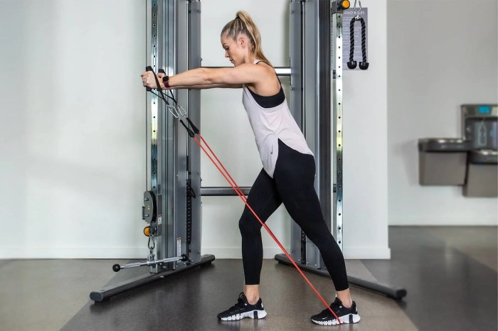
    • Neck stretches with resistance bands or weights: If the resistance is too high, these can put undue strain on the neck, potentially leading to injury. Start with very light resistance and gradually increase it as your neck strengthens and tolerance improves.
    • Sudden, jerky movements: Any movement that involves abrupt or forceful changes in position, especially in the neck, should be avoided. These movements are likely to cause inflammation and further pain.

    Modified Exercise Table

    This table Artikels common neck exercises and provides modifications for individuals with neck arthritis. It’s essential to consult with a healthcare professional before starting any new exercise regimen.

    Avoiding certain neck arthritis exercises is crucial, especially if you have a sensitivity to germs, like in the case of mysophobia, a phobia of germs. This phobia can make you anxious about potentially contaminated equipment or tools. So, it’s essential to choose exercises that minimize this risk and maintain good hygiene, like using clean mats and equipment for neck stretches and avoiding crowded exercise spaces.

    Proper precautions will help you stay focused on your recovery and avoid unnecessary discomfort.

    Exercise Description Modification for Arthritis Precautions
    Neck Flexion Gentle bending of the neck forward Maintain a slow, controlled motion, avoid sudden movements. Use a resistance band if necessary but keep the resistance light. Do not push beyond a comfortable range of motion. Stop if pain occurs.
    Neck Extension Gentle bending of the neck backward Maintain a slow, controlled motion. Limit the range of motion to avoid hyperextension. Avoid any jerky or sudden movements.
    Neck Rotation Rotating the head from side to side Maintain a slow, controlled motion. Keep the rotation limited to a comfortable range. Avoid forceful or rapid rotations.
    Neck Side Bending Bending the head to the sides Maintain a slow, controlled motion. Limit the range of motion to avoid straining the neck muscles. Avoid excessive or jerky movements.

    Alternative Exercises and Treatments

    Beyond avoiding certain exercises, a comprehensive approach to neck arthritis management involves exploring alternative strategies for strengthening neck muscles and alleviating pain. This includes a range of exercises, therapies, and lifestyle adjustments, each with its own set of benefits and limitations. A personalized approach, guided by a healthcare professional, is crucial for optimal results.Effective management of neck arthritis often requires a multifaceted strategy, blending exercise modifications with complementary therapies.

    Understanding the specific benefits and drawbacks of various approaches allows individuals to make informed decisions regarding their treatment plans.

    Strengthening Exercises for the Neck

    Strengthening neck muscles without exacerbating pain is key to maintaining stability and reducing discomfort. Gentle, progressive exercises, focusing on controlled movements, are vital. These exercises should be performed with proper form and gradually increased in intensity. Examples include isometric exercises, where muscles are contracted without significant movement, and exercises that involve controlled range-of-motion movements.

    • Isometric exercises: These involve contracting neck muscles against resistance without significant movement. Examples include gently pressing your head against a wall or resistance band, holding for a few seconds, and repeating several times. Isometric exercises are generally considered safe and effective for building neck strength without putting excessive strain on the joints.
    • Gentle range-of-motion exercises: These exercises focus on gradually increasing the movement of the neck in different directions. For example, slowly tilting your head to the left and right, forward and backward, and rotating your head in a circular motion. It is crucial to maintain a slow and controlled tempo to avoid stressing the neck.
    • Neck stretches: Gentle stretches can help alleviate stiffness and improve flexibility. Examples include stretching the front of the neck by gently pulling your chin toward your chest and holding for a few seconds, and stretching the back of the neck by gently tilting your head back. These should be performed slowly and without pushing beyond a comfortable range.

    Alternative Treatment Approaches

    Beyond exercise, various treatment options can help manage neck arthritis symptoms. These options, when used in conjunction with exercise modifications, can provide comprehensive relief.

    • Medications: Over-the-counter pain relievers, such as ibuprofen or naproxen, can help reduce inflammation and pain. Prescription medications, like certain muscle relaxants or corticosteroids, might be necessary in more severe cases. These medications can be helpful but may have side effects.
    • Physical therapy: A physical therapist can provide tailored exercises, manual therapy techniques, and advice on posture and lifestyle changes. Specific exercises and techniques can be beneficial in strengthening supporting muscles and improving range of motion. Physical therapy is a crucial aspect of management, often offering a structured approach to recovery and long-term well-being.
    • Lifestyle modifications: Maintaining a healthy weight, avoiding prolonged periods of inactivity, and adopting good posture habits are important lifestyle factors. Proper posture helps distribute weight evenly, reducing strain on the neck. Regular breaks from work or other activities that involve prolonged sitting or straining the neck can significantly reduce pain and discomfort.

    Comparison of Neck Arthritis Treatments

    Treatment Type Description Benefits Limitations
    Exercise Modifications Tailored exercises focusing on controlled movements and strengthening without causing pain. Improved neck strength, reduced pain, enhanced range of motion. Requires patience and consistent practice. Incorrect form can worsen symptoms.
    Physical Therapy Personalized exercises, manual therapy, and lifestyle advice. Targeted treatment for specific needs, professional guidance, long-term management strategies. May require multiple sessions. Cost can be a factor.
    Medications Over-the-counter or prescription pain relievers, muscle relaxants, or corticosteroids. Quick relief from pain and inflammation. Potential side effects, may not address underlying issues, long-term use may be problematic.

    Importance of Professional Guidance: Neck Arthritis Exercises To Avoid

    Neck arthritis exercises to avoid

    Taking care of neck arthritis requires a cautious approach, and one crucial aspect is seeking professional guidance. Simply finding exercises online or from friends might not be the best strategy. A personalized plan, tailored to your specific condition and limitations, is essential for preventing further injury and promoting healing.Professional guidance is paramount because neck arthritis presents a complex interplay of pain, stiffness, and potential underlying conditions.

    A healthcare professional can accurately assess your specific needs, identify potential risks, and create a safe and effective exercise program. This personalized approach maximizes the benefits and minimizes the risks associated with exercise.

    Crucial Role of Healthcare Professionals, Neck arthritis exercises to avoid

    Healthcare professionals, including physical therapists and physicians, play a vital role in developing safe and effective exercise programs for neck arthritis. They possess the expertise to evaluate your condition, understand your limitations, and design exercises that target specific areas of weakness or pain while avoiding harmful movements. Physical therapists are trained in manual therapy, exercise prescription, and patient education, making them well-suited to guide you through your recovery journey.

    Doctors can assess your overall health, rule out other potential causes of neck pain, and ensure the exercises are compatible with any other medical conditions you may have.

    Questions to Ask a Doctor or Physical Therapist

    It’s essential to ask the right questions to ensure you receive the best possible care. These questions can help you understand your condition and the exercises prescribed. Example questions to ask include: “What are the specific exercises recommended for my type of neck arthritis?”, “What are the potential risks associated with these exercises?”, “What are the expected outcomes and timelines for improvement?”, and “What modifications should I make if I experience pain or discomfort during the exercises?”.

    Steps to Take Before Starting an Exercise Program

    Understanding the process before embarking on an exercise program is critical. This structured approach helps ensure safety and effectiveness. The table below Artikels the key steps and their importance in managing neck arthritis.

    Step Action Importance
    1 Consult a Healthcare Professional Crucial for proper diagnosis, personalized exercise prescription, and risk assessment.
    2 Thorough Evaluation Allows the professional to identify specific needs, limitations, and potential complications.
    3 Understand Exercise Instructions Ensures correct form and technique to prevent injury and maximize benefits.
    4 Gradual Progression Avoids overexertion and allows the body to adapt to the exercises.
    5 Regular Monitoring and Adjustment Enables the professional to assess progress, identify any emerging issues, and modify the program as needed.

    Illustrative Examples of Exercises to Avoid

    Neck arthritis can be aggravated by certain exercises if not performed correctly. Understanding which movements to avoid is crucial for preventing further pain and discomfort. Choosing appropriate alternatives is equally important for maintaining neck health and promoting healing.Identifying exercises that strain the neck is key to protecting your joints. Many seemingly harmless movements can become problematic when the spine is already compromised.

    This section will present examples of exercises to avoid and illustrate safe alternatives, helping you make informed choices.

    Specific Exercises to Avoid

    Many exercises, while seemingly beneficial for overall fitness, can exacerbate neck arthritis symptoms if not performed with extreme caution. Certain movements can put undue stress on the already vulnerable cervical spine, leading to increased pain and inflammation. Understanding these movements is crucial to protecting your neck.

    • Neck Rotations: Full-range neck rotations can place excessive strain on the delicate joints of the neck. Repeated rotation can irritate the inflamed areas and lead to increased pain and stiffness. The repetitive motion can contribute to further degeneration and potentially cause instability.
    • Sudden Jerky Movements: Rapid or jerky head movements, even when seemingly insignificant, can cause sudden stress and impact on the neck, particularly when the neck is already prone to inflammation. These movements can quickly lead to acute pain episodes. Examples include sudden turns of the head or quick, forceful shrugs.
    • Heavy Weight Lifting with Unsupported Neck: Lifting heavy weights without proper support, particularly when the head is held in a vulnerable position, can place a significant load on the neck. This can exacerbate existing arthritis pain and even potentially lead to herniated discs.
    • High-Impact Exercises with Neck Strain: Exercises that involve a high degree of impact, such as high-impact aerobics or sports where the head is frequently in motion, can cause undue stress on the neck. If not performed cautiously, these can lead to further inflammation and pain. The high impact can lead to micro-trauma in the cervical spine, which can be significant in the context of existing arthritis.

    Illustrative Examples of Proper and Incorrect Form

    Understanding the correct and incorrect form of exercises is crucial in preventing further damage to the neck. This section will illustrate the differences between safe and harmful movements.

    Exercise Correct Form Incorrect Form Description
    Neck Flexion Gentle, controlled bending of the head towards the chest, keeping the spine straight. Rapid, jerky bending of the head towards the chest, rounding the shoulders. Incorrect form places excessive strain on the cervical spine, increasing the risk of pain and further damage. Correct form keeps the spine aligned and minimizes stress on the neck.
    Neck Extension Gentle, controlled tilting of the head back, maintaining a straight posture. Jerky tilting of the head back, arching the back. Incorrect form overextends the cervical spine, causing strain and potential injury. Correct form keeps the neck aligned, protecting the delicate joints.
    Neck Side Bending Slow, controlled side-to-side movement of the head, maintaining a straight posture. Rapid, jerky side bending of the head, tilting the head to one side. Incorrect form puts excessive pressure on the neck’s side joints, causing strain and potential inflammation. Correct form keeps the spine straight and the neck aligned.
    Neck Rotation Slow, controlled rotation of the head, keeping the spine straight. Rapid, forceful rotation of the head, twisting the neck. Incorrect form places stress on the neck’s rotation joints, potentially leading to further inflammation. Correct form maintains spine alignment and minimizes stress on the neck.

    Final Wrap-Up

    Neck arthritis exercises to avoid

    In conclusion, avoiding certain neck exercises is vital for managing neck arthritis effectively. By understanding the mechanics of neck pain, identifying problematic exercises, and exploring alternative treatments, you can actively participate in your recovery. Remember, consulting with a healthcare professional is paramount before starting any exercise program. This guide provides a starting point, but personalized advice from a doctor or physical therapist is essential for optimal results.

  • Physical Therapy for Pectus Excavatum A Deep Dive

    Physical Therapy for Pectus Excavatum A Deep Dive

    Physical therapy for pectus excavatum provides a vital pathway to managing this condition, offering a blend of exercises and techniques to address both the physical and emotional aspects of the condition. It’s a crucial component in the overall treatment plan, helping individuals regain strength, improve posture, and enhance breathing capacity. This comprehensive guide delves into the specifics of physical therapy, outlining the exercises, techniques, and expected outcomes.

    Pectus excavatum, often characterized by a sunken chest, can impact daily activities and self-esteem. Understanding the condition’s various aspects, from causes and symptoms to treatment options, is essential for a comprehensive approach to managing it. This article provides a detailed exploration of physical therapy’s role in this process, covering everything from exercise types to long-term management strategies.

    Introduction to Pectus Excavatum

    Physical therapy for pectus excavatum

    Pectus excavatum, often called funnel chest, is a congenital chest wall deformity. It’s characterized by a noticeable depression or inward sinking of the lower portion of the sternum, creating a “funnel” shape. This condition affects the structural integrity of the chest and can impact the development and function of the respiratory system.The condition is visually apparent as a concave depression in the chest, typically located in the lower portion of the sternum.

    This inward indentation of the chest wall can vary in depth and width, affecting the aesthetic appearance and potentially impacting the individual’s overall well-being. The appearance of pectus excavatum can range from a subtle inward curve to a significant, pronounced depression.

    Age of Diagnosis

    Pectus excavatum is frequently detected during childhood or adolescence. Most cases are diagnosed between the ages of 6 and 16, although it can present earlier or later in some individuals. Early detection is important for proper management and treatment, which often involves physical therapy and, in some cases, surgical intervention.

    Severity Levels

    Pectus excavatum varies in severity, impacting the level of impact on respiratory function and aesthetic appearance. Understanding the different degrees of severity helps guide appropriate treatment strategies.

    Severity Level Visual Description Potential Symptoms Treatment Options
    Mild A subtle inward curve of the lower sternum, barely noticeable. Mild discomfort, occasional shortness of breath during strenuous activity. May not experience noticeable symptoms. Observation and close monitoring for potential progression. Physical therapy may be recommended to improve posture and flexibility.
    Moderate A moderate inward depression of the lower sternum, clearly visible. The depth of the depression is significant enough to affect the aesthetic appearance. Mild to moderate shortness of breath, particularly during exercise or physical exertion. Potential for reduced lung capacity. Physical therapy, bracing, and in some cases, surgical intervention.
    Severe A deep and pronounced inward depression of the lower sternum. Significant chest wall deformity. Shortness of breath, reduced lung capacity, significant discomfort, potential for cardiac complications. Respiratory problems may be more pronounced. Surgical intervention is often necessary to correct the deformity. Physical therapy may be a component of the post-operative recovery.

    Causes and Risk Factors

    Understanding the causes and risk factors of pectus excavatum is crucial for both prevention and treatment. While a precise cause isn’t always identifiable, research has shed light on potential contributing elements, enabling better strategies for management and potentially early intervention. This knowledge empowers individuals and healthcare providers to approach this condition with a comprehensive understanding.The development of pectus excavatum is a complex process, often involving a combination of genetic predispositions, developmental factors, and potentially environmental influences.

    Precisely how these elements interact to create the condition remains a subject of ongoing research. However, identifying these factors helps in predicting the condition’s potential severity and tailoring treatment plans accordingly.

    Genetic Predispositions and Family History

    A significant factor in pectus excavatum is the presence of a family history of the condition. Studies suggest a hereditary component, implying that genetic predisposition plays a role in the development of pectus excavatum. Individuals with a family history of the condition have a higher likelihood of developing it themselves. This genetic predisposition is not always fully understood, but it highlights the importance of considering family medical history when assessing risk.

    Environmental Factors

    Environmental factors, while not always directly causative, can potentially influence the development of pectus excavatum. Factors like nutritional deficiencies during critical developmental stages, or exposure to certain environmental toxins, may contribute to the condition. However, definitive links between specific environmental exposures and pectus excavatum are still under investigation. The impact of environmental factors on the development of the condition is an area of active research.

    Known Risk Factors

    Several factors have been linked to an increased risk of pectus excavatum. These factors can range from the presence of other medical conditions to specific developmental stages. For instance, certain connective tissue disorders may be associated with an increased risk. Also, the presence of other developmental abnormalities might contribute to the condition. It’s important to note that these risk factors do not definitively cause pectus excavatum but rather increase the likelihood of its occurrence.

    Table of Potential Causes

    Cause Category Specific Cause Supporting Evidence Potential Impact on Treatment
    Genetic Inherited predisposition Family history studies showing higher incidence in affected families. Genetic counseling and increased monitoring for individuals with a family history.
    Developmental Abnormal chest wall development during fetal or early childhood stages. Observed variations in chest wall development in affected individuals. Early diagnosis and potential surgical intervention to correct structural abnormalities.
    Environmental Exposure to certain environmental toxins or nutritional deficiencies. Studies investigating possible correlations, though definitive evidence is lacking. Addressing potential environmental risk factors, including nutritional support and minimizing exposure to toxins.
    Associated Conditions Presence of other medical conditions, such as connective tissue disorders. Some research suggesting correlations between specific conditions and increased risk. Comprehensive medical evaluation to identify potential underlying conditions and tailor treatment accordingly.

    Symptoms and Diagnosis

    Pectus excavatum, often referred to as sunken chest, is a visible and sometimes symptomatic condition. Understanding its symptoms and the diagnostic process is crucial for appropriate management and treatment. Accurate diagnosis hinges on a comprehensive evaluation of physical signs, patient history, and often, imaging studies.The symptoms of pectus excavatum can vary greatly in severity, depending on the depth and extent of the chest wall depression.

    These symptoms can range from minor discomfort to more significant issues impacting quality of life. Diagnosis typically involves a careful physical examination and potentially, imaging studies.

    Physical Examination Findings

    A thorough physical examination is the initial step in evaluating pectus excavatum. Clinicians assess the patient’s overall appearance, paying particular attention to the chest wall configuration. The degree of chest wall depression is a key indicator. A significant depression may be accompanied by other observable features like a visible indentation or a noticeable asymmetry in the chest.

    The physician will also assess the patient’s breathing mechanics and overall physical condition.

    Imaging Techniques

    Imaging plays a vital role in confirming the diagnosis and evaluating the severity of pectus excavatum. Radiographic imaging, including X-rays, is often the first step. X-rays provide a two-dimensional view of the chest, enabling visualization of the sternum and rib cage. These images help determine the extent of the inward depression. More detailed anatomical information can be obtained through CT scans.

    A CT scan generates cross-sectional images, offering a comprehensive view of the bony structures, which can aid in the accurate measurement of the deformity.

    Comparison of Diagnostic Tools

    Different imaging modalities offer varying levels of detail and resolution. X-rays are relatively inexpensive and readily available, providing a basic assessment of the chest wall. CT scans offer a more comprehensive and detailed view, particularly useful in assessing the degree of deformity and its impact on surrounding structures. In cases where a more precise measurement of the deformity is necessary, a CT scan is often preferred over an X-ray.

    The choice of imaging modality depends on the specific clinical situation and the information needed.

    Table of Symptoms and Diagnostic Correlations

    Symptom Category Specific Symptom Severity Correlation Diagnostic Tool Correlation
    Discomfort Chest pain, especially with deep breathing or exertion Moderate to severe X-ray, CT scan
    Breathing Issues Shortness of breath, especially during physical activity Moderate to severe X-ray, CT scan, physical exam
    Aesthetic Concerns Visible indentation of the chest wall Mild to severe X-ray, CT scan, physical exam
    Physical Examination Visible asymmetry in the chest Moderate to severe Physical exam, X-ray, CT scan
    Physical Examination Limited chest expansion Moderate to severe Physical exam, X-ray, CT scan

    Physical Therapy for Pectus Excavatum

    Physical therapy plays a crucial role in managing pectus excavatum, offering a non-invasive approach to address both the physical and emotional aspects of the condition. It focuses on improving posture, breathing mechanics, and strengthening the chest muscles, contributing to improved quality of life for individuals with pectus excavatum.Physical therapy is a personalized and tailored treatment approach, carefully designed to address the specific needs and goals of each patient.

    It helps in restoring symmetry, improving function, and boosting self-confidence. By focusing on exercises and techniques, physical therapists work collaboratively with patients to achieve optimal results and maximize their potential for recovery.

    Physical therapy for pectus excavatum focuses on strengthening chest muscles and improving posture. Sometimes, underlying genetic factors can play a role in the condition, and understanding the potential genetic component might involve exploring a DNA test. For example, a what is a dna test could reveal if a genetic predisposition is present, which could inform the physical therapy approach.

    Ultimately, a tailored physical therapy plan, potentially incorporating genetic insights, is crucial for managing pectus excavatum effectively.

    Role of Physical Therapy in Pectus Excavatum Management

    Physical therapy for pectus excavatum is not intended to fully correct the deformity but rather to improve the patient’s overall well-being and quality of life. It addresses the limitations in posture and breathing, which are often accompanied by psychological distress, anxiety, and decreased self-esteem. The goal is to help patients adapt to their condition and maintain healthy breathing and movement patterns.

    Types of Exercises and Techniques

    A variety of exercises are employed in physical therapy for pectus excavatum. These techniques focus on stretching, strengthening, and improving posture and breathing mechanics. Exercises may include specific chest stretches, exercises to build chest muscle strength, and respiratory exercises to improve lung capacity.

    Addressing Physical and Emotional Aspects

    Physical therapy addresses both the physical and emotional aspects of pectus excavatum. The physical benefits include improved posture, enhanced chest muscle strength, and better breathing patterns. Importantly, the therapy also helps to build confidence and reduce anxiety associated with the condition, which are crucial for a patient’s psychological well-being.

    Improving Posture and Breathing

    Exercises to improve posture and breathing are central to physical therapy for pectus excavatum. Postural exercises aim to correct the inward curvature of the chest, while breathing exercises focus on increasing lung capacity and improving the efficiency of respiration. This often involves proper breathing techniques and exercises that engage the muscles of the chest and abdomen to promote deeper and more efficient breaths.

    Strengthening Chest Muscles

    Strengthening chest muscles is essential to improve the overall support structure of the chest wall. Exercises targeting the pectoral muscles, as well as other muscles surrounding the chest, help restore balance and provide a sense of stability. These exercises, combined with other therapies, help to minimize the outward visibility of the deformity.

    Exercises for Pectus Excavatum

    Physical therapy incorporates a range of exercises to address the specific needs of each patient. The following table provides a summary of some common exercises used in physical therapy for pectus excavatum.

    Exercise Type Description Targeted Muscles Expected Outcomes
    Chest Expansion Exercises Involves gently expanding the chest cavity by pulling the arms apart or raising the arms overhead. Pectoral muscles, intercostal muscles, and shoulder muscles Improved chest expansion, increased lung capacity, and better breathing mechanics.
    Wall Slides Involves leaning against a wall with the back straight and sliding up and down. Back muscles, shoulder muscles, and core muscles Improved posture, increased core strength, and reduced stress on the chest wall.
    Bent-Over Rows Involves bending over and pulling weights towards the chest. Back muscles, latissimus dorsi, and rhomboids Strengthened back muscles, improved posture, and reduced stress on the chest wall.
    Breathing Exercises Involves techniques to improve lung capacity and breathing patterns. Respiratory muscles (diaphragm, intercostals) Increased lung capacity, improved breathing efficiency, and reduced shortness of breath.

    Treatment Options and Procedures

    Pectus excavatum, while often manageable with physical therapy, sometimes requires more extensive interventions. Understanding the various treatment options, their potential benefits and risks, and the importance of early intervention is crucial for making informed decisions. This section will delve into the diverse approaches to correcting this condition, from non-surgical therapies to surgical procedures.Surgical procedures are often necessary to address more severe cases of pectus excavatum, where the chest wall depression significantly impacts breathing and overall well-being.

    These interventions aim to restore the natural shape and function of the chest, improving both physical and psychological health. Furthermore, the effectiveness of these treatments can be significantly enhanced by early intervention, especially when considering the potential long-term consequences of the condition.

    Surgical Procedures for Pectus Excavatum

    Surgical procedures for pectus excavatum aim to reshape the sternum and surrounding cartilage to correct the inward depression. Common surgical techniques include Nuss procedure and Ravitch procedure.

    • Nuss Procedure: This is a minimally invasive procedure where a metal bar is inserted behind the sternum to gradually push it outward. The bar is usually left in place for 1-2 years, allowing the chest wall to reshape naturally. The bar is then removed in a subsequent procedure. This approach is generally considered safe and effective for a wide range of cases.

      For example, in a 2018 study, the Nuss procedure demonstrated high satisfaction rates and minimal complications in a cohort of adolescents and adults.

    • Ravitch Procedure: This procedure involves reshaping the sternum and costal cartilages directly. It is typically performed on younger patients with less severe deformities. The technique involves separating the sternum, reshaping it, and securing it with sutures. This method can offer a more rapid correction but may have a higher risk of complications compared to the Nuss procedure.

    Comparison of Treatment Options

    Choosing the right treatment for pectus excavatum involves careful consideration of factors such as the severity of the condition, age of the patient, and individual preferences. Physical therapy, while valuable in early stages, may not be sufficient for more pronounced cases.

    Treatment Type Description Success Rate Potential Risks
    Physical Therapy Focuses on strengthening chest muscles, improving posture, and promoting overall flexibility. High success rate for mild cases, but may not be sufficient for significant deformities. Limited effectiveness in severe cases, potential for muscle soreness and discomfort.
    Nuss Procedure Involves placing a metal bar behind the sternum to push it outward, allowing for gradual reshaping. High success rate, typically minimal complications. Possible infection, bar migration, and discomfort at the insertion site.
    Ravitch Procedure Directly reshapes the sternum and costal cartilages, typically performed on younger patients. High success rate, but potentially higher risk of complications than the Nuss procedure. Risk of infection, bleeding, and potential damage to surrounding tissues.

    Importance of Early Intervention, Physical therapy for pectus excavatum

    Early intervention in pectus excavatum is crucial for maximizing the effectiveness of treatment. Children and adolescents with the condition often experience better outcomes with early diagnosis and intervention. The younger the patient, the better the chances for successful correction, and potentially fewer complications. Furthermore, early treatment can reduce the likelihood of long-term physical and psychological issues associated with the condition.

    Long-Term Management and Outcomes

    Physical therapy for pectus excavatum

    Pectus excavatum, while often treatable, requires ongoing care and attention to ensure optimal long-term outcomes. Successful management involves a proactive approach to preventing complications and maintaining the benefits achieved through initial treatment. This involves a combination of regular follow-ups, lifestyle adjustments, and proactive monitoring of potential issues.Long-term management is crucial because pectus excavatum, while often not life-threatening, can impact quality of life.

    Physical therapy for pectus excavatum is a great way to improve chest wall function and overall well-being. While it’s important to focus on the specific symptoms of pectus excavatum, it’s also crucial to be aware of other potential health concerns. For example, understanding the signs and symptoms of chlamydia signs and symptoms of chlamydia is vital for comprehensive health management.

    Ultimately, physical therapy for pectus excavatum can help restore a patient’s comfort and mobility, making daily life easier.

    Individuals with pectus excavatum may experience ongoing discomfort, reduced physical activity, or psychological distress if their condition isn’t managed effectively. Proactive care helps prevent these issues and fosters a positive impact on well-being.

    Importance of Follow-up Appointments

    Regular follow-up appointments are vital for monitoring the progression of the condition and detecting any potential complications early. These appointments allow the healthcare team to assess the effectiveness of the initial treatment and make necessary adjustments to the care plan. They also provide a platform for addressing any concerns or discomfort the patient may experience. Thorough evaluations ensure that any potential complications are caught early, allowing for timely intervention.

    Potential Long-Term Outcomes of Different Treatment Options

    The long-term outcomes of pectus excavatum treatment vary depending on the chosen approach. Surgical interventions, such as Nuss procedures, typically result in significant improvement in chest wall morphology and function. However, the long-term effectiveness of non-surgical approaches like physical therapy is often influenced by the individual’s adherence to the prescribed exercises and lifestyle modifications. Patient compliance and commitment to the prescribed care plan are key factors in achieving optimal results.

    Potential Complications After Treatment

    While pectus excavatum treatment is generally safe, some potential complications can arise. These can include, but are not limited to, infection at the surgical site, persistent pain, and recurrence of the deformity in some cases. Proper post-operative care and ongoing monitoring can mitigate these risks. Furthermore, psychological impacts, such as body image concerns, can arise, requiring careful consideration and support.

    Physical therapy for pectus excavatum is all about strengthening the chest muscles and improving posture, right? Interestingly, similar principles apply to addressing other musculoskeletal issues, like migraine headaches. For example, chiropractic therapy for migraine headache can help by focusing on spinal alignment and relieving tension. Ultimately, though, the core goal of physical therapy for pectus excavatum remains the same: to restore proper chest structure and function.

    Importance of Ongoing Care and Lifestyle Adjustments

    Maintaining a healthy lifestyle plays a significant role in achieving long-term success. This includes maintaining a balanced diet, regular exercise, and stress management techniques. These factors contribute to overall well-being and support the body’s ability to heal and recover from treatment. For example, regular cardiovascular exercise can improve lung function and overall fitness.

    Follow-up Schedule and Potential Complications

    Follow-up Strategy Frequency Expected Outcomes Potential Complications
    Post-operative checkups Initially weekly, then monthly, then every 3-6 months Monitoring for infection, wound healing, and deformity recurrence. Adjustment of treatment plan if needed. Infection, persistent pain, recurrence of deformity, scar tissue issues, psychological distress.
    Regular physical therapy sessions As recommended by the physical therapist Strengthening of chest muscles, improved posture, and pain management. Muscle strain, fatigue, potential discomfort during exercises.
    Lifestyle modifications (diet, exercise) Ongoing Improved overall health, reduced risk of complications, and better quality of life. Dietary restrictions, lack of motivation, potential social isolation, difficulty in maintaining healthy habits.
    Psychological support (if needed) As needed Addressing body image concerns, stress, and anxiety. Limited effectiveness without patient engagement, potential for relapse.

    Impact on Daily Life

    Living with pectus excavatum can significantly impact daily activities and overall well-being. The visible chest deformity can affect self-esteem and body image, potentially leading to social anxieties and avoidance of certain activities. Understanding how this condition impacts daily life is crucial for developing appropriate coping strategies and maximizing quality of life.The impact of pectus excavatum extends beyond the physical.

    The condition can affect various aspects of daily life, from simple tasks like dressing to more complex activities like sports and social interactions. Individuals with pectus excavatum may experience discomfort or pain during certain activities, potentially limiting their participation. Addressing these challenges with tailored physical therapy and support strategies is vital for enhancing the quality of life for those affected.

    Impact on Self-Esteem and Body Image

    Pectus excavatum can significantly affect self-esteem and body image. The visible chest deformity can lead to feelings of self-consciousness, embarrassment, and social anxiety. These emotional challenges can affect a person’s confidence and participation in social activities, impacting their overall quality of life. The impact of pectus excavatum on body image is often significant, potentially leading to avoidance of activities that involve physical exertion or social interaction.

    Support from family, friends, and healthcare professionals can play a critical role in helping individuals cope with these challenges and develop a positive body image.

    Challenges in Daily Activities

    Individuals with pectus excavatum may experience various challenges in their daily activities. Certain movements, positions, or physical exertion can cause discomfort or pain. Understanding these challenges is crucial for developing appropriate adaptations and support strategies. Activities requiring deep breathing, such as singing or playing wind instruments, may also be affected.

    Examples of Challenging Activities

    A variety of activities can present challenges for individuals with pectus excavatum. These challenges can range from simple tasks to more strenuous activities. Examples include:

    • Sports participation: Contact sports, high-impact activities, and certain exercises might be difficult or painful.
    • Physical exertion: Activities requiring sustained physical effort or repetitive movements can lead to discomfort.
    • Clothing selection: Finding clothing that comfortably accommodates the chest deformity can be challenging.
    • Social interactions: The visible chest deformity may lead to self-consciousness and avoidance of social situations.

    Adaptation and Support Strategies

    Addressing the challenges associated with pectus excavatum requires a multi-faceted approach, combining adaptations to activities and support strategies. This involves finding ways to modify activities, using assistive devices, and seeking support from healthcare professionals and loved ones. This section provides practical examples to help individuals with pectus excavatum overcome these challenges.

    Activity Type Potential Challenges Adaptations Support Strategies
    Sports (e.g., running, swimming) Pain, discomfort during exertion, difficulty with breathing Modifying intensity and duration of exercise, choosing low-impact activities, using supportive gear Working with a physical therapist to develop an exercise plan, seeking guidance from coaches or trainers, discussing pain management strategies with a physician
    Lifting heavy objects Pain and discomfort in the chest area Using proper lifting techniques, seeking assistance when lifting heavy items, avoiding strenuous lifting activities Consulting with a physical therapist for guidance on safe lifting techniques, discussing pain triggers with a physician, seeking assistance from family or friends
    Wearing certain clothing Discomfort or difficulty finding suitable clothing that accommodates the chest deformity Choosing loose-fitting clothing, using supportive undergarments, seeking advice from clothing retailers Seeking advice from physical therapists or healthcare professionals on proper clothing selection, exploring adaptive clothing options, joining support groups for individuals with pectus excavatum
    Social interactions Self-consciousness, embarrassment, anxiety about the chest deformity Developing self-confidence through positive self-talk, focusing on personal strengths, seeking support from family and friends Participating in support groups for individuals with pectus excavatum, seeking therapy to address social anxiety, discussing concerns with a healthcare professional

    Wrap-Up: Physical Therapy For Pectus Excavatum

    In conclusion, physical therapy for pectus excavatum is a vital part of the treatment journey. By addressing the physical and emotional aspects of the condition, physical therapy empowers individuals to regain strength, improve breathing, and enhance their quality of life. This detailed exploration of the exercises, techniques, and potential outcomes provides a clear picture of how physical therapy plays a pivotal role in managing pectus excavatum, highlighting its importance in achieving optimal outcomes and overall well-being.

  • What to Wear to Physical Therapy Your Guide

    What to Wear to Physical Therapy Your Guide

    What to wear to physical therapy is more important than you might think. Choosing the right clothes can significantly impact your comfort, performance, and even your mindset during your sessions. This guide will equip you with the knowledge to select clothing that optimizes your therapy experience, from comfortable fabrics to temperature regulation and specific exercise considerations.

    From choosing breathable fabrics for optimal comfort to considering the type of exercises you’ll be doing, this guide covers everything you need to know. We’ll also explore the importance of inclusive styles and how to find budget-friendly options without sacrificing quality.

    Clothing Comfort and Mobility

    Choosing the right clothing for physical therapy is crucial for maximizing your session’s effectiveness and minimizing discomfort. Appropriate attire supports proper movement, range of motion, and overall well-being during exercises and stretches. The correct fabrics and styles can significantly impact your experience, allowing you to focus on the therapeutic process rather than feeling restricted or uncomfortable.Comfort and mobility are key factors when selecting clothing for physical therapy.

    Fabric choices that prioritize breathability, flexibility, and moisture-wicking properties will contribute to a more comfortable and productive session. Proper clothing can also reduce distractions, allowing you to concentrate on your therapy goals.

    Fabric Selection for Physical Therapy

    Selecting the right fabrics for physical therapy sessions is important for comfort and optimal performance. Breathable fabrics allow air circulation, preventing overheating and maintaining a comfortable temperature. Flexibility in the fabric ensures ease of movement and range of motion during exercises. Moisture-wicking properties draw sweat away from the skin, keeping you dry and comfortable, which is crucial to avoid chafing or skin irritation.

    Types of Clothing for Enhanced Mobility

    Several types of clothing promote ease of movement and range of motion during physical therapy exercises. Loose-fitting garments, such as athletic shorts or leggings, allow for unrestricted movement, enabling a full range of motion during stretches and exercises. Moisture-wicking fabrics like polyester or nylon are excellent choices for their ability to keep you dry and comfortable during intense workouts.

    Consider athletic tops or shirts that are made of similar materials, providing a similar benefit.

    Fabric Comparison for Physical Therapy Attire

    Fabric Type Comfort Mobility Price
    Polyester Good; wicks moisture effectively Excellent; stretchy and flexible Moderate
    Nylon Good; wicks moisture effectively Excellent; durable and supportive Moderate
    Cotton Good; soft and comfortable Fair; can become restrictive when wet Low
    Spandex Excellent; very flexible and stretchy Excellent; allows for great range of motion Moderate to High
    Bamboo Excellent; naturally breathable and soft Good; offers a balance between comfort and mobility Moderate to High

    This table provides a general comparison of common fabric types used in physical therapy attire. Prices can vary depending on the brand and specific design. Factors like comfort, mobility, and the desired level of support should guide your decision-making process.

    Avoiding Restrictive Clothing

    Restrictive clothing can hinder movement and cause discomfort during physical therapy sessions. Tight-fitting clothing, such as jeans or non-stretchy fabrics, can restrict range of motion and impede proper exercise execution. Avoid garments with zippers or tight closures that might rub against the skin or cause discomfort. Opt for clothing that allows for a full range of motion without constricting or binding.

    This will ensure a more productive and comfortable therapy session.

    Temperature Regulation

    What to wear to physical therapy

    Staying comfortable during physical therapy sessions is crucial for optimal performance and recovery. This involves more than just choosing clothes that fit well; temperature regulation plays a vital role. Understanding how clothing affects body temperature, especially during varying exercise intensities and room temperatures, can significantly enhance your therapy experience.Maintaining the right temperature balance during physical therapy helps prevent overheating or chilling, which can impact your body’s ability to heal and perform exercises effectively.

    Proper clothing choices allow you to adapt to the specific needs of each session, ensuring comfort and safety throughout the process.

    Managing Body Temperature in Different Conditions

    The ideal temperature for physical therapy varies depending on the exercise intensity and the surrounding environment. Clothing choices should be tailored to help maintain a comfortable internal body temperature. For example, if the room is cool, layering clothes allows you to adjust to the temperature changes during different parts of the session. Conversely, if the room is warm, lightweight, moisture-wicking fabrics are key to preventing overheating.

    Clothing Options for Different Temperatures

    Different climates and exercise intensities require different clothing approaches. Layers are essential for cold weather. Starting with a moisture-wicking base layer next to the skin, followed by a mid-layer for insulation, and a lightweight, breathable outer layer for protection from wind and rain, allows you to adjust to varying temperatures and exercise intensities. In warmer weather, loose-fitting, breathable fabrics like cotton or linen, in light colors, are excellent choices.

    These fabrics allow air to circulate and help keep you cool.

    Choosing Appropriate Clothing Layers

    The best approach to temperature regulation is layering. This allows you to adjust to changes in temperature and activity level throughout your session.

    Weather Condition Base Layer Mid Layer Outer Layer
    Cool/Chilly Moisture-wicking thermal top and bottom Fleece jacket or sweater Light waterproof or windbreaker jacket
    Warm/Mild Moisture-wicking t-shirt or tank top Lightweight long-sleeved shirt or jacket Light, breathable jacket or light vest
    Hot/Humid Moisture-wicking athletic shirt or tank top Lightweight athletic shirt or singlet Loose-fitting, breathable shorts or pants

    Practical Considerations for Specific Exercises

    Choosing the right clothing for your physical therapy sessions is crucial for optimal performance and safety. Beyond comfort and temperature regulation, the type of exercise you’re performing dictates specific clothing needs. Consideration of these factors can enhance your therapy experience and ensure a safe and effective treatment plan.Understanding the specific requirements for different exercises, from gentle stretching to intense strength training, is essential for maximizing the benefits of your physical therapy.

    This allows for greater ease of movement, reduces the risk of injury, and ensures proper hygiene during each session.

    Clothing for Stretching

    Stretching exercises require clothing that allows for a full range of motion without restriction. Loose-fitting fabrics like breathable cotton or athletic wear with good flexibility are ideal. Avoid restrictive clothing that might impede your movement or cause discomfort during stretches. This can lead to muscle strain or tears if the clothing limits movement. Proper stretching attire enables a full range of motion, reducing the risk of injury.

    Clothing for Strength Training

    Strength training exercises demand clothing that offers support and stability while allowing for free movement. Moisture-wicking fabrics, like polyester or nylon, are recommended to keep you dry and comfortable during intense workouts. Consider athletic shorts or leggings that provide adequate support for your muscles without restricting movement. Choosing the right clothing can prevent injuries and improve performance during strength training sessions.

    Clothing for Aquatic Therapy

    Aquatic therapy, often performed in pools or water tanks, necessitates specialized clothing. Swimsuits or athletic clothing designed for water activities are crucial for safety and comfort. These suits usually provide excellent buoyancy and prevent the risk of slipping or getting caught on equipment. The fabric should be water-resistant to maintain dryness and prevent discomfort.

    Clothing for Safety and Hygiene

    Safety and hygiene are paramount during physical therapy sessions. Clothing should not contain loose threads or materials that could get caught on equipment. For example, avoid clothing with loose zippers or long, dangling straps. Fabric should be free of any sharp or rough edges that might cause irritation to the skin. This is important to prevent skin abrasions or discomfort.

    Select clothing that is easy to wash and disinfect to maintain hygiene standards.

    Clothing Selection Table

    Exercise Type Ideal Clothing Type Key Considerations
    Stretching Loose-fitting, breathable fabrics (e.g., cotton, athletic wear) Full range of motion, comfort
    Strength Training Moisture-wicking fabrics (e.g., polyester, nylon), supportive athletic wear Support, stability, dryness
    Aquatic Therapy Swimsuits or water-resistant athletic clothing Buoyancy, water resistance, comfort in water

    Accessibility and Inclusivity

    Choosing the right clothing for physical therapy isn’t just about comfort; it’s about inclusivity and accessibility for everyone. A wide range of body types and needs require adaptable clothing that supports movement and confidence. This section dives into the crucial role of inclusive apparel in physical therapy, ensuring that patients of all sizes and abilities can participate fully and comfortably.Physical therapy should be a journey of empowerment, and accessible clothing plays a significant role in creating that environment.

    Inclusive designs are crucial for patients to feel confident and supported, which in turn promotes a more positive and effective therapy experience.

    Choosing comfortable clothes for physical therapy is key, especially if you’re dealing with conditions like metastatic castration-resistant prostate cancer symptoms. metastatic castration resistant prostate cancer symptoms can sometimes make movement challenging, so opting for loose-fitting, breathable fabrics like cotton or linen is a great idea. You’ll want something that allows for a full range of motion, so avoid anything too restrictive.

    Think athletic wear or comfortable sweatpants – the goal is to feel free and supported, not constricted, during your therapy sessions.

    Importance of Inclusive Clothing Choices, What to wear to physical therapy

    Inclusive clothing choices are vital for patients of diverse body types and needs. A variety of styles and fits are needed to accommodate varying sizes, shapes, and conditions. This allows patients to move freely and comfortably, reducing any potential barriers to their recovery.

    Clothing Styles for Specific Needs

    Several clothing styles can be adjusted or modified to meet specific conditions or requirements. Stretchy, athletic fabrics, for instance, allow for a wide range of movement and are often preferred. Clothing with adjustable features like zippers, elastic waistbands, or Velcro closures can accommodate varying levels of mobility and support.

    Significance for Patients with Mobility Limitations

    Comfortable and accessible clothing is particularly important for patients with mobility limitations. Clothes that are easy to put on and take off, without requiring significant effort, can greatly improve the patient experience. The ease of dressing can directly impact a patient’s independence and self-esteem during the recovery process. For example, patients with limited upper body strength may find clothing with wide necklines or easy-access zippers beneficial.

    So, what to wear to physical therapy? Comfort is key! Loose-fitting clothes that allow for easy movement are best. Thinking about how a plant-based diet might influence longevity, I recently read an interesting article about whether vegans live longer and age better. Do vegans live longer and age better ? Regardless of dietary choices, comfortable, breathable clothing is crucial for a successful therapy session.

    Opt for moisture-wicking fabrics to stay dry and prevent discomfort during exercises.

    Clothing Options for Various Body Types and Sizes

    Body Type/Size Clothing Features Examples
    Petite Short inseams, shorter torso lengths, easy access closures Fitted athletic tops and bottoms with adjustable drawstrings, leggings with smaller sizes.
    Plus Size Roomy fits, stretchy materials, generous sizing Loose-fitting athletic tops and bottoms, clothing with wide necklines and waistbands.
    Activewear with built-in support Supportive materials, adjustable straps Sports bras, compression shirts, and shorts
    Patients with Limited Mobility Easy-to-wear closures, wide necklines, loose fitting garments Clothes with zippers, elastic waistbands, and wide sleeves

    “Inclusive clothing choices are critical for patients of all body types and abilities to feel confident and comfortable during physical therapy.”

    Appearance and Confidence

    Feeling good about yourself can significantly impact your physical therapy journey. Positive self-perception and a sense of comfort in your attire can boost your motivation and engagement in exercises. This, in turn, can accelerate your recovery and overall well-being. When you feel confident and comfortable, you’re more likely to participate actively and enthusiastically in your sessions.Beyond the practical aspects of comfort and mobility, clothing choices play a crucial role in shaping a patient’s mindset.

    A well-chosen outfit can foster a sense of empowerment and readiness to tackle the therapy ahead. Conversely, ill-fitting or uncomfortable clothing can create anxiety and self-consciousness, potentially hindering progress. The right apparel can make all the difference in fostering a positive and encouraging atmosphere.

    Choosing Clothing for a Positive Experience

    Choosing clothing for physical therapy goes beyond simple practicality. It involves considering how your attire impacts your self-image and your overall therapy experience. Select clothing that makes you feel good about yourself, confident, and ready to engage fully in your exercises. This includes factors like fit, fabric, and color choices. Consider how these factors contribute to your self-perception and the environment in which you engage in therapy.

    So, what to wear to physical therapy? Comfort is key! Think loose-fitting, breathable fabrics, like athletic wear. And, while you’re focusing on your recovery, remember that low vitamin D and K levels can sometimes contribute to seasonal depression, which can impact your overall well-being. For more information on how vitamin D and vitamin K can help with seasonal blues, check out this informative article on vitamin d vitamin k for seasonal depression.

    Ultimately, choosing comfortable clothing that allows for easy movement is the best approach for a productive therapy session.

    Style Suggestions for Physical Therapy Wear

    The following table provides style suggestions for physical therapy wear, emphasizing comfort and confidence. Each style option is designed to support a positive and encouraging atmosphere during sessions.

    Style Description Comfort Considerations Confidence Boosting Factors
    Activewear Comprised of stretchy, moisture-wicking fabrics. Excellent movement range, quick-drying to prevent overheating or chilling. Modern, trendy options, allowing you to express your personal style.
    Athletic Leggings or Shorts Tight-fitting but comfortable bottoms. Excellent range of motion, minimizing distractions. Can feel powerful and streamlined.
    Capris or Yoga Pants Slightly loose, allowing for more freedom of movement. Comfortable and flexible, allowing for a wider range of motion. Versatile and stylish, often in a variety of colors and prints.
    Comfortable T-shirts or Tank Tops Simple and soft fabrics. Easy to move in, breathable. A sense of ease and comfort.
    Light and Breathable Sweatshirts or Jackets Suitable for cooler environments or for layering. Breathable and comfortable. Adds an element of warmth and protection.
    Versatile Dress Pants or Shorts Easy to move in, suitable for a variety of exercises. Durable and versatile, suitable for various activities. Can provide a sense of preparedness and professionalism.

    Specific Equipment Considerations: What To Wear To Physical Therapy

    Choosing the right clothing for physical therapy goes beyond comfort and mobility; it significantly impacts the effectiveness and safety of your sessions. Considering the specific equipment used in your therapy, such as weights, resistance bands, or water, is crucial for optimal performance and injury prevention. Proper attire allows for seamless movement and prevents interference with the equipment, ultimately enhancing your overall therapy experience.Effective physical therapy often involves the use of various equipment to target specific muscle groups and improve physical function.

    Therefore, the clothing you wear must accommodate these tools, enabling a full range of motion and preventing discomfort or safety hazards. The appropriate clothing allows you to focus on the therapeutic exercises rather than being distracted by clothing restrictions.

    Clothing for Weights and Resistance Bands

    Selecting clothing suitable for weight training or resistance band exercises is important for unrestricted movement. Fabric that clings or restricts movement can impede your range of motion and potentially cause discomfort. For these types of exercises, loose-fitting, moisture-wicking fabrics are ideal. Avoid clothing with zippers or drawstrings that might snag or cause irritation. Lightweight athletic shorts or leggings paired with a breathable athletic top are generally recommended.

    Clothing for Water Therapy

    Water therapy presents unique considerations for clothing selection. The primary concern is maintaining comfort and safety while in the water. Fabric that holds water can impede mobility and potentially cause discomfort. Moisture-wicking, quick-drying fabrics are essential to prevent chafing and maintain comfort. Swimsuits or athletic shorts and tops designed for water activities are excellent choices for water therapy.

    Avoid clothing that can easily bunch up or get caught on equipment. Lightweight, fast-drying fabrics are crucial to prevent the risk of hypothermia.

    Comparison of Clothing Options

    Therapy Equipment Clothing Recommendation Compatibility Ease of Use
    Weights/Resistance Bands Loose-fitting athletic shorts/leggings, moisture-wicking athletic top Excellent; allows for a full range of motion without restriction High; easy to move and exercise in
    Water Therapy Swimsuit, or moisture-wicking athletic shorts/top designed for water Excellent; allows for freedom of movement in the water High; quick-drying and prevents discomfort

    Hygiene and Maintenance

    What to wear to physical therapy

    Maintaining the cleanliness and hygiene of your physical therapy attire is crucial for preventing infections and ensuring a comfortable and safe therapy experience. Proper care of your clothing also extends the lifespan of your garments, saving you money in the long run. This section details the importance of hygiene in physical therapy clothing and provides practical washing and care instructions.

    Importance of Clean Physical Therapy Clothing

    Physical therapy often involves close contact with equipment and other individuals. Clean clothing minimizes the risk of spreading bacteria or viruses, thereby reducing the likelihood of infections or discomfort. This is particularly important for individuals with weakened immune systems or those undergoing specific therapies that increase susceptibility to infection. The practice of maintaining clean physical therapy attire contributes to a healthier and safer environment for everyone involved in the process.

    Washing and Drying Methods

    Proper washing and drying techniques are vital for maintaining the quality and effectiveness of physical therapy clothing. The type of fabric, the level of activity involved, and the presence of any specific treatments or lotions can influence how to handle clothing items. Following care instructions ensures that the clothing retains its shape, comfort, and longevity, and protects from fading or damage.

    Washing Instructions for Different Fabrics

    Fabric Type Washing Instructions Drying Instructions
    Cotton Machine wash in cold water with similar colors. Use a mild detergent. Tumble dry on low heat or hang dry.
    Polyester Machine wash in cold water with similar colors. Use a mild detergent. Tumble dry on low heat or hang dry.
    Nylon Hand wash in cold water with a mild detergent, or machine wash on a delicate cycle. Line dry or tumble dry on low heat.
    Mesh Machine wash in cold water on a delicate cycle. Use a mild detergent. Line dry or tumble dry on low heat. Avoid excessive heat to prevent distortion.
    Specialized/Moisture-wicking Fabrics Check manufacturer instructions for specific care guidelines. Follow manufacturer instructions for appropriate drying methods.

    Washing physical therapy clothing regularly prevents the buildup of sweat, dirt, and potentially harmful microorganisms.

    Budget-Friendly Options

    Finding suitable physical therapy attire doesn’t have to break the bank. With a little creativity and smart shopping, you can find comfortable, quality options without sacrificing your budget. This section explores various ways to stretch your clothing budget while maintaining the necessary comfort and functionality for your therapy sessions.

    Stretching Your Clothing Budget

    Finding affordable options for physical therapy clothing requires a shift in perspective. Instead of focusing on designer labels, prioritize quality materials and durable construction. Look for items that can withstand frequent washing and wear, extending their lifespan. Consider buying versatile pieces that can be mixed and matched to create different outfits, minimizing the need for a large wardrobe.

    Thrift stores, online marketplaces, and sales are excellent places to discover hidden treasures. Many brands offer discounts or promotions, and checking for these opportunities is key.

    Affordable Brands and Materials

    Several brands cater to budget-conscious consumers without compromising quality. Look for brands known for durable materials and classic designs. Brands focusing on athletic wear often offer competitive prices. Polyester blends and breathable fabrics are good choices for physical therapy attire. These materials are typically more affordable than specialized performance fabrics while still providing necessary comfort and breathability.

    Affordable Physical Therapy Clothing Options

    Item Brand Recommendation Estimated Cost (USD)
    Workout Shorts Target, Old Navy $15-$25
    Moisture-wicking T-shirt Nike, Adidas $15-$25
    Workout Leggings Lululemon (Outlet or previous season), Athleta $25-$40
    Compression Socks Medline, other medical supply stores $10-$20
    Stretchy sports bra Target, Walmart, Amazon Basics $10-$20
    Lightweight Hoodie Uniqlo, Old Navy $20-$35

    Note: Prices are estimates and may vary depending on size, style, and current promotions. Outlet stores and online marketplaces often offer further discounts.

    Final Wrap-Up

    In conclusion, dressing appropriately for physical therapy goes beyond just looking good; it’s about maximizing your comfort, performance, and overall well-being during your sessions. By considering factors like fabric type, temperature, exercise type, and personal needs, you can choose clothing that enhances your experience and supports your recovery journey. Remember, comfortable, practical, and inclusive clothing choices are key to feeling confident and prepared for each therapy session.

  • Physical Therapy After Ankle Fracture Your Recovery Guide

    Physical Therapy After Ankle Fracture Your Recovery Guide

    Physical therapy after ankle fracture is crucial for a successful recovery. This comprehensive guide provides a detailed overview of the entire process, from initial rehabilitation to advanced exercises and returning to pre-injury activities. We’ll explore the different phases, common exercises, and important considerations for managing pain and potential complications.

    Understanding the specific exercises, the progression through different phases, and the role of assistive devices like braces and crutches will be crucial in your journey back to full function. We’ll cover everything from initial range of motion exercises to advanced strengthening and balance training.

    Introduction to Ankle Fracture Physical Therapy

    Recovering from an ankle fracture is a journey that involves a carefully structured physical therapy program. The process focuses on restoring strength, flexibility, and range of motion in the ankle, while preventing further injury. This comprehensive approach is crucial for a successful return to daily activities and desired levels of function.The importance of physical therapy after an ankle fracture cannot be overstated.

    Recovering from an ankle fracture takes time and dedication to physical therapy. Building back strength and flexibility is crucial, but sometimes other health concerns pop up. For example, if you’re dealing with skin conditions like rosacea, finding the right medication can be a challenge. Fortunately, there are many options available, and researching them can help you feel more in control of your recovery.

    Check out this helpful resource for information on rosacea medication what are my options rosacea medication what are my options. Once you’ve got your skin health sorted, you can get back on track with your physical therapy and get back to feeling your best.

    It’s a vital component of the healing and rehabilitation process. Physical therapists guide patients through exercises and techniques tailored to their specific needs and recovery progress. These interventions help regain strength, balance, and mobility, minimizing long-term complications.

    Typical Recovery Process

    The recovery period following an ankle fracture varies depending on the severity of the fracture and the individual’s healing capacity. It typically involves several phases, each with its own set of goals and exercises. Early stages focus on minimizing pain and swelling, followed by gradually increasing range of motion and strengthening exercises. The ultimate goal is a complete return to pre-injury activity levels.

    Goals of Physical Therapy

    The primary goals of physical therapy after an ankle fracture are multifaceted. They encompass pain management, restoring full range of motion, enhancing strength and stability, and improving balance and coordination. These goals are crucial for enabling a safe and effective return to normal activities, including walking, running, and participating in sports.

    Timeline for Physical Therapy

    A typical physical therapy timeline for an ankle fracture is generally between 6 to 12 weeks, although individual timelines can vary. Factors influencing the duration include the type of fracture, the patient’s overall health, and adherence to the prescribed therapy plan. The therapist will carefully monitor progress and adjust the program as needed to ensure optimal outcomes.

    Phases of Physical Therapy

    Understanding the different phases of physical therapy is essential for managing expectations and staying motivated. Each phase builds upon the previous one, gradually increasing the intensity and complexity of exercises.

    Recovering from an ankle fracture involves diligent physical therapy, focusing on rebuilding strength and range of motion. While physical therapy is key, it’s also important to consider factors like hydration and blood sugar levels that might influence recovery. For instance, some people experience headaches when fasting, potentially due to fluctuating blood sugar levels. Learning about how fasting can cause a headache here can help you understand your body better during this crucial recovery period.

    Ultimately, listening to your body and following your physical therapist’s instructions are vital for a successful recovery.

    Phase Goals
    Initial Phase (Weeks 1-4) Focus on pain management, minimizing swelling, and restoring ankle range of motion. Exercises will include gentle range-of-motion exercises, light ankle pumps, and isometric exercises to maintain muscle tone.
    Intermediate Phase (Weeks 5-8) Gradually increasing strength and stability. This phase will incorporate exercises that target specific muscles around the ankle, improving balance, and potentially using resistance bands or weights.
    Advanced Phase (Weeks 9-12) Preparing for return to activities. Exercises will focus on functional movements like walking, stair climbing, and sports-specific drills. Emphasis on proprioception (awareness of body position) and agility training.

    Initial Phase of Physical Therapy

    The initial phase of physical therapy after an ankle fracture is crucial for restoring function and preventing long-term complications. This phase focuses on regaining range of motion, managing pain effectively, and establishing a safe foundation for future rehabilitation. Early intervention and adherence to the prescribed exercises are vital for a successful recovery.

    Recovering from an ankle fracture requires diligent physical therapy, focusing on regaining strength and mobility. But did you know that some vitamins might interact with your blood pressure medication? For example, some supplements can affect blood pressure, so it’s important to check with your doctor before taking any new vitamins, especially if you have high blood pressure. Understanding potential interactions like this is crucial for a smooth recovery, ensuring the physical therapy sessions are as effective as possible.

    So, if you’re looking for a deeper dive into vitamins to avoid with blood pressure, check out this informative guide: vitamins to avoid with blood pressure. Properly managing your vitamin intake, alongside your physical therapy, will help you heal faster and stronger.

    Range of Motion Exercises

    Regaining ankle range of motion (ROM) is paramount during the initial phase. This involves gentle, controlled movements to restore flexibility and prevent stiffness. Exercises typically begin with passive movements, where a therapist assists in moving the ankle joint through its available range. As pain and swelling subside, active exercises are introduced, encouraging the patient to actively participate in the movements.

    Progressive exercises, like ankle pumps and circles, gradually increase the range of motion.

    Pain Management Strategies, Physical therapy after ankle fracture

    Effective pain management is essential for patient comfort and optimal progress. Strategies often include over-the-counter pain relievers, ice packs, and elevation of the injured ankle. In some cases, your physical therapist might prescribe stronger pain medications or recommend other techniques such as transcutaneous electrical nerve stimulation (TENS) for more intense pain. Close communication with your healthcare provider is key to managing pain effectively and safely.

    Role of Assistive Devices

    Assistive devices, such as crutches or braces, play a significant role in the initial phase. They provide support and reduce stress on the injured ankle, allowing for controlled weight-bearing and preventing further injury. Crutches are commonly used to limit weight on the ankle, while braces provide stability and support. Proper use of these devices is crucial to avoid complications and promote healing.

    The progression from crutches to minimal support or no support is carefully monitored by the physical therapist, guided by the patient’s pain tolerance and progress.

    Progression of Exercises

    Exercise progression is gradual and tailored to the individual’s recovery. Starting with basic exercises, the intensity and complexity of the movements increase as the patient’s condition improves. Examples include simple ankle pumps, dorsiflexion and plantarflexion exercises, and gradually increasing weight-bearing activities. The goal is to build strength and endurance in the surrounding muscles, ensuring a safe return to daily activities.

    Comparison of Ankle Braces

    Type of Brace Description Benefits (Initial Phase)
    Air-Stirrup Ankle Brace Provides a lightweight, breathable design with adjustable compression. Excellent for early recovery, allows for comfortable movement while providing support, and reduces pain.
    Rigid Ankle Brace Offers substantial support and stability due to its firm structure. Provides more support than air-stirrup braces, crucial for initial recovery, and helps to prevent further injury during early weight-bearing activities.
    Lace-up Ankle Brace Offers a custom fit with laces for precise adjustment and support. Provides a highly customizable fit, suitable for individuals with specific needs. Allows for precise control and support, which can be important in the initial phase of recovery.

    Note: The choice of brace will depend on individual needs and the severity of the fracture.

    Intermediate Phase of Physical Therapy

    Physical therapy after ankle fracture

    The intermediate phase of ankle fracture physical therapy builds upon the foundation laid in the initial phase. This stage focuses on progressing from basic range of motion and pain management to strengthening, balance, and functional mobility. It’s a crucial period for restoring the ankle’s full capacity and preparing for return to normal activities.This phase emphasizes progressive resistance exercises, targeted balance training, and the introduction of activities that mimic everyday movements.

    The goal is to gradually increase the ankle’s strength, stability, and proprioception (the body’s awareness of its position in space) to prevent future injury.

    Strengthening Exercises

    This phase introduces resistance exercises to build strength in the ankle, foot, and surrounding muscles. These exercises progressively increase the load on the ankle joint, improving its ability to handle stress. Crucial muscles targeted include the anterior tibialis, posterior tibialis, peroneals, and gastrocnemius. These muscles are essential for proper ankle function.

    • Ankle Pumps: Using resistance bands or weights, perform ankle pumps in various directions (dorsiflexion, plantar flexion, inversion, eversion) to strengthen the involved muscles. Start with light resistance and gradually increase the load as strength improves. The number of repetitions and sets should be adjusted based on individual progress and pain tolerance.
    • Toe Raises and Curls: These exercises target the intrinsic foot muscles. They can be performed with or without weights, and focus on strengthening muscles responsible for lifting and moving the toes.
    • Resistance Band Exercises: Incorporate resistance band exercises like ankle plantar flexion, dorsiflexion, and eversion/inversion. Resistance bands provide a controlled way to progressively increase resistance.
    • Weight-bearing Exercises: Progress to weight-bearing exercises such as calf raises, heel raises, and toe raises on a stable surface to gradually increase load.

    Improving Balance and Proprioception

    Improving balance and proprioception is crucial for preventing falls and restoring confidence in using the ankle. Exercises should focus on challenging the ankle’s ability to adapt to changing positions and support the body.

    • Single-Leg Balance Exercises: Practice balancing on one leg with eyes open and closed, progressively increasing the duration and difficulty. This can be done on a stable surface or a slightly unstable surface like a foam pad. The duration of each balance attempt should increase with progress.
    • Balance Board Exercises: Use balance boards to challenge the ankle’s ability to adapt to various positions and forces. Begin with basic standing exercises and gradually increase the instability of the board.
    • Walking on Uneven Surfaces: Gradually transition to walking on uneven surfaces like grass or gravel to further challenge balance and proprioception. This provides real-world practice.

    Increasing Ankle Stability

    This involves exercises that improve the joint’s ability to withstand stress and maintain its position. It’s essential for preventing future injuries.

    • Theraband Exercises: Incorporate theraband exercises for inversion and eversion to improve the strength and stability of the ankle ligaments. This enhances the ankle’s ability to withstand stress and maintain its position.
    • Proprioceptive Exercises: Exercises like ankle taps, wobble board exercises, and balance exercises on unstable surfaces improve the ankle’s awareness of its position and movement. This helps to develop the body’s sense of where its limbs are in space.

    Improving Functional Mobility

    This phase introduces activities that mimic everyday movements to improve the ankle’s functional capacity.

    • Walking and Stair Climbing: Gradually increase the distance and duration of walking, incorporating stair climbing as tolerated. The focus is on proper gait mechanics and ensuring minimal pain.
    • Light Jogging: Introduce light jogging on flat surfaces, monitoring for pain and adjusting the intensity as needed. This is important to restore functional mobility and prepares for higher-impact activities.
    • Sport-Specific Activities: If applicable, gradually incorporate sport-specific movements, like cutting, pivoting, and jumping, under supervision, ensuring proper technique and gradual progression.

    Progression of Exercises

    Phase Initial Phase Intermediate Phase
    Strengthening Exercises Isometric exercises, ankle pumps, and gentle ROM Progressive resistance exercises, using resistance bands and weights, calf raises, toe raises
    Balance and Proprioception Basic balance exercises with eyes open Single-leg balance exercises, balance board exercises, and walking on uneven surfaces
    Ankle Stability Gentle ankle mobilization and stretches Theraband exercises for inversion and eversion, proprioceptive exercises
    Functional Mobility Weight-bearing activities like walking Increased walking distance, stair climbing, and light jogging

    Advanced Phase of Physical Therapy

    The advanced phase of ankle fracture physical therapy focuses on restoring full function and preparing for a return to pre-injury activities. This stage builds upon the foundation laid in the initial and intermediate phases, emphasizing functional exercises and progressive challenges. The goal is to not only regain strength and range of motion but also to develop the agility, balance, and coordination necessary for safe and effective participation in various activities.

    Exercises for Returning to Pre-Injury Activities

    This phase transitions from isolated exercises to more functional movements. Patients begin practicing activities that mirror their pre-injury routines. This includes exercises such as single-leg squats, lunges, and plyometrics (exercises involving jumping and hopping) tailored to the patient’s specific needs and progress. These exercises are crucial for building confidence and preparing for the demands of daily life and potential return to sports.

    Strategies for Improving Agility and Coordination

    Agility and coordination are vital for safe and effective movement. Strategies used to improve these skills involve balance exercises on unstable surfaces like foam pads or wobble boards, agility drills such as shuttle runs, and coordination exercises involving tasks that combine movement and cognitive input, such as catching a ball while moving. These drills progressively challenge the ankle’s ability to react and adapt to changing demands, leading to improved proprioception (the body’s awareness of its position in space).

    Methods for Addressing Remaining Limitations in Mobility and Strength

    Remaining limitations in mobility and strength are addressed through targeted exercises. These exercises may include resistance training using weights or resistance bands, specific stretching routines, and balance exercises designed to enhance joint stability. Progressing these exercises from low to high intensity ensures that the ankle is gradually strengthened and conditioned to withstand the demands of daily activities.

    Comparison of Physical Activities for Gradual Return to Sports or Activities

    The gradual return to sports or activities necessitates a tailored approach. Different activities place varying demands on the ankle, necessitating different progression strategies. For example, low-impact activities like swimming or cycling might be introduced earlier than high-impact sports like basketball or soccer. Carefully assessing the patient’s progress and listening to their body is crucial during this transition.

    Expected Improvements in Functional Capacity

    Week Expected Improvement Example
    Weeks 8-12 Significant increase in single-leg balance and hop tests Patient able to perform 10 consecutive single-leg hops without significant wobble or pain.
    Weeks 12-16 Improved agility and coordination in functional tasks. Patient successfully completes agility drills such as shuttle runs without loss of balance.
    Weeks 16-20 Increased strength and endurance in the ankle and surrounding muscles. Patient can perform multiple sets of plyometric exercises without significant pain or fatigue.
    Weeks 20-24 Return to pre-injury activity levels with appropriate modifications Patient is able to participate in non-impact sports, or engage in activities such as walking, climbing stairs, or playing tennis with minimal pain or discomfort.

    Common Complications and Considerations

    Navigating the recovery journey after an ankle fracture can be challenging. While physical therapy is crucial for regaining function, it’s essential to understand potential obstacles and how to address them proactively. This section will delve into common complications, the significance of patient adherence, pain management strategies, and considerations for specific medical conditions. Understanding these factors can empower you to make informed decisions and optimize your recovery.Addressing potential complications and anticipating challenges is key to a smooth and successful recovery.

    Recognizing the possibility of stiffness, pain, or delayed healing, for example, allows for proactive measures to be implemented. This section provides a comprehensive overview of these considerations to support patients throughout their physical therapy journey.

    Potential Complications

    Potential complications following an ankle fracture can significantly impact recovery time and the final outcome. Common complications include pain, stiffness, delayed healing, and the potential for developing chronic ankle instability. Understanding these potential issues is crucial for developing a proactive approach to recovery.

    • Pain: Persistent or worsening pain can be a significant setback. This could be due to factors like improper healing, ongoing inflammation, or the development of nerve impingement. Early intervention and management strategies are essential to minimize pain and maximize mobility.
    • Stiffness: Limited range of motion is a frequent concern. This can arise from a combination of factors, including inflammation, scar tissue formation, and lack of appropriate exercises. Addressing stiffness early through targeted exercises and mobilization techniques is critical for regaining full function.
    • Delayed Healing: Sometimes, the fracture may not heal as expected. This could be due to various factors such as poor blood supply, inadequate immobilization, or underlying medical conditions. Careful monitoring and adherence to the prescribed treatment plan are vital to facilitate proper healing.
    • Chronic Ankle Instability: This condition can develop if the supporting ligaments are not adequately repaired or rehabilitated during the recovery process. Early identification and treatment strategies are essential to prevent this complication.

    Patient Education and Adherence

    Patient education plays a pivotal role in successful recovery. Understanding the prescribed therapy program and actively participating in the rehabilitation process is essential.

    • Importance of Understanding: Clear communication between the physical therapist and the patient is critical. The patient should understand the rationale behind each exercise, the expected progression, and the importance of consistent participation.
    • Adherence to the Plan: A crucial element of successful recovery is consistent adherence to the prescribed physical therapy program. Missing sessions or not performing exercises as instructed can significantly hinder progress and potentially lead to complications. Regular follow-ups and consistent effort are vital.

    Pain Management Strategies, Physical therapy after ankle fracture

    Managing pain during physical therapy is essential for maintaining motivation and progress.

    • Utilizing Pain Relief Medications: Over-the-counter pain relievers can be helpful, but it’s important to use them as directed by a healthcare professional.
    • Applying Ice or Heat: Applying ice packs or heat packs as appropriate can help manage pain and inflammation.
    • Relaxation Techniques: Techniques like deep breathing exercises and meditation can be beneficial for managing anxiety and pain levels.
    • Communication with the Physical Therapist: Open communication with the physical therapist is crucial. Reporting any unusual or persistent pain is essential for adjusting the treatment plan.

    Considerations for Specific Medical Conditions

    Individuals with pre-existing medical conditions, such as diabetes or cardiovascular issues, may require specific considerations during physical therapy.

    • Individualized Approach: Physical therapy should be tailored to the individual’s specific needs and medical history.
    • Monitoring Vital Signs: Close monitoring of vital signs, such as blood pressure and heart rate, may be necessary during exercise sessions.
    • Modifying Exercises: Exercises may need to be modified to accommodate any limitations or restrictions related to pre-existing conditions.

    Factors Impacting Recovery

    Several factors can influence the recovery process and therapy duration.

    • Severity of the Fracture: The severity of the fracture directly impacts the complexity of the recovery process and the expected duration of physical therapy.
    • Patient Age and Overall Health: Age and overall health can influence healing time and the ability to participate actively in the physical therapy program.
    • Compliance with Treatment Plan: Consistent participation in the prescribed therapy plan is a significant factor in recovery time.

    Specific Exercise Demonstrations: Physical Therapy After Ankle Fracture

    Recovering from an ankle fracture requires a gradual and progressive approach to exercise. Proper technique and careful progression are crucial to prevent further injury and ensure a safe and effective recovery. This section details specific exercises, their benefits, and modifications for various levels of ability and injury severity. Understanding these exercises and their proper execution will significantly contribute to a successful rehabilitation journey.The exercises presented here are designed to improve ankle mobility, strength, and stability.

    Each exercise should be performed under the guidance of a physical therapist, who can assess your individual progress and tailor the exercises to your specific needs. Remember, consistency and proper form are key to achieving optimal results.

    Ankle Pumps

    Ankle pumps are essential for regaining ankle mobility and flexibility. These exercises are generally the first steps in rehabilitation, focusing on the ankle joint itself.

    • Sitting or standing, slowly flex and extend your ankle, lifting your toes and then pointing your foot downward. Maintain a slow and controlled motion, ensuring your movement is smooth and doesn’t cause any pain.
    • Repeat this motion 10-15 times, 3-4 times a day. Gradually increase the repetitions as your range of motion improves.
    • Modification: For individuals with limited mobility or severe pain, the exercise can be performed with a light resistance band around the foot to aid movement.

    Range of Motion Exercises

    These exercises aim to restore the full range of motion in the ankle joint, enabling you to perform daily tasks more easily.

    • Dorsiflexion: Gently pull your toes upwards towards your shin. Hold for a few seconds, then release.
    • Plantarflexion: Gently point your toes downwards away from your shin. Hold for a few seconds, then release.
    • Inversion: Turn your foot inward, bringing the sole of your foot towards the other leg. Hold for a few seconds, then release.
    • Eversion: Turn your foot outward, bringing the outside of your foot away from the other leg. Hold for a few seconds, then release.
    • Modification: For individuals with limited mobility, use a towel or strap to assist in the movement. Gradually increase the range of motion as pain subsides and mobility improves.

    Strengthening Exercises

    These exercises are crucial for building strength in the muscles surrounding the ankle joint, improving stability and preventing future injuries.

    • Ankle Plantarflexion and Dorsiflexion with Resistance Band: Wrap a resistance band around the ball of your foot. Resist the band as you lift your foot up (dorsiflexion) and push your foot down (plantarflexion). Repeat 10-15 times, 2-3 sets.
    • Ankle Abduction and Adduction with Resistance Band: Wrap the resistance band around the ankle and move your foot out (abduction) and in (adduction). Repeat 10-15 times, 2-3 sets.
    • Modification: For those with limited strength, use a lighter resistance band or perform the exercises without any resistance. Increase the resistance gradually as strength improves.

    Exercise Demonstrations Table

    Exercise Description Visual Representation (Description) Target Muscle Groups
    Ankle Pumps Flex and extend ankle Imagine a foot moving up and down like a pump, with the toes going up and then down. Ankle joint muscles, Gastrocnemius, Soleus
    Dorsiflexion Lifting toes up Imagine pulling the toes upwards towards the shin. Anterior tibialis, extensor digitorum longus
    Plantarflexion Pointing toes down Imagine pointing the toes downwards away from the shin. Gastrocnemius, Soleus
    Ankle Inversion Turning foot inward Imagine turning the sole of the foot towards the other leg. Tibialis posterior, peroneus longus
    Ankle Eversion Turning foot outward Imagine turning the outside of the foot away from the other leg. Peroneus longus, brevis

    Patient Education and Self-Management

    Physical therapy after ankle fracture

    Taking an active role in your recovery is crucial for a successful outcome after an ankle fracture. Proper home exercise programs, pain management strategies, and adherence to recommendations are essential for optimal healing and preventing complications. This section focuses on empowering you with the knowledge and tools to manage your recovery effectively at home.Effective self-management significantly influences the healing process.

    By understanding the importance of proper home exercises, pain monitoring, and supportive footwear choices, you can actively participate in your rehabilitation and minimize potential setbacks.

    Home Exercise Program Strategies

    A well-structured home exercise program complements physical therapy sessions. These exercises reinforce the gains made in therapy and promote muscle strength and range of motion. Consistency is key to achieving optimal results.

    • Understanding the Program: Carefully review and understand the exercises prescribed by your physical therapist. If you have any questions, don’t hesitate to contact your therapist or physical therapy clinic for clarification. This includes proper form and technique demonstrations.
    • Frequency and Duration: Adhere to the recommended frequency and duration of exercises as Artikeld by your therapist. Consistency is more important than intensity, so aim to complete the exercises as directed, even if you feel slightly fatigued. Starting with shorter durations and gradually increasing is recommended.
    • Progression: Your home program should gradually increase in difficulty as your ankle heals and strengthens. Your physical therapist will guide you on how to progress the exercises and will provide new exercises as your recovery progresses.

    Pain Monitoring and Reporting

    Pain management is an integral part of your recovery. Learning to monitor your pain levels and promptly report any concerns to your physical therapist is crucial for preventing complications and ensuring your comfort.

    • Understanding Pain Scales: Familiarize yourself with a pain scale, such as a 0-10 scale, to objectively track your pain levels. A 0 represents no pain, and a 10 represents the worst possible pain.
    • Recognizing Patterns: Pay attention to any patterns or triggers that seem to increase your pain. This awareness will help you identify potential issues and discuss them with your therapist.
    • Communication is Key: Don’t hesitate to contact your physical therapist if you experience unexpected or significant pain increases, changes in pain characteristics, or if you have any concerns about your recovery.

    Examples of Home Exercise Programs

    Here are a few examples of exercises you might incorporate into your home program. These examples are for illustrative purposes and should be adapted based on your individual needs and guidance from your therapist.

    • Ankle Pumps: Slowly and gently flex and extend your ankle. Repeat this exercise 10-15 times, several times a day.
    • Toe Raises: Raise your toes up and down, working to improve dorsiflexion. Repeat 10-15 times, several times a day.
    • Ankle Circles: Gently move your ankle in circles, both clockwise and counter-clockwise. Perform 10-15 circles in each direction.

    Footwear and Activity Restrictions

    Proper footwear is essential for ankle support and preventing further injury. Avoiding activities that exacerbate pain is equally important.

    • Supportive Footwear: Wear supportive shoes or ankle braces, as recommended by your physical therapist. Avoid high heels or shoes with minimal support.
    • Activity Modification: Refrain from activities that cause pain or discomfort. This includes any strenuous activities, such as running or jumping, until your ankle is fully recovered.

    Progress Tracking Checklist

    Tracking your progress and adherence to your home exercise program is a vital part of your recovery.

    Date Exercises Completed Pain Level (0-10) Notes/Observations
    [Date] [List exercises completed] [Pain level] [Any observations, e.g., pain increase, fatigue]

    End of Discussion

    In conclusion, navigating the recovery process after an ankle fracture requires a structured approach. By following a tailored physical therapy program, diligently performing exercises, and actively managing pain, you can significantly improve your chances of a full recovery and return to your desired activities. Remember, patient education and consistent effort are key to a successful rehabilitation journey.

  • Exercises for Ankylosing Spondylitis A Guide

    Exercises for Ankylosing Spondylitis A Guide

    Exercises for ankylosing spondylitis are crucial for managing this chronic inflammatory condition. This guide delves into the types of exercises, routines, and considerations needed to effectively incorporate movement into your life with AS. We’ll explore everything from personalized programs to lifestyle integration and how exercise impacts your overall well-being.

    Understanding the specific needs of those living with ankylosing spondylitis is paramount. This comprehensive guide will empower you with the knowledge and tools to develop a tailored exercise plan that prioritizes your individual needs and goals, promoting both physical and mental well-being.

    Ankylosing Spondylitis: Understanding the Condition and Management

    Ankylosing spondylitis (AS) is a chronic inflammatory condition primarily affecting the spine and sacroiliac joints. Characterized by inflammation and stiffness, AS can lead to significant pain and reduced mobility over time. Understanding the nature of this disease, its progression, and the crucial role of exercise is vital for effective management.

    Finding the right exercises for ankylosing spondylitis can be tricky, but incorporating regular walking into your routine is a fantastic way to start. Studies show that physical activity, like walking, can significantly reduce symptoms and improve overall well-being. This is especially important since walking cuts depression risk , a key factor for managing the emotional aspects of this condition.

    So, as you explore different exercises for ankylosing spondylitis, remember that simple activities like walking can have a powerful impact.

    Defining Ankylosing Spondylitis

    Ankylosing spondylitis is a type of arthritis that primarily targets the spine and sacroiliac joints. Inflammation in these areas causes pain, stiffness, and, over time, fusion of the vertebrae. This fusion, or ankylosis, can restrict movement and posture, impacting daily activities.

    Common Symptoms of Ankylosing Spondylitis

    Symptoms of AS often begin subtly, making early diagnosis challenging. Common symptoms include:

    • Low back pain, typically starting insidiously and worsening over time.
    • Stiffness, particularly in the morning or after periods of inactivity.
    • Pain and stiffness in the sacroiliac joints, located at the base of the spine.
    • Limited range of motion in the spine, affecting flexibility and posture.
    • Pain and stiffness in the hips, shoulders, and other joints in more advanced stages.

    These symptoms can vary significantly in severity from person to person, and some individuals may experience more severe symptoms than others.

    Typical Progression of Ankylosing Spondylitis

    The progression of AS is highly variable. Some individuals experience a gradual increase in symptoms over many years, while others may experience a more rapid progression. Early intervention and appropriate management strategies can significantly slow the progression of the disease.

    • Early stages often feature intermittent pain and stiffness, particularly in the morning.
    • As the disease progresses, pain and stiffness become more persistent and widespread.
    • Over time, the inflammation can lead to fusion of the vertebrae, resulting in reduced spinal mobility and posture changes.

    It’s crucial to recognize that the progression is not uniform, and individual experiences vary greatly.

    The Role of Exercise in Managing Ankylosing Spondylitis

    Regular exercise plays a vital role in managing AS, helping to maintain joint mobility, improve flexibility, and reduce pain. A carefully designed exercise program can help individuals maintain a healthy lifestyle and manage the condition effectively.

    • Maintaining a consistent exercise routine can help strengthen muscles supporting the spine and improve posture.
    • Aerobic exercise can help improve cardiovascular health and overall well-being.
    • Flexibility exercises are essential to maintain spinal mobility and prevent stiffness.

    The Importance of Personalized Exercise Programs

    Given the diverse range of symptoms and responses to treatment in individuals with AS, personalized exercise programs are crucial. Generic exercise routines may not be effective or safe for everyone.

    • A healthcare professional, such as a physical therapist specializing in AS, should develop a personalized exercise plan tailored to the individual’s specific needs and abilities.
    • The plan should consider the individual’s pain levels, current physical limitations, and overall health.
    • Regular monitoring and adjustments to the program are essential to ensure its effectiveness and safety.

    This personalized approach ensures that exercise is both beneficial and safe for each individual with AS.

    Types of Exercises for Ankylosing Spondylitis

    Finding the right exercise routine is crucial for managing ankylosing spondylitis (AS). Consistent physical activity can significantly improve flexibility, strength, and overall well-being, helping to alleviate pain and stiffness. Understanding the different types of exercises and their suitability for various stages of the disease is key to creating a personalized and effective management plan.Understanding the specific types of exercises tailored for AS is essential for developing a comprehensive management strategy.

    This approach involves targeting areas affected by the condition, considering the varying stages of the disease, and emphasizing low-impact activities to minimize stress on the joints.

    Exercise Types Suitable for Ankylosing Spondylitis

    A structured exercise program should be tailored to individual needs and disease progression. Choosing appropriate exercises can significantly improve quality of life for those with AS.

    Exercise Type Description Benefits Precautions
    Gentle Stretching Involves slow, controlled movements to increase flexibility and range of motion in the spine and surrounding joints. Improves posture, reduces stiffness, and enhances overall mobility. Avoid sudden or forceful movements. Listen to your body and stop if you feel sharp pain.
    Aerobic Exercise (Low-Impact) Activities like walking, swimming, cycling, or using an elliptical trainer. Improves cardiovascular health, strengthens muscles, and boosts endurance. Choose activities that do not put excessive stress on the spine. Start slowly and gradually increase intensity.
    Strengthening Exercises Focuses on building core and back strength to support the spine and reduce pain. These can include exercises like planks, bridges, and seated rows. Improves posture, stability, and overall strength. Avoid exercises that cause significant pain or discomfort. Use proper form to prevent injury.
    Yoga and Pilates Gentle movements and postures that improve flexibility, balance, and core strength. Enhances spinal mobility, improves posture, and promotes relaxation. Adapt poses to accommodate individual needs and limitations. Avoid poses that cause excessive stress on the spine.
    Tai Chi A series of slow, flowing movements that enhance balance, flexibility, and mindfulness. Improves posture, balance, and reduces stress. Beginners should start with simpler forms and gradually increase complexity.

    Targeting Specific Areas Affected by AS

    Specific exercises can be focused on areas commonly affected by AS, such as the spine, hips, and shoulders. Exercises that target these regions can help to maintain flexibility and strength. Gentle spinal stretches, hip openers, and shoulder rotations can help maintain a healthy range of motion.

    Staying active is crucial for managing ankylosing spondylitis, and specific exercises can really help. But sometimes, alongside the physical discomfort, you might experience a migraine without aura – a debilitating headache type. Understanding what a migraine without aura is can be helpful in connecting potential symptoms and figuring out the best approach to managing your overall health, especially when considering exercises for ankylosing spondylitis, like gentle stretching and swimming, which can be very beneficial for pain relief.

    Finding the right balance between exercise and managing potential health complications is key. what is a migraine without aura A good physical therapist can guide you on the best exercise program for your needs.

    Comparing Exercise Modalities

    Different exercise modalities can offer varying levels of benefit in managing AS. For instance, low-impact aerobic exercises, like swimming, can improve cardiovascular health without putting excessive stress on the spine. Strengthening exercises, on the other hand, help to build muscle support around the spine, which can reduce pain and improve stability. Yoga and Pilates routines offer a holistic approach by combining stretching, strengthening, and mindfulness practices.

    Importance of Low-Impact Exercises

    Low-impact exercises are particularly important for individuals with AS due to the potential for joint inflammation and pain. Activities like swimming, walking, and cycling put less stress on the spine and joints, allowing for continued participation without exacerbating existing symptoms.

    Examples of Exercises for Different Stages

    The choice of exercises should be adapted to the individual’s stage of AS. For example, in the early stages, gentle stretching and low-impact aerobics can be beneficial to maintain flexibility and overall health. As the disease progresses, a more tailored approach involving strengthening exercises, yoga, or Pilates might be needed to address specific areas of weakness or pain.

    Exercise Routines and Programs

    Exercises for ankylosing spondylitis

    Finding the right exercise routine is crucial for managing ankylosing spondylitis (AS). Consistency and appropriate progression are key to achieving pain relief, maintaining mobility, and improving overall well-being. This section Artikels sample routines for different fitness levels, emphasizing modifications and progressions to ensure safety and effectiveness.Understanding your current fitness level and any specific limitations is vital before starting any new exercise program.

    Consult with your healthcare provider or a physical therapist to tailor a program that aligns with your individual needs and goals. Remember, listen to your body and adjust the routines as necessary.

    Finding the right exercises for ankylosing spondylitis can be tricky, but consistency is key. It’s fascinating how our bodies work, and while the genetics of eye color genetics of eye color are influenced by similar complex biological factors, the physical impact of these exercises on spinal mobility is the crucial focus for those with this condition. Ultimately, tailored exercises are essential for managing the symptoms and improving overall well-being.

    Beginner Exercise Routine

    This routine focuses on building a foundation of movement and flexibility, gradually introducing core strengthening exercises.

    • Warm-up (5-10 minutes): Gentle cardiovascular activities like walking, arm circles, and leg swings. This prepares the body for more strenuous activity and reduces the risk of injury.
    • Core Exercises (10-15 minutes): Plank variations (on knees or forearms), bird-dog exercises, and pelvic tilts. These exercises strengthen the core muscles, which are essential for posture and spinal stability.
    • Stretching (15-20 minutes): Focus on spinal stretches like spinal twists, forward folds, and gentle back extensions. Hold each stretch for 20-30 seconds, emphasizing controlled movements. Avoid bouncing or forcing the stretch.
    • Cool-down (5-10 minutes): Slow down the cardiovascular activity, returning to gentle movements like walking and deep breathing. This helps the body recover and reduces muscle soreness.

    Intermediate Exercise Program

    This routine builds upon the beginner’s program, incorporating more challenging exercises and modifications for increasing intensity and range of motion.

    • Warm-up (10-15 minutes): Increase the intensity of the warm-up, potentially adding light jogging or cycling. This will help prepare the body for more advanced exercises.
    • Core Exercises (15-20 minutes): Introduce advanced plank variations, Russian twists, and bicycle crunches. Gradually increase the duration and repetitions of each exercise.
    • Stretching (20-25 minutes): Incorporate more dynamic stretches, such as arm swings and leg swings, to improve flexibility and range of motion. Hold static stretches for 30-60 seconds.
    • Cool-down (10-15 minutes): Continue with gentle cardiovascular activity and static stretches to aid in muscle recovery and reduce the risk of post-exercise stiffness.

    Advanced Exercise Program

    This routine is designed for individuals with a higher fitness level, incorporating more challenging exercises and specific modifications for potential issues.

    • Warm-up (15-20 minutes): Include activities like jumping jacks, high knees, and butt kicks. This prepares the body for more intense exercise.
    • Core Exercises (20-30 minutes): Perform advanced exercises such as weighted planks, medicine ball slams, and hanging leg raises. These exercises target core strength and stability.
    • Stretching (25-30 minutes): Include yoga poses that target specific areas of stiffness or pain. Focus on holding stretches for a longer duration (60-90 seconds).
    • Cool-down (15-20 minutes): Engage in activities like walking and gentle stretching, emphasizing relaxation and recovery.

    Incorporating Flexibility Exercises into Daily Routines

    Regular flexibility exercises are essential for managing AS. Incorporate stretches into your daily routine, even if it’s just for a few minutes.

    • Chair Stretches: Simple stretches can be performed while sitting in a chair. Forward bends, side stretches, and shoulder rolls can be incorporated throughout the day.
    • Standing Stretches: Standing stretches, like torso twists and leg stretches, can be done during work breaks or while waiting in line.
    • Activities to Adapt: Activities like swimming, walking, and cycling can be adapted for use in exercise programs. Consult with your physical therapist for suitable modifications.

    Exercise Considerations for Specific Needs

    Adjusting exercise routines for ankylosing spondylitis (AS) requires careful consideration of individual needs. Different levels of mobility, pain sensitivities, and flare-ups necessitate modifications to maintain progress and prevent setbacks. Understanding these factors allows for personalized programs that support both physical and emotional well-being.Tailoring exercises to specific needs is crucial for optimal results and injury prevention. This involves adjusting intensity, duration, and type of exercise based on individual capabilities and limitations.

    Understanding how to modify exercises for different needs ensures that the exercises remain beneficial and safe.

    Modifying Exercises for Varying Mobility and Flexibility

    Individual differences in mobility and flexibility are significant factors in exercise design. Those with limited range of motion need modifications to avoid strain and potential injury. For example, instead of full forward bends, partial bends or stretches against a wall or with a chair can be used to maintain flexibility without excessive stress. Gentle, controlled movements are paramount.

    Progressive increases in range of motion are essential but must be gradual to prevent setbacks.

    Managing Pain During Exercise Sessions

    Pain management is crucial during exercise sessions. Listen to your body; stop if pain increases significantly. Using heat packs or ice before and after exercise can help reduce pain and inflammation. Employing pain-relieving medication as prescribed by a healthcare professional can also be helpful. Proper warm-up and cool-down routines are essential to prevent sudden pain spikes.

    Adjusting Exercise Routines Based on Flare-ups or Exacerbations

    Flare-ups and exacerbations are common in AS. During these periods, it’s essential to modify or temporarily suspend exercise routines. Listen to your body, and reduce the intensity, duration, or type of exercise. Rest and gentle stretching, like gentle range of motion exercises, may be more appropriate. Consult with a healthcare professional to determine the best course of action.

    Consider incorporating activities that can help manage stress, such as meditation or relaxation techniques, to support overall well-being during flare-ups.

    Preventing Injuries During Exercise

    Injury prevention is paramount in an exercise program for AS. Warm-up adequately before any exercise session. Proper form is critical; avoid pushing yourself beyond your limits. Using supportive equipment, such as a chair or wall for balance, can be helpful for exercises requiring stability. Listen to your body and avoid pushing through pain.

    Cool down thoroughly after each session to aid in muscle recovery.

    The Role of Rest and Recovery in Exercise Programs, Exercises for ankylosing spondylitis

    Rest and recovery are just as important as exercise itself. Adequate rest allows the body to repair and rebuild tissues, promoting overall health and well-being. Establish a regular sleep schedule, ensuring sufficient sleep. Include rest days into your exercise routine to avoid overexertion. Pay attention to signs of fatigue and adjust accordingly.

    Listen to your body’s signals and prioritize rest when needed. Adequate rest, along with proper nutrition, aids in faster recovery and reduces the risk of injury.

    Exercise and Lifestyle Integration

    Embarking on an exercise program for ankylosing spondylitis (AS) is a crucial step toward managing the condition effectively. However, exercise alone isn’t sufficient. Integrating a healthy lifestyle alongside your exercise routine is paramount for long-term well-being and symptom control. This approach fosters a holistic approach to managing AS, recognizing the intricate connection between physical activity, nutrition, and overall health.Maintaining a consistent exercise routine is vital for managing AS symptoms.

    However, it’s equally important to create a lifestyle that complements and supports this routine. This holistic approach recognizes that AS isn’t just a physical condition but one that impacts various aspects of daily life, requiring a multi-faceted approach to effective management.

    Strategies for Integrating Exercise into Daily Routines

    Successful exercise integration hinges on consistency and practicality. Finding activities you enjoy and can incorporate seamlessly into your daily schedule is key. This may involve scheduling exercise blocks, incorporating physical activity into your commute, or finding ways to be more active throughout the day. Small, consistent efforts add up over time, leading to significant improvements in overall health and well-being.

    Scheduling exercise blocks in your daily planner and viewing them as appointments helps establish routine.

    Importance of Maintaining a Healthy Lifestyle Alongside Exercise Programs

    A healthy lifestyle, encompassing proper nutrition, adequate sleep, and stress management, is essential for optimizing the benefits of exercise programs. Nutrition provides the fuel for your body to recover and repair, while adequate sleep allows for muscle repair and overall restoration. Stress management techniques, such as meditation or yoga, can help reduce inflammation and promote a sense of calm, which can significantly impact AS symptoms.

    Relationship Between Exercise, Nutrition, and Managing Ankylosing Spondylitis

    Nutrition plays a significant role in managing AS. A balanced diet rich in fruits, vegetables, and whole grains can help reduce inflammation and provide essential nutrients for optimal health. Avoiding processed foods, sugary drinks, and excessive saturated fats is crucial for reducing potential inflammatory triggers. The right nutrition fuels your body to recover and adapt to the demands of exercise, further enhancing your overall health and well-being.

    Role of Support Groups and Physical Therapists in Exercise Programs

    Support groups provide a valuable network of individuals who understand the challenges of living with AS. Sharing experiences, advice, and encouragement with others facing similar conditions can be empowering and motivating. Physical therapists are crucial in developing tailored exercise programs and providing guidance on proper form and technique. They can assess individual needs and create personalized routines, minimizing the risk of injury and maximizing the effectiveness of exercise.

    Lifestyle Factors Affecting Ankylosing Spondylitis and Recommendations

    Lifestyle Factor Impact on AS Recommendations Examples
    Nutrition Poor nutrition can exacerbate inflammation and reduce energy levels, negatively impacting exercise adherence and overall well-being. Focus on a balanced diet rich in fruits, vegetables, and whole grains. Limit processed foods, sugary drinks, and excessive saturated fats. Include fruits and vegetables in every meal, choose lean protein sources, and opt for whole-grain breads and cereals.
    Sleep Adequate sleep is crucial for muscle repair and recovery, reducing inflammation, and improving overall mood. Insufficient sleep can worsen fatigue and increase pain. Aim for 7-9 hours of quality sleep per night. Establish a regular sleep schedule and create a relaxing bedtime routine. Create a calming bedtime routine, ensure your bedroom is dark and quiet, and avoid screens before bed.
    Stress Management Stress can exacerbate inflammation and pain, making it harder to adhere to an exercise program. Incorporate stress-reducing activities such as yoga, meditation, or spending time in nature. Practice deep breathing exercises, engage in hobbies, or spend time in nature to promote relaxation.
    Hydration Proper hydration is vital for joint health and overall function. Dehydration can worsen pain and stiffness. Drink plenty of water throughout the day, and adjust intake based on activity levels. Carry a water bottle and sip on water regularly throughout the day.

    Exercise for Specific Body Parts

    Staying active is crucial for managing Ankylosing Spondylitis, and targeted exercises for specific body parts can significantly improve your quality of life. This section delves into exercises designed to enhance spinal mobility, strengthen core muscles, improve shoulder and hip flexibility, and build overall body strength and endurance. Proper form and safety are paramount, so follow the instructions carefully.Effective exercise programs for Ankylosing Spondylitis require a tailored approach.

    Understanding how to exercise specific body parts safely and effectively is key to managing pain and stiffness while building strength and improving overall function.

    Improving Spinal Mobility and Flexibility

    Maintaining spinal mobility is essential for preventing stiffness and pain associated with Ankylosing Spondylitis. Gentle stretching and specific exercises can help improve flexibility and range of motion.

    • Cat-Cow Pose: Begin on your hands and knees. Inhale, arch your back like a cow, dropping your belly towards the floor, and tilting your head back. Exhale, round your spine like a cat, tucking your chin towards your chest. Repeat 10-15 times. This exercise gently mobilizes the spine and improves spinal extension and flexion.

    • Spinal Rotations: Sit or stand tall. Slowly rotate your torso from side to side, keeping your core engaged and maintaining a controlled movement. Repeat 10-15 times per side. This helps increase spinal rotation and flexibility.
    • Knee-to-Chest Stretches: Lie on your back. Gently pull one knee towards your chest, holding for 20-30 seconds. Repeat with the other leg. This stretches the hamstrings and lower back, enhancing flexibility in the lumbar spine.

    Strengthening Core Muscles

    A strong core provides stability and support for the spine, helping to reduce pain and improve posture.

    • Plank: Hold a plank position, supporting your body weight on your forearms and toes. Maintain a straight line from head to heels. Hold for 15-30 seconds, gradually increasing the duration as your strength improves. This strengthens the core, including the abdominal muscles, back muscles, and shoulders.
    • Bird Dog: Start on your hands and knees. Extend one arm forward and the opposite leg backward, keeping your body in a straight line. Hold for a few seconds, then return to the starting position and repeat with the other arm and leg. Repeat 10-15 times per side. This strengthens the deep core muscles and stabilizes the spine.

    • Crunches: Lie on your back with knees bent and feet flat on the floor. Engage your core and slowly lift your head and shoulders off the floor. Lower yourself back down slowly. Repeat 10-15 times. This targets the abdominal muscles and helps improve core strength.

    Targeting Shoulder and Hip Flexibility

    Maintaining flexibility in the shoulders and hips is crucial for overall body movement and reducing pain.

    • Shoulder Stretches: Reach one arm across your body, using your opposite hand to gently pull on your extended arm. Hold for 20-30 seconds. Repeat with the other arm. This improves shoulder mobility and reduces tightness.
    • Hip Flexor Stretches: Stand with one leg forward and the other leg back. Bend your front knee and keep your back knee on the floor. Hold for 20-30 seconds. Repeat with the other leg. This targets the hip flexors, important for maintaining mobility.

    • Figure-Four Stretch: Lie on your back. Cross one ankle over the opposite knee and gently pull on the bent knee towards your chest. Hold for 20-30 seconds. Repeat with the other leg. This stretches the hips and glutes, enhancing flexibility and reducing stiffness.

    Improving Overall Body Strength and Endurance

    Building overall body strength and endurance can significantly improve your ability to manage daily activities and reduce fatigue.

    • Wall Push-Ups: Stand facing a wall with your hands shoulder-width apart on the wall. Slowly move your body towards the wall, bending your elbows until your chest nearly touches the wall. Push yourself back to the starting position. Repeat 10-15 times. This exercise builds upper body strength and endurance.

    • Walking: A simple yet effective exercise. Start with short walks and gradually increase the duration and distance as tolerated. Walking improves cardiovascular health and strengthens leg muscles.
    • Swimming: A low-impact exercise that can improve cardiovascular health and build overall body strength. Consult with your physician before starting a swimming program.

    Exercise and Medications

    Exercises for ankylosing spondylitis

    Understanding how exercise interacts with medications for ankylosing spondylitis (AS) is crucial for optimizing treatment and managing the condition effectively. This interplay can significantly impact the effectiveness of medications and requires careful consideration. Properly managing exercise routines in conjunction with medication regimens is vital for overall well-being and symptom control.Medication adherence and exercise are both essential components of an AS management plan.

    However, the effects of exercise on medication efficacy, and vice-versa, are not always straightforward. Careful monitoring and adjustments are often necessary to ensure the best possible outcomes.

    Impact of Exercise on Medication Effectiveness

    Exercise can influence the effectiveness of medications for AS by enhancing their absorption, distribution, or metabolism. Certain medications might be more effective when coupled with specific exercise routines, potentially leading to reduced inflammation and improved mobility. Conversely, some exercises might interact negatively with medication, potentially increasing side effects or diminishing the therapeutic benefits.

    Medication Interactions with Exercise

    Specific medications used for AS can have various side effects that impact exercise routines. For instance, some medications might cause fatigue, muscle weakness, or gastrointestinal issues, which could limit the intensity or duration of exercise sessions. Careful consideration of these potential interactions is crucial for tailoring exercise programs.

    Adjusting Exercise Routines Based on Medication Side Effects

    Medication side effects can significantly impact exercise routines. For example, if a medication causes fatigue, exercise sessions should be shorter and less intense. If a medication causes gastrointestinal distress, adjustments to the timing of exercise in relation to meals might be needed. It is also essential to modify the intensity and type of exercise based on the individual’s tolerance and experience.

    Successful Exercise Programs Implemented by Individuals on Medication

    Several individuals with AS have successfully implemented exercise programs while on medication. A common example is a patient who initially experienced increased fatigue with a specific medication. They adjusted their routine by exercising at lower intensities and shorter durations. They also prioritized rest days to manage fatigue effectively. This led to sustained adherence to the exercise program and improved overall well-being.

    Another example might be a patient who found that morning exercise helped to minimize gastrointestinal discomfort caused by their medication.

    Tips for Communication with Healthcare Professionals Regarding Exercise Routines

    Open communication with healthcare professionals is essential when managing exercise routines alongside medication. Patients should inform their doctors about any changes to their exercise program, any new or worsening side effects, and any adjustments they make to their medication dosage. This allows healthcare professionals to monitor the impact of exercise on medication effectiveness and provide personalized guidance. This collaborative approach helps optimize the management of AS.

    Exercise and Preventing Complications: Exercises For Ankylosing Spondylitis

    Staying active is crucial for managing ankylosing spondylitis (AS). Beyond improving flexibility and range of motion, regular exercise plays a vital role in preventing potential complications associated with the condition. A proactive approach to exercise can significantly enhance the quality of life for individuals living with AS.Exercise, when appropriately tailored, can mitigate the risk of various complications arising from AS, including osteoporosis, cardiovascular issues, and spinal deformities.

    Understanding the interplay between exercise and these potential complications is essential for developing a comprehensive management strategy.

    Potential Complications of Ankylosing Spondylitis and Exercise Mitigation

    AS can lead to a range of complications. These complications can significantly impact quality of life, making preventative measures through exercise crucial. Regular exercise can help in reducing the severity of these complications.

    • Osteoporosis: A common complication of AS, osteoporosis is characterized by reduced bone density, increasing the risk of fractures. Weight-bearing exercises, such as walking, jogging, and stair climbing, stimulate bone formation, helping to maintain bone density and reducing the risk of fractures. Resistance training, using weights or resistance bands, also strengthens bones, further enhancing bone health.
    • Cardiovascular Issues: AS can increase the risk of cardiovascular problems. Aerobic exercises, such as swimming, cycling, and brisk walking, improve cardiovascular fitness, reducing the risk of heart disease, stroke, and high blood pressure. Consistent cardiovascular activity helps maintain a healthy heart and circulation, mitigating potential cardiovascular complications.
    • Spinal Deformities: Over time, AS can lead to spinal deformities, potentially causing pain and mobility issues. Specific exercises, such as stretching and core strengthening, can help maintain spinal alignment and flexibility, reducing the risk of developing these deformities. Regular movement helps to counteract the effects of stiffness and maintain a healthy range of motion.

    Reducing the Risk of Osteoporosis Through Exercise

    Maintaining bone density is essential for preventing fractures and other complications related to osteoporosis. Exercise plays a key role in this process.Weight-bearing exercises are particularly effective in stimulating bone growth and increasing bone density. Examples include brisk walking, jogging, stair climbing, and dancing. These activities force the bones to work against gravity, encouraging bone formation. Resistance training, utilizing weights or resistance bands, also strengthens bones, further enhancing bone health.

    A balanced diet rich in calcium and vitamin D, coupled with regular exercise, creates a comprehensive strategy for bone health.

    Exercise and Cardiovascular Health

    Cardiovascular health is vital for overall well-being, and AS patients need to be particularly mindful of their cardiovascular health. Exercise plays a crucial role in maintaining healthy cardiovascular function.Aerobic exercises, such as swimming, cycling, and brisk walking, significantly improve cardiovascular fitness. These activities strengthen the heart muscle, improve blood circulation, and help regulate blood pressure. Consistent cardiovascular activity helps maintain a healthy heart and circulation, reducing the risk of heart disease, stroke, and high blood pressure.

    Monitoring heart rate during exercise and adjusting intensity as needed is vital to prevent strain on the heart.

    Preventing Spinal Deformities Through Exercise

    Maintaining spinal health is crucial in managing AS. Regular exercise can significantly contribute to preventing spinal deformities.Specific exercises focused on stretching and core strengthening are vital in maintaining spinal alignment and flexibility. These exercises help to counteract the stiffness associated with AS and maintain a healthy range of motion. Yoga, Pilates, and tai chi are examples of exercises that can help improve flexibility and core strength, contributing to spinal health.

    By maintaining flexibility and strength, spinal deformities can be prevented or mitigated.

    Monitoring Exercise Intensity and Duration

    Proper monitoring of exercise intensity and duration is essential for preventing injuries and maximizing the benefits of exercise.

    It is crucial to start slowly and gradually increase the intensity and duration of exercises over time.

    Listening to the body’s signals is paramount. Rest and recovery are equally important as exercise. Consult with a healthcare professional or physical therapist to develop a personalized exercise plan that is appropriate for individual needs and fitness levels. Regular monitoring allows for adjustments to the exercise plan, ensuring safety and effectiveness. Overexertion can be detrimental and should be avoided.

    Final Wrap-Up

    In conclusion, managing ankylosing spondylitis through exercise is a personalized journey. By understanding the different types of exercises, creating suitable routines, and integrating them into a healthy lifestyle, you can effectively manage symptoms, improve your quality of life, and prevent complications. Remember that consistency, proper form, and listening to your body are key. This guide serves as a valuable resource, but always consult your healthcare provider for personalized advice.

  • Muscle Stiffness Symptoms, Causes & Treatment

    Muscle Stiffness Symptoms, Causes & Treatment

    Muscle stiffness symptoms causes and treatment – Muscle stiffness symptoms, causes, and treatment: Understanding this complex issue is crucial for managing discomfort and restoring mobility. From everyday aches to more serious conditions, muscle stiffness can stem from various factors, ranging from minor injuries to underlying medical problems. This comprehensive guide delves into the intricacies of muscle stiffness, exploring its symptoms, potential causes, and effective treatment options.

    We’ll cover everything from the physiological mechanisms behind muscle contraction to the diagnostic procedures used to identify the root cause.

    This article provides a thorough overview of muscle stiffness, exploring its multifaceted nature. We’ll analyze the different types of muscle stiffness, examining how they manifest and what factors contribute to their development. By understanding the symptoms, causes, and treatment options, you can take proactive steps towards managing and preventing muscle stiffness.

    Introduction to Muscle Stiffness

    Muscle stiffness is a common complaint, often characterized by a feeling of tightness, soreness, or a restricted range of motion in one or more muscles. It’s a frequently encountered ailment, affecting individuals of all ages and activity levels. Understanding the distinction between muscle stiffness and related conditions like spasms or cramps is crucial for accurate diagnosis and effective treatment.Muscle stiffness differs from muscle spasms and cramps in its duration and intensity.

    Spasms are sudden, involuntary muscle contractions, while cramps are characterized by more prolonged and painful muscle contractions. Stiffness, on the other hand, presents as a sustained feeling of tightness and reduced flexibility, often without the accompanying pain or sharp contractions. This difference is important because the underlying causes and appropriate treatment strategies can vary.The physiological processes of muscle contraction and relaxation play a vital role in the development of stiffness.

    Muscles contract and relax in response to nerve impulses. If the relaxation process is disrupted, this can lead to sustained muscle tension, resulting in stiffness. Factors like inadequate blood supply, imbalances in electrolytes, and metabolic issues can contribute to this disruption. Over time, this can cause a buildup of lactic acid, contributing to the feeling of stiffness and soreness.Muscle stiffness can arise from a multitude of causes.

    Overuse of muscles, injuries, and underlying medical conditions are among the most common contributors. Certain medications, dehydration, and poor posture can also play a role. Addressing the underlying cause is key to effective management of muscle stiffness.

    Common Causes of Muscle Stiffness

    Various factors can contribute to muscle stiffness. Understanding these causes allows for targeted interventions and appropriate management strategies.

    Cause Symptoms Risk Factors Treatment Options
    Overuse Muscle soreness, tenderness, reduced range of motion, tightness. Symptoms may worsen with activity and improve with rest. Excessive exercise, repetitive movements, improper warm-up or cool-down routines, lack of flexibility. Rest, ice packs, stretching, over-the-counter pain relievers, physical therapy, gradual increase in exercise intensity.
    Muscle Strain/Injury Localized pain, swelling, bruising, limited movement in the affected area. Symptoms may be accompanied by sharp pain. Sudden movements, falls, accidents, improper lifting techniques, insufficient warm-up before exercise. Rest, ice, compression, elevation (RICE), pain medication, physical therapy, rehabilitation exercises.
    Underlying Medical Conditions Stiffness accompanied by other symptoms like fever, fatigue, or joint pain. Conditions such as arthritis, fibromyalgia, multiple sclerosis, neurological disorders, hypothyroidism. Certain medications can also cause stiffness as a side effect. Diagnosis and treatment of the underlying condition. This may include medications, physical therapy, or other appropriate interventions.
    Dehydration General muscle soreness, cramping, and stiffness, often accompanied by thirst and dry mouth. Insufficient fluid intake, excessive sweating during exercise, hot weather, certain medical conditions. Increased fluid intake, electrolyte replacement drinks, avoiding strenuous activities in extreme heat.

    Symptoms of Muscle Stiffness

    Muscle stiffness symptoms causes and treatment

    Muscle stiffness, a common ailment, manifests in various ways, impacting daily activities and overall well-being. Understanding the diverse symptoms, their potential causes, and how they vary across different parts of the body is crucial for effective diagnosis and treatment. This section will delve into the spectrum of symptoms associated with muscle stiffness, highlighting the nuances of this often-overlooked condition.Muscle stiffness can range from a mild, nagging discomfort to severe, debilitating pain.

    The intensity and specific characteristics of the symptoms often depend on the underlying cause and the severity of the stiffness itself. For example, a mild strain might produce a localized, dull ache, while a more significant injury or systemic condition could lead to widespread pain and accompanying symptoms.

    Symptom Variations

    Muscle stiffness isn’t a monolithic experience; its presentation varies considerably based on the affected area and the root cause. Symptoms can manifest as a persistent ache, a feeling of tightness, or a limited range of motion. The discomfort might be constant or intermittent, worsening with activity or rest.

    Types of Symptoms

    A wide array of symptoms can accompany muscle stiffness, influencing the patient’s experience and overall quality of life. These additional symptoms can include:

    • Fatigue: Persistent tiredness, often coupled with stiffness, can significantly impact daily activities and overall well-being.
    • Limited Range of Motion: Stiffness can restrict movement in the affected area, making tasks like bending, stretching, or twisting difficult and potentially painful.
    • Pain: Muscle stiffness frequently coexists with pain, ranging from mild soreness to intense throbbing, depending on the severity and cause.
    • Tenderness: Localized tenderness, often accompanied by pain, is a common symptom. The area might feel more sensitive to touch than usual.
    • Muscle Spasms: Involuntary muscle contractions, often characterized by sudden, sharp pain, can occur alongside stiffness, further complicating the experience.
    • Swelling: In some cases, muscle stiffness may be accompanied by swelling or inflammation in the affected area, exacerbating the discomfort and restricting movement.

    Symptom Variation by Body Part

    The location of muscle stiffness can also influence the specific symptoms experienced. For example, stiffness in the neck often presents as a restricted range of motion and a dull ache, potentially radiating to the shoulders and head. Stiffness in the lower back might manifest as a persistent, aching pain, often worsened by prolonged sitting or standing.

    Symptom Severity and Associated Conditions

    The severity of symptoms varies greatly depending on the underlying condition causing the stiffness. For instance, mild stiffness after exercise may resolve within a few days, while chronic stiffness, indicative of conditions like fibromyalgia or rheumatoid arthritis, may require ongoing management.

    Symptom Description Severity Level Associated Conditions
    Muscle Pain Aching, throbbing, or sharp sensations in the muscles. Mild to Severe Muscle strains, sprains, fibromyalgia, arthritis
    Limited Range of Motion Difficulty moving a joint or muscle through its full range of motion. Mild to Severe Arthritis, bursitis, tendinitis, nerve compression
    Fatigue Feeling excessively tired and lacking energy. Mild to Severe Fibromyalgia, chronic fatigue syndrome, certain medical conditions
    Tenderness Sensitivity to touch in the affected muscle. Mild to Severe Muscle strains, sprains, myofascial pain syndrome

    Causes of Muscle Stiffness: Muscle Stiffness Symptoms Causes And Treatment

    Muscle stiffness, a common ailment, can stem from a multitude of factors. Understanding these underlying causes is crucial for effective diagnosis and treatment. From minor injuries to serious medical conditions, pinpointing the root cause is the first step toward alleviating discomfort and restoring function.

    Trauma-Related Causes

    Injuries are a significant contributor to muscle stiffness. These injuries can range from minor strains and sprains to more severe fractures and dislocations. Direct trauma, such as a blow or fall, can lead to muscle damage and subsequent stiffness. The body’s natural healing response often involves inflammation, which further contributes to the discomfort. Repetitive micro-trauma, like overuse in sports or certain occupations, can also result in muscle stiffness and pain over time.

    Inflammation-Related Causes

    Inflammation is a key player in many cases of muscle stiffness. Conditions like tendonitis and bursitis involve inflammation of the tendons and bursae, respectively, which are crucial for smooth joint movement. This inflammation can lead to pain and stiffness in the affected area. Autoimmune disorders, such as rheumatoid arthritis, can also cause widespread muscle stiffness as part of the inflammatory process.

    Neurological Disorders

    Neurological conditions can disrupt the signals between the brain and muscles, leading to stiffness. Multiple sclerosis, cerebral palsy, and stroke are examples of conditions that can impair nerve function and result in muscle stiffness. Spinal cord injuries can also cause muscle stiffness and weakness in the affected area, depending on the level and severity of the injury.

    Metabolic Issues

    Metabolic disorders can contribute to muscle stiffness. Conditions like hypothyroidism, characterized by low thyroid hormone levels, can lead to muscle weakness and stiffness. Similarly, electrolyte imbalances, such as low levels of calcium or potassium, can also cause muscle stiffness and cramps.

    Environmental Factors

    Environmental factors can also influence muscle stiffness. Exposure to extreme temperatures, dehydration, and poor posture can all contribute to muscle tension and stiffness. Prolonged periods of inactivity, particularly in sedentary individuals, can lead to muscle stiffness and reduced flexibility.

    Age and Lifestyle Choices

    Age plays a significant role in muscle stiffness. As we age, our muscles naturally lose elasticity and flexibility, making them more susceptible to stiffness. Furthermore, lifestyle choices, such as a sedentary lifestyle, poor posture, and lack of regular exercise, can accelerate the development of muscle stiffness.

    Acute vs. Chronic Stiffness

    Differentiating between acute and chronic muscle stiffness is essential for appropriate diagnosis and treatment. Acute stiffness typically arises suddenly and resolves within a few days, often related to injuries or sudden exertion. Chronic stiffness, on the other hand, persists for weeks or months, often associated with underlying medical conditions. A thorough medical evaluation is crucial for distinguishing between these two types.

    Table of Causes of Muscle Stiffness, Muscle stiffness symptoms causes and treatment

    Category Cause Contributing Factors Diagnostic Considerations
    Trauma Muscle strain, Sprain, Fracture Sudden impact, overuse, improper form History of injury, physical examination, imaging studies (X-rays, MRI)
    Inflammation Tendinitis, Bursitis, Rheumatoid arthritis Repetitive movements, infection, autoimmune response Physical examination, inflammatory markers, imaging studies (ultrasound, MRI)
    Neurological Disorders Multiple sclerosis, Stroke, Spinal cord injury Genetic predisposition, trauma, lifestyle factors Neurological examination, nerve conduction studies, imaging studies (MRI)
    Metabolic Issues Hypothyroidism, Electrolyte imbalance Hormonal imbalance, dietary deficiencies, medications Blood tests (thyroid function, electrolytes), physical examination
    Environmental Factors Cold exposure, Dehydration, Poor posture Environmental conditions, lack of hydration, prolonged sitting Detailed history, physical examination, ruling out other conditions

    Diagnosis and Evaluation

    Muscle stiffness, while often a minor inconvenience, can sometimes signal a more serious underlying condition. Accurate diagnosis is crucial for developing an effective treatment plan. A thorough evaluation involves a combination of clinical assessments and potentially, specialized tests. This process aims to pinpoint the cause of the stiffness and differentiate it from other conditions with overlapping symptoms.Pinpointing the precise cause of muscle stiffness is vital for developing an appropriate and effective treatment strategy.

    This necessitates a multifaceted approach that combines a detailed medical history, a comprehensive physical examination, and potentially, diagnostic imaging and laboratory tests. The evaluation process prioritizes differentiating muscle stiffness from other conditions that may present with similar symptoms, ensuring an accurate diagnosis and personalized treatment plan.

    Diagnostic Procedures

    A comprehensive approach to diagnosing muscle stiffness involves several key procedures. These procedures are designed to uncover the root cause of the stiffness and distinguish it from other conditions with similar symptoms. A careful medical history, along with a thorough physical examination, provides crucial initial insights.

    Medical History Review

    Gathering a detailed medical history is a fundamental aspect of the diagnostic process. This involves inquiries about the onset, duration, and location of the stiffness; any associated symptoms like pain, weakness, or changes in sensation; recent illnesses or injuries; and any medications or supplements the patient is currently taking. This information helps to identify potential risk factors and provides valuable context for the physical examination.

    A detailed medical history helps to determine the potential underlying cause of the stiffness.

    Physical Examination

    A physical examination is an essential component of the diagnostic process. It involves assessing the patient’s range of motion, muscle strength, and reflexes. Specific areas of stiffness are noted, and the examiner will look for any signs of inflammation, swelling, or other abnormalities. The examiner will also look for any associated neurological or musculoskeletal abnormalities, such as muscle atrophy or joint deformities.

    This information helps to narrow down the potential causes and guide the next steps in the diagnostic process.

    Muscle stiffness can be a real pain, right? It’s often caused by overuse, injury, or even certain medical conditions. While addressing the root cause is key, sometimes a seemingly unrelated issue like belly button leaking fluid belly button leaking fluid might actually be a symptom of a larger problem that’s impacting your overall health and potentially causing the stiffness.

    Ultimately, consulting a doctor for a proper diagnosis and treatment plan is crucial for muscle stiffness, regardless of any other potential symptoms.

    Imaging Tests

    Imaging tests, such as X-rays, CT scans, or MRIs, can be used to visualize the musculoskeletal system and identify potential structural abnormalities. These tests are often used to assess for conditions like fractures, dislocations, or tumors that may be contributing to the stiffness. Specific imaging techniques can be employed to further evaluate soft tissue structures and rule out conditions like muscle tears or strains.

    These tests aid in identifying structural or anatomical issues contributing to muscle stiffness.

    Differentiating Muscle Stiffness from Other Conditions

    Accurate diagnosis necessitates differentiating muscle stiffness from other conditions with similar symptoms. Conditions like fibromyalgia, myositis, or neurological disorders can present with stiffness, and distinguishing them from simple muscle stiffness is crucial for appropriate management. The evaluation process aims to distinguish muscle stiffness from other conditions like arthritis, lupus, or multiple sclerosis. This process ensures the correct diagnosis and subsequent treatment.

    Potential Diagnostic Tests

    Depending on the suspected underlying cause, additional tests might be required. These may include blood tests to check for inflammatory markers, electrolyte levels, or specific muscle enzymes. Electromyography (EMG) studies can assess the electrical activity of muscles, helping to evaluate nerve and muscle function. Nerve conduction studies are conducted to evaluate the speed and strength of nerve impulses.

    Muscle stiffness can stem from various factors, like stress or injury. Sometimes, though, persistent tightness might be linked to deeper emotional issues, like unresolved grief. Learning to cope with those feelings, as explored in resources like letting go of grief , can surprisingly alleviate physical tension. Physical therapy and gentle stretching, alongside addressing underlying emotional burdens, can often lead to significant improvements in managing muscle stiffness symptoms.

    These procedures aid in determining the specific cause of muscle stiffness.

    Determining Severity

    Severity of muscle stiffness is assessed based on factors like the duration of the stiffness, the level of discomfort, and the impact on daily activities. The presence of associated symptoms like pain, weakness, or fever can also influence the assessment of severity. Factors such as the extent of functional impairment also help determine the severity of the stiffness.

    Table of Diagnostic Methods

    Diagnostic Method Procedure Expected Results Potential Limitations
    Medical History Review Detailed questioning about symptoms, medical history, and lifestyle Identification of potential risk factors and contributing factors Reliance on patient accuracy and recall
    Physical Examination Assessment of range of motion, muscle strength, and reflexes Identification of physical abnormalities and associated signs Subjectivity in assessment and potential for overlooking subtle findings
    Imaging Tests (X-ray, CT, MRI) Visualization of bones, joints, and soft tissues Identification of fractures, dislocations, tumors, or other structural abnormalities Potential for radiation exposure and limited visualization of soft tissues
    Blood Tests Analysis of blood samples for markers of inflammation, electrolyte imbalances, or muscle damage Identification of specific conditions or underlying causes May not always reveal the direct cause of stiffness and require further investigation

    Treatment Options for Muscle Stiffness

    Muscle stiffness symptoms causes and treatment

    Muscle stiffness, a common ailment affecting various populations, can significantly impact daily life. Understanding the diverse treatment options available, from simple home remedies to more involved medical interventions, is crucial for effective management. Tailoring treatment to the specific cause and severity of stiffness is paramount for optimal results.Effective treatment for muscle stiffness requires a multifaceted approach. This involves identifying the root cause of the stiffness, whether it stems from an underlying condition, injury, or repetitive strain.

    Addressing the underlying cause alongside targeted treatment methods often leads to the most successful outcomes. This section explores the spectrum of treatment options, ranging from conservative approaches to more intensive interventions, emphasizing their respective effectiveness and potential side effects.

    Home Remedies and Self-Care Techniques

    Simple lifestyle adjustments and home remedies can often alleviate mild muscle stiffness. Gentle stretching, regular exercise (especially low-impact activities), and maintaining good posture can promote flexibility and reduce stiffness. Applying heat or cold packs to the affected area can also provide temporary relief by increasing blood flow or reducing inflammation. Hydration is key to overall muscle function, and a balanced diet can also support healthy muscle tissue.

    Physical Therapy

    Physical therapy plays a vital role in managing muscle stiffness, particularly for conditions like musculoskeletal injuries or chronic pain. A physical therapist can develop a personalized exercise program that targets specific muscle groups and improves flexibility, strength, and range of motion. Manual therapy techniques, such as massage and mobilization, can also be incorporated to address tightness and pain.

    The effectiveness of physical therapy often depends on adherence to the prescribed exercises and consistent follow-up sessions.

    Medications

    Various medications can be used to address muscle stiffness, depending on the underlying cause. Over-the-counter pain relievers like ibuprofen or naproxen can help reduce inflammation and pain associated with muscle stiffness. In more severe cases, prescription medications, such as muscle relaxants, may be necessary. However, muscle relaxants can have side effects like drowsiness and dizziness, and their use should be carefully considered and monitored by a healthcare professional.

    The choice of medication should always be guided by a physician’s assessment.

    Other Treatment Approaches

    Other treatment options for muscle stiffness include acupuncture, massage therapy, and alternative therapies. Acupuncture, for instance, involves inserting thin needles into specific points on the body, which is believed to stimulate the flow of energy and alleviate pain. Massage therapy can also be effective in reducing muscle tension and promoting relaxation. The effectiveness of these therapies varies from person to person, and it’s essential to discuss these options with a healthcare provider to determine if they are suitable for the individual’s specific needs.

    Treatment Comparison Table

    Treatment Type Description Effectiveness Potential Side Effects
    Home Remedies (stretching, heat/cold packs) Simple, non-invasive methods for managing mild stiffness. Generally effective for mild cases, but may not be sufficient for severe or chronic stiffness. Minimal, if any.
    Physical Therapy Personalized exercise and manual therapy programs. Highly effective for musculoskeletal conditions and injuries. Potential for muscle soreness or discomfort during initial sessions.
    Medications (pain relievers, muscle relaxants) Prescription and over-the-counter medications. Effective for reducing pain and inflammation, but may have side effects. Drowsiness, dizziness, stomach upset (for some medications).
    Alternative Therapies (acupuncture, massage) Non-pharmacological approaches. Varied effectiveness, potentially beneficial for some individuals. Potential for allergic reactions or discomfort.

    Prevention of Muscle Stiffness

    Muscle stiffness, a common ailment affecting individuals of all ages, can be significantly mitigated through proactive measures. Understanding the contributing factors and implementing preventative strategies are crucial for maintaining optimal muscle health and function. These preventative measures not only reduce the risk of stiffness but also contribute to overall well-being and athletic performance.Maintaining healthy muscles involves a multifaceted approach, encompassing various lifestyle choices and habits.

    By prioritizing proper warm-up and cool-down routines, good posture, regular exercise, and a balanced diet, individuals can significantly decrease the likelihood of experiencing muscle stiffness.

    Warm-up and Cool-down Routines

    A proper warm-up prepares the muscles for activity, gradually increasing blood flow and tissue temperature. This improved blood circulation delivers oxygen and nutrients essential for efficient muscle function, reducing the risk of stiffness. A cool-down, on the other hand, gradually reduces muscle tension and promotes the removal of metabolic waste products. This process aids in preventing lactic acid buildup, a common contributor to muscle soreness and stiffness.

    Consistent warm-up and cool-down routines are crucial for injury prevention and optimal muscle performance.

    Maintaining Good Posture and Body Mechanics

    Maintaining good posture and employing proper body mechanics are essential for minimizing stress on muscles and joints. Slouching or poor posture can lead to muscle imbalances and strain, potentially resulting in stiffness. Consciously striving for upright posture, especially during prolonged periods of sitting or standing, helps distribute weight evenly and prevents undue strain on specific muscle groups. Appropriate lifting techniques, such as bending at the knees rather than the waist, further protect muscles from strain and potential stiffness.

    Regular Exercise and Stretching

    Regular exercise and stretching play a critical role in maintaining muscle flexibility and preventing stiffness. Exercise strengthens muscles, improves blood circulation, and promotes overall muscle health. Stretching, in turn, increases range of motion and flexibility, preventing muscle tightness and stiffness. A combination of strength training and flexibility exercises is ideal for comprehensive muscle health. Incorporating regular exercise and stretching into a daily or weekly routine is crucial for maintaining muscle health and preventing stiffness.

    Muscle stiffness can be a real pain, and figuring out the causes and best treatments can be tricky. Sometimes, simple things like staying hydrated can make a difference. Drinking hot water, for example, has various potential benefits, including aiding in muscle relaxation benefits of drinking hot water. Ultimately, though, addressing the root cause of the stiffness through proper diagnosis and a tailored treatment plan is key to long-term relief.

    Nutrition and Hydration

    Adequate nutrition and hydration are essential for optimal muscle function and the prevention of stiffness. A balanced diet rich in protein, essential vitamins, and minerals supports muscle repair and growth. Sufficient hydration ensures proper fluid balance within muscles, aiding in nutrient delivery and waste removal. Dehydration can lead to muscle fatigue and stiffness, so maintaining adequate fluid intake is critical.

    The specific nutritional needs may vary depending on individual factors, such as activity level and overall health.

    Prevention Strategies Table

    Prevention Strategy Description Practical Application
    Proper Warm-up and Cool-down Gradually increase blood flow and prepare muscles for activity; gradually reduce muscle tension and remove metabolic waste after activity. Dynamic stretches (arm circles, leg swings) for 5-10 minutes before exercise; static stretches (holding a stretch for 15-30 seconds) for 5-10 minutes after exercise.
    Good Posture and Body Mechanics Maintain an upright posture; use proper lifting techniques. Use ergonomic furniture; consciously maintain an upright posture while sitting or standing; lift objects with your legs, not your back.
    Regular Exercise and Stretching Strengthen muscles, improve circulation, and maintain flexibility. Incorporate a combination of strength training and flexibility exercises into your routine; aim for at least 150 minutes of moderate-intensity aerobic activity per week.
    Nutrition and Hydration Consume a balanced diet rich in protein, vitamins, and minerals; maintain adequate fluid intake. Include lean proteins, fruits, vegetables, and whole grains in your diet; drink plenty of water throughout the day.

    Illustrations and Visualizations

    Understanding muscle stiffness requires more than just words. Visual aids can significantly enhance our comprehension of the complexities involved. Illustrations and visualizations provide a concrete way to grasp the anatomy, progression, and relationships inherent in this condition.

    Muscle Anatomy and Stiffness

    A detailed illustration of a muscle would highlight key components crucial to understanding stiffness. The diagram should clearly depict the muscle fibers, connective tissues (tendons and ligaments), and the surrounding nerves and blood vessels. Crucially, the illustration should indicate the specific areas within the muscle where stiffness is likely to originate, such as inflamed tendons or compressed nerves.

    For example, a highlighted section of the muscle could show a build-up of scar tissue, which is often associated with stiffness following an injury. This visual representation would help pinpoint potential sources of discomfort and dysfunction.

    Muscle Stiffness Progression Over Time

    Visualizing the progression of muscle stiffness over time can be achieved through a graph or a series of images. The graph would plot the severity of stiffness against time. It could show a gradual increase in stiffness over weeks or months, or a sudden onset followed by a plateau. Alternatively, a series of images could depict the muscle at different stages of stiffness, showcasing changes in appearance and range of motion.

    For instance, one image might depict a healthy muscle, another a slightly stiff muscle with reduced flexibility, and a third, a significantly stiff muscle with limited movement. This would demonstrate the dynamic nature of the condition.

    Causes and Symptoms Relationship Diagram

    A diagram illustrating the relationship between causes and symptoms of muscle stiffness would be beneficial. This could be a flow chart or a more complex network diagram. The diagram should link various causes (e.g., injury, overuse, inflammation) to specific symptoms (e.g., pain, reduced range of motion, tenderness). For example, a connection could be drawn between prolonged immobilization and muscle atrophy, leading to stiffness.

    This visual representation would help patients and healthcare providers understand the interconnectedness of these factors. A table format could be used to visually represent the relationship, listing potential causes in one column and their associated symptoms in another.

    Diagnostic Process Flowchart

    A flowchart demonstrating the diagnostic process for muscle stiffness would clearly Artikel the steps involved. It should begin with the initial patient complaint and follow a logical sequence of assessments. This could include physical examination, medical history review, and potentially imaging studies (such as X-rays or MRIs). The flowchart would indicate when further investigations are necessary, such as blood tests to rule out underlying conditions.

    For instance, a specific box in the flowchart could indicate a referral to a specialist if the initial assessments suggest a potential underlying neurological condition.

    Comparing Muscle Group Susceptibility

    To compare the susceptibility of different muscle groups to stiffness, a series of images would be helpful. Each image would represent a specific muscle group (e.g., quadriceps, hamstrings, lower back). The images could be accompanied by a table listing the muscle group, its typical functions, common causes of stiffness, and examples of related injuries or conditions. For example, a specific image could highlight the postural muscles of the back and their susceptibility to stiffness due to prolonged sitting or poor posture.

    This visualization would facilitate a better understanding of the different factors affecting various muscle groups. The table would allow for easy comparison and identification of patterns.

    Final Wrap-Up

    In conclusion, muscle stiffness, while a common ailment, can have various underlying causes. This guide has explored the spectrum of symptoms, from mild discomfort to severe pain, and Artikeld the diagnostic procedures to pinpoint the source of the problem. Treatment options, ranging from home remedies to medical interventions, are tailored to the specific cause and severity. Proactive measures, like proper warm-up and cool-down routines, good posture, and regular exercise, play a vital role in preventing muscle stiffness.

    Remember, early diagnosis and appropriate treatment are key to managing and alleviating muscle stiffness.

  • Physical Therapy After Proximal Humeral Fracture A Guide

    Physical Therapy After Proximal Humeral Fracture A Guide

    Physical therapy after a proximal humeral fracture is crucial for regaining strength, mobility, and function in the shoulder. This comprehensive guide delves into the evaluation, rehabilitation phases, and considerations for various patient populations, ensuring a smooth recovery journey. We’ll cover everything from initial assessments to advanced exercises, addressing common complications and outlining successful return-to-activity strategies.

    The process involves a multi-stage approach tailored to individual needs. Early rehabilitation focuses on regaining range of motion and pain management, while intermediate phases progressively increase strength and stability. Ultimately, the goal is a safe and effective return to daily activities, with a focus on injury prevention.

    Introduction to Proximal Humeral Fracture

    A proximal humeral fracture is a break in the upper part of the humerus bone, the long bone extending from the shoulder to the elbow. This type of fracture can occur at various points along the proximal humerus, leading to significant pain and functional limitations. Understanding the causes, types, and symptoms is crucial for effective diagnosis and treatment.Proximal humeral fractures are often the result of significant trauma, such as a fall or a high-impact collision.

    Recovering from a proximal humeral fracture means a dedicated physical therapy regimen. It’s about more than just strengthening muscles; it’s about regaining full range of motion. While working on exercises, it’s fascinating to consider unusual ways to burn calories, like dancing or even playing a sport like badminton. This can help maintain your overall fitness during the physical therapy process, and ultimately speed up the recovery process from the proximal humeral fracture.

    Unusual ways to burn calories can definitely add a fun element to your healing journey! I’m finding that a variety of activities, not just traditional exercises, can be helpful.

    However, they can also occur in individuals with underlying osteoporosis or other conditions that weaken the bone structure. Factors such as age, activity level, and the presence of pre-existing medical conditions influence the likelihood of such a fracture.

    Types of Proximal Humeral Fractures

    The proximal humerus has a complex anatomical structure, and fractures can involve various parts of the bone. Anatomical classifications help surgeons and physical therapists to better understand the injury and develop an appropriate treatment plan. These classifications typically categorize fractures based on the specific anatomical location of the break and the associated bone fragments.

    Common Causes of Proximal Humeral Fractures

    Falls, especially from a height, are the most common cause of proximal humeral fractures. High-impact trauma, such as a motor vehicle accident, can also result in these fractures. In some cases, fractures may be a consequence of a direct blow to the shoulder. Additionally, underlying osteoporosis or other bone-weakening conditions can make individuals more susceptible to this type of fracture, even with relatively minor trauma.

    Symptoms of Proximal Humeral Fractures

    The symptoms of a proximal humeral fracture often include significant pain and tenderness in the shoulder region, particularly around the greater tuberosity and the surgical neck. The patient may experience pain when attempting to move the shoulder, or even with simple movements like raising their arm. There may also be visible swelling and bruising around the shoulder joint.

    In some cases, the shoulder may appear deformed or out of alignment.

    Comparison of Different Fracture Types

    Type Cause Symptoms Common Treatment
    Surgical Neck Fracture Falls, direct trauma, or sports injuries. Pain, swelling, bruising around the shoulder, limited range of motion, and potential deformity. Nonsurgical treatment with immobilization, physical therapy, and pain management are common for less severe cases. Surgical intervention, such as pinning or plating, may be necessary for displaced fractures.
    Greater Tuberosity Fracture Direct trauma, falls, or sports injuries. Pain, swelling, tenderness at the top of the shoulder, and potential difficulty in lifting the arm. Nonsurgical management, such as immobilization and pain relief, may be sufficient for some patients. Surgical repair may be necessary if the fracture is unstable or displaced.
    Lesser Tuberosity Fracture Direct trauma, falls, or sports injuries. Pain, swelling, tenderness at the front of the shoulder. The pain may radiate to the elbow or arm. Nonsurgical management is often attempted, but surgery may be necessary for displaced fractures to restore proper shoulder function.
    Humeral Shaft Fracture High-energy trauma, such as motor vehicle accidents or falls from significant heights. Severe pain, swelling, bruising, and significant loss of shoulder function. Surgical intervention, including plating or intramedullary nailing, is typically necessary for stability and proper healing.

    Physical Therapy Evaluation Following Fracture

    Navigating the recovery journey after a proximal humeral fracture requires a meticulous approach, especially in the initial physical therapy evaluation. This phase is crucial for establishing a baseline understanding of the patient’s condition and tailoring a treatment plan that promotes optimal healing and restoration of function. A thorough evaluation helps predict the patient’s progress and potential complications.

    Initial Physical Therapy Evaluation Process

    The initial evaluation involves a systematic assessment of the patient’s physical abilities, pain levels, and overall well-being. This process is designed to gather comprehensive data to guide the physical therapist in developing an individualized treatment plan.

    Assessment of Range of Motion

    Assessing range of motion (ROM) is paramount in evaluating the affected shoulder joint. This involves measuring the extent of movement in all directions, including flexion, extension, abduction, adduction, internal and external rotation. This evaluation helps determine the limitations imposed by the fracture and any potential adhesions or soft tissue restrictions.

    • Active range of motion (AROM): The patient performs the movements independently, indicating their current level of muscle control and joint mobility.
    • Passive range of motion (PROM): The therapist guides the patient’s limb through the available range of motion, revealing limitations that may be due to pain, muscle tightness, or joint stiffness.
    • Resisted range of motion (PROM): Resistance is applied during movement to assess the strength of the surrounding muscles and their ability to control the joint.

    Assessment of Strength

    Evaluating muscle strength is essential to determine the functional capacity of the shoulder musculature. This assessment is critical for determining the patient’s ability to perform activities of daily living and return to their previous level of activity.

    • Manual Muscle Testing (MMT): A standardized method for grading muscle strength by assessing the patient’s ability to resist the therapist’s force against specific movements. Each muscle group is assessed individually.
    • Isometric Exercises: The patient contracts the muscles against resistance without any visible movement, offering a measure of the muscle’s static strength.

    Assessment of Pain Levels

    Pain management is a crucial component of the rehabilitation process. Understanding the patient’s pain levels and their response to different modalities is vital for creating an effective pain management strategy.

    • Visual Analog Scale (VAS): A simple scale that allows the patient to rate their pain level on a scale of 0 to 10, with 0 representing no pain and 10 representing the worst pain imaginable.
    • Numeric Rating Scale (NRS): A numerical scale that provides a similar assessment of pain intensity as the VAS.
    • Patient-reported outcome measures (PROMs): Questionnaires or interviews to gauge the impact of pain on the patient’s daily life, functional abilities, and overall well-being.

    Specific Tests/Measures

    Several specific tests are used to assess the condition of the shoulder, including stability and neurovascular function.

    • Apley scratch test: Assesses glenohumeral (shoulder) range of motion, particularly for internal and external rotation.
    • Hawkins-Kennedy test: Evaluates for impingement syndrome.
    • Empty Can test: Assesses the supraspinatus muscle strength.

    Pre- and Post-Treatment Evaluation Results

    Evaluation Parameter Pre-Treatment Example Post-Treatment Example (after 2 weeks)
    Active ROM (flexion) 60 degrees 90 degrees
    Strength (supraspinatus) 3/5 4/5
    Pain (VAS) 7 3

    Key Considerations in the Evaluation

    Factor Description
    Patient’s Age Younger patients may have a faster recovery rate compared to older patients.
    Activity Level Patients with higher activity levels may require a more intense rehabilitation program.
    Fracture Severity More severe fractures may require longer rehabilitation periods and more extensive interventions.

    Phase 1: Early Rehabilitation

    Physical therapy after a proximal humeral fracture

    The initial phase of physical therapy following a proximal humeral fracture focuses on protecting the healing bone and regaining essential functions, such as pain management, gentle range of motion, and early mobilization. This crucial phase lays the foundation for a successful recovery, preventing complications like stiffness and muscle atrophy. The primary goal is to reduce pain, improve joint mobility, and promote healing while minimizing the risk of further injury.

    Goals of Early Physical Therapy

    The goals of early physical therapy after a proximal humeral fracture are multifaceted. These include pain management, preservation of joint mobility, and the prevention of muscle atrophy. A comprehensive approach to these goals is essential for a positive recovery trajectory.

    Typical Exercises and Activities

    During the first few weeks following surgery or reduction, exercises focus on gentle range of motion and pain-free movements. These exercises aim to prevent stiffness and maintain muscle function. Active and passive range of motion exercises are essential components.

    • Passive range of motion (PROM): A therapist gently moves the affected arm through its full range of motion. This is particularly helpful in the early stages when active movement is painful or limited. This approach helps to maintain joint flexibility and prevent adhesions.
    • Active assisted range of motion (AAROM): The patient actively participates in moving the affected arm, with assistance from the therapist or a device. This type of exercise increases the patient’s involvement and builds strength, while still ensuring pain-free movement.
    • Gentle active range of motion (AROM): As pain allows, the patient performs movements independently. This gradually increases the patient’s control and participation in their recovery process.
    • Isometric exercises: These exercises involve contracting the muscles without moving the joint. They help maintain muscle strength and tone while avoiding excessive stress on the healing fracture site. Examples include squeezing a hand-held device or contracting shoulder muscles against resistance. These exercises are important for preventing muscle atrophy and maintaining strength.

    Pain Management Strategies

    Pain management is paramount during the early rehabilitation phase. This often involves a combination of strategies, including medication, ice, heat, and techniques to help the patient cope with the discomfort.

    • Medication: Pain relievers, as prescribed by the physician, can significantly reduce discomfort. This allows for more effective and comfortable therapy sessions.
    • Ice: Applying ice packs to the affected area can help reduce swelling and inflammation. This can be used in conjunction with other pain management techniques.
    • Heat: Applying heat can help relax muscles and improve blood flow. This can be a beneficial adjunct to other pain management strategies, especially as the patient progresses in therapy.
    • Relaxation techniques: Techniques like deep breathing exercises and mindfulness can help patients manage pain and anxiety associated with the recovery process.

    Progression of Exercises

    The progression of exercises is carefully monitored and adjusted based on the individual’s progress, pain levels, and healing. It is important to gradually increase the intensity and complexity of exercises to maintain and improve range of motion and strength.

    Week Primary Focus Typical Exercises
    1-2 Pain management, PROM, AAROM Gentle range of motion exercises, isometric exercises, passive stretching, light hand exercises
    3-4 Increased AROM, light strengthening Increasing active range of motion, light resistance exercises, gradually increasing intensity of isometric exercises
    5-6 Improving strength and endurance More challenging active exercises, increasing resistance, light functional tasks (e.g., controlled reaching)
    7-8 Functional activities, gradual return to ADLs Functional exercises, gradually increasing activity level as tolerated

    Phase 2: Intermediate Rehabilitation

    Physical therapy after a proximal humeral fracture

    Welcome back to our journey through proximal humeral fracture recovery! Phase 2 of your physical therapy marks a significant step toward regaining full shoulder function. This phase focuses on progressively challenging your shoulder, building strength and stability, and preparing for more complex movements. It’s crucial to remember that each individual’s recovery timeline is unique, and your therapist will tailor exercises and activities to your specific needs and progress.

    Goals of Intermediate Rehabilitation

    The primary goals in this phase are to enhance shoulder strength, increase range of motion, improve joint stability, and begin incorporating functional activities. These activities help patients transition from passive exercises to active, controlled movements, allowing for a more gradual and controlled return to daily activities.

    Progressive Strength Training

    Increasing strength gradually is essential to avoid re-injury. Begin with lighter weights or resistance bands and gradually increase the load as tolerated. Focus on controlled movements and avoid jerky motions. Listen to your body and don’t push through pain. Your therapist will guide you on appropriate weight progressions.

    Shoulder Stability and Mobility Exercises

    Improving shoulder stability and mobility is crucial for preventing future problems and enhancing overall function. Here are some examples:

    • External Rotation Strengthening: Using resistance bands or light weights, perform external rotation exercises with the arm at various angles. This strengthens the muscles that stabilize the shoulder joint and help with tasks like reaching behind you.
    • Internal Rotation Strengthening: Similar to external rotation, use resistance to build strength in the internal rotator muscles. This helps with tasks like reaching across your body.
    • Shoulder Blade Exercises (Scapular Stabilization): Exercises focusing on strengthening the muscles around the shoulder blade are crucial for proper shoulder mechanics. These exercises often involve scapular retractions, protractions, and upward/downward rotations. This helps ensure the shoulder blade moves correctly during various movements, reducing stress on the joint.
    • Range of Motion Exercises: Gentle but consistent range of motion exercises are vital to maintain and increase flexibility. This can include passive and active-assisted exercises that gradually restore the full range of motion.

    Addressing Lingering Pain or Discomfort

    Persistent pain or discomfort should be promptly addressed with your physical therapist. They will help you identify the source of the pain and adjust your exercises or activities accordingly. Rest and ice can be used to manage discomfort between therapy sessions. Remember, it’s essential to prioritize pain management to avoid setbacks in your recovery.

    Functional Activities

    As your strength and stability improve, functional activities will be introduced. These activities mimic everyday movements and help you prepare for returning to your normal activities. Examples include:

    • Reaching and Grasping: Practice reaching for objects at different heights and grasping items with varying sizes and shapes.
    • Dressing and Grooming: Gradually reintroduce tasks like putting on clothes and performing personal care routines. This helps with daily living activities.
    • Light Household Chores: Depending on your progress, you may begin incorporating light household tasks like loading/unloading dishes, or folding clothes, gradually increasing the intensity and duration as your condition allows. Consult with your physical therapist to ensure the activities are safe and appropriate.

    Phase 3: Return to Function and Activities

    This final phase of physical therapy focuses on restoring your pre-injury function and preparing you for a safe and successful return to daily activities. It’s a crucial period for fine-tuning movement patterns, building confidence, and preventing future problems. This stage emphasizes gradual progression, tailored to your individual needs and recovery trajectory.

    Goals of Physical Therapy in the Final Phase

    The primary goals in this phase are to achieve a full range of motion, restore strength and endurance comparable to pre-injury levels, and regain optimal functional use of the affected arm. This includes tasks like lifting, carrying, and reaching. Success also hinges on establishing proper body mechanics to prevent future injuries.

    Transitioning Back to Pre-Injury Activities

    A gradual and progressive return to your pre-injury activities is paramount. Avoid rushing the process. Start with light activities and gradually increase the intensity and duration as your strength and mobility improve. This phased approach minimizes the risk of re-injury and allows for proper adaptation. Examples include light household chores, such as washing dishes or dusting, initially performed with reduced weight or effort.

    Addressing Functional Limitations

    Identifying and addressing any functional limitations is crucial. This may involve modifications to daily tasks, adaptive equipment, or further therapy sessions to target specific areas of weakness or pain. For example, if you have difficulty lifting objects, using a grabber tool can make the task easier and safer. It’s essential to seek guidance from your physical therapist to tailor strategies to your individual limitations.

    Strategies for Preventing Future Injuries

    Preventing future injuries is a vital aspect of this phase. Proper body mechanics and posture are key. This includes maintaining good posture during daily tasks and using supportive equipment if needed. Strengthening the surrounding shoulder muscles beyond the immediate fracture site is also essential for a strong and stable shoulder joint. A personalized exercise program designed by your therapist will be vital.

    Progressing Activities to Simulate Daily Tasks

    To prepare you for your daily routine, activities are progressively modified to simulate real-life scenarios. For example, practicing carrying groceries, lifting a child, or reaching for objects at varying heights can be incorporated into your therapy sessions. These activities should be performed under the supervision of your physical therapist to ensure proper form and technique. Gradual increases in the weight or resistance of the activities will simulate the demands of daily tasks.

    A detailed program will guide your progression from simple tasks to more complex activities.

    Considerations for Specific Patient Populations

    Rehabilitating a proximal humeral fracture requires tailoring the approach to individual patient needs. Different factors, such as age, activity level, and pre-existing conditions, significantly influence the recovery process. Understanding these variations is crucial for developing effective and personalized treatment plans. This section will explore considerations for specific patient populations, focusing on the nuances of rehabilitation for various groups.Effective rehabilitation programs recognize the importance of adapting strategies to individual patient needs.

    By acknowledging the unique characteristics of different patient groups, therapists can create more personalized and effective treatment plans. This approach not only enhances the patient’s chances of recovery but also promotes a positive and supportive therapeutic environment.

    Elderly Patient Considerations

    Elderly patients often present with comorbidities, reduced bone density, and decreased muscle mass, which can affect their healing and recovery. A slower progression through rehabilitation phases is often necessary, focusing on maintaining functional independence. Careful consideration of balance and fall prevention is paramount. Exercises should emphasize strength training to maintain bone density and muscle mass, as well as activities that promote balance and coordination.

    This patient population often requires more frequent monitoring and support during the recovery period.

    Athlete Patient Considerations

    Athletes require a more intensive and specialized approach to rehabilitation, aiming for a swift return to pre-injury activity levels. The focus is on regaining strength, power, and agility, along with a rigorous regimen of functional exercises that mimic their specific sport. This necessitates a thorough understanding of the athlete’s training regimen and injury history. Close monitoring is crucial to prevent re-injury and to ensure a safe return to sport.

    A structured program of progressive overload is essential for athletes, gradually increasing the intensity and complexity of exercises as they recover.

    Patient Compliance and Motivation

    Patient compliance and motivation play a pivotal role in the success of any rehabilitation program. Patient education and clear communication are essential for fostering a positive attitude and understanding of the recovery process. Establishing realistic goals and providing regular feedback and encouragement are crucial to maintaining motivation. Therapists should tailor their approach to address individual patient needs and concerns, actively fostering a collaborative partnership.

    Recovering from a proximal humeral fracture involves a lot of physical therapy, focusing on regaining strength and range of motion. A crucial part of the initial recovery process often involves the use of Kirschner wires, which are small, thin wires inserted to stabilize the bone. Learning more about these wires can be helpful for understanding the recovery process what is a Kirschner wire.

    Physical therapy after the wires are removed continues to be key in restoring full function and avoiding long-term issues.

    Potential Complications and Management

    Potential complications during rehabilitation can range from delayed healing to stiffness and pain. Regular assessment and prompt intervention are crucial for managing these complications. Identifying and addressing potential issues early can significantly improve the patient’s overall outcome. Early intervention for complications like shoulder impingement, adhesive capsulitis, or persistent pain can prevent long-term problems.

    Patient Education and Self-Management Strategies

    Patient education empowers individuals to actively participate in their recovery. Providing clear instructions on exercises, home programs, and activity modifications is essential. Detailed handouts, videos, and online resources can further aid in self-management strategies. Empowering patients with the knowledge and tools to manage their rehabilitation independently improves long-term outcomes.

    Assistive Devices and Incorporation

    Assistive devices, such as slings, braces, and walkers, can aid in managing pain, supporting the injured limb, and promoting functional mobility. Appropriate assistive devices are selected based on the patient’s specific needs and functional limitations. Incorporating these devices into the rehabilitation program ensures a safe and effective approach to recovery. Gradual and supervised progression in the use and removal of assistive devices is crucial to prevent complications and restore independence.

    Recovering from a proximal humeral fracture requires diligent physical therapy, focusing on regaining strength and range of motion. While focusing on exercises, it’s also important to consider things like oral hygiene. For example, if you’re wondering about gargling with peroxide, it’s always best to consult a medical professional. can you gargle with peroxide This will help you understand the safest and most effective way to care for your mouth while you’re undergoing physical therapy for your arm.

    The key is to keep up with your therapy to get back to your usual activities as quickly as possible.

    Common Complications and Management: Physical Therapy After A Proximal Humeral Fracture

    Navigating the road to recovery after a proximal humeral fracture often involves unexpected hurdles. While physical therapy plays a crucial role in restoring function, potential complications can arise. Understanding these complications and their management strategies is essential for successful rehabilitation and preventing long-term problems. This section delves into common challenges and how to address them effectively.

    Potential Complications

    Post-fracture complications can significantly impact recovery. These range from seemingly minor issues like stiffness and pain to more complex problems such as instability. Addressing these complications proactively is vital for a smooth recovery trajectory.

    Stiffness Management

    Joint stiffness is a frequent complication following proximal humeral fractures. It arises from a combination of disuse, inflammation, and scar tissue formation. Early mobilization, gentle range-of-motion exercises, and manual therapy techniques are crucial in mitigating stiffness. A well-structured exercise program, tailored to the patient’s specific needs, plays a key role in maintaining joint flexibility.

    Pain Management Strategies

    Pain is a common and often debilitating consequence of proximal humeral fractures. The severity of pain can vary considerably, and management strategies need to be individualized. This often involves a multi-faceted approach, including medication, physical therapy modalities (like heat or ice), and pain-reducing exercises. Patient education on pain management techniques is essential for empowering them to actively participate in their recovery.

    Instability Considerations, Physical therapy after a proximal humeral fracture

    Instability, in the context of a proximal humeral fracture, refers to a compromised joint stability. This can be a result of damage to the surrounding ligaments and muscles. Restorative exercises, bracing, and, in some cases, surgical intervention, may be necessary to restore joint stability. Close monitoring and a gradual progression of exercises are essential to prevent further injury.

    Interdisciplinary Care

    Successful management of complications requires a collaborative approach. Interdisciplinary care, involving physical therapists, physicians, nurses, and other healthcare professionals, is crucial. The diverse expertise of these professionals ensures that the patient receives comprehensive care tailored to their specific needs. This ensures a holistic and effective approach to treatment.

    Table of Common Complications and Treatment Strategies

    Common Complications Treatment Strategies
    Stiffness Early mobilization, range-of-motion exercises, manual therapy, patient education
    Pain Medication, physical therapy modalities (heat/ice), pain-reducing exercises, patient education
    Instability Restorative exercises, bracing, possible surgical intervention, close monitoring
    Delayed Union/Nonunion Continued physical therapy, potential surgical intervention, nutritional support

    Ongoing Follow-up and Addressing Concerns

    Post-discharge follow-up care is essential for monitoring progress and addressing any concerns. Regular check-ins with the physical therapist and physician are crucial. Open communication channels between the patient and healthcare team are vital for prompt intervention and problem-solving. Addressing concerns promptly prevents potential complications and ensures optimal recovery.

    Tools and Techniques

    Physical therapy for proximal humeral fractures employs a variety of tools and techniques to promote healing, restore function, and prevent complications. This section details the specific approaches used, including modalities, manual therapy, and assistive devices, to guide the patient through the rehabilitation process effectively.Restoring mobility and strength after a proximal humeral fracture requires a multifaceted approach. The choice of tools and techniques is tailored to the individual patient’s needs and progress, ensuring optimal outcomes.

    Specific Tools and Techniques

    The therapeutic approach involves a combination of active and passive techniques. Passive techniques, like mobilization, help restore range of motion, while active exercises enhance strength and function. Early mobilization is crucial to prevent stiffness and maintain joint integrity.

    Modalities

    Modalities like ultrasound, ice, and heat play a significant role in managing pain, inflammation, and promoting tissue healing. These tools are used in conjunction with other therapeutic interventions to provide comprehensive care.

    • Ultrasound: Ultrasound therapy uses high-frequency sound waves to promote tissue healing. It can help reduce pain and inflammation by increasing blood flow to the injured area. Therapeutic ultrasound can be particularly useful in the early stages of rehabilitation to aid in the healing process. It’s important to follow the specific parameters recommended by the physical therapist, as over-application can be detrimental.

    • Ice: Cryotherapy, or the application of ice, is often used to manage acute pain and inflammation in the immediate aftermath of the fracture. Applying ice packs for 15-20 minutes at a time, several times a day, can help control swelling and reduce discomfort.
    • Heat: Heat therapy can be beneficial in later stages of rehabilitation. It can improve blood flow to the area, easing muscle stiffness and promoting relaxation. Heat is usually applied for 15-20 minutes, and the specific type of heat application (e.g., hot packs, paraffin wax baths) will be guided by the physical therapist.

    Manual Therapy Techniques

    Manual therapy techniques, such as soft tissue mobilization and joint mobilization, are essential for restoring normal range of motion and function. These techniques address specific restrictions or adhesions that may have developed as a result of the fracture. A physical therapist’s skilled hands can gently manipulate the injured tissues, restoring flexibility and promoting healing.

    Assistive Devices and Adaptive Techniques

    Assistive devices and adaptive techniques are crucial for supporting the patient during the rehabilitation process. These tools are often necessary to aid in performing daily tasks and activities while the patient recovers strength and function. Examples include slings, splints, and adaptive utensils. Proper instruction on how to use these devices is essential to prevent further injury and promote optimal function.

    • Slings: Slings are used to support the arm and shoulder, reducing stress on the healing humerus. Proper sling use allows the patient to perform activities of daily living while minimizing strain on the fracture site. The physical therapist will demonstrate the correct way to wear the sling and provide guidance on safe activities.
    • Splints: Splints provide additional support and protection to the injured area, particularly during the early stages of rehabilitation. They can limit excessive movement and allow for gradual restoration of range of motion. Splints are often used in conjunction with other modalities to ensure a complete and holistic approach to rehabilitation.
    • Adaptive Utensils: These devices can assist with eating and other tasks, minimizing the strain on the affected arm. They are especially useful in the intermediate phases of recovery when the patient is working on regaining strength and function. Adaptive utensils are designed to support the patient in performing daily activities safely and efficiently.

    Modality Comparison

    Modality Effectiveness Potential Side Effects
    Ultrasound Promotes tissue healing, reduces pain and inflammation Skin irritation, discomfort if parameters are incorrect
    Ice Reduces pain, inflammation, and swelling Skin numbness, cold intolerance
    Heat Improves blood flow, reduces muscle stiffness Skin burns, discomfort if applied incorrectly

    End of Discussion

    In conclusion, navigating physical therapy after a proximal humeral fracture requires a multifaceted approach. Understanding the various phases, patient considerations, and potential complications empowers both patients and therapists to work collaboratively towards a successful recovery. A strong partnership, proactive management, and a personalized plan are key elements in achieving optimal outcomes. Remember, consistent effort and adherence to the prescribed therapy are essential for a full recovery.

  • Physical Therapy After Fracture Hardware Removal Your Recovery Guide

    Physical Therapy After Fracture Hardware Removal Your Recovery Guide

    Physical therapy after fracture hardware removal is crucial for a successful recovery. This comprehensive guide provides a detailed overview of the process, from initial assessments to returning to daily activities. We’ll explore the different types of fractures requiring hardware removal, common exercises, pain management strategies, and the importance of gradual mobility restoration. We’ll also discuss potential complications and how to prevent them.

    Understanding the specifics of physical therapy after fracture hardware removal can empower you to make informed decisions about your recovery journey. This guide will equip you with the knowledge and tools to navigate each stage of rehabilitation effectively and confidently.

    Introduction to Physical Therapy Post-Hardware Removal

    After the removal of fracture hardware, a carefully structured physical therapy program is crucial for restoring function and preventing long-term complications. This process focuses on regaining strength, mobility, and flexibility, ensuring a smooth transition back to normal activities. The specific program depends on the type and location of the fracture and the patient’s individual needs.

    Typical Physical Therapy Process

    Post-hardware removal physical therapy typically begins with gentle range-of-motion exercises to improve joint flexibility and reduce stiffness. These exercises are progressively intensified as the patient’s strength and tolerance increase. Therapeutic exercises, including strengthening and endurance training, are also incorporated to rebuild muscle mass and improve overall functional capacity. Manual therapy techniques may also be used to address any lingering pain or restrictions in movement.

    Common Goals of Physical Therapy

    The primary goals of physical therapy after hardware removal are to regain full range of motion, restore strength and endurance, and reduce pain. This includes regaining the ability to perform daily activities without limitations, such as walking, climbing stairs, or lifting objects. Functional training plays a critical role in preparing the patient for returning to their pre-injury activities and preventing future injury.

    Importance of Proper Rehabilitation

    Proper rehabilitation after fracture hardware removal is essential for a successful recovery. It minimizes the risk of complications like muscle atrophy, joint stiffness, and chronic pain. Adequate rehabilitation helps to ensure that the patient regains their pre-injury level of function and prevents potential future issues. This is especially important for fractures in weight-bearing areas.

    Examples of Fractures Requiring Hardware Removal

    Hardware removal is necessary in various fracture scenarios. Common examples include fractures of the femur, clavicle, humerus, and tibia. The specific type of hardware used varies depending on the fracture type and location. For instance, plates and screws are often used for larger bone fractures, while pins and wires might be employed for smaller or more complex fractures.

    In all cases, the goal is to remove the hardware once it is no longer necessary for the healing process.

    Rehabilitation Protocols for Different Fracture Types

    Fracture Type Rehabilitation Focus Expected Timeline
    Femur Fracture Initially focusing on regaining range of motion in the hip and knee joints, followed by strengthening exercises for the surrounding muscles. Weight-bearing exercises are gradually introduced as tolerated, with a strong emphasis on restoring gait and balance. Typically 6-12 months, depending on the severity of the fracture and the patient’s response to treatment.
    Clavicle Fracture Emphasis on restoring shoulder range of motion and strengthening the surrounding muscles, particularly those involved in shoulder elevation and stabilization. Gentle exercises are started early, and progressive resistance training is implemented as tolerated. Usually 6-8 weeks, with complete recovery taking around 3 months.
    Humerus Fracture Focuses on restoring shoulder range of motion, elbow flexibility, and upper extremity strength. This includes exercises to improve shoulder function, elbow extension, and grip strength. Generally 3-6 months, depending on the location and severity of the fracture.
    Tibia Fracture Rehabilitation initially focuses on restoring ankle and knee range of motion, and strengthening lower leg muscles. Gradual introduction of weight-bearing exercises and functional training is critical to restore normal gait. Typically 6-12 months, depending on the severity and location of the fracture.

    Pre-Therapy Assessment and Evaluation

    Following the removal of fracture hardware, a comprehensive pre-therapy assessment is crucial for establishing a personalized treatment plan. This initial evaluation serves as a baseline, identifying the patient’s current functional status and limitations. It allows the physical therapist to tailor exercises and interventions to address specific needs, optimizing recovery and preventing complications.

    Initial Evaluation Process

    The evaluation process begins with a thorough history taking, encompassing details about the injury, surgical procedure, pain levels, and any pre-existing medical conditions. This information forms the foundation for understanding the patient’s unique situation and expected recovery trajectory. The therapist also assesses the patient’s overall functional ability and daily activities, considering how the injury and hardware removal affect their independence.

    Assessments to Determine Baseline Function and Limitations

    Several assessments are employed to determine the patient’s baseline function and limitations. These assessments provide quantifiable data, allowing the therapist to track progress throughout the rehabilitation process. The evaluations typically encompass a range of measurements and observations, enabling a holistic understanding of the patient’s physical status.

    Key Factors Considered

    Pain levels, range of motion (ROM), strength, and balance are paramount factors in the assessment. Pain is evaluated using standardized scales like the Visual Analog Scale (VAS) or Numerical Rating Scale (NRS). ROM is measured using goniometry, assessing the extent of movement at various joints. Strength is assessed using manual muscle testing, evaluating the patient’s ability to exert force against resistance.

    Balance is evaluated through specific tests, such as the Berg Balance Scale, which identifies potential risk factors for falls.

    Specific Tests and Measurements

    Assessment Description Purpose
    ROM Assessment Using a goniometer, the therapist measures the active and passive range of motion at affected joints. This involves carefully positioning the joint and measuring the angle of movement. Examples include elbow flexion/extension, shoulder abduction/adduction, and hip flexion/extension. To determine the extent of joint mobility and identify any limitations or restrictions in movement.
    Strength Testing Manual muscle testing assesses the patient’s ability to contract muscles against resistance. Specific muscle groups are tested, and the therapist grades the strength using a standardized scale, ranging from 0 (no muscle contraction) to 5 (normal strength). Examples include testing shoulder abduction, quadriceps strength, and wrist extensor strength. To evaluate the patient’s muscular strength and identify areas requiring strengthening exercises.
    Pain Assessment Pain is assessed using a visual analog scale (VAS) or numerical rating scale (NRS). The patient rates their pain level on a scale, typically from 0 to 10, with 0 representing no pain and 10 representing the worst possible pain. To quantify pain levels and track changes in pain throughout the therapy process.
    Functional Capacity Assessment Activities of daily living (ADLs) are assessed to determine how the patient performs basic tasks such as dressing, bathing, eating, and transferring. This assessment provides insights into the impact of the injury and hardware removal on daily activities. To identify functional limitations and tailor therapy to improve independence in ADLs.
    Balance Assessment Tests such as the Berg Balance Scale or Timed Up and Go (TUG) are used to evaluate the patient’s balance and risk of falls. To identify balance deficits and develop strategies to improve stability and reduce fall risk.

    Post-Surgical Physical Therapy Exercises

    Recovering from a fracture and hardware removal requires a structured physical therapy program. This program is crucial for restoring strength, mobility, and function in the affected area. The exercises detailed below are tailored to gradually increase the load on the healing tissues while minimizing the risk of complications. A diligent approach and adherence to the prescribed program are vital for optimal recovery.

    Common Exercises in Post-Fracture Physical Therapy

    This section details common exercises used in physical therapy after fracture hardware removal. These exercises are designed to improve range of motion, strength, and overall function.

    • Range of Motion Exercises: These exercises focus on restoring the full range of motion (ROM) in the affected joint. They include passive movements (therapist guided) and active assisted movements (patient and therapist collaborate). Active range of motion exercises are essential for promoting muscle activation and joint mobility. Examples include gentle flexion, extension, abduction, and adduction exercises for the affected limb.

      These movements are essential to prevent stiffness and promote healthy joint function.

    • Isometric Exercises: These exercises involve contracting muscles without changing the length of the muscle. They are crucial in the early stages of recovery because they help build muscle strength without putting excessive stress on the healing bone and soft tissues. Examples include squeezing a hand-held ball, or pushing against a wall or resistance band. Isometric exercises improve muscle tone and help prevent muscle atrophy, a significant concern during immobility.

    • Progressive Resistive Exercises: As the healing progresses, these exercises gradually increase the resistance applied to the muscles. These exercises use resistance bands, weights, or other tools to strengthen the muscles surrounding the affected area. Progressive resistance exercises are crucial for restoring functional strength and improving the ability to perform daily activities.
    • Balance and Proprioception Exercises: Balance and proprioception exercises focus on regaining the body’s awareness of its position in space and maintaining equilibrium. These exercises are vital for preventing falls and improving overall stability. Examples include standing on one leg, heel-toe walking, or performing exercises on an unstable surface like a balance board. Improved balance is essential for safe and efficient movement.

    • Functional Exercises: These exercises focus on restoring the ability to perform daily activities. These exercises involve activities such as walking, climbing stairs, and getting in and out of chairs. These exercises help patients transition from the therapy setting to their everyday lives with confidence and ease.

    Sample Exercise Program for Tibial Fracture Post-Hardware Removal

    A sample exercise program for a patient recovering from a tibial fracture with hardware removal is presented below. This program is a guideline and should be adjusted based on the patient’s individual needs and progress. It’s essential to consult with a physical therapist for personalized guidance.

    Recovering from fracture hardware removal often involves physical therapy to regain strength and range of motion. While focusing on those exercises, it’s also important to consider your overall well-being, such as managing any chest congestion. For example, exploring natural remedies like those found in home remedies for chest congestion can be a great way to address any discomfort.

    This approach, combined with diligent physical therapy, can accelerate your recovery journey.

     
    Exercise Program: Tibial Fracture Post-Hardware Removal
    Day 1: Light range of motion exercises, isometric exercises
    Day 2:  Increase range of motion exercises, add ankle pumps and toe exercises
    Day 3:  Continue with previous exercises, introduce light weight-bearing exercises with assistive devices
    Day 4:  Continue with previous exercises, increase weight-bearing time and intensity
    Day 5:  Continue with previous exercises, introduce balance exercises (e.g., standing on one leg)
    Day 6:  Continue with previous exercises, increase functional exercises (e.g., walking)
    Day 7:  Continue with previous exercises, increase duration of functional exercises and introduce more complex functional activities.
    Day 8-14:  Continue with all exercises, gradually increase intensity and duration based on tolerance and progress.
    Day 15-28: Focus on functional activities, gradually introduce activities of daily living (ADLs)
    Day 29-42:  Increase exercise intensity and duration, focus on advanced balance and coordination exercises.
    Day 43+: Continue with all exercises as tolerated, progress to sport-specific activities if applicable.


    Addressing Pain and Swelling

    Post-surgery, managing pain and swelling is crucial for a smooth recovery. Effective pain and swelling management techniques can significantly improve comfort, facilitate healing, and promote optimal range of motion. Understanding these strategies empowers you to actively participate in your recovery journey.

    Pain and swelling are common after fracture hardware removal surgery. These reactions are the body’s natural response to the trauma of the procedure and the healing process. The key is to implement strategies that effectively reduce and manage these symptoms.

    Pain Management Strategies

    Managing pain effectively allows you to engage in physical therapy exercises and activities more comfortably, promoting faster healing and recovery. The strategies include utilizing pain medication as prescribed, along with complementary therapies to alleviate discomfort.

    • Medication Adherence: Strictly follow the prescribed dosage and schedule of pain medication to control pain effectively. Consult your doctor if you experience any adverse effects or if the medication is not adequately managing your pain.
    • Non-Pharmacological Techniques: Explore non-pharmacological approaches such as relaxation techniques, guided imagery, or gentle massage to complement medication and reduce discomfort.

    Swelling Reduction Techniques

    Reducing swelling is vital to prevent complications and allow for optimal function. Proper techniques, including elevation and compression, are crucial for minimizing swelling and promoting healing.

    • Elevation: Elevating the affected limb above the heart reduces the gravitational pull on blood and fluid, minimizing swelling. Maintain this position as much as possible, especially during rest periods. Ensure the elevated position is comfortable and allows for good circulation.
    • Compression: Compression wraps or bandages support the tissues and help reduce fluid buildup. Use elastic bandages or compression sleeves as prescribed by your physical therapist. Proper application ensures effective compression without compromising circulation.

    Modality Application, Physical therapy after fracture hardware removal

    Modalities like ice and heat play significant roles in managing pain and inflammation. Understanding how to effectively apply these modalities is key for optimal healing.

    • Ice Application: Applying ice packs to the affected area helps reduce inflammation and pain. Wrap the ice pack in a thin towel to prevent direct skin contact and avoid frostbite. Apply ice for 15-20 minutes at a time, several times a day, as prescribed by your physical therapist. Avoid prolonged exposure.
    • Heat Application: Heat therapy can improve blood flow to the area, easing muscle soreness and stiffness. Apply heat using a heating pad or warm compress for 15-20 minutes at a time. Ensure the heat is not too intense and that you use a towel to protect your skin from direct contact.

    Positioning for Swelling Reduction

    Proper positioning is essential for reducing swelling and promoting healing. Specific positions can assist in optimal fluid drainage.

    Recovering from fracture hardware removal often involves diligent physical therapy. Building strength and regaining range of motion is crucial, but sometimes other health concerns can impact recovery. For example, understanding the broader implications of conditions like outside the exam room metastatic breast cancer can provide a deeper perspective on the challenges faced by patients navigating the complexities of their treatment and recovery journeys.

    Ultimately, a personalized physical therapy plan is key to successful rehabilitation after hardware removal.

    • Elevation: Maintaining the elevated position of the affected limb as described previously aids in reducing swelling.
    • Leg Elevation: Elevating the leg while lying down is another crucial strategy to promote venous return. Maintain this position as needed to manage swelling effectively.

    Restoring Mobility and Function

    Physical therapy after fracture hardware removal

    Recovering lost mobility and function after hardware removal is a crucial part of the physical therapy process. This phase focuses on regaining strength, flexibility, and the ability to perform daily activities without limitations. It’s important to understand that the rate of recovery varies greatly depending on individual factors, including the type of fracture, the extent of the injury, and the patient’s overall health.

    Patience and a consistent approach are key to success.

    The goal is not just to get back to the pre-injury level of function, but also to build resilience and prevent future problems. This involves careful progression of exercises, appropriate use of assistive devices, and a strong emphasis on patient education.

    Recovering from a fracture and having the hardware removed? Physical therapy is crucial for regaining strength and mobility. It’s all about carefully rebuilding muscle and joint function. While on this journey, you might also be researching treatments for other health concerns, such as those related to hormone balance. For instance, some men explore options like estrogen blockers for men, like these , but physical therapy after hardware removal remains a key focus.

    The goal is to get back to your active lifestyle as quickly and safely as possible.

    Regaining Lost Mobility

    Regaining lost mobility involves a combination of targeted exercises, stretching, and gradual increases in activity levels. Early exercises focus on restoring range of motion in the affected joint. This is achieved by gentle movements and stretches, performed within the pain-free range. As the patient progresses, the exercises become more complex, emphasizing strength and endurance.

    Assistive Devices and Adaptive Techniques

    Assistive devices can play a vital role in restoring mobility and function. They provide support and stability, allowing patients to perform activities they might otherwise find challenging. Examples include crutches, walkers, and forearm crutches.

    • Crutches provide support for the upper body, reducing the load on the injured limb. They are particularly helpful for patients with fractures in the lower extremities, allowing them to bear less weight initially and gradually increase as their condition improves.
    • Walkers offer greater stability than crutches, making them suitable for patients who have difficulty with balance or require more support. They are generally used for patients with fractures in both lower extremities, or those who need more support for balance issues.
    • Forearm crutches provide more support and balance compared to standard crutches, particularly for those who require more stability or have pain or discomfort in their wrists or hands. This is often a good choice for patients with upper body injuries.

    Proper technique for using assistive devices is crucial for safety and effectiveness. Patients should be carefully instructed on how to use each device correctly to avoid injury and maximize support. Adaptive techniques, such as using raised toilet seats or grab bars, can also make daily tasks easier and safer.

    Gradual Increase in Activity Levels

    A gradual increase in activity levels is essential for restoring function. Starting with low-impact activities and gradually increasing the intensity and duration is crucial to avoid setbacks. This approach minimizes the risk of reinjury and allows the body to adapt progressively. This can include simple tasks like walking short distances, then gradually increasing the distance and duration.

    It’s crucial to monitor the patient’s response and adjust the program accordingly.

    Patient Education and Compliance

    Patient education plays a significant role in the success of the therapy program. Understanding the importance of exercises, the proper use of assistive devices, and the importance of gradual increases in activity levels empowers the patient to actively participate in their recovery. Clear communication and a supportive environment foster compliance with the therapy program.

    Comparison of Assistive Devices

    Device Pros Cons
    Crutches Lightweight, easy to transport, good for short distances Less stable than walkers, can cause wrist or shoulder pain if not used properly
    Walkers More stable than crutches, provide support for both legs Less portable than crutches, can be cumbersome for some patients
    Forearm crutches More stability than standard crutches, better for patients with wrist or hand issues Can be more challenging to learn, might require more upper body strength

    Crutches vs. walkers – The choice between crutches and walkers depends on the patient’s individual needs and abilities. Factors such as the type of fracture, pain level, and balance issues influence the optimal device selection.

    Return to Activity and Sports

    Returning to normal activities and, ultimately, sports after fracture hardware removal is a gradual process. It’s crucial to prioritize healing and restoring full strength and range of motion before pushing the limits. A tailored approach, guided by your physical therapist, is essential to prevent setbacks and ensure a safe and successful return.

    Criteria for Returning to Normal Activities

    Determining readiness for returning to normal activities involves a multifaceted evaluation. Factors considered include the type of fracture, the location of the hardware, the extent of healing, the patient’s pain tolerance, and the patient’s ability to perform specific functional tasks. Your physical therapist will assess your progress in each of these areas. For instance, if you had a wrist fracture, the therapist will closely monitor your grip strength and wrist flexibility before allowing you to resume activities like using a computer or writing.

    Gradual Increase in Activity Levels

    A gradual increase in activity levels is paramount for a safe return to normal activities. Starting with low-impact activities and progressively increasing intensity and duration is crucial. This approach minimizes the risk of re-injury and allows the body to adapt to the increased demands. For example, if you previously participated in running, the physical therapist might recommend starting with walking for short periods, gradually increasing the duration and distance.

    Factors Influencing Return to Sports

    Several factors influence the timeframe for returning to sports or strenuous activities. The complexity of the fracture, the type of hardware used, and the patient’s overall physical condition play a significant role. For instance, a patient with a complex tibial plateau fracture will require a longer recovery period than someone with a simple distal radius fracture. Individual responses to therapy and the adherence to the prescribed exercise program also influence the return to sports timeline.

    Furthermore, the intensity and demands of the specific sport will also affect the timeframe for a successful return.

    Preparing for Return to Sports

    Specific activities and exercises can help prepare for a return to sports. These exercises should be tailored to the specific demands of the sport. For instance, a basketball player might incorporate exercises that strengthen the muscles used for jumping, shooting, and pivoting. Examples include plyometrics, agility drills, and sport-specific drills, like shooting practice with light weights. These activities help to progressively load the tissues and improve coordination.

    Regular strength training is also crucial to building the necessary muscle strength and endurance for a successful return to sport. Incorporating these activities will help prevent potential injuries and ensure a safe return to your sport.

    Common Complications and Prevention

    Recovering from fracture hardware removal surgery is a journey of restoration, but it’s crucial to understand potential setbacks to proactively address them. This section delves into common complications that can arise during the recovery process, Artikels preventive strategies, and explains how physical therapy plays a vital role in managing these issues. Understanding these potential challenges empowers patients and therapists to work collaboratively toward a smooth and successful recovery.

    Post-operative complications, while not inevitable, are possible after any surgical procedure. By understanding the potential risks and implementing preventive measures, patients can significantly reduce the likelihood of encountering these complications. Physical therapy, through its tailored exercises and interventions, plays a key role in both preventing and managing these complications, enabling a more efficient and effective recovery process. Furthermore, consistent monitoring and follow-up appointments with healthcare professionals are essential in mitigating any potential problems and ensuring optimal outcomes.

    Potential Complications

    The removal of fracture hardware can sometimes lead to complications like infection, pain that persists or worsens, delayed healing, stiffness, or altered range of motion. The severity of these complications can vary greatly, and the recovery time can be prolonged in certain cases. The exact nature and severity of these complications can depend on individual factors, including the patient’s overall health, the extent of the initial fracture, and the specific type of hardware used.

    Infection Prevention

    Maintaining meticulous hygiene is paramount to preventing infection. This includes diligently following post-operative wound care instructions, ensuring proper hand hygiene, and promptly reporting any signs of infection, such as redness, swelling, warmth, or pus drainage, to the healthcare team. The use of prescribed antibiotics, if necessary, is critical in preventing and managing infections. Adherence to the prescribed medication regimen is crucial for effective treatment.

    Early detection and prompt treatment are key to preventing complications and ensuring a faster recovery.

    Pain Management Strategies

    Persistent or worsening pain after hardware removal can be a significant concern. Managing pain effectively involves a multifaceted approach that combines medication, physical therapy interventions, and lifestyle adjustments. This may include over-the-counter pain relievers, prescription medications, and alternative pain relief methods such as heat therapy or cold packs. Physical therapy techniques like gentle range-of-motion exercises and targeted muscle releases can help alleviate pain and promote healing.

    Crucially, regular communication with the healthcare team is vital to adjust the pain management strategy as needed and to promptly address any concerns about pain progression.

    Maintaining Mobility and Range of Motion

    Delayed healing or reduced range of motion can occur due to stiffness or lack of movement. Early mobilization, initiated by the physical therapist, is crucial to maintain and improve range of motion. The prescribed exercises, including passive and active range-of-motion exercises, are tailored to the patient’s specific needs and the type of fracture hardware removed. Consistency in performing these exercises is key to preventing stiffness and improving mobility.

    Patients should actively participate in their rehabilitation program and report any unusual limitations or pain experienced during exercises.

    Ongoing Monitoring and Follow-up

    Regular follow-up appointments are essential for monitoring the recovery process and identifying any potential complications early. This includes evaluating the healing progress, assessing pain levels, and monitoring for any signs of infection or other complications. The frequency of these appointments may vary depending on the individual’s progress and the healthcare provider’s recommendations. This ongoing monitoring allows for early intervention if any problems arise, ensuring the best possible outcomes.

    Follow-up appointments are crucial for proactive management of any potential issues.

    Ultimate Conclusion: Physical Therapy After Fracture Hardware Removal

    Physical therapy after fracture hardware removal

    In conclusion, physical therapy after fracture hardware removal is a personalized journey demanding patience and adherence to the prescribed program. Proper assessment, targeted exercises, pain management techniques, and gradual restoration of mobility are key elements for successful recovery. By understanding the specific needs of your fracture type, you can work closely with your physical therapist to maximize your recovery and return to a fulfilling life.

    Remember, consistent effort and open communication with your healthcare team are vital for achieving optimal results.

  • Functional Reach Test in Physical Therapy A Comprehensive Guide

    Functional Reach Test in Physical Therapy A Comprehensive Guide

    The functional reach test in physical therapy sets the stage for understanding a crucial assessment tool for evaluating functional mobility. This test is used to measure the distance a person can reach forward from a standing position, providing valuable insights into balance and potential risks of falls. Understanding the test’s purpose, administration, interpretation, and limitations is essential for physical therapists and their patients.

    This comprehensive guide explores the functional reach test, delving into its various applications, including specific patient populations. We will discuss the necessary equipment, step-by-step procedures, and standardized measurements. Furthermore, we’ll analyze how to interpret results, identify potential limitations, and highlight safety precautions.

    Introduction to the Functional Reach Test

    The Functional Reach Test (FRT) is a simple, yet insightful, clinical assessment tool used in physical therapy to evaluate an individual’s ability to reach forward. It measures the maximum distance a person can reach forward while maintaining a stable stance. This test provides valuable information about postural stability and functional mobility, particularly in individuals with conditions that impact balance and movement.The FRT is valuable because it assesses a person’s ability to maintain balance during a functional movement.

    This is crucial for identifying potential impairments that might affect daily activities like reaching for objects, walking, or transferring. The test’s simplicity and quick administration make it a practical tool for both initial assessments and monitoring progress during rehabilitation.

    Purpose and Rationale

    The FRT’s purpose is to quantitatively assess the functional mobility of an individual, focusing on the ability to maintain balance while performing a forward reach. This is particularly relevant in evaluating individuals who experience postural instability or decreased mobility due to various conditions. By understanding the individual’s functional reach, clinicians can identify areas needing improvement and tailor rehabilitation strategies accordingly.

    Contexts of Utilization

    The FRT is commonly used in various clinical settings. It’s frequently employed in assessing individuals with balance disorders, such as those resulting from stroke, Parkinson’s disease, multiple sclerosis, and other neurological conditions. It is also helpful in evaluating individuals with musculoskeletal conditions that affect balance and movement, like osteoarthritis and spinal injuries. Further, it is used in geriatric assessments to evaluate mobility limitations associated with aging.

    Moreover, it is utilized in pre and post-operative evaluations to monitor recovery from surgical procedures affecting balance and movement.

    Clinical Significance in Assessing Functional Mobility

    The FRT’s clinical significance lies in its ability to provide objective data about an individual’s functional reach. This data aids in identifying potential impairments in postural stability and functional mobility, helping clinicians tailor rehabilitation strategies. A shorter functional reach distance often indicates a higher risk of falls, highlighting the importance of interventions to improve balance and mobility. Furthermore, monitoring changes in functional reach over time allows clinicians to track the effectiveness of interventions and adjust treatment plans accordingly.

    Equipment and Materials Required

    The FRT requires minimal equipment, making it a practical and cost-effective assessment tool.

    Item Description Quantity Notes
    Measuring Tape or Ruler A flexible measuring device, marked in centimeters or inches. 1 Ensure accuracy in measurement.
    Marking Point A non-movable marker to mark the starting point of the reach. 1 e.g., a piece of tape or a pen.
    Standardized Instructions Written or verbal instructions for the test. 1 set Ensure clear and consistent directions.
    Safe and Stable Surface A flat, non-slippery surface to perform the test. 1 e.g., a floor or a table.

    Administration of the Functional Reach Test

    The Functional Reach Test (FRT) is a simple yet valuable assessment tool in physical therapy, providing insights into a patient’s balance and forward reach capabilities. Understanding how to administer the FRT accurately is crucial for obtaining reliable and meaningful results. This section details the step-by-step procedure, standardized protocols, and essential considerations for both the tester and the patient.The FRT evaluates an individual’s ability to reach forward while maintaining balance.

    This capacity is essential for everyday activities like reaching for objects or preventing falls. A reduced functional reach distance often correlates with an increased risk of falls and impaired mobility.

    Step-by-Step Procedure

    The FRT procedure involves a series of specific steps. First, ensure the patient is standing with feet shoulder-width apart, maintaining a stable base. Then, instruct the patient to maintain an upright posture and relaxed stance, with arms hanging naturally at their sides. The tester should stand behind the patient, positioning the reacher at a distance of 20-30cm away from the patient’s center of gravity, ensuring it is aligned with their center of gravity and at the level of their mid-chest.

    The functional reach test, a simple yet insightful assessment in physical therapy, measures how far a person can reach forward while maintaining balance. While focusing on balance and mobility, it’s important to consider dietary factors that can impact overall health, including the sometimes-overlooked issue of too much fiber in cholesterol lowering diets. A balanced approach to nutrition, as well as targeted exercises, is key to optimizing results, and the functional reach test can help gauge the effectiveness of those strategies.

    This distance allows for a clear and precise measurement. Next, the patient is instructed to reach forward as far as possible while maintaining their balance. The distance is measured from the marked starting position to the furthest point reached by the patient’s fingertips, in centimeters.

    Standardized Procedures

    Standardized procedures for the FRT are crucial for consistency and comparability of results. These include specific instructions for the patient, ensuring that they understand the test’s purpose and how to perform it correctly. Consistent positioning of the reacher and the measurement tool are also vital for accurate results. Use of a calibrated and appropriate reacher is essential to maintain the accuracy of the test.

    The functional reach test, a simple yet insightful physical therapy assessment, measures balance and mobility. It’s a great tool for gauging a patient’s ability to reach and maintain their balance. For those facing challenges after breast cancer treatment, finding the right support can be crucial, and ask an expert breast cancer patient resources is a fantastic place to connect with experienced professionals and gain valuable insights.

    Ultimately, the functional reach test helps therapists understand a patient’s functional capabilities, paving the way for tailored rehabilitation plans.

    Positioning and Body Mechanics

    Correct positioning is essential for both the tester and the patient to ensure accuracy and safety. The tester should stand behind the patient, maintaining a stable stance and ensuring their body position does not interfere with the patient’s reach. The patient should be instructed to maintain a neutral posture, relaxed and centered, to avoid undue stress or strain on their joints or muscles.

    Clear instructions and support can enhance the patient’s confidence and reduce any anxiety.

    Variations and Modifications

    Variation Description Rationale Considerations
    Modified FRT for patients with impaired mobility The patient may use assistive devices like a walker or cane. Adapting the test to individual needs ensures a fair and meaningful assessment. Proper usage of assistive devices should be supervised to avoid interference or undue strain.
    FRT in a different environment (e.g., uneven surface) Perform the test in a simulated real-life setting, considering factors like stairs or uneven terrain. Provides a more realistic evaluation of the patient’s balance in various situations. Ensure the environment is safe and controlled, considering potential fall risks.
    FRT with visual or auditory distractions Introduce controlled distractions during the test. Assess the patient’s ability to maintain balance under conditions mimicking real-world scenarios. Ensure the distractions are appropriate and not overly overwhelming for the patient.
    FRT with different starting positions Perform the test from different starting positions (e.g., standing on one leg). Provides a more comprehensive assessment of balance capabilities. Consider the patient’s capabilities and adjust accordingly.

    Recording and Documentation

    Accurate documentation of the FRT results is essential for clinical decision-making and tracking progress. Record the patient’s name, date, time, and any relevant demographic information. Note the specific instructions provided to the patient. Document the patient’s starting position, the reacher position, and the distance reached. Include any observed deviations from the standardized procedure.

    Any assistive devices used during the test and any visible signs of discomfort or distress should be documented.

    Interpretation of Functional Reach Test Results

    The Functional Reach Test (FRT) provides valuable insights into a patient’s postural stability and their ability to maintain balance during everyday activities. Understanding how to interpret FRT scores is crucial for developing effective treatment plans and predicting potential falls. Accurate interpretation involves considering the test’s limitations and potential influencing factors, alongside other assessments used in physical therapy.FRT scores are interpreted by comparing the measured reach distance to established norms or the individual’s baseline scores.

    A lower reach distance generally indicates a greater risk of falls and functional limitations. This interpretation requires a holistic approach, taking into account the patient’s specific clinical presentation, medical history, and other functional assessments. The FRT is not a stand-alone assessment but a piece of a larger picture.

    Factors Influencing FRT Results

    Several factors can influence a patient’s FRT score, impacting the interpretation of the results. These factors include:

    • Age and Gender: Older adults and females, generally, tend to have shorter functional reach distances compared to younger adults and males. This is a known physiological variation and should be considered when comparing scores.
    • Underlying Medical Conditions: Conditions like arthritis, Parkinson’s disease, stroke, and other neurological impairments can significantly impact balance and postural stability, leading to lower FRT scores.
    • Medication Effects: Certain medications can affect balance and coordination, potentially influencing the FRT results. This requires careful consideration in interpretation, especially for patients taking such medications.
    • Previous Falls or Injuries: Patients with a history of falls or balance-related injuries often demonstrate lower FRT scores, reflecting a compromised ability to maintain equilibrium.
    • Individual Variability: Individual variations in body composition, muscle strength, and flexibility also play a role in determining an individual’s FRT score. A patient’s baseline score and their improvements or declines over time are critical to the interpretation.

    Comparison with Other Assessments, The functional reach test in physical therapy

    The FRT complements other balance assessments, providing a more comprehensive picture of a patient’s functional status. Comparing FRT results with assessments like the Timed Up and Go (TUG) test, Berg Balance Scale (BBS), and the Single-Leg Stance Test can enhance the overall understanding of a patient’s balance abilities.

    • TUG Test: A longer TUG time may correlate with a lower FRT score, suggesting greater difficulty with mobility and balance tasks.
    • Berg Balance Scale: A lower BBS score often coincides with a lower FRT score, highlighting a significant relationship between balance confidence and reach ability.
    • Single-Leg Stance Test: A shorter single-leg stance time frequently accompanies a lower FRT score, signifying a reduced ability to maintain balance in dynamic situations.

    FRT Score Interpretation and Functional Limitations

    The following table provides a general guideline for classifying FRT scores and their corresponding functional limitations. Remember, this is a general guideline, and individualized interpretation is crucial.

    FRT Score (cm) Functional Limitations Example Implications Clinical Considerations
    < 15 cm Significant functional limitations, increased risk of falls, difficulty with daily activities. A patient with a score below 15 cm might struggle with tasks like getting up from a chair, reaching for objects, or walking independently. Close monitoring, increased support, and targeted interventions are necessary.
    15 – 20 cm Moderate functional limitations, potential for falls, and some difficulty with daily tasks. A patient with a score between 15 and 20 cm might experience some difficulty with balance during activities like stair climbing or navigating cluttered environments. Balance training, gait retraining, and assistive devices might be helpful.
    21 – 25 cm Mild functional limitations, less risk of falls, and minimal difficulty with daily tasks. A patient with a score between 21 and 25 cm might have only slight balance challenges during everyday activities. Continued monitoring and balance exercises are advisable.
    > 25 cm Minimal or no functional limitations, good balance, and minimal risk of falls. A patient with a score above 25 cm likely has adequate balance and can perform daily activities with ease. Focus on maintaining balance and preventing future issues.

    Applications and Limitations of the Functional Reach Test

    The Functional Reach Test (FRT) is a valuable tool in physical therapy for assessing balance and fall risk. Understanding its specific applications, limitations, and comparison to other tests is crucial for clinicians to accurately interpret results and tailor interventions. This discussion will delve into the practical uses of the FRT across various patient populations, explore potential limitations, and examine its reliability and validity.The FRT, a simple yet informative test, provides clinicians with a quantitative measure of a patient’s ability to maintain balance during a forward reach.

    This allows for a practical assessment of postural control and fall risk, particularly useful in identifying patients at risk for falls and evaluating the effectiveness of interventions. However, it is vital to understand the specific contexts in which the FRT is most appropriate and its inherent limitations.

    Clinical Applications in Different Patient Populations

    The FRT’s application extends to a wide range of patient populations. It’s particularly useful for individuals with balance disorders, such as those experiencing vestibular dysfunction, stroke, or Parkinson’s disease. The FRT can also be applied to older adults at risk for falls, athletes with balance deficits, and patients recovering from orthopedic surgeries. Its simplicity and quick administration make it suitable for frequent assessments within a variety of rehabilitation settings.

    In geriatric populations, the FRT can identify individuals who may benefit from balance training exercises to improve stability and reduce fall risk.

    Potential Limitations of the FRT

    The FRT, while valuable, has certain limitations. One major limitation is its reliance on a single measurement. A single FRT score may not fully capture the complex nature of balance and postural control. External factors, such as the patient’s mood, fatigue level, or the specific day, can influence the results. The test environment itself can also affect performance.

    For example, a cluttered or uneven surface could influence the results, and the test is not suitable for patients with severe cognitive impairments or visual deficits.

    Reliability and Validity of the FRT

    The reliability and validity of the FRT are crucial considerations for clinical use. Reliability refers to the consistency of the test, while validity refers to its ability to accurately measure what it intends to measure. Studies have shown that the FRT demonstrates good to excellent test-retest reliability, meaning that patients tend to obtain similar scores on repeated testing.

    The FRT’s validity, however, is somewhat dependent on the specific population being tested and the context of the assessment.

    Clinical Utility Compared to Other Balance Tests

    The FRT complements other balance tests, rather than replacing them entirely. It offers a quick, simple, and portable assessment of functional reach, while other tests like the Timed Up and Go (TUG) or Berg Balance Scale (BBS) provide a more comprehensive evaluation of balance and functional mobility. The FRT can be used as a screening tool to identify individuals needing further assessment with more comprehensive balance tests.

    For example, the FRT might suggest a patient requires a TUG to assess their overall mobility and functional balance.

    Potential Areas for Future Research

    Further research could focus on expanding the FRT’s application to different patient populations, such as those with specific neurological conditions. Investigating the impact of various environmental factors on FRT scores is another important area for research. Exploring the relationship between FRT scores and falls in diverse populations could also yield valuable insights into fall prevention strategies. Ultimately, continuing to refine the understanding of the FRT’s clinical utility and its limitations through research will enhance its application in clinical practice.

    Safety Precautions and Considerations

    The functional reach test in physical therapy

    The Functional Reach Test (FRT) is a valuable assessment tool, but proper safety precautions are crucial to ensure the well-being of the patient. Understanding potential risks and implementing mitigating strategies are paramount to a safe and accurate evaluation. This section details the necessary safety measures and considerations for administering the FRT.

    The functional reach test, a simple yet insightful physical therapy assessment, measures your ability to reach forward. It’s all about how far you can comfortably extend your arm. Knowing this can help physical therapists gauge your balance and overall mobility. This ties in nicely with the concept of feeling full and satisfied after eating, which is something I’ve been exploring lately.

    For example, foods high in fiber, like those listed on this great resource about foods that make you feel full , can help you feel more satiated. Ultimately, a strong sense of balance, both literally and figuratively, contributes to a more functional and active life, which is the core principle behind the functional reach test.

    Safety Precautions During Administration

    Careful observation and appropriate support are essential during the FRT. Ensure the patient has a stable and secure surface to perform the test. Provide adequate support to prevent falls or injuries, particularly for individuals with balance impairments. A physical therapist or trained professional should always be present during the FRT, capable of quickly intervening if necessary. Monitoring the patient’s posture and balance throughout the test is critical to identify any signs of instability or discomfort.

    Potential Risks and Complications

    Potential risks associated with the FRT include falls, injuries to the back or extremities, and exacerbation of existing medical conditions. Individuals with a history of falls, severe balance disorders, or recent surgeries may be at increased risk. Falls can result in fractures, sprains, or other musculoskeletal injuries. For example, a patient with a history of vertebral compression fractures might experience further pain or instability during the reach.

    The FRT should be performed with caution, especially for individuals at high risk.

    Minimizing Risks During the Test

    Several strategies can be employed to minimize the risk of injury during the FRT. Providing a stable surface and ensuring the patient feels secure is critical. Assisting the patient with maintaining their balance during the test and offering verbal encouragement can significantly reduce the risk of falls. Proper positioning, especially in individuals with limited mobility, is vital.

    The therapist should be prepared to intervene promptly if necessary. This includes having a means of immediate support and assistance readily available.

    Contraindications for the Functional Reach Test

    The following table Artikels potential contraindications for performing the FRT. Careful consideration of these factors is essential to ensure patient safety.

    Category Specific Condition/Reason Explanation Recommendations
    Neurological Conditions Recent stroke, severe Parkinson’s disease, or uncontrolled seizures Patients with these conditions may experience sudden balance changes or loss of coordination, increasing fall risk. Avoid FRT; consider alternative balance assessments.
    Musculoskeletal Conditions Acute back pain, recent fractures, or severe arthritis These conditions can cause pain and instability, increasing the risk of further injury. Consult with the physician before administering; modify or avoid the test.
    Cardiovascular Conditions Uncontrolled hypertension or recent cardiac events Sudden exertion during the FRT might pose a risk to cardiovascular stability. Consult with the physician; monitor vital signs closely.
    Cognitive Impairments Severe cognitive deficits or confusion Patients may have difficulty understanding instructions or maintaining balance. Avoid FRT; consider alternative assessment methods.

    Adapting the FRT for Specific Disabilities

    The FRT can be adapted for individuals with specific disabilities or conditions to ensure a safe and accurate assessment. For example, individuals with visual impairments may require verbal guidance and close supervision. Individuals with limited mobility may require assistance with positioning or support. Modifications should be made to accommodate the patient’s specific needs, always prioritizing safety. Using assistive devices, like a walker or cane, can be necessary for certain individuals to maintain stability during the test.

    Clear communication and understanding of the patient’s limitations are essential for adapting the FRT.

    Visual Representation of the Test: The Functional Reach Test In Physical Therapy

    The Functional Reach Test (FRT) is a simple yet insightful assessment tool for evaluating a patient’s forward reach ability. Visualizing the test procedure and the patient’s posture is crucial for accurate administration and interpretation. This section provides a detailed, step-by-step guide to the FRT, highlighting key positions and observations.

    Patient Setup and Positioning

    Proper patient positioning is paramount for a reliable FRT. The patient stands with their feet shoulder-width apart, maintaining a comfortable and stable stance. Their arms are relaxed at their sides, and their gaze is directed forward. The patient should be positioned in a way that allows for a full, unimpeded forward reach. This is crucial for accurate measurement.

    A stable surface, like a solid floor or a sturdy examination table, is needed to ensure the patient maintains balance.

    Step-by-Step Demonstration of the FRT

    The following steps detail the procedure for performing the FRT:

    1. The examiner stands facing the patient, approximately 12 inches away from the patient’s mid-line.
    2. The patient is instructed to stand with their feet shoulder-width apart and to maintain a relaxed posture, arms hanging loosely at their sides.
    3. The examiner marks a starting point on the wall, usually at the level of the patient’s mid-chest.
    4. The patient is instructed to extend their arm forward, maintaining a relaxed grip, as far as they comfortably can without losing their balance.
    5. The examiner marks the furthest point of the patient’s reach on the wall.
    6. The distance between the starting point and the farthest reach point is measured using a measuring tape or a ruler.

    Visual Representation of Measurement

    Imagine a patient standing, feet shoulder-width apart, facing a wall. A vertical line represents the patient’s midline, and a horizontal line marks the starting point at their mid-chest level. The patient extends their arm forward, touching the wall at their furthest reach. The distance between the starting point on the wall and the marked point of the patient’s reach is the functional reach measurement.

    A ruler or measuring tape is used to quantify this distance.

    Visual Cues to Observe During the Test

    Several visual cues are essential during the FRT:

    • Patient’s posture: Observe the patient’s stance and balance throughout the test. Maintaining a stable posture is crucial for accurate results.
    • Arm extension: Note the range of motion of the patient’s arm during the reach. The arm should extend smoothly and without any apparent limitations. Any pain or stiffness should be noted.
    • Balance: Watch for any signs of instability or swaying during the reach. This could indicate a need for additional support or modifications to the test procedure.
    • Grip: Observe the patient’s grip. It should be relaxed and natural, allowing for the full range of motion.

    Final Review

    The functional reach test in physical therapy

    In conclusion, the functional reach test provides a valuable assessment tool for physical therapists. Understanding its intricacies, from administration to interpretation, empowers clinicians to tailor interventions and support patients’ functional mobility. While the test has limitations, its reliability and clinical utility make it a significant component in the overall assessment of balance and fall risk. Further research into specific applications and populations will continue to refine its use in the field of physical therapy.

  • Reverse Walking in Physical Therapy A Deep Dive

    Reverse Walking in Physical Therapy A Deep Dive

    Reverse walking in physical therapy is a powerful technique gaining traction for improving balance, gait, and strength. This approach challenges the typical forward motion, offering unique benefits for various patient populations. We’ll explore the biomechanics, benefits, and safety considerations, along with exercise progressions and modifications.

    This comprehensive guide dives into the specifics of reverse walking, outlining its application in various physical therapy settings. We’ll discuss how this exercise impacts muscle activation, balance, and overall functional improvement, alongside the important factors of safety and assessment.

    Introduction to Reverse Walking in Physical Therapy

    Reverse walking in physical therapy

    Reverse walking, a seemingly simple activity, plays a crucial role in physical therapy. It’s not just about walking backward; it’s a carefully structured exercise that targets specific muscle groups, improves balance, and aids in the rehabilitation process for a variety of conditions. This exercise is often used in conjunction with other therapies to help patients regain lost function and mobility.Reverse walking in physical therapy is a controlled exercise, meticulously designed to challenge and strengthen specific muscle groups, often in a way that forward walking does not.

    By reversing the movement pattern, therapists can focus on different muscle activation patterns and address specific functional deficits. This controlled environment allows for personalized adjustments and close monitoring, making it a valuable tool in rehabilitation programs.

    Definition of Reverse Walking

    Reverse walking is the act of walking backward. It’s a deliberate movement pattern, not simply a reversal of forward walking. The specific technique and control involved make it a distinct exercise with different therapeutic applications.

    Purpose of Reverse Walking in Physical Therapy

    Reverse walking exercises in physical therapy are used to enhance balance, coordination, and strength. By requiring the body to engage different muscle groups and adapt to a novel movement pattern, it helps improve proprioception (the body’s awareness of its position in space) and reaction time. The controlled environment allows therapists to assess and modify the exercise to suit individual needs and goals.

    Reverse walking in physical therapy can be a surprisingly effective technique for improving balance and gait. It’s a great way to build strength and coordination, but sometimes it’s tough to explain to someone why it’s necessary. Just like when explaining skin conditions like vitiligo to others, explaining vitiligo to others requires patience and clear communication. Ultimately, though, understanding the process behind reverse walking, like understanding the specifics of any physical therapy, helps patients focus on the positive outcomes.

    It’s all about a tailored approach to healing.

    Common Conditions Where Reverse Walking is Used

    Reverse walking is often incorporated into rehabilitation programs for various conditions affecting balance, coordination, and lower body strength. These conditions include stroke, traumatic brain injury, multiple sclerosis, and neurological disorders that affect motor function. It’s also utilized for improving gait patterns after surgery or injury.

    Stages of Reverse Walking Exercises

    Stage Description Key Movements Therapeutic Benefits
    Stage 1: Basic Reverse Steps Initial phase focusing on establishing the proper backward gait pattern. Maintaining a controlled and slow pace, ensuring proper posture and alignment. Small, deliberate steps with minimal arm swing. Improves proprioception and balance. Builds foundational strength and stability in the lower extremities.
    Stage 2: Controlled Reverse Walking Progressing to longer strides and increasing the speed while maintaining stability. Increasing step length and pace, while maintaining upright posture and balance. Gradual increase in arm swing (controlled). Develops more advanced balance and coordination skills. Enhances endurance and strengthens postural muscles.
    Stage 3: Advanced Reverse Walking Incorporating turns and changes in direction to enhance agility. Including turns, pivots, and changes in direction within the backward walking pattern. Maintaining speed and balance. Improves agility, responsiveness, and dynamic balance. Develops neuromuscular control and reaction time.
    Stage 4: Functional Reverse Walking Integrating reverse walking into everyday tasks or activities. Reverse walking in real-world scenarios such as navigating obstacles, stair climbing, or carrying objects. Improves functional mobility, confidence, and independence. Translates therapy gains into everyday activities.

    Biomechanics of Reverse Walking

    Reverse walking, a seemingly simple act, presents unique biomechanical challenges compared to forward walking. Understanding these differences is crucial for physical therapists working with patients recovering from injuries or neurological conditions. The altered loading patterns and muscle activation strategies demand a nuanced approach to rehabilitation.Reverse walking requires a significant reorganization of the body’s movement patterns. The interplay between muscle activation, joint angles, and ground reaction forces differs substantially from the established biomechanics of forward walking.

    This necessitates a deeper understanding of the specific adaptations needed for effective reverse walking rehabilitation.

    Comparison of Forward and Reverse Walking

    Forward and reverse walking, while both utilizing gait cycles, exhibit fundamental differences in their biomechanical characteristics. The direction of progression significantly alters the distribution of forces and the recruitment of muscles. In forward walking, the body propels itself forward, relying on a specific sequence of muscle contractions. Reverse walking, conversely, necessitates a reversal of these forces and movements, demanding a distinct set of muscular actions to move the body backward.

    Muscle Activation During Reverse Walking

    The muscle activation patterns in reverse walking are distinct from those in forward walking. Muscles primarily responsible for forward propulsion, such as the gluteus maximus and hamstrings, experience altered roles and activation timings in reverse walking. Instead of pushing the body forward, these muscles now work to counteract the tendency to fall backward. Furthermore, muscles responsible for stabilizing the body, such as the core muscles and hip abductors, become even more crucial in reverse walking to maintain balance and control.

    Muscle Activation Patterns

    Muscle Group Forward Walking Activation Reverse Walking Activation
    Gluteus Maximus Primary extensor of the hip, propelling the body forward Significant activation to stabilize and counteract backward movement; may also be less prominent compared to forward walking
    Hamstrings Assist in hip extension and knee flexion during the swing phase Assist in maintaining balance and controlling the backward movement of the leg, may experience higher activation in the early swing phase to compensate for the opposite movement.
    Quadriceps Femoris Primary extensor of the knee, crucial for maintaining forward momentum. Less prominent compared to forward walking; primarily involved in controlling knee extension to maintain balance.
    Gastrocnemius Assists in plantarflexion, maintaining stability and propelling the body forward Important for maintaining balance during the backward movement; plantarflexion occurs during the stance phase but is modified to stabilize against the backward movement.
    Tibialis Anterior Assists in dorsiflexion, contributing to a smooth stride and forward progression. Crucial for controlling ankle movement to maintain stability during the backward movement.
    Core Muscles Crucial for trunk stability and maintaining posture during the entire gait cycle Even more crucial for maintaining balance and posture; increased activation during the backward movement to counteract the tendency to lean forward.

    Benefits and Applications

    Reverse walking, a seemingly simple exercise, holds significant potential for rehabilitation and improving overall physical function. It offers a unique challenge to the body, promoting balance, strength, and coordination in a controlled environment. This approach can be especially beneficial for patients recovering from injuries or conditions that affect their gait.The exercise’s effectiveness lies in its ability to engage multiple muscle groups, promoting functional strength and balance, crucial for everyday activities.

    Furthermore, the controlled nature of reverse walking allows therapists to adapt the exercise to suit the specific needs and abilities of individual patients.

    Benefits of Reverse Walking Exercises

    Reverse walking provides a multifaceted approach to rehabilitation. It actively challenges the body’s balance mechanisms and strengthens crucial muscle groups, thereby improving stability and gait. The emphasis on controlled movement reduces the risk of falls and injuries, allowing patients to progress safely and effectively. Patients often experience a noticeable improvement in their confidence and ability to perform everyday tasks.

    Role in Improving Balance and Gait

    Reverse walking directly impacts balance and gait by requiring the body to compensate for the atypical movement pattern. This process enhances proprioception, the body’s awareness of its position in space. By practicing reverse walking, patients refine their strategies for maintaining balance, which can translate to improvements in their overall gait pattern during forward walking. Consistent practice strengthens the neural pathways responsible for coordinated movement.

    Role in Strengthening Specific Muscle Groups

    Reverse walking targets a wide array of muscle groups, contributing to overall functional strength. The exercise particularly strengthens muscles in the legs, core, and back, improving stability and reducing the risk of falls. This increased strength also enhances the ability to perform daily activities such as climbing stairs or rising from a chair. By working against gravity in an atypical manner, reverse walking strengthens postural muscles and deep core stabilisers.

    Patient Populations Who Could Benefit

    Reverse walking can be beneficial for a variety of patient populations, including individuals recovering from stroke, neurological conditions, or orthopedic injuries. The controlled environment of a therapy session allows for gradual progression and tailoring of the exercise to meet the individual’s specific needs. It can also be a valuable tool for older adults to maintain or improve their balance and mobility.

    Reverse walking in physical therapy can be a surprisingly effective exercise, but it’s crucial to manage any pain that comes with it. Sometimes, you might need ibuprofen to ease the discomfort, but it’s essential to understand how much is too much. Knowing the safe dosage is key to avoiding potential side effects, and you can find helpful information on that front at how much ibuprofen is too much.

    Remember to listen to your body and adjust your reverse walking routine accordingly, prioritizing pain management and recovery for optimal results.

    Moreover, patients with cerebral palsy, multiple sclerosis, or Parkinson’s disease could benefit from reverse walking, given careful consideration and adaptation to their individual conditions.

    Adapting Reverse Walking for Different Levels of Ability

    The exercise’s versatility allows for adaptations to accommodate different levels of ability. Patients with limited mobility can begin with shorter distances or reduced speed, while those with greater capacity can gradually increase the intensity and duration of their training. The environment, support, and guidance provided by a physical therapist are crucial for a safe and effective progression. Furthermore, patients can use assistive devices, such as walkers or canes, to maintain stability and safety during reverse walking.

    Potential Adaptations for Different Patient Needs

    • Reduced Speed and Distance: Starting with short distances and slow speeds allows patients to gradually acclimate to the exercise without overwhelming them. This is particularly important for patients with limited mobility or balance issues.
    • Assisted Walking: Using handrails, walkers, or other assistive devices can help maintain balance and safety, especially for patients with significant balance deficits. The level of assistance should be adjusted as the patient progresses.
    • Varying Terrain: Practicing reverse walking on different surfaces, such as flat ground, inclines, or declines, can progressively challenge balance and coordination. The level of difficulty should be adjusted based on the patient’s capabilities.
    • Resistance Training: Incorporating resistance bands or weights can enhance the strengthening effect of reverse walking, increasing the challenge and promoting functional strength for specific muscle groups.
    • Focus on Balance: During reverse walking, focusing on maintaining balance and control is essential. Visual cues, such as stationary points, can be used to help patients maintain their balance, as well as improve proprioception.

    Exercise Progressions and Modifications

    Reverse walking, while beneficial, requires a structured approach to ensure safety and effectiveness. Progression should be tailored to each patient’s individual needs and abilities, building upon previous successes and accommodating limitations. This careful gradation minimizes risk of injury and maximizes therapeutic gains.

    Exercise Progression Stages

    Gradual progression is crucial in reverse walking exercises. Starting with simple movements and progressively increasing the complexity ensures patients build strength and confidence before tackling more challenging tasks. This approach allows for optimal adaptation and prevents frustration or discouragement.

    Stage Description Equipment Therapeutic Goals
    Stage 1: Initiation Short, controlled steps backward, maintaining a stable stance. Focus on maintaining balance and proper posture. None initially, but potentially a wall or railing for support. Develop balance and proprioception in the reverse direction. Establish a safe starting point for the exercise.
    Stage 2: Controlled Movement Increasing the distance of reverse walking steps, maintaining a steady pace. Maintaining a neutral spine and avoiding excessive hip flexion or extension is important. Potentially light ankle weights. Enhance motor control and coordination in reverse walking. Improve strength and endurance in posterior chain muscles.
    Stage 3: Increased Pace Increasing the speed of reverse walking while maintaining proper form. Gradual increase in speed is essential to prevent falls or injuries. None, or resistance bands for increased challenge. Improve cardiovascular fitness and endurance. Develop dynamic balance and coordination.
    Stage 4: Advanced Challenges Adding variations such as reverse walking with arm movements, or stepping over obstacles. Resistance bands, balance pads, cones for obstacles. Increase functional capacity. Develop more complex motor patterns for functional activities.

    Modifications for Specific Impairments

    Modifications are essential for accommodating patients with diverse impairments, ensuring they can safely participate in reverse walking exercises. Adapting the exercise ensures effectiveness while mitigating risk.

    Impairment Modification Explanation
    Balance Issues Perform reverse walking near a wall or support surface for added stability. Begin with shorter distances and gradually increase the steps. Providing a stable environment reduces the risk of falls and allows the patient to focus on proper form.
    Knee Pain Use a cane or walker for support during reverse walking. Consider using a knee brace for added stability. Minimizing stress on the knee joint through support structures.
    Hip Weakness Use resistance bands around the knees or ankles to increase the resistance and strengthen the hip muscles. Perform reverse walking with a slight bend in the knees to reduce the strain on the hips. Utilizing resistance increases the challenge while focusing on hip strengthening and proper form.
    Foot Drop Use a foot drop brace to maintain proper foot position. Ensure the patient has appropriate footwear for support and stability. Maintaining a functional foot position is critical to maintaining balance and avoiding falls.
    Reduced Range of Motion Use assistive devices to support the affected limb. Modify the exercise to avoid positions that exacerbate pain or stiffness. Avoiding exacerbating the impairment is paramount for effective therapy.

    Safety Considerations and Precautions

    Reverse walking, while beneficial for rehabilitation, necessitates careful consideration of safety protocols. Implementing these precautions minimizes the risk of injury and maximizes the effectiveness of the exercise program. A thorough understanding of potential risks and proactive strategies for prevention are crucial for both therapists and patients.Proper supervision and meticulous patient assessment are paramount to successful reverse walking. An individualized approach, tailored to the patient’s specific needs and limitations, is essential for optimizing safety and progress.

    Importance of Proper Supervision

    Thorough supervision is crucial to ensure patient safety and prevent potential injuries during reverse walking exercises. Qualified physical therapists or trained professionals should closely monitor patients, adjusting the exercise program as needed. Constant observation allows for immediate intervention in case of balance issues, pain, or discomfort. This ensures that the patient remains within their capabilities and avoids overexertion.

    Patient Assessment and Evaluation

    Pre-exercise assessments are vital for identifying potential risks and tailoring the reverse walking program. A comprehensive evaluation should include a thorough medical history, gait analysis, range of motion assessment, strength testing, and balance evaluations. These assessments help determine the patient’s baseline capabilities, identify any pre-existing conditions, and establish appropriate exercise progressions. Identifying and addressing potential issues like joint instability, muscle weakness, or balance problems proactively reduces the risk of injuries.

    Potential Risks and Complications

    Reverse walking, while generally safe, carries potential risks. These include falls, muscle strains, joint pain, and exacerbation of pre-existing conditions. Patients with conditions like osteoporosis, arthritis, or recent injuries are at a higher risk. A careful evaluation and gradual progression are crucial to mitigate these risks. For example, a patient with knee osteoarthritis might experience increased pain if the reverse walking exercises are too vigorous.

    Preventing Common Injuries

    Preventing injuries during reverse walking exercises involves several strategies. First, ensuring appropriate footwear is essential for proper support and balance. Secondly, ensuring the exercise environment is safe and free of obstacles is vital. Thirdly, a gradual increase in intensity and duration is recommended. This gradual progression allows the body to adapt to the new movement pattern and reduces the risk of overuse injuries.

    Mitigation Strategies, Reverse walking in physical therapy

    Several strategies can mitigate risks associated with reverse walking. Using assistive devices, such as canes or walkers, can provide additional support and stability. Modifying the exercise environment, like reducing the surface area, can decrease the risk of falls. Implementing proper warm-up and cool-down routines is essential to prepare the muscles and joints for the exercise and prevent muscle soreness.

    Communication is key, encouraging patients to report any pain or discomfort promptly allows for immediate intervention.

    Reverse walking in physical therapy can be a surprisingly effective exercise, especially for those with specific mobility challenges. While focusing on proper form is key, sometimes underlying health conditions can impact progress. For example, if you’re experiencing hypothyroidism, exploring options like using armour thyroid for hypothyroidism might be beneficial alongside your physical therapy routine. Ultimately, reverse walking, when approached thoughtfully, can lead to improved balance and overall physical well-being.

    Assessment and Evaluation

    Reverse walking, while beneficial, requires careful assessment to ensure patient safety and optimal progress. A thorough evaluation helps determine the appropriate exercise intensity, modifications, and overall program design for each individual. This process allows for personalized treatment plans, maximizing outcomes and minimizing risks.Understanding a patient’s readiness for reverse walking is crucial for successful rehabilitation. This involves evaluating various factors, from their physical capabilities to their cognitive understanding of the exercise.

    A structured approach, combining objective measurements and subjective feedback, ensures a safe and effective rehabilitation journey.

    Patient Readiness Assessment

    A comprehensive assessment prior to introducing reverse walking exercises is paramount. This assessment should not be a cursory check but a detailed evaluation of the patient’s current physical and cognitive state. This will determine their suitability for the exercise, identifying any potential limitations or risks.

    • Medical History Review: A detailed review of the patient’s medical history is essential. This includes any pre-existing conditions, recent surgeries, medications, and any known allergies. This ensures that reverse walking is appropriate and does not exacerbate underlying health issues. For example, a patient with a history of heart conditions might require closer monitoring during the exercise.
    • Range of Motion Assessment: Assessing the patient’s range of motion (ROM) in the hips, knees, ankles, and spine is crucial. Limited ROM in any joint can significantly impact the ability to perform reverse walking. A thorough examination of the patient’s flexibility in these areas is important for successful exercise execution.
    • Balance and Gait Analysis: Evaluating the patient’s balance and gait is fundamental. This involves observing their posture, stance, and walking pattern in a variety of settings, including flat surfaces and potentially uneven terrain. Identifying existing gait abnormalities, such as a tendency to lean, can be addressed through exercise progressions.
    • Strength Assessment: Evaluating the patient’s lower extremity strength is vital. This includes assessing the strength of muscles involved in propulsion and balance during walking. A strength deficit in the lower limbs may necessitate modifications or a slower progression.
    • Cognitive Functioning Evaluation: Assessing the patient’s cognitive ability to understand and follow instructions is critical. Clear communication and proper instruction are essential for patient compliance and safety during exercise.

    Patient Assessment Checklist

    A standardized checklist aids in ensuring a thorough assessment. This provides structure and helps track key data points for each patient.

    Assessment Item Criteria Evaluation
    Medical History Any pre-existing conditions, recent surgeries, medications, allergies Reviewed and documented
    Range of Motion (ROM) Hip, knee, ankle, spine Measured and recorded
    Balance Posture, stance, walking pattern on various surfaces Observed and documented
    Strength Lower extremity muscles (e.g., quadriceps, hamstrings, glutes) Evaluated using standardized tests (e.g., manual muscle testing)
    Cognitive Function Understanding instructions, following commands Observed and documented

    Progress Evaluation Metrics

    Tracking progress is crucial for adapting the exercise program and motivating the patient. Monitoring key metrics provides evidence of improvement.

    • Gait Speed and Distance: Measuring the patient’s walking speed and distance covered in a set time frame allows for objective tracking of improvement in mobility. For example, a patient who initially walks 20 meters in 30 seconds might improve to 30 meters in 30 seconds over time.
    • Balance Metrics: Using tools like the Berg Balance Scale or Timed Up and Go test, allows for objective quantification of balance improvement. Changes in these scores reflect progress in regaining balance control.
    • Gait Analysis: Analyzing gait patterns before and after the intervention can provide detailed insights into improvements in stride length, cadence, and posture. Video recordings, supplemented with gait analysis software, are useful.
    • Patient Reported Outcomes: Collecting patient feedback on their perceived ability to perform reverse walking, pain levels, and any other relevant factors, offers a crucial subjective perspective. This subjective information is vital in evaluating overall improvement.

    Monitoring Exercise Effectiveness

    Monitoring the effectiveness of reverse walking exercises requires a structured approach. Regular evaluation is key to ensuring the exercise program remains appropriate for the patient’s evolving needs.

    • Regular Follow-up Appointments: Scheduling regular follow-up appointments allows for consistent monitoring of progress, adjustments to the exercise program, and identification of any emerging issues.
    • Objective Data Collection: Collecting objective data (e.g., gait speed, balance scores) at each follow-up appointment provides quantifiable evidence of the exercise’s impact.
    • Subjective Feedback: Encouraging patients to provide feedback on their experience with reverse walking exercises helps gauge their comfort level and identify any areas where modifications might be needed.
    • Documentation and Analysis: Thoroughly documenting all assessment findings and progress notes facilitates analysis and adaptation of the rehabilitation plan as needed.

    Equipment and Resources: Reverse Walking In Physical Therapy

    Reverse walking, while a beneficial exercise, can be tailored and supported by various equipment and resources. Understanding the tools available can enhance the safety and effectiveness of the therapy process. Choosing the right equipment is crucial for optimizing patient outcomes.This section details essential equipment for reverse walking exercises, along with valuable supplementary resources. Knowledge of assistive devices can greatly improve the patient experience and therapeutic approach.

    Equipment for Reverse Walking Exercises

    Appropriate equipment can significantly impact the effectiveness and safety of reverse walking exercises. This includes ensuring proper support, stability, and guidance during the exercise.

    • Walking Aids: Canes, walkers, and crutches are crucial for patients with reduced mobility or balance issues. These tools provide additional support, allowing for more controlled and confident reverse walking. The type of assistive device selected will depend on the individual’s specific needs and limitations. For example, a quad cane might be more appropriate for someone with hip or knee pain, while a walker could be better suited for someone with significant lower extremity weakness.

    • Resistance Bands: These bands can be used to increase the difficulty of the reverse walking exercise, particularly for patients who are already able to perform reverse walking without assistance. Resistance bands can be strategically positioned to target specific muscle groups, enhancing the effectiveness of the exercise. For example, a resistance band placed around the ankles can increase the demand on the leg muscles, promoting strength and endurance.

    • Balance Boards: These boards can be used to challenge the patient’s balance and coordination, making reverse walking more challenging. This type of exercise is beneficial for patients with a history of falls or balance issues. For example, a balance board can be incorporated into reverse walking exercises to challenge the stability of the movement, gradually increasing the difficulty over time.

    • Adjustable Height Platforms: These platforms allow for varying degrees of elevation, useful for reverse walking exercises focused on strengthening or stretching specific muscle groups. For instance, by adjusting the height, the exercise can be modified to address different needs or progress to more advanced levels.

    Additional Resources for Learning More

    Several resources are available to expand knowledge about reverse walking in physical therapy. Staying updated with the latest research and techniques is crucial for providing optimal care.

    • Professional Journals: Publications like the Journal of Physical Therapy Science and the Archives of Physical Medicine and Rehabilitation regularly publish research articles on various aspects of physical therapy, including reverse walking. These journals provide valuable insights into the latest research findings and best practices.
    • Online Courses and Workshops: Numerous online platforms offer courses and workshops on physical therapy techniques, including reverse walking. These resources provide practical training and knowledge exchange opportunities.
    • Conferences and Seminars: Attending conferences and seminars dedicated to physical therapy provides an opportunity to learn from leading experts in the field. These events often feature presentations and workshops on topics such as reverse walking, enhancing understanding and skills.
    • Expert Consultations: Consulting with experienced physical therapists specializing in gait rehabilitation can provide personalized guidance and insights into reverse walking techniques for specific patient cases. This individualized approach is particularly important for complex or challenging cases.

    Examples of Assistive Devices

    Assistive devices play a vital role in supporting patients during reverse walking exercises.

    • Walkers: Walkers provide substantial support and stability, enabling individuals with reduced mobility or balance issues to perform reverse walking with greater confidence. Different walker types exist, each with varying levels of support. The choice of walker will depend on the patient’s specific needs.
    • Canes: Canes offer a lighter form of support than walkers, assisting with balance and reducing strain on specific joints during reverse walking. Single-point and quad canes provide different levels of support, aiding the patient in maintaining their balance while performing the exercise.
    • Crutches: Crutches are primarily used for patients with upper body or shoulder impairments, offering support and assisting in weight-bearing during reverse walking exercises. They can be used to reduce stress on the affected limb, facilitating the exercise safely and effectively.

    Last Recap

    Reverse walking in physical therapy

    Reverse walking in physical therapy presents a valuable tool for rehabilitation, offering a nuanced approach to improving gait and balance. By understanding the biomechanics, benefits, and safety precautions, therapists and patients can effectively utilize this technique to achieve optimal outcomes. We’ve covered the key aspects, from initial assessments to advanced progressions, enabling a more comprehensive understanding of this method.