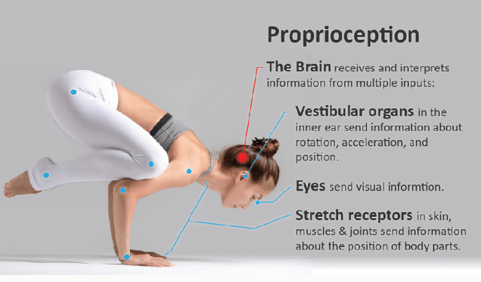
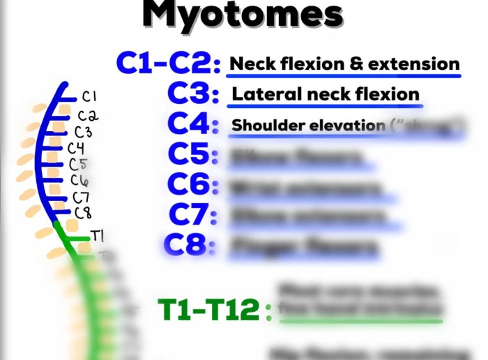
Tag: neurological disorders

  • What is a Myotome A Deep Dive

    What is a Myotome A Deep Dive

    What is a myotome? It’s a crucial concept in understanding how our muscles are controlled by the nervous system. Imagine a specific group of muscles, all working together, coordinated by a particular segment of the spinal cord. That’s essentially a myotome. This isn’t just an academic curiosity; myotome assessment plays a vital role in diagnosing neurological conditions.

    From pinpointing the source of muscle weakness to understanding the intricate pathways of the nervous system, exploring myotomes reveals a fascinating interplay between structure and function.

    This comprehensive guide delves into the world of myotomes, examining their definition, anatomy, function, clinical relevance, and assessment methods. We’ll uncover how these intricate muscle groups are organized, the role of nerve roots, and how evaluating myotomes helps diagnose and understand various neurological issues. Get ready to explore the fascinating science behind your body’s movement!

    Definition and Scope

    A myotome is a group of muscles innervated by a specific spinal nerve root. Understanding myotomes is crucial for clinicians, as they provide a pathway to assess the integrity of the motor pathways of the nervous system. It’s a fundamental concept in neurology, helping pinpoint the location of potential nerve damage.Myotomes and dermatomes, while both associated with spinal nerves, differ in their function.

    Dermatomes map sensory innervation, while myotomes map motor innervation. Damage to a specific nerve root can manifest as weakness or paralysis in the associated myotome, allowing clinicians to pinpoint the affected area. This localized weakness helps determine the level of spinal cord or nerve root compromise.

    Myotome Definition and Relationship to Dermatomes

    A myotome is a group of muscles that are controlled by a single spinal nerve root. This contrasts with dermatomes, which map the sensory areas served by the same nerve root. The relationship between the two is based on the common spinal nerve root origin. Clinical correlation between sensory and motor deficits, observed in dermatomes and myotomes respectively, allows clinicians to pinpoint the affected spinal segment and the extent of the damage.

    A myotome is basically a group of muscles that a single spinal nerve root controls. Understanding these muscle groups is crucial for diagnosing and treating pain, especially when considering active and passive pain management strategies. For example, if you’re experiencing pain in a specific area, evaluating the corresponding myotome can help pinpoint the source of the issue. This kind of assessment can help you determine the best course of action, such as exploring different approaches to pain relief, including active and passive pain management techniques.

    Ultimately, knowing your myotomes can give you a better understanding of your body and how to manage any discomfort effectively.

    This integrative approach is vital in diagnosing and treating various neurological conditions.

    Clinical Significance of Myotomes

    Myotomes are essential in clinical settings for diagnosing nerve root or spinal cord lesions. Weakness or paralysis in a specific myotome can indicate a problem with the corresponding spinal nerve root, often associated with conditions like herniated discs, spinal stenosis, or multiple sclerosis. The pattern of muscle weakness can help determine the precise location and extent of the neurological damage.

    Description of Myotomes

    Myotomes can be described in various ways. An anatomical description focuses on the specific muscles within the group. A functional description emphasizes the movements the myotome enables. Different methods of testing can also provide a detailed description, focusing on the specific movements and muscles involved in the examination. These methods are integral to the clinical evaluation.

    Table of Myotomes

    Myotome Name Associated Muscles Nerve Root Clinical Significance
    Cervical Myotome C5 Deltoid, biceps brachii, brachialis C5 spinal nerve root Weakness in shoulder abduction and elbow flexion can suggest C5 nerve root compression.
    Cervical Myotome C6 Biceps brachii, wrist extensors C6 spinal nerve root Weakness in wrist extension and elbow flexion indicates possible C6 nerve root impingement.
    Lumbar Myotome L2 Hip flexors L2 spinal nerve root Paralysis in hip flexion may point to L2 nerve root compression.
    Lumbar Myotome L4 Knee extensors L4 spinal nerve root Weakness in knee extension often suggests L4 nerve root damage, potentially due to disc herniation.
    Sacral Myotome S1 Plantar flexors, ankle evertors S1 spinal nerve root Weakness in plantar flexion and ankle eversion can indicate S1 nerve root pathology.

    Anatomy and Structure: What Is A Myotome

    What is a myotome

    Myotomes are fascinating segments of the body, directly linked to the intricate network of nerves and muscles. Understanding their anatomical arrangement is crucial for diagnosing and treating neuromuscular disorders. This section delves into the detailed structure of myotomes, illustrating their organization and the specific components that contribute to their function.The arrangement of myotomes reflects the segmental organization of the nervous system.

    They are essentially functional units, each controlled by a specific group of nerve roots. The specific muscles controlled by each myotome are essential in coordinating movement, posture, and other functions. This knowledge is fundamental in assessing neurological integrity.

    Anatomical Arrangement of Myotomes, What is a myotome

    The spinal cord segments and corresponding nerve roots organize myotomes in a precise pattern. Imagine a vertical arrangement of segments, each responsible for controlling muscles in a specific region of the body. This organization allows for precise control and coordination of movement, enabling us to perform a vast array of actions. This arrangement also allows for efficient communication between the brain and different parts of the body.

    Components of a Myotome

    A myotome comprises the nerve root(s) that innervate it and the specific muscle groups it controls. These components are interconnected, forming a functional unit. The nerve roots carry signals from the spinal cord to the muscles, enabling contraction and movement. The muscle groups are the effectors of this signal, performing the intended actions. Together, these elements create a complete functional unit.

    Nerve Root Innervation

    The nerve roots are the essential communication channels connecting the spinal cord to the muscles. Each myotome receives its innervation from specific nerve roots, which originate from the spinal cord. These nerve roots relay signals from the central nervous system to the muscle fibers, triggering contractions. The precise location of these roots is essential in determining the specific myotome.

    Spinal Cord Segments Involved

    The spinal cord is divided into segments, each corresponding to a specific region of the body. These segments are crucial in the formation of myotomes, as they are the source of the nerve roots. Each segment sends signals to the corresponding muscle groups, controlling their actions. The precise location and function of each spinal cord segment are essential in understanding myotome function.

    This information is used to diagnose and treat various neuromuscular conditions.

    Myotome Table

    This table illustrates the relationship between myotome, nerve root(s), and the muscle groups controlled. It highlights the crucial role of nerve roots in activating specific muscle groups, which together create the function of a myotome. This information is critical in assessing neurological function and diagnosing neuromuscular disorders.

    Myotome Nerve Root(s) Innervating It Muscle Groups Controlled
    Cervical Myotomes (C1-C8) C1-C8 Neck, shoulder, arm, hand muscles
    Thoracic Myotomes (T1-T12) T1-T12 Intercostal muscles, back muscles
    Lumbar Myotomes (L1-L5) L1-L5 Hip, thigh, knee muscles
    Sacral Myotomes (S1-S5) S1-S5 Leg, foot muscles

    Neural Pathways Involved in Myotome Function

    The neural pathways involved in myotome function are complex, but they can be described as follows. Sensory input from the muscle and surrounding tissues is conveyed to the spinal cord through sensory nerves. Motor commands are then sent from the spinal cord via motor nerves to the muscle fibers. This two-way communication allows for precise control and coordination of movement.

    The intricate nature of these pathways underscores the importance of a well-functioning nervous system.

    Function and Clinical Relevance

    Myotomes, as we’ve explored, represent a specific group of muscles innervated by a single spinal nerve root. Understanding their function is crucial in evaluating neurological health. This section delves into the practical applications of myotome assessment in clinical settings.Myotome assessment provides valuable insights into the integrity of the peripheral nervous system. Clinicians utilize this information to pinpoint potential nerve root impingements, spinal cord lesions, or peripheral neuropathies.

    The accuracy of this assessment is pivotal in formulating a diagnosis and developing a targeted treatment plan.

    Myotome Function

    Myotomes are responsible for specific movements or actions. Each myotome corresponds to a particular set of muscles, and the coordinated activation of these muscles allows for a range of body movements. For example, the C5 myotome controls shoulder abduction, while the L4 myotome facilitates dorsiflexion of the foot. Understanding the specific movements controlled by each myotome helps clinicians assess the extent of potential neurological damage.

    Myotome Assessment in Clinical Practice

    Clinicians evaluate myotomes through a series of standardized muscle strength tests. These tests involve asking the patient to perform specific movements, and the examiner assesses the strength and range of motion of the corresponding muscles. The examiner grades the strength using a standardized scale, often a 0-5 scale, where 0 indicates no muscle contraction and 5 represents normal strength.

    The precise movements and muscle groups tested depend on the suspected area of neurological impairment. Thorough and careful examination is crucial to ensure accurate assessment.

    Clinical Uses of Myotome Testing

    Myotome testing serves as a vital diagnostic tool in identifying and evaluating neurological conditions. It helps pinpoint the level of spinal cord or nerve root involvement, aiding in differential diagnosis. This testing is particularly useful in cases of suspected nerve root compression, such as from herniated discs, or in conditions like spinal stenosis. Furthermore, myotome assessment can help monitor the progression or response to treatment in various neurological disorders.

    A myotome is a group of muscles that are innervated by a single spinal nerve root. Maintaining good kidney health is crucial for overall well-being, and a balanced diet plays a key role. Eating foods rich in antioxidants and low in sodium, such as those highlighted in this resource on foods for kidney health , can contribute significantly to kidney function.

    Understanding myotomes is important for diagnosing and treating neuromuscular conditions.

    Conditions Where Myotome Assessment is Important

    Myotome assessment is critical in several neurological conditions. The information gathered aids in pinpointing the location and extent of the damage to the nervous system. By evaluating muscle strength, clinicians can identify the specific nerve roots or spinal cord segments that are affected. This allows for a more accurate diagnosis and facilitates appropriate treatment.

    Examples of Conditions and Expected Myotome Weakness

    Condition Expected Myotome Weakness Diagnostic Significance
    Cervical Radiculopathy (C6-C7) Weakness in elbow flexion and wrist extension Indicates nerve root compression at the C6-C7 level, potentially from a herniated disc.
    Lumbar Radiculopathy (L5) Weakness in foot dorsiflexion and great toe extension Suggests nerve root compression at the L5 level, possibly due to spinal stenosis or a herniated disc.
    Spinal Cord Injury (Thoracic Level) Weakness in shoulder abduction, elbow flexion, and wrist extension. Indicates a spinal cord lesion, and the level of the lesion can be determined by the specific myotomes affected.
    Peripheral Neuropathy Gradual, symmetric weakness in multiple myotomes Suggests a diffuse or systemic process affecting the peripheral nerves.

    Assessment and Testing

    Assessing myotome function is crucial for evaluating neuromuscular health and identifying potential nerve or muscle pathologies. Proper myotome testing involves a systematic approach to evaluate the strength and power of specific muscle groups innervated by particular spinal nerve roots. This allows clinicians to pinpoint the location of potential damage or dysfunction.

    Methods for Assessing Myotome Function

    Various methods are employed to assess myotome function, each with its strengths and limitations. These methods range from simple manual muscle testing to more sophisticated electromyographic (EMG) studies. Manual muscle testing is a widely used, cost-effective method that relies on the clinician’s observation of the patient’s ability to perform specific movements against resistance. EMG studies provide a more detailed evaluation of muscle electrical activity, allowing for a deeper understanding of muscle function and identifying any neuromuscular disorders.

    Steps Involved in a Myotome Examination

    A comprehensive myotome examination involves several key steps. First, a thorough patient history and neurological examination are essential to understand the patient’s presenting complaint and potential contributing factors. Second, the examination should focus on identifying the specific muscle groups associated with the relevant myotomes. Third, standardized manual muscle testing procedures should be meticulously followed to ensure accuracy and reliability.

    Lastly, interpretation of the findings should be performed with careful consideration of the patient’s overall clinical presentation.

    Importance of Proper Technique in Myotome Testing

    Proper technique is paramount in myotome testing to ensure accurate and reliable results. Inconsistent or improper techniques can lead to inaccurate assessment and potentially misdiagnosis. Clinicians should adhere to standardized procedures and ensure that the patient is properly positioned and relaxed during the test. Accurate measurement and consistent evaluation criteria are crucial for reliable results. Standardized protocols should be used to maintain consistency and reproducibility.

    Step-by-Step Procedure for Testing Specific Myotomes

    1. Biceps Myotome (C5-C6): Assess the patient’s ability to flex the elbow against resistance. The examiner should position the patient supine or sitting with the elbow flexed at 90 degrees. The examiner applies resistance to the patient’s forearm while the patient attempts to maintain elbow flexion. Note the degree of resistance the patient can overcome. The examiner should observe for any weakness, atrophy, or tremors during the test.

    2. Triceps Myotome (C6-C7): Assess the patient’s ability to extend the elbow against resistance. The examiner should position the patient supine or sitting with the elbow flexed at 90 degrees. The examiner applies resistance to the patient’s forearm while the patient attempts to extend the elbow. Observe the degree of resistance the patient can overcome. Look for any weakness, atrophy, or tremors during the test.

      So, what exactly is a myotome? Basically, it’s a group of muscles that are innervated by a single spinal nerve root. Understanding these muscle groups is crucial for diagnosing nerve damage, and for people exploring natural health options like AHCC supplements, for example, AHCC benefits and safety is an important consideration. Knowing how AHCC might interact with your myotomes is key to potential therapeutic benefits.

      Ultimately, a good understanding of myotomes is essential for anyone working with the body’s complex systems.

      Note that a weak triceps response can suggest damage to the radial nerve.

    3. Wrist Extensors (C6-C7): Assess the patient’s ability to extend the wrist against resistance. The patient should be positioned supine or sitting with the forearm supported and the wrist in a neutral position. The examiner applies resistance to the dorsal aspect of the hand as the patient attempts to extend the wrist. Assess the strength and note any weakness. This test evaluates the integrity of the radial nerve.

    4. Iliopsoas Myotome (L2-L3): Assess hip flexion. The patient should be supine with the knee flexed and the hip at a 90-degree angle. The examiner should apply resistance to the patient’s thigh while they attempt to maintain hip flexion. Record the patient’s ability to maintain the flexion against resistance. Assess for any weakness, atrophy, or other abnormalities.

      This is crucial for assessing the integrity of the lumbar plexus and the associated nerves.

    Clinical Cases and Examples

    Myotome testing plays a crucial role in diagnosing and localizing neurological impairments. By assessing the strength and function of specific muscle groups, clinicians can pinpoint the level of the nervous system affected, whether it’s a peripheral nerve, spinal cord, or brain. Understanding the clinical presentation and interpretation of myotome weakness is vital for developing an accurate diagnosis and appropriate treatment plan.Interpreting myotome testing results requires careful consideration of the patient’s overall neurological presentation.

    The presence of other symptoms, such as sensory changes, reflexes, and cognitive impairments, provides valuable context and helps differentiate between various neurological conditions. These combined findings contribute to a comprehensive picture of the patient’s neurological status.

    Myotome Weakness in Cervical Radiculopathy

    Cervical radiculopathy, often caused by nerve root compression in the neck, frequently leads to specific myotome weakness. The affected myotomes are typically those innervated by the affected nerve roots. For example, C5 and C6 nerve root involvement might result in weakness of the biceps and deltoid muscles, affecting shoulder abduction and flexion.

    Myotome Weakness in Spinal Cord Injury

    Spinal cord injuries can result in varying degrees of myotome weakness, depending on the level and extent of the damage. A complete spinal cord lesion at the C5 level, for example, would likely produce significant weakness in the muscles innervated by the C5 nerve root and below, leading to weakness in shoulder abduction, elbow flexion, wrist extension, and hand movements.

    Myotome Weakness in Peripheral Neuropathy

    Peripheral neuropathies encompass a broad range of conditions affecting peripheral nerves. These conditions can lead to varying degrees of myotome weakness depending on the affected nerves. For instance, a diabetic neuropathy can lead to distal muscle weakness affecting the muscles of the feet and legs, impacting ambulation and balance.

    Interpreting Myotome Testing Results

    Interpreting myotome testing results in conjunction with other neurological findings is crucial for accurate diagnosis. For example, if a patient presents with weakness in the C7 myotome (triceps) but normal reflexes, the cause might be localized to the peripheral nerve rather than the spinal cord. Consideration of the patient’s medical history, symptoms, and other neurological examination findings further enhances diagnostic accuracy.

    Case Studies of Myotome Involvement

    Case Scenario Myotome Affected Symptoms Diagnosis
    Patient with neck pain, radiating down the arm, and weakness in biceps and wrist extensors. C5, C6, C7 Pain, numbness, tingling in the arm and hand, weakness in elbow flexion and wrist extension. Cervical radiculopathy
    Patient with a history of a fall, presenting with decreased sensation in the lower extremities and weakness in hip flexion and knee extension. L2, L3, L4 Numbness and tingling in the lower extremities, weakness in hip flexion, knee extension, and dorsiflexion. Spinal cord injury
    Patient with a history of diabetes, experiencing gradual weakness in the foot and ankle. L4, L5, S1 Gradual weakness and numbness in the feet and ankles, difficulty with ambulation. Diabetic neuropathy

    Variations and Considerations

    Myotomes, while providing a valuable framework for assessing neuromuscular function, aren’t a perfect fit for every individual. Variations in their organization and the influence of various factors can significantly affect how a myotome is assessed and interpreted. Understanding these variations and considerations is crucial for accurate diagnoses and effective treatment plans.The consistency in myotome organization, though a useful guide, is not absolute.

    Individual variations in muscle fiber type, size, and innervation patterns can affect the specific muscles that make up a myotome. This variability in anatomy necessitates a careful and nuanced approach to myotome assessment, always considering the individual’s unique characteristics.

    Variations in Myotome Organization

    Individual differences in muscle anatomy and innervation can lead to variations in myotome boundaries. These variations can sometimes result in a muscle being innervated by a nerve root slightly different from the expected pattern. This can cause a myotome to be slightly larger or smaller in some individuals than in others. Factors like genetic predisposition and previous injuries can also play a role in these anatomical variations.

    Factors Influencing Myotome Assessment

    Several factors can impact the accuracy of myotome testing. Patient cooperation and motivation are critical, as muscle strength is affected by psychological factors like fear and anxiety. Pain, fatigue, and other medical conditions can also influence the ability to perform muscle tests, potentially leading to inaccurate or misleading results. Furthermore, the examiner’s skill and experience in performing the tests directly impacts the reliability of the assessment.

    Careful consideration of these influencing factors is paramount to obtaining meaningful and reliable results.

    Limitations of Myotome Testing

    Myotome testing, despite its usefulness, has limitations. It provides a general indication of nerve root function but doesn’t offer a precise measurement of the extent of the nerve damage. The test’s resolution is limited, and subtle deficits may be missed. Additionally, myotome testing relies on the patient’s ability to cooperate, which can be affected by factors like pain or cognitive impairment.

    It’s important to recognize that myotome testing is a component of a broader neurological assessment, not a stand-alone diagnostic tool.

    Considerations for Assessing Myotomes in Different Age Groups

    Age significantly impacts muscle strength and the ability to perform motor tasks. Children and older adults may have different baseline strength levels, requiring adjusted assessment methods and interpretations. The developmental stages of children, for example, need to be taken into account. In children, the myotome assessment might focus on milestones and developmental norms, rather than precise strength measurements.

    Table: Age Group Considerations for Myotome Assessment

    Age Group Potential Variations Assessment Considerations
    Infants (0-1 year) Immature neuromuscular system, limited voluntary movement Focus on developmental milestones, use of reflexes, parental input
    Children (2-12 years) Varying developmental stages, rapid growth spurts Use age-appropriate tasks, consider developmental norms, assess cooperation
    Adults (18-65 years) Stable muscle mass, generally consistent motor abilities Standard myotome testing methods are generally applicable
    Older Adults (65+) Decreased muscle mass, decreased strength, potential comorbidities Adjust testing to accommodate limitations, assess for confounding factors, use caution

    Imaging and Techniques

    What is a myotome

    Visualizing myotomes, the groups of muscles innervated by a single spinal nerve root, presents unique challenges. While direct visualization of muscle function isn’t possible with standard imaging, these techniques can provide valuable information about the structures involved, aiding in the diagnosis and understanding of neuromuscular conditions. Understanding the limitations of these techniques is equally crucial for accurate interpretation.Imaging modalities offer insights into the anatomy of myotomes, but don’t directly measure their functional capacity.

    This is critical to remember when interpreting results, as imaging findings must be correlated with other assessments like electromyography (EMG) and nerve conduction studies (NCS) to gain a comprehensive picture. Indirect assessments of myotome involvement are possible through visualization of neural pathways and structures related to muscle function.

    MRI and CT Imaging

    Magnetic resonance imaging (MRI) and computed tomography (CT) scans are valuable tools for visualizing the anatomy of the spine, spinal cord, and surrounding tissues. These techniques can depict the spinal nerves, which are crucial for understanding myotome function. MRI excels at providing detailed soft tissue images, highlighting potential spinal cord compression or nerve root impingement, which can affect the associated myotomes.

    CT, on the other hand, provides excellent bone detail, aiding in identifying bony structures that might contribute to nerve root compression.

    Limitations of Imaging Techniques

    Imaging techniques primarily visualize thestructures* involved in myotome function, not the function itself. For example, an MRI might reveal a herniated disc compressing a nerve root, but it doesn’t directly demonstrate the resultant weakness in the muscles of the corresponding myotome. Other factors, like the severity of the compression or the individual’s response to the condition, can also affect the myotome’s function and are not directly measurable by these techniques.

    Visualizing Neural Pathways

    Visualization of neural pathways involves advanced techniques. Advanced MRI techniques, such as diffusion tensor imaging (DTI), can map the pathways of nerve fibers, providing insights into the integrity of neural connections. This is crucial in cases of suspected nerve damage or disease, aiding in identifying the affected myotomes.

    Electrodiagnostic Studies (EMG and NCS)

    Electromyography (EMG) and nerve conduction studies (NCS) are essential for evaluating the electrical activity of muscles and nerves. EMG measures the electrical activity of muscle fibers, while NCS assesses the speed and amplitude of nerve impulses. These studies can directly assess the functional integrity of the myotomes, detecting issues like denervation (loss of nerve supply) or axonal damage.

    For instance, if a specific myotome shows reduced or absent electrical activity, it suggests a potential problem with the nerve root or the muscles it supplies.

    Comparison of Imaging Modalities

    Imaging Modality Strengths Limitations Application in Myotome Assessment
    MRI Excellent soft tissue detail, visualization of spinal cord and nerve roots Limited visualization of bone structures, can be expensive Identifying spinal cord compression, nerve root impingement
    CT Excellent bone detail, rapid acquisition Limited soft tissue contrast, higher radiation exposure Identifying bony structures contributing to nerve root compression
    DTI Visualization of neural pathways Requires specialized software and interpretation, more expensive than standard MRI Assessing nerve fiber integrity, identifying areas of potential damage
    EMG/NCS Direct assessment of muscle and nerve function Invasive procedure, requires specialized expertise for interpretation Evaluating the functional integrity of the myotomes, detecting denervation or axonal damage

    Ending Remarks

    In conclusion, understanding myotomes provides a critical lens through which we can appreciate the intricate relationship between the nervous system and our muscles. From a clinical standpoint, myotome assessment is invaluable for pinpointing the source of neurological issues. This guide has explored the key aspects of myotomes, offering a clear understanding of their definition, anatomy, function, and clinical applications.

    We’ve seen how these muscle groups, coordinated by specific nerve roots, are crucial for our everyday movements and how their assessment is a valuable tool in diagnosing various conditions. Myotomes are more than just anatomical structures; they’re essential for our health and well-being.

  • Understanding What is a Small Stroke

    Understanding What is a Small Stroke

    What is a small stroke? This in-depth exploration delves into the intricacies of this often-overlooked medical condition, highlighting its defining characteristics, potential causes, diagnostic procedures, and treatment options. We’ll examine the subtle signs and symptoms, distinguish it from a TIA, and explore the long-term effects and recovery strategies.

    From the initial definition and medical terminology to the crucial role of lifestyle choices, underlying health conditions, and preventative measures, this comprehensive guide provides a complete understanding of small strokes.

    Defining a Small Stroke: What Is A Small Stroke

    What is a small stroke

    A small stroke, sometimes referred to as a ministroke, is a temporary interruption of blood flow to the brain, resulting in a localized area of reduced brain function. These events, while often less severe than larger strokes, can still have significant consequences and require prompt medical attention. Recognizing the signs and understanding the distinctions between small strokes and other conditions is crucial for timely intervention.A small stroke, medically termed a “transient ischemic attack” (TIA), or “small cerebrovascular accident” (CVA), is characterized by the temporary interruption of blood flow to the brain.

    This interruption leads to temporary neurological symptoms, often lasting from a few minutes to several hours. Key to understanding small strokes is the distinction between their temporary nature and the permanent damage associated with a larger stroke.

    Characteristics of a Small Stroke

    A small stroke is a temporary interruption of blood flow to the brain, causing localized symptoms that typically resolve within 24 hours. Unlike a larger stroke, the brain tissue damage is usually reversible, although residual effects are possible. Understanding the nuances between a small stroke and a transient ischemic attack (TIA) is essential for proper diagnosis and management.

    Difference Between Small Stroke and TIA

    While both small strokes and TIAs involve temporary disruptions in blood flow, there are subtle but significant distinctions. A TIA is a brief episode of neurological dysfunction caused by temporary ischemia (reduced blood flow) in the brain. The symptoms resolve completely within 24 hours, with no lasting neurological deficits. A small stroke, though temporary, may have residual effects.

    Common Signs and Symptoms of a Small Stroke, What is a small stroke

    Small strokes manifest in various ways, depending on the affected brain region. Common signs include numbness or weakness in the face, arm, or leg, especially on one side of the body; confusion or difficulty speaking; trouble seeing in one or both eyes; dizziness, loss of balance, or coordination problems; and sudden severe headache. Prompt recognition of these symptoms is crucial for timely intervention.

    Types of Small Strokes

    Stroke Type Symptoms Duration Risk Factors
    Ischemic Small Stroke Numbness or weakness, difficulty speaking, vision problems, dizziness, headache. Minutes to hours. Symptoms typically resolve within 24 hours. High blood pressure, high cholesterol, smoking, diabetes, heart disease, and a family history of stroke.
    Hemorrhagic Small Stroke Sudden severe headache, stiff neck, vomiting, altered consciousness, weakness, numbness, or vision changes. Minutes to hours. Symptoms typically resolve within 24 hours. High blood pressure, blood clotting disorders, and use of blood thinners.

    Causes and Risk Factors

    Small strokes, while often overlooked, can have significant consequences. Understanding the causes and risk factors is crucial for prevention and early intervention. Identifying these factors allows individuals to make informed choices about their health and potentially reduce their risk. Proactive steps can significantly lower the likelihood of experiencing a small stroke.Lifestyle choices play a pivotal role in the development of small strokes.

    Poor dietary habits, lack of physical activity, and smoking significantly increase the risk. These behaviors contribute to the buildup of plaque in blood vessels, narrowing them and hindering blood flow.

    Primary Causes of Small Strokes

    Small strokes, also known as transient ischemic attacks (TIAs), are often caused by a temporary disruption of blood flow to the brain. This disruption can be due to various factors, including blood clots, plaque buildup, or a burst blood vessel. The most common cause is atherosclerosis, a condition characterized by the hardening and narrowing of arteries due to plaque buildup.

    This process is frequently associated with poor lifestyle choices and underlying health conditions. Another crucial cause is the formation of blood clots within the arteries leading to the brain.

    Role of Lifestyle Choices

    Unhealthy lifestyle choices are major contributors to small stroke risk. A diet high in saturated and trans fats, cholesterol, and sodium can contribute to high blood pressure and high cholesterol, increasing the likelihood of plaque buildup in blood vessels. A sedentary lifestyle reduces blood circulation, further contributing to the risk of clot formation. Smoking damages blood vessels and increases the risk of blood clots, significantly impacting overall vascular health.

    These lifestyle choices, when combined, create a potent cocktail of risk factors that can lead to a small stroke.

    Importance of Underlying Health Conditions

    Several underlying health conditions significantly increase the risk of small strokes. High blood pressure (hypertension) is a critical risk factor, as it puts stress on blood vessels and increases the risk of both plaque formation and blood clot formation. High cholesterol, diabetes, and heart disease all contribute to the buildup of plaque, narrowing arteries, and making the brain vulnerable to blood flow disruption.

    For example, individuals with poorly controlled diabetes often have higher blood pressure and cholesterol levels, further exacerbating the risk of small strokes.

    Significance of Age and Genetics

    Age is a significant risk factor for small strokes. As we age, our blood vessels may naturally experience some degree of damage or hardening, increasing the risk of plaque buildup and clot formation. Genetics also play a role. Family history of stroke, particularly in younger individuals, can signal a genetic predisposition to conditions that increase the risk of small strokes.

    However, while age and genetics contribute, lifestyle modifications can significantly mitigate the risk.

    Comparison of Risk Factors for Different Small Stroke Types

    Risk Factor Small Stroke Type A (Embolic) Small Stroke Type B (Hemorrhagic)
    High Blood Pressure High Moderate
    High Cholesterol High Moderate
    Smoking High Moderate
    Diabetes High Moderate
    Heart Disease High Moderate
    Age Moderate High
    Genetics Moderate Moderate
    Lifestyle choices High Moderate

    Note: This table provides a general comparison and does not represent all possible stroke types or variations in risk. Consulting a healthcare professional is crucial for personalized risk assessment and management.

    Diagnosis and Evaluation

    Pinpointing a small stroke, often characterized by subtle symptoms, requires a meticulous diagnostic approach. Accurate identification is crucial for prompt treatment and minimizing long-term complications. The process typically involves a combination of clinical assessments and specialized imaging techniques to pinpoint the location and extent of the brain injury. This detailed evaluation helps healthcare providers tailor treatment strategies to the specific needs of each patient.The diagnostic journey for a suspected small stroke begins with a thorough evaluation of the patient’s medical history, current symptoms, and risk factors.

    This initial assessment lays the groundwork for subsequent investigations and helps healthcare professionals develop a focused diagnostic plan. A comprehensive understanding of the patient’s overall health, including pre-existing conditions and medications, is essential.

    Diagnostic Procedures

    A range of procedures are employed to identify and characterize a small stroke. These methods help in determining the cause, extent, and location of the suspected ischemic event. The procedures include detailed patient history, physical examination, and advanced imaging techniques.

    Imaging Techniques

    Neuroimaging plays a critical role in diagnosing small strokes. These techniques provide detailed visualizations of the brain’s structures and blood vessels, allowing healthcare professionals to identify areas of damage or disruption.

    • Computed Tomography (CT) Scans: CT scans are rapid and readily available, enabling quick assessment of the brain for signs of bleeding or significant tissue damage. CT scans are particularly valuable in the initial stages of diagnosis, offering a preliminary view of the brain’s structure and helping rule out other conditions.
    • Magnetic Resonance Imaging (MRI) Scans: MRI scans provide more detailed anatomical information, allowing for better visualization of the brain’s soft tissues and blood vessels. These scans are particularly useful in identifying subtle ischemic changes that might be missed on a CT scan, offering a higher resolution view of the affected area. Diffusion-weighted MRI (DWI) and perfusion-weighted MRI (PWI) are specific types of MRI that highlight areas of reduced blood flow, helping to pinpoint the location and extent of the stroke.

    • Magnetic Resonance Angiography (MRA): MRA utilizes MRI to visualize the blood vessels in the brain. This technique helps identify blockages or narrowing in the arteries, which can be the cause of a small stroke.

    Neurological Examinations

    Neurological examinations are essential for assessing the patient’s neurological function. These assessments evaluate the patient’s motor skills, sensory perception, cognitive abilities, and reflexes. This evaluation helps determine the extent of the neurological deficits and aids in identifying the affected areas of the brain.

    • Motor Function Assessment: This involves evaluating muscle strength, tone, and coordination. Examples include assessing arm and leg strength, and testing fine motor skills. Difficulties with movement, weakness, or paralysis are noted.
    • Sensory Function Assessment: This includes testing the patient’s ability to perceive touch, pain, temperature, and vibration. Any loss or alteration in sensation is documented.
    • Cognitive Function Assessment: This involves assessing the patient’s memory, attention span, language skills, and problem-solving abilities. Changes in cognitive function, including confusion, disorientation, or difficulty with communication, are noted.
    • Cranial Nerve Assessment: This evaluates the function of the 12 cranial nerves, which control various sensory and motor functions. Assessment of these nerves aids in identifying the specific brain regions affected by the stroke.

    Step-by-Step Procedure for Diagnosing a Small Stroke

    A systematic approach to diagnosis is critical. A combination of the mentioned methods is used.

    A small stroke, sometimes called a transient ischemic attack (TIA), can be a tricky thing. It’s essentially a mini-stroke, where blood flow to the brain is briefly interrupted. This can manifest in various ways, but one less-obvious symptom is persistent hunger, potentially linked to the underlying health issues that contribute to a higher risk of a small stroke.

    For a deeper dive into why you might be experiencing constant hunger, check out this informative article on why am i always hungry. Ultimately, if you’re concerned about any potential health issues, it’s best to consult a doctor to rule out anything serious, including a small stroke.

    1. Patient History and Physical Examination: Detailed history, symptom description, and a complete physical examination are performed.
    2. Initial Imaging (CT Scan): A rapid CT scan is often performed to quickly rule out hemorrhagic stroke or other conditions.
    3. Further Imaging (MRI): If the initial CT scan is inconclusive, an MRI, including DWI and PWI, is employed to identify subtle ischemic changes.
    4. Neurological Examination: A comprehensive neurological examination is conducted to assess the extent of neurological deficits.
    5. Further Investigations: Additional tests, such as blood tests and carotid ultrasound, might be necessary to determine the underlying cause of the stroke.

    Flowchart for Small Stroke Diagnosis

    [A detailed flowchart illustrating the steps in diagnosing a small stroke, including the different imaging modalities and neurological tests, could be added here. It would visually represent the sequence of events and the branching possibilities depending on the results of the tests. A diagram is beyond the scope of this text-based format.]

    Treatment and Management

    Navigating a small stroke can feel overwhelming, but understanding the treatment and management strategies is crucial for recovery. Early intervention and a multi-faceted approach, combining medical care with lifestyle adjustments, significantly impact the patient’s journey towards regaining function and quality of life. The goal is not just to stop the stroke, but to minimize its impact and empower the patient to regain lost abilities.

    Immediate Treatment Approaches

    Immediate action following a suspected small stroke is critical. Emergency medical services should be contacted immediately. Prompt evaluation and diagnosis are vital for determining the appropriate course of action. Timely treatment can potentially limit the extent of brain damage and improve recovery outcomes. This includes assessing the patient’s symptoms, vital signs, and neurological function to establish the severity and nature of the event.

    Role of Medication in Managing Small Strokes

    Medication plays a significant role in managing small strokes. Antiplatelet drugs, such as aspirin or clopidogrel, are often prescribed to prevent further blood clots from forming. These medications help to thin the blood and reduce the risk of another stroke. Anticoagulants, like warfarin or newer oral anticoagulants (NOACs), may be used in specific cases, such as when the small stroke is linked to a blood clot.

    Blood pressure medications are often prescribed to manage and maintain healthy blood pressure levels. This approach aims to minimize the risk of future events and improve the patient’s overall health. Managing underlying conditions, such as high blood pressure, high cholesterol, and diabetes, is equally important to prevent future strokes.

    Importance of Lifestyle Modifications

    Lifestyle modifications are essential for stroke recovery and prevention. A healthy diet low in saturated and trans fats, rich in fruits, vegetables, and whole grains, is crucial. Regular exercise, such as walking, swimming, or cycling, helps improve cardiovascular health and reduces the risk of future strokes. Maintaining a healthy weight and quitting smoking are also important lifestyle choices.

    Managing stress through relaxation techniques, like meditation or yoga, is beneficial for overall well-being. These lifestyle changes can contribute significantly to a patient’s long-term health and recovery.

    Common Rehabilitation Strategies

    Rehabilitation is crucial for regaining lost functions after a small stroke. Physical therapy helps to improve motor skills, balance, and coordination. Occupational therapy focuses on adapting daily activities and tasks to meet the patient’s current abilities. Speech therapy is important for recovering communication and swallowing skills. These therapies are tailored to the individual’s specific needs and goals, enabling them to regain lost independence and function.

    Psychological support is also crucial to address emotional and mental well-being, providing coping mechanisms for the challenges of stroke recovery.

    Treatment Options Summary Table

    Stroke Type Immediate Treatment Medications Rehabilitation
    Ischemic (due to blood clot) Rapid evaluation and diagnosis, supportive care Antiplatelets (aspirin, clopidogrel), anticoagulants (warfarin, NOACs) Physical therapy, occupational therapy, speech therapy, psychological support
    Hemorrhagic (due to bleeding) Controlling blood pressure, addressing underlying cause, supportive care Medications to control blood pressure, manage underlying conditions Physical therapy, occupational therapy, speech therapy, psychological support
    Transient Ischemic Attack (TIA) Similar to ischemic, focus on identifying cause Antiplatelets, blood pressure medications, lipid-lowering drugs Physical therapy, occupational therapy, speech therapy, lifestyle modification, monitoring

    Long-Term Effects and Recovery

    A small stroke, while often less severe than a large-scale stroke, can still have a significant and lasting impact on a person’s life. Understanding the potential long-term effects and developing strategies for recovery is crucial for navigating this experience. The path to recovery is unique for each individual, influenced by the specific area of the brain affected and the individual’s overall health.The long-term effects of a small stroke can vary widely, ranging from mild functional impairments to more significant challenges.

    Factors like the location and size of the affected brain region, the individual’s pre-stroke health, and the promptness and quality of treatment play key roles in determining the extent of recovery.

    A small stroke, sometimes called a transient ischemic attack (TIA), is a temporary interruption of blood flow to the brain. While it often has no lasting effects, understanding the potential causes is crucial. One such potential cause, that can affect your overall health and potentially lead to weight changes, is the use of lithium for certain mental health conditions.

    To learn more about whether lithium might contribute to weight gain, check out this helpful resource on does lithium cause weight gain. Recognizing these potential connections can help you and your doctor better understand your health and treatment options, leading to a better understanding of what a small stroke might be.

    Potential Long-Term Effects

    Small strokes can lead to a variety of long-term effects. These include difficulties with movement, speech, swallowing, vision, and cognitive functions. Some individuals may experience problems with memory, attention, problem-solving, or decision-making. It is crucial to remember that not everyone experiences all these effects, and the severity can differ greatly.

    Impact on Daily Life Activities

    A small stroke can disrupt daily life activities in significant ways. For instance, tasks such as dressing, eating, bathing, and performing household chores might become challenging. Communication difficulties can also impact social interactions and relationships. The individual’s ability to work or maintain their usual level of independence may be affected. It’s essential to address these difficulties with appropriate strategies and support systems.

    Importance of Support Systems

    Strong support systems are vital for small stroke survivors. Family, friends, and support groups provide emotional comfort, practical assistance, and encouragement. Professional therapists, such as occupational therapists, physical therapists, and speech-language pathologists, play a crucial role in helping individuals adapt to their new limitations and regain lost skills. Support from healthcare professionals is equally critical.

    A small stroke, sometimes called a transient ischemic attack (TIA), is a temporary interruption of blood flow to the brain. While often a warning sign of a larger stroke, understanding the causes and treatment of related issues like CPAP-related gas buildup is crucial. For example, if you’re using CPAP for sleep apnea, learning about the causes and treatment of cpap gas can help you manage potential complications and avoid further issues.

    Ultimately, recognizing the subtle symptoms of a small stroke is key to early intervention and preventing more serious problems.

    Strategies for Managing Long-Term Complications

    Effective management of long-term complications requires a multi-faceted approach. This involves adapting daily routines to accommodate limitations, utilizing assistive devices as needed, and seeking ongoing medical care. Regular check-ups and follow-up appointments with healthcare providers are essential for monitoring progress and addressing any emerging issues. Developing coping mechanisms and stress management techniques can also help improve overall well-being.

    Resources for Support and Recovery

    • Support Groups: Support groups provide a forum for sharing experiences, exchanging coping strategies, and gaining encouragement from others facing similar challenges. These groups offer valuable emotional support and practical advice.
    • Occupational Therapy: Occupational therapists can assess daily living skills and develop strategies to adapt tasks to accommodate limitations. They can provide guidance on assistive devices and modifications to the home environment.
    • Physical Therapy: Physical therapists focus on restoring physical function and mobility. They design exercises and rehabilitation programs tailored to the individual’s needs, helping regain lost strength and coordination.
    • Speech-Language Pathology: Speech-language pathologists address communication and swallowing difficulties. They develop strategies for improving speech clarity, and teach techniques for safer swallowing, improving the individual’s ability to communicate and consume food effectively.
    • Mental Health Professionals: Mental health professionals are crucial in addressing the emotional and psychological impact of a stroke. They can help manage stress, anxiety, and depression, and provide support during the recovery process.

    Prevention Strategies

    Preventing a small stroke hinges on proactive lifestyle choices and early intervention. By understanding the risk factors and taking steps to mitigate them, individuals can significantly reduce their likelihood of experiencing a small stroke. A healthy lifestyle, encompassing diet, exercise, and stress management, forms the cornerstone of stroke prevention.Adopting preventative measures isn’t about eliminating risk entirely, but about managing it effectively.

    By addressing modifiable risk factors, individuals can significantly lower their chances of suffering a small stroke and improve their overall well-being.

    Lifestyle Modifications

    A healthy lifestyle is paramount in stroke prevention. Consistent physical activity, a balanced diet, and stress management techniques play vital roles in reducing the risk. Maintaining a healthy weight, limiting alcohol consumption, and quitting smoking are also crucial steps. These actions not only decrease the risk of small strokes but also contribute to overall cardiovascular health.

    • Regular Exercise: Aim for at least 150 minutes of moderate-intensity aerobic exercise per week, such as brisk walking, swimming, or cycling. Even shorter bursts of activity throughout the day can be beneficial. Regular exercise helps control blood pressure, cholesterol levels, and blood sugar, all of which are critical in stroke prevention.
    • Balanced Diet: Focus on a diet rich in fruits, vegetables, whole grains, and lean protein. Limit processed foods, saturated fats, and excessive sodium intake. A balanced diet helps maintain healthy weight, blood pressure, and cholesterol levels.
    • Stress Management: Chronic stress can elevate blood pressure and increase the risk of stroke. Incorporate stress-reducing activities like yoga, meditation, deep breathing exercises, or spending time in nature. Finding healthy ways to manage stress is essential for overall health and well-being.
    • Weight Management: Maintaining a healthy weight is crucial for stroke prevention. Excess weight puts extra strain on the cardiovascular system, increasing the risk of high blood pressure, high cholesterol, and diabetes, all of which are significant risk factors for stroke.
    • Moderation in Alcohol Consumption: If you choose to drink alcohol, do so in moderation. Excessive alcohol consumption can raise blood pressure and increase the risk of stroke. Understanding your personal risk factors and consulting with a healthcare professional can provide personalized guidance on alcohol consumption.
    • Smoking Cessation: Smoking significantly damages blood vessels, increasing the risk of blood clots and stroke. Quitting smoking is one of the most significant steps you can take to reduce your stroke risk. Support groups, counseling, and nicotine replacement therapies can aid in this process.

    Managing Risk Factors

    Early detection and management of risk factors are essential in stroke prevention. Regular checkups with your doctor are critical for monitoring blood pressure, cholesterol, blood sugar, and other risk factors.

    • Regular Checkups: Schedule regular appointments with your doctor to monitor your health. This allows for early detection and management of risk factors, enabling you to take proactive steps to prevent a stroke. Be proactive in discussing your family history, lifestyle, and any concerns with your physician.
    • Blood Pressure Control: High blood pressure is a major risk factor for stroke. If you have high blood pressure, work with your doctor to develop a treatment plan. Lifestyle modifications and medications can effectively control blood pressure and reduce stroke risk.
    • Cholesterol Management: High cholesterol levels increase the risk of blood clots and stroke. Your doctor can recommend lifestyle changes and medications to manage your cholesterol levels, significantly reducing your risk of a stroke.
    • Blood Sugar Control: Uncontrolled blood sugar levels, often associated with diabetes, can damage blood vessels and increase the risk of stroke. Working closely with your doctor to manage your blood sugar levels is vital.

    Actionable Steps

    Taking proactive steps to reduce your risk of a small stroke can significantly improve your overall health.

    1. Establish a Healthy Diet: Prioritize a balanced diet rich in fruits, vegetables, whole grains, and lean protein. Limit processed foods, saturated fats, and excessive sodium.
    2. Regular Physical Activity: Engage in at least 150 minutes of moderate-intensity aerobic exercise per week. Incorporate activities like brisk walking, swimming, or cycling into your routine.
    3. Manage Stress Effectively: Practice stress-reducing techniques like yoga, meditation, or deep breathing exercises.
    4. Maintain a Healthy Weight: Work with your doctor to establish a healthy weight management plan.
    5. Quit Smoking: Smoking is a significant risk factor for stroke. Seek support and resources to quit.
    6. Control Blood Pressure, Cholesterol, and Blood Sugar: Follow your doctor’s recommendations for managing these risk factors.
    7. Regular Checkups: Schedule regular appointments with your doctor to monitor your health and address any concerns.

    Illustrative Cases

    Understanding the impact of a small stroke requires looking at real-life examples. These case studies highlight the variability in symptoms, treatment approaches, and recovery trajectories. Each scenario underscores the importance of prompt diagnosis and individualized care plans.

    Case Study 1: Sudden Weakness and Speech Difficulty

    A 65-year-old woman experienced sudden weakness in her right arm and leg, accompanied by slurred speech. She reported the onset of symptoms while preparing breakfast. Initial evaluation revealed a small ischemic stroke affecting the left hemisphere of the brain, specifically impacting the language and motor areas. The diagnosis was confirmed by a combination of neurological examination, CT scan, and blood tests.

    Treatment included intravenous thrombolysis (if appropriate and within the time window) followed by aspirin and antiplatelet medications. Physical therapy and speech therapy were crucial components of her rehabilitation program. After several months of intensive therapy, she regained significant motor function and speech clarity, although some residual weakness remained. This illustrates the importance of rapid medical intervention and rehabilitation in optimizing recovery from a small stroke.

    Case Study 2: Vision Loss and Difficulty with Balance

    A 72-year-old man presented with a sudden loss of vision in his left eye and difficulty maintaining balance. Symptoms began while walking in the park. Neurological examination, along with imaging studies (CT scan and MRI), revealed a small stroke in the right occipital lobe and cerebellum, impacting areas responsible for visual processing and coordination. Due to the patient’s age and the specific areas affected, treatment focused on managing blood pressure, cholesterol, and blood sugar levels.

    He received rehabilitation therapies, including physical therapy for balance and occupational therapy for adapting to visual impairments. Recovery was slower than in the first case, but he showed improvement in vision and balance over several months, regaining significant independence.

    Case Study 3: Lacunar Stroke and Cognitive Impairment

    A 58-year-old woman experienced subtle cognitive changes, including difficulty with concentration and memory. These symptoms were accompanied by mild weakness in her left hand. Extensive diagnostic testing, including imaging and neurological assessments, revealed a lacunar stroke in the basal ganglia. Lacunar strokes, unlike other types, affect deeper brain structures, potentially leading to cognitive impairments. Treatment focused on managing underlying risk factors, such as hypertension, and supportive care.

    While some cognitive deficits persisted, she regained significant functional independence with supportive care and rehabilitation. This case emphasizes the importance of comprehensive assessments, as subtle cognitive changes can be an indicator of a small stroke, especially in lacunar strokes.

    Visual Representation of Small Stroke Progression

    Imagine a river (blood flow) encountering a small obstruction (blood clot). Initially, the flow is reduced in a localized area (the obstruction). This disruption deprives the surrounding brain tissue of oxygen and nutrients, leading to cell dysfunction and damage. The extent of damage depends on the duration of the blockage and the brain area affected. Over time, the impact on brain function becomes more evident.

    Visual aids like color-coded maps of the brain highlighting affected areas can illustrate the progression. Further, diagrams showing the shrinking of brain tissue in the affected area are illustrative.

    Diagram of Blood Flow Disruption in a Small Stroke

    A diagram depicting a blood vessel in the brain should show a healthy blood vessel with normal blood flow. Next to it, an image of the same blood vessel with a blood clot partially obstructing the flow. The diagram should highlight the reduced blood flow in the affected area, leading to a lack of oxygen and nutrients to the brain cells.

    The image should also indicate the potential for further clot formation or enlargement, which could exacerbate the damage. The diagram should label the blood vessel, the clot, the disrupted blood flow, and the affected brain tissue. Additional elements in the diagram should include a schematic representation of the affected areas of the brain and the surrounding blood vessels. A legend explaining the different components of the diagram would be beneficial.

    Concluding Remarks

    What is a small stroke

    In conclusion, understanding what is a small stroke is vital for early detection, prevention, and effective management. By recognizing the symptoms, comprehending the causes, and embracing the available treatments and support systems, individuals can navigate this condition with greater confidence and improve their long-term outcomes. Remember, early intervention is key.

  • Understanding Atonic Seizures What You Need to Know

    Understanding Atonic Seizures What You Need to Know

    What are atonic seizures? These are a type of seizure characterized by a sudden loss of muscle tone, leading to a brief period of falling or collapsing. Understanding their causes, symptoms, and treatment is crucial for those affected and their loved ones. This comprehensive guide delves into the intricacies of atonic seizures, providing valuable insights into diagnosis, management, and the impact they have on daily life.

    Atonic seizures can vary significantly in their presentation, frequency, and duration. They can range from mild, almost imperceptible episodes to more dramatic, incapacitating falls. Factors like underlying medical conditions, triggers, and individual responses play a significant role in shaping the experience of atonic seizures. This detailed look will cover everything from the basic definitions to the long-term impact and available support systems.

    Definition and Characteristics

    Atonic seizures are a type of seizure characterized by a sudden loss of muscle tone. This loss of muscle control can manifest in various ways, from a momentary slump to a complete collapse. Understanding their characteristics is crucial for diagnosis and appropriate management.Atonic seizures are a complex neurological event, and their presentation can vary significantly from person to person.

    They often occur without any warning signs, which can make them difficult to predict and manage. Recognition of the distinctive features is vital for prompt intervention and support.

    Typical Presentation

    Atonic seizures are typically characterized by a sudden, brief loss of muscle tone. This can result in a person collapsing, dropping objects, or losing their balance. The loss of muscle tone may affect the entire body or just a portion of it. The duration of the loss of muscle tone can range from a few seconds to a minute or less.

    This rapid onset and offset is a distinguishing feature compared to other seizure types.

    Different Types of Atonic Seizures

    While not formally categorized into distinct types, atonic seizures can manifest in different ways, based on the body parts affected. Some individuals might experience a sudden drop of the head or arms, while others may have a complete body collapse. The variability in presentation can be influenced by factors such as the individual’s age, overall health, and the specific brain region involved.

    Common Causes and Potential Triggers

    Atonic seizures, like other seizure types, can have a variety of potential causes. These can range from genetic predisposition to acquired conditions like brain injury or metabolic disorders. Specific triggers may exist for some individuals, such as stress, fatigue, or certain medications. Identifying potential triggers is essential for seizure management and potentially reducing the frequency of attacks.

    Frequency and Duration

    The frequency and duration of atonic seizures vary significantly between individuals. Some people experience infrequent seizures, while others may have multiple seizures within a day. Similarly, the duration of individual episodes can differ. The variability highlights the complexity of these events and the need for individualized treatment strategies.

    Comparison to Other Seizure Types

    Seizure Type Description Symptoms Duration
    Atonic Sudden loss of muscle tone Collapse, dropping objects, loss of balance Seconds to a minute or less
    Tonic Sudden stiffening of muscles Falling backward, arching of the back Seconds to minutes
    Clonic Rapid, repetitive jerking movements Muscle spasms, convulsions Seconds to minutes
    Absence Brief loss of awareness Staring, lip smacking, blinking Few seconds

    Diagnosis and Evaluation

    Diagnosing atonic seizures, characterized by sudden loss of muscle tone, requires a multifaceted approach. Accurate diagnosis hinges on a thorough understanding of the patient’s medical history, a comprehensive physical examination, and often, various diagnostic tests. This process aims to identify the underlying cause of the seizures and guide appropriate treatment strategies.A key element in diagnosing atonic seizures is gathering detailed information about the patient’s health history.

    This includes any previous medical conditions, medications, family history of neurological disorders, and a detailed account of the seizure events themselves. Factors like the frequency, duration, and circumstances surrounding the seizures provide crucial insights.

    Medical History

    A thorough medical history is essential for evaluating the potential causes of atonic seizures. This involves questioning the patient and/or their family about past illnesses, injuries, and any existing neurological conditions. Information about the timing and circumstances of the seizures, such as whether they occur during specific activities or after a particular event, are particularly helpful. This historical perspective assists in identifying potential triggers or underlying conditions that may be contributing to the episodes.

    Physical Examinations

    A comprehensive neurological examination is crucial in evaluating a patient experiencing atonic seizures. This examination includes assessment of the patient’s muscle strength, coordination, reflexes, and sensory functions. Observation of any physical abnormalities, such as muscle weakness or tremors, can also provide valuable clues. The physical examination helps in identifying any signs or symptoms that might point towards a specific underlying cause of the seizures.

    Atonic seizures are characterized by a sudden loss of muscle tone, often causing a person to drop or collapse. While the exact causes of these episodes are still being researched, some studies suggest a link between neurological health and dietary choices. For example, nutritional factors like consuming enough omega-3 fatty acids, which research indicates can slow aging processes omega 3 slows aging , might play a role in reducing the frequency and severity of these seizures.

    This, however, is just one potential contributing factor, and more research is needed to definitively establish a direct relationship between these factors and atonic seizures.

    Diagnostic Tests, What are atonic seizures

    Several diagnostic tests may be used to aid in evaluating atonic seizures. These tests help determine the presence of structural brain abnormalities, metabolic imbalances, or other conditions that might be associated with the seizures.

    • Electroencephalography (EEG): This non-invasive test records the electrical activity in the brain. An EEG can help identify abnormal patterns that might be indicative of seizures, such as spike-wave discharges. An EEG is particularly helpful in identifying the types of brain wave activity that occur during and around the seizure events.
    • Magnetic Resonance Imaging (MRI): An MRI uses magnetic fields and radio waves to create detailed images of the brain. This test can reveal structural abnormalities, such as tumors or lesions, which might be contributing to the seizures.
    • Computed Tomography (CT) Scan: A CT scan uses X-rays to generate cross-sectional images of the brain. While less detailed than an MRI, a CT scan can quickly identify significant structural abnormalities, like bleeding or major injuries.
    • Blood Tests: Blood tests are often performed to evaluate for metabolic disorders, infections, or other systemic conditions that could potentially trigger or be associated with atonic seizures.
    • Genetic Testing: In some cases, genetic testing may be necessary to identify specific genetic mutations that might be linked to the development of atonic seizures.

    Confirmation Procedures

    Confirmation of the diagnosis of atonic seizures often involves a combination of the aforementioned methods. The specific procedures employed depend on the individual patient’s circumstances and the suspected underlying causes.

    Step Procedure Purpose
    1 Detailed Medical History To identify potential triggers, previous conditions, and family history.
    2 Neurological Examination To evaluate muscle strength, coordination, reflexes, and sensory functions.
    3 Electroencephalography (EEG) To detect abnormal electrical activity in the brain that may be associated with seizures.
    4 Neuroimaging (MRI or CT Scan) To identify structural abnormalities in the brain that could be contributing to the seizures.
    5 Blood Tests To rule out metabolic disorders, infections, or other systemic conditions.
    6 Review of Diagnostic Results To integrate all findings and formulate a diagnosis.

    Treatment and Management

    Atonic seizures, characterized by sudden loss of muscle tone, can significantly impact a person’s daily life. Effective treatment strategies aim to reduce seizure frequency, minimize their severity, and improve the overall quality of life for individuals affected by these episodes. Understanding the various treatment options available is crucial for navigating this complex condition.

    Medication Strategies

    Atonic seizures, often triggered by underlying conditions, can be managed with medications. Anti-epileptic drugs (AEDs) are frequently prescribed to reduce seizure activity. These medications work by altering the abnormal electrical activity in the brain. Different AEDs have varying mechanisms of action and side effects, so careful selection is essential. A doctor will tailor the choice and dosage to the individual’s specific needs and response.

    For example, some AEDs may be more effective in suppressing generalized tonic-clonic seizures, while others might be better suited for atonic seizures.

    Non-Pharmacological Interventions

    Beyond medication, various non-pharmacological interventions can play a supportive role in managing atonic seizures. These strategies focus on creating a safer environment and mitigating potential risks associated with the seizures. Strategies include ensuring the individual’s surroundings are free from hazards like loose rugs or sharp objects. Also, teaching fall prevention techniques, and using appropriate assistive devices can dramatically reduce the likelihood of injury during a seizure.

    These preventive measures are particularly important, as the sudden loss of muscle tone can lead to falls and injuries.

    Lifestyle Modifications

    Lifestyle modifications are an integral part of managing atonic seizures. Dietary adjustments, stress management techniques, and regular sleep patterns can significantly impact seizure frequency and severity. For instance, a balanced diet can help maintain optimal brain function, and stress reduction methods like yoga or meditation can contribute to overall well-being. Regular sleep patterns can also regulate brain activity and potentially reduce seizure risk.

    Strategies for Seizure Prevention

    Implementing strategies for seizure prevention can contribute to minimizing the impact of atonic seizures on daily life. Understanding potential triggers and avoiding them can help prevent future episodes. Triggers can vary from person to person, so careful observation and documentation of patterns are vital. Regular monitoring and proactive measures can help in preventing falls, injuries, and other potential complications associated with atonic seizures.

    Comparison of Treatment Options

    Treatment Mechanism Effectiveness
    Anti-epileptic Drugs (AEDs) Alter abnormal brain electrical activity Variable; effectiveness depends on the individual and the specific AED.
    Non-pharmacological interventions Creating safer environments, mitigating risks Supportive, not curative; often enhances the effects of medication.
    Lifestyle Modifications Improving overall health and well-being Can contribute to reducing seizure frequency and severity; impact is highly individualized.

    Impact on Daily Life: What Are Atonic Seizures

    Atonic seizures, characterized by sudden loss of muscle tone, can significantly disrupt daily life. These episodes, often unpredictable, can lead to falls, injuries, and a range of challenges in various aspects of an individual’s routine. Understanding the multifaceted impact on daily activities is crucial for developing effective support strategies.

    Impact on Activities of Daily Living

    Atonic seizures can interrupt nearly every aspect of daily life. The sudden loss of muscle tone can result in falls, potentially causing injuries like fractures or head trauma. This can impact activities like walking, eating, dressing, and using the restroom. The unpredictability of these seizures can also make it difficult to participate in school, work, or social events.

    For example, a child experiencing atonic seizures might miss school due to injuries or fear of further episodes.

    Challenges Faced by Individuals

    Individuals experiencing atonic seizures face numerous challenges. The unpredictability of the seizures can lead to anxiety, fear, and avoidance of certain situations. This avoidance can limit opportunities for social interaction, educational advancement, and professional growth. Physical injuries resulting from falls can require extensive recovery time, leading to further disruptions in daily routines. Cognitive impairments may also occur, impacting memory, attention, and processing speed.

    Atonic seizures are a type of seizure characterized by a sudden loss of muscle tone. Understanding these can be crucial, especially when considering recent advancements in stroke prevention strategies, like the new stroke prevention guidelines. These guidelines are essential for those looking to reduce their risk of stroke and help manage related conditions, like seizures, more effectively.

    Ultimately, further research into atonic seizures and their connection to stroke risk factors is still needed.

    Emotional and Psychological Effects

    Atonic seizures can have a profound emotional and psychological impact. The fear of falling and potential injury can lead to anxiety and depression. The unpredictable nature of seizures can create feelings of isolation and frustration. Social stigma and misunderstanding can also contribute to emotional distress. Self-esteem may suffer as individuals grapple with the limitations imposed by their condition.

    Social Implications

    The social implications of atonic seizures can be significant. The unpredictable nature of the seizures can make it challenging for individuals to maintain relationships and participate in social activities. Misunderstandings or lack of awareness about atonic seizures can lead to social isolation or discrimination. Open communication and education about the condition are vital for fostering understanding and acceptance within social circles.

    Accommodations for Individuals with Atonic Seizures

    Making accommodations can significantly improve the quality of life for individuals with atonic seizures. These may include modifications to the home environment, such as removing tripping hazards or installing grab bars. Adaptive equipment, such as specialized seating or assistive devices, can also be beneficial. Modifications to school or work environments, like flexible scheduling or designated rest areas, may be necessary.

    Providing clear communication about seizure triggers and warning signs to family members and caregivers is essential.

    Coping Strategies

    Developing coping strategies is vital for managing the impact of atonic seizures. These strategies may include relaxation techniques, stress management exercises, and mindfulness practices. Maintaining a healthy lifestyle, including regular exercise and a balanced diet, can help mitigate stress and improve overall well-being. Seeking support from support groups or therapists can provide valuable guidance and emotional support.

    Atonic seizures, characterized by sudden loss of muscle tone, can be pretty scary. Understanding what triggers them is key, and sometimes, focusing on your overall health, like what you eat after a tonsillectomy, can play a part in preventing them. For example, following a specific diet after surgery is really important. Post-tonsillectomy, you might want to know what can i eat after a tonsillectomy here to ensure a smooth recovery.

    Ultimately, if you’re experiencing these episodes, it’s best to consult with your doctor. This way, you can address any underlying health concerns related to atonic seizures effectively.

    Building a strong support network of family, friends, and healthcare professionals is crucial.

    Support Resources

    Resource Description Contact
    National Institute of Neurological Disorders and Stroke (NINDS) Provides information and research on neurological disorders, including epilepsy. (301) 496-5751
    Epilepsy Foundation Offers resources, support groups, and educational materials for individuals with epilepsy and their families. (800) 332-1000
    The Epilepsy Action Network (EAN) Advocates for individuals with epilepsy, raising awareness and promoting research. (800) 332-1000
    Local Epilepsy Support Groups Provide peer support, education, and networking opportunities. Local listings

    Complications and Prognosis

    What are atonic seizures

    Atonic seizures, characterized by sudden loss of muscle tone, can have significant impacts on an individual’s health and well-being beyond the immediate episode. Understanding the potential complications and long-term prognosis is crucial for effective management and support. This section delves into the possible difficulties associated with atonic seizures, factors that influence outcomes, and the overall outlook for those affected.

    Potential Complications

    Atonic seizures, due to their sudden and unpredictable nature, can lead to a range of complications. These complications can arise from the seizures themselves or from the injuries sustained during a seizure.

    • Head Injuries: Falls during atonic seizures are a significant concern. These falls can result in head trauma, ranging from minor bumps and bruises to more severe concussions or skull fractures. The risk of such injuries is directly proportional to the frequency and severity of the seizures, and the environment in which the seizures occur. For example, a fall on a hard surface during a seizure can lead to more significant injury compared to a fall on a soft surface.

      Proper seizure precautions, such as using protective barriers in the home, are essential to minimize this risk.

    • Dental Injuries: Biting the tongue or cheek during an atonic seizure can cause injuries. These injuries can lead to bleeding, infection, and pain, impacting oral health and quality of life. Prompt medical attention is vital for appropriate treatment and preventing long-term complications.
    • Fractures: Falls associated with atonic seizures can result in fractures in various parts of the body, including the arms, legs, and spine. These fractures can lead to prolonged pain, reduced mobility, and the need for rehabilitation. The severity of fractures can vary greatly, from minor hairline fractures to more serious breaks requiring surgery.
    • Psychological Impact: The unpredictability and potential for injury associated with atonic seizures can lead to significant psychological distress. Anxiety, fear, and a decreased sense of self-efficacy are common responses. Addressing these psychological concerns through counseling and support groups is crucial for a positive prognosis.

    Long-Term Prognosis

    The long-term prognosis for individuals with atonic seizures is influenced by several factors, including the frequency, severity, and underlying cause of the seizures. While some individuals may experience a relatively stable course, others may face ongoing challenges.

    • Variability: The prognosis for atonic seizures is highly variable. Some individuals may experience infrequent seizures with minimal impact on daily life, while others may experience frequent and severe seizures, leading to significant limitations. This variability highlights the importance of personalized care plans tailored to each individual’s needs.
    • Factors Affecting Prognosis: The underlying cause of the seizures significantly impacts the long-term prognosis. If a treatable condition is identified and effectively managed, the prognosis is generally more favorable. Furthermore, the age of onset, the presence of associated medical conditions, and the individual’s overall health status are factors influencing the prognosis.

    Comparison to Other Seizure Types

    Comparing the prognosis of atonic seizures to other seizure types requires careful consideration of various factors. While the specific complications and long-term outcomes can vary, the potential for injuries and psychological distress remains a concern across different seizure types.

    • General Comparison: Atonic seizures, often characterized by sudden loss of muscle tone, present a unique set of challenges in comparison to other seizure types. For instance, generalized tonic-clonic seizures, characterized by a combination of muscle stiffening and jerking movements, typically have a different set of potential complications, such as biting the tongue or sustaining injuries during the convulsive phase.

      Understanding these distinctions is vital for developing tailored management strategies.

    Factors Influencing Prognosis

    Several factors play a crucial role in determining the long-term prognosis for individuals with atonic seizures. Early diagnosis, prompt and appropriate treatment, and adherence to medication regimens are all crucial for minimizing the risk of complications.

    • Early Diagnosis: Early diagnosis of the underlying cause of atonic seizures is critical for appropriate treatment and management. Early interventions often lead to better outcomes. A quick diagnosis is important to start treatment and to identify potential underlying conditions.
    • Treatment Effectiveness: The effectiveness of the chosen treatment plan is a key determinant in the long-term prognosis. The individual’s response to treatment varies significantly, emphasizing the need for close monitoring and adjustments to the treatment plan as needed. Regular follow-up appointments with the neurologist are essential to monitor progress and adjust the treatment plan accordingly.

    Summary of Potential Outcomes

    The potential outcomes for individuals with atonic seizures span a wide range, reflecting the variability in the condition. Some individuals may experience infrequent seizures with minimal impact on daily life, while others may face more significant challenges. The prognosis hinges on the underlying cause, the effectiveness of treatment, and the individual’s overall health.

    Importance of Ongoing Medical Care

    Ongoing medical care is essential for individuals with atonic seizures. Regular follow-up appointments, adherence to prescribed medications, and proactive management of potential complications are vital for optimizing the long-term prognosis. Open communication with healthcare providers about any concerns or changes in seizure frequency or severity is crucial.

    Illustrations and Visual Aids

    Visual aids are crucial for understanding complex neurological conditions like atonic seizures. Illustrations, graphs, and diagrams help to clarify the sequence of events, the brain regions involved, and the overall impact on the individual. These tools can aid in education, communication, and even in identifying potential warning signs or triggers.

    Illustration of an Atonic Seizure

    This illustration would depict a person suddenly losing muscle tone, collapsing, or falling to the ground. The illustration should clearly show the sudden loss of postural control, with the body slumping or dropping. Important details to include would be the lack of conscious effort or control over the body during the seizure, and the person’s lack of coordination, such as the arms and legs falling limply.

    The background should show the environment the seizure occurred in, highlighting the context of the event. The figure should be in a neutral position (not in the act of running, jumping, or other movements) to clearly emphasize the sudden loss of muscle tone.

    Graph Depicting Progression of an Atonic Seizure

    A graph depicting the progression of an atonic seizure would plot the loss of muscle tone over time. The x-axis would represent time (e.g., seconds). The y-axis would represent the muscle tone (measured in units or visually represented as high/low). The graph would show a rapid and steep decline in muscle tone, indicating the sudden onset of the seizure.

    The graph would clearly demonstrate the duration of the seizure and the rapid return to normal muscle tone after the seizure. It’s important to note that the graph would show variability in seizure duration across individuals. Some seizures may be very brief, while others may last for a few seconds. The graph should clearly show the sharp, sudden dip and the quick return to baseline.

    Diagram Illustrating Brain Regions Involved in Atonic Seizures

    This diagram would illustrate the interconnected areas of the brain that are likely to be involved in atonic seizures. It would visually depict the cerebral cortex, cerebellum, and brainstem. The connections between these regions would be highlighted to emphasize the complex neural pathways involved in motor control and coordination. The diagram should include a clear key to identify the specific areas, such as the motor cortex, cerebellum, and brainstem.

    The diagram should not only highlight the regions but also showcase the pathways connecting these regions. This is essential for understanding the interplay of these regions in controlling muscle tone. It would show how disruption or abnormal activity in these areas can contribute to the sudden loss of muscle tone. Specific pathways responsible for muscle tone regulation should be emphasized.

    For example, the pathways from the motor cortex to the spinal cord.

    Final Wrap-Up

    What are atonic seizures

    In conclusion, understanding atonic seizures requires a multifaceted approach. From recognizing the symptoms and causes to navigating diagnosis, treatment, and the impact on daily life, this exploration provides a thorough overview. It’s essential to remember that each individual’s experience with atonic seizures is unique. Seeking professional medical guidance is crucial for accurate diagnosis and personalized management strategies. The information presented here aims to enhance understanding and empower those affected by this condition.

  • Migraine and Multiple Sclerosis Connection Unveiling the Link

    Migraine and Multiple Sclerosis Connection Unveiling the Link

    Migraine and multiple sclerosis connection: This exploration delves into the intriguing relationship between these two neurological conditions. Migraines, characterized by debilitating headaches, and multiple sclerosis, a chronic autoimmune disease affecting the central nervous system, often share overlapping symptoms. Understanding this connection is crucial for accurate diagnosis and effective treatment strategies.

    The potential overlap in symptoms, triggers, and underlying mechanisms between migraines and multiple sclerosis presents significant challenges for healthcare professionals. This article examines the potential links, highlighting the challenges in diagnosis, and discussing potential treatment strategies and future research avenues. It will provide a clear comparison of symptoms and explore potential mechanisms behind the connection.

    Introduction to Migraine and Multiple Sclerosis Connection

    Migraine headaches are debilitating neurological disorders characterized by severe, throbbing head pain, often accompanied by nausea, vomiting, and sensitivity to light and sound. Multiple sclerosis (MS) is a chronic autoimmune disease affecting the central nervous system, leading to a wide range of neurological symptoms. While seemingly disparate, there’s growing evidence suggesting a potential connection between these two conditions.

    This connection is complex and not fully understood, but research continues to shed light on the potential overlap in their underlying mechanisms and symptoms.Understanding this potential link is crucial for both diagnosis and treatment. Early identification of potential overlapping symptoms can lead to more effective management strategies for individuals experiencing both conditions. The following sections explore the common symptoms and potential shared triggers, as well as the challenges in diagnosis.

    Common Symptoms of Migraine and MS

    Migraine and MS share some overlapping symptoms, making it challenging to distinguish between the two in some cases. Both conditions can manifest as neurological dysfunction, affecting various parts of the body. Headaches, including migraines, are a frequently reported symptom of MS, while MS can also cause a range of neurological symptoms that mimic those of migraine, such as vision problems, fatigue, and cognitive impairment.

    Comparing Migraine and MS Symptoms

    The table below highlights the similarities and differences in symptoms, triggers, and diagnosis of migraine and MS.

    Feature Migraine Multiple Sclerosis
    Symptoms Severe, throbbing head pain, often on one side of the head; nausea, vomiting; sensitivity to light and sound; aura (visual disturbances, tingling, numbness) preceding the headache. Varying symptoms, including numbness, tingling, vision problems, muscle weakness, balance issues, fatigue, cognitive difficulties, and pain. Symptoms can fluctuate and come and go.
    Triggers Stress, changes in sleep patterns, certain foods (e.g., aged cheeses, processed meats), caffeine withdrawal, hormonal fluctuations, weather changes. Exact triggers are not fully understood, but factors like stress, infections, and environmental exposures are suspected. Genetic predisposition plays a role.
    Diagnosis Based on a thorough evaluation of headache history, physical examination, and sometimes diagnostic tests (e.g., brain imaging). No single test definitively confirms migraine. Diagnosis involves a comprehensive evaluation, including neurological examination, medical history review, and often MRI scans of the brain and spinal cord to identify characteristic lesions. No single test definitively confirms MS.

    Potential Mechanisms of the Connection

    Migraine and multiple sclerosis connection

    The link between migraine and multiple sclerosis (MS) remains a complex and fascinating area of research. While the precise mechanisms connecting these two neurological disorders are not fully understood, several potential pathways are being explored. Understanding these potential mechanisms could lead to improved diagnostic tools and targeted therapies for both conditions.Exploring the underlying mechanisms of this connection is crucial to unraveling the shared pathophysiology and developing effective treatment strategies.

    This exploration involves looking at the roles of inflammation, neurotransmitters, and the immune system, and the interplay of genetic and environmental factors.

    Inflammation

    Inflammation plays a significant role in both migraine and MS. Chronic inflammation in the central nervous system (CNS) is a hallmark of MS, and similar inflammatory processes have been observed in migraineurs. Studies suggest that the inflammatory response in the brain may trigger or exacerbate migraine attacks. This inflammatory response can involve the activation of immune cells, the release of pro-inflammatory cytokines, and the disruption of the blood-brain barrier.

    Neurotransmitters

    Neurotransmitters are chemical messengers in the nervous system that regulate various functions, including pain perception. Imbalances in neurotransmitter systems, such as serotonin, glutamate, and calcitonin gene-related peptide (CGRP), have been implicated in both migraine and MS. Disruptions in these systems may contribute to the development and progression of both conditions. For example, altered levels of serotonin have been observed in both migraine patients and those with MS, suggesting a potential shared pathway.

    Immune System

    The immune system plays a central role in MS, where the body’s own immune cells attack the myelin sheath surrounding nerve fibers. Some research suggests a possible link between immune system dysfunction and migraine, although the specific mechanisms are not fully understood. Immune system dysregulation could lead to the release of inflammatory molecules that contribute to migraine attacks.

    This link is an active area of research, focusing on identifying specific immune pathways that may be involved.

    Genetic and Environmental Factors

    Genetic predisposition and environmental factors likely interact to influence the development of both migraine and MS. Individuals with a family history of either condition may be more susceptible. Environmental factors, such as infections, stress, and lifestyle choices, might also contribute to the onset or exacerbation of these disorders. The interplay of these factors is complex and warrants further investigation.

    Table of Theories Explaining the Connection

    Theory Mechanism Evidence
    Shared Inflammatory Pathways Chronic inflammation in the CNS, potentially triggered by similar factors in both conditions, leading to migraine attacks. Studies showing increased inflammatory markers in both migraine and MS patients; observed overlap in immune cell activation.
    Neurotransmitter Imbalances Disruptions in neurotransmitter systems (e.g., serotonin, glutamate, CGRP) in both conditions, leading to pain perception and other symptoms. Observed altered levels of neurotransmitters in migraine and MS patients; studies demonstrating the role of these neurotransmitters in migraine mechanisms.
    Immune System Dysregulation Possible link between immune system dysfunction and migraine, potentially involving the release of inflammatory molecules. Studies exploring the role of immune cells and cytokines in both conditions; need for further investigation to elucidate specific pathways.
    Genetic Predisposition and Environmental Factors Genetic predisposition combined with environmental factors (infections, stress, lifestyle) might contribute to the development of both conditions. Family history studies showing increased risk of both conditions; observations of environmental triggers for both migraine and MS.

    Symptoms and Diagnosis Challenges

    Migraine and multiple sclerosis connection

    Navigating the complexities of migraine and multiple sclerosis (MS) presents a significant diagnostic hurdle. The overlapping symptoms between these conditions can lead to misdiagnosis or delayed treatment, impacting the quality of life for those affected. Understanding these overlaps is crucial for healthcare professionals to ensure accurate diagnoses and appropriate management strategies.The intricate interplay of neurological symptoms in migraine and MS can make distinguishing one from the other challenging.

    The connection between migraines and multiple sclerosis is fascinating, but often poorly understood. While the exact mechanisms are still being researched, some theories suggest a role for the nucleus pulposus, the gelatinous center of the intervertebral discs. Understanding this structure, as detailed in this resource on what is the nucleus pulposus , could potentially shed light on the pain pathways involved in both conditions.

    Ultimately, further research into these intricate connections is crucial for developing more effective treatments for both migraines and multiple sclerosis.

    Both conditions can manifest with similar symptoms, creating a diagnostic puzzle for healthcare providers. A thorough neurological evaluation, coupled with a detailed patient history, is paramount to correctly identifying the underlying condition.

    Mimicking Symptoms and Overlap

    Migraine and MS share a range of neurological symptoms, which can lead to diagnostic confusion. Headaches, visual disturbances, numbness, tingling, and weakness are common to both conditions. The episodic nature of migraine attacks can mimic the unpredictable presentation of MS symptoms, further complicating diagnosis.

    Diagnostic Challenges in MS Patients with Migraine

    Identifying migraine in individuals with MS is particularly challenging due to the overlapping symptoms. The episodic nature of migraine headaches can be mistaken for exacerbations of MS, potentially delaying or misdirecting appropriate treatment for the migraine. Furthermore, the neurological symptoms of MS, such as sensory disturbances and motor weakness, can obscure the presence of a migraine attack. For instance, a patient experiencing a migraine aura with visual disturbances might be misdiagnosed with an MS attack.

    Diagnostic Scenarios

    The following table highlights some common diagnostic scenarios where both conditions are present, emphasizing the importance of a comprehensive neurological evaluation:

    Scenario Symptoms Potential Misdiagnosis
    Patient with MS experiencing frequent headaches, accompanied by nausea and photophobia. Headaches, nausea, photophobia, neurological symptoms associated with MS. Migraine might be overlooked, leading to a focus solely on MS management.
    Patient with a history of MS experiencing a sudden onset of severe headache with visual disturbances. Severe headache, visual disturbances, neurological symptoms associated with MS. The visual disturbances could be misinterpreted as an MS attack, potentially delaying treatment for migraine.
    Patient presenting with MS symptoms and episodic headaches that worsen with stress or lack of sleep. MS symptoms, episodic headaches worsening with stress or sleep deprivation. MS symptoms could overshadow the potential migraine component.

    Importance of Thorough Neurological Evaluation

    A thorough neurological evaluation is crucial in differentiating migraine from MS. This evaluation should include a detailed patient history, a comprehensive neurological examination, and appropriate diagnostic tests. Neuroimaging, such as MRI scans, can help rule out other neurological conditions and identify potential structural abnormalities. Electroencephalography (EEG) can help assess brain activity, which may offer further insights into the nature of the symptoms.

    Neurological testing focusing on specific neurological functions like vision and motor skills can also be helpful.

    While the connection between migraines and multiple sclerosis is still being researched, some sufferers experience both conditions. Finding effective treatments for skin issues like facial eczema can be challenging, and fortunately, there are resources available to help. For example, learning about eczema on face treatment strategies can improve overall well-being. Ultimately, understanding the potential overlap between these conditions is crucial for comprehensive care.

    A detailed patient history, comprehensive neurological examination, and appropriate diagnostic tests are essential in differentiating migraine from MS.

    Ever wondered about the connection between migraines and multiple sclerosis? It’s a complex area, but research is ongoing. Interestingly, some folks with chronic pain conditions, like fibromyalgia, find that using mobility aids can significantly improve their quality of life. For instance, exploring options like mobility aids with fibromyalgia could help manage symptoms and allow for greater independence.

    Ultimately, understanding these connections can help in better managing the symptoms associated with both migraine and multiple sclerosis.

    Treatment Strategies and Management: Migraine And Multiple Sclerosis Connection

    Managing migraine alongside multiple sclerosis (MS) presents unique challenges. The overlapping symptoms and potential for drug interactions require a meticulous and personalized approach. Treatment plans must consider both conditions, aiming for effective migraine relief without exacerbating MS symptoms or increasing the risk of adverse effects. This necessitates a collaborative effort between neurologists specializing in both conditions to develop the most suitable strategy.Treatment approaches for migraine in individuals with MS often involve a combination of therapies.

    This approach aims to address the underlying causes of migraine while minimizing potential interactions with MS medications. A crucial aspect of this process is careful monitoring of both conditions to ensure treatment effectiveness and safety.

    Comparison of Treatment Approaches

    Migraine treatments for individuals without MS often include over-the-counter pain relievers, triptans, and preventive medications. For those with MS, these options are often considered, but with extra caution due to potential drug interactions. Sometimes, alternative or complementary therapies are incorporated to provide a holistic approach, which might include acupuncture, biofeedback, or dietary modifications. Tailored treatment plans are essential to account for the specific needs and sensitivities of each patient.

    Potential Drug Interactions

    Careful consideration of drug interactions is paramount. Triptans, frequently used for migraine attacks, can interact with medications used to treat MS. For example, some MS medications may influence the metabolism of triptans, potentially altering their effectiveness or increasing the risk of side effects. Similarly, some preventive migraine medications may have interactions with immunosuppressants or disease-modifying therapies used in MS management.

    Open communication between the neurologist and the patient about all medications is essential to avoid potential problems.

    Table of Common Migraine Treatments and Potential Effects on MS Symptoms, Migraine and multiple sclerosis connection

    Treatment Migraine Impact MS Impact
    Over-the-counter pain relievers (e.g., ibuprofen, naproxen) Effective for mild to moderate migraines; may cause gastrointestinal upset. Generally well-tolerated; can cause stomach issues in some individuals. No significant interactions with common MS medications are reported.
    Triptans (e.g., sumatriptan) Effective in aborting acute migraine attacks; potential for vasoconstriction; contraindicated in individuals with cardiovascular conditions. May interact with certain MS medications, potentially affecting their effectiveness or increasing the risk of side effects; close monitoring is essential.
    Preventive medications (e.g., beta-blockers, anti-epileptics) Can reduce migraine frequency and severity; may have side effects such as fatigue or dizziness. Some preventive migraine medications may interact with MS medications, potentially affecting their effectiveness or increasing the risk of side effects; close monitoring is essential.
    Botox Effective for chronic migraine; may cause localized muscle weakness. Generally safe; requires careful consideration of potential interactions with MS medications, especially those that affect muscle function.

    Importance of Lifestyle Modifications

    Managing both migraine and MS effectively often requires lifestyle modifications beyond medication. Maintaining a consistent sleep schedule, managing stress levels, and adhering to a healthy diet can significantly impact both conditions. These lifestyle adjustments can contribute to reducing the frequency and intensity of migraine attacks and potentially alleviate some MS symptoms. Recognizing triggers and implementing strategies to avoid them is crucial.

    Lifestyle Factors Exacerbating Migraine in MS

    Several lifestyle factors can exacerbate migraine attacks in individuals with MS. These include:

    • Poor sleep quality or inconsistent sleep schedule: Disrupted sleep patterns are known triggers for migraine in general and are often worsened in MS patients. Maintaining a regular sleep schedule is important for both conditions.
    • Stress and anxiety: Stress can trigger both migraine attacks and MS symptoms. Strategies for stress reduction, such as meditation, yoga, or mindfulness exercises, can be beneficial.
    • Dehydration: Dehydration can trigger migraine headaches. Staying well-hydrated is important for overall health, including migraine management.
    • Changes in weather patterns: Fluctuations in barometric pressure and temperature can sometimes act as migraine triggers. Understanding these patterns can help individuals with MS anticipate potential attacks.
    • Dietary triggers: Certain foods and drinks, such as aged cheeses, processed meats, and caffeine, can trigger migraine attacks. Identifying individual sensitivities through careful dietary tracking is essential.

    Research and Future Directions

    Unraveling the complex relationship between migraine and multiple sclerosis (MS) requires dedicated research efforts. Current studies are exploring potential mechanisms, identifying biomarkers, and developing targeted therapies. The potential for improved diagnostics and more effective treatments hinges on advancing our understanding of this often-overlooked connection.Understanding the underlying biological mechanisms linking migraine and MS is crucial. Future research should aim to identify shared genetic predispositions, common immune system dysregulation, and potential neural pathways that contribute to both conditions.

    This knowledge will pave the way for more precise diagnostic tools and potentially novel therapeutic strategies.

    Current Research Areas

    Research into the migraine-MS connection is multifaceted, encompassing various disciplines. Studies are investigating the role of inflammation, immune system dysfunction, and neurotransmitter imbalances in the development and exacerbation of both conditions. Genetic studies are exploring shared genetic vulnerabilities, while neuroimaging techniques are mapping potential structural and functional brain changes.

    Potential Avenues for Future Research

    Future research should focus on developing innovative approaches to investigate the underlying mechanisms. This includes leveraging large-scale biobanks to identify specific biomarkers associated with migraine in MS patients. Further exploration of the role of gut microbiota and its potential interaction with the immune system in both conditions is a promising area of investigation.

    Importance of Clinical Trials and Observational Studies

    Ongoing clinical trials are essential to evaluate the efficacy and safety of novel treatments targeting both migraine and MS. Observational studies tracking long-term outcomes in patients with both conditions will provide invaluable insights into the progression of the diseases and the impact of potential treatments. This data will be crucial for informing clinical practice guidelines.

    Recent Research Findings

    Recent studies have highlighted the potential for shared genetic factors in migraine and MS. Research also indicates a possible correlation between certain immune responses and the frequency and severity of migraine attacks in individuals with MS. These findings underscore the need for further investigation into the underlying mechanisms.

    Key Research Questions and Potential Future Studies

    Research Question Methodology Expected Outcome
    Are specific genetic variants associated with increased susceptibility to both migraine and MS? Genome-wide association studies (GWAS) on large cohorts of individuals with both conditions. Gene expression profiling of immune cells in both conditions. Identification of potential susceptibility genes and pathways, leading to the development of targeted therapies.
    Does gut microbiome dysbiosis play a role in migraine exacerbation in MS patients? Comparative analysis of gut microbiome composition in migraine-prone MS patients versus those without migraine. Investigate the impact of probiotics and prebiotics on migraine frequency and severity. Understanding the potential link between gut health and neurological symptoms in MS, potentially identifying new therapeutic targets.
    Can neuroimaging techniques identify specific brain structural or functional changes associated with migraine in MS patients? Advanced MRI techniques, including diffusion tensor imaging (DTI) and functional MRI (fMRI), on MS patients with and without migraine. Potential identification of specific brain regions or pathways that contribute to migraine symptoms in MS, aiding in improved diagnostic tools.
    Do specific immune cells or inflammatory markers contribute to the exacerbation of migraine in MS patients? Detailed analysis of immune cell populations and inflammatory markers in blood and cerebrospinal fluid samples from MS patients with and without migraine. Identification of specific inflammatory pathways and biomarkers that can be targeted for treatment development.

    End of Discussion

    In conclusion, the connection between migraine and multiple sclerosis is complex and multifaceted. While research continues to unravel the intricacies of this link, a deeper understanding of the shared symptoms, underlying mechanisms, and diagnostic challenges is crucial for improving patient outcomes. By acknowledging the complexities and the potential for misdiagnosis, we can work towards more effective treatment and management strategies for individuals affected by both conditions.

    Further research is vital to unlock the full potential of personalized care and tailored interventions.