Right bundle branch block rbbb – Right bundle branch block (RBBB) is a common heart condition where the electrical signal traveling through the heart’s conduction system encounters a delay or obstruction. Understanding the intricacies of RBBB involves exploring its causes, symptoms, diagnosis, and management. This comprehensive guide will take you through the anatomy, clinical presentation, ECG characteristics, underlying causes, management strategies, and differential diagnosis of RBBB.
From the normal ECG patterns to the specific ECG findings indicative of RBBB, we’ll delve into the details of recognizing this condition. We’ll also explore the potential risk factors, the importance of patient history, and how RBBB might present differently in various populations. By understanding RBBB in depth, you’ll gain a better appreciation for its clinical implications and how it impacts patients.
Introduction to Right Bundle Branch Block (RBBB)
Right Bundle Branch Block (RBBB) is a heart condition characterized by a delay or block in the electrical impulse traveling through the right bundle branch, a component of the heart’s electrical conduction system. This delay causes a specific pattern of electrical activity observable on an electrocardiogram (ECG). Understanding RBBB involves appreciating the normal conduction pathway and the mechanisms behind this block.
While often benign, RBBB can sometimes indicate underlying heart conditions, making its recognition crucial for accurate diagnosis and management.
Anatomy and Function of the Bundle Branches, Right bundle branch block rbbb
The heart’s electrical conduction system ensures coordinated contraction of the heart chambers. The sinoatrial (SA) node initiates the electrical impulse, which spreads through the atria causing them to contract. The impulse then travels to the atrioventricular (AV) node, which acts as a crucial gatekeeper, delaying the impulse slightly before transmitting it to the bundle of His. The bundle of His bifurcates into the left and right bundle branches.
These branches further subdivide into a network of Purkinje fibers that spread the impulse throughout the ventricles, triggering their contraction. This intricate system ensures a synchronized heartbeat.
Mechanism of RBBB Formation
RBBB typically arises from a problem within the right bundle branch itself, often due to: structural abnormalities of the heart, like scarring or fibrosis; inflammatory processes affecting the conduction system; or increased pressure within the right ventricle. These factors can cause a delay or block in the electrical signal’s propagation along the right bundle branch. Sometimes, RBBB is an isolated finding, while in other cases, it may be a manifestation of more extensive cardiac disease.
Normal ECG Characteristics
A healthy individual’s ECG exhibits specific waveforms and intervals reflecting normal electrical activity. These characteristics provide a baseline for comparing with an ECG exhibiting RBBB. Understanding the normal patterns is fundamental to recognizing deviations. The primary features that define a healthy ECG include consistent P waves, QRS complexes, and T waves, along with appropriate intervals and durations.
ECG Changes in RBBB
| ECG Lead | Normal Finding | RBBB Finding | Explanation |
|---|---|---|---|
| V1 | Normally, a dominant R wave | Wide, slurred or notched R wave; sometimes an initial RSR’ or rsR’ pattern | The right ventricular depolarization is delayed, causing the R wave to be wider and more complex. |
| V5, V6 | Normally, a dominant R wave | Normally shaped QRS complex, no significant changes | Left ventricular depolarization isn’t directly affected by the block in the right bundle branch. |
| II, III, aVF | Normally, a dominant R wave | Normally shaped QRS complex, but there may be a slight terminal S wave. | Electrical activity from the left ventricle will still be dominant in these leads. |
| aVR | Normally, a dominant negative QRS complex | Normally shaped QRS complex, but may be slightly distorted | The block in the right bundle branch has a less significant impact on the activity in the lead. |
Clinical Presentation and Diagnosis

Right bundle branch block (RBBB) is often an incidental finding on an electrocardiogram (ECG) performed for other reasons. While frequently asymptomatic, it can sometimes present with symptoms that vary in severity and frequency. Understanding these symptoms is crucial for accurate diagnosis and appropriate management. This section details the common presentations and the diagnostic approach for RBBB.
Right bundle branch block (RBBB) is a heart condition that can sometimes be a bit concerning, but often doesn’t cause significant issues. Interestingly, some individuals with RBBB might also exhibit traits associated with “quiet borderline personality disorder,” a less outwardly noticeable form of the condition. While the connection between these two isn’t fully understood, further research into the potential link between RBBB and quiet borderline personality disorder could shed light on the complex interplay between physical and mental health.
Ultimately, it’s crucial to remember that RBBB is just one piece of a potentially larger puzzle.
Common Symptoms Associated with RBBB
RBBB itself rarely causes noticeable symptoms. However, the underlying heart condition that may be present with RBBB can lead to symptoms. These symptoms may include palpitations, dizziness, shortness of breath, chest pain, or fatigue. These symptoms can range from mild to severe, depending on the severity of the underlying heart condition.
Significance of Symptoms in RBBB Diagnosis
The presence and nature of symptoms are essential considerations in the diagnostic process. Symptoms such as chest pain, particularly if accompanied by shortness of breath or diaphoresis (excessive sweating), could indicate a more serious condition like acute myocardial infarction (heart attack). Dizziness or syncope (fainting) could suggest a rhythm disturbance or other cardiac problem. The absence of symptoms, however, does not rule out the presence of RBBB, especially when discovered incidentally.
A detailed patient history is critical to evaluate the possible relationship between symptoms and the presence of RBBB.
Importance of Detailed Patient History
A thorough patient history plays a vital role in evaluating the significance of symptoms and the overall cardiac health of the patient. This includes information about past medical history, family history of heart conditions, lifestyle factors (smoking, diet, exercise), and any medications currently being taken. Symptoms should be meticulously documented, noting their onset, duration, frequency, intensity, and any associated factors (e.g., exertion, stress, position).
The doctor must carefully consider these factors alongside the physical examination and ECG findings.
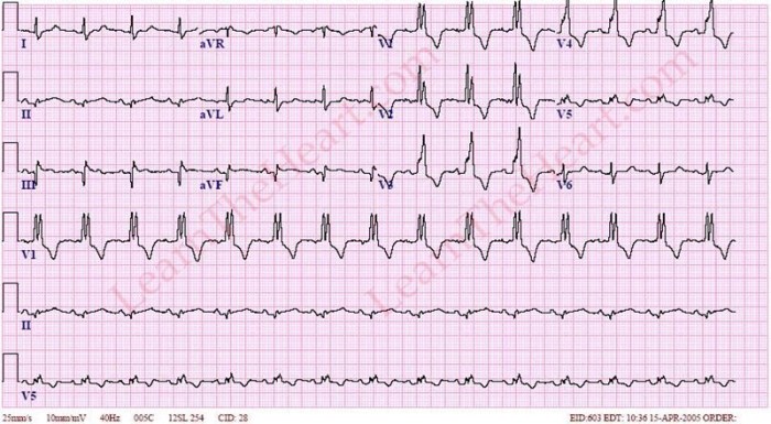
Diagnosing RBBB Using an ECG
Diagnosing RBBB relies heavily on the interpretation of an electrocardiogram (ECG). The characteristic ECG features of RBBB include a widened QRS complex, usually greater than 0.12 seconds, and specific changes in the morphology of the QRS complex’s terminal portion. The right bundle branch depolarization pattern is delayed, causing the characteristic appearance on the ECG. The specific features are a dominant R wave in V1, and a slurred or notched S wave in V1 and V2.
A careful and systematic analysis of the ECG tracing is necessary for accurate diagnosis.
ECG Findings in RBBB
| Symptom | Frequency | Severity | Clinical Significance |
|---|---|---|---|
| Palpitations | Variable | Mild to moderate | May indicate underlying cardiac arrhythmia or other conditions. |
| Dizziness | Variable | Mild to severe | Suggests potential rhythm disturbances or other circulatory issues. |
| Shortness of Breath | Variable | Mild to severe | Could indicate underlying heart disease or pulmonary issues. |
| Chest Pain | Variable | Mild to severe | Requires immediate evaluation as it could indicate acute myocardial infarction (heart attack) or other cardiac conditions. |
| Fatigue | Variable | Mild to moderate | May be related to underlying heart conditions or other factors. |
ECG Characteristics of RBBB
Right bundle branch block (RBBB) is a common conduction abnormality of the heart. It’s characterized by a delay in the electrical impulse as it travels through the right bundle branch, affecting the ventricular depolarization. Understanding the ECG characteristics of RBBB is crucial for accurate diagnosis and appropriate management.
ECG Findings in RBBB
The characteristic ECG findings in RBBB arise from the prolonged conduction time through the right ventricle. This results in specific patterns in various leads that distinguish it from other cardiac conditions. Key features include a widened QRS complex and specific morphology changes. The morphology of the QRS complex is a fundamental factor in recognizing RBBB on an ECG.
Key Features Distinguishing RBBB
Several features on the ECG differentiate RBBB from other conditions. A prolonged and widened QRS complex is a primary indicator. The pattern of this widening is typically distinct from other conduction disturbances. The morphology of the QRS complex, particularly in leads like V1 and V2, is crucial for diagnosis. Careful analysis of the RSR’ or rsR’ pattern in these leads is vital.
Other clues may include a prominent terminal R wave in V1 or V2 and a secondary S wave in V6.
Right bundle branch block (RBBB) is a heart condition that can sometimes be associated with other health issues. Recent advancements in understanding the link between weight and heart health are fascinating, particularly the progress of anti obesity drugs. These advancements are shedding light on how weight management can potentially impact the progression of RBBB. Further research is needed to fully understand the complex relationship between obesity and this heart condition.
Comparison with LBBB
RBBB and left bundle branch block (LBBB) are both significant conduction disturbances. However, they present with different ECG characteristics. The QRS complex widening is a key difference, with RBBB exhibiting a characteristic right-axis deviation. The morphology of the QRS complex in the precordial leads, such as V1 and V2, differs markedly between the two. LBBB typically demonstrates a different pattern of QRS complex widening and morphology, usually with a wider QRS complex and a different morphology in the precordial leads, especially V5 and V6.
ECG Table for RBBB
| Lead | Finding | Description | Significance in RBBB |
|---|---|---|---|
| V1 | RSR’ pattern | A large R wave followed by a small S wave, then a larger R wave. | A hallmark of RBBB, indicative of right ventricular depolarization. |
| V2 | RSR’ pattern or rSR’ pattern | Similar to V1, but the R waves might be smaller. | Further evidence supporting RBBB diagnosis. |
| V5 | Normal or slightly abnormal | The precordial leads on the left side may show a normal or slightly abnormal pattern. | This provides a comparison to the right-sided leads (V1-V2), reinforcing the diagnosis. |
| V6 | Slightly abnormal S wave | A small or slightly abnormal S wave in lead V6. | Consistency with the characteristic pattern of right-sided ventricular depolarization. |
| I, aVL | Normal or slightly abnormal | Leads reflecting left-sided ventricular activity often show normal or minimally abnormal characteristics. | Provides contrast with the significant changes observed in right-sided leads. |
Recognizing RBBB on an ECG Tracing
To recognize RBBB on an ECG tracing, carefully examine the QRS complex, focusing on the morphology in leads V1 and V2. Look for the characteristic RSR’ or rsR’ pattern. Consider the duration of the QRS complex; it should be widened. Compare the morphology of leads V1-V2 with those in V5-V6 to note the differences. Pay attention to the axis deviation.
These combined observations help in distinguishing RBBB from other cardiac conditions.
Underlying Causes and Risk Factors
Right bundle branch block (RBBB) isn’t always a cause for immediate concern, but it can be a marker for underlying heart conditions. Understanding the potential causes and risk factors associated with RBBB is crucial for proper diagnosis and management. This section delves into the various conditions linked to RBBB, focusing on common causes, associated risk factors, and the influence of age on its occurrence.Various factors can contribute to the development of RBBB, ranging from relatively benign conditions to more serious underlying heart diseases.
Identifying these causes is vital for appropriate patient management and potential interventions.
Common Causes of RBBB
RBBB can arise from a multitude of conditions, impacting the electrical pathways in the heart. Some causes are structural, while others are related to the function of the heart muscle. Common causes include:
- Structural heart disease: Conditions affecting the heart’s structure can disrupt the normal electrical conduction, leading to RBBB. Examples include valvular heart disease, congenital heart defects, and cardiomyopathies. These structural abnormalities can physically obstruct or alter the pathways of electrical impulses within the heart.
- Myocardial ischemia: Reduced blood flow to the heart muscle, often due to coronary artery disease, can cause RBBB. The lack of oxygen and nutrients can damage the heart muscle, affecting its electrical conduction system.
- Pulmonary hypertension: Increased pressure in the blood vessels of the lungs can lead to RBBB. This condition can strain the heart and disrupt its electrical activity.
- Heart conditions associated with aging: The natural aging process can sometimes lead to subtle changes in the heart’s electrical system, which can manifest as RBBB. This is often associated with the development of other conditions, such as left ventricular hypertrophy, which are more prevalent in older populations.
- Other conditions: Conditions such as infiltrative diseases, pericarditis, and some forms of chronic lung disease can also contribute to RBBB. These conditions can lead to inflammation or damage to the heart muscle or surrounding structures, interfering with normal electrical conduction.
Risk Factors for RBBB Development
Several factors can increase the likelihood of developing RBBB. Identifying these risk factors can aid in proactive screening and monitoring for individuals at higher risk.
- Age: The prevalence of RBBB tends to increase with age. This is likely due to the cumulative effects of aging on the heart’s electrical system, as well as the increased likelihood of developing related conditions such as coronary artery disease.
- Underlying heart conditions: Individuals with a history of structural heart diseases, such as valvular heart disease or congenital heart defects, are at higher risk of developing RBBB.
- High blood pressure: Chronic high blood pressure can strain the heart and contribute to the development of RBBB.
- Smoking: Smoking is a well-established risk factor for cardiovascular diseases, including those that can cause RBBB.
- Diabetes: Diabetes is linked to increased risk of heart conditions and can impact the heart’s electrical conduction.
Age and RBBB Occurrence
The relationship between age and RBBB occurrence is significant. Studies have shown an increase in the incidence of RBBB with advancing age. This is often correlated with the development of other cardiovascular conditions.
Prevalence of RBBB Causes
The following table provides an overview of the various causes of RBBB, their descriptions, and a general estimate of their prevalence. Note that precise prevalence figures can vary depending on the population studied and the specific diagnostic criteria used.
| Cause | Description | Prevalence |
|---|---|---|
| Structural Heart Disease | Conditions affecting the heart’s structure, including valvular defects, congenital issues, and cardiomyopathies. | Moderate to High |
| Myocardial Ischemia | Reduced blood flow to the heart muscle, often due to coronary artery disease. | High |
| Pulmonary Hypertension | Increased pressure in the blood vessels of the lungs. | Moderate |
| Aging-Related Heart Conditions | Natural changes in the heart’s electrical system associated with age. | Moderate |
| Other Conditions | Infiltrative diseases, pericarditis, and some chronic lung diseases. | Variable |
Management and Prognosis
Right bundle branch block (RBBB) is often a benign finding, meaning it doesn’t necessarily cause significant health problems in many individuals. However, its presence can indicate underlying heart conditions, and in such cases, appropriate management is crucial. Understanding the management strategies and potential prognosis is vital for patients and healthcare providers.
Management Approaches
The management of RBBB depends largely on the underlying cause and the presence of any associated symptoms or complications. For asymptomatic individuals with no evidence of heart disease, observation is usually sufficient. However, if RBBB is a symptom of a more significant heart condition, the management plan will focus on addressing the underlying problem. This may include medications, lifestyle modifications, or, in some cases, interventions such as cardiac catheterization or surgery.
Treatment Strategies
Treatment strategies for RBBB are primarily focused on managing the underlying cause. Medical management might include medications to control blood pressure, cholesterol, or heart rate, depending on the specific condition. Lifestyle modifications, such as a heart-healthy diet and regular exercise, are also crucial. In cases of structural heart disease, interventions such as cardiac catheterization or surgical procedures may be necessary.
The goal of treatment is not just to alleviate the symptoms of RBBB, but also to improve overall heart health and prevent potential complications.
Expected Prognosis
The prognosis for patients with RBBB is generally favorable, especially in cases where it’s an isolated finding and there’s no underlying heart disease. However, the prognosis can be affected by the presence of associated conditions. For example, if RBBB is a manifestation of coronary artery disease, the prognosis might be less favorable, depending on the severity and extent of the disease.
Individual outcomes vary, and a thorough evaluation by a cardiologist is essential to determine the specific prognosis for each patient.
Potential Complications
While RBBB itself is not usually life-threatening, it can be a sign of more serious cardiac conditions. These conditions include heart failure, myocardial infarction, and arrhythmias. The potential for these complications highlights the importance of prompt diagnosis and appropriate management. Early detection and intervention can significantly reduce the risk of adverse outcomes.
Treatment Summary Table
| Treatment | Description | Effectiveness | Potential Side Effects |
|---|---|---|---|
| Observation (Asymptomatic Patients) | Regular monitoring and follow-up to assess for any changes or progression. | Generally effective in preventing complications if no underlying condition exists. | Minimal or no side effects. |
| Medical Management (e.g., Beta-blockers, ACE inhibitors) | Medication to control blood pressure, heart rate, or other associated conditions. | Effective in managing underlying conditions, improving symptoms, and reducing risk of complications. | Possible side effects such as dizziness, fatigue, or nausea; specific side effects vary by medication. |
| Cardiac Catheterization/Interventions | Procedures to assess and treat the underlying heart condition. | Effective in addressing structural heart disease and preventing progression. | Potential risks associated with any invasive procedure, including bleeding, infection, or complications related to the specific procedure. |
| Surgery | Surgical procedures to correct structural heart abnormalities. | Effective in treating specific structural problems and improving overall heart health. | Risks associated with major surgery, including infection, bleeding, and potential complications. |
Differential Diagnosis

Right bundle branch block (RBBB) can sometimes mimic other cardiac conditions. Accurate diagnosis relies on careful consideration of both clinical presentation and electrocardiogram (ECG) findings. A thorough understanding of the ECG patterns associated with RBBB and other conditions is crucial for appropriate patient management. Misdiagnosis can lead to delayed or inappropriate interventions, so distinguishing RBBB from other potential causes is essential.
Conditions Mimicking RBBB
Several conditions can present with ECG findings similar to RBBB. These conditions often require careful evaluation to differentiate them from true RBBB. The subtle variations in ECG patterns and clinical context are key to accurate diagnosis.
- Left Anterior Fascicular Block (LAFB): LAFB is a conduction disturbance in the left anterior fascicle of the left bundle branch. Its ECG pattern often shows a characteristic widening of the QRS complex, particularly in the precordial leads, which can be similar to RBBB. However, the presence of a characteristic QRS morphology in leads I, aVL, and V5-V6 distinguishes LAFB from RBBB.
The presence of a left axis deviation is also a differentiating feature.
- Left Posterior Fascicular Block (LPFB): LPFB, another left bundle branch block variant, involves a conduction delay in the left posterior fascicle. The ECG pattern may show a prolonged QRS complex, resembling RBBB, particularly in the inferior leads. A right axis deviation is a crucial indicator, distinguishing it from RBBB which generally shows a normal or slightly leftward axis.
- Intraventricular Conduction Delay (IVCD): This is a broader term encompassing various degrees of conduction delay within the ventricles. The ECG patterns can vary widely, exhibiting prolonged QRS duration, and may sometimes mimic RBBB, but additional clinical factors are needed for proper distinction.
- Acute Myocardial Infarction (AMI): AMI can sometimes cause RBBB, particularly in the context of anterior or inferior infarctions. The ECG changes associated with AMI, such as ST-segment elevation, are critical to distinguish it from isolated RBBB. The presence of chest pain, dyspnea, or other symptoms associated with AMI can also aid in the differential diagnosis.
- Ventricular Preexcitation Syndromes (e.g., Wolff-Parkinson-White syndrome): These conditions often exhibit a characteristic delta wave on the ECG, easily differentiating them from RBBB. The presence of a short PR interval and delta wave is the most important distinguishing feature.
ECG Pattern Comparison
The ECG patterns of RBBB and other conditions can be subtly different, requiring careful analysis. Comparing the characteristic features of each condition can aid in differentiating them. The QRS complex morphology, axis deviation, and presence of other abnormalities are all crucial for distinguishing one condition from another.
| Condition | ECG Pattern | Key Differentiating Features |
|---|---|---|
| Right Bundle Branch Block (RBBB) | Widened QRS complex (>0.12 seconds), rsR’ morphology in V1, and a slightly slurred or notched S wave in V1. Normal or slightly leftward axis. | Absence of other characteristic ECG changes (e.g., ST-segment elevation, inverted T waves), absence of preexcitation pattern. |
| Left Anterior Fascicular Block (LAFB) | Widened QRS complex, characteristic QRS morphology in leads I, aVL, and V5-V6. | Left axis deviation, absence of rsR’ morphology in V1. |
| Left Posterior Fascicular Block (LPFB) | Widened QRS complex, right axis deviation. | Presence of right axis deviation, absence of rsR’ morphology in V1. |
| Acute Myocardial Infarction (AMI) | Widened QRS complex (potentially), ST-segment elevation, T-wave inversion. | Presence of ST-segment elevation, T-wave inversion, or other characteristic AMI changes. |
Special Considerations in Specific Populations
Right bundle branch block (RBBB) can manifest differently across various populations, demanding tailored diagnostic and management approaches. Factors like age, pregnancy status, and underlying comorbidities significantly influence the interpretation of RBBB and the subsequent treatment strategy. Understanding these nuances is crucial for optimal patient care.
Pediatric Patients
Diagnosing RBBB in pediatric patients requires careful consideration of normal variations in the electrocardiogram (ECG) compared to adults. Pediatric hearts are still developing, and subtle changes in conduction pathways can mimic RBBB. The presence of RBBB in a child may be transient or part of a broader congenital heart condition. Thorough evaluation of the child’s medical history, physical examination, and a detailed ECG interpretation are essential.
The management of RBBB in children often involves serial ECG monitoring to assess for any progression of the condition or associated complications. The use of medications to manage underlying conditions is essential, alongside regular follow-ups to evaluate the cardiac status.
Pregnant Women
Pregnancy places additional physiological demands on the cardiovascular system. These changes can influence the ECG appearance and potentially mask or exaggerate underlying cardiac conditions, including RBBB. RBBB in pregnant women often presents as a benign finding, but careful consideration is essential to rule out more serious cardiac complications. The increased blood volume and cardiac output during pregnancy may influence the conduction system.
Careful monitoring and management, focusing on the underlying cause and its impact on the pregnancy, are necessary.
Elderly Patients
Age-related changes in the heart, such as fibrosis and calcification of the conduction system, can lead to RBBB in elderly patients. The presence of other age-related conditions, such as hypertension or coronary artery disease, needs careful consideration. A comprehensive geriatric assessment, including evaluation of cognitive function and functional status, is crucial in elderly patients with RBBB. Careful monitoring and management should be tailored to the patient’s overall health and life expectancy.
Patients with Other Comorbidities
RBBB may occur in conjunction with other medical conditions, significantly impacting its management. For example, patients with diabetes or hypertension may experience RBBB as a result of vascular damage or other underlying cardiac conditions. Management strategies for RBBB in patients with comorbidities must address both the RBBB and the associated conditions. Careful monitoring of both the RBBB and the comorbidity is paramount, and interventions should be tailored to the individual patient’s needs.
Table of Special Considerations
| Population | Unique Considerations | Management Strategies |
|---|---|---|
| Pediatric Patients | Normal variations in ECG, potential for transient RBBB, possible association with congenital heart conditions. | Serial ECG monitoring, evaluation for underlying conditions, and regular follow-ups. Consideration of medication management for underlying conditions. |
| Pregnant Women | Physiological changes during pregnancy can influence ECG appearance, potential for masking underlying cardiac conditions. | Careful monitoring, consideration of underlying causes, and tailored management strategies that address the pregnancy and the RBBB. |
| Elderly Patients | Age-related changes in the heart (fibrosis, calcification), presence of other age-related conditions. | Comprehensive geriatric assessment, monitoring for complications, and management tailored to the patient’s overall health and life expectancy. |
| Patients with Comorbidities | RBBB may be a manifestation of underlying conditions like diabetes or hypertension. | Management of both RBBB and associated comorbidities. Careful monitoring of both conditions is essential. |
Illustrative Case Studies
Right Bundle Branch Block (RBBB) is a relatively common cardiac condition. While often asymptomatic, RBBB can be a marker of underlying heart disease or a consequence of other conditions. Understanding the diverse presentations and potential outcomes of RBBB is crucial for clinicians to provide appropriate management. Illustrative case studies help to contextualize the diagnosis, treatment, and prognosis, highlighting the importance of clinical judgment in patient care.RBBB case studies often reveal a spectrum of presentations, from entirely asymptomatic individuals to those experiencing symptoms related to underlying heart conditions.
Careful analysis of patient history, physical examination, and electrocardiographic findings is paramount in determining the appropriate course of action. These cases highlight the importance of considering both the immediate and long-term implications for the patient’s health.
Case Study Examples
A crucial aspect of understanding RBBB is the variability in its presentation. Different underlying causes can influence the severity and prognosis. The following table presents illustrative case studies that demonstrate this variability.
| Case Study | Patient Characteristics | Clinical Outcome |
|---|---|---|
| Case 1: Asymptomatic RBBB | A 55-year-old male with a history of hypertension and occasional chest pain, but no prior cardiac events. Electrocardiogram (ECG) revealed RBBB without any accompanying symptoms. | The patient was monitored closely. Further cardiac testing was negative for significant underlying heart disease. The patient remained asymptomatic and was advised on lifestyle modifications to manage his hypertension. Long-term follow-up demonstrated stable RBBB with no adverse events. |
| Case 2: RBBB associated with Coronary Artery Disease (CAD) | A 68-year-old female presenting with exertional dyspnea and chest pain. ECG showed RBBB and ST-segment changes suggestive of ischemia. Cardiac catheterization revealed significant coronary artery stenosis. | The patient underwent percutaneous coronary intervention (PCI) to restore coronary blood flow. The RBBB persisted after the procedure, but the patient’s symptoms improved significantly. Long-term management focused on cardiac rehabilitation and medication to control risk factors. |
| Case 3: RBBB in a Young Athlete | A 22-year-old male athlete with a sudden onset of palpitations and dizziness. ECG showed RBBB. The patient had a family history of sudden cardiac death. | Further cardiac evaluation, including a comprehensive cardiac work-up, did not reveal any structural abnormalities. The patient’s symptoms resolved, and he continued to participate in athletics. Ongoing cardiac monitoring was implemented. Genetic testing for inherited cardiac conditions was considered. |
Importance of Clinical Judgment
Recognizing the variability in RBBB cases is crucial. Clinical judgment plays a pivotal role in evaluating patients with RBBB. A thorough history, physical examination, and ECG interpretation are essential for determining the underlying cause and risk stratification. Management strategies should consider the patient’s overall health, lifestyle, and the presence of associated conditions.This requires a careful consideration of all the available information, including the patient’s age, medical history, symptoms, and the results of any diagnostic tests.
It is critical to understand that RBBB can be an isolated finding or a marker of more significant cardiac disease. The decision regarding further investigations and treatment should be individualized.
Right bundle branch block (RBBB) is a heart condition that can sometimes be a bit concerning. While it’s not directly related to getting sick with mono, understanding the nuances of conditions like RBBB can be fascinating. Learning about the causes and potential treatments of RBBB is valuable, and if you’re curious about whether you can get mono twice, it’s definitely worth checking out some reliable sources like this article on can you get mono twice.
Ultimately, RBBB requires careful monitoring and, if necessary, medical intervention.
Advanced Topics (Optional)
Diving deeper into right bundle branch block (RBBB) reveals fascinating complexities beyond the basic ECG findings. Understanding the role of electrophysiology, the latest research on pathophysiology, and advanced treatment techniques provides a more comprehensive picture of this condition. This section will explore these advanced aspects, offering insights into the nuances of RBBB management.Electrophysiological studies play a critical role in evaluating the intricacies of RBBB.
These studies directly measure the electrical activity of the heart, offering a precise assessment of conduction abnormalities and their impact on cardiac function. They provide detailed information about the timing and sequence of electrical impulses, enabling a more nuanced understanding of the underlying mechanisms contributing to RBBB.
Electrophysiological Studies in Evaluating RBBB
Electrophysiological studies (EPS) are crucial in evaluating the underlying mechanisms of RBBB. These studies use catheters to record electrical activity within the heart chambers, providing a detailed picture of conduction pathways. By mapping the electrical activity, EPS can identify specific sites of conduction delay or block, thereby revealing the precise location of the conduction disturbance associated with RBBB.
This detailed assessment is especially valuable in cases where the cause of RBBB is uncertain or where more targeted treatment strategies are required. For example, in cases of suspected congenital heart conditions or when evaluating the suitability of specific pacing strategies.
Latest Research on the Pathophysiology of RBBB
Recent research has shed light on the intricate mechanisms behind RBBB development. Researchers are exploring the role of various factors, including fibrosis, inflammation, and autonomic nervous system dysfunction, in contributing to the block in the right bundle branch. Understanding these pathophysiological mechanisms is essential for developing more targeted and effective therapeutic interventions. For instance, studies suggest that inflammation might play a role in the progression of RBBB, and anti-inflammatory therapies could potentially slow its progression.
Advanced Techniques Used to Treat RBBB
Beyond traditional pacing therapies, advanced techniques are being investigated and applied to address RBBB. These techniques focus on restoring normal electrical conduction pathways, often through minimally invasive procedures. Radiofrequency ablation, for example, is a technique that targets and eliminates specific areas of abnormal electrical activity. This technique can potentially restore normal conduction patterns, reducing the need for long-term pacing.
Different Types of Pacing Procedures
Various pacing procedures are available to manage RBBB, each with its own advantages and considerations. Understanding these different types is crucial for selecting the most appropriate approach for individual patients.
- Permanent Pacemakers: These devices are implanted to provide electrical stimulation to the heart, ensuring a consistent heart rhythm. Different types of permanent pacemakers cater to various pacing needs, ranging from basic rate control to more complex functions.
- Temporary Pacemakers: These devices are used for short-term pacing, providing temporary support during acute situations or procedures. They are commonly used during diagnostic testing or procedures to evaluate cardiac function.
- Cardiac Resynchronization Therapy (CRT): In cases where RBBB is associated with left ventricular dysfunction, CRT may be considered. This therapy involves pacing both ventricles to improve cardiac output and reduce symptoms of heart failure. CRT devices are often combined with other pacing systems to deliver coordinated electrical signals to the heart.
Summary: Right Bundle Branch Block Rbbb
In conclusion, right bundle branch block (RBBB), while often not life-threatening, warrants careful evaluation and management. Recognizing the characteristic ECG patterns, understanding the underlying causes, and implementing appropriate treatment strategies are crucial for optimal patient outcomes. This guide has provided a comprehensive overview, from the basic to the more advanced aspects of RBBB, offering a thorough understanding of this important cardiac condition.