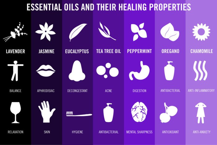
Blog

  • Why Do People Die in Their Sleep? Unveiling the Causes

    Why Do People Die in Their Sleep? Unveiling the Causes

    Why do people die in their sleep? This question touches upon a deeply unsettling aspect of human life, prompting us to explore the various factors that can contribute to sudden death during slumber. From underlying medical conditions to sleep disorders, drug interactions, and environmental influences, the causes are multifaceted and often complex. Understanding these factors is crucial for raising awareness and potentially preventing such tragedies.

    This exploration delves into the intricate interplay of medical conditions, sleep disturbances, and external factors that can lead to sleep-related fatalities. We’ll examine the role of genetics, lifestyle choices, and even environmental conditions in increasing the risk of death during sleep. By understanding these factors, we can better appreciate the importance of preventative measures and early intervention.

    Underlying Medical Conditions

    Sudden death during sleep, though often alarming, can be linked to various underlying medical conditions. Understanding these conditions is crucial for recognizing potential risks and implementing preventative measures. Early detection and appropriate medical interventions can significantly reduce the likelihood of such tragic events.

    Common Medical Conditions Increasing Sleep-Related Death Risk

    Several medical conditions can increase the risk of sudden death during sleep. These conditions often involve disruptions to the heart’s rhythm, breathing patterns, or the body’s ability to regulate essential functions during sleep.

    • Cardiovascular Diseases: Conditions like coronary artery disease, heart failure, and arrhythmias can compromise the heart’s ability to pump blood effectively. Disruptions in blood flow, especially during sleep when the body is at rest, can lead to sudden cardiac arrest. For example, a patient with undiagnosed coronary artery disease might experience a fatal arrhythmia during sleep, interrupting the normal rhythm and causing the heart to stop beating.

      Early detection and management of cardiovascular conditions through regular check-ups and lifestyle modifications can greatly reduce the risk.

    • Respiratory Conditions: Obstructive sleep apnea (OSA) is a common sleep disorder characterized by pauses in breathing during sleep. These pauses can reduce the amount of oxygen reaching the brain and other vital organs. In severe cases, OSA can lead to cardiac arrhythmias and, in some cases, sudden death. Asthma and other respiratory illnesses can also increase the risk of sleep-related deaths due to impaired breathing and decreased oxygen levels.

      For instance, an individual with untreated asthma experiencing a severe attack during sleep might struggle to breathe, leading to oxygen deprivation and potentially fatal consequences.

    • Neurological Conditions: Some neurological disorders, such as stroke, can disrupt the brain’s control over vital functions, including breathing and heart rate. A stroke affecting areas controlling breathing or the cardiovascular system during sleep can lead to fatal complications. Furthermore, certain neurological conditions can directly impact the heart’s rhythm and function, resulting in sudden cardiac arrest. For example, a patient with a history of stroke might experience a fatal arrhythmia triggered by sleep-related changes in blood pressure and oxygen levels.

    Importance of Routine Check-ups and Preventative Measures

    Regular medical check-ups play a vital role in identifying and managing underlying conditions that increase the risk of sleep-related fatalities. Routine screenings can detect potential problems early, allowing for prompt interventions and preventive measures. This proactive approach can significantly reduce the likelihood of sudden death during sleep.

    • Lifestyle Modifications: Maintaining a healthy lifestyle, including a balanced diet, regular exercise, and avoiding smoking, can significantly reduce the risk of various medical conditions. For instance, adopting a heart-healthy diet can reduce the risk of cardiovascular diseases, a major contributor to sleep-related deaths.
    • Medication Adherence: Proper adherence to prescribed medications is essential for managing underlying conditions and minimizing complications. Consistent medication use helps to control blood pressure, cholesterol levels, and other factors associated with cardiovascular and respiratory diseases. Furthermore, it is important to consult with a healthcare provider if there are concerns about side effects or drug interactions.
    • Sleep Hygiene Practices: Establishing healthy sleep habits, such as maintaining a regular sleep schedule, creating a conducive sleep environment, and avoiding caffeine and alcohol before bedtime, can promote better sleep quality and reduce the risk of sleep disorders like OSA.

    Risk Assessment Table

    Condition Risk Factors Symptoms Preventive Measures
    Cardiovascular Disease High blood pressure, high cholesterol, smoking, obesity Chest pain, shortness of breath, dizziness Regular check-ups, medication adherence, healthy diet, exercise
    Obstructive Sleep Apnea (OSA) Obesity, large neck circumference, family history Loud snoring, gasping for air during sleep, daytime sleepiness Weight loss, CPAP therapy, lifestyle modifications
    Neurological Disorders History of stroke, brain injury, neurological conditions Headache, weakness, numbness, loss of coordination Regular check-ups, medication adherence, management of underlying conditions

    Sleep Disorders: Why Do People Die In Their Sleep

    Sleep, a fundamental human need, is often overlooked as a crucial factor in overall health. Beyond simply feeling rested, adequate sleep plays a vital role in regulating numerous bodily functions, including breathing, heart rate, and hormone production. Disruptions to this delicate balance, often stemming from sleep disorders, can increase the risk of serious health problems, some of which can lead to sudden death.

    Sleep Apnea and Sudden Death

    Sleep apnea, characterized by pauses in breathing during sleep, is a serious sleep disorder. These pauses, called apneas, can drastically reduce the amount of oxygen reaching the brain and body. Repeated episodes of oxygen deprivation over time can strain the cardiovascular system, increasing the risk of high blood pressure, heart disease, and stroke. Furthermore, the constant disruption of normal breathing patterns during sleep can trigger irregular heart rhythms, potentially leading to sudden cardiac death.

    The risk is particularly elevated in individuals with pre-existing cardiovascular conditions. A significant number of sudden unexplained deaths have been linked to sleep apnea.

    Impact of Other Sleep Disorders

    Beyond sleep apnea, other sleep disorders can also negatively affect sleep-related mortality. Narcolepsy, a neurological disorder characterized by excessive daytime sleepiness and sudden attacks of sleep, can disrupt normal sleep patterns and increase the risk of accidents. Restless legs syndrome, marked by uncomfortable sensations in the legs and an irresistible urge to move them, can lead to poor sleep quality, affecting overall health.

    Both narcolepsy and restless legs syndrome, while not directly causing sudden death in the same way as sleep apnea, can contribute to a decline in health, increasing vulnerability to other life-threatening conditions.

    Disruptions to Breathing Patterns and Heart Function

    Sleep disorders can severely disrupt breathing patterns and heart function during sleep. In sleep apnea, the repeated pauses in breathing cause a significant drop in oxygen levels, placing a strain on the cardiovascular system. This can lead to irregular heart rhythms and high blood pressure, increasing the risk of heart attack or stroke. Narcolepsy can cause sudden, unexpected episodes of muscle weakness, which can compromise breathing.

    Similarly, restless legs syndrome can disrupt sleep, reducing the time spent in restorative deep sleep and leading to fatigue and potential health issues.

    Comparison of Sleep Disorders

    Sleep Disorder Symptoms Diagnostic Procedures
    Sleep Apnea Loud snoring, gasping or choking sounds during sleep, daytime sleepiness, morning headaches, dry mouth, and waking up with a sore throat. Polysomnography (sleep study) to measure breathing patterns, brain activity, heart rate, and oxygen levels during sleep. Medical history and physical examination.
    Narcolepsy Excessive daytime sleepiness, sudden attacks of sleep, cataplexy (sudden loss of muscle tone), sleep paralysis, and hypnagogic hallucinations. Detailed medical history, sleep diaries, multiple sleep latency test (MSLT) to measure the speed of falling asleep during daytime naps, and polysomnography.
    Restless Legs Syndrome Uncomfortable sensations in the legs, an irresistible urge to move the legs, and worsening symptoms at night. Symptoms are often worse in the evening and at rest. Detailed medical history, sleep diaries, physical examination, and a neurological examination to rule out other conditions. Sleep study (polysomnography) may be used to assess the sleep disruption.

    Drug Interactions and Substance Abuse

    Sleep-related deaths can tragically be linked to a complex interplay of factors, including pre-existing conditions and sleep disorders. However, another significant contributor is the interaction of medications and substances with the body’s natural sleep processes. Understanding how various drugs and substances affect respiratory function and sleep patterns is crucial for recognizing potential risks. This section will explore the potential dangers of drug interactions and substance abuse in relation to sleep-related fatalities.Drug interactions can disrupt the delicate balance of physiological processes, particularly during sleep, potentially leading to fatal consequences.

    Sometimes, people die peacefully in their sleep, but other times, underlying health conditions play a role. It’s easy to overlook the toll that caring for someone can take, leading to burnout. This can manifest in various ways, like exhaustion, irritability, or a lack of motivation. Understanding the signs of caregiver burnout is crucial for ensuring both the caregiver and the cared-for individual receive the necessary support.

    Ultimately, a multitude of factors can contribute to unexpected deaths during sleep, highlighting the importance of prioritizing health and well-being.

    This disruption can involve multiple systems, including the respiratory and cardiovascular systems, both of which are crucial for maintaining life during sleep. The effects of drug interactions and substance abuse on sleep-related deaths are often compounded by the individual’s pre-existing health conditions and lifestyle choices.

    Potential Drug Interactions

    Many medications, when taken together or with alcohol, can have unpredictable and potentially dangerous effects on sleep. This includes medications for various conditions, including but not limited to, pain relief, anxiety, and high blood pressure. The risk is amplified when these drugs are taken in combination or at higher dosages than recommended.

    Role of Alcohol and Substance Abuse

    Alcohol significantly impacts sleep architecture, often leading to fragmented sleep and impaired respiratory function. This impairment is particularly dangerous during sleep, as the body’s natural defenses are reduced. Furthermore, combining alcohol with other sedatives or depressants can result in a synergistic effect, increasing the risk of respiratory depression and potentially fatal consequences. Similarly, the abuse of other substances, such as opioids, can have a detrimental impact on respiratory function during sleep.

    Effects of Different Drugs on Sleep Patterns and Respiratory Function, Why do people die in their sleep

    Different drugs affect sleep patterns and respiratory function in various ways. Some drugs, such as benzodiazepines, can promote relaxation and sleep but also depress respiratory function, increasing the risk of sleep apnea and cessation. Opioids, known for their pain-relieving properties, can also depress breathing, especially when combined with other sedatives or alcohol. Stimulants, while often used to combat sleepiness, can lead to insomnia and disrupt sleep patterns, potentially increasing stress on the respiratory system.

    Table of Drugs and Potential Risks During Sleep

    Drug Type Potential Risks During Sleep
    Benzodiazepines Increased risk of respiratory depression, sleep-disordered breathing, and fatal outcomes, especially when combined with alcohol or other sedatives.
    Opioids Significant risk of respiratory depression, potentially leading to cessation during sleep, particularly in combination with other central nervous system depressants.
    Sedatives (e.g., barbiturates) Highly increased risk of respiratory depression, significantly impacting breathing during sleep and posing a life-threatening risk, especially when combined with alcohol or other sedatives.
    Alcohol Disrupts sleep architecture, leading to fragmented sleep, impaired respiratory function, and increased risk of sleep-related death, especially when combined with other sedative substances.
    Stimulants May cause insomnia and disrupt sleep patterns, potentially increasing stress on the respiratory system, but not typically leading to respiratory arrest in isolation.

    External Factors and Environmental Influences

    Sleep-related deaths aren’t always due to internal medical issues. External factors, including environmental conditions and sleep positions, can significantly impact breathing and heart function during sleep, sometimes leading to fatal outcomes. Understanding these external factors is crucial for preventative measures and promoting safer sleep environments.Certain environmental conditions can create an environment that negatively affects the body’s ability to maintain healthy respiration and circulation during sleep.

    This can be particularly dangerous for individuals with underlying health issues or those who are already vulnerable.

    Environmental Conditions Affecting Breathing

    Environmental conditions significantly influence sleep quality and safety. Factors like temperature, humidity, and even air quality can alter breathing patterns and heart rate, potentially leading to respiratory distress and sleep-related fatalities. For instance, extremely cold temperatures can constrict airways, making breathing more difficult. Conversely, excessive heat can lead to hyperthermia, further compromising breathing function. Exposure to allergens or pollutants in the air can exacerbate existing respiratory conditions or trigger allergic reactions during sleep.

    Sleep Positions and Their Impact

    Sleep positions can also play a critical role in sleep-related deaths. Certain positions can obstruct breathing pathways, particularly for individuals who are predisposed to sleep apnea or respiratory issues. For example, sleeping on the stomach can compress the chest and abdomen, hindering the natural expansion of the lungs. Similarly, sleeping in positions that put pressure on the airways can disrupt breathing patterns, increasing the risk of sleep apnea and related complications.

    It is also important to note that infants and children are particularly vulnerable to positional asphyxia, emphasizing the need for safe sleep practices.

    Impact of Temperature, Humidity, and Other Factors

    Temperature fluctuations, humidity levels, and even the presence of certain substances in the air can impact the body’s physiological responses during sleep. For example, extreme temperatures can trigger physiological responses that interfere with breathing and heart function. High humidity can also affect respiratory function and increase the risk of respiratory infections, potentially exacerbating sleep-related problems. Additionally, the presence of irritants or pollutants in the air can negatively affect breathing and cardiovascular function.

    Summary Table of External Factors and Their Potential Effects

    External Factor Potential Effect on Sleep
    Extreme Temperatures (High/Low) Increased risk of hyperthermia or hypothermia, affecting breathing and heart rate.
    High Humidity Increased risk of respiratory infections, potential interference with breathing.
    Air Quality (Pollutants, Allergens) Exacerbation of existing respiratory conditions, triggering allergic reactions, and affecting breathing.
    Sleep Positions (e.g., Stomach Sleeping) Potential airway obstruction, hindering breathing and oxygen intake.
    Exposure to Irritants/Substances Negative impact on respiratory and cardiovascular function.

    Genetic Predisposition and Family History

    Understanding sleep-related deaths often requires looking beyond immediate factors. Genetic predisposition plays a significant role, influencing an individual’s susceptibility to certain conditions that can lead to unexpected sleep-related mortality. A family history of sudden death, particularly in seemingly healthy individuals, can signal an underlying genetic vulnerability that warrants further investigation.Family history of sudden death, especially in young or seemingly healthy individuals, can serve as a crucial warning sign.

    This heightened risk often points to a genetic component that may predispose individuals to a range of sleep-related disorders or conditions. While environmental and lifestyle factors are crucial, a genetic predisposition can significantly increase the chances of such occurrences.

    Genetic Factors in Sleep-Related Deaths

    Genetic factors can contribute to a range of sleep disorders and conditions that increase the risk of sleep-related mortality. Variations in genes can affect the function of the heart, respiratory system, and brain, making individuals more prone to abnormalities during sleep. These variations can be passed down through generations, creating a familial pattern of sudden death.

    Examples of Genetic Conditions

    Certain genetic conditions are directly linked to sleep-related deaths, impacting the body’s ability to regulate vital functions during sleep. These conditions can manifest subtly, and individuals may not exhibit any symptoms during wakefulness.

    • Long QT Syndrome: This genetic disorder affects the electrical activity of the heart. Prolonged QT intervals can increase the risk of sudden cardiac arrest, often occurring during sleep. Examples include cases where young athletes unexpectedly collapse during exercise or sleep, or individuals with a family history of sudden cardiac arrest without clear cause.
    • Arrhythmogenic Right Ventricular Cardiomyopathy (ARVC): This condition leads to structural abnormalities in the heart, particularly the right ventricle. These abnormalities can disrupt the heart’s rhythm, potentially causing fatal arrhythmias during sleep. Individuals with a family history of unexplained cardiac events, especially those involving the right ventricle, should be evaluated for this condition.
    • Sudden Infant Death Syndrome (SIDS): While the exact cause of SIDS remains largely unknown, some genetic predispositions may play a role in the vulnerability to this condition, especially in certain ethnic groups. This is a crucial area of ongoing research. A family history of SIDS in multiple children can suggest a need for genetic testing and careful monitoring of subsequent children.

    Table of Genetic Conditions Associated with Sleep-Related Deaths

    The following table Artikels some genetic conditions associated with sleep-related deaths, highlighting their potential impact on cardiac function and the importance of familial history assessment.

    Sometimes, people die in their sleep due to underlying conditions. While a seemingly unrelated issue like shoulder pain when throwing shoulder pain when throwing might not directly cause death, it can be a symptom of more serious health problems. These issues, if left unchecked, can contribute to unexpected fatalities during sleep. So, while sleep is often a time of rest, it’s crucial to address potential health concerns to improve your overall well-being.

    Genetic Condition Potential Impact on Sleep Impact on Mortality
    Long QT Syndrome Disrupts heart’s electrical activity, potentially causing fatal arrhythmias during sleep. Increased risk of sudden cardiac arrest, often during sleep.
    Arrhythmogenic Right Ventricular Cardiomyopathy (ARVC) Structural abnormalities in the heart disrupt rhythm, potentially causing fatal arrhythmias during sleep. Increased risk of sudden cardiac arrest, often during sleep.
    Familial Hypertrophic Cardiomyopathy Thickening of heart muscle can interfere with blood flow and potentially lead to abnormal heart rhythms during sleep. Increased risk of sudden cardiac arrest, especially during strenuous physical activity or stress, but also potentially in sleep.

    Age and Life Stage

    The delicate balance of life, from infancy to old age, often impacts the susceptibility to sleep-related deaths. Different stages of life present varying vulnerabilities due to developmental factors, physiological changes, and environmental exposures. Understanding how age influences sleep-related fatalities is crucial for preventative measures and appropriate interventions.The risk of sleep-related deaths is not uniform across all age groups.

    Factors like the immaturity of the nervous system in infants, the ongoing development of the body and brain in children, and the physiological changes associated with aging in adults and the elderly all contribute to the distinct patterns of sleep-related fatalities.

    Infancy

    Infant mortality is unfortunately a significant concern, and sleep-related deaths in this stage are often associated with Sudden Infant Death Syndrome (SIDS). Factors like unsafe sleep environments, soft bedding, and overheating contribute to this risk. The immaturity of the infant’s nervous system, respiratory system, and thermoregulation mechanisms make them vulnerable to disruptions in breathing during sleep. Premature babies and those with underlying health conditions are at an even higher risk.

    Childhood

    Childhood sleep-related deaths, though less common than in infancy, can be associated with underlying medical conditions, sleep disorders, or accidental suffocation. Children may experience sleep apnea, which can disrupt breathing during sleep. They might also be susceptible to accidents if the sleep environment is not adequately monitored or safe. Furthermore, conditions like certain types of epilepsy or heart conditions can increase risk.

    Adulthood

    Sleep-related deaths in adulthood encompass a wider range of potential causes. Cardiovascular conditions, sleep apnea, and drug interactions or substance abuse can all play significant roles. As adults age, the risk of developing conditions like heart failure or stroke increases, and these can manifest as sleep-related deaths. Sleep disorders such as sleep apnea, narcolepsy, or restless leg syndrome can increase vulnerability.

    Old Age

    In older adults, sleep-related deaths can be linked to various factors. Chronic conditions, like heart disease, respiratory issues, or neurological disorders, are more prevalent. These conditions can increase the risk of respiratory problems during sleep. Additionally, medications or interactions between medications can cause adverse reactions, leading to sleep-related fatalities. Cognitive decline and mobility issues can also contribute to sleep-related risks in this age group.

    Correlation Between Age and Sleep-Related Deaths

    Age Group Potential Causes Specific Examples
    Infancy (0-1 year) SIDS, unsafe sleep environments, underlying medical conditions, prematurity Improper bedding, overheating, respiratory issues
    Childhood (1-18 years) Sleep apnea, suffocation, underlying medical conditions, accidents Sleep disorders, unsafe sleep environments, accidental suffocation
    Adulthood (19-64 years) Cardiovascular conditions, sleep apnea, drug interactions, substance abuse, underlying medical conditions Heart disease, stroke, sleep apnea, prescription drug interactions, alcohol abuse
    Old Age (65+ years) Chronic conditions, medications, cognitive decline, mobility issues, respiratory issues Heart failure, respiratory problems, neurological disorders, medication side effects

    Lifestyle Factors

    Why tho? - 9GAG

    Our daily habits profoundly impact our sleep quality and, consequently, our risk of sleep-related deaths. A balanced lifestyle, encompassing diet, exercise, and stress management, plays a crucial role in maintaining healthy sleep patterns. Poor sleep hygiene, in turn, can contribute to various health problems, increasing vulnerability to sleep-related complications. Understanding the link between lifestyle choices and sleep-related mortality is essential for preventative measures.

    The Influence of Diet on Sleep

    Diet significantly influences sleep quality. Consuming excessive amounts of caffeine, alcohol, and processed foods can disrupt sleep patterns. Conversely, a balanced diet rich in fruits, vegetables, and whole grains can promote better sleep. Foods high in magnesium and tryptophan, crucial for melatonin production, are beneficial for sleep. For example, a diet rich in sugar can lead to energy crashes and subsequent difficulty falling asleep.

    Similarly, a late-night dinner laden with heavy proteins or fats can create digestive discomfort and impede sleep. A balanced diet is key to maintaining stable blood sugar levels, promoting restful sleep.

    The Importance of Regular Exercise

    Regular physical activity is crucial for overall health, and it significantly impacts sleep quality. Exercise releases endorphins, which have mood-boosting effects. However, exercising too close to bedtime can energize the body, making it difficult to fall asleep. Finding a suitable time for exercise, ideally several hours before bedtime, is essential for optimal sleep. For instance, a vigorous workout right before sleep can interfere with sleep onset and duration.

    Conversely, regular exercise, especially aerobic activity, has been shown to improve sleep quality and duration in many studies.

    Stress Management and Sleep

    Chronic stress is a significant contributor to sleep disturbances. Stress hormones like cortisol interfere with the body’s natural sleep-wake cycle. Stress management techniques, such as meditation, yoga, and deep breathing exercises, can help regulate stress levels and improve sleep quality. For instance, individuals experiencing high levels of stress may find it difficult to relax and fall asleep, often resulting in insomnia and other sleep disorders.

    Incorporating stress-reducing activities into daily routines can significantly improve sleep quality and reduce the risk of sleep-related issues.

    Sleep Hygiene and Mortality

    Maintaining good sleep hygiene is paramount for preventing sleep-related mortality. Consistent sleep schedules, a conducive sleep environment, and avoiding excessive screen time before bed are key elements of good sleep hygiene. Individuals who maintain consistent sleep schedules are more likely to experience quality sleep and reduce the risk of sleep-related mortality. For instance, irregular sleep schedules can disrupt the body’s natural circadian rhythm, making individuals more susceptible to sleep-related problems.

    Correlation Between Lifestyle Choices and Sleep-Related Deaths

    Lifestyle Choice Potential Impact on Sleep Potential Impact on Sleep-Related Mortality
    High caffeine intake Difficulty falling asleep, disrupted sleep Increased risk of sleep-related complications
    Irregular sleep schedule Disrupted circadian rhythm, poor sleep quality Increased risk of sleep-related deaths
    High stress levels Difficulty relaxing, insomnia Increased vulnerability to sleep-related complications
    Lack of regular exercise Poor sleep quality, reduced energy levels Potential increase in sleep-related deaths
    Unhealthy diet Digestive issues, poor blood sugar regulation Increased risk of sleep-related issues and health problems

    Illustrative Case Studies

    Unveiling the mysteries surrounding sudden unexpected deaths in sleep requires a deep dive into specific case studies. These real-world examples, while tragic, offer valuable insights into the diverse factors contributing to sleep-related fatalities. Understanding the diagnostic process and outcomes can help medical professionals refine their approaches to prevention and treatment.Analyzing specific cases illuminates the complex interplay of underlying medical conditions, sleep disorders, and external factors that can lead to fatal consequences.

    The detailed accounts highlight the critical role of accurate diagnosis and prompt intervention in mitigating the risk of sleep-related deaths.

    Case Study 1: Obstructive Sleep Apnea

    Obstructive sleep apnea (OSA) is a prevalent sleep disorder characterized by pauses in breathing during sleep. These pauses can lead to a dangerous drop in blood oxygen levels and disrupt the body’s natural restorative processes.This case involves a 55-year-old male with a history of hypertension and obesity. He presented with progressively worsening sleepiness and daytime fatigue. Sleep studies revealed severe OSA, with frequent apneic episodes and low oxygen saturation.

    Sudden death in sleep can be a mystery, often linked to underlying health issues. Factors like high blood pressure and, significantly, cholesterol heart disease, particularly prevalent in African Americans, cholesterol heart disease and african americans , can contribute to these tragic events. This highlights the importance of regular check-ups and proactive health management to minimize such risks.

    Despite lifestyle modifications and CPAP therapy, the patient experienced a sudden cardiac arrest during sleep. Autopsy confirmed the presence of severe cardiac fibrosis and significant pulmonary hypertension, directly attributable to the chronic sleep apnea. The diagnostic process involved a thorough medical history, physical examination, and polysomnography. The outcome was a tragic and untimely death, highlighting the importance of early diagnosis and aggressive management of OSA.

    Case Study 2: Underlying Cardiovascular Disease

    Certain underlying cardiovascular conditions can increase the risk of sudden death during sleep. These conditions may manifest with subtle symptoms or remain undiagnosed until a critical event.A 68-year-old female with a history of coronary artery disease, hypertension, and diabetes presented with intermittent chest pain and shortness of breath. She reported experiencing palpitations and lightheadedness before the incident. An electrocardiogram (ECG) and cardiac enzyme tests were performed.

    While the ECG revealed some abnormalities, the results were not immediately alarming. However, further investigation and analysis of her medical history revealed a significant risk for cardiac events. Sadly, she succumbed to a cardiac arrhythmia during sleep. The diagnostic process focused on her symptoms, medical history, and cardiac assessments. The outcome underscored the need for careful monitoring and prompt intervention for patients with known cardiovascular risk factors.

    Case Study 3: Drug-Induced Respiratory Depression

    The interaction of medications with natural sleep processes can sometimes have fatal consequences. Patients taking certain medications may be at a heightened risk of respiratory depression, leading to cessation of breathing during sleep.A 70-year-old male taking a combination of opioid painkillers and sedatives for chronic pain experienced sudden respiratory arrest during sleep. The patient’s medical history revealed a history of chronic pain and pre-existing respiratory conditions.

    Initial investigations indicated a probable interaction between the prescribed medications, leading to respiratory depression. The diagnostic process included analyzing the patient’s medication regimen, reviewing medical history, and performing blood gas analysis. The outcome emphasized the importance of careful medication management and close monitoring in patients with pre-existing respiratory conditions.

    Summary of Case Studies

    Case Study Patient Demographics Medical History Diagnostic Process Outcome
    1 55-year-old male Hypertension, obesity Sleep studies, medical history Sudden cardiac arrest
    2 68-year-old female Coronary artery disease, hypertension, diabetes ECG, cardiac enzyme tests, medical history Cardiac arrhythmia
    3 70-year-old male Chronic pain, pre-existing respiratory conditions Medication review, blood gas analysis Respiratory arrest

    Prevention Strategies

    Why do people die in their sleep

    Reducing the risk of sleep-related deaths requires a multi-faceted approach that addresses both individual and societal factors. Proactive measures, combined with increased public awareness, can significantly lower the incidence of these tragic events. This involves understanding the potential contributing factors and implementing strategies to mitigate their impact.

    Public Awareness Campaigns and Education Programs

    Effective prevention relies heavily on educating the public about the risks associated with various sleep-related conditions and behaviors. Public awareness campaigns play a crucial role in disseminating information about recognizing the warning signs and taking preventive measures. These campaigns can highlight the importance of maintaining healthy sleep hygiene, identifying potential sleep disorders, and seeking professional help when necessary. They can also address misconceptions surrounding sleep and encourage individuals to prioritize sleep health.

    Targeted educational programs can be delivered in schools, workplaces, and community centers, reaching a wide range of demographics. For example, a campaign focusing on sleep apnea in older adults could emphasize the connection between untreated sleep apnea and increased risk of cardiovascular issues.

    Early Detection and Intervention in Sleep Disorders

    Prompt identification and treatment of sleep disorders are vital for preventing sleep-related deaths. Early detection allows for intervention, reducing the progression of potentially life-threatening conditions. Recognizing the symptoms of sleep disorders, such as snoring, gasping for air during sleep, daytime sleepiness, and difficulty concentrating, is crucial. This early identification is often the key to successful treatment and the prevention of severe consequences.

    A thorough sleep study by a qualified professional can accurately diagnose sleep disorders, including conditions like sleep apnea and narcolepsy, enabling appropriate treatment plans to be implemented. For example, an early diagnosis of sleep apnea could lead to the use of a CPAP machine, which significantly reduces the risk of sleep-related complications.

    Preventive Measures and Lifestyle Adjustments

    Maintaining healthy sleep habits and lifestyle choices can significantly reduce the risk of sleep-related deaths. These include establishing a regular sleep schedule, creating a relaxing bedtime routine, optimizing the sleep environment, and avoiding excessive caffeine or alcohol consumption close to bedtime. Also, a balanced diet and regular exercise can contribute to better sleep quality. A structured approach to improving sleep habits, coupled with professional guidance when needed, is essential.

    For example, encouraging regular physical activity in adolescents can lead to better sleep quality and reduced risk of sleep-related breathing problems.

    Examples of Successful Prevention Programs

    Several successful prevention programs have been implemented in various communities and settings. These programs often combine educational components with targeted interventions for specific populations at risk. One program might focus on providing sleep hygiene education in senior centers, while another could partner with schools to promote healthy sleep habits in adolescents. For instance, a program designed to help people with pre-existing medical conditions understand the potential for sleep-related deaths would emphasize the importance of consistent medical monitoring and prompt reporting of symptoms.

    The success of such programs hinges on their adaptability to diverse populations and the involvement of relevant healthcare providers and community organizations. Moreover, ongoing evaluation and adjustment based on data and feedback are critical for maximizing the impact of these prevention initiatives.

    Final Review

    In conclusion, the reasons behind sleep-related deaths are diverse and multifaceted. From underlying medical issues and sleep disorders to drug interactions, environmental factors, and genetic predispositions, a range of factors contribute to this often-unpredictable outcome. While some causes are unavoidable, understanding the contributing factors is vital in developing preventive strategies and promoting public awareness. Early detection and intervention remain crucial for mitigating risks and ultimately saving lives.

  • What to Eat After Food Poisoning A Guide

    What to Eat After Food Poisoning A Guide

    What to eat after food poisoning? This guide provides a comprehensive overview of safe foods to consume after experiencing food poisoning. It delves into the types of food poisoning, immediate actions to take, and dietary considerations. We’ll explore foods to eat and avoid, covering everything from hydration to recovery, and even preventative measures to avoid future incidents.

    From understanding the different types of food poisoning and their symptoms to knowing the crucial steps after suspicion, this guide is your complete resource. We’ll examine the importance of proper nutrition and hydration during recovery and highlight specific dietary needs for different individuals and age groups.

    Understanding Food Poisoning

    Food poisoning, a common ailment, occurs when harmful bacteria, viruses, or parasites contaminate food. This contamination can lead to a range of symptoms, from mild discomfort to severe illness. Understanding the different types, causes, and symptoms is crucial for prevention and proper management.Food poisoning is a serious issue, impacting individuals of all ages. The diverse range of pathogens and contaminated food items make prevention challenging.

    Proper food handling practices and recognizing early warning signs are key to minimizing the risks.

    Definition and Types of Food Poisoning

    Food poisoning, scientifically known as foodborne illness, is a condition resulting from the consumption of contaminated food or water. This contamination can be caused by a variety of factors, including bacteria (like Salmonella, E. coli, and Listeria), viruses (like norovirus), and parasites. Each type of pathogen has unique characteristics and causes different symptoms. For instance, certain bacteria might produce toxins that affect the body in specific ways.

    Common Symptoms of Food Poisoning

    Common symptoms of food poisoning include nausea, vomiting, diarrhea, stomach cramps, and fever. These symptoms typically appear within a few hours to several days after consuming contaminated food. Mild cases may present with only mild nausea and diarrhea, while severe cases can involve severe dehydration, bloody stools, and high fevers, necessitating immediate medical attention. The intensity of symptoms varies depending on the pathogen and the individual’s immune response.

    Importance of Seeking Immediate Medical Attention

    Severe cases of food poisoning require immediate medical attention. Symptoms like persistent high fever, severe dehydration (indicated by decreased urination, dry mouth, and dizziness), bloody stools, or signs of shock warrant immediate medical intervention. Prompt treatment can prevent complications and ensure a faster recovery. Delayed treatment can lead to more serious health problems, including kidney failure in some cases.

    Common Misconceptions about Food Poisoning

    One common misconception is that food poisoning only affects the digestive system. While digestive issues are prominent, other systems can also be affected, potentially leading to more widespread health concerns. Furthermore, the belief that food poisoning is always severe is incorrect. Mild cases are common and resolve without medical intervention. The critical aspect is recognizing the symptoms and taking appropriate measures.

    Table of Common Food Poisoning Types

    Type of Food Poisoning Common Symptoms Typical Duration of Illness
    Salmonella Fever, diarrhea (often bloody), stomach cramps, vomiting 1 to 3 days
    Norovirus Vomiting, diarrhea, stomach cramps, nausea 1 to 3 days
    E. coli O157:H7 Bloody diarrhea, severe stomach cramps, vomiting 2 to 8 days
    Staphylococcus aureus Nausea, vomiting, diarrhea, stomach cramps 1 to 6 hours

    Immediate Actions After Suspecting Food Poisoning

    Food poisoning can be a distressing experience, but swift and appropriate action can significantly reduce its impact. Understanding the immediate steps to take is crucial in mitigating symptoms and preventing further complications. This section details the critical actions to take if you suspect you’ve contracted food poisoning.Proper management of food poisoning often hinges on immediate actions. The initial response can significantly affect the severity and duration of the illness.

    By taking the right steps promptly, you can minimize discomfort and ensure a faster recovery.

    Safety First: Immediate Actions

    A swift and careful approach to handling food poisoning is key to reducing its impact. Immediate actions, like separating yourself from potential sources of further contamination, are vital. This includes isolating yourself from others and taking steps to prevent further contamination of food or surfaces.

    • Isolate yourself to prevent spreading the illness. Limit contact with others, particularly those with weakened immune systems, to minimize the risk of transmission. Avoid handling food or utensils until symptoms subside and the illness has passed. This will help protect others and yourself from further exposure.
    • Thoroughly wash your hands with soap and water for at least 20 seconds. This is paramount for preventing the spread of harmful bacteria. Pay particular attention to washing hands after using the bathroom, before preparing food, and after touching potentially contaminated surfaces.
    • Drink plenty of clear liquids to combat dehydration. Water, clear broths, and electrolyte solutions are excellent choices. Avoid sugary drinks, as they can worsen dehydration.

    Managing Vomiting and Diarrhea

    Vomiting and diarrhea are common symptoms of food poisoning, and effective management is essential. Addressing these symptoms can help restore fluid balance and prevent complications.

    • Avoid solid foods initially. When vomiting and diarrhea occur, the stomach and intestines need time to recover. Stick to clear liquids like water, broth, or electrolyte drinks. Gradually reintroduce bland, easily digestible foods as symptoms subside.
    • Monitor your fluid intake. Dehydration is a significant concern during food poisoning. Regularly monitor your fluid intake and seek medical attention if dehydration becomes severe. Keep track of your fluid intake and observe for signs of dehydration, such as dark urine or extreme thirst.

    Foods to Avoid and Those to Consider

    Certain foods can exacerbate food poisoning symptoms. Avoiding specific foods during the illness is essential for promoting recovery.

    • Avoid greasy, spicy, or acidic foods. These can irritate the stomach and worsen digestive discomfort. Choose bland foods that are easy to digest and do not contain any ingredients that could worsen your symptoms.
    • Consider BRAT diet options. A BRAT diet (Bananas, Rice, Applesauce, Toast) is often recommended for its easily digestible nature. These foods can help soothe the digestive tract and minimize further irritation.

    Comparison of Methods to Manage Vomiting and Diarrhea

    The following table compares different methods for managing vomiting and diarrhea, emphasizing effectiveness and safety.

    Method Effectiveness Safety Considerations
    Clear Liquids (Water, Broth) High High Replenishes lost fluids and electrolytes
    Electrolyte Solutions High High Replaces essential minerals lost through vomiting and diarrhea
    BRAT Diet Moderate High Bland, easily digestible foods that can help soothe the digestive tract
    Medication (Anti-emetics, anti-diarrheals) Variable Moderate Consult a doctor before taking any medication. These medications may not be suitable for all cases and can have side effects.

    Foods to Eat After Food Poisoning: What To Eat After Food Poisoning

    What to eat after food poisoning

    Recovering from food poisoning can be a delicate process. Your digestive system is likely inflamed and sensitive, making certain foods harder to tolerate. Choosing the right foods can significantly aid in your recovery by providing the necessary nutrients without further irritating your system. This guide Artikels the types of foods generally considered safe and easily digestible after food poisoning, emphasizing their nutritional properties and ease of digestion.After experiencing food poisoning, your body needs gentle nourishment.

    So, you’ve got food poisoning? Listen up! Bland foods are key – think toast, crackers, and clear broths. And while you’re recovering, incorporating some gentle exercise like benefits of walking every day can help get your digestive system back on track. Just remember to avoid anything greasy or spicy until you’re feeling better.

    This involves focusing on foods that are low in fiber, easily digestible, and won’t further stress your compromised digestive system. Hydration is crucial for replenishing lost fluids and electrolytes, playing a vital role in your overall recovery.

    Safe and Easily Digestible Foods

    A key aspect of recovery from food poisoning is selecting foods that are gentle on the digestive system. This often involves choosing foods low in fiber and easily absorbed by the body. The following list provides examples of such foods.

    • Plain, cooked rice: Rice is a staple for a reason. Its smooth texture and easily digestible nature make it a perfect choice for a sensitive stomach. It’s also a good source of carbohydrates for energy.
    • Plain, cooked pasta: Similar to rice, pasta provides easily digestible carbohydrates. Opt for plain, unseasoned pasta and avoid sauces with high-fat or fiber content.
    • Well-cooked, plain chicken broth: Chicken broth is gentle on the stomach and provides essential electrolytes lost during diarrhea or vomiting. Avoid broth with excessive spices or added flavorings.
    • Plain yogurt (low-fat): Plain yogurt, especially low-fat, can be soothing to the digestive tract. The probiotics in some yogurts may further aid in restoring gut health.
    • White bread (toast): White bread is easily digestible, but moderation is key. A small piece of toast, lightly buttered, can be a source of simple carbohydrates.
    • Bananas: Bananas are a good source of potassium, an important electrolyte. Their soft texture makes them easy on the digestive system.
    • Applesauce (smooth, unsweetened): The smooth, mashed texture of applesauce is gentle on the stomach. Avoid chunky or sweetened varieties.

    Importance of Hydration and Electrolyte Balance

    Fluid loss through vomiting and diarrhea is a significant concern during food poisoning. Replenishing lost fluids and electrolytes is crucial for restoring normal bodily functions and preventing dehydration.

    • Water: Plain water is essential for hydration. Drink small sips frequently rather than large gulps at once.
    • Electrolyte drinks: Electrolyte drinks can help replenish lost minerals like sodium, potassium, and chloride. Look for ones that are low in sugar and have no added flavors.
    • Broths: Chicken or vegetable broth is a great way to rehydrate and replace lost electrolytes.

    Digestibility and Nutritional Content Comparison

    Different foods have varying levels of digestibility and nutritional value. The following table provides a basic comparison of some of the foods mentioned.

    Food Ease of Digestion Nutritional Value
    Plain Cooked Rice High Good source of carbohydrates
    Plain Cooked Pasta High Good source of carbohydrates
    Plain Yogurt (low-fat) Moderate Source of probiotics and protein
    Well-cooked Plain Chicken Broth High Source of electrolytes and hydration
    Bananas Moderate Good source of potassium

    Foods to Avoid After Food Poisoning

    Your digestive system is already struggling after food poisoning, so it’s crucial to give it a break by avoiding certain foods. This is not about a permanent diet change, but rather a temporary measure to allow your body to recover and repair itself. The key is to choose foods that are gentle on your stomach and easy to digest.The best approach is to start with simple, easily digestible foods and gradually introduce more complex options as your symptoms improve.

    Recovering from food poisoning can be tricky, but focusing on easily digestible foods is key. Bananas and avocados are great options, but don’t forget the importance of replenishing lost electrolytes, particularly potassium. To ensure you’re getting enough of this vital nutrient, consider incorporating foods high in potassium, like sweet potatoes and spinach. These foods help your body recover more quickly after a bout of food poisoning by replenishing essential minerals.

    Look for a comprehensive list of potassium-rich foods at foods high in potassium for more ideas. Ultimately, sticking to bland, easily processed foods will help your stomach settle and get you back on your feet faster.

    Your body will tell you what it needs, so listen to its cues. Avoid anything that causes discomfort or increases nausea.

    Foods That Exacerbate Digestive Discomfort, What to eat after food poisoning

    Foods high in fat, acidity, or spice can significantly irritate the digestive tract when it’s already compromised. This can worsen nausea, vomiting, and diarrhea, delaying recovery.

    • Fatty foods, including fried foods, greasy meats, and high-fat dairy products, can be difficult for the stomach to process, potentially triggering further discomfort and digestive upset.
    • Highly acidic foods, such as citrus fruits (oranges, lemons, grapefruits), tomatoes, and vinegar-based dressings, can irritate the stomach lining and exacerbate existing symptoms.
    • Spicy foods, including chili peppers, hot sauces, and curry dishes, can increase stomach irritation and worsen diarrhea.

    Foods That Can Cause Further Complications

    Certain foods can be more challenging for your system to process after food poisoning. They might contain components that can lead to further complications or delays in recovery.

    • Foods high in fiber, such as beans, lentils, and whole grains, can be difficult to digest when your digestive system is already inflamed or weakened. This can result in gas, bloating, and increased diarrhea.
    • Caffeinated beverages like coffee, tea, and colas can stimulate the digestive system and worsen diarrhea, increasing dehydration risks.
    • Alcohol should be strictly avoided. It can irritate the stomach lining and hinder the body’s ability to recover from the illness.

    Importance of Avoiding Highly Acidic or Spicy Foods

    The stomach lining can be severely inflamed after food poisoning. Highly acidic or spicy foods can exacerbate this inflammation, leading to increased pain, nausea, and discomfort.

    • Acidic foods like citrus fruits, tomatoes, and vinegar-based sauces can irritate the already sensitive stomach lining, making symptoms worse.
    • Spicy foods can increase inflammation and irritation, further intensifying digestive distress.

    Safe vs. Unsafe Foods

    This table provides a simple comparison of foods that are generally safe and those that are best avoided after food poisoning. It’s important to remember that individual responses can vary, and consulting with a healthcare professional is always recommended.

    Safe Foods Unsafe Foods
    Plain boiled chicken breast Fried chicken
    Plain rice Spicy curries
    Bananas Citrus fruits (oranges, lemons)
    Oatmeal High-fat dairy products
    Plain toast Fried foods
    Scrambled eggs (plain) Tomatoes

    Dietary Considerations and Special Cases

    Food poisoning can affect everyone differently, and individuals with specific health conditions or sensitivities may require more careful dietary management during recovery. Understanding these unique needs is crucial for a swift and safe recovery. This section will provide tailored recommendations for managing food poisoning in various circumstances.Dietary needs vary significantly based on underlying health conditions, age, and medications.

    Proper nutrition is essential for recovery, but certain foods can worsen symptoms or even trigger complications. A personalized approach is crucial, and always consulting a doctor for guidance is highly recommended.

    Dietary Considerations for Individuals with Specific Health Conditions

    Individuals with pre-existing conditions, such as diabetes, celiac disease, or kidney problems, must take extra precautions. Dietary modifications during food poisoning are vital to prevent further complications. For instance, someone with diabetes might need to adjust their carbohydrate intake carefully to avoid blood sugar fluctuations. Likewise, someone with celiac disease must avoid gluten-containing foods, which can irritate the digestive tract.

    Individuals with kidney issues need to monitor their protein intake to prevent further strain on their kidneys.

    Managing Food Poisoning in Children and the Elderly

    Children and the elderly are particularly vulnerable to the effects of food poisoning. Children’s developing digestive systems and the elderly’s reduced immune responses make them more susceptible to severe symptoms. It’s crucial to provide appropriate care and dietary support.For children, small, frequent meals of easily digestible foods are recommended. Avoid overly spicy or fatty foods, which can exacerbate discomfort.

    The elderly may require softer, easily chewed foods. Carefully monitor their fluid intake to prevent dehydration. Always consult a pediatrician or geriatrician for personalized advice.

    Importance of Following a Doctor’s Advice

    If you have underlying health conditions, following a doctor’s advice is paramount. Doctors can provide personalized recommendations tailored to your specific needs and health history. This personalized approach ensures that your dietary choices support your recovery while mitigating any potential risks. A healthcare professional can assess your condition and recommend specific foods to avoid or consume, along with any necessary medication.

    Importance of Consulting a Doctor for Personalized Advice

    Ultimately, consulting a doctor is essential for personalized advice. They can evaluate your specific situation, including underlying health conditions, current medications, and the severity of your food poisoning. A doctor can tailor a dietary plan to your needs, ensuring a safe and effective recovery. Avoid self-treating, as this can worsen your condition.

    Dietary Considerations Table

    Age Group/Health Condition Dietary Recommendations
    Children (under 12) Small, frequent meals of easily digestible foods. Avoid spicy or fatty foods. Prioritize fluids to prevent dehydration.
    Elderly (over 65) Soft, easily chewed foods. Monitor fluid intake. Avoid foods that are difficult to digest.
    Individuals with Diabetes Monitor carbohydrate intake closely to prevent blood sugar fluctuations. Consult with a doctor about appropriate carbohydrate choices.
    Individuals with Celiac Disease Strictly avoid gluten-containing foods. Ensure all foods are gluten-free.
    Individuals with Kidney Issues Monitor protein intake to prevent strain on the kidneys. Consult a doctor for specific dietary recommendations.
    Pregnant Women Follow a doctor’s advice closely. Focus on nutrient-rich foods and avoid potentially harmful foods.

    Recovery and Long-Term Effects

    Food poisoning, while typically a temporary ailment, can sometimes leave lingering effects. Understanding the typical recovery time, potential complications, and the importance of proper nutrition is crucial for a swift and complete recovery. This section delves into these aspects, offering guidance for a healthy return to normal life.

    Typical Recovery Time

    The duration of recovery from food poisoning varies significantly depending on the severity of the illness and the specific pathogen involved. Most cases resolve within 24 to 72 hours. Symptoms like nausea, vomiting, and diarrhea usually subside within this timeframe, although fatigue may linger for a few days. A person with a robust immune system and who adheres to the recommended dietary guidelines will likely recover faster.

    In some instances, more severe cases may take longer to resolve, potentially requiring a longer period of rest and recovery.

    After food poisoning, your stomach is likely pretty sensitive. It’s crucial to choose foods that won’t further irritate your system. Bland, easily digestible options are key, like white rice and toast. You might also want to consider how high stomach acid symptoms could be affecting your digestive recovery. High stomach acid symptoms can make eating anything challenging, so start slow and listen to your body.

    Stick to small, frequent meals of easily absorbed foods to help your stomach heal and avoid further discomfort.

    Signs Indicating Need for Further Medical Attention

    Certain signs warrant immediate medical attention. These include persistent high fever (above 101°F or 38.3°C), severe dehydration (dry mouth, sunken eyes, decreased urination), blood in vomit or stool, persistent and severe abdominal pain, and signs of neurological complications like confusion or seizures. If you experience any of these symptoms, seek medical help immediately.

    Potential Long-Term Effects

    While food poisoning typically resolves without lasting health consequences, certain rare cases can lead to more severe complications. Dehydration, if not properly managed, can cause electrolyte imbalances. These imbalances can impact various bodily functions and require medical intervention. Furthermore, certain types of foodborne illnesses, like those caused by

    • Salmonella* or
    • E. coli*, may in some rare cases lead to long-term health problems, including reactive arthritis or Guillain-Barré syndrome. However, these complications are uncommon.

    Importance of Proper Nutrition for Speedy Recovery

    Proper nutrition plays a vital role in accelerating recovery. Focus on consuming easily digestible foods like bananas, rice, applesauce, and toast (BRAT diet). These foods are gentle on the digestive system and help restore the gut microbiome. Hydration is equally important. Consume clear broths, electrolyte drinks, or water to replenish lost fluids.

    Avoid overly greasy, spicy, or acidic foods that can further irritate the digestive tract. Include foods rich in vitamins and minerals to support the body’s recovery process.

    Preventing Future Food Poisoning Incidents

    Maintaining food safety is crucial to prevent future episodes of food poisoning. Thoroughly cook meats, poultry, and seafood to their recommended internal temperatures. Store perishable foods properly at safe temperatures. Wash fruits and vegetables thoroughly before consumption. Practice good hand hygiene before and after handling food.

    Be mindful of expiration dates and discard food that shows signs of spoilage. These preventative measures significantly reduce the risk of future food poisoning incidents.

    Preventing Food Poisoning

    Food poisoning, a distressing experience, can be largely prevented through adherence to meticulous food safety practices. Understanding the principles behind food safety and diligently following proper storage and preparation techniques are crucial in minimizing the risk of contamination and illness. By incorporating these preventative measures into your daily routines, you can significantly reduce the likelihood of encountering this unpleasant outcome.

    Key Principles of Food Safety

    Safe food handling practices form the cornerstone of preventing food poisoning. These principles are designed to limit the growth of harmful bacteria and other microorganisms that can cause illness. Proper temperature control, thorough cooking, and the avoidance of cross-contamination are key strategies in achieving this goal. Adherence to these principles significantly lowers the risk of harmful bacteria proliferating in food.

    Food Storage and Preparation

    Proper food storage is paramount in maintaining food safety. Store perishable foods, such as meat, poultry, and seafood, at temperatures below 40°F (4°C) to inhibit bacterial growth. Refrigerate promptly after purchase or preparation. Ensure that all food items are properly covered to prevent contamination. Avoid leaving food out at room temperature for extended periods, as this is an ideal environment for bacteria to multiply.

    Remember, timely refrigeration is essential for preventing foodborne illness.

    Safe Food Handling Practices

    Safe food handling practices encompass a range of procedures aimed at preventing contamination. Wash your hands thoroughly with soap and water before and after handling food, especially after touching raw meat or poultry. Use separate cutting boards and utensils for raw and cooked foods to prevent cross-contamination. Avoid using food that has passed its expiration date. Thorough cleaning of surfaces and utensils after use is critical in maintaining a hygienic environment.

    Always prioritize proper food handling procedures to mitigate the risk of foodborne illnesses.

    Importance of Clean Utensils and Surfaces

    Maintaining cleanliness in the kitchen is vital for food safety. Use clean utensils and cutting boards for all food preparation tasks. Wash surfaces and utensils thoroughly with hot, soapy water after each use, especially after handling raw foods. Sanitize surfaces using a suitable sanitizer solution to eliminate any remaining bacteria or microorganisms. This practice minimizes the chances of cross-contamination and prevents the spread of harmful pathogens.

    Safe Food Preparation Procedure

    A step-by-step procedure for safe food preparation can help prevent contamination.

    1. Thoroughly wash your hands with soap and water for at least 20 seconds before and after handling food, particularly after touching raw meats or poultry.
    2. Clean and sanitize all surfaces, cutting boards, and utensils that will come into contact with food.
    3. Cook food to its recommended internal temperature, using a food thermometer to ensure safety. Use appropriate cooking times for different food types.
    4. Store cooked foods immediately in the refrigerator at a safe temperature to prevent bacterial growth.
    5. Avoid cross-contamination by using separate cutting boards and utensils for raw and cooked foods.
    6. Discard any food that shows signs of spoilage, such as unusual odors, discoloration, or mold.
    7. Refrigerate leftovers promptly and consume within the recommended time frame.

    “Consistent adherence to these practices can significantly minimize the risk of foodborne illnesses.”

    Closing Summary

    What to eat after food poisoning

    In conclusion, navigating food poisoning requires careful consideration of what you eat and how you manage your symptoms. This guide provides a roadmap to safe recovery, emphasizing the importance of hydration, easily digestible foods, and avoiding potentially problematic items. Remember, consulting a doctor is crucial for personalized advice, especially in severe cases or for individuals with underlying health conditions.

    By following these guidelines, you can significantly improve your recovery and prevent future episodes.

  • Sleep Apnea What Happens If You Stop Breathing?

    Sleep Apnea What Happens If You Stop Breathing?

    What happens if you stop breathing in your sleep? This is a crucial question for understanding sleep apnea, a common yet often overlooked sleep disorder. It’s a condition where breathing repeatedly stops and starts during sleep, affecting everything from your physical health to your daily life. This exploration will delve into the physiological processes behind sleep apnea, the different types, potential consequences, and methods for diagnosis and management.

    Sleep apnea isn’t just about feeling tired. It can lead to a cascade of serious health problems if left untreated. We’ll cover the types of apnea, including obstructive, central, and mixed, along with their specific symptoms and risk factors.

    Understanding Sleep Apnea

    Sleep apnea is a sleep disorder characterized by pauses in breathing during sleep. These pauses can last from a few seconds to minutes and disrupt the normal sleep cycle, leading to significant health consequences. Understanding the physiological processes involved, the different types, and associated symptoms is crucial for early diagnosis and effective treatment.

    Physiological Process of Breathing During Sleep

    Breathing during sleep is regulated by a complex interplay of signals from the brain and the respiratory system. The brain stem, a crucial part of the brain, sends signals to the respiratory muscles to control the rhythm and depth of breathing. These signals are influenced by various factors, including carbon dioxide levels in the blood, oxygen levels, and signals from other parts of the brain.

    During sleep, the brain’s activity changes, which in turn affects the respiratory control center, potentially leading to irregularities in breathing. The process involves a delicate balance between the respiratory system and the brain’s control mechanisms.

    Stopping breathing during sleep, or sleep apnea, can be a serious issue. It can lead to a whole host of problems, including fatigue and potentially more severe health concerns. Sometimes, this sleep disruption can manifest in other ways, like shoulder pain when throwing, a common complaint among athletes. Shoulder pain when throwing might be linked to underlying sleep issues, though it’s crucial to consult a doctor for proper diagnosis.

    Regardless, consistent poor sleep, from whatever cause, is not something to ignore. It can significantly impact your overall well-being.

    Types of Sleep Apnea

    Sleep apnea manifests in various forms, each with distinct characteristics. The primary types are obstructive, central, and mixed. Understanding these distinctions is essential for proper diagnosis and treatment.

    Obstructive Sleep Apnea, What happens if you stop breathing in your sleep

    Obstructive sleep apnea (OSA) is the most common type. It occurs when the airway passages become blocked during sleep, typically due to relaxation of the muscles in the throat and soft palate. This blockage prevents air from entering the lungs, leading to a temporary cessation of breathing. Common symptoms include loud snoring, gasping or choking sounds during sleep, daytime sleepiness, and morning headaches.

    Central Sleep Apnea

    Central sleep apnea (CSA) arises from a problem with the brain’s signals to the respiratory muscles. The brain fails to send the appropriate signals to initiate breathing, resulting in pauses in breathing during sleep. This type of sleep apnea often occurs in individuals with neurological conditions, heart failure, or those taking certain medications. Typical symptoms encompass daytime sleepiness, fatigue, and difficulty concentrating.

    Mixed Sleep Apnea

    Mixed sleep apnea combines features of both obstructive and central sleep apnea. This complex form involves a combination of airway obstruction and impaired brain signals controlling breathing. Symptoms of mixed sleep apnea typically overlap with those of both obstructive and central sleep apnea. It’s often challenging to differentiate mixed sleep apnea from other types, requiring a comprehensive sleep study for diagnosis.

    Risk Factors for Sleep Apnea

    Several factors can increase a person’s risk of developing sleep apnea. Obesity is a significant risk factor, as excess weight can lead to increased tissue mass around the airway, potentially obstructing it. Certain medical conditions, such as high blood pressure and diabetes, are also associated with a higher risk. Age, genetics, and certain medications can also contribute to the development of sleep apnea.

    Comparison of Sleep Apnea Types

    Type Causes Symptoms Treatments
    Obstructive Relaxation of throat muscles, enlarged tonsils or adenoids, nasal congestion, or anatomical abnormalities in the airway. Loud snoring, gasping or choking sounds during sleep, daytime sleepiness, morning headaches, irritability, and difficulty concentrating. Weight loss, continuous positive airway pressure (CPAP) therapy, oral appliances, surgery (in severe cases).
    Central Problems with brain signals to respiratory muscles, heart failure, stroke, or certain medications. Daytime sleepiness, fatigue, difficulty concentrating, and morning headaches. CPAP therapy (sometimes less effective than in OSA), medications to treat underlying conditions, and in severe cases, surgery.
    Mixed A combination of airway obstruction and impaired brain signals controlling breathing. A combination of symptoms of obstructive and central sleep apnea, often more severe. Treatment typically focuses on addressing both aspects of the disorder. CPAP therapy, oral appliances, and in some cases, surgery may be necessary.

    Consequences of Sleep Apnea

    Sleep apnea, a condition characterized by pauses in breathing during sleep, can have significant repercussions on both immediate well-being and long-term health. These interruptions, often unnoticed by the sufferer, can lead to a cascade of adverse effects, impacting everything from daytime alertness to vital organ function. Understanding these consequences is crucial for early diagnosis and effective treatment.Interrupted breathing during sleep, even for brief periods, disrupts the normal restorative process.

    This can manifest in a variety of short-term effects, including daytime fatigue, difficulty concentrating, and irritability. Individuals with sleep apnea may experience headaches, morning sore throats, and even memory problems. These immediate effects can significantly impair quality of life, impacting work performance, social interactions, and overall well-being.

    Short-Term Effects of Sleep Apnea

    The short-term consequences of sleep apnea stem directly from the repeated interruptions of breathing during sleep. These interruptions deprive the body of adequate oxygen, leading to a variety of symptoms. Daytime fatigue is a common complaint, affecting cognitive function and impacting daily tasks. Difficulty concentrating and remembering information is also frequently observed. Irritability and mood swings are also potential side effects of the sleep deprivation caused by sleep apnea.

    These symptoms can impact relationships and professional life.

    Long-Term Health Implications of Untreated Sleep Apnea

    Untreated sleep apnea carries serious long-term health implications. Prolonged periods of oxygen deprivation can contribute to a range of health problems, impacting various organ systems. The heart, in particular, is highly vulnerable to the chronic stress of repeated oxygen fluctuations. The consequences can be significant and far-reaching, extending to other areas of health.

    Potential Impact on Cardiovascular Health

    Sleep apnea significantly increases the risk of cardiovascular complications. The repeated drops in blood oxygen levels during apneic episodes can strain the cardiovascular system, increasing the risk of hypertension (high blood pressure), stroke, and heart attack. The chronic stress on the heart caused by these episodes can lead to the development of heart disease. For example, a study in the Journal of the American Heart Association showed a strong correlation between sleep apnea severity and increased risk of cardiovascular events.

    Correlation Between Sleep Apnea and Other Medical Conditions

    Sleep apnea is linked to a variety of other medical conditions. The chronic inflammation and oxidative stress associated with sleep apnea can contribute to the development of diabetes, obesity, and even certain types of cancers. The disruption of normal bodily functions can create a cascade of adverse effects, impacting overall health and well-being. For example, the link between sleep apnea and type 2 diabetes is well-documented, with studies showing that individuals with sleep apnea have a higher risk of developing this condition.

    Potential Health Complications of Sleep Apnea

    Health Complication Severity Frequency Description
    Hypertension (High Blood Pressure) Moderate to Severe High Chronic high blood pressure can lead to heart disease, stroke, and kidney problems.
    Stroke Severe Moderate Interruptions in blood flow to the brain can cause brain damage, leading to long-term disabilities.
    Heart Attack Severe Moderate Reduced oxygen supply to the heart can lead to damage to the heart muscle and potentially fatal consequences.
    Type 2 Diabetes Moderate to Severe High Sleep apnea can disrupt insulin sensitivity, increasing the risk of developing or worsening diabetes.
    Obesity Moderate to Severe High Sleep apnea and obesity often have a reciprocal relationship, with one condition exacerbating the other.
    Depression Moderate Moderate Chronic sleep deprivation associated with sleep apnea can lead to symptoms of depression.
    Anxiety Moderate Moderate Sleep apnea can contribute to feelings of anxiety and difficulty relaxing.

    Diagnosing Sleep Apnea

    Discovering if you have sleep apnea often starts with a conversation with your doctor. They’ll assess your symptoms, medical history, and lifestyle factors. This initial evaluation helps determine if further testing is necessary and identifies potential risk factors. A thorough understanding of your sleep patterns and any underlying medical conditions is crucial for accurate diagnosis.Sleep apnea isn’t always immediately apparent.

    Symptoms can be subtle, mimicking other sleep disorders or even general fatigue. A comprehensive approach to diagnosis involves a combination of clinical evaluation and specialized testing. This allows healthcare providers to pinpoint the specific cause of the sleep disturbances and determine the best course of treatment.

    Methods Used to Diagnose Sleep Apnea

    A thorough medical evaluation is the first step in diagnosing sleep apnea. This includes a detailed discussion of symptoms, medical history, and lifestyle factors. The doctor will ask about snoring, pauses in breathing, daytime sleepiness, and any other related issues. They’ll also consider any pre-existing medical conditions, medications, and family history. Physical examination is also important, as certain physical characteristics might indicate a higher risk.

    Types of Sleep Studies

    Various sleep studies are used to confirm a sleep apnea diagnosis. These studies provide objective data about breathing patterns and sleep quality during the night.

    Stopping breathing during sleep, or sleep apnea, can be a serious issue, impacting overall health. While it might seem like a minor problem, it can lead to a range of health concerns, including high blood pressure and an increased risk of heart disease. Understanding the potential long-term effects is crucial. For instance, knowing the life expectancy of someone with stage 3 lung cancer is important for making informed decisions about treatment and lifestyle changes what is stage 3 lung cancer life expectancy.

    Ultimately, addressing sleep apnea is key to preventing these complications and improving overall well-being.

    • Polysomnography (PSG): This is the gold standard for sleep apnea diagnosis. It’s an overnight sleep study that monitors several physiological parameters simultaneously. These parameters include brain waves, eye movements, muscle activity, breathing patterns, and oxygen levels in the blood. The PSG provides comprehensive data about the sleep stages and the presence of apnea events.
    • Home Sleep Tests (HSTs): These are portable tests conducted at home. They monitor breathing and oxygen levels, but typically do not record brain wave activity. HSTs are often used as an initial screening tool, particularly when the doctor suspects mild sleep apnea. Results from an HST may require further evaluation with a full PSG. If a home sleep test indicates a high probability of sleep apnea, the next step often involves a comprehensive PSG.

    Equipment Used in Sleep Studies

    A range of equipment is used to monitor physiological functions during sleep studies. The specific equipment used varies depending on the type of study.

    • Electrodes: These sensors are attached to the scalp, eyes, and chin to record brain waves, eye movements, and muscle activity. Electrodes help identify different sleep stages and track any abnormal patterns.
    • Nasal Cannula: This device gently fits into the nose and delivers a continuous flow of oxygen to maintain blood oxygen levels. The cannula is a crucial part of the sleep study apparatus for monitoring oxygen saturation.
    • Pulse Oximeter: This small device clips onto a finger or toe to measure the blood’s oxygen saturation level. It provides continuous readings of oxygen levels in the blood, which helps detect periods of oxygen desaturation.
    • Electrocardiogram (ECG) leads: These leads are used to monitor the heart’s electrical activity. Monitoring heart activity is crucial in sleep studies, as sleep apnea can sometimes impact heart rhythm.
    • Snore Monitor: Some home sleep tests include a device to measure the intensity of snoring. This helps in evaluating the severity of apnea events, though it is less accurate compared to other methods.

    Role of a Physician in the Diagnosis Process

    A physician plays a critical role in the diagnosis process, beyond just ordering a sleep study. They interpret the data collected from the sleep study, considering the patient’s medical history and symptoms. The physician determines if the observed apnea events meet the diagnostic criteria for sleep apnea. They also consider other sleep disorders that might be present and correlate the findings with other medical conditions.

    Step-by-Step Procedure for a Typical Sleep Study

    The process for a typical sleep study, typically a polysomnography (PSG), follows these steps:

    1. Preparation: The patient is given instructions about dietary and activity restrictions the night before the study. They may be advised to avoid caffeine and alcohol.
    2. Setup: Sensors are attached to various body parts to monitor brain waves, eye movements, muscle activity, breathing, and blood oxygen levels.
    3. Recording: The equipment records data throughout the night, capturing sleep stages and any breathing pauses.
    4. Analysis: Trained sleep specialists analyze the collected data, identifying apnea events and other sleep disturbances.
    5. Interpretation: The physician interprets the results and discusses the findings with the patient. The doctor determines the severity of sleep apnea, if present, and discusses treatment options.

    Managing Sleep Apnea: What Happens If You Stop Breathing In Your Sleep

    What happens if you stop breathing in your sleep

    Successfully managing sleep apnea involves a multifaceted approach that goes beyond just addressing the symptoms. It requires a collaborative effort between the patient, their healthcare provider, and any support systems in place. The goal is not only to alleviate the immediate discomfort but also to improve overall health and quality of life.Effective management strategies often combine medical interventions with lifestyle adjustments, tailoring the approach to individual needs and circumstances.

    Stopping breathing during sleep, or sleep apnea, can be a serious issue. It disrupts your sleep and can lead to a range of health problems. Fortunately, some natural remedies, like certain dietary changes and herbal supplements, might help manage some conditions. For instance, some people find relief from the discomfort associated with conditions like endometriosis using natural treatments for endometriosis.

    natural treatments for endometriosis can be a potential avenue for exploring supportive therapies. However, it’s crucial to remember that sleep apnea needs professional medical attention for proper diagnosis and treatment.

    This personalized approach ensures that the chosen treatment plan is not only effective but also sustainable over the long term.

    Treatment Options for Sleep Apnea

    Various treatment options are available for sleep apnea, each with its own set of advantages and disadvantages. The most effective approach is typically determined through a comprehensive evaluation by a healthcare professional.

    CPAP Therapy

    Continuous Positive Airway Pressure (CPAP) therapy is a widely recognized and often recommended treatment for sleep apnea. CPAP machines deliver a continuous flow of air pressure through a mask worn during sleep. This pressure helps keep the airways open, preventing the collapse that leads to apnea episodes. The effectiveness of CPAP depends largely on patient compliance. For those who can tolerate and consistently use it, CPAP is generally very effective in reducing apnea events and improving sleep quality.

    A well-fitted mask and consistent use are critical for maximizing benefits.

    Oral Appliances

    Oral appliances are custom-fitted devices worn during sleep that reposition the jaw and tongue to keep the airway open. These appliances are often a good alternative for those who find CPAP therapy uncomfortable or difficult to tolerate. While they can be effective in some cases, their effectiveness may vary depending on the severity of the apnea and the individual’s specific anatomy.

    Success rates are often lower for more severe cases compared to CPAP.

    Surgery

    Surgical procedures for sleep apnea aim to address structural abnormalities in the airway, such as enlarged tonsils, a deviated septum, or a narrow airway. The type of surgery depends on the specific cause of the apnea. Surgical interventions can be effective in reducing apnea episodes, but they carry potential risks and recovery times. For instance, some surgeries may require a longer recovery period, and the effectiveness of the procedure can vary significantly depending on the individual and the nature of the obstruction.

    Lifestyle Changes

    Lifestyle modifications play a crucial role in managing sleep apnea, often acting as a supportive component to medical treatments. They are especially valuable for individuals who may not be suitable candidates for other treatment options or for those seeking to minimize their reliance on specific treatments.

    • Maintaining a healthy weight:
    • Regular exercise:
    • Avoiding alcohol and sedatives before bed:
    • Sleeping on your side or back with appropriate support:
    • Quitting smoking:

    These changes can contribute significantly to improved sleep quality and reduced apnea events.

    Comparison of Treatment Options

    Treatment Pros Cons
    CPAP Generally highly effective, widely available, and well-studied; can significantly improve sleep quality and reduce daytime sleepiness. Can be uncomfortable for some users; requires consistent use and adherence to treatment protocol.
    Oral Appliances Often well-tolerated; can be a good alternative for those who can’t tolerate CPAP; less intrusive than surgery. May not be effective for all patients; effectiveness can vary depending on the severity of apnea and individual anatomy.
    Surgery Potentially curative for some cases of sleep apnea, correcting underlying structural issues; long-term solution for some. Higher risk of complications; significant recovery time; may not be appropriate for all patients.

    Impact on Daily Life

    What happens if you stop breathing in your sleep

    Sleep apnea, a surprisingly common sleep disorder, significantly impacts daily life, often in ways that extend beyond simple fatigue. The repeated interruptions of breathing during sleep disrupt the restorative process, leading to a cascade of effects that can affect everything from cognitive function to emotional well-being. Understanding these impacts is crucial for both diagnosis and effective management strategies.The consistent lack of restorative sleep due to sleep apnea directly translates to decreased productivity and overall well-being.

    This is a significant concern, impacting not just personal lives, but also professional performance and overall societal productivity. Individuals with sleep apnea often find themselves struggling with tasks requiring focus and concentration.

    Effects on Daily Activities and Productivity

    Sleep apnea disrupts the sleep cycle, preventing deep, restorative sleep. This results in daytime fatigue, difficulty concentrating, and reduced alertness. Individuals may experience decreased productivity at work or school, increased errors, and diminished overall performance in daily activities. For example, a person with untreated sleep apnea might find it challenging to concentrate during a meeting, leading to missed deadlines or decreased output at work.

    Impact on Cognitive Function

    The repeated awakenings and oxygen deprivation associated with sleep apnea can impair cognitive functions. This can manifest as difficulty with memory, learning, and problem-solving. Studies have shown a correlation between sleep apnea and increased risk of cognitive decline, including dementia. A common example is a person experiencing forgetfulness or difficulty recalling recent events.

    Emotional and Psychological Effects

    Sleep apnea can significantly impact emotional and psychological well-being. The chronic fatigue and irritability caused by inadequate sleep can lead to mood swings, anxiety, and depression. The frustration and embarrassment associated with sleep apnea symptoms can also contribute to feelings of isolation and low self-esteem. For instance, a person with sleep apnea might experience increased irritability and difficulty managing stress.

    Impact on Relationships

    The effects of sleep apnea extend beyond the individual to impact relationships. The daytime fatigue, mood swings, and difficulty concentrating can strain interpersonal interactions. Partners and family members may experience frustration and misunderstandings due to the impact of sleep apnea on the affected individual. A couple might find communication challenging due to sleep apnea-related irritability.

    Strategies to Mitigate the Impact

    Addressing the impact of sleep apnea on daily life requires a multifaceted approach. Effective strategies focus on both medical treatment and lifestyle adjustments. These strategies can significantly improve quality of life for those affected by sleep apnea.

    • Medical Treatment: Consistent adherence to prescribed treatment plans, such as CPAP therapy, is crucial. Regular follow-ups with healthcare providers are essential for monitoring progress and adjusting treatment as needed.
    • Lifestyle Modifications: Maintaining a healthy weight, adopting a regular sleep schedule, and avoiding alcohol and sedatives before bed can significantly improve sleep quality.
    • Stress Management Techniques: Incorporating stress-reducing techniques, such as meditation or yoga, can help manage mood swings and improve overall well-being.
    • Communication and Support: Open communication with partners, family members, and support groups can foster understanding and reduce feelings of isolation.

    Last Word

    In summary, understanding what happens if you stop breathing in your sleep is critical for your overall health. Sleep apnea can have far-reaching consequences, impacting cardiovascular health, cognitive function, and daily life. Early diagnosis and effective management strategies are vital to mitigate these impacts and improve quality of life. This discussion highlights the importance of seeking professional medical advice if you suspect you might have sleep apnea.

  • Is There a Link Between Sneezing and Migraines?

    Is There a Link Between Sneezing and Migraines?

    Is there a link between sneezing and migraines? This question delves into the fascinating intersection of two seemingly disparate bodily responses. Sneezing, a reflex triggered by irritants in the nasal passages, involves a complex chain of physiological events. Migraines, on the other hand, are debilitating headaches characterized by throbbing pain, often accompanied by nausea and sensitivity to light and sound.

    While seemingly unrelated, we’ll explore the potential connection between these two conditions, examining possible neural pathways, shared triggers, and the body’s responses.

    The exploration will uncover potential correlations, comparing the physiological mechanisms of each condition. This investigation will use a structured approach, utilizing tables to present the findings clearly. We’ll discuss the various types of sneezing and migraines, analyze their symptoms, and evaluate potential causal links. This will lead to a comprehensive understanding of the potential connection between these two seemingly disparate phenomena.

    Introduction to Sneezing and Migraines: Is There A Link Between Sneezing And Migraines

    Sneezing, a common yet often involuntary bodily response, is a complex physiological process triggered by irritants in the nasal passages. This intricate reflex involves a rapid expulsion of air from the lungs, forcefully propelling mucus and foreign particles out of the respiratory system. Understanding the intricate mechanisms behind sneezing is key to appreciating its connection (or lack thereof) to other health conditions.

    Migraines, on the other hand, are debilitating headaches characterized by pulsating pain, typically on one side of the head. They vary in intensity and duration, impacting daily life significantly.Migraines manifest in diverse forms, each with unique characteristics. Some migraines are accompanied by aura, a sensory disturbance that precedes the headache, while others lack this visual or sensory warning.

    While I’m still researching if there’s a direct link between sneezing and migraines, it’s fascinating how different bodily functions can impact our health. Interestingly, similar pressure changes in the body can also be a contributing factor in conditions like calcific tendonitis of the shoulder, for which treatment options for calcific tendonitis of the shoulder are often focused on managing pain and restoring mobility.

    So, the mystery of sneezing and migraines continues, but maybe there’s more to the connection than we initially thought.

    The severity and frequency of migraines can range from occasional mild discomfort to severe, incapacitating episodes. Understanding these nuances is crucial for recognizing and addressing the condition effectively. Common symptoms associated with both sneezing and migraines often include pain, but the specific location and intensity differ.

    While the connection between sneezing and migraines is still being researched, it’s interesting to consider how different bodily functions can be interconnected. Thinking about how digestive issues can affect overall health, a balanced diet and foods to relieve constipation, like those found in this helpful guide foods to relieve constipation , might actually play a role in reducing the frequency of migraine symptoms.

    Ultimately, more research is needed to confirm any link between sneezing and migraines.

    Sneezing Physiology

    Sneezing is a complex reflex initiated by the trigeminal nerve, a cranial nerve responsible for sensation in the face and head. When foreign particles or irritants stimulate the nasal mucosa, nerve impulses travel to the brainstem, triggering a coordinated response. This response involves the contraction of various muscles in the chest and throat, culminating in the forceful expulsion of air.

    Ever wondered if sneezing could trigger a migraine? While the connection isn’t definitively proven, accessing medical records can be crucial in exploring potential links between seemingly disparate symptoms. Unfortunately, denied access to medical records can hinder research into these connections, leaving us with unanswered questions about the possible relationship between sneezing fits and migraine attacks.

    So, the question of a link between sneezing and migraines remains a bit of a mystery, at least until better access to patient data becomes more readily available.

    The force and speed of the sneeze are crucial in clearing the nasal passages of irritants. The sneeze reflex is a protective mechanism to remove irritants from the respiratory system.

    Migraine Types and Characteristics

    Migraines are categorized into various types based on the presence or absence of aura, the location of the pain, and the associated symptoms. Common types include migraine with aura, migraine without aura, and chronic migraine. Migraines with aura are often characterized by visual disturbances, such as flashing lights or blind spots, preceding the headache. Migraines without aura present as intense, throbbing headaches, often accompanied by nausea and sensitivity to light and sound.

    Chronic migraines are characterized by headache episodes lasting 15 days or more per month. This persistent pain can significantly impact daily activities.

    Common Symptoms of Sneezing and Migraines

    Both sneezing and migraines can involve symptoms that overlap, although the precise nature and intensity of these symptoms differ significantly. Sneezing often involves a sudden, sharp expulsion of air, while migraines involve a throbbing or pulsating headache. The common thread lies in the potential for discomfort and impact on daily activities.

    Symptom Overlap Table

    Sneezing Type Migraine Type Symptoms Overlap Potential Causal Link
    Simple Migraine without aura Head discomfort, potential pressure Unknown
    Allergic Migraine with aura Facial pressure, potential sensitivity to light/sound Unknown
    Frequent Chronic migraine Head pain, possible sensitivity to stimuli Unknown

    Potential Mechanisms Linking Sneezing and Migraines

    Is there a link between sneezing and migraines

    The connection between seemingly disparate bodily functions like sneezing and migraines can be intriguing. While a direct causal link isn’t definitively established, several potential mechanisms offer explanations for how these two conditions might be related. Understanding these possible pathways could lead to better diagnostic and treatment strategies for both.The intricate network of nerves within the body plays a vital role in transmitting signals between the nasal cavity and the brain.

    This neural communication system isn’t confined to the sensory aspects of smell or touch; it extends to areas associated with pain perception and headache generation. This suggests a potential pathway where a sneeze, involving a forceful expulsion of air through the nose and throat, might trigger or exacerbate existing migraine triggers.

    Neural Pathways and Headache Generation

    The trigeminal nerve is a crucial player in the process of headache generation, carrying sensory information from the head and face to the brain. This nerve is also involved in the reflex arc of sneezing, which initiates a complex cascade of muscular contractions. The close proximity of these pathways in the brainstem, along with shared trigger points, creates a possibility for overlap or interaction.

    For example, increased intracranial pressure, a common factor in migraine triggers, could potentially influence the pathways related to sneezing, or vice-versa. This interplay could lead to a cascade of events culminating in a migraine attack.

    Potential Shared Triggers

    Several factors can act as triggers for both sneezing and migraines. These include environmental irritants, such as dust, pollen, or smoke. Stress, dehydration, and changes in barometric pressure can also contribute to both conditions.

    Physiological Responses, Is there a link between sneezing and migraines

    Comparing the physiological responses during sneezing and a migraine attack reveals some interesting parallels. Sneezing involves a rapid, involuntary contraction of muscles in the chest and face, accompanied by a forceful expulsion of air. Migraine attacks, on the other hand, are characterized by throbbing pain, often one-sided, and associated with other symptoms like nausea, vomiting, and sensitivity to light and sound.

    While the symptoms are vastly different, the shared element of involuntary bodily response suggests that underlying mechanisms might be interacting.

    Comparison Table

    Trigger Sneezing Response Migraine Response Potential Link
    Environmental irritants (dust, pollen) Involuntary contraction of facial and chest muscles, expulsion of air Inflammation and activation of pain pathways in the head, often accompanied by nausea and sensitivity to light
    Stress Increased muscle tension, rapid breathing Increased sensitivity to pain stimuli, release of stress hormones
    Dehydration Potential for dryness and irritation in nasal passages Potential for vasoconstriction and inflammation in the brain
    Changes in barometric pressure Possible influence on nasal cavity pressure Potential effect on intracranial pressure

    Conclusion

    Is there a link between sneezing and migraines

    In conclusion, while a definitive link between sneezing and migraines remains elusive, our exploration has revealed intriguing possibilities. The intricate workings of the nervous system, coupled with potential shared triggers, suggest a possible connection that warrants further investigation. The tables presented provide a concise overview of the potential correlations and further research is necessary to fully understand the complex interplay between these two conditions.

  • Worst Blood Pressure Drugs A Critical Look

    Worst Blood Pressure Drugs A Critical Look

    Worst blood pressure drugs are a concern for many. This article dives deep into common complaints, potential risks, alternative treatments, patient experiences, and specific drug examples. We’ll examine the side effects, potential long-term consequences, and the importance of open communication with healthcare providers.

    Understanding the potential downsides of blood pressure medications is crucial for informed decision-making. This exploration aims to provide a balanced perspective, helping readers navigate this complex health issue.

    Identifying Common Complaints About Blood Pressure Medications

    Navigating the world of blood pressure medications can be challenging. Many individuals experience side effects, and understanding these common complaints can help patients and healthcare providers make informed decisions. This exploration delves into the frequent issues associated with various blood pressure drug classes, offering insights into reported experiences and potential solutions.

    Common Side Effects of Blood Pressure Medications

    A variety of side effects are associated with blood pressure medications. These side effects can range from mild and manageable to severe and potentially requiring discontinuation of the medication. Understanding the spectrum of possible side effects is crucial for proactive management and open communication with healthcare providers.

    Ugh, worst blood pressure drugs, right? They can really mess with your system. It got me thinking about how vital organs are, and how you can sometimes live without parts of your body that seem essential. For example, you might be surprised to learn about how someone can live a full life without a pancreas, check out this article on can you live without your pancreas.

    That got me back to thinking about blood pressure meds and how important it is to find ones that work without causing a whole host of side effects.

    • Diuretics often lead to increased urination, potentially causing dehydration, dizziness, and electrolyte imbalances. These imbalances can manifest as muscle cramps, fatigue, or irregular heartbeat. Some individuals report headaches and nausea as well.
    • ACE inhibitors, while frequently prescribed, are known to cause a persistent cough, sometimes described as dry and irritating. This cough is a noteworthy side effect that distinguishes ACE inhibitors from other classes of drugs. Additionally, some patients experience a loss of taste or a change in taste perception. Headaches, fatigue, and dizziness are also reported, sometimes accompanied by a feeling of lightheadedness when standing up quickly.

    • Beta-blockers can slow the heart rate and lower blood pressure, potentially resulting in fatigue, sleep disturbances, and a decreased heart rate that may feel too slow. Cold extremities and depression are also reported in some patients.
    • Calcium channel blockers, frequently prescribed for their efficacy, can cause swelling in the ankles and feet, dizziness, and headaches. Constipation, nausea, and a flushed face are also reported side effects.

    Negative Patient Experiences with Blood Pressure Medications

    Patient experiences with blood pressure medications vary considerably. While many find the medications effective, some encounter significant negative experiences that impact their quality of life. These experiences highlight the importance of careful monitoring and open communication between patients and their healthcare providers.

    • One patient reported experiencing persistent dry cough after starting an ACE inhibitor, making it difficult to sleep at night and affecting their overall well-being. This illustrates the importance of recognizing and documenting potential side effects.
    • Another patient found that a diuretic caused severe dehydration, leading to dizziness and lightheadedness, necessitating adjustments to the dosage or switching to a different medication. This example underscores the importance of individualized treatment plans.
    • A third patient reported significant fatigue and sleep disturbances after starting a beta-blocker, impacting their ability to work and perform daily tasks. The severity and duration of these symptoms varied from patient to patient.

    Reported Issues with Different Blood Pressure Medication Classes

    Different classes of blood pressure medications exhibit varying patterns of side effects. Recognizing these patterns can guide patients and healthcare providers in selecting the most appropriate medication and managing potential complications.

    Drug Type Common Side Effects Frequency Severity
    Diuretics Increased urination, dehydration, dizziness, electrolyte imbalances (e.g., low potassium), headaches, nausea High Mild to Moderate
    ACE Inhibitors Persistent cough, loss of taste, headaches, fatigue, dizziness, lightheadedness Moderate Mild to Moderate
    Beta-blockers Fatigue, sleep disturbances, decreased heart rate, cold extremities, depression Moderate Mild to Moderate
    Calcium Channel Blockers Peripheral edema (swelling in ankles/feet), dizziness, headaches, constipation, nausea, flushed face Moderate Mild to Moderate

    Exploring Potential Risks

    Worst blood pressure drugs

    Blood pressure medications, while crucial for managing hypertension, can carry potential risks. Understanding these risks is vital for informed decision-making and open communication with healthcare providers. Proper management of these risks often involves careful monitoring, lifestyle adjustments, and proactive discussions with medical professionals.Certain blood pressure medications can have side effects that extend beyond the immediate. Long-term use might lead to complications or interactions with other medications, necessitating careful monitoring and adjustment by medical professionals.

    Long-Term Health Risks

    Prolonged use of some blood pressure medications can potentially contribute to specific health issues. These risks vary depending on the medication and individual factors. For instance, some diuretics can lead to electrolyte imbalances over time, potentially affecting heart function. Similarly, beta-blockers, while effective in lowering blood pressure, may have long-term effects on blood sugar levels. Regular checkups and open communication with a doctor are key to monitoring for these potential issues.

    Drug Interactions

    Medication interactions are a significant concern. Some blood pressure medications can interfere with other drugs, supplements, or even over-the-counter medications. These interactions can range from mild to severe, impacting effectiveness and causing adverse reactions. A thorough review of all medications, including prescriptions, over-the-counter drugs, and supplements, is crucial for preventing potential issues.

    Factors Increasing Adverse Reactions

    Several factors can increase the likelihood of adverse reactions to blood pressure medications. Pre-existing health conditions, such as kidney disease or liver problems, can influence how the body processes these drugs. Age, lifestyle choices, and even concurrent use of other medications can also contribute. It’s important to discuss all personal health factors with a doctor to ensure the chosen medication is appropriate.

    Potential Drug Interactions Table

    Medication A Medication B Potential Interaction Severity
    Diuretics (e.g., furosemide) Lithium Increased lithium levels, potentially leading to lithium toxicity. Moderate to Severe
    ACE Inhibitors (e.g., lisinopril) Potassium supplements Increased potassium levels, potentially leading to hyperkalemia. Moderate to Severe
    Calcium Channel Blockers (e.g., amlodipine) Grapefruit juice Increased blood levels of calcium channel blockers, potentially leading to side effects. Moderate
    Beta-blockers (e.g., metoprolol) Insulin or oral diabetes medications Increased risk of hypoglycemia (low blood sugar) Moderate

    Examining Alternatives and Strategies

    High blood pressure, a silent killer, often requires medication. However, managing it effectively can extend beyond pills. Alternative approaches, ranging from lifestyle modifications to natural remedies, can complement or even replace medication in some cases. Understanding these options and their potential effectiveness is crucial for personalized hypertension management.Lifestyle changes are a cornerstone of blood pressure control. A healthy diet rich in fruits, vegetables, and whole grains, coupled with regular exercise, can significantly impact blood pressure levels.

    These lifestyle adjustments not only lower blood pressure but also improve overall health and well-being.

    Ugh, those worst blood pressure drugs can really mess with your system. It got me thinking about the importance of a healthy diet, especially for little ones. For example, knowing how much vitamin C babies need is crucial for their development. To learn more about the recommended intake for infants, check out this helpful resource on how much vitamin c do babies need.

    Ultimately, finding the right balance of nutrients, including vitamin C, and managing blood pressure effectively is key for a healthy life, even for adults.

    Lifestyle Modifications for Blood Pressure Management

    Adopting a healthier lifestyle often involves dietary changes, increased physical activity, and stress reduction techniques. These strategies, when combined, can lead to substantial improvements in blood pressure levels.

    • Dietary Changes: A balanced diet low in sodium, saturated fats, and cholesterol is crucial. Reducing salt intake, a primary driver of elevated blood pressure, can lead to substantial improvements. Increasing potassium-rich foods, like bananas and leafy greens, can further support healthy blood pressure levels. Examples of dietary patterns that are beneficial for blood pressure management include the DASH (Dietary Approaches to Stop Hypertension) diet and Mediterranean diet.

      These diets focus on whole foods and limit processed foods.

    • Regular Exercise: Physical activity is essential for blood pressure regulation. Aerobic exercises like brisk walking, jogging, swimming, or cycling can significantly lower blood pressure and improve cardiovascular health. Consistency in exercise is key. The American Heart Association recommends at least 150 minutes of moderate-intensity aerobic activity or 75 minutes of vigorous-intensity aerobic activity per week.
    • Stress Management: Chronic stress can contribute to elevated blood pressure. Effective stress management techniques like yoga, meditation, deep breathing exercises, or spending time in nature can help regulate blood pressure.

    Natural Remedies and Supplements

    Natural remedies and supplements, such as hibiscus tea, garlic, and coenzyme Q10, have shown promise in managing blood pressure. However, it’s crucial to consult with a healthcare professional before incorporating them into your treatment plan. These remedies may not be suitable for everyone and should not replace prescribed medication.

    Ugh, worst blood pressure drugs, right? They can really mess with your system. Speaking of things that can mess with your body, did you know there are different types of meniscus tears? Understanding types of meniscus tears can help you better understand the impact on your joints. Finding the right medication for high blood pressure is a tricky business, though.

    It’s a constant balancing act between side effects and effectiveness.

    • Hibiscus Tea: This popular beverage has shown potential in lowering blood pressure. Some studies suggest hibiscus tea can help reduce blood pressure, but more research is needed.
    • Garlic: Garlic has been traditionally used to improve cardiovascular health. Some evidence suggests it can help lower blood pressure, but further research is needed.
    • Coenzyme Q10: Coenzyme Q10 (CoQ10) is an antioxidant involved in energy production in cells. Some studies indicate a potential role in lowering blood pressure, but more research is required to establish a clear link.

    Navigating Side Effects and Medication Adjustments

    Side effects of blood pressure medications can vary. A healthcare provider can help identify potential side effects and tailor a medication adjustment plan. For example, if a patient experiences dizziness or fatigue from a particular medication, the dosage or type of medication might be adjusted. It’s essential to discuss any concerns or side effects with your doctor.

    “Regular communication with your doctor is key to effectively managing high blood pressure.”

    Understanding Patient Experiences

    High blood pressure, a prevalent health concern, often leads patients to seek medication as a primary solution. However, numerous individuals have successfully managed their condition through lifestyle adjustments and alternative therapies. This section delves into the experiences of those who have achieved positive outcomes without or with limited reliance on pharmaceuticals.Patient experiences with high blood pressure management are diverse and often reflect individual needs and preferences.

    Success stories highlight the potential for non-pharmacological approaches, which can complement or even replace medication in some cases. Understanding these experiences provides valuable insights into personalized strategies for hypertension control.

    Patient Success Stories and Approaches

    Many individuals have effectively managed high blood pressure through comprehensive lifestyle modifications. These approaches often include a combination of dietary changes, regular exercise, stress reduction techniques, and adherence to a consistent sleep schedule. The success of these strategies depends on factors like individual commitment, the severity of hypertension, and potential underlying health conditions.

    Patient Approach Success Rate Comments
    Sarah Dietary changes (low sodium, increased fruits and vegetables), regular exercise (walking 30 minutes daily), stress management techniques (meditation), and adequate sleep (7-8 hours nightly). Significant reduction in blood pressure readings, no longer requiring medication. Sarah reported that the lifestyle changes improved her overall well-being and energy levels.
    David Combination of a DASH diet, regular yoga and Tai Chi practice, and reducing caffeine and alcohol intake. Moderate reduction in blood pressure, allowing for a decrease in medication dosage. David found the holistic approach to be very beneficial for managing stress and improving overall health.
    Maria Herbal remedies (consult a doctor before starting any herbal therapy) combined with a healthy diet and daily walks. No significant changes in blood pressure readings, but improvements in overall health reported. Maria’s experience highlights the importance of consulting healthcare professionals before incorporating herbal remedies into blood pressure management.

    Importance of Open Communication

    Open and honest communication between patients and healthcare providers is crucial in managing high blood pressure effectively. Patients should feel comfortable discussing their concerns, questions, and experiences related to blood pressure medication and alternative therapies. This dialogue enables healthcare professionals to tailor treatment plans to individual needs and address potential side effects or concerns. Furthermore, patients can actively participate in their own care by asking questions and seeking clarification.

    Questions Patients Should Ask

    Patients should proactively engage in discussions with their healthcare providers to ensure appropriate management of their high blood pressure. The following questions encourage informed decision-making and help to optimize treatment plans.

    • What are the potential side effects of this medication, and how can they be managed?
    • Are there any alternative therapies that could complement or potentially replace this medication?
    • How can I adjust my lifestyle to better manage my blood pressure?
    • What are the long-term implications of this treatment plan?
    • What are the potential risks and benefits of the alternative therapies I am considering?

    Analyzing Specific Drug Examples

    Worst blood pressure drugs

    Understanding the intricacies of blood pressure medications requires delving into specific drug examples. This allows us to grasp the nuances of their mechanisms, potential side effects, and how they compare to similar treatments. This in-depth analysis provides a more comprehensive understanding of the various options available to patients and empowers them to make informed decisions with their healthcare providers.

    Lisinopril: A Detailed Look

    Lisinopril, a widely prescribed ACE inhibitor, works by relaxing blood vessels. This relaxation lowers blood pressure by reducing the constriction of the arteries. Its mechanism of action involves inhibiting the production of angiotensin II, a hormone that narrows blood vessels. By blocking this hormone, lisinopril promotes vasodilation, leading to a decrease in blood pressure.

    Mechanism of Action, Worst blood pressure drugs

    Lisinopril works by blocking the conversion of angiotensin I to angiotensin II, a potent vasoconstrictor. This leads to vasodilation and reduced blood pressure.

    This process contributes to the overall effectiveness of lisinopril in managing hypertension. However, it’s crucial to understand that individual responses to medication can vary.

    Potential Side Effects

    Common side effects associated with lisinopril include dizziness, lightheadedness, and a persistent cough. More serious, though less frequent, side effects can include kidney problems, angioedema (swelling of the face, throat, or tongue), and low blood pressure. Patients should promptly report any unusual or concerning symptoms to their healthcare provider.

    Patient Reviews and Testimonials

    Patient experiences with lisinopril vary significantly. Some individuals report experiencing marked improvements in their blood pressure and overall well-being. Others have noted adverse reactions, highlighting the importance of careful monitoring and open communication with their physician. Online forums and patient support groups can offer valuable insights into the diverse experiences with this medication.

    Comparison to Similar Medications

    Several medications share similar mechanisms of action with lisinopril, such as other ACE inhibitors (e.g., ramipril, enalapril) and angiotensin receptor blockers (ARBs). Comparisons of efficacy and tolerability are often complex and depend on individual factors such as age, overall health, and the presence of other medical conditions. Direct comparisons between these classes of drugs should be guided by a healthcare professional.

    Epilogue: Worst Blood Pressure Drugs

    In conclusion, while blood pressure medication is often necessary, understanding potential downsides is vital. This analysis highlights the importance of open communication with doctors, exploring alternatives, and thoroughly researching individual drug options. Ultimately, a personalized approach that considers individual needs and potential risks is key to effective high blood pressure management.

  • Tuberculous Meningitis Overview and More

    Tuberculous Meningitis Overview and More

    Tuberculous meningitis overview and more delves into the complexities of this often-overlooked neurological disease. From its insidious origins to the intricate diagnostic processes, we’ll explore the multifaceted nature of this infection, highlighting its unique challenges and the critical need for early intervention.

    This comprehensive overview covers everything from the defining symptoms and diagnostic methods to the intricate pathophysiology, treatment approaches, and ultimately, the potential long-term outcomes. We’ll examine the crucial role of a multidisciplinary approach in patient care and offer a glimpse into the challenges of managing such a complex case.

    Introduction to Tuberculous Meningitis

    Tuberculous meningitis (TBM) is a serious and potentially life-threatening infection of the membranes surrounding the brain and spinal cord. It’s a form of meningitis, caused by the bacteriumMycobacterium tuberculosis*, the same organism responsible for tuberculosis. Understanding its causes, symptoms, and diagnosis is crucial for timely intervention and improved patient outcomes.Tuberculous meningitis arises when theMycobacterium tuberculosis* bacteria, typically present in the lungs, spreads to the central nervous system (CNS).

    The exact pathogenesis, or how the disease develops, is complex, involving the body’s immune response to the infection. In essence, the immune system’s attempts to fight off the bacteria can cause inflammation and damage to the meninges, leading to a range of neurological symptoms.

    Definition of Tuberculous Meningitis

    Tuberculous meningitis (TBM) is an inflammation of the meninges (the protective membranes surrounding the brain and spinal cord) caused by the bacteriumMycobacterium tuberculosis*. This inflammatory response leads to a range of neurological symptoms and complications.

    Etiology and Pathogenesis

    Tuberculosis, caused byMycobacterium tuberculosis*, is the underlying cause of TBM. The bacteria often initially infect the lungs, but they can spread to other parts of the body, including the central nervous system. This dissemination happens through the bloodstream, leading to the inflammation of the meninges. The immune system’s response to the infection plays a crucial role in the pathogenesis.

    While the immune system attempts to control the infection, it can also contribute to the damage of the meninges, resulting in the characteristic symptoms of TBM.

    Typical Presentation of Symptoms

    Patients with TBM typically present with a gradual onset of symptoms. Early symptoms may mimic other illnesses, making diagnosis challenging. Common symptoms include:

    • Headache: Often persistent and worsening over time, sometimes described as a throbbing or band-like sensation around the head.
    • Fever: Usually high, and often accompanied by chills and sweating.
    • Nausea and vomiting: Can be severe and occur frequently.
    • Stiff neck (meningismus): A common symptom indicative of meningeal irritation.
    • Neurological dysfunction: Varying symptoms, including confusion, lethargy, seizures, focal neurological deficits (e.g., weakness on one side of the body), and cranial nerve palsies (e.g., impaired vision or hearing).

    These symptoms often develop subtly, with a progression that can be insidious.

    Common Diagnostic Methods

    Diagnosis of TBM relies on a combination of clinical evaluation, laboratory tests, and imaging studies. Key methods include:

    • Lumbar puncture (spinal tap): A crucial diagnostic procedure that involves obtaining cerebrospinal fluid (CSF) for analysis. Examination of the CSF for presence of bacteria, inflammatory cells, and other markers can aid in diagnosis.
    • Chest X-ray: Used to look for evidence of active pulmonary tuberculosis, a common source of the infection.
    • Blood tests: May reveal evidence of an infection, but aren’t specific to TBM.
    • Polymerase Chain Reaction (PCR) testing: A molecular test that can detect the presence of
      -Mycobacterium tuberculosis* DNA in CSF, providing a rapid and highly sensitive method for diagnosis.

    Comparison of Tuberculous Meningitis Symptoms with Other Meningitis Types, Tuberculous meningitis overview and more

    Symptom Tuberculous Meningitis Viral Meningitis Bacterial Meningitis
    Headache Often persistent, worsening May occur, often less severe Sudden, severe
    Fever Often high, gradual onset May occur, often mild Often high, rapid onset
    Neck Stiffness Present May be present Present
    Neurological Dysfunction Common, progressive Less common, generally mild Common, potentially severe and rapidly evolving
    CSF Findings Lymphocytic pleocytosis, elevated protein, low glucose Lymphocytic pleocytosis, normal or slightly elevated protein, normal glucose Neutrophilic pleocytosis, elevated protein, low glucose

    Note that the CSF findings (cerebrospinal fluid) are particularly important in differentiating between different types of meningitis. Variations in cell counts, protein levels, and glucose levels provide clues about the underlying cause of the meningitis.

    Pathophysiology

    Tuberculous meningitis (TBM) arises from the complex interplay between Mycobacterium tuberculosis (Mtb) infection and the host’s immune response within the central nervous system (CNS). The infection’s progression isn’t a straightforward process but involves a cascade of events, ultimately leading to inflammation and neurological damage. Understanding the pathophysiology is crucial for developing effective diagnostic and therapeutic strategies.The immune system’s initial response to Mtb in the CNS is characterized by the activation of macrophages and the release of inflammatory cytokines.

    These initial inflammatory responses, while crucial in containing the infection, can also contribute to the development of the disease. The subsequent immune response within the CNS leads to the formation of granulomas, which can cause localized damage. The presence of Mtb within the CNS triggers a series of events that ultimately lead to the clinical manifestations of TBM.

    Immune Response to Mycobacterium tuberculosis in the CNS

    The immune response to Mtb in the CNS is multifaceted and involves various cell types and mediators. Macrophages, a crucial part of the innate immune system, engulf Mtb and initiate an inflammatory response. This response, while intended to eliminate the pathogen, can lead to significant inflammation and tissue damage within the CNS. The subsequent activation of T cells and the release of cytokines further contribute to the inflammatory cascade.

    Inflammatory Processes in the Development of TBM

    The inflammatory processes in TBM are complex and involve multiple signaling pathways. The release of cytokines like TNF-α, IL-1β, and IFN-γ, crucial components of the immune response, promotes inflammation. These cytokines can cause increased vascular permeability, leading to edema and further tissue damage within the CNS. The presence of inflammatory cells, such as neutrophils and lymphocytes, contributes to the inflammatory infiltrate within the CNS, further exacerbating the damage.

    Mechanisms of Neurological Damage

    Neurological damage in TBM stems from several factors. The inflammatory process itself causes direct damage to neuronal cells and their supporting structures. The accumulation of inflammatory cells and the resulting edema lead to compression of brain structures, causing neurological dysfunction. The presence of granulomas and the formation of fibrous tissue can obstruct cerebrospinal fluid flow and contribute to hydrocephalus.

    In severe cases, this damage can lead to permanent neurological sequelae.

    Role of Granuloma Formation

    Granuloma formation is a key characteristic of TBM. These structures, formed by immune cells and containing Mtb, attempt to contain the infection. However, the presence of granulomas within the CNS can cause mechanical damage, disrupting brain tissue and leading to neurological deficits. The size and location of granulomas significantly influence the severity of the neurological manifestations. Granulomas can also cause compression of vital structures within the brain, potentially resulting in focal neurological deficits.

    Stages of Tuberculous Meningitis Progression

    Stage Characteristics Clinical Manifestations
    Early Stage Initial infection and mild inflammation; often asymptomatic or with nonspecific symptoms Headache, fever, malaise, anorexia, lethargy
    Intermediate Stage Progression of inflammation; increased intracranial pressure Severe headache, stiff neck (meningismus), photophobia, nausea, vomiting, seizures, cranial nerve palsies
    Late Stage Extensive inflammation and neurological damage; potential for complications Focal neurological deficits (e.g., hemiparesis, aphasia), hydrocephalus, coma, death

    The progression of TBM is highly variable, influenced by the host’s immune response, the virulence of the infecting Mtb strain, and the timely initiation of treatment. Monitoring and tracking the disease’s progression are crucial to ensure appropriate therapeutic intervention and minimize long-term neurological sequelae.

    Clinical Manifestations

    Tuberculous meningitis (TBM) presents a complex tapestry of neurological and systemic symptoms, often varying significantly between individuals and even within the same patient over time. Recognizing these manifestations, both subtle and dramatic, is crucial for timely diagnosis and appropriate management. The spectrum of presentations underscores the importance of a high index of suspicion, particularly in individuals with risk factors for TB exposure.

    Early diagnosis and intervention are vital to mitigate long-term neurological sequelae.The clinical picture of TBM is often characterized by insidious onset, with initial symptoms frequently subtle and easily overlooked. This insidious nature often leads to delayed diagnosis. Recognizing the potential for these subtle manifestations and the possibility of TBM in vulnerable populations is critical. Prompt evaluation is crucial to avoid potential neurological complications.

    Neurological Symptoms

    A wide array of neurological symptoms can emerge in TBM. These range from mild headache and fever to more severe manifestations like seizures, cranial nerve palsies, and altered mental status. The progression of neurological dysfunction is often gradual, but can also exhibit sudden exacerbations.

    • Headache: A persistent headache, often described as throbbing or severe, is a frequent initial symptom. It may be accompanied by fever and stiffness of the neck. Its intensity may fluctuate, and its localization may be diffuse or focal, mimicking other neurological conditions.
    • Fever: High fever is a common presenting symptom. Its presence, coupled with other symptoms, may heighten suspicion for TBM.
    • Neck Stiffness (Nuchal Rigidity): This symptom often accompanies meningitis, reflecting inflammation of the meninges. It is usually a late symptom in the progression of TBM.
    • Cranial Nerve Palsies: Inflammation around the cranial nerves can lead to a range of impairments. These can include blurred vision, difficulty with eye movements, facial weakness, and difficulty swallowing. The specific cranial nerve affected can offer clues about the location of the inflammatory process.
    • Seizures: These can be focal or generalized, reflecting the underlying inflammatory process and potential involvement of brain tissue.
    • Altered Mental Status: This can manifest as confusion, drowsiness, lethargy, or even coma. The degree of mental status change can fluctuate, sometimes correlating with the severity of the infection.
    • Focal neurological deficits: These can include weakness, numbness, or sensory loss in specific body parts, indicating localized brain involvement.

    Associated Systemic Symptoms

    Beyond neurological manifestations, TBM can also present with systemic symptoms, which can often mimic other infectious illnesses. These may be present in varying degrees of severity.

    • Weight loss: This can be significant, reflecting the body’s response to the chronic inflammatory process.
    • Fatigue: Persistent fatigue and weakness are common, contributing to the overall decline in function.
    • Anorexia: Loss of appetite is often present, compounding the patient’s overall decline.
    • Night sweats: These can be a prominent symptom, adding to the diagnostic considerations.
    • Cough: In some cases, a cough may be present, possibly reflecting pulmonary involvement in the disease.
    • General malaise: This is a non-specific symptom encompassing a feeling of discomfort and unease, often accompanying various illnesses.

    Variability in Clinical Presentation

    The clinical presentation of TBM can vary considerably, depending on factors such as age, immune status, and the extent of the disease. This variability can complicate early diagnosis.

    • Age: Infants and young children may present with nonspecific symptoms such as irritability, feeding difficulties, and lethargy, making diagnosis even more challenging.
    • Immunocompromised individuals: Patients with weakened immune systems, such as those with HIV/AIDS, may have atypical or muted presentations, making it harder to differentiate TBM from other infections.
    • Co-morbidities: Pre-existing conditions can influence the presentation and severity of TBM symptoms.

    Importance of Early Diagnosis

    Early diagnosis of TBM is paramount. Prompt treatment significantly reduces the risk of long-term neurological complications. Delay in diagnosis can lead to irreversible brain damage and even death.

    • Reduced neurological sequelae: Early treatment can help minimize permanent neurological damage.
    • Improved patient outcomes: Treatment initiated early can significantly enhance patient survival and functional recovery.

    Differentiating from Other Neurological Conditions

    Differentiating TBM from other neurological conditions requires a comprehensive evaluation, including detailed history, physical examination, and appropriate laboratory investigations.

    • Other types of meningitis: Bacterial meningitis, viral meningitis, and fungal meningitis may present with overlapping symptoms, necessitating careful laboratory investigation.
    • Brain tumors: Some brain tumors can mimic the symptoms of meningitis, but usually present with more persistent and slowly evolving symptoms.
    • Neurodegenerative diseases: Certain neurodegenerative conditions may exhibit symptoms that overlap with those of TBM. However, the progression and presentation are usually different.

    Diagnosis

    Diagnosing tuberculous meningitis (TBM) can be challenging, often requiring a multifaceted approach. The presentation can mimic other types of meningitis, making a definitive diagnosis crucial for timely and appropriate treatment. Accurate diagnosis hinges on a thorough history, physical examination, and a battery of laboratory investigations, particularly cerebrospinal fluid (CSF) analysis.A crucial aspect of the diagnostic process is recognizing the subtle but important clues in the patient’s history and physical findings.

    Early recognition and prompt investigation are essential to reduce morbidity and mortality associated with this condition.

    Diagnostic Criteria

    Tuberculous meningitis diagnosis relies on a combination of clinical and laboratory findings. No single test definitively confirms TBM. The diagnosis often involves ruling out other potential causes of meningitis while accumulating suggestive evidence. The presence of granulomas and acid-fast bacilli (AFB) in the cerebrospinal fluid (CSF) is highly suggestive, though not always present.

    Importance of History and Physical Examination

    A detailed history, focusing on recent or prolonged exposure to individuals with tuberculosis (TB), is crucial. This includes evaluating any potential contact with individuals exhibiting symptoms of pulmonary TB, or even asymptomatic individuals. Physical examination should meticulously assess neurological status, looking for signs of meningeal irritation, such as nuchal rigidity, or focal neurological deficits. These subtle clinical findings, combined with the patient’s history, can significantly aid in the diagnostic process.

    Laboratory Investigations

    Several laboratory investigations are employed to aid in the diagnosis of TBM. A comprehensive blood work-up, including a complete blood count (CBC), erythrocyte sedimentation rate (ESR), and C-reactive protein (CRP), can offer insights into the patient’s overall health and inflammatory state. Importantly, the cerebrospinal fluid (CSF) analysis is paramount.

    CSF Analysis

    Cerebrospinal fluid (CSF) analysis is a cornerstone of TBM diagnosis. It’s crucial to analyze the CSF for various parameters. This includes an examination for cells, protein, glucose, and other biomarkers. The typical CSF findings in TBM often include an elevated white blood cell count (WBC), with a predominance of lymphocytes, an elevated protein level, and a decreased glucose level.

    These findings, when considered alongside the clinical picture, provide valuable clues.

    CSF Findings Comparison Table

    | Feature | Tuberculous Meningitis | Other Meningitis Types ||—|—|—|| Cell Count | Lymphocytic pleocytosis (predominantly lymphocytes) | Variable, can include neutrophils or lymphocytes || Protein | Elevated | Variable, can be elevated || Glucose | Decreased | Normal or slightly decreased (in some cases) || Gram Stain | Negative | Variable, may be positive || Culture | Positive for

    Mycobacterium tuberculosis* | Variable |

    | Acid-Fast Bacilli (AFB) | Positive | Negative |

    Radiological Findings

    Neuroimaging plays a crucial role in evaluating the extent of the disease. Computed tomography (CT) scans and magnetic resonance imaging (MRI) scans are valuable tools in assessing the brain and meninges. CT scans may reveal hydrocephalus, which is often observed in severe cases. MRI scans can depict the presence of inflammatory lesions, which are characteristic of TBM.

    In addition, the location of the lesions within the brain can offer clues to the underlying pathology.

    Treatment and Management

    Tuberculous meningitis, a severe form of infection, necessitates a multifaceted approach to treatment and management. Early diagnosis and aggressive intervention are crucial to minimizing neurological damage and improving patient outcomes. This includes a combination of anti-tubercular drugs, supportive care, and careful monitoring. Successful management relies heavily on adherence to the prescribed treatment regimen.Standard treatment for tuberculous meningitis typically involves a combination of multiple drugs targeting the causative bacteria, Mycobacterium tuberculosis.

    This multi-drug therapy (MDT) approach is vital in combating the development of drug resistance. Supportive care plays a significant role in addressing the symptoms and complications that often accompany this disease.

    Standard Treatment Regimens

    The standard treatment for tuberculous meningitis encompasses a multi-drug regimen, typically lasting for 12 to 24 months. This extended duration reflects the need for thorough eradication of the bacteria from the central nervous system. The specific medications and dosages are carefully tailored to individual patient needs and response. A crucial aspect of treatment is close monitoring of the patient’s condition throughout the duration of therapy.

    Importance of Multi-Drug Therapy

    Multi-drug therapy (MDT) is crucial in the treatment of tuberculous meningitis. It significantly reduces the risk of developing drug resistance in Mycobacterium tuberculosis, a critical factor in the long-term effectiveness of the treatment. This approach utilizes a combination of medications with different mechanisms of action, which hinders the emergence of resistant strains and enhances the overall success rate of the treatment.

    Failure to adhere to MDT can lead to treatment failure and potentially life-threatening complications.

    Role of Supportive Care

    Supportive care is integral to the management of tuberculous meningitis. It addresses the various symptoms and complications that may arise, including fever, headache, seizures, and altered mental status. Close monitoring of vital signs, fluid balance, and nutritional status is essential. Appropriate management of seizures, if they occur, is crucial to prevent further neurological damage. Adequate hydration and nutrition are paramount to supporting the body’s response to treatment.

    Tuberculous meningitis, a serious infection, requires careful medical attention. Understanding the causes and symptoms is crucial for early diagnosis and treatment. While researching this, I stumbled upon an interesting question: can blood donation lower cholesterol levels? The answer, as explored in this resource, can blood donation lower cholesterol levels , is complex and multifaceted. Ultimately, the best approach to managing tuberculous meningitis, and overall health, is a holistic one involving medical guidance and proactive lifestyle choices.

    Potential Complications and Management

    Tuberculous meningitis can lead to a range of complications, including hydrocephalus, cranial nerve palsies, and cerebral infarction. Hydrocephalus, characterized by the accumulation of cerebrospinal fluid in the brain, may necessitate surgical intervention to relieve pressure. Cranial nerve palsies can result in impaired motor or sensory function, requiring close monitoring and supportive care. Cerebral infarction, a blockage of blood flow to the brain, can lead to permanent neurological deficits.

    Treatment focuses on addressing the underlying cause, managing symptoms, and preventing further complications. Early detection and prompt intervention are vital in mitigating the severity of these complications.

    Specific Medications in the Treatment Regimen

    Medication Mechanism of Action Potential Side Effects
    Isoniazid (INH) Inhibits the synthesis of mycolic acids, essential components of the mycobacterial cell wall. Peripheral neuropathy, liver damage
    Rifampicin (RIF) Inhibits RNA synthesis in mycobacteria. Hepatotoxicity, discoloration of body fluids
    Pyrazinamide (PZA) Inhibits bacterial growth at an acidic pH. Hepatotoxicity, hyperuricemia
    Ethambutol (EMB) Disrupts the synthesis of the mycobacterial cell wall. Optic neuritis
    Streptomycin (STR) Inhibits protein synthesis in mycobacteria. Ototoxicity, nephrotoxicity

    Note: This table provides a general overview. Specific dosages and durations of treatment should be determined by a qualified healthcare professional based on individual patient factors and response to therapy.

    Prognosis and Outcomes

    Tuberculous meningitis (TBM) is a serious and potentially devastating infection of the brain and spinal cord. The prognosis for TBM varies significantly depending on several factors, including the speed of diagnosis and treatment initiation, the severity of the initial infection, and the individual’s overall health. Understanding these factors is crucial for developing effective management strategies and improving patient outcomes.

    Factors Influencing Prognosis

    The prognosis of TBM is significantly influenced by several key factors. These include the time taken to diagnose and initiate appropriate treatment, the extent of brain involvement, the presence of complications like hydrocephalus, and the patient’s pre-existing health conditions. Early detection and prompt initiation of therapy are paramount to minimize long-term neurological damage. Patients with more extensive involvement of the meninges or brain parenchyma tend to have a poorer prognosis.

    Furthermore, the presence of hydrocephalus, a buildup of cerebrospinal fluid, often complicates the clinical course and necessitates surgical intervention to reduce pressure on the brain. Pre-existing medical conditions, such as HIV infection or malnutrition, can exacerbate the severity of TBM and negatively impact the prognosis.

    Potential Long-Term Neurological Sequelae

    TBM can lead to a range of long-term neurological sequelae, affecting various cognitive, motor, and sensory functions. These sequelae can include cognitive impairment, such as memory loss, difficulty concentrating, or learning disabilities. Motor deficits, like paralysis or weakness in limbs, are also common. Sensory disturbances, including vision or hearing problems, can also occur. The severity of these sequelae is highly dependent on the extent of brain damage caused by the infection and the effectiveness of treatment.

    For instance, a patient with minimal involvement and prompt treatment might experience only mild cognitive difficulties, whereas someone with widespread infection and delayed treatment could suffer significant, long-lasting neurological impairments.

    Importance of Early Intervention and Appropriate Treatment

    Early diagnosis and treatment are crucial for minimizing the risk of long-term neurological damage. Prompt initiation of anti-tuberculosis therapy, often administered intravenously, is essential to control the infection and prevent further damage to the brain. Early recognition of symptoms, such as headache, fever, stiff neck, and altered mental status, is vital to initiate timely intervention. Proper adherence to the prescribed treatment regimen is equally important to ensure the effectiveness of therapy and prevent relapse.

    A well-structured treatment plan, carefully monitored by medical professionals, is crucial for optimizing patient outcomes.

    Monitoring Procedures for Assessing Treatment Effectiveness and Prognosis

    Monitoring the effectiveness of treatment and prognosis involves a combination of clinical assessments and laboratory tests. Neurological examinations, including assessments of motor function, sensory perception, and cognitive abilities, are routinely performed to evaluate the progression or improvement in the patient’s condition. Serial cerebrospinal fluid (CSF) analyses are essential to monitor the decline in the number of inflammatory cells and the reduction in the presence of bacteria.

    Tuberculous meningitis, a serious infection, often starts subtly like a typical cold. But unlike a common cold, it requires immediate medical attention. Learning the subtle signs your cold is getting better, like a reduced cough or a clearer nose, can be a helpful comparison, but it’s crucial to remember that tuberculous meningitis needs a doctor’s diagnosis. Understanding the overview and more about this condition is vital for early detection and treatment.

    For a detailed guide on recognizing cold improvement, check out this helpful resource: signs your cold is getting better. Ultimately, any unusual symptoms should prompt a visit to your doctor.

    Regular monitoring of vital signs, such as temperature, blood pressure, and heart rate, is also crucial for identifying any complications or deterioration. Radiological imaging, such as CT scans or MRI, may be used to evaluate brain structure and identify any signs of complications.

    Correlation Between Treatment Duration and Recovery Outcomes

    The duration of treatment for TBM significantly correlates with the likelihood of full recovery. Patients who adhere to the complete treatment regimen, typically lasting for several months, have a higher chance of achieving a full recovery. A shorter duration of treatment might be associated with a higher risk of relapse or incomplete recovery. Early intervention, prompt initiation of therapy, and consistent monitoring are critical to maximizing the chances of a successful outcome.

    Treatment Duration (Months) Recovery Outcome
    6-9 Good recovery, minimal sequelae
    9-12 Moderate recovery, some sequelae
    >12 Poor recovery, significant sequelae

    The table above provides a general guideline. Individual responses to treatment vary, and the actual outcomes may differ based on the specific patient’s condition and adherence to treatment.

    Prevention and Public Health

    Tuberculous meningitis, a devastating complication of tuberculosis, underscores the critical need for proactive prevention and robust public health strategies. Effective interventions at individual and societal levels are essential to minimize the risk of transmission and the devastating consequences of this disease. This section will Artikel key preventive measures, emphasizing the importance of early detection and treatment, and highlighting public health strategies for controlling outbreaks.Effective prevention hinges on understanding the transmission pathways and implementing appropriate measures.

    The transmission ofMycobacterium tuberculosis* occurs primarily through respiratory droplets. This means individuals who are infected and coughing or sneezing can spread the bacteria to those nearby. Preventing transmission involves mitigating exposure to these droplets.

    Measures to Prevent Transmission

    Preventing the transmission ofMycobacterium tuberculosis* relies on multiple strategies. These strategies aim to reduce exposure to the bacteria and to ensure prompt treatment of active cases. Individuals with active tuberculosis must take precautions to prevent the spread of the disease. This often involves the use of respiratory protection, such as masks, and isolation to minimize contact with others.

    Role of Vaccination

    Vaccination plays a crucial role in preventing tuberculosis, particularly in high-risk populations. The BCG vaccine, while not completely protective against all forms of tuberculosis, has proven effective in reducing the incidence of severe forms of the disease, including meningitis. This vaccine is frequently used in many parts of the world, especially in children.

    Importance of Early Detection and Treatment

    Early detection and treatment of tuberculosis are critical in preventing the development of tuberculous meningitis. Early diagnosis allows for prompt initiation of treatment, reducing the risk of transmission and the development of severe complications. Regular screening and monitoring of high-risk populations are essential components of this strategy.

    Public Health Strategies for Controlling Tuberculosis Outbreaks

    Public health strategies for controlling tuberculosis outbreaks encompass a wide range of measures. These strategies are multifaceted and involve community engagement, contact tracing, and the provision of appropriate healthcare services. Comprehensive tuberculosis control programs are essential for effective disease management and prevention of outbreaks. Such programs often include:

    • Contact Tracing: Identifying and monitoring individuals who have had close contact with individuals with active tuberculosis is crucial for preventing further transmission. This involves interviewing individuals who were exposed to determine their risk and providing them with necessary preventative measures or testing.
    • Screening and Testing: Regular screening and testing of high-risk populations, such as healthcare workers and individuals with HIV, are important for early detection of tuberculosis and preventing the development of severe complications like meningitis.
    • Treatment Adherence Support: Ensuring patients with tuberculosis complete their treatment regimen is essential to prevent the emergence of drug-resistant strains and the spread of the disease. This includes addressing social and economic barriers that may hinder treatment adherence.
    • Improved Infrastructure and Resources: Strengthening healthcare infrastructure and ensuring adequate resources for tuberculosis diagnosis and treatment are vital for effective disease control. This includes ensuring access to diagnostic tools and trained healthcare personnel.

    Preventive Measures for Individuals at Risk

    Individuals at increased risk of developing tuberculosis, such as those with weakened immune systems or those with close contact to individuals with active tuberculosis, should take proactive measures to minimize their risk of infection. These measures include:

    • Hygiene Practices: Maintaining good hygiene practices, such as frequent handwashing, covering coughs and sneezes, and avoiding close contact with individuals with active tuberculosis, is crucial for preventing the spread of the disease.
    • Healthy Lifestyle Choices: Maintaining a healthy lifestyle, including adequate nutrition and regular exercise, can strengthen the immune system and make individuals less susceptible to tuberculosis infection.
    • Regular Check-ups: Regular check-ups with healthcare providers, particularly for those with underlying health conditions or known exposures, can facilitate early detection of any potential tuberculosis infection.
    • Vaccination (if eligible): Individuals eligible for vaccination, particularly those in high-risk populations, should consider vaccination with BCG.

    Illustrative Case Studies

    Tuberculous meningitis overview and more

    Tuberculous meningitis (TBM) presents a complex diagnostic and therapeutic challenge, often requiring a multidisciplinary approach. Early recognition and prompt treatment are crucial to minimize neurological sequelae. The following case studies illustrate the diverse presentations, diagnostic dilemmas, and management strategies involved in TBM.

    Tuberculous meningitis, a serious infection of the brain’s membranes, requires careful medical attention. Understanding the various symptoms is crucial, but sometimes similar symptoms can arise from other conditions. For instance, back pain and nerve-related issues, like those stemming from herniated discs, what are herniated disc symptoms , can mimic certain aspects of meningitis. This is why a proper diagnosis is essential to ensure the right treatment.

    Further research into the different causes and management strategies for tuberculous meningitis is important to promote awareness and effective care.

    Case Study 1: A Young Adult with Gradual Neurological Deterioration

    This case involves a 25-year-old male presenting with a progressive headache, fever, and gradually worsening confusion over a 3-week period. He denied any recent travel or contact with individuals with known infections. Physical examination revealed meningeal signs, including stiff neck and Brudzinski’s sign. A lumbar puncture yielded cerebrospinal fluid (CSF) analysis showing elevated protein, lymphocytic pleocytosis, and a positive acid-fast bacilli smear.

    PCR for Mycobacterium tuberculosis was also positive. The patient was diagnosed with tuberculous meningitis and started on a four-drug regimen (isoniazid, rifampicin, pyrazinamide, and ethambutol). He was also administered corticosteroids to reduce inflammation.Challenges encountered included the insidious onset of symptoms, which delayed the diagnosis. The absence of travel history or obvious risk factors further complicated the initial assessment.

    The patient’s progressive neurological deterioration required intensive monitoring and supportive care, including respiratory support.A multidisciplinary approach, involving neurologists, infectious disease specialists, and pulmonologists, was crucial for optimal patient management. The team coordinated treatment, monitored for side effects, and addressed the patient’s psychosocial needs.The patient responded well to treatment, showing gradual improvement in neurological function and resolution of symptoms.

    Follow-up care and monitoring were essential to ensure long-term recovery.

    Case Study 2: A Child with Acute Meningitis

    A 6-year-old child presented with high fever, severe headache, and vomiting. The child exhibited stiff neck, altered consciousness, and a positive Kernig’s sign. The CSF analysis revealed elevated protein, lymphocytic pleocytosis, and a positive culture for Mycobacterium tuberculosis. Diagnosis was further confirmed with PCR. The patient received the same four-drug regimen as in Case Study 1, supplemented with close monitoring for complications.

    The child’s condition was initially critical, requiring intensive care and close observation for signs of increased intracranial pressure.The challenges in this case included the difficulty in distinguishing TBM from other forms of acute meningitis, especially in a pediatric population. Early diagnosis was crucial to prevent severe neurological damage. The multidisciplinary approach ensured close collaboration between pediatric specialists, neurologists, and infectious disease specialists.The child responded to treatment with a positive trend in the CSF analysis.

    However, significant neurological sequelae, including cognitive impairment, could not be entirely ruled out, highlighting the importance of long-term rehabilitation.

    Tuberculous meningitis requires a comprehensive approach encompassing early diagnosis, aggressive treatment, and a strong multidisciplinary team. Factors like the patient’s age, underlying health conditions, and adherence to treatment play crucial roles in the outcome. Proper follow-up care is essential to assess and address potential complications.

    Differential Diagnosis: Tuberculous Meningitis Overview And More

    Tuberculous meningitis overview and more

    Tuberculous meningitis (TBM) can mimic other neurological conditions, making accurate diagnosis crucial. The overlapping symptoms and subtle differences in presentation can lead to diagnostic challenges. Early and precise identification of TBM is vital for timely treatment and favorable outcomes. This necessitates a thorough understanding of the differential diagnoses and the specific diagnostic tests used to distinguish TBM from other causes of meningitis.Differentiating TBM from other forms of meningitis and neurological illnesses is essential to provide appropriate and effective treatment.

    Failure to accurately identify TBM can lead to delays in initiating crucial anti-tuberculosis therapy, potentially resulting in poor neurological outcomes. A systematic approach, considering the clinical presentation, epidemiological factors, and specific diagnostic tests, is paramount in arriving at a definitive diagnosis.

    Comparison with Other Meningitis Causes

    Identifying the specific cause of meningitis is a critical aspect of patient management. Different types of meningitis have varying clinical presentations, diagnostic findings, and treatment strategies. Accurate differentiation is crucial for initiating the appropriate management plan.

    • Viral Meningitis: Viral meningitis is frequently characterized by a more benign course compared to TBM. Symptoms are often less severe and typically resolve within a few weeks. Laboratory findings, such as cerebrospinal fluid (CSF) analysis, often show a lymphocytic pleocytosis, with a higher proportion of lymphocytes compared to TBM. The presence of characteristic viral pathogens in the CSF can further aid in diagnosis.

      Viral meningitis is typically self-limiting, and treatment focuses on symptom management.

    • Bacterial Meningitis: Bacterial meningitis, often caused by pathogens like
      -Streptococcus pneumoniae* or
      -Neisseria meningitidis*, presents with a more rapid and severe onset of symptoms compared to TBM. CSF analysis typically reveals a neutrophilic pleocytosis, high protein levels, and low glucose levels. Rapid initiation of broad-spectrum antibiotics is crucial for bacterial meningitis. The clinical presentation, particularly the rapid progression and severe symptoms, helps distinguish it from TBM.

    • Fungal Meningitis: Fungal meningitis, often caused by
      -Cryptococcus neoformans*, may present with a subacute or chronic course. CSF analysis may reveal lymphocytic pleocytosis, but the presence of specific fungal elements can aid in diagnosis. Treatment strategies differ significantly from TBM and involve antifungal agents.
    • Other Neurological Conditions: Conditions such as brain abscesses, stroke, and encephalitis can present with symptoms overlapping with TBM, especially concerning neurological deficits. Imaging studies, like CT scans and MRIs, can aid in distinguishing these conditions from TBM. The presence of focal neurological deficits or specific imaging findings is key to differentiating them from the more diffuse involvement seen in TBM.

    Diagnostic Tests for Differential Diagnosis

    A comprehensive diagnostic approach is crucial in distinguishing TBM from other neurological conditions. This involves a combination of clinical evaluation, laboratory tests, and imaging studies.

    • Cerebrospinal Fluid (CSF) Analysis: CSF analysis is a fundamental diagnostic tool. The presence of lymphocytes, low glucose levels, and elevated protein levels, along with the presence of acid-fast bacilli (AFB) in the CSF, is indicative of TBM. Comparing these findings with other causes of meningitis helps establish a differential diagnosis. Furthermore, the cellular composition and other markers in the CSF help differentiate between the various causes of meningitis.

    • Imaging Studies: Imaging studies, such as CT scans and MRIs, are essential to rule out other neurological conditions. These scans can reveal abnormalities indicative of brain abscesses, stroke, or other focal lesions. CT scans can help identify signs of intracranial pressure elevation, which is common in TBM, and MRIs can further delineate the extent of the pathology and identify specific regions of involvement.

    • Sputum and Tissue Culture: Culture of sputum or tissue samples for
      -Mycobacterium tuberculosis* can confirm the diagnosis of TBM. This is important for accurate identification of the causative agent, which is critical for effective treatment.

    Overlap of Symptoms and Challenges

    The overlapping symptoms between TBM and other neurological conditions can make differentiation challenging. For example, both TBM and viral meningitis can manifest with fever, headache, and altered mental status. A meticulous clinical history, a thorough neurological examination, and the combination of laboratory findings are crucial for distinguishing TBM from other causes.

    Table of Key Differences

    Characteristic Tuberculous Meningitis Viral Meningitis Bacterial Meningitis
    CSF Findings Lymphocytic pleocytosis, low glucose, elevated protein, AFB Lymphocytic pleocytosis, normal glucose, normal or slightly elevated protein Neutrophilic pleocytosis, low glucose, elevated protein
    Course Insidious, subacute Acute, self-limiting Rapid, severe
    Symptoms Headache, fever, altered mental status, neurological deficits Headache, fever, altered mental status, malaise Severe headache, high fever, stiff neck, altered mental status

    Conclusion

    In conclusion, tuberculous meningitis, though a formidable adversary, is not insurmountable. By understanding its multifaceted nature, from its insidious onset to the potential for long-term sequelae, we can better equip ourselves to combat this disease. Early diagnosis and appropriate treatment remain paramount, underscoring the vital role of public health initiatives and research in prevention and improved outcomes.

  • Parkinsons Dystonia vs Dyskinesia A Deep Dive

    Parkinsons Dystonia vs Dyskinesia A Deep Dive

    Dystonia vs dyskinesia in parkinsons is a crucial distinction for understanding and managing this complex neurological condition. These movement disorders, often presenting with involuntary muscle contractions and abnormal movements, can significantly impact a Parkinson’s patient’s quality of life. This exploration delves into the specifics of each, their differences, and how they manifest in Parkinson’s disease. We’ll examine their underlying causes, diagnostic methods, and treatment approaches, ultimately offering a clearer understanding of these challenging symptoms.

    This comprehensive guide aims to equip readers with a deeper understanding of the nuances between dystonia and dyskinesia in Parkinson’s. We will cover the various types of each, their prevalence, diagnostic criteria, and typical presentations. Moreover, we’ll delve into potential risk factors, treatment strategies, and long-term management, ultimately providing a well-rounded perspective on these often-overlooked aspects of Parkinson’s disease.

    Defining Dystonia and Dyskinesia

    Dystonia vs dyskinesia in parkinsons

    Understanding the differences between dystonia and dyskinesia is crucial for accurate diagnosis and effective management in neurological conditions like Parkinson’s disease. Both conditions involve involuntary movements, but their underlying mechanisms and clinical presentations differ significantly. This section delves into the specifics of each, highlighting their characteristics, symptoms, and the mechanisms driving them.

    Defining Dystonia

    Dystonia is a neurological movement disorder characterized by sustained or intermittent muscle contractions. These contractions cause twisting, repetitive movements or abnormal postures. Symptoms can vary significantly in severity and location, affecting different parts of the body. For example, someone might experience twisting of the neck, abnormal postures of the hands or feet, or even widespread muscle contractions across multiple areas.

    The spasms often cause pain and discomfort, and can significantly impact daily activities. The movements can be slow, sustained, or rapid, depending on the individual and the specific type of dystonia.

    Defining Dyskinesia

    Dyskinesia encompasses a group of neurological movement disorders characterized by involuntary, irregular, and often repetitive movements. These movements can range from subtle fidgeting to severe, uncontrollable jerking. The movements are typically non-rhythmic and can affect various parts of the body, including the face, arms, legs, and trunk. Dyskinesia is often associated with certain medical conditions, like Parkinson’s disease, and can be triggered or worsened by medications or other factors.

    For instance, individuals with Parkinson’s disease may experience dyskinesia as a side effect of levodopa treatment.

    Comparing Clinical Presentations

    Dystonia and dyskinesia, while both involving involuntary movements, differ in their presentation. Dystonia often manifests as sustained, patterned movements that produce abnormal postures. Dyskinesia, on the other hand, typically presents as irregular, unpredictable movements, sometimes described as writhing or jerking. The timing and location of these movements in dyskinesia are often variable and can fluctuate over time.

    The speed and pattern of dyskinetic movements can change rapidly. Distinguishing between the two conditions often relies on careful observation of the patient’s movements, noting their consistency and pattern.

    Underlying Mechanisms, Dystonia vs dyskinesia in parkinsons

    The precise mechanisms underlying dystonia and dyskinesia are still being investigated. However, current research suggests that dystonia may arise from abnormal neuronal activity in the basal ganglia, leading to sustained muscle contractions. This imbalance can be triggered by various factors, including genetic predisposition, environmental toxins, or brain injury. Dyskinesia, in contrast, may be linked to abnormal dopamine signaling in the brain, particularly in the basal ganglia.

    Understanding the difference between dystonia and dyskinesia in Parkinson’s can be tricky, but it’s crucial for effective management. While exploring ways to manage these symptoms, it’s important to remember that sometimes lifestyle changes, like focusing on losing weight with IBS , can significantly impact overall well-being and potentially ease certain symptoms. This can, in turn, influence the progression and severity of dystonia and dyskinesia in Parkinson’s.

    This can be a consequence of prolonged or excessive dopamine receptor stimulation, as seen with certain medications used to treat Parkinson’s disease.

    Differences Summarized

    Feature Dystonia Dyskinesia
    Movement Pattern Sustained, twisting, repetitive, often producing abnormal postures Irregular, unpredictable, often repetitive, jerking or writhing
    Muscle Involvement Localized or widespread muscle contractions Variable, can affect various body parts
    Symptoms Sustained muscle contractions, abnormal postures, pain Involuntary, irregular movements, varying in severity
    Underlying Mechanism Abnormal neuronal activity in the basal ganglia Abnormal dopamine signaling in the basal ganglia

    Dystonia in Parkinson’s Disease

    Dystonia, a movement disorder characterized by sustained muscle contractions, can significantly impact the quality of life for individuals with Parkinson’s disease (PD). While tremor and rigidity are more commonly associated with PD, dystonia can co-occur, often adding a complex layer to the clinical presentation and management. Understanding the nuances of dystonia in PD is crucial for accurate diagnosis and effective treatment strategies.Parkinson’s disease itself is a progressive neurodegenerative disorder, primarily affecting dopamine-producing neurons in the brain.

    This dopamine deficiency leads to motor symptoms such as tremors, slowness of movement (bradykinesia), and rigidity. Dystonia, in this context, represents an additional motor complication, often manifesting as twisting or repetitive movements.

    Types of Dystonia in Parkinson’s

    Dystonia in Parkinson’s disease can manifest in various forms. Common types include focal dystonia, affecting a specific muscle group, and segmental dystonia, involving adjacent muscle groups. Generalized dystonia, which affects multiple muscle groups, and cranial dystonia, impacting the muscles of the head and neck, can also occur. The specific type of dystonia in a given patient can vary and may evolve over time.

    Prevalence and Incidence of Dystonia in Parkinson’s

    The prevalence of dystonia in Parkinson’s disease is estimated to range from 10% to 40% across different studies. The incidence, meaning the rate at which new cases occur, varies depending on the specific subtype and the patient population studied. Factors like disease duration and age of onset can influence the likelihood of developing dystonia. It is important to note that these figures represent estimates and can differ based on study methodology and inclusion criteria.

    Diagnostic Criteria for Dystonia in Parkinson’s

    Diagnosing dystonia in Parkinson’s involves a multi-faceted approach. Clinicians consider the patient’s medical history, physical examination findings, and potentially supporting diagnostic tests. The diagnostic criteria emphasize the presence of sustained muscle contractions, twisting movements, or postures, and their impact on daily functioning. Differentiating dystonia from other movement disorders in PD is crucial for appropriate management.

    Typical Presentation of Dystonia in Parkinson’s Patients

    The presentation of dystonia in Parkinson’s patients can vary. It often presents as twisting or repetitive movements, such as twisting of the neck, or abnormal postures, like a fixed position of the arm or leg. These symptoms can fluctuate in severity and may be exacerbated by factors like stress, fatigue, or changes in medication dosage. The symptoms may also be more prominent during specific times of the day.

    Methods Used to Diagnose Dystonia in Parkinson’s

    Various methods are used to diagnose dystonia in Parkinson’s disease, including a comprehensive neurological examination, which assesses muscle tone, reflexes, and range of motion. Electrodiagnostic studies, like electromyography (EMG), can help differentiate dystonia from other movement disorders by evaluating the electrical activity in muscles. Brain imaging techniques, such as MRI or CT scans, can assist in ruling out other potential causes of movement disorders.

    Ever wondered about the difference between dystonia and dyskinesia in Parkinson’s? It’s a tricky one, but essentially, dystonia involves sustained muscle contractions, while dyskinesia is more about involuntary movements. Trying to figure out the best ways to manage these symptoms can be tough, and it’s important to talk to your doctor. This can often involve careful monitoring and adjustments to treatment plans, just like understanding how long you can safely stay in a sauna can involve knowing your body’s limits.

    How long can you stay in a sauna depends on various factors, and similarly, managing Parkinson’s symptoms requires a personalized approach. Ultimately, the goal is to find what works best for you and your individual needs, and the key to this is thorough consultation with your healthcare professional.

    Risk Factors for Dystonia in Parkinson’s

    Risk Factor Description
    Age of Onset Earlier age of Parkinson’s onset may increase the risk of developing dystonia.
    Disease Duration Longer duration of Parkinson’s disease is often associated with a higher chance of developing dystonia.
    Medication Dosage Specific medications for Parkinson’s disease, especially levodopa, can potentially trigger or worsen dystonia in some individuals.
    Genetic Predisposition A family history of dystonia or other neurological disorders might increase the risk of developing dystonia in Parkinson’s patients.
    Underlying Medical Conditions Presence of other medical conditions can interact with Parkinson’s and potentially increase the likelihood of dystonia.

    Dyskinesia in Parkinson’s Disease

    Parkinson’s disease, a progressive neurodegenerative disorder, often presents with motor symptoms like tremors and rigidity. However, a significant proportion of individuals with Parkinson’s also experience dyskinesias, involuntary movements that can significantly impact their quality of life. Understanding the different types, prevalence, and diagnostic criteria of dyskinesia is crucial for effective management and support.

    Types of Dyskinesia in Parkinson’s

    Dyskinesias in Parkinson’s can manifest in various forms, impacting different parts of the body and varying in severity. Common types include:

    • Chorea: Characterized by rapid, jerky, and unpredictable movements, often involving multiple parts of the body simultaneously. These movements can be subtle or quite prominent.
    • Athetosis: Involves slow, writhing, and involuntary movements, primarily affecting the extremities and face. The movements tend to be more continuous than chorea.
    • Dystonia: Involves sustained muscle contractions, resulting in twisting or repetitive movements. These movements can lead to abnormal postures and impair function.
    • Myoclonus: Brief, shock-like muscle contractions, often affecting the limbs. These movements are typically less severe than chorea or athetosis.

    Prevalence and Incidence of Dyskinesia

    The prevalence of dyskinesia in Parkinson’s disease is significant, impacting a substantial portion of patients over the long term. Studies indicate that the risk of developing dyskinesia increases with the duration of levodopa therapy, a common medication used to manage Parkinson’s symptoms. The precise incidence varies depending on factors such as age at diagnosis, the specific type of dyskinesia, and the individual’s response to treatment.

    It’s important to note that not all Parkinson’s patients develop dyskinesia, and the severity can differ widely.

    Diagnostic Criteria for Dyskinesia

    A diagnosis of dyskinesia in Parkinson’s relies on a combination of clinical evaluation and observation. Clinicians assess the nature, frequency, and distribution of involuntary movements. A detailed medical history, including the duration and type of Parkinson’s symptoms and medication history, is crucial. No single test definitively diagnoses dyskinesia; instead, the diagnosis is based on a thorough evaluation of symptoms.

    The observation of abnormal involuntary movements is central to diagnosis.

    Typical Presentation of Dyskinesia in Parkinson’s

    The presentation of dyskinesia in Parkinson’s patients varies greatly. Symptoms often emerge after long-term levodopa therapy, becoming more pronounced as the disease progresses. Patients may experience dyskinesia during periods of activity or rest. Some individuals experience dyskinesia that is intermittent, while others have continuous, fluctuating movements. Symptoms are typically assessed using standardized scales, which evaluate the severity and impact of the involuntary movements.

    Methods Used to Diagnose Dyskinesia

    Diagnosis involves a comprehensive clinical assessment. A neurological examination, focused on observing involuntary movements, is essential. Neuroimaging techniques, such as magnetic resonance imaging (MRI) or computed tomography (CT), can sometimes help rule out other neurological conditions. Moreover, detailed documentation of the movements, including frequency, location, and intensity, is crucial. Specific scales are used to quantitatively assess the severity and impact of dyskinesia on daily activities.

    Potential Risk Factors for Dyskinesia

    Factors influencing the development of dyskinesia are complex and not fully understood. Here’s a table summarizing potential risk factors:

    Risk Factor Description
    Levodopa Dose and Duration Higher doses and longer durations of levodopa therapy are strongly associated with a greater risk of dyskinesia.
    Age at Parkinson’s Onset Younger age at diagnosis might be a factor, although more research is needed to confirm this.
    Disease Duration The progression of the disease itself, measured in time, may play a role in the development of dyskinesia.
    Genetic Predisposition Some genetic variations may increase the susceptibility to dyskinesia.
    Other Medications Concurrent use of other medications, particularly those that interact with levodopa, can influence dyskinesia risk.

    Distinguishing Dystonia from Dyskinesia in Parkinson’s

    Differentiating between dystonia and dyskinesia in Parkinson’s disease can be challenging, as both can manifest with involuntary movements. Accurate diagnosis is crucial for appropriate management and treatment. These conditions, though often co-occurring, have distinct characteristics that can be identified with careful neurological examination and, sometimes, imaging studies. Understanding these differences is vital for optimizing patient care.While both dystonia and dyskinesia involve involuntary movements, their underlying mechanisms and clinical presentation differ significantly.

    Dystonia is characterized by sustained muscle contractions, often leading to twisting or repetitive movements, while dyskinesia involves involuntary, irregular movements. These differences are key in establishing a precise diagnosis.

    Key Differences in Clinical Presentation

    Dystonia and dyskinesia differ in the nature of the involuntary movements they produce. Dystonia typically involves sustained muscle contractions, resulting in postures or twisting movements that are often asymmetrical and can be painful. Dyskinesia, on the other hand, presents as irregular, unpredictable, and often more rapid movements. These movements can be choreiform (dance-like), athetoid (slow, writhing), or ballism (violent, jerky).

    Recognizing these patterns helps distinguish between the two conditions.

    Neurological Examination

    A comprehensive neurological examination plays a crucial role in differentiating dystonia from dyskinesia. A thorough assessment involves observing the patient’s posture, range of motion, and involuntary movements. The examiner should carefully evaluate the distribution of the involuntary movements, their speed, and the presence of any associated features such as pain or discomfort. For example, a patient with dystonia may exhibit a characteristic posture, while a patient with dyskinesia may have more generalized, irregular movements.

    Imaging Studies

    Imaging studies, such as Magnetic Resonance Imaging (MRI), may provide additional insights. While MRI typically doesn’t offer definitive diagnoses, it can help rule out other neurological conditions that might mimic dystonia or dyskinesia. MRI may be helpful in cases where the clinical presentation is ambiguous, particularly to identify structural brain abnormalities that could contribute to the symptoms.

    Treatment Strategies

    Treatment approaches for dystonia and dyskinesia in Parkinson’s disease often differ. For dystonia, strategies might include medication adjustments, botulinum toxin injections, or surgical interventions like deep brain stimulation (DBS). Dyskinesia management often involves medication adjustments, such as lowering the dose of levodopa, or exploring other medication strategies. Individualized treatment plans are essential, as the effectiveness of various therapies can vary greatly among patients.

    Figuring out dystonia versus dyskinesia in Parkinson’s can be tricky, but it’s important for proper treatment. While those are neurological issues, sometimes foot pain can be a red herring. For instance, Morton’s neuroma, a common cause of foot pain, mortons neuroma common cause of foot pain , can mimic the symptoms of movement disorders. Ultimately, accurate diagnosis of dystonia and dyskinesia is crucial for effective management of Parkinson’s symptoms.

    Progression Over Time

    The progression of dystonia and dyskinesia in Parkinson’s disease can vary considerably. Dystonia may initially manifest as intermittent spasms that gradually become more frequent and sustained. Dyskinesia, often associated with long-term levodopa therapy, typically appears later in the disease course, potentially increasing in severity over time. Monitoring the progression of these conditions is crucial for tailoring treatment strategies and managing potential complications.

    Treatment Approaches

    Dystonia vs dyskinesia in parkinsons

    Managing dystonia and dyskinesia in Parkinson’s disease requires a multifaceted approach tailored to the individual patient. The effectiveness of interventions varies greatly, highlighting the need for personalized strategies. Treatment aims to alleviate symptoms, improve quality of life, and minimize the impact of these debilitating motor fluctuations.Addressing these motor complications often involves a combination of pharmacological and non-pharmacological strategies.

    Finding the optimal balance and adjusting treatments over time is crucial for managing these conditions effectively.

    Treatment Strategies for Dystonia in Parkinson’s Disease

    Various treatment strategies are employed to manage dystonia in Parkinson’s disease. These approaches aim to reduce the severity and frequency of dystonic spasms.

    • Pharmacological Interventions: Adjustments to Parkinson’s medications, such as increasing levodopa dosage or adding dopamine agonists, are frequently attempted. Sometimes, increasing the dose of medications that are known to help with dystonia can provide relief. However, these adjustments can also increase the risk of dyskinesia.
    • Botulinum Toxin (Botox): Injections of botulinum toxin are a common treatment option. These injections can temporarily relax muscles affected by dystonia, reducing spasms and improving movement. However, the effects are temporary, requiring repeated injections. Side effects from Botox include pain at the injection site and bruising.
    • Surgical Interventions: Deep brain stimulation (DBS) is a surgical option that may be considered in cases of severe, medication-resistant dystonia. DBS involves implanting electrodes in specific brain regions to deliver electrical impulses, modulating abnormal neuronal activity. The effectiveness of DBS in managing dystonia in Parkinson’s disease can vary considerably.
    • Physical Therapy and Occupational Therapy: These non-pharmacological therapies can significantly improve functional ability and reduce the impact of dystonia. Strategies like stretching, range-of-motion exercises, and assistive devices can enhance daily activities and reduce discomfort. Occupational therapy can adapt daily routines to accommodate dystonic movements and improve independence.

    Treatment Strategies for Dyskinesia in Parkinson’s Disease

    Managing dyskinesia in Parkinson’s disease focuses on reducing the severity and frequency of involuntary movements.

    • Pharmacological Adjustments: Reducing levodopa dosage or adjusting the timing of medication administration can sometimes reduce dyskinesia. Adding medications such as amantadine or adjusting the dose of other medications may also be considered. This approach aims to strike a balance between managing Parkinson’s symptoms and minimizing dyskinesia. However, reducing levodopa can worsen Parkinson’s symptoms.
    • Lowering Levodopa Dose: Reducing the dosage of levodopa, the primary medication for Parkinson’s disease, is a common strategy to manage dyskinesia. This reduction aims to decrease the intensity of dopamine fluctuations, thereby reducing the frequency and severity of dyskinesia. However, this reduction often leads to worsening of Parkinson’s motor symptoms.
    • Combination Therapy: Combining different medications, including those that affect dopamine receptors or other neurotransmitters, may provide better symptom control and minimize dyskinesia. This approach often involves careful monitoring and adjustments to dosage and timing.
    • Surgical Interventions: Deep brain stimulation (DBS) can also be considered in cases of severe, medication-resistant dyskinesia. As with dystonia, the effectiveness of DBS in managing dyskinesia can vary significantly.

    Comparison of Treatment Effectiveness

    The effectiveness of treatments for dystonia and dyskinesia in Parkinson’s varies depending on individual factors and the specific characteristics of each condition.

    Treatment Side Effects
    Levodopa Dose Adjustments Parkinson’s symptom worsening, dyskinesia fluctuations
    Botulinum Toxin (Botox) Pain at injection site, bruising
    Deep Brain Stimulation (DBS) Surgical risks, infection, device malfunction
    Amantadine Confusion, dizziness, hallucinations, nausea, sleep disturbances

    Examples of Medication

    Examples of medications used to manage dystonia include botulinum toxin (Botox), and adjustments in the dosage of Parkinson’s medications.Examples of medications used to manage dyskinesia include amantadine, and adjustments in the dosage of Parkinson’s medications.

    Illustrative Case Studies

    Understanding dystonia and dyskinesia in Parkinson’s disease requires looking at real-life examples. These case studies, while hypothetical, illustrate the diverse ways these movement disorders can manifest and affect individuals. They highlight the challenges in diagnosis and the importance of personalized treatment approaches.The following case studies detail the symptoms, diagnosis, and treatment for patients with dystonia and dyskinesia, respectively, in the context of Parkinson’s disease.

    They emphasize the physical and emotional toll these conditions can take on patients and their families.

    Case Study 1: Dystonia in Parkinson’s Disease

    This case involves a 62-year-old male patient, Mr. A, diagnosed with Parkinson’s disease five years prior. He initially presented with the typical Parkinsonian symptoms, including tremors, rigidity, and bradykinesia. However, over time, he started experiencing progressively worsening dystonic spasms in his left foot. These spasms were particularly pronounced during periods of stress or fatigue.

    The spasms made walking difficult and painful, and his gait became unsteady.Mr. A’s symptoms were initially attributed to worsening Parkinson’s disease. However, a detailed neurological examination, including specific tests for dystonia, revealed the separate nature of the spasms. A diagnosis of dystonia co-occurring with Parkinson’s disease was made.Treatment involved a combination of strategies. Medication adjustments, including levodopa optimization, were attempted.

    Physical therapy focused on improving flexibility and range of motion, along with exercises to address the specific muscle groups affected by dystonia. Botulinum toxin injections into the affected muscles provided temporary relief from the spasms. Support groups and counseling helped Mr. A cope with the emotional and psychological impact of the disease.Long-term outcomes for patients with Parkinson’s-related dystonia can vary significantly.

    While some individuals experience periods of remission or significant improvement, others may experience persistent symptoms that impact their quality of life. Mr. A, in this example, continued to experience fluctuating dystonic episodes despite treatment.

    Case Study 2: Dyskinesia in Parkinson’s Disease

    A 58-year-old female patient, Ms. B, experienced dyskinesia as a complication of long-term levodopa therapy for her Parkinson’s disease. Her initial symptoms, including tremors and rigidity, were effectively managed with medication. However, after several years of stable control, Ms. B developed increasingly frequent and severe dyskinetic movements, particularly in her arms and legs.

    These movements were involuntary, jerky, and unpredictable, making everyday tasks challenging.Ms. B’s symptoms were further assessed by a specialist to differentiate the dyskinesia from other movement disorders. A detailed medical history, including medication records and a neurological examination, confirmed the diagnosis of levodopa-induced dyskinesia.Treatment strategies focused on managing the dyskinesia while maintaining adequate Parkinson’s disease symptom control. This included adjusting the timing and dosage of levodopa, as well as exploring alternative medications that might minimize dyskinetic movements.

    Deep brain stimulation (DBS) was also considered, but its application depended on the severity and responsiveness to other treatments. The emotional impact of the dyskinesia on Ms. B’s daily life was addressed through counseling and support groups.Long-term outcomes for patients with levodopa-induced dyskinesia are variable. While some patients may experience partial or complete remission with medication adjustments, others may continue to experience significant challenges with their dyskinetic movements.

    Ms. B, in this example, saw some improvement in the frequency and severity of her dyskinesia with medication adjustments, but the symptoms persisted.

    Prognosis and Long-Term Management: Dystonia Vs Dyskinesia In Parkinsons

    Living with Parkinson’s disease (PD) can be challenging, and the presence of dystonia or dyskinesia adds another layer of complexity. Understanding the long-term outlook and developing effective management strategies is crucial for maintaining a good quality of life for individuals affected by these conditions. This section delves into the prognosis and management approaches for both dystonia and dyskinesia in the context of Parkinson’s.The long-term management of dystonia and dyskinesia in Parkinson’s involves a multifaceted approach encompassing medication adjustments, lifestyle modifications, and supportive therapies.

    The goal is to mitigate symptoms, improve functional abilities, and enhance the overall well-being of the patient.

    Long-Term Prognosis for Dystonia in Parkinson’s

    Dystonia in Parkinson’s is often a progressive condition. While the progression varies greatly from person to person, it typically involves a gradual worsening of symptoms over time. The severity of the dystonia, its location, and the overall progression of Parkinson’s disease are all contributing factors. Some individuals may experience periods of stability, while others may see a more rapid decline.

    The long-term prognosis is highly dependent on the individual’s specific response to treatment and the underlying progression of Parkinson’s disease.

    Long-Term Prognosis for Dyskinesia in Parkinson’s

    Dyskinesia, a common side effect of levodopa therapy in Parkinson’s, is often characterized by involuntary movements. The severity and type of dyskinesia can fluctuate significantly. Some individuals may experience mild, intermittent dyskinesia, while others may face more severe and debilitating movements. The long-term prognosis is influenced by the duration and dose of levodopa therapy, as well as the individual’s response to treatment adjustments.

    The progression of Parkinson’s itself also plays a role in the evolution of dyskinesia.

    Strategies for Long-Term Management of Dystonia and Dyskinesia

    Effective long-term management of dystonia and dyskinesia requires a personalized approach tailored to the individual’s needs. Medication adjustments are often a cornerstone of treatment. This might involve altering the dosage, timing, or type of medication to minimize symptoms while maintaining adequate Parkinson’s control. Furthermore, non-pharmacological strategies are also important, including physical therapy, occupational therapy, and speech therapy.

    These therapies can help improve motor skills, functional abilities, and communication, mitigating the impact of dystonia and dyskinesia. In addition, lifestyle modifications such as exercise, a balanced diet, and stress management techniques are important components of a comprehensive approach.

    Importance of Patient Education and Support

    Patient education plays a critical role in managing dystonia and dyskinesia. Educating patients about their condition, potential triggers, and treatment options empowers them to actively participate in their care. This knowledge empowers patients to make informed decisions and adapt their lifestyle to manage symptoms more effectively. Support groups and counseling services can provide valuable emotional and practical support, helping patients cope with the emotional toll of these conditions.

    Understanding the emotional impact and providing support mechanisms can significantly improve the patient’s overall quality of life.

    Potential Impact on Quality of Life

    Dystonia and dyskinesia can significantly impact a patient’s quality of life. The involuntary movements, pain, and limitations in daily activities can lead to feelings of frustration, isolation, and reduced independence. The impact varies greatly from individual to individual, depending on the severity and type of symptoms. Strategies to address these impacts, including therapy, social support, and adjustments in daily routines, can enhance the quality of life.

    Summary

    In conclusion, differentiating dystonia from dyskinesia in Parkinson’s requires a nuanced understanding of their distinct characteristics and presentations. While both can significantly impact a patient’s life, their underlying mechanisms, symptoms, and treatment strategies differ. This in-depth look at dystonia vs dyskinesia in parkinsons aims to provide a clearer picture for patients, caregivers, and healthcare professionals. By understanding these complexities, we can better support those living with Parkinson’s disease.

  • Yeast Infection vs UTI Understanding the Differences

    Yeast Infection vs UTI Understanding the Differences

    Yeast infection vs UTI: This guide delves into the often-confused conditions of yeast infections and urinary tract infections (UTIs). We’ll explore their distinct symptoms, causes, and treatment approaches, helping you understand the key differences and how to approach diagnosis and care. Knowing the specifics is crucial for getting the right treatment.

    From common symptoms like burning and itching to the potential causes, like bacteria or fungi, this detailed comparison will help you navigate the complexities of these infections. We’ll also examine risk factors, diagnostic methods, and various treatment options available. Finally, we’ll cover preventive measures and when to seek medical attention.

    Understanding Yeast Infections and UTIs

    Yeast infections and urinary tract infections (UTIs) are common conditions that can cause discomfort and disrupt daily life. While both can manifest with similar symptoms, they differ significantly in their underlying causes and affected areas. Knowing the distinctions between these conditions is crucial for seeking appropriate medical care and initiating effective treatment.

    Key Differences in Symptoms, Causes, and Affected Areas

    Yeast infections and UTIs are distinct conditions, although both can present with uncomfortable symptoms. A key difference lies in their location and the underlying causes. Yeast infections primarily affect the vagina and surrounding areas, whereas UTIs affect the urinary tract, including the bladder, ureters, and kidneys.

    Symptoms of Yeast Infections and UTIs

    Recognizing the symptoms of both conditions is vital for prompt diagnosis and treatment. Yeast infections often present with itching, burning, and a thick, white discharge. UTIs, on the other hand, frequently involve pain or burning during urination, frequent urination, and sometimes cloudy or bloody urine.

    Trying to figure out if it’s a yeast infection or a UTI can be tricky, right? Symptoms can be similar, but understanding the underlying causes is key. While exploring natural remedies, I’ve been researching how gelatin can support overall health, and its potential role in different aspects of wellness. For example, a recent article on gelatin uses nutrition and more highlighted its potential benefits.

    Ultimately, consulting a doctor is crucial for accurate diagnosis and personalized treatment plans for yeast infections or UTIs.

    Causes of Yeast Infections and UTIs

    Understanding the causes of these conditions is essential for prevention and management. Yeast infections are typically caused by an overgrowth of a type of fungus, called Candida, which is naturally present in the body. UTIs, conversely, are usually caused by bacteria entering the urinary tract, often from the outside, through the urethra.

    Treatment for Yeast Infections and UTIs

    Proper treatment for yeast infections and UTIs is essential for resolving symptoms and preventing recurrence. Effective treatments for yeast infections often include antifungal medications, either creams or oral pills. UTIs, conversely, typically require antibiotics to eliminate the bacterial infection.

    Comparison Table

    Condition Symptoms Causes Treatment
    Yeast Infection Itching, burning, thick white discharge, redness, swelling Overgrowth of Candida yeast, often due to changes in vaginal pH or immune system factors Antifungal medications (creams, oral pills)
    UTI Pain or burning during urination, frequent urination, cloudy or bloody urine, lower abdominal pain Bacteria entering the urinary tract (often E. coli), poor hygiene, sexual activity Antibiotics

    Symptoms

    Recognizing the symptoms of yeast infections and UTIs is crucial for prompt and effective treatment. Understanding the differences and similarities between these conditions can help you pinpoint the problem and seek appropriate medical advice. Accurate identification of symptoms allows for the right diagnosis and treatment, potentially preventing complications.Symptoms of yeast infections and UTIs can overlap, leading to confusion in self-diagnosis.

    However, key differences exist in the location and character of the discomfort, allowing for a more nuanced understanding of the condition. Differentiating between these infections is important to ensure appropriate treatment and prevent potential complications.

    Common Symptoms

    Many individuals experience similar initial symptoms for both yeast infections and UTIs. These shared symptoms can make diagnosis challenging, highlighting the importance of a thorough evaluation by a healthcare professional. Careful attention to accompanying symptoms and the location of discomfort can offer clues for accurate identification.

    • Burning sensation during urination: This is a common symptom in both conditions. In yeast infections, the burning sensation is typically localized to the vaginal area, whereas in UTIs, it’s more prominent during urination and may extend to the urethra.
    • Pain or discomfort in the pelvic area: This can be present in both conditions, though the degree and location of discomfort can vary. In yeast infections, pain may be concentrated in the vulva and vagina, whereas in UTIs, the pain might radiate to the lower abdomen or back.
    • Increased frequency of urination: Both yeast infections and UTIs can lead to a heightened need to urinate, particularly at night. The frequency of urination might be more pronounced with a UTI.

    Less Common Symptoms

    While the common symptoms can overlap, certain less common symptoms can offer further clues in differentiating between yeast infections and UTIs. Observing these less common symptoms can help provide a more complete picture for medical professionals.

    • Vaginal discharge: In yeast infections, the discharge is typically thick, white, and clumpy, resembling cottage cheese. In UTIs, there may be no noticeable discharge or the discharge may be clear or slightly cloudy.
    • Itching and irritation: Itching and irritation in the vaginal area are more prominent in yeast infections. These symptoms can be mild to severe and often worsen during the day or when exposed to heat.
    • Pain during sexual intercourse: Pain during sexual intercourse (dyspareunia) can be a symptom in both conditions. However, in yeast infections, the pain is usually localized to the vulva and vagina, whereas in UTIs, the pain might be more generalized or associated with urination.

    Severity and Duration

    The severity and duration of symptoms can vary greatly between individuals and even within the same individual. Factors such as the individual’s immune system, the underlying cause, and the severity of the infection all play a role in the presentation and duration of symptoms. Accurate assessment of symptoms is essential for appropriate treatment.

    Symptom Comparison

    Symptom Description (Yeast Infection vs UTI)
    Burning sensation during urination Common in both, but more localized to vaginal area in yeast infections; more prominent during urination and may extend to urethra in UTIs.
    Vaginal discharge Thick, white, clumpy (cottage cheese-like) in yeast infections; absent or clear/slightly cloudy in UTIs.
    Itching and irritation More prominent and often worsening in yeast infections; may or may not be present in UTIs.
    Pain/discomfort in pelvic area Concentrated in vulva and vagina in yeast infections; may radiate to lower abdomen or back in UTIs.
    Increased frequency of urination Can occur in both, but more pronounced in UTIs.
    Pain during sexual intercourse Usually localized to vulva and vagina in yeast infections; may be more generalized in UTIs.

    Causes

    Understanding the causes of yeast infections and UTIs is crucial for effective prevention and treatment. Different factors contribute to these conditions, and knowing these factors can help you take proactive steps to maintain your health. Knowing the root causes helps you identify potential triggers and adjust your lifestyle accordingly.Yeast infections and urinary tract infections (UTIs) are common, yet distinct conditions.

    Both can cause discomfort and require attention, but their underlying causes differ significantly. While both can be frustrating, understanding their origins can lead to better management and prevention.

    Primary Factors of Yeast Infections

    Yeast infections, primarily caused by the overgrowth of

    • Candida albicans*, are often linked to several factors. A disruption in the delicate balance of the vaginal microbiome is a key contributor. This imbalance can result from various influences. For example, antibiotics, which can kill beneficial bacteria, can create an environment where
    • Candida* flourishes. Furthermore, hormonal changes, such as those experienced during pregnancy or menopause, can also affect the vaginal pH, making it easier for
    • Candida* to proliferate. A diet high in sugar, or a weakened immune system can also play a role.

    Various Causes of UTIs

    Urinary tract infections (UTIs) are predominantly caused by bacteria, most commonlyEscherichia coli* (E. coli). The bacteria typically enter the urinary tract through the urethra and multiply in the bladder. Factors like sexual activity, poor hygiene, and holding urine for extended periods can increase the risk of bacterial colonization. Also, pre-existing conditions such as kidney stones or anatomical abnormalities can also increase the susceptibility to UTIs.

    Figuring out if it’s a yeast infection or a UTI can be tricky, especially with overlapping symptoms. Sometimes, a positive herpes IGG test result can be confusing, leading to worry and questions about other potential conditions. Understanding what a positive herpes IGG test means is crucial to accurately diagnosing the underlying issue, and ultimately helping to determine if you have a yeast infection or a UTI.

    Thankfully, there are resources to help you understand the nuances of these conditions. For a detailed explanation of what a positive herpes IGG test result signifies, check out this helpful resource: what does a positive herpes igg test mean. Ultimately, accurate diagnosis and treatment are key, and knowing the difference between a yeast infection and a UTI remains important.

    Certain medications and medical devices can also disrupt the normal urinary tract environment.

    Bacteria vs. Fungi

    The roles of bacteria and fungi in these conditions differ significantly. Yeast infections are primarily caused by an overgrowth of a fungal species,

    • Candida*, while UTIs are predominantly caused by bacteria.
    • Candida* normally exists in small numbers in the body, and its overgrowth occurs due to an imbalance in the vaginal microbiome. On the other hand, bacteria like E. coli are typically not part of the normal vaginal flora and enter the urinary tract, causing infection when the conditions allow. The differing mechanisms of infection and the types of organisms involved distinguish the two conditions.

    Comparison of Common Causes

    Characteristic Yeast Infections UTIs
    Primary Pathogen Candida albicans (fungus) Escherichia coli (bacteria)
    Contributing Factors Antibiotics, hormonal changes, high sugar diet, weakened immune system, tight clothing Sexual activity, poor hygiene, holding urine, pre-existing conditions, certain medications, medical devices
    Entry Point Vagina Urethra

    Risk Factors

    Understanding the risk factors for yeast infections and UTIs is crucial for prevention and early intervention. Knowing what increases your susceptibility allows you to make informed lifestyle choices to minimize your chances of developing these common conditions. Different factors play a role in each condition, and often, these factors overlap.

    Factors Increasing Yeast Infection Risk

    Certain factors significantly increase the risk of yeast infections. A weakened immune system, often due to illness or stress, allows the overgrowth of yeast, a naturally occurring fungus in the body. Antibiotics, by disrupting the natural balance of bacteria in the vagina, can also create an environment favorable to yeast proliferation. Wearing tight, non-breathable clothing, particularly in warm, humid conditions, can trap moisture, creating an ideal breeding ground for yeast.

    Furthermore, hormonal changes, such as those associated with pregnancy or menopause, can alter the vaginal environment, increasing the risk. Poor hygiene practices, while not a primary cause, can contribute to the growth of yeast if not coupled with other preventative measures.

    Factors Increasing UTI Risk

    Several factors can elevate the risk of urinary tract infections (UTIs). Sexual activity, particularly unprotected sex, can introduce bacteria into the urinary tract. Holding urine for extended periods can also increase the risk, as bacteria can multiply in the stagnant urine. Certain medical conditions, such as diabetes, can impair the body’s ability to fight off infection. Furthermore, anatomical factors, such as a shorter urethra in women, can make it easier for bacteria to reach the bladder.

    The use of spermicides or diaphragms can alter the vaginal environment and may slightly increase the risk of UTI.

    Comparison of Risk Factors

    Risk Factor Explanation (Yeast Infection vs. UTI)
    Weakened Immune System Yeast Infection: Allows overgrowth of existing yeast.
    UTI: Reduces the body’s ability to fight off bacteria.
    Antibiotic Use Yeast Infection: Disrupts the natural vaginal bacterial balance, promoting yeast overgrowth.
    UTI: Can kill beneficial bacteria, making the body more susceptible to infection.
    Tight Clothing Yeast Infection: Creates a warm, moist environment for yeast to thrive.
    UTI: Not a direct cause, but may contribute to discomfort and improper hygiene practices.
    Hormonal Changes Yeast Infection: Alters the vaginal environment, making it more conducive to yeast growth.
    UTI: Not a direct cause, but can contribute to changes in vaginal pH.
    Sexual Activity Yeast Infection: Not a direct cause, but sexual activity may increase risk if other factors are present.
    UTI: Can introduce bacteria into the urinary tract.
    Holding Urine Yeast Infection: Not a direct cause.
    UTI: Stagnant urine provides an environment for bacterial growth.
    Medical Conditions (e.g., Diabetes) Yeast Infection: May impact immune response, increasing risk.
    UTI: Compromised immune function makes the body more susceptible to infection.
    Anatomical Factors (e.g., Short Urethra) Yeast Infection: Not a direct cause.
    UTI: Makes it easier for bacteria to reach the bladder.
    Use of Spermicides or Diaphragms Yeast Infection: May indirectly affect vaginal pH, but not a primary cause.
    UTI: May slightly alter the vaginal environment, increasing risk.

    Diagnosis

    Figuring out if you have a yeast infection or a UTI can be tricky, as the symptoms can overlap. Accurate diagnosis is crucial for getting the right treatment and preventing potential complications. A healthcare professional can distinguish between these conditions and guide you towards the best course of action.Proper diagnosis involves a careful evaluation of symptoms, medical history, and sometimes, additional tests.

    Understanding the specific diagnostic methods for each condition helps in ensuring the right treatment plan.

    Diagnostic Methods for Yeast Infections, Yeast infection vs uti

    A healthcare provider typically begins by asking about your symptoms, including their duration, severity, and any other associated issues. They will also want to know your medical history and any medications you’re currently taking. A physical examination may be conducted to look for any visible signs of the infection, such as redness, swelling, or discharge. In some cases, a sample of the discharge might be collected for laboratory analysis to confirm the presence of yeast.

    Diagnostic Methods for UTIs

    Similar to yeast infections, a healthcare provider will gather information about your symptoms, medical history, and any medications. A physical examination, often including checking for tenderness in the lower abdomen or pelvis, may be performed. For a definitive diagnosis, a urine sample is crucial. The sample is sent to a lab to look for signs of infection, such as bacteria, white blood cells, or unusual substances.

    Some cases might require additional tests, such as a urine culture, to identify the specific type of bacteria causing the infection and its susceptibility to antibiotics.

    Importance of a Medical Professional

    A healthcare provider is essential in diagnosing yeast infections and UTIs. Their expertise allows them to accurately assess symptoms, rule out other potential conditions, and determine the appropriate diagnostic tests. They can interpret the results of those tests and make informed decisions about treatment options. Without professional guidance, you might misdiagnose yourself, leading to delayed or inappropriate treatment, which could worsen the condition or cause complications.

    Comparison of Diagnostic Procedures

    Characteristic Yeast Infection UTI
    Initial Assessment Symptom evaluation, medical history, physical examination (visual inspection for signs of infection) Symptom evaluation, medical history, physical examination (checking for tenderness in the lower abdomen or pelvis)
    Key Diagnostic Test Microscopic examination of discharge sample, possibly culture Urine sample analysis (looking for bacteria, white blood cells, or unusual substances), urine culture (if needed)
    Additional Considerations History of recent antibiotic use, sexual activity, or other factors that might influence the infection History of recent sexual activity, recent urinary tract procedures, or other factors that might influence the infection
    Importance of Professional Diagnosis Accurate identification of the cause, preventing complications from improper treatment, and ruling out other potential conditions. Accurate identification of the causative bacteria, selection of the appropriate antibiotic, preventing complications from improper treatment, and ruling out other potential conditions.

    Treatment Options

    Treating yeast infections and UTIs effectively depends on accurate diagnosis. Different treatment approaches are tailored to the specific type of infection and its severity. This section Artikels common treatment options for both conditions, highlighting the distinctions in approach.

    Yeast Infection Treatments

    Yeast infections, typically caused byCandida albicans*, are often manageable with over-the-counter (OTC) antifungal medications. These medications work by inhibiting the growth of the yeast.

    Trying to figure out if it’s a yeast infection or a UTI can be tricky, especially when you’re also dealing with other symptoms. Sometimes, those symptoms can mimic other illnesses, like the flu. For example, children experiencing flu symptoms, like fever and body aches, might also have similar symptoms to a yeast infection or UTI. But, importantly, if you’re concerned about a possible yeast infection or UTI, it’s always best to consult a healthcare professional for accurate diagnosis and treatment.

    Check out this resource for more info on flu symptoms in children to get a better idea of the differences in symptoms.

    • Topical Antifungal Creams/Ointments: These creams are applied directly to the affected area. Common examples include clotrimazole, miconazole, and butoconazole. They are generally effective for mild to moderate infections. Patients should follow the instructions on the package carefully, ensuring consistent application for the recommended duration to achieve full eradication of the infection.
    • Oral Antifungal Medications: For more severe or recurring infections, oral antifungal medications, such as fluconazole, are prescribed by a healthcare provider. These medications are systemic, targeting the infection throughout the body. The dosage and duration of treatment vary depending on the individual case.

    UTI Treatments

    UTIs, often caused by bacteria like

    Escherichia coli*, require different approaches depending on the severity.

    • Antibiotics: The cornerstone of UTI treatment is antibiotic therapy. Various antibiotics are used, with the selection depending on the specific bacteria causing the infection and the patient’s overall health. Examples include trimethoprim/sulfamethoxazole (TMP/SMZ), nitrofurantoin, and cephalexin. Strict adherence to the prescribed dosage and duration is crucial to eliminate the bacteria and prevent recurrence.
    • Over-the-Counter Pain Relievers: Pain and discomfort associated with UTIs can be alleviated with over-the-counter pain relievers like ibuprofen or acetaminophen. These medications can help manage symptoms, but they do not treat the underlying infection.
    • Cranberry Products: Some individuals use cranberry products to prevent UTIs. While some studies suggest that cranberry products may help prevent recurrent UTIs, more research is needed to definitively establish their efficacy. These should not be considered a replacement for professional medical advice.

    Treatment Differences Based on Severity

    Mild infections often respond well to OTC treatments, while severe or recurrent infections may necessitate stronger, prescribed medications. This is where a healthcare professional’s guidance is crucial. Factors such as the patient’s medical history and the severity of symptoms guide the appropriate treatment.

    Treatment Option Description (Yeast Infection vs UTI)
    Topical Antifungal Creams/Ointments Effective for mild yeast infections. Applied directly to the affected area.
    Oral Antifungal Medications Prescribed for more severe or recurring yeast infections. Systemic treatment.
    Antibiotics Essential for UTI treatment, targeting the causative bacteria.
    Over-the-Counter Pain Relievers Alleviate pain and discomfort associated with both yeast infections and UTIs, but do not treat the infection itself.
    Cranberry Products May help prevent recurrent UTIs, but more research is needed. Not a substitute for professional medical advice.

    Prevention: Yeast Infection Vs Uti

    Staying healthy and preventing yeast infections and UTIs is achievable with the right knowledge and lifestyle adjustments. Simple changes in your daily routine can significantly reduce your risk of these common issues. By understanding the triggers and adopting preventative measures, you empower yourself to maintain optimal vaginal and urinary health.Maintaining a healthy balance of good bacteria in the vagina and urinary tract is key to preventing both yeast infections and UTIs.

    These conditions often arise when the natural environment is disrupted, allowing harmful microorganisms to overgrow.

    Strategies for Preventing Yeast Infections

    Understanding the factors that contribute to yeast overgrowth is crucial for prevention. Maintaining a healthy vaginal pH, avoiding excessive use of harsh soaps or douches, and wearing breathable clothing are important steps.

    • Maintaining a Healthy Vaginal pH: A balanced vaginal pH, typically around 3.8-4.5, helps prevent the overgrowth of yeast. Factors like stress, antibiotics, and hormonal changes can disrupt this balance. Avoiding overly alkaline products like scented soaps and douches, and wearing cotton underwear, which allows for better airflow, can help maintain a healthy pH.
    • Avoiding Tight Clothing: Wearing tight-fitting clothing, especially synthetic fabrics, can trap moisture and heat in the genital area, creating a breeding ground for yeast. Opting for loose-fitting, breathable cotton clothing is beneficial.
    • Managing Stress: Stress can impact the immune system, making you more susceptible to yeast infections. Incorporating stress-reducing activities like exercise, yoga, or meditation can be helpful.
    • Probiotics: Taking probiotics, which contain beneficial bacteria, can help restore the balance of vaginal flora and prevent yeast overgrowth. Consult with a healthcare professional before adding probiotics to your routine.

    Methods for Preventing UTIs

    Preventing UTIs often involves maintaining good hygiene practices and making smart dietary choices.

    • Proper Hygiene: Maintaining good hygiene practices is essential in preventing UTIs. Wiping from front to back after urination is crucial to avoid transferring bacteria from the anal area to the urethra.
    • Staying Hydrated: Drinking plenty of water helps flush out bacteria from the urinary tract, reducing the risk of infection. Aim for 8 glasses of water per day, or more depending on activity level.
    • Avoiding Tight Clothing: Similar to yeast infections, tight clothing can restrict blood flow and increase the risk of UTIs. Wearing loose-fitting clothing, especially in the groin area, can help.
    • Cranberry Juice: Consuming cranberry juice or products containing cranberries may help prevent UTIs by preventing bacteria from sticking to the walls of the urinary tract. However, some people may experience digestive discomfort from consuming high amounts of cranberry products.

    Lifestyle Changes for Minimizing Risk

    Implementing certain lifestyle changes can significantly reduce the risk of both yeast infections and UTIs.

    • Diet: A balanced diet rich in fruits, vegetables, and whole grains can support overall health, potentially reducing the risk of infections. Avoid excessive sugar intake, as it can promote yeast overgrowth.
    • Exercise: Regular exercise can improve blood circulation and overall health, potentially decreasing the risk of infections. Aim for at least 30 minutes of moderate-intensity exercise most days of the week.
    • Hygiene: Practicing good hygiene, such as wiping from front to back after urination, and avoiding harsh soaps or douches, can help maintain a healthy environment and reduce the risk of infection.

    Preventive Measures

    Preventive Measure Explanation (Yeast Infection vs UTI)
    Maintaining a healthy vaginal pH For yeast infections, a balanced pH helps prevent yeast overgrowth. For UTIs, a healthy pH in the urinary tract is important to avoid bacterial growth.
    Avoiding tight clothing Tight clothing traps moisture and heat, increasing the risk of both yeast infections and UTIs.
    Drinking plenty of water Drinking water helps flush bacteria from the urinary tract, preventing UTIs. While not directly impacting yeast, it’s a general health benefit.
    Practicing good hygiene Good hygiene practices, like wiping from front to back, are crucial to prevent the spread of bacteria.
    Avoiding excessive sugar intake High sugar intake can promote yeast overgrowth, increasing the risk of yeast infections.

    When to Seek Medical Attention

    Yeast infection vs uti

    Navigating the complexities of yeast infections and urinary tract infections (UTIs) can be daunting. Knowing when to seek medical attention is crucial for prompt and effective treatment, preventing complications, and ensuring your overall well-being. Proper medical guidance can differentiate between manageable conditions and those requiring immediate intervention.

    Urgent Situations for Yeast Infections

    Yeast infections, while often manageable at home, can sometimes signal underlying issues or become more severe. Seeking medical attention is important if symptoms worsen rapidly or if you experience complications. Persistent, severe pain that significantly impacts daily life warrants a visit to a healthcare provider. This is particularly true if the infection spreads to other areas of the body, such as the vagina or surrounding skin.

    Similarly, if you experience fever, chills, or other systemic symptoms alongside the infection, prompt medical evaluation is recommended.

    Urgent Situations for UTIs

    Urinary tract infections (UTIs) can progress quickly and cause serious complications if left untreated. Immediate medical attention is crucial in certain scenarios. If you experience high fever (above 101°F), severe flank pain (pain in the side of the back), or painful urination that is accompanied by blood in the urine, seek immediate medical care. These symptoms may indicate a kidney infection, a more serious condition that needs prompt treatment to prevent further complications.

    Additionally, if you have a compromised immune system or have a history of recurrent or complicated UTIs, consulting a healthcare professional is vital.

    Importance of Consulting a Healthcare Professional

    Regardless of the perceived severity of symptoms, consulting a healthcare professional for any concerns related to yeast infections or UTIs is highly recommended. A healthcare provider can accurately diagnose the condition, determine the appropriate treatment, and prevent potential complications. Self-treating can sometimes mask underlying issues or delay necessary care. A healthcare professional can tailor a treatment plan to your specific needs and circumstances.

    Comparison Table: Yeast Infection vs. UTI

    Symptoms/Situations Action (Yeast Infection vs UTI)
    Severe, persistent pain; rapid symptom worsening; fever; chills; spread to other areas Seek medical attention for yeast infection
    High fever (above 101°F); severe flank pain; painful urination with blood; compromised immune system; recurrent UTIs Seek immediate medical attention for UTI
    Unusually strong odor; vaginal discharge; persistent itching; burning sensation during urination; frequent urination Consult a healthcare professional for both conditions
    Symptoms not improving or worsening after home remedies; pain interfering with daily activities Consult a healthcare professional for both conditions

    Visualizing the Affected Areas

    Yeast infection vs uti

    Understanding the precise location of a yeast infection or a urinary tract infection (UTI) is crucial for accurate diagnosis and effective treatment. Different anatomical sites affect symptom presentation and dictate the best course of action. This section provides a visual representation of the areas commonly affected by these conditions.

    Yeast Infections: A Closer Look

    Yeast infections, also known as candidiasis, primarily affect the moist, warm areas of the body where fungi thrive. These infections are typically found in the genital area, mouth, and skin folds. The most common location is the vagina in women, causing vaginal irritation and discharge. In men, yeast infections can affect the penis, often appearing as redness and swelling.

    In other parts of the body, yeast infections can manifest as skin rashes, redness, and itching in the folds of skin, such as under the breasts, in the groin, or in between the toes. The skin’s moisture content plays a significant role in the infection’s development.

    Urinary Tract Infections: Anatomical Considerations

    Urinary tract infections (UTIs) are infections that affect any part of the urinary system, including the kidneys, ureters, bladder, and urethra. The most common site of infection is the bladder (cystitis), followed by the urethra (urethritis). Kidney infections (pyelonephritis) are less common but can be severe if left untreated. The urethra, the tube that carries urine out of the body, is a common entry point for bacteria, leading to infections in the lower urinary tract.

    How Location Affects Symptoms and Treatment

    The location of the infection significantly impacts the symptoms experienced. Vaginal yeast infections often present with itching, burning, and a thick, white discharge. A UTI in the bladder can cause frequent urination, painful urination, and cloudy or strong-smelling urine. The symptoms and severity of the infection can vary depending on the location and the individual. Treatment strategies also differ based on the affected area.

    A vaginal yeast infection might be treated with topical antifungal medications, while a UTI might require oral antibiotics.

    Importance of Recognizing the Affected Area for Accurate Diagnosis

    Precise identification of the affected body part is essential for accurate diagnosis. For example, symptoms like burning and itching in the vaginal area strongly suggest a yeast infection, whereas frequent urination and painful urination are more indicative of a UTI. Misdiagnosis can lead to inappropriate treatment and potentially delay proper management of the underlying condition. A thorough understanding of the anatomical locations allows healthcare professionals to tailor the diagnostic process and treatment plan.

    Illustrative Table: Infection, Location, Symptoms, and Treatment

    Condition Body Part Affected Symptoms Treatment Approach
    Vaginal Yeast Infection Vagina Itching, burning, thick white discharge Topical antifungal creams or oral medications
    Urinary Tract Infection (UTI) Bladder, Urethra, Kidneys Frequent urination, painful urination, cloudy/strong-smelling urine Oral antibiotics
    Skin Yeast Infection Skin folds (e.g., under breasts, groin) Redness, itching, rash Topical antifungal creams or lotions
    Penile Yeast Infection Penis Redness, swelling, itching Topical antifungal creams or oral medications

    Epilogue

    In conclusion, recognizing the differences between yeast infections and UTIs is essential for prompt and effective treatment. This comprehensive overview highlights the unique characteristics of each condition, empowering you with knowledge to seek the right medical care. Remember, accurate diagnosis and appropriate treatment are key to resolving these infections and maintaining your overall well-being.

  • Metastatic Breast Cancer Care Team A Comprehensive Guide

    Metastatic Breast Cancer Care Team A Comprehensive Guide

    Metastatic breast cancer care team: navigating the complexities of treatment and support for those facing this challenging journey. This guide delves into the intricate roles, responsibilities, and collaborations within a comprehensive care team, highlighting the importance of effective communication and patient-centered approaches.

    From defining the various disciplines represented, such as oncologists, surgeons, and nurses, to exploring the intricate communication strategies, this resource provides a comprehensive overview of the multifaceted aspects of metastatic breast cancer care. Understanding the treatment options, supportive care, and ethical considerations is crucial for navigating the complexities of this disease.

    Defining the Metastatic Breast Cancer Care Team

    Navigating the complexities of metastatic breast cancer requires a comprehensive and coordinated approach. This involves a dedicated team of specialists, each bringing unique expertise to ensure the best possible outcomes for patients. The team’s collective knowledge and collaborative efforts are crucial for managing the disease effectively.The metastatic breast cancer care team is not a static entity; it’s a dynamic network of professionals who adapt to the evolving needs of each patient.

    This adaptability is essential for managing the diverse challenges presented by the disease. From initial diagnosis to ongoing treatment and support, the team provides a comprehensive and personalized approach to care.

    Navigating metastatic breast cancer can feel overwhelming, but a strong care team is crucial. They’re your support system, guiding you through treatments and helping manage side effects. Knowing when to take apple cider vinegar, for instance, can be part of your overall well-being plan, when to take apple cider vinegar , but ultimately, your care team is your best resource for personalized advice.

    They’ll help you tailor your approach to your specific needs, ensuring you get the best possible care throughout this journey.

    Roles and Responsibilities of Team Members

    The success of metastatic breast cancer treatment hinges on the seamless collaboration of various medical professionals. Understanding the roles and responsibilities of each member is vital for patients and their families. This allows them to actively participate in their care and make informed decisions.

    Disciplines Represented on the Team

    A metastatic breast cancer care team typically includes oncologists, surgeons, radiotherapists, nurses, social workers, and nutritionists. Each discipline plays a critical role in managing the disease.

    • Oncologists are medical doctors specializing in cancer treatment. They play a pivotal role in developing and overseeing treatment plans, including chemotherapy, targeted therapies, and hormone therapy. They work closely with other team members to create a holistic plan tailored to each patient’s specific needs.
    • Surgeons, in certain cases, may be involved in procedures such as biopsies or the removal of tumors, especially in the early stages of the disease. They often work closely with oncologists to ensure the surgical approach complements the overall treatment strategy.
    • Radiotherapists use radiation therapy to target and destroy cancer cells. They tailor radiation treatments to minimize side effects and maximize effectiveness. Their expertise is crucial for managing local recurrence or relieving symptoms.
    • Nurses are essential for patient care, providing direct support, education, and emotional support. They administer medications, monitor side effects, and educate patients about their treatment plan. They act as a bridge between the medical team and the patient, ensuring understanding and compliance.
    • Social workers provide psychosocial support to patients and their families. They address emotional and practical challenges, helping patients navigate the emotional toll of the disease. They connect patients with resources, support groups, and other helpful services.
    • Nutritionists play a vital role in optimizing patient nutrition. They offer personalized dietary recommendations to support treatment, minimize side effects, and improve overall well-being. Their expertise is crucial for maintaining strength and energy throughout treatment.

    Collaboration and Interaction

    Effective communication and collaboration among team members are critical. This ensures a cohesive and patient-centered approach to care. For example, an oncologist might consult with a radiotherapist to determine the optimal radiation treatment schedule in conjunction with chemotherapy. A social worker might meet with a patient to discuss financial concerns related to treatment.

    Member Key Functions
    Oncologist Develops treatment plan, oversees care, coordinates with other specialists.
    Surgeon Performs procedures (if needed), collaborates with oncologists on surgical aspects of treatment.
    Radiotherapist Provides radiation therapy, manages side effects, coordinates with other specialists.
    Nurse Administers medications, monitors vital signs, educates patients, provides emotional support.
    Social Worker Provides psychosocial support, connects patients with resources, addresses emotional and practical challenges.
    Nutritionist Provides personalized dietary recommendations, educates on nutrition, supports treatment.

    Communication and Coordination within the Team

    Metastatic breast cancer care team

    Effective communication and coordination are crucial for providing optimal care to patients with metastatic breast cancer. A well-functioning team, comprised of diverse specialists, relies on seamless information sharing and collaborative decision-making to address the complex needs of these patients. This involves not only understanding the disease’s progression but also the patient’s emotional and practical challenges.

    Importance of Effective Communication

    A unified front in managing metastatic breast cancer is paramount. Each member of the care team plays a vital role in this process, from oncologists and surgeons to nurses, social workers, and dieticians. Clear communication ensures that everyone is on the same page regarding the patient’s treatment plan, side effects, and overall well-being. This collaborative approach maximizes the potential benefits of treatment and minimizes the negative impacts of the disease.

    A well-coordinated approach enhances patient satisfaction and outcomes.

    Methods of Communication

    Effective communication relies on various methods tailored to the specific needs of the patient and the team. These include shared patient portals, regular team meetings, and email communication.

    • Shared Patient Portals: These platforms offer a centralized location for storing medical records, test results, and communication logs. Patients can access their information, facilitating proactive participation in their care. Real-time updates and secure messaging between team members also improve coordination. This method promotes transparency and ensures everyone has the most up-to-date information.
    • Regular Team Meetings: Dedicated meetings, whether in person or virtual, are essential for discussing complex cases and developing unified treatment strategies. This structured approach allows for open dialogue, addressing concerns and challenges collaboratively. Regular review of patient progress, treatment adjustments, and potential complications are discussed.
    • Email Communication: Email remains a valuable tool for disseminating updates, sharing important documents, and scheduling appointments. Clear and concise communication, using a standardized format, helps maintain consistency and reduces misinterpretations. Important information regarding appointments and updates are sent promptly via email.

    Challenges in Coordinating Care

    Coordinating care for patients with metastatic breast cancer presents several challenges. The complexity of the disease, its diverse treatment options, and the need for multidisciplinary involvement can be overwhelming. Different healthcare providers may have varying communication styles, leading to potential miscommunication or misunderstandings. The fluctuating nature of the disease also necessitates continuous adjustments to the treatment plan, requiring quick and effective coordination.

    Scheduling conflicts, varying work schedules, and geographical distances can also pose challenges to seamless communication.

    Strategies for Improving Communication and Coordination

    Several strategies can enhance communication and coordination within the metastatic breast cancer care team. Establishing clear communication protocols, promoting a culture of collaboration, and utilizing technology effectively are crucial.

    • Standardized Communication Protocols: Creating standardized protocols for information sharing, including templates for documenting patient progress and communicating updates, streamlines the process. This approach reduces ambiguity and improves the efficiency of communication between different team members.
    • Encouraging Collaboration: Fostering a culture of collaboration, where team members actively engage in shared decision-making and problem-solving, is essential. This collaborative approach improves treatment outcomes and promotes patient well-being.
    • Utilizing Technology Effectively: Leveraging technology such as shared patient portals, video conferencing, and project management tools can enhance communication and coordination. This reduces delays and ensures timely access to critical information.

    Comparison of Communication Methods

    Communication Method Advantages Disadvantages
    Shared Patient Portals Centralized information, real-time updates, secure communication Requires technical proficiency, potential for overload, dependence on technology
    Regular Team Meetings Face-to-face interaction, discussion of complex cases, unified decisions Time-consuming, scheduling conflicts, potential for missed updates
    Email Communication Convenient, accessible, allows for documentation Potential for misinterpretation, lack of real-time feedback, difficulty tracking responses

    Patient-Centered Care Approach

    Navigating the complexities of metastatic breast cancer requires a profound understanding of the patient’s experience. A patient-centered approach prioritizes the patient’s values, preferences, and needs, ensuring that treatment decisions are made collaboratively and empower the patient to actively participate in their care. This holistic perspective recognizes that the journey with metastatic breast cancer is multifaceted, impacting not only physical health but also emotional, social, and spiritual well-being.A patient-centered approach fosters a partnership between the care team and the patient, acknowledging the patient’s expertise in their own life and health journey.

    This collaborative environment allows for the identification and consideration of all factors relevant to the patient’s situation.

    Principles of a Patient-Centered Approach

    The core principles of a patient-centered approach in metastatic breast cancer care involve respecting patient autonomy, promoting shared decision-making, and fostering a supportive and empathetic environment. This approach acknowledges the patient’s unique experience, preferences, and values, empowering them to actively participate in their care.

    Key Elements of Shared Decision-Making

    Shared decision-making involves a collaborative process where the care team and the patient work together to make treatment decisions. This involves presenting information clearly and comprehensively, acknowledging the patient’s concerns and preferences, and facilitating the patient’s understanding of treatment options and potential outcomes. Open communication and active listening are crucial components. Patients are not simply recipients of information; they are active participants in the decision-making process.

    This shared responsibility fosters trust and empowers patients to feel confident in their choices.

    Importance of Patient Education and Empowerment

    Patient education plays a vital role in empowering patients to navigate their metastatic breast cancer journey. Comprehensive education about the disease, treatment options, potential side effects, and available resources equips patients with the knowledge necessary to make informed decisions. This empowerment extends beyond knowledge; it includes fostering self-management skills, promoting coping mechanisms, and providing access to support networks.

    Education should be ongoing and tailored to the individual patient’s needs and learning style. This ongoing support ensures patients feel prepared to handle challenges and remain active participants in their care.

    Examples of Patient-Centered Care Interventions

    Several interventions exemplify a patient-centered approach. These include actively listening to patient concerns, incorporating patient preferences into treatment plans, providing access to diverse support services, and ensuring clear and accessible communication channels. Care teams can utilize tools like decision aids, which provide patients with structured information to facilitate informed decision-making.

    Patient-Centered Care Strategies and Potential Impact

    Patient-Centered Care Strategy Potential Impact
    Providing comprehensive information about treatment options, including potential benefits, risks, and side effects Empowers patients to make informed decisions aligned with their values and preferences.
    Facilitating open communication channels between the patient and care team Builds trust, fosters understanding, and addresses patient concerns promptly.
    Offering various support services, such as counseling, support groups, and financial assistance Reduces emotional distress, enhances coping mechanisms, and improves overall well-being.
    Incorporating patient preferences into treatment plans Increases patient satisfaction, adherence to treatment, and quality of life.
    Utilizing decision aids to support shared decision-making Enhances patient understanding, reduces decisional conflict, and promotes confidence in treatment choices.

    Treatment Strategies and Options

    Navigating the complexities of metastatic breast cancer requires a multifaceted approach, and understanding the available treatment strategies is crucial for informed decision-making. This exploration delves into the various treatment options, highlighting the factors influencing selection and providing a comparative overview of their efficacy and side effects.Treatment decisions for metastatic breast cancer are highly personalized, taking into account numerous factors, including the specific type of breast cancer, the extent of the metastasis, the patient’s overall health, and their preferences.

    A comprehensive understanding of these variables is vital for choosing the most appropriate course of action.

    Overview of Treatment Modalities

    Treatment options for metastatic breast cancer encompass a range of modalities, each with its own strengths and limitations. These include chemotherapy, targeted therapy, hormone therapy, and immunotherapy. Choosing the right combination and sequence is often a careful balancing act.

    • Chemotherapy: Chemotherapy drugs target rapidly dividing cells, including cancer cells. This approach is frequently used to shrink tumors, relieve symptoms, and slow disease progression. It can be administered intravenously or orally, depending on the specific drug and patient tolerance.
    • Targeted Therapy: Targeted therapies are designed to attack specific molecules or pathways within cancer cells, often with fewer side effects than traditional chemotherapy. Examples include drugs that block the growth of blood vessels that feed tumors or inhibit the action of growth factors that stimulate cancer cell proliferation.
    • Hormone Therapy: Hormone therapy is effective for hormone receptor-positive breast cancers. These therapies work by reducing the amount of hormones available to the cancer cells, thereby inhibiting their growth and spread. Specific medications block the production or action of estrogen and progesterone.
    • Immunotherapy: Immunotherapy aims to harness the body’s immune system to fight cancer cells. This approach often involves drugs that stimulate the immune response or block the signals that suppress it. While showing promise in certain cases, it is not a standard treatment for all patients.

    Factors Influencing Treatment Selection

    Several factors play a significant role in determining the most suitable treatment plan. These include the patient’s overall health, the specific characteristics of the cancer, and the presence of other medical conditions. Treatment decisions must always be made in consultation with the patient and their healthcare team.

    • Patient’s Overall Health: The patient’s general health status, including their age, comorbidities, and functional capacity, significantly influences the type and intensity of treatment possible. A careful assessment of the patient’s tolerance for treatment is crucial.
    • Cancer Characteristics: The specific type of breast cancer, the presence of hormone receptors (estrogen and progesterone receptors), HER2 status, and the extent of the metastasis all play a role in selecting appropriate treatment options. The tumor’s response to prior therapies is also considered.
    • Presence of Other Medical Conditions: Existing medical conditions, such as heart disease or kidney problems, can influence the choice of treatment and the potential side effects. Carefully weighing the benefits against the risks is paramount.

    Comparative Efficacy and Side Effects

    A comprehensive comparison of treatment options requires considering efficacy (how well the treatment works) and potential side effects.

    Treatment Modality Efficacy Potential Side Effects
    Chemotherapy Generally effective in shrinking tumors and slowing disease progression, but effectiveness varies by individual and cancer type. Nausea, vomiting, hair loss, fatigue, mouth sores, and potential damage to other organs.
    Targeted Therapy Effective in specific subtypes of breast cancer, often with fewer side effects than chemotherapy. Fatigue, skin rashes, and potential effects on the heart, liver, or kidneys.
    Hormone Therapy Highly effective for hormone receptor-positive cancers, often with long-term disease control. Hot flashes, weight gain, vaginal dryness, and potential effects on bone density.
    Immunotherapy Emerging as a promising treatment option, showing activity in certain cases, but not yet a standard treatment for all. Fatigue, flu-like symptoms, skin reactions, and potential immune-related adverse events.

    Supportive Care and Palliative Care

    Metastatic breast cancer care team

    Navigating metastatic breast cancer requires a multifaceted approach that extends beyond treatment strategies. Supportive and palliative care play crucial roles in enhancing the quality of life for patients and their families during this journey. These services address the physical, emotional, and psychological needs often overlooked in the face of aggressive treatment plans.Supportive care focuses on managing the side effects of treatment, while palliative care aims to alleviate symptoms and improve overall well-being, regardless of the intent to cure.

    Integrating these specialized services into the overall care plan empowers patients to better cope with the challenges of metastatic breast cancer and promotes a holistic approach to their health.

    Role of Supportive Care in Managing Side Effects

    Supportive care interventions are tailored to address the specific side effects experienced by each patient. These interventions aim to minimize discomfort and maximize the patient’s ability to tolerate treatment, thus improving their overall quality of life. Examples include nutritional counseling, pain management strategies, and strategies for managing fatigue and nausea.

    Importance of Palliative Care for Symptom Management

    Palliative care is essential for symptom management in metastatic breast cancer. It provides a comprehensive approach to addressing the diverse physical, emotional, and spiritual needs of patients and their families. This approach prioritizes comfort and well-being, focusing on improving the quality of life regardless of the disease’s trajectory. It emphasizes a team-oriented approach, involving physicians, nurses, social workers, and other healthcare professionals.

    Integration of Supportive and Palliative Care into the Overall Care Plan

    Integrating supportive and palliative care into the overall care plan is crucial. This integration should occur early in the diagnosis and treatment process, allowing for proactive management of potential side effects and symptom escalation. Collaboration between oncologists, palliative care specialists, and other healthcare providers is essential to ensure a coordinated and patient-centered approach. Regular communication and shared decision-making are paramount to effectively addressing the unique needs of each patient.

    Examples of Supportive Care Interventions

    Supportive care interventions are varied and personalized. They encompass a range of strategies, including:

    • Nutritional Counseling: A registered dietitian can assess dietary needs and recommend dietary modifications to manage appetite changes, nutrient deficiencies, and other nutritional issues.
    • Symptom Management Programs: These programs provide education and support for managing symptoms like pain, fatigue, nausea, and anxiety.
    • Physical Therapy and Exercise Programs: These programs can help improve strength, mobility, and overall physical function, which are often impacted by treatment.
    • Psychological Support: This can involve counseling, support groups, or other interventions to address emotional distress and coping mechanisms.

    Common Side Effects and Supportive Care Strategies

    The table below Artikels common side effects of metastatic breast cancer treatments and corresponding supportive care strategies:

    Side Effect Supportive Care Strategy
    Fatigue Regular exercise, rest periods, energy conservation techniques, nutritional support, and potentially medication
    Nausea and Vomiting Anti-emetic medications, dietary modifications, and relaxation techniques
    Pain Pharmacological pain management (e.g., opioids, non-opioids), non-pharmacological pain relief (e.g., heat, cold, massage), and psychological support
    Loss of Appetite Nutritional supplements, small, frequent meals, and appetite stimulants, and addressing underlying causes
    Anxiety and Depression Counseling, support groups, and potentially medication
    Skin Irritation/Changes Moisturizing creams, skin protection measures, and close monitoring

    Ethical Considerations

    Navigating the complexities of metastatic breast cancer requires a deep understanding of the ethical dilemmas that arise. These dilemmas often involve balancing patient autonomy with the realities of a progressive and often life-limiting disease. This section explores the key ethical considerations, emphasizing the importance of patient-centered care and informed decision-making throughout the treatment journey.

    Patient Autonomy and Informed Consent

    Respecting patient autonomy is paramount in metastatic breast cancer care. Patients have the right to make informed decisions about their treatment, including the choice to decline or modify treatment options. Informed consent is a critical process that ensures patients understand the potential benefits, risks, and alternatives associated with each treatment choice. This involves clear communication, active listening, and providing comprehensive information in a language the patient comprehends.

    Clinicians must also acknowledge and respect the patient’s right to refuse treatment, even if it may seem contrary to medical advice. The process of informed consent is not a one-time event but an ongoing dialogue that adapts to the evolving needs and understanding of the patient.

    End-of-Life Care Considerations

    Decisions surrounding end-of-life care are frequently encountered in metastatic breast cancer. These decisions often involve complex discussions about the goals of care, symptom management, and the use of life-sustaining treatments. Clinicians must navigate these discussions with sensitivity and empathy, recognizing the emotional distress and uncertainty that patients and families may experience. Advanced care planning, including advance directives and durable power of attorney for health care, is crucial for ensuring patients’ wishes are honored when they are unable to communicate their preferences.

    Role of Ethical Guidelines in Decision-Making

    Ethical guidelines and principles, such as beneficence (acting in the patient’s best interest), non-maleficence (avoiding harm), and justice (fairness in resource allocation), provide a framework for navigating challenging decisions. These guidelines are not rigid rules but rather principles that should guide clinicians in their decision-making processes, taking into account the specific circumstances of each patient. Professional organizations, such as the American Society of Clinical Oncology (ASCO), offer resources and guidance for ethical decision-making in oncology care.

    This guidance aims to ensure high-quality care that respects patient values and preferences.

    Table of Ethical Considerations and Potential Solutions

    Ethical Consideration Potential Solutions
    Patient expresses a desire for aggressive treatment, despite a low probability of benefit and high risk of side effects. Thorough discussion about realistic expectations, potential side effects, and alternative treatment options. Exploration of patient values and goals of care. Collaboration with a palliative care team for comprehensive symptom management.
    Family members disagree on treatment decisions for a patient who lacks decision-making capacity. Mediation by a social worker or ethicist. Documentation of all discussions and decisions. Emphasis on communication and shared understanding. Consideration of the patient’s wishes, if known.
    Limited access to specific medications or treatments due to cost. Exploring alternative treatment options. Advocating for patient access to necessary medications through financial assistance programs or clinical trials. Collaboration with insurance companies to find appropriate solutions.
    Patient requests information about experimental treatments, despite potential risks. Providing accurate and unbiased information about the treatment’s efficacy and potential side effects. Discussion of realistic expectations and the need for informed consent. Guidance on accessing clinical trials.

    Research and Advancements in Care

    Metastatic breast cancer, while challenging, is not without hope. Constant research efforts are yielding exciting breakthroughs, improving treatment options, and ultimately enhancing the quality of life for those affected. Understanding these advancements is crucial for patients and their care teams.The field of metastatic breast cancer research is dynamic and ever-evolving. New discoveries are regularly being made, leading to more targeted therapies and improved outcomes.

    These advancements are not just theoretical; they are transforming the way we approach treatment and support, giving patients access to more effective and less toxic options.

    Navigating metastatic breast cancer can feel overwhelming, but a strong care team is crucial. They’re your support system, helping you understand treatment options and manage side effects. Sometimes, though, physical discomfort like pain in the lumbosacral joint or l5 s1 the lumbosacral joint or l5 s1 can significantly impact quality of life. Fortunately, your care team can help connect you with specialists to address these issues, ensuring you get the holistic support you need during this challenging journey.

    Current Research Summary

    Research on metastatic breast cancer encompasses a wide range of approaches, from exploring new drug combinations to investigating the role of specific genetic markers. Scientists are meticulously studying the complex biology of the disease, aiming to identify novel therapeutic targets and predict patient responses to different treatments. This knowledge allows for personalized medicine, where treatments are tailored to individual patients based on their specific genetic profile and disease characteristics.

    Navigating the complexities of a metastatic breast cancer care team can be overwhelming. Understanding the various roles and responsibilities is crucial, but so too is awareness of potential co-occurring conditions. For instance, did you know that celiac disease facts and statistics highlight a surprising link between digestive issues and other health concerns? Ultimately, a strong care team that considers all aspects of a patient’s health, from potential celiac disease to breast cancer treatment, is key to the best possible outcomes.

    Emerging Trends in Treatment

    Several promising trends are shaping the future of metastatic breast cancer treatment. Immunotherapy, which harnesses the body’s own immune system to fight cancer cells, is showing remarkable potential. Targeted therapies, specifically designed to attack cancer cells while minimizing harm to healthy tissues, are becoming increasingly sophisticated. These treatments are often combined with traditional chemotherapy regimens, leading to more effective outcomes.

    Importance of Research in Improving Outcomes, Metastatic breast cancer care team

    Research plays a critical role in improving patient outcomes by identifying more effective therapies, predicting treatment responses, and reducing side effects. By understanding the underlying mechanisms of the disease, researchers can develop more targeted and personalized treatment strategies. This knowledge also informs the development of supportive care approaches, which are crucial for managing the physical and emotional challenges associated with metastatic breast cancer.

    Recent Breakthroughs in Care

    Recent breakthroughs include the development of novel targeted therapies that effectively block specific growth pathways in cancer cells. These therapies have significantly improved response rates and overall survival for some patients. Furthermore, advancements in genomic profiling are allowing for more precise identification of the genetic drivers of the disease, enabling physicians to select the most appropriate treatment for each individual patient.

    The use of liquid biopsies, which analyze circulating tumor DNA, is revolutionizing the monitoring of disease progression and response to treatment.

    Key Research Areas and Potential Impact

    Research Area Potential Impact on Patient Care
    Genomic profiling and personalized medicine Tailored treatment strategies based on individual patient characteristics, leading to improved response rates and reduced side effects.
    Immunotherapy Enhanced ability to harness the body’s immune system to fight cancer cells, potentially leading to longer survival and improved quality of life.
    Targeted therapies Precise targeting of cancer cells with minimal harm to healthy tissues, leading to more effective treatments with fewer side effects.
    Combination therapies Synergistic effects of combining different therapies, potentially leading to higher response rates and longer progression-free survival.
    Liquid biopsies Real-time monitoring of disease progression and response to treatment, enabling earlier detection of resistance and prompt adjustments to therapy.

    Navigating the Healthcare System

    The journey of metastatic breast cancer is often marked by navigating a complex and sometimes overwhelming healthcare system. Patients face numerous appointments, specialists, and procedures, which can be emotionally and logistically challenging. This often requires a significant amount of time, effort, and support from family and friends, adding another layer of complexity. Effective navigation is crucial for optimal treatment and quality of life.

    The Complexity of the System

    The healthcare system for metastatic breast cancer is multifaceted. Patients may need to see oncologists, radiologists, surgeons, and other specialists. Coordinating appointments, understanding treatment plans, and managing insurance complexities can be daunting tasks. Each specialist may have their own set of procedures, protocols, and terminology, adding to the difficulty of keeping track of all the details and ensuring everything is properly documented and communicated.

    The Importance of Patient Advocacy

    A crucial aspect of navigating the system is patient advocacy. Patients are experts in their own health journey and should take an active role in their care. This involves asking questions, seeking clarification, and documenting all communications. It’s essential to keep detailed records of appointments, test results, and treatment plans. This documentation becomes invaluable for coordinating care and understanding the progression of the disease.

    Advocating for oneself can improve the quality of communication and ensure that all relevant information is considered in treatment decisions.

    Support Networks and Resources

    Navigating the metastatic breast cancer journey can be made significantly easier with access to supportive networks and resources. These resources offer crucial information, emotional support, and practical assistance. Knowing what resources are available can help patients feel empowered and less isolated.

    Table of Resources and Support Networks

    Category Resource/Network Description
    Patient Advocacy Groups National Breast Cancer Foundation, Susan G. Komen Provide information, support groups, and resources for patients and their families.
    Cancer Centers/Hospitals MD Anderson Cancer Center, Memorial Sloan Kettering Cancer Center Offer specialized care, support services, and clinical trials for metastatic breast cancer.
    Support Groups Local support groups (often facilitated by hospitals or community organizations) Provide emotional support and a sense of community among patients facing similar challenges.
    Online Forums Cancer-specific online forums Offer a platform for patients to connect, share experiences, and seek advice from others.
    Financial Assistance Patient assistance programs (often through pharmaceutical companies) Offer financial aid to help cover the costs of treatment and related expenses.
    Legal Aid Organizations offering legal aid Can provide support with navigating insurance claims, financial aid, and other legal matters related to the disease.

    Last Recap: Metastatic Breast Cancer Care Team

    In conclusion, a metastatic breast cancer care team is a vital network of support, offering a multi-faceted approach to care. By understanding the roles, responsibilities, communication strategies, and patient-centered approaches, individuals facing metastatic breast cancer can better navigate their treatment journey. This guide provides a solid foundation for comprehending the challenges and opportunities in this complex field, emphasizing the importance of collaboration, communication, and a holistic approach to care.

  • Lump Under Skin Causes & When to Worry

    Lump Under Skin Causes & When to Worry

    Lump under skin causes and when to worry about it is a crucial topic for anyone experiencing such a concern. Skin lumps can arise from a variety of factors, ranging from harmless cysts to more serious conditions. Understanding the potential causes and knowing when to seek medical attention can be vital for a quick diagnosis and appropriate treatment.

    This post explores the different types of skin lumps, common causes, and the signs that suggest you should consult a doctor. We’ll cover everything from benign growths to potential indicators of more serious issues. Armed with knowledge, you’ll be better equipped to navigate this potentially unsettling experience.

    Introduction to Skin Lumps

    Skin lumps, also known as skin lesions, are abnormal growths or swellings that appear on the surface of the skin. They can vary significantly in size, shape, and texture, and their appearance can be a clue to their underlying cause. Understanding the characteristics of skin lumps, including their location, appearance, and potential causes, is crucial for determining whether further medical evaluation is necessary.Skin lumps can arise from a multitude of causes, ranging from harmless, benign conditions to more serious, potentially cancerous ones.

    Recognizing the different types of skin lumps and their potential implications is essential for appropriate action. This section will Artikel the common types of skin lumps, their characteristics, and when it’s important to seek professional medical advice.

    Lumps under the skin can be a real worry, but often they’re completely harmless. They can stem from a variety of things, like cysts, infections, or even just a buildup of fluid. However, if you’re concerned, it’s always best to see a doctor. Knowing the potential risks of imaging tests like CT scans is also important; for example, the potential cancer risks associated with CT scans are something you should be aware of, especially if they are frequently used.

    ct scan cancer risk is a factor to consider alongside other potential causes when evaluating a lump. Ultimately, the best way to determine the cause of a lump and decide if you need to be concerned is to consult with a healthcare professional.

    Types of Skin Lumps

    Skin lumps can be broadly categorized into benign, malignant, and inflammatory types. Benign lumps are typically harmless and do not spread to other parts of the body. Malignant lumps, conversely, are cancerous and can metastasize. Inflammatory lumps are caused by an underlying inflammatory process. The precise classification of a lump often requires a thorough examination and possibly a biopsy.

    Common Locations of Skin Lumps

    Skin lumps can appear virtually anywhere on the body. Common locations include the face, neck, back, chest, arms, legs, and scalp. The location of the lump can offer clues to the potential cause. For example, lumps on the scalp might be related to hair follicle issues, while those on the legs could be related to subcutaneous fat deposits or other factors.

    Appearance and Texture of Skin Lumps

    The appearance and texture of a skin lump can provide valuable clues about its potential nature. Some common characteristics include:

    • Size and Shape: Lumps can range from tiny bumps to larger masses. The shape can be round, oval, irregular, or nodular.
    • Color: Skin lumps can be the same color as the surrounding skin, or they might appear red, pink, brown, black, or even bluish.
    • Texture: The texture can vary from smooth and soft to firm, hard, or even bumpy.
    • Pain: Some lumps may be painless, while others may be tender or cause discomfort.
    • Mobility: The mobility of a lump, meaning how easily it can be moved under the skin, can provide information about its depth and potential nature.

    Table of Common Skin Lumps

    This table summarizes different types of skin lumps, their appearance, potential causes, and when medical attention is recommended.

    Lump Type Appearance Common Causes When to Seek Medical Attention
    Benign Cyst Smooth, firm, or soft, often round, typically painless. Blocked hair follicles, sebaceous glands, or other fluid-filled sacs. If the lump is rapidly growing, painful, or causing significant discomfort.
    Skin Tag Small, fleshy, and often stalk-like, usually painless. Skin folds, friction, or aging. If the skin tag is causing irritation or bleeding.
    Moles (Melanocytic Nevi) Various colors, often flat or raised, can be small or large, usually painless. Genetic predisposition, sun exposure. If the mole changes in size, shape, color, or texture, or if it bleeds or itches.
    Basal Cell Carcinoma Often pearly or waxy, with a slightly raised border, sometimes with a central ulcer. Exposure to ultraviolet (UV) radiation from the sun or tanning beds. Immediately if the lump appears to be growing or changing.

    Potential Causes of Skin Lumps

    Lump under skin causes and when to worry

    Skin lumps, while often benign, can sometimes signal underlying health concerns. Understanding the potential causes, ranging from common, harmless conditions to more serious possibilities, is crucial for appropriate evaluation and treatment. This section delves into the diverse factors contributing to skin lump formation.Identifying the cause of a skin lump is essential for determining the appropriate course of action.

    Accurate diagnosis requires a comprehensive evaluation, considering the lump’s characteristics (size, shape, color, location, and any associated symptoms) and the individual’s medical history.

    Common Benign Causes

    Many skin lumps are harmless and result from benign conditions. These conditions typically do not pose a significant health risk. Examples include:

    • Epidermal Cysts: These are common, fluid-filled cysts that develop beneath the skin’s surface. They are typically painless and slow-growing, often appearing as a small, firm bump. They are usually caused by trapped skin cells.
    • Sebaceous Cysts: These cysts are formed from blocked sebaceous glands, which produce oil for the skin. They typically present as a soft, fluctuating lump, often filled with a cheesy material.
    • Lipomas: These are benign fatty tumors that often develop beneath the skin. They are usually soft, painless, and movable. They are frequently found in the arms, shoulders, and back.
    • Skin Tags: These are small, benign growths that hang from the skin by a stalk. They are typically flesh-colored and painless. Skin tags often develop in areas with friction, such as the neck, armpits, or groin.

    Inflammatory Conditions

    Inflammation can lead to various skin lumps. These conditions often present with redness, warmth, and tenderness around the affected area.

    • Acne: While often associated with bumps and pimples, severe acne can result in inflamed lumps under the skin. These lumps can be painful and may require treatment to resolve.
    • Cellulitis: This bacterial infection of the skin and underlying tissues can cause painful, red, and swollen lumps. Cellulitis requires prompt medical attention.
    • Contact Dermatitis: An allergic reaction to an irritant or allergen can cause inflamed lumps and bumps on the skin. The affected area may be itchy and painful.
    • Insect Bites/Stings: A localized reaction to insect bites or stings can cause inflamed lumps, often with itching and swelling.

    Cysts

    Cysts are sac-like structures that can contain various substances, including fluid, air, or solid material. Different types of cysts can lead to skin lumps.

    • Dermoid Cysts: These cysts contain various tissues, such as hair follicles and teeth. They are often found in the face or scalp and are slow-growing.
    • Pilar Cysts: These cysts develop from hair follicles and appear as small, firm lumps, usually painless and slow-growing.
    • Galactoceles: These are cysts that develop in the milk ducts, particularly in women who have recently given birth or are breastfeeding.

    Infections

    Infections, particularly bacterial or fungal infections, can lead to skin lumps.

    • Furuncles (Boils): These are localized bacterial infections that cause painful, pus-filled lumps. They are commonly found on the skin’s surface.
    • Abscesses: Deeper skin infections that can result in pus-filled lumps that may need drainage.
    • Skin infections caused by certain fungi: Some fungal infections can lead to inflamed lumps and bumps, often with itching and scaling.

    Comparison of Benign and Malignant Skin Lump Causes

    Characteristic Benign Causes Malignant Causes
    Growth Rate Slow and gradual Can be rapid
    Pain Often painless May be painful
    Appearance Smooth, firm, or soft Irregular, ulcerated, or bleeding
    Texture Typically firm or soft Can be hard and irregular
    Mobility Usually movable Often fixed

    Underlying Medical Conditions

    Certain underlying medical conditions can contribute to skin lump formation.

    • Lupus: This autoimmune disorder can manifest with skin lumps and rashes. Lupus symptoms vary significantly.
    • Sarcoidosis: This inflammatory condition can cause lumps in various parts of the body, including the skin.
    • Certain types of cancer: While less common, certain cancers can cause skin lumps. Skin cancer is characterized by specific patterns.

    When to Seek Medical Attention

    Spotting a lump on your skin can be unsettling. While many skin lumps are harmless, some can signal underlying health concerns. Understanding when to seek medical attention is crucial for timely diagnosis and treatment. Knowing the potential warning signs can empower you to take proactive steps towards your well-being.Knowing when to seek medical attention for a skin lump is vital.

    Ignoring potentially serious signs could delay appropriate care, potentially affecting the outcome. Factors like the lump’s growth rate, appearance, and location can all contribute to the need for professional evaluation.

    Symptoms Warranting Immediate Medical Attention

    Prompt medical attention is essential for certain skin lump characteristics. Rapid growth, significant pain, changes in the skin around the lump, and unusual bleeding or discharge are all cause for concern. These symptoms may indicate an infection, a cancerous growth, or another underlying medical condition. If you notice any of these immediate warning signs, it’s crucial to schedule an appointment with a dermatologist or other qualified healthcare professional.

    Factors Increasing the Risk of a Serious Skin Lump, Lump under skin causes and when to worry

    Certain factors can increase the likelihood of a skin lump being a cause for concern. A history of skin cancer in the family, a weakened immune system, or exposure to excessive sunlight or harmful substances can heighten the risk. Furthermore, lumps that appear in unusual locations, such as the armpit or groin, may require more immediate evaluation. Knowing these factors can help you determine if a skin lump requires more immediate attention.

    Tracking Changes in Lump Characteristics

    Regularly monitoring a skin lump’s size, shape, and appearance is vital. Note any changes, such as increasing size, altered borders, or color variations. Changes in the lump’s texture or the surrounding skin, such as redness, inflammation, or tenderness, should also be documented. These observations can aid in determining if the lump is progressing or potentially becoming more serious.

    Detailed records can be crucial for communication with your healthcare provider.

    Location-Specific Concerns

    The location of a skin lump can also be a factor in determining the need for medical evaluation. Lumps appearing on sun-exposed areas like the face, neck, or hands might warrant closer examination due to their increased risk of being a skin cancer. Lumps located near joints or areas prone to injury might suggest a different underlying cause.

    Understanding the location’s potential significance can help guide your approach.

    Table of Key Characteristics Requiring Immediate Medical Evaluation

    Characteristic Description
    Rapid Growth A noticeable increase in size within a short period.
    Significant Pain Persistent or intense pain associated with the lump.
    Change in Skin Around the Lump Redness, inflammation, or other changes in the skin surrounding the lump.
    Bleeding or Discharge Uncommon bleeding or discharge from the lump.
    Unusual Location Appearance in unusual areas like the armpits, groin, or on sun-exposed skin.
    Family History of Skin Cancer Presence of a family history of skin cancer.

    Diagnostic Methods for Skin Lumps

    Understanding the cause of a skin lump is crucial for appropriate treatment. Accurate diagnosis relies on a combination of methods, from a simple physical examination to more sophisticated imaging techniques and biopsies. This process helps determine the nature of the lump, its potential impact on health, and the most effective course of action.

    Physical Examination

    A thorough physical examination is the first step in assessing a skin lump. This involves careful observation of the lump’s size, shape, color, consistency, location, and any associated symptoms. The dermatologist or physician will look for signs such as tenderness, bleeding, or ulceration. The location of the lump, for example, near a joint or in an area with increased sun exposure, can provide valuable clues about potential underlying conditions.

    The examination also assesses the surrounding skin for any abnormalities or changes in texture.

    Imaging Techniques

    Imaging techniques play a significant role in evaluating skin lumps, particularly when physical examination alone isn’t sufficient to determine the nature of the lesion. Dermatologists and other specialists often use techniques like ultrasound and dermatoscopic imaging to obtain detailed visual information about the lump’s structure and depth.Ultrasound, a non-invasive method, uses high-frequency sound waves to create images of internal structures.

    It’s particularly helpful in distinguishing between solid and cystic masses and assessing the size and depth of the lump. Dermatoscopic imaging uses a specialized microscope to magnify and analyze the skin’s surface, allowing for the identification of subtle features that might be missed during a routine examination. This is especially useful for assessing pigmented lesions, which could indicate skin cancer.

    Computed tomography (CT) scans or magnetic resonance imaging (MRI) scans might be employed in certain cases to obtain a more detailed cross-sectional view of the skin and surrounding tissues.

    Biopsies

    Biopsies are a critical step in diagnosing skin lumps, especially when the physical examination and imaging findings are inconclusive. A biopsy involves removing a small sample of the lump tissue for microscopic examination by a pathologist. This allows for a definitive diagnosis of the type of cell present in the lump and can identify conditions such as benign tumors, skin cancers, infections, or inflammatory processes.The type of biopsy performed depends on the size, location, and suspected nature of the lump.

    Excisional biopsies involve removing the entire lump, while incisional biopsies remove only a portion of it. The choice between these two types depends on the clinical judgment of the physician.

    Summary of Diagnostic Methods

    Diagnostic Method Description Application
    Physical Examination Visual inspection, palpation, assessment of size, shape, color, and location. Initial assessment, identifying suspicious features, guiding further investigations.
    Imaging Techniques (e.g., Ultrasound, Dermatoscopy) Non-invasive methods to visualize internal structures and skin surface details. Assessing the structure and depth of the lump, aiding in distinguishing between solid and cystic masses, evaluating pigmented lesions.
    Biopsy (e.g., Excisional, Incisional) Removal of a tissue sample for microscopic examination. Providing a definitive diagnosis when other methods are inconclusive, identifying the type of cells present.

    Treatment Options for Skin Lumps: Lump Under Skin Causes And When To Worry

    Treating skin lumps requires careful consideration of their underlying cause and potential for harm. A thorough evaluation by a dermatologist or other qualified healthcare professional is crucial to determine the most appropriate course of action. Different types of skin lumps necessitate different treatment strategies, ranging from simple observation to surgical intervention.Effective treatment plans aim to address the underlying issue, whether it’s a benign growth, an infection, or a more serious condition.

    Factors such as the lump’s size, location, and potential for growth or spread influence the chosen treatment. It’s essential to understand that self-treating skin lumps can be risky and potentially worsen the condition.

    Common Treatments for Benign Skin Lumps

    Benign skin lumps, which are typically harmless, often require less aggressive treatment options. These options aim to either remove the lump or manage its symptoms.

    A lump under the skin can be concerning, but often benign. Sometimes, it’s just a harmless cyst. However, if you’re experiencing chest congestion, trying some soothing home remedies like those found in this helpful guide on home remedies for chest congestion might help. But if the lump is growing rapidly, painful, or accompanied by other symptoms, it’s always best to see a doctor to rule out anything more serious.

    • Excision: Surgical removal is a common method for benign skin lumps. The procedure involves cutting out the lump and surrounding tissue. Excision is often used for skin tags, moles, and other benign tumors. The specific technique used depends on the size and location of the lump, and the surgeon’s preference. For example, a small, superficial skin tag might be removed with a simple scalpel incision, while a larger, deeper lesion might require a more extensive surgical approach.

    • Cryotherapy: This non-surgical technique involves freezing the skin lump to destroy it. Liquid nitrogen is typically used for this purpose. Cryotherapy is often effective for treating certain types of warts and small skin lesions. Potential side effects include temporary skin discoloration or discomfort.
    • Electrocautery: This method uses heat to destroy the skin lump. A heated instrument, often an electric needle, is used to burn away the abnormal tissue. Electrocautery is a useful technique for treating small, superficial skin lesions. Possible complications include scarring.
    • Laser therapy: Laser surgery uses a highly focused beam of light to vaporize or remove the skin lump. This approach is particularly useful for treating skin lesions that are difficult to access or remove with other methods. Laser therapy often results in minimal scarring and is effective for various skin conditions.

    Treatment Approaches Based on Underlying Causes

    The chosen treatment strategy for a skin lump often depends on the underlying cause. For example, an infected skin lump might require antibiotic treatment, while a cancerous lump might necessitate more aggressive therapies.

    • Infections: Infected skin lumps may respond to antibiotic therapy. The specific antibiotic and duration of treatment will depend on the causative organism. Antibiotics may be taken orally or applied topically.
    • Inflammation: Inflammation-related skin lumps may be treated with anti-inflammatory medications or topical creams. The choice of treatment depends on the severity and cause of the inflammation.
    • Cancerous Lumps: Cancerous skin lumps require specialized treatments like chemotherapy, radiation therapy, or surgery. These treatments are often part of a broader cancer treatment plan and are determined by the type, stage, and location of the cancer.

    Surgical Procedures for Removing Skin Lumps

    Surgical removal is a common treatment option for skin lumps. The specific surgical technique used depends on the type, size, and location of the lump.

    • Simple excision: This is a straightforward procedure involving the removal of the lump and a small margin of surrounding tissue. It’s often used for benign skin lesions.
    • Mohs surgery: This specialized surgical technique is used for skin cancers. Thin layers of tissue are removed and examined under a microscope until no cancerous cells are detected. It’s a precise approach that aims to minimize the removal of healthy tissue.
    • Curettage and electrodessication: This involves scraping away the lump with a curette and then using an electric current to destroy the remaining tissue. It’s often used for superficial skin lesions.

    Role of Medication in Managing Skin Lumps

    Medication can play a significant role in managing certain skin lumps, particularly those related to infections or inflammation.

    Lumps under the skin can stem from various things, like cysts or infections. But sometimes, a lump could be a sign of something more serious, like a tumor. For instance, a lump in the pelvic area, especially in men, might warrant further investigation, particularly if it’s accompanied by other symptoms. It’s crucial to consider more serious conditions like metastatic castration resistant prostate cancer, which, in some cases, can manifest as a lump.

    If you’re concerned about a lump, it’s always best to see a doctor for a proper diagnosis and to rule out any potential underlying health issues. Remember, early detection is key when it comes to lumps under the skin. metastatic castration resistant prostate cancer is a serious condition that requires prompt medical attention.

    • Antibiotics: Antibiotics are used to treat infections that cause skin lumps. The choice of antibiotic depends on the specific bacteria causing the infection.
    • Anti-inflammatory medications: Nonsteroidal anti-inflammatory drugs (NSAIDs) or corticosteroids may be used to reduce inflammation associated with certain skin lumps. The dosage and type of medication are determined by the healthcare professional.

    Treatment Options Table

    Treatment Option Suitability Potential Side Effects
    Excision Benign lumps, some cancers Bleeding, infection, scarring
    Cryotherapy Small, superficial lumps Discomfort, temporary skin discoloration
    Electrocautery Small, superficial lumps Scarring, pain
    Laser therapy Various skin lesions, difficult-to-reach lumps Pain, temporary redness, scarring
    Antibiotics Infected skin lumps Allergic reactions, digestive upset
    Anti-inflammatory medications Inflammation-related lumps Stomach upset, headache, other side effects

    Prevention Strategies for Skin Lumps

    Lump under skin causes and when to worry

    Skin lumps, while sometimes benign, can be a source of concern. Proactive measures can significantly reduce the risk of developing them. Implementing healthy lifestyle choices and practicing regular self-examination are crucial steps in this preventative approach.Early detection and prompt treatment are key to managing skin lumps effectively. A preventative approach focuses on minimizing risk factors and promoting overall skin health, leading to a lower likelihood of developing skin abnormalities.

    Lifestyle Factors to Reduce Skin Lump Risk

    Maintaining a healthy lifestyle plays a vital role in overall well-being and can reduce the risk of various skin conditions, including the development of skin lumps. A balanced diet rich in fruits, vegetables, and whole grains provides essential nutrients for healthy skin tissue repair and function. Regular exercise promotes blood circulation, supporting skin health and immune function. Stress management techniques, such as meditation or yoga, can also contribute to a healthier skin environment.

    Importance of Regular Skin Checks and Self-Examinations

    Regular skin checks are crucial for early detection of skin changes. This includes both professional examinations by dermatologists and self-examinations performed at home. Self-exams are simple yet effective tools for identifying potential problems early on. By regularly checking your skin, you can detect any unusual growths, changes in existing moles, or other irregularities that might signal a developing skin lump.

    This early detection allows for prompt medical intervention and treatment if necessary.

    Sun Protection in Preventing Skin Lumps

    Protecting your skin from excessive sun exposure is paramount in preventing various skin conditions, including skin lumps. The sun’s ultraviolet (UV) radiation is a significant risk factor for skin cancer, a leading cause of skin lumps. Sun protection measures include using broad-spectrum sunscreen with an SPF of 30 or higher, wearing protective clothing, and seeking shade during peak sun hours.

    Regular use of sunscreen, along with other protective measures, helps significantly reduce the risk of skin damage and subsequent skin lump development.

    Managing Underlying Medical Conditions

    Certain medical conditions can increase the risk of developing skin lumps. For example, individuals with weakened immune systems may be more susceptible to various skin infections and growths. Individuals with autoimmune diseases might experience skin manifestations as part of their condition. Managing underlying medical conditions effectively can help minimize the risk of skin lumps. This may involve adhering to prescribed medications, attending regular checkups, and maintaining a healthy lifestyle.

    Preventative Measures

    • Balanced Diet: A diet rich in fruits, vegetables, and whole grains provides essential nutrients for skin health, promoting repair and function.
    • Regular Exercise: Physical activity promotes blood circulation, supporting skin health and immune function.
    • Stress Management: Techniques like meditation or yoga can create a healthier skin environment.
    • Regular Skin Checks: Self-exams and professional dermatological examinations are crucial for early detection of skin changes.
    • Sun Protection: Use broad-spectrum sunscreen with an SPF of 30 or higher, wear protective clothing, and seek shade during peak sun hours.
    • Manage Underlying Conditions: Adhering to medical treatments, regular checkups, and a healthy lifestyle are vital for minimizing risks associated with pre-existing conditions.

    Last Point

    In conclusion, dealing with a lump under the skin can be concerning, but a proactive approach is key. By understanding the possible causes, recognizing warning signs, and knowing when to seek medical help, you can effectively manage the situation. Remember, early detection and proper diagnosis are paramount to ensuring the best possible outcome. Always consult a healthcare professional for personalized advice and treatment.