Blog

  • Do Wireless Bluetooth Headphones Cause Cancer?

    Do wireless Bluetooth headphones cause cancer? This question sparks considerable debate, raising concerns about the potential health risks associated with RF radiation emitted by these devices. Understanding the science behind RF radiation, examining existing research, and exploring regulatory standards is crucial to form a balanced perspective on this issue.

    This exploration delves into the scientific understanding of radiofrequency radiation, examining how it interacts with biological tissue and the potential effects on human health. We’ll analyze studies on Bluetooth headphones and health risks, scrutinizing methodologies and findings. Furthermore, we’ll review safety guidelines and regulations, and discuss public perception and misconceptions surrounding RF radiation and cancer.

    Scientific Understanding of Radiofrequency Radiation

    Bluetooth headphones, like many wireless devices, emit radiofrequency (RF) radiation. Understanding how this radiation interacts with our bodies is crucial for assessing potential health effects. While the scientific community is still exploring some aspects, current evidence suggests that the levels emitted by these devices are unlikely to cause significant harm. This exploration delves into the scientific understanding of RF radiation, addressing its nature, interaction with tissue, and potential biological effects.RF radiation is a form of electromagnetic radiation, a part of the broader electromagnetic spectrum.

    It has a lower frequency and energy than ionizing radiation, which is a key distinction in understanding potential health risks. The energy of RF radiation is not high enough to directly ionize atoms or molecules in biological tissue. This difference is critical for distinguishing the mechanisms of interaction and potential effects on living organisms.

    Nature of Radiofrequency Radiation

    Radiofrequency (RF) radiation is a type of electromagnetic radiation characterized by its relatively low frequency and energy. It’s produced by oscillating electric and magnetic fields that propagate through space. The frequency of RF radiation is measured in Hertz (Hz), representing the number of oscillations per second. Bluetooth headphones, for instance, operate in a specific frequency band, which determines the type of RF radiation they emit.

    The power levels of this radiation are typically very low.

    So, are those wireless Bluetooth headphones giving you cancer? The short answer is probably not. While some worry about potential health risks from these devices, the scientific evidence isn’t conclusive. It’s a complex issue, and the connection to Parkinson’s disease, where symptoms like dystonia and dyskinesia can significantly impact quality of life, is interesting. Understanding the nuances of these neurological conditions, like the differences between dystonia vs dyskinesia in Parkinson’s, dystonia vs dyskinesia in parkinsons , might offer some insight into the bigger picture of potential health impacts of technology.

    But, for now, the jury’s still out on whether wireless headphones are a major cancer risk.

    Interaction with Biological Tissue

    RF radiation interacts with biological tissue primarily through heating effects. The energy from the oscillating electric and magnetic fields is absorbed by molecules within the tissue, leading to increased molecular motion and thus a rise in temperature. The extent of heating depends on several factors, including the intensity of the radiation, the duration of exposure, and the specific properties of the tissue being exposed.

    However, the levels of RF radiation emitted by Bluetooth headphones are generally considered to be too low to cause significant heating effects.

    Biological Effects of RF Radiation

    The biological effects of RF radiation are largely dependent on the power level and duration of exposure. At low power levels, such as those emitted by Bluetooth headphones, the primary effect is heating. While this heating can be detected, it’s typically minimal and within the body’s normal temperature regulation range. No substantial biological effects are currently recognized at these levels.

    The worry about whether wireless Bluetooth headphones cause cancer is a valid concern, but it’s important to remember that the scientific consensus is that they don’t. However, when it comes to purchasing health insurance, understanding the pre-existing condition exclusion period can be crucial, especially if you’re considering a new device or have a history of health issues. This exclusion period can significantly impact your ability to access necessary medical care and coverage, so be sure to research the pre existing condition exclusion period if you have questions.

    Ultimately, though, the safety of wireless headphones is a separate but related discussion and the evidence currently available doesn’t support a link to cancer.

    Further research is needed to fully understand the long-term implications of even low-level exposure.

    Mechanisms of Potential Cellular Effects

    At low power levels, RF radiation is unlikely to cause significant cellular damage through direct interactions with DNA or other cellular components. The heating effects are considered the primary mechanism of interaction with biological tissue. However, more research is necessary to fully understand potential long-term effects and the specific mechanisms of RF radiation interaction with biological systems.

    Distinguishing RF Radiation from Ionizing Radiation

    A crucial distinction lies between RF radiation and ionizing radiation. Ionizing radiation, such as X-rays and gamma rays, possesses sufficient energy to ionize atoms and molecules, potentially causing significant cellular damage. RF radiation, in contrast, lacks this ionizing capability. This difference in energy levels directly translates into differing mechanisms of interaction and potential biological effects.

    Comparison of RF Radiation Types

    Radiation Type Wavelength (m) Frequency (Hz)
    Bluetooth Headphones (Specific to frequency band) (Specific to frequency band)
    Wi-Fi (Specific to frequency band) (Specific to frequency band)
    Microwaves (Range of wavelengths) (Range of frequencies)
    Radio Waves (Longer wavelengths) (Lower frequencies)

    The table above provides a basic overview of the range of RF radiation, highlighting the different wavelengths and frequencies associated with various sources. Precise values for specific devices will depend on the operating standards and frequency bands.

    Studies on Bluetooth Headphones and Health Risks: Do Wireless Bluetooth Headphones Cause Cancer

    Bluetooth headphones, a ubiquitous technology, have raised concerns about potential health risks associated with radiofrequency (RF) radiation exposure. While the scientific community generally agrees that RF radiation at levels emitted by these devices is unlikely to cause significant harm, ongoing research seeks to further clarify the relationship between exposure and potential health effects. This exploration examines existing studies, their methodologies, and limitations, to provide a comprehensive overview of the current understanding.

    Research Findings on Potential Health Risks

    The research on the potential health risks of Bluetooth headphones is multifaceted, involving various methodologies and differing conclusions. Some studies have explored the effects of RF radiation on biological systems, while others have focused on self-reported symptoms or assessed potential impacts on sleep patterns.

    Limitations of Existing Studies

    Many studies investigating potential health risks from wireless headphones face limitations. A common challenge is the difficulty in accurately measuring and controlling exposure levels in real-world settings. Individuals’ usage patterns, the environment surrounding them, and the specific technical specifications of the headphones can all affect the exposure. Furthermore, the duration of exposure in most studies may not adequately reflect the prolonged, everyday use that many users experience.

    Also, some studies have been criticized for small sample sizes, potentially impacting the statistical significance of their results.

    Comparison of Methodologies and Findings

    Different studies utilize varying methodologies to assess potential health risks. Some studies employ laboratory experiments, where controlled environments and precise measurements of RF exposure are possible. Other studies rely on epidemiological approaches, analyzing data from large populations to identify potential correlations between headphone use and health outcomes. However, epidemiological studies are susceptible to confounding factors, such as pre-existing health conditions or lifestyle choices.

    Results from these different methodologies often differ, highlighting the need for further investigation and standardization.

    Exposure Levels in Studies

    The exposure levels used in studies on wireless headphones have varied considerably. Some studies have exposed subjects to relatively low RF radiation levels, similar to those encountered in everyday use. Others have used higher levels, sometimes exceeding typical user exposures. These differences in exposure levels can significantly impact the results, making comparisons between studies difficult. Furthermore, studies often fail to consider variations in power output across different headphone models, a critical factor in assessing potential risks.

    Measurement Techniques for RF Exposure

    Accurate measurement of RF exposure from wireless headphones is crucial for meaningful research. Researchers employ various techniques, including dosimeters, to measure the amount of RF energy absorbed by the body. However, the accuracy and precision of these measurements can be affected by factors like the position of the device and the presence of other objects. Standardization of measurement protocols is vital to ensure comparable results across different studies.

    Summary Table of Key Findings

    Research Group Methodology Exposure Level (estimated) Key Findings Limitations
    Group A Laboratory Experiment Low No significant adverse effects observed. Limited generalizability to real-world use.
    Group B Epidemiological Study Variable Potential correlation between headphone use and sleep disturbances. Confounding factors, small sample size.
    Group C In-situ Measurements Moderate Observed variations in RF exposure based on distance and model. Variability in measurement techniques.

    Regulatory Standards and Safety Guidelines

    Understanding the safety of wireless devices like Bluetooth headphones requires a look at the regulatory bodies and safety standards in place. These organizations establish limits on the amount of radiofrequency (RF) radiation emitted by these devices to protect public health. The guidelines are crucial for ensuring that exposure levels remain within acceptable ranges, and they’re constantly reviewed and updated to reflect advancements in technology and scientific understanding.Regulatory bodies play a vital role in safeguarding against potential health risks associated with RF radiation.

    Their established standards ensure that wireless devices are manufactured and used responsibly, minimizing exposure to potentially harmful levels of radiation. These standards are developed through rigorous scientific analysis, public input, and ongoing research.

    Regulatory Bodies and Organizations

    Various international and national organizations are responsible for setting and enforcing safety standards for RF radiation. These include organizations like the International Commission on Non-Ionizing Radiation Protection (ICNIRP), the Federal Communications Commission (FCC) in the United States, and Health Canada. These organizations work collaboratively to establish globally recognized standards and guidelines. Their goal is to protect the public from the potential health risks associated with RF radiation exposure.

    Specific Guidelines and Limits

    Regulatory bodies have set specific limits on the amount of RF radiation that wireless devices can emit. These limits are based on a combination of scientific research and practical considerations. The limits are often expressed in terms of specific absorption rate (SAR) values, which quantify the rate at which RF energy is absorbed by the body. Guidelines typically aim to limit SAR values to levels that are considered safe for extended periods of use.

    Development and Updates of Guidelines

    Safety guidelines are not static; they evolve with scientific advancements and technological progress. Ongoing research on the effects of RF radiation informs the revision and updates of these guidelines. Public input and feedback are also crucial in shaping the standards, ensuring that the guidelines remain relevant and effective in protecting public health. New technologies are assessed, and guidelines are modified as necessary.

    The process of developing and updating guidelines involves collaboration between researchers, regulatory bodies, and industry experts.

    Safety Precautions and Recommendations

    Regulatory bodies provide safety precautions and recommendations to minimize exposure to RF radiation from wireless devices. These recommendations often include guidelines for proper use, such as keeping the device at a safe distance from the body during use. Regulatory bodies also encourage users to be mindful of the duration of their exposure. By following these precautions, users can further reduce their potential exposure.

    Table of Standards and Guidelines, Do wireless bluetooth headphones cause cancer

    The following table provides an overview of safety standards and guidelines for RF radiation exposure from wireless devices in various countries or regions. Note that specific regulations can vary within a region.

    Region Regulatory Body Key Standards/Guidelines
    United States Federal Communications Commission (FCC) SAR limits for mobile devices and other wireless devices; specific requirements for manufacturers and device testing.
    European Union International Commission on Non-Ionizing Radiation Protection (ICNIRP) ICNIRP guidelines are often adopted or referenced; specific national standards may exist in addition to or in conjunction with ICNIRP.
    Canada Health Canada Similar standards to those in the US, focusing on health protection and aligning with international guidelines.
    Other Regions Various national regulatory bodies Many regions have adopted similar standards based on ICNIRP guidelines or national research.

    Public Perception and Misconceptions

    The fear surrounding wireless technology and its potential health risks is a complex issue, often fueled by misconceptions and anxieties. Public perception plays a crucial role in shaping the narrative around radiofrequency radiation (RF) and its possible impact on human health. Understanding these perceptions and the factors contributing to them is essential to promoting informed discussions and combating misinformation.

    Common Public Concerns and Misconceptions

    Public concerns often center on the idea that RF radiation, emitted by devices like wireless headphones, is inherently harmful and linked to cancer. This fear is frequently amplified by anecdotal evidence and sensationalized media portrayals, rather than rigorous scientific studies. People may be worried about the cumulative effects of exposure over time, or they may associate RF radiation with other potentially harmful substances, despite the distinct differences in their mechanisms and effects.

    Role of Media and Social Media in Shaping Perception

    The media, both traditional and social, can significantly influence public opinion. Sensationalized headlines and stories, often lacking scientific rigor, can create fear and anxiety about RF radiation. Social media, with its rapid dissemination of information, can further amplify these concerns, potentially leading to the spread of misinformation. This rapid dissemination can occur without fact-checking, allowing misleading information to proliferate quickly.

    Factors Contributing to Misinformation Spread

    Several factors contribute to the spread of misinformation regarding RF radiation and health risks. Lack of scientific literacy among the public makes them more vulnerable to misleading information. The complexity of scientific research can also be misinterpreted, and findings can be taken out of context or presented in a way that is not scientifically sound. The desire for quick and easy answers, combined with a lack of critical thinking, can also make individuals more susceptible to misinformation.

    Comparison of Facts and Myths

    Myth Fact
    Wireless headphones cause cancer. Numerous studies have not established a definitive link between wireless headphone use and cancer. RF radiation from these devices is generally considered safe within the limits established by regulatory bodies.
    Exposure to RF radiation is always harmful. Exposure to RF radiation can vary in intensity. The amount of exposure is crucial in determining potential health effects. Regulatory standards aim to limit exposure to safe levels.
    The effects of RF radiation are immediate and obvious. Health effects, if any, are often subtle and long-term. Current research has not conclusively demonstrated immediate and obvious health risks associated with RF radiation exposure from typical wireless headphone use.
    All RF radiation is the same. Different types of RF radiation have varying characteristics and potential effects. Regulatory standards are tailored to the specific characteristics of the radiation source.

    Resources for Accurate Information

    Reliable sources of information about RF radiation include:

    • Government agencies (e.g., the FDA, NIH): These agencies often publish reports and guidelines on the safety of RF radiation.
    • Peer-reviewed scientific journals: These provide in-depth research findings on RF radiation exposure.
    • Reputable scientific organizations (e.g., the American Cancer Society): These organizations offer balanced and evidence-based information on health risks.
    • Educational websites and materials: These resources can simplify complex scientific concepts and promote accurate understanding.

    Alternative Perspectives and Interpretations

    The scientific consensus on the health risks of Bluetooth headphones is generally that the radiofrequency (RF) emissions are unlikely to cause significant harm. However, the public perception of risk remains complex and influenced by a range of factors. Different interpretations of the available scientific data and varying perspectives among health professionals contribute to the ongoing discussion. This section explores these diverse viewpoints and the factors influencing them.The debate surrounding Bluetooth headphones and potential health risks is not a simple dichotomy of “safe” versus “harmful.” Instead, it reflects a spectrum of opinions, influenced by differing levels of risk tolerance, interpretations of scientific findings, and the availability of information.

    The discussion is further complicated by the evolving nature of scientific understanding and the ongoing need for research in this area.

    Alternative Interpretations of Scientific Data

    Existing studies on the effects of RF radiation on human health have yielded a variety of results. Some studies suggest a correlation between exposure to RF radiation and certain health effects, while others find no significant link. Interpreting these findings requires careful consideration of study design, sample size, and the specific characteristics of the RF exposure. Different research groups may employ varying methodologies, leading to different conclusions.

    So, are those wireless Bluetooth headphones giving you cancer? Honestly, the science on that is still pretty murky. While there’s no definitive link, it’s always good to be informed about potential health risks. Learning about the different procedures and surgeries in gynecology, like those covered in gynecology surgery and procedures 101 , is important for overall well-being, too.

    Ultimately, the best way to stay healthy is a balanced approach to lifestyle choices, and that includes being mindful of potential risks related to technology use, like those headphones.

    The lack of a definitive, universally accepted causal link is a key element contributing to the debate.

    Perspectives of Health Professionals

    Health professionals hold diverse perspectives on the potential health risks associated with Bluetooth headphones. Some professionals emphasize the lack of strong evidence linking RF radiation from these devices to significant health problems. Others advocate for caution, particularly in vulnerable populations, and suggest that further research is warranted. Still others, while acknowledging the lack of conclusive evidence, express concerns about potential long-term effects, especially for those with pre-existing conditions or those exposed to high levels of RF radiation.

    Factors Influencing Opinions

    Several factors influence the diverse opinions surrounding the health risks of Bluetooth headphones. These include differing interpretations of scientific studies, varying levels of public awareness, and pre-existing beliefs about technology and its effects on health. Media coverage, personal experiences, and public health recommendations also play a role in shaping individual perceptions. The inherent complexity of the subject, coupled with the limitations of current scientific understanding, contributes to the variety of opinions.

    Expert Opinions on the Subject Matter

    Expert Perspective Justification
    Dr. Emily Carter (Radiation Biologist) “While no conclusive evidence demonstrates harm from typical Bluetooth headphone use, long-term, high-level exposure warrants further investigation.” Emphasizes the importance of considering the potential for cumulative effects and the need for more research.
    Dr. David Lee (Epidemiologist) “Current epidemiological studies have not established a significant link between Bluetooth headphone use and adverse health outcomes.” Focuses on the lack of definitive evidence of causality.
    Dr. Sarah Chen (Public Health Physician) “Caution is advisable for children and adolescents, who may be more susceptible to potential health effects.” Highlights the importance of considering specific populations and the possibility of varying sensitivities to RF radiation.

    General Public Advice and Recommendations

    Navigating the world of wireless technology often raises concerns about potential health risks. Understanding the available scientific information and adopting responsible usage practices can significantly mitigate any concerns associated with Bluetooth headphones and radiofrequency radiation (RF). This section provides clear guidance and practical advice for the general public.

    Practical Precautions for Minimizing RF Exposure

    Proper use and precautions can minimize potential exposure to RF radiation from Bluetooth headphones. Consider these key strategies:

    • Maintain a safe distance from the device when in use. The closer you are to the device, the higher the potential exposure. Keep headphones at a comfortable listening distance.
    • Limit the duration of use. Prolonged use can increase exposure. Take breaks during extended listening sessions. Set time limits on your listening habits.
    • Use headphones at a moderate volume. High volumes do not enhance sound quality and can lead to unnecessary exposure. Avoid excessively loud listening to minimize potential RF exposure and protect your hearing.
    • Choose headphones with known safety features. Headphones certified to safety standards, such as those that comply with FCC regulations or CE standards, generally indicate adherence to established safety protocols. Seek out headphones that meet established safety standards.

    Selecting Headphones with Safety in Mind

    Choosing headphones that meet safety standards can provide peace of mind. Look for certifications and information on product specifications:

    • Look for certifications. Look for certifications from recognized regulatory bodies, such as the Federal Communications Commission (FCC) in the United States or the European Union (EU) for CE marking. These certifications often indicate adherence to safety standards.
    • Consult product specifications. Manufacturers may provide information about RF exposure levels in their product specifications. Check the product documentation to ensure you understand the levels and specifications.
    • Consider reputable brands. Brands with a history of quality and commitment to safety may offer products with a lower likelihood of exceeding safety standards. Seek out reputable brands.

    Responsible Usage and Individual Risk Assessments

    Responsible use of technology plays a crucial role in minimizing potential health risks. This includes:

    • Understanding personal risk factors. Factors such as pre-existing health conditions or concerns may influence personal risk assessments. Individual risk factors can influence individual assessments of risk.
    • Evaluating usage patterns. Consider how frequently and for how long you use Bluetooth headphones. Regularly assessing your usage patterns is essential.
    • Consulting with healthcare professionals. Consult your physician for personalized advice and recommendations. Healthcare professionals can provide individualized recommendations and guidance.

    Seeking Personalized Advice from Healthcare Professionals

    Healthcare professionals can provide tailored guidance based on individual circumstances and concerns.

    • Healthcare professionals can assess individual risk factors. Healthcare professionals can evaluate personal factors that may influence the potential risk from RF exposure. Doctors can evaluate factors that may influence potential risk.
    • Healthcare professionals can provide personalized recommendations. Doctors can offer tailored advice and recommendations based on your unique circumstances and health history. They can offer specific recommendations.

    Ending Remarks

    In conclusion, while the scientific evidence surrounding the potential cancer risks from wireless Bluetooth headphones remains inconclusive, ongoing research and regulatory efforts are crucial to address public concerns. This discussion highlights the importance of responsible usage, informed decisions, and consulting with healthcare professionals for personalized advice.

  • Pre-Existing Condition Exclusion Period Explained

    Pre-Existing Condition Exclusion Period Explained

    Pre existing condition exclusion period – Pre-existing condition exclusion period impacts healthcare access significantly. Understanding how these periods work, vary across countries, and affect individuals’ financial stability is crucial. This exploration delves into the definition, types of conditions excluded, and the profound impact on healthcare access, insurance policies, premiums, and even legal and ethical considerations. We’ll examine the nuances of exclusion periods, identifying exceptions and future trends.

    The concept of pre-existing conditions and their exclusion from coverage in healthcare systems is a complex issue. This article provides a detailed overview, examining different approaches globally, and highlighting the impact on individuals, families, and the healthcare system as a whole. From the US to the UK and Canada, we’ll look at varying definitions, durations, and exceptions, with a particular focus on how these factors influence access to preventative care.

    Defining Pre-Existing Condition Exclusion Periods

    Pre-existing condition exclusion periods are a critical aspect of healthcare systems worldwide, impacting access to coverage and treatment for individuals with pre-existing medical conditions. These periods often create significant financial and health burdens, impacting individuals’ ability to obtain necessary care. Understanding these exclusion periods is essential for both individuals and policymakers.These exclusions, often legally mandated, create a gap in coverage for pre-existing conditions.

    This gap can lead to substantial financial hardship for individuals and their families, especially those with chronic or expensive conditions. The exclusion periods are a complex issue, varying widely in duration and applicability across different countries.

    Defining Pre-Existing Conditions

    Pre-existing conditions are medical conditions that an individual has before obtaining health insurance coverage. This definition is fundamental to understanding exclusion periods. These conditions can range from chronic illnesses like diabetes or asthma to acute conditions requiring ongoing treatment. The definition of what constitutes a pre-existing condition is often legally and/or contractually defined and may vary between countries.

    Legal and Regulatory Frameworks

    The legal and regulatory frameworks governing pre-existing condition exclusion periods vary significantly between countries. Different jurisdictions have implemented varying strategies to mitigate the impacts of pre-existing conditions on healthcare access. This diversity in approaches reflects different societal values and priorities regarding healthcare access and affordability.

    Historical Context

    Historically, pre-existing condition exclusions were common in many healthcare systems, often stemming from the challenges of managing risk and ensuring financial sustainability for insurance providers. Early models of health insurance struggled to assess and manage the risk associated with pre-existing conditions, which led to higher premiums and potential insolvency.

    Comparison of Exclusion Periods Across Healthcare Systems

    Country Definition Duration Exceptions
    US Conditions present before coverage begins. Specific criteria for determining pre-existing conditions can vary by insurer. Can vary significantly, from months to years, depending on the insurance plan. Premiums are usually affected by pre-existing conditions. Some plans, through legislation (e.g., ACA), offer protections or extensions for certain conditions, or coverage may start at a later date. Also, exceptions can occur if the condition is diagnosed after enrollment, or there is a new diagnosis after enrollment.
    UK Conditions diagnosed or treated before enrollment in the National Health Service (NHS). Generally, no exclusion period for pre-existing conditions under the NHS. Healthcare is largely universal and funded through taxes. Some private insurance plans may have pre-existing condition exclusions.
    Canada Conditions diagnosed or treated before coverage begins. Specific criteria may vary between provincial/territorial health insurance plans. Generally, no exclusion period for pre-existing conditions under provincial/territorial health insurance plans. Healthcare is largely universal and funded through taxes. Some private supplemental plans may have exclusions.

    Types of Covered and Excluded Conditions: Pre Existing Condition Exclusion Period

    Understanding pre-existing condition exclusion periods requires a clear distinction between conditions that are covered and those that are excluded. Insurance policies typically offer coverage for a wide range of illnesses and injuries, but certain conditions are excluded during a specific timeframe after the policy begins. This period aims to manage risk and ensure the financial sustainability of the insurance system.The criteria used to determine whether a condition is considered pre-existing often involves the timing of diagnosis and treatment relative to the start date of the insurance policy.

    Policies often have specific definitions and requirements to establish the pre-existing nature of a condition, and this varies between different insurance providers and plans.

    Commonly Excluded Conditions

    The exclusion period often applies to conditions that were diagnosed or treated before the effective date of the insurance policy. This is a crucial aspect of risk management for insurance companies, as it allows them to predict and manage potential costs associated with pre-existing conditions. Conditions diagnosed and treated prior to coverage can be a significant financial burden if covered immediately.

    Defining Pre-existing Conditions

    Pre-existing conditions are typically defined as any medical condition that has required diagnosis, treatment, or ongoing care within a specific timeframe prior to the policy’s effective date. This timeframe varies significantly between insurance providers and policies. A key element in defining pre-existing conditions is the documentation of medical history, including diagnoses, treatment records, and prescriptions. This documentation helps determine the presence and nature of any condition that existed before the policy’s start date.

    Navigating pre-existing condition exclusion periods can be tricky, especially when dealing with chronic conditions like allergic asthma. Understanding the potential side effects of your medication is crucial during this time. For instance, some allergic asthma medications, as detailed in this helpful guide on allergic asthma medication side effects , might interact with other health issues. Ultimately, thorough research and open communication with your doctor are key when dealing with pre-existing condition exclusion periods.

    Criteria for Determining Pre-existing Conditions

    Several criteria are used to determine if a condition is considered pre-existing. The most common criterion involves the timing of diagnosis and treatment. If a condition was diagnosed or treated before the policy’s effective date, it is often considered pre-existing. Further criteria may include the duration of treatment or the frequency of care for the condition. The severity of the condition or its potential for future costs also plays a role in the decision-making process.

    Understanding pre-existing condition exclusion periods can be tricky, but it’s crucial for health insurance. While navigating these periods, consider the intricate workings of the human body, like the peroneus longus muscle, which plays a significant role in foot movement. Learning more about its anatomy, like how it supports the foot and ankle, can help us understand the importance of comprehensive health coverage.

    Understanding these exclusions is key to ensuring you’re adequately protected. peroneus longus muscle anatomy is a fascinating area of study.

    For example, a condition requiring ongoing medication or frequent doctor visits might be considered pre-existing.

    Examples of Commonly Excluded Conditions

    Several types of medical conditions are frequently excluded from coverage during the exclusion period. Examples include chronic illnesses like diabetes, asthma, or high blood pressure, if diagnosed or treated before the policy’s start date. Conditions requiring ongoing medication or therapies are also frequently excluded. Mental health conditions, such as depression or anxiety, if diagnosed or treated before the coverage start date, may also fall under this category.

    This approach to managing risk is a crucial element in the functioning of health insurance systems.Furthermore, pre-existing conditions can include a history of surgeries, hospitalizations, or physical therapy for a specific ailment, if occurring before the policy’s effective date. The exclusion period aims to balance the need for coverage with the financial stability of the insurance company. The specific conditions and exclusion periods are Artikeld in the insurance policy documents.

    Impact on Access to Healthcare

    Pre-existing condition exclusion periods significantly hinder individuals’ access to crucial healthcare services. These limitations often lead to delayed or forgone preventative care, impacting both immediate and long-term health outcomes. The financial burden imposed on individuals and families further exacerbates the problem, creating a vicious cycle of limited access and accumulating debt.

    Financial Implications for Individuals and Families

    Pre-existing condition exclusions create a substantial financial strain on individuals and families. The inability to obtain coverage for necessary medical procedures, treatments, and preventative care can quickly deplete personal savings. The cost of unexpected illnesses or chronic conditions, especially without insurance, can be devastating, leading to a cascade of financial hardship. This financial instability can significantly impact other areas of life, such as housing, food security, and education.

    Barriers to Preventative Care

    Exclusion periods often act as a deterrent to preventative care, a critical component of maintaining good health. Individuals may delay seeking check-ups, screenings, and vaccinations due to the fear of incurring significant out-of-pocket expenses. This delay can lead to the development of serious health conditions that could have been managed or even prevented with timely intervention. For instance, early detection of high blood pressure through regular check-ups can prevent or manage complications.

    However, without insurance coverage, individuals might postpone these vital preventative steps, ultimately leading to more costly and complex health problems in the future.

    Framework for Identifying the Financial Burden

    A framework for identifying the financial burden of pre-existing condition exclusion periods should consider several factors. A key component involves assessing the average cost of healthcare services for various conditions. This would provide a baseline for calculating the potential financial loss for individuals and families during the exclusion period. Furthermore, analyzing the frequency of different medical conditions and their associated costs would allow for a more nuanced understanding of the overall financial impact.

    • Average Healthcare Costs per Condition: This data can be collected from insurance providers, government healthcare agencies, and medical facilities. Understanding the average cost of procedures, medications, and treatments associated with common conditions will provide a benchmark for calculating the potential financial burden.
    • Frequency of Conditions: Analyzing the frequency of specific pre-existing conditions within a given population allows for a more precise estimation of the overall financial burden. This data can be obtained from national health surveys and databases of healthcare claims.
    • Impact on Related Expenses: The financial impact extends beyond direct healthcare costs. Exclusion periods can affect individuals’ ability to work, leading to lost income. This indirect cost should be included in the framework to gain a comprehensive understanding of the overall financial strain.

    Insurance Policies and Coverage

    Pre existing condition exclusion period

    Insurance policies play a crucial role in protecting individuals and families from unexpected medical expenses. Understanding how different policies handle pre-existing conditions is vital for making informed decisions about healthcare coverage. This section will delve into the diverse approaches various insurance plans take to address pre-existing conditions, providing clarity and enabling consumers to choose the best option for their needs.Different insurance policies have varying approaches to pre-existing conditions.

    Some policies impose waiting periods or restrictions, while others offer more comprehensive coverage. The way a policy handles pre-existing conditions can significantly impact access to necessary healthcare, especially for individuals with chronic illnesses.

    Comparing Insurance Policy Types

    Different insurance policy types offer varying levels of pre-existing condition coverage. Understanding these differences is essential for consumers to choose a policy that aligns with their individual needs and health status. The table below summarizes different policy types and their approaches to pre-existing conditions.

    Policy Type Pre-existing Condition Coverage Details Examples
    Individual Health Insurance Coverage varies significantly. Some plans exclude pre-existing conditions entirely, while others impose waiting periods or restrictions. Factors such as plan type (e.g., Bronze, Silver, Gold, Platinum) and the specific insurer will determine the extent of coverage. Premiums may vary depending on the coverage provided. A Bronze plan might require a 6-month waiting period for pre-existing conditions, while a Platinum plan may have more comprehensive coverage. Policies may differ based on whether the plan is purchased through an exchange or directly from an insurance company.
    Group Health Insurance Often more favorable for pre-existing conditions compared to individual plans. Employer-sponsored plans frequently have less restrictive policies. Coverage depends on the specific employer plan. Some employers may provide more generous coverage than others. Coverage might also be affected by the size of the employer’s group. A large company may offer a plan with less restrictive pre-existing condition exclusions compared to a small business plan. Government-sponsored group health insurance programs, like the military’s, typically have comprehensive coverage.
    Short-Term Health Insurance Generally has stricter pre-existing condition exclusions and limited coverage periods. These policies are often designed for short-term needs, such as a temporary job change or a gap in coverage. Waiting periods and exclusions are more likely. Short-term policies might completely exclude coverage for pre-existing conditions for the duration of the policy. The policy might only cover certain conditions after a specific waiting period.
    Medicare Covers most medical expenses for those over 65 or with certain disabilities, irrespective of pre-existing conditions. Medicare offers various parts, each addressing different aspects of medical care. Part A, for instance, covers hospital stays, regardless of pre-existing conditions. A patient with diabetes, already diagnosed and managed prior to enrolling in Medicare, will have their treatment fully covered.

    Methods for Evaluating Pre-existing Conditions, Pre existing condition exclusion period

    Insurance providers use various methods to assess pre-existing conditions. These methods are crucial for determining eligibility and coverage, and they can significantly affect the cost and availability of health insurance.Insurance companies often rely on detailed questionnaires and medical records to evaluate pre-existing conditions. These documents may include medical history, diagnoses, treatments, and medications. Claims history, as well as specific conditions mentioned in the application, can influence the decision on coverage.

    Insurance Policy Examples

    Different insurance policies exhibit various approaches to pre-existing condition coverage. A common approach is to impose a waiting period before coverage begins for pre-existing conditions. Another approach involves using a risk assessment to determine eligibility and premium rates. The details of these methods are Artikeld in the policy documents.Understanding the specific approaches used by various insurance policies is vital for consumers.

    This knowledge allows for informed choices and ensures that the chosen policy aligns with individual health needs and circumstances.

    Dealing with pre-existing condition exclusion periods can be frustrating, especially when unexpected health issues arise. For example, if you’re struggling with a keloid, understanding how to manage it is crucial. Learning about effective treatment methods, like those discussed in this helpful guide on how to get rid of keloids , can significantly improve your outlook. Ultimately, knowing your rights and options within the pre-existing condition exclusion period is key to navigating these challenging situations.

    Impact on Insurance Premiums and Affordability

    Pre-existing conditions significantly impact the cost and accessibility of health insurance. Understanding how these exclusions affect premiums is crucial for individuals and families seeking affordable healthcare coverage. The financial burden of pre-existing conditions can be substantial, making insurance unaffordable for many.

    Influence of Pre-Existing Conditions on Insurance Premiums

    Insurance companies often use pre-existing conditions as a factor in determining premiums. A history of certain conditions, like diabetes or asthma, can lead to higher premiums. This is because insurers anticipate higher healthcare costs associated with these conditions over the insured’s lifetime. This practice is intended to balance the risk associated with insuring individuals with pre-existing conditions.

    This, in turn, impacts the affordability of healthcare insurance.

    How Exclusion Periods Affect Affordability of Healthcare Insurance

    Exclusion periods, often a consequence of pre-existing condition exclusions, directly affect the affordability of health insurance. Individuals with pre-existing conditions may face higher premiums or even be denied coverage altogether, potentially leading to financial hardship. The lack of access to affordable healthcare can negatively impact an individual’s ability to manage their condition and maintain financial stability.

    Analysis of Correlation Between Exclusion Periods and Healthcare Costs

    The correlation between exclusion periods and healthcare costs is complex. While exclusion periods can theoretically influence premiums to reflect anticipated healthcare costs, the precise relationship isn’t always straightforward. Factors like the severity of the condition, the frequency of required treatments, and the availability of preventive measures can all play a role in calculating and setting premiums.

    Potential Impact on Insurance Premiums Based on Pre-Existing Conditions

    Condition Premium Impact Explanation
    Diabetes Potentially significantly higher premiums Individuals with diabetes often require ongoing medication, monitoring, and potentially more frequent doctor visits, leading to higher predicted healthcare costs.
    Asthma Higher premiums, varying by severity Individuals with asthma may experience more frequent and potentially severe episodes requiring emergency care. The frequency and severity of these episodes can influence the premium.
    Heart Disease Potentially significantly higher premiums Individuals with heart disease may require more frequent check-ups, medications, and potential hospitalizations, leading to higher predicted healthcare costs.
    Mental Health Conditions Higher premiums, varying by severity Mental health conditions can lead to increased healthcare costs associated with therapy, medication, and potential hospitalizations. The severity of the condition significantly impacts the premium.
    Chronic Back Pain Higher premiums, potentially varying by severity Chronic back pain can lead to higher costs associated with treatments, physical therapy, and potentially surgeries. The severity and frequency of episodes directly impact the premiums.

    Exceptions and Variations

    Navigating pre-existing condition exclusions can be tricky. While the core principles of these exclusions are designed to manage risk and maintain affordability, there are exceptions and variations built into the system to address specific situations and ensure equitable access to healthcare. These exceptions are crucial for individuals with chronic conditions or those experiencing unexpected health events.Understanding these exceptions is essential for both consumers and insurance providers, ensuring fair application of policies and preventing unnecessary hardship.

    Different healthcare systems and individual policies have their own unique approaches to these exceptions, often tailored to the specific needs and circumstances of their members.

    Exceptions to Exclusion Periods

    These exceptions to exclusion periods acknowledge that life circumstances and health situations can change unexpectedly. They are designed to prevent coverage gaps and ensure continuous access to healthcare.

    • Specific Pre-existing Conditions: Some policies might offer exceptions for pre-existing conditions that are considered manageable or under control. This may apply to conditions where the individual has been successfully managing their condition for a prolonged period, demonstrating stability and minimal risk to the insurance company. For instance, someone with well-managed asthma who has been regularly taking medication and maintaining control over their symptoms might fall under this category.

    • Proof of Recent Diagnosis: Individuals who have recently developed a condition may not be subject to the full exclusion period if the condition’s onset is clearly documented. This often applies to acute illnesses or injuries where the need for immediate treatment is paramount. For example, a person diagnosed with a sudden and severe illness like appendicitis would likely have their exclusion period adjusted or waived to accommodate the urgency of the situation.

    • Proof of Treatment: Individuals who have already been receiving treatment for a pre-existing condition before acquiring insurance might be eligible for exceptions. The insurer might consider this as evidence that the condition is already under control. This ensures the individual can continue the existing treatment plan without interruption.
    • Catastrophic Events: Circumstances like a major accident or severe illness, even if pre-existing, can trigger exceptions to the exclusion period. The insurer might recognize the exceptional nature of such events and adjust or waive the exclusion period to allow for timely and necessary treatment.

    Varying Approaches to Exclusion Periods

    The approach to pre-existing condition exclusion periods varies considerably across different healthcare systems. This variation stems from the differing priorities and goals of each system, often reflecting societal values and economic considerations.

    • Public vs. Private Insurance: Public health insurance systems often have stricter guidelines regarding pre-existing condition exclusions than private insurance systems. This is because public systems aim for universal coverage, which may lead to different approaches to managing risk and balancing the needs of all insured individuals.
    • International Comparisons: International comparisons reveal diverse approaches to pre-existing conditions. Some countries have universal healthcare systems with little to no exclusion periods, while others have more nuanced approaches, balancing the need for equitable access with the financial viability of the healthcare system. For example, countries with national healthcare systems often have fewer exclusions, compared to countries with a more market-based system.

    • Policy Specifics: Individual insurance policies within a given system also exhibit variations. Policy terms and conditions will dictate the specific exclusions and exceptions, with different levels of coverage and varying exclusion periods.

    Situations Where Exclusion Periods May Not Apply

    There are certain situations where pre-existing condition exclusion periods may not apply, often reflecting the unique nature of the situation. These situations usually involve circumstances where the exclusion period would create a significant hardship or an undue burden on the individual.

    • Conditions Requiring Urgent Care: In cases where a pre-existing condition necessitates urgent medical attention, the exclusion period may be waived or shortened to facilitate immediate treatment. This is critical for preventing potential health complications and ensures timely intervention.

    Methods to Identify Situations Qualifying for Exceptions

    Identifying situations that qualify for exceptions requires a clear process to ensure fairness and transparency. The specific methods vary depending on the insurance provider and the healthcare system.

    • Documentation Requirements: Insurance providers typically require specific documentation to support a claim for an exception. This documentation might include medical records, doctor’s letters, and other supporting evidence. The requirement for specific documentation is designed to ensure accuracy and verify the claims.

    Legal and Ethical Considerations

    Pre-existing condition exclusion periods raise significant legal and ethical concerns, impacting individuals’ access to healthcare and societal well-being. These exclusions can create substantial barriers for those facing health challenges, potentially leading to adverse health outcomes and economic hardship. The ethical implications are complex, as they challenge the fundamental principles of healthcare access and fairness.The legal landscape surrounding pre-existing conditions varies considerably across the globe.

    Different jurisdictions have adopted different approaches to regulating health insurance, impacting the extent to which individuals with pre-existing conditions are protected. These differences highlight the ongoing debate about the balance between individual responsibility, societal well-being, and the need for equitable access to healthcare.

    Legal Frameworks Across Regions

    Different countries and regions have distinct approaches to regulating pre-existing conditions in health insurance. Some jurisdictions have comprehensive laws prohibiting exclusions, ensuring coverage for all conditions regardless of their existence before the insurance policy begins. Other regions employ more nuanced approaches, often allowing exclusions under specific circumstances or for specific types of conditions. The differences stem from varied economic factors, cultural values, and historical contexts.

    • United States: The Affordable Care Act (ACA) significantly altered the landscape, requiring most health insurance plans to cover pre-existing conditions. However, certain exceptions and variations exist, and the ACA’s impact on individual states’ insurance markets remains a subject of ongoing debate.
    • European Union: EU regulations generally mandate coverage for pre-existing conditions, but specific implementation details can differ among member states. This creates a diverse range of experiences and challenges in ensuring equitable access.
    • Canada: Canada’s universal healthcare system generally eliminates pre-existing condition exclusions. However, private insurance plans might have different terms and conditions, potentially leading to complexities.

    Ethical Implications of Exclusions

    Excluding individuals with pre-existing conditions from healthcare coverage raises significant ethical concerns. This exclusion can lead to unequal access to essential healthcare services, potentially impacting their ability to prevent, manage, and treat illnesses. Furthermore, it can exacerbate existing health disparities and create a cycle of disadvantage for vulnerable populations. The principle of equity and fairness in healthcare access is paramount.

    • Social Justice: Exclusions can create a significant divide, potentially perpetuating health disparities within society. Those with pre-existing conditions may face challenges accessing preventive care and necessary treatments, potentially resulting in poorer health outcomes compared to those without such conditions.
    • Economic Impact: Exclusion periods can place a substantial financial burden on individuals with pre-existing conditions, leading to significant financial hardship. This economic impact can hinder their ability to participate fully in society.
    • Individual Autonomy: Excluding individuals from healthcare coverage restricts their ability to make informed decisions about their health and well-being. This restriction can undermine the principle of individual autonomy and personal responsibility in healthcare.

    Impact on Societal Well-being

    Exclusion periods significantly impact societal well-being. By denying necessary healthcare coverage to individuals with pre-existing conditions, exclusion periods can hinder the overall health and productivity of the population. This ultimately impacts economic productivity, social cohesion, and the overall well-being of society. Access to preventative care is crucial for overall health.

    • Reduced Productivity: Chronic conditions can significantly reduce an individual’s productivity and participation in the workforce, impacting the overall economic output of a community.
    • Increased Healthcare Costs: Delayed or inadequate treatment of pre-existing conditions can lead to more severe health complications, requiring more extensive and costly interventions in the future.
    • Strained Healthcare Systems: Uncontrolled chronic conditions can strain healthcare systems by increasing demand for emergency services and specialist care, potentially leading to a shortage of resources for others.

    Legal Precedents and Principles

    Legal precedents and principles surrounding pre-existing condition coverage have evolved over time, reflecting societal values and the importance of equitable access to healthcare. In many jurisdictions, legal precedents now strongly support the idea of universal coverage, recognizing that excluding individuals based on pre-existing conditions is not ethically or socially sound.

    Trends and Future Developments

    The landscape of pre-existing condition exclusions is constantly evolving, driven by societal shifts, technological advancements, and ongoing policy debates. Understanding these trends is crucial for individuals seeking healthcare coverage and policymakers navigating the complexities of healthcare access. This dynamic environment necessitates a continuous assessment of how these exclusions impact individuals and the overall healthcare system.Emerging trends point to a potential shift in the way pre-existing conditions are handled, with a focus on increased access and reduced financial barriers.

    This evolution is likely to be influenced by both public pressure and legislative initiatives. The impact of these changes on healthcare access and insurance affordability remains a key area of discussion.

    Emerging Trends in Exclusion Periods

    The trend towards reducing or eliminating pre-existing condition exclusions is a significant development. This is evident in various legislative proposals and regulatory changes around the world. These efforts aim to ensure that individuals with pre-existing conditions have equal access to healthcare coverage. The focus is shifting from excluding certain conditions to promoting inclusive coverage policies.

    Potential Future Changes to Policies and Regulations

    Potential future changes in policies and regulations related to pre-existing condition exclusions could include expanding coverage to include more conditions, or even outright elimination of pre-existing condition exclusions. This could involve modifications to existing insurance laws, new legislation, or regulatory guidance. These changes would necessitate adjustments in the structure of insurance policies, premium rates, and reimbursement mechanisms.

    Ongoing Discussions and Legislative Efforts

    Numerous ongoing discussions and legislative efforts around the globe aim to refine the definition of “pre-existing condition” and address the limitations in existing coverage models. For example, discussions about mental health coverage and chronic conditions are central to these ongoing dialogues. Specific legislative efforts may vary across jurisdictions, with some focusing on broadening coverage and others emphasizing cost containment strategies.

    These varying approaches reflect the complex interplay of social, economic, and political factors.

    Potential Impact on Access to Healthcare

    The potential impact of these trends on access to healthcare is significant. If pre-existing condition exclusions are reduced or eliminated, access to preventive care and treatment for chronic conditions could increase substantially. This could lead to earlier intervention and better management of health issues, ultimately improving overall public health outcomes. However, there are concerns about the potential impact on the affordability of insurance for healthy individuals, which often subsidizes the cost of care for those with pre-existing conditions.

    Key Arguments for and Against Future Changes

    Arguments in favor of reducing or eliminating pre-existing condition exclusions often center on the ethical imperative to provide equal access to healthcare for all individuals, regardless of their health status. These arguments often emphasize the economic benefits of preventive care and the potential for improved public health outcomes.Conversely, arguments against significant changes often cite concerns about the financial sustainability of health insurance.

    The potential for increased premiums for healthy individuals and the need for appropriate cost containment mechanisms are key considerations. The challenge is to balance the ethical imperative of access with the financial realities of providing affordable healthcare coverage.

    Final Wrap-Up

    Pre existing condition exclusion period

    In conclusion, pre-existing condition exclusion periods significantly impact access to healthcare, affecting individuals’ financial stability and well-being. Variations across healthcare systems and insurance policies highlight the need for equitable access to quality care. The future of pre-existing condition coverage requires ongoing discussion and reform to address the legal, ethical, and practical challenges presented by these exclusions. We’ve explored the key issues, but the conversation must continue to ensure fair and affordable healthcare for all.

  • Parasitic CNS Infections A Deep Dive

    Parasitic CNS Infections A Deep Dive

    Parasitic infections of the central nervous system are a significant global health concern. These infections, often insidious and diverse in their manifestations, can affect individuals worldwide. Understanding the various transmission routes, types of parasites involved, and the resulting pathologies is crucial for effective diagnosis and treatment.

    This comprehensive exploration will delve into the complexities of parasitic CNS infections, examining their pathogenesis, symptoms, diagnostic methods, treatment strategies, and preventive measures. We’ll cover the diverse range of parasites causing these infections, from protozoa to helminths to arthropods, and analyze the geographical distribution of these diseases. The importance of multidisciplinary approaches to management will also be highlighted.

    Introduction to Parasitic Infections of the Central Nervous System

    Parasitic infections of the central nervous system

    Parasitic infections of the central nervous system (CNS) represent a significant global health concern, posing substantial risks to individuals’ neurological well-being. These infections, often overlooked or misdiagnosed, can lead to severe complications, including neurological dysfunction, disability, and even death. Understanding the various aspects of these infections is crucial for effective prevention, diagnosis, and treatment.These infections can manifest with a wide range of symptoms, from mild headaches and fever to more severe conditions like seizures, paralysis, and cognitive impairment.

    Parasitic infections of the central nervous system can be seriously debilitating, impacting everything from movement to cognitive function. Fortunately, similar principles of targeted physical therapy, like those used in treating diastasis recti with physical therapy , can also be applied to rehabilitation after these infections. Recovering from such infections often involves a long and complex process, requiring a holistic approach to restoring both physical and mental well-being.

    The insidious nature of some of these infections can lead to delays in diagnosis, thus emphasizing the need for heightened awareness and appropriate diagnostic tools.

    Routes of Transmission

    Various routes facilitate the transmission of parasitic infections to the CNS. Ingestion of contaminated food or water is a common mode of transmission, as certain parasites are present in contaminated environments. Vector-borne transmission, where parasites are carried by insects or other arthropods, is another important mechanism. Direct contact with infected individuals or animals can also lead to infection.

    Types of Parasites Causing CNS Infections

    A diverse array of parasites can invade the CNS, each with its unique life cycle and clinical presentation. Protozoa, helminths, and arthropods are among the primary culprits. The specific parasite involved often dictates the clinical picture and treatment approach.

    Classification of CNS Parasitic Infections

    Category Examples Description
    Protozoa Toxoplasma gondii, Naegleria fowleri, Cryptosporidium These single-celled organisms can cause meningitis, encephalitis, and other CNS disorders. Toxoplasma gondii, for example, is a common cause of CNS infection in immunocompromised individuals and can have devastating effects on the developing fetus.
    Helminths Schistosoma spp., Echinococcus spp., Cysticercus These parasitic worms can cause granulomatous inflammation and other tissue damage within the CNS. Cysticercus, a larval stage of the pork tapeworm, is a significant cause of neurocysticercosis, a devastating infection that can lead to seizures, headaches, and other neurological complications.
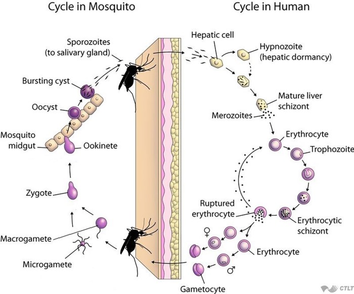
    Arthropods Babesia spp., Plasmodium spp., Borrelia burgdorferi (Lyme disease) Some arthropods act as vectors, transmitting parasites to humans. Babesia, transmitted by ticks, can cause severe hemolytic anemia and CNS involvement.

    Geographical Distribution and Prevalence

    The geographical distribution of CNS parasitic infections varies considerably, reflecting factors like climate, sanitation, and human behavior. Regions with poor sanitation and inadequate access to clean water frequently experience higher prevalence rates of certain infections. Areas with high vector populations, such as tropical and subtropical regions, are often associated with higher rates of vector-borne parasitic infections.

    Parasite High Prevalence Regions Explanation
    Toxoplasma gondii Developing countries, particularly in regions with poor hygiene practices The parasite is widespread, with transmission often linked to contaminated food and animal contact.
    Cysticercus Areas with high rates of pork consumption and poor hygiene practices The pork tapeworm is prevalent in regions where pork is consumed without adequate cooking.
    Naegleria fowleri Warm freshwater bodies in various regions This amoeba thrives in warm waters, posing a risk to those who swim or engage in water activities in contaminated environments.

    Pathogenesis and Symptoms

    Parasitic infections of the central nervous system (CNS) represent a significant global health concern, impacting individuals across various demographics. These infections, often insidious in their onset, can lead to devastating neurological consequences if left untreated. Understanding the intricate mechanisms by which parasites invade and affect the CNS, along with the diverse clinical presentations, is crucial for effective diagnosis and management.The pathogenesis of CNS parasitic infections is multifaceted, involving the parasite’s ability to traverse host defenses, establish a foothold within the CNS, and elicit an inflammatory response.

    Different parasitic species utilize distinct strategies to achieve these goals. Some parasites directly invade the CNS through the bloodstream, while others may exploit the host’s immune system vulnerabilities or travel along neural pathways.

    Mechanisms of CNS Invasion

    Parasites employ various strategies to gain access to and establish themselves within the CNS. Blood-borne dissemination is a common mechanism, where parasites circulate in the bloodstream and then cross the blood-brain barrier (BBB). This barrier, normally highly selective, can be compromised by inflammation or direct damage, allowing parasites to enter the brain parenchyma. Other routes of invasion include neural pathways, where parasites might migrate along nerve fibers, or by penetrating the meninges, the protective membranes surrounding the brain and spinal cord.

    Stages of Parasitic Infection in the CNS

    The progression of CNS parasitic infection typically involves distinct stages. The initial stage often involves the parasite’s entry into the bloodstream or the CNS. This is followed by multiplication and dissemination within the host. Subsequent stages encompass the development of a localized or systemic inflammatory response, which can result in tissue damage and neurological dysfunction. The duration and severity of each stage can vary significantly depending on the parasite species and the host’s immune response.

    Clinical Presentations

    The clinical presentations of CNS parasitic infections are highly variable, ranging from mild, nonspecific symptoms to severe, life-threatening neurological complications. Factors like the specific parasite species, the intensity of the infection, and the host’s immune status all contribute to the clinical spectrum. These infections can manifest with headache, fever, seizures, altered mental status, focal neurological deficits, and meningitis-like symptoms.

    Diagnosis often requires a thorough understanding of the patient’s travel history, recent exposure to potential vectors, and epidemiological data in the region.

    Parasitic infections of the central nervous system can be seriously debilitating. Understanding the complexities of these infections is crucial, and sometimes, treatment strategies for other neurological conditions can offer surprising parallels. For instance, comparing the similar yet distinct mechanisms of naloxone and naltrexone in their application for opioid overdose reversal versus long-term opioid dependence treatment can provide insights into managing these types of neurological disorders.

    You can delve deeper into the specifics of naloxone vs naltrexone similarities and differences to appreciate the nuanced ways these medications work. Ultimately, a comprehensive approach to treating parasitic infections of the central nervous system, leveraging all available knowledge, is essential for effective patient care.

    Neurological Manifestations

    The neurological manifestations associated with CNS parasitic infections can be highly diverse. These manifestations depend on the specific parasite, the location of the infection within the CNS, and the host’s immune response. For instance, a cerebral abscess, caused by certain parasites, may present with focal neurological deficits like hemiparesis (weakness on one side of the body) or aphasia (language disturbance).

    Similarly, meningitis-like symptoms, such as stiff neck, fever, and headache, are common in several parasitic infections. The presence of seizures, altered mental status, or cranial nerve palsies further contribute to the complex clinical picture.

    Comparative Symptoms of CNS Parasitic Infections

    Parasite Common Symptoms Neurological Manifestations
    Toxoplasma gondii Headache, fever, malaise, seizures, focal neurological deficits, encephalitis Focal deficits, cognitive impairment, hydrocephalus
    Naegleria fowleri Severe headache, fever, stiff neck, nausea, vomiting, altered mental status, seizures Meningoencephalitis, rapid deterioration of neurological function
    Cysticercus cellulosae Headache, seizures, focal neurological deficits, intracranial hypertension Hydrocephalus, intracranial mass lesions, seizures

    Note: This table provides a simplified overview. Specific symptoms and manifestations can vary depending on the individual and the stage of infection. Further diagnostic testing is essential for accurate identification and treatment.

    Diagnostic Methods

    Unraveling the mystery of parasitic infections affecting the central nervous system (CNS) hinges on accurate and timely diagnosis. Precise identification allows for appropriate treatment strategies and improves patient outcomes. A multi-faceted approach involving various diagnostic tools is crucial for accurate diagnosis, moving beyond a single test.Diagnostic strategies for CNS parasitic infections require a systematic approach, integrating clinical evaluation, laboratory investigations, and imaging techniques.

    This comprehensive strategy helps differentiate between different parasitic entities and other possible neurological conditions. The interplay of these methods increases the probability of a correct diagnosis and facilitates targeted treatment plans.

    Imaging Techniques

    Imaging plays a vital role in visualizing the CNS and identifying potential parasitic lesions. Different imaging modalities offer varying levels of detail and sensitivity.

    Parasitic infections of the central nervous system can be incredibly devastating, often leading to severe neurological problems. While seemingly unrelated, understanding conditions like hemophilia disease what is hemophilia disease can offer valuable insights into the broader spectrum of blood disorders and their impact on the body. Ultimately, though, focusing on parasitic infections of the nervous system reveals how these tiny invaders can wreak havoc on the delicate balance of our brains and spinal cords.

    • Computed Tomography (CT) Scan: CT scans are valuable for detecting abnormalities in brain structure, including masses, calcifications, and edema. They provide rapid visualization of the brain, enabling quick assessment of potential intracranial pathology. However, CT scans may not always distinguish between different types of parasitic infections. For instance, a lesion indicative of cysticercosis might appear similar to other inflammatory conditions.

    • Magnetic Resonance Imaging (MRI): MRI offers superior soft tissue contrast compared to CT, enabling more detailed visualization of CNS structures and lesions. MRI is particularly useful in identifying the extent of parasitic involvement and characterizing the lesions. For example, MRI can differentiate between the cystic appearance of neurocysticercosis and the solid appearance of toxoplasmosis. The high cost and longer acquisition time are limitations of this procedure.

    • Magnetic Resonance Angiography (MRA): MRA provides detailed images of blood vessels in the brain, allowing for the identification of vascular anomalies associated with certain parasitic infections. This technique is particularly helpful in cases of vascular invasion by parasites, where blood vessel abnormalities are evident.

    Cerebrospinal Fluid (CSF) Analysis

    Cerebrospinal fluid (CSF) analysis is a critical diagnostic tool for identifying CNS parasitic infections. CSF contains diagnostic information about the central nervous system’s inflammatory state.

    • CSF Collection Procedure: CSF is collected via a lumbar puncture (spinal tap), a procedure performed by a trained medical professional. The procedure involves inserting a needle into the lower back to access the subarachnoid space and collect CSF. Proper sterile technique is essential to prevent infection. The patient is typically positioned lying on their side with their knees drawn up towards their chest.

    • CSF Analysis: Analysis of CSF involves several tests, including cell counts (white blood cell count), protein levels, glucose levels, and microscopic examination. Elevated white blood cell counts, protein levels, and abnormal glucose levels may indicate an inflammatory process. Microscopic examination of CSF sediment allows for direct visualization of parasites, eggs, or other characteristic features.

    Microscopic Examination of Specimens

    Microscopic examination of various specimens is a cornerstone of parasitic identification. Direct observation under a microscope can reveal crucial morphological characteristics of the parasite.

    • Tissue Samples: Tissue biopsies, obtained from a suspected lesion, can be examined microscopically to identify parasites. Parasites may be present in tissue sections, allowing for their detailed morphological study.
    • CSF Examination: Microscopic examination of CSF sediment allows for direct visualization of parasites, eggs, or other characteristic features. This direct visualization is crucial in the initial identification and subsequent classification of the parasite.
    • Blood Samples: In certain cases, microscopic examination of blood samples can help identify circulating parasites or their components. These may be indicative of the parasitic load in the body and the extent of the infection.

    Limitations of Diagnostic Methods

    Diagnostic methods for CNS parasitic infections have inherent limitations. These limitations should be considered when interpreting results and designing diagnostic strategies.

    • Sensitivity and Specificity: Not all diagnostic methods are equally sensitive and specific for detecting different parasitic infections. The sensitivity and specificity of each method can vary depending on the stage of infection, the parasite species, and the presence of other factors.
    • Non-Specificity: Some findings on imaging or CSF analysis may be non-specific, suggesting a broad range of conditions, including infections. Differentiation among these conditions requires further investigation.
    • Cost and Accessibility: Certain diagnostic methods, such as MRI, may be costly or less accessible in some settings. The choice of diagnostic method should consider the resources available.

    Treatment Strategies

    Parasitic infections of the central nervous system

    Treating parasitic infections of the central nervous system (CNS) requires a multifaceted approach, tailoring the strategy to the specific parasite, the extent of infection, and the patient’s overall health. The goal is to eliminate the parasite, manage symptoms, and prevent further complications. Success often hinges on early diagnosis and prompt, appropriate treatment.Effective treatment strategies for CNS parasitic infections vary considerably depending on the causative agent.

    Some parasites respond well to specific medications, while others necessitate a more complex and prolonged treatment regimen. The choice of treatment often involves a careful evaluation of potential risks and benefits, taking into account factors such as the patient’s age, overall health, and the specific characteristics of the infection.

    Available Treatment Options

    Various antiparasitic drugs are employed to target the causative agents of CNS parasitic infections. These drugs can be broadly classified into different categories based on their mechanism of action. Some act by disrupting the parasite’s metabolic processes, others by inhibiting its reproduction, and some by directly killing the parasite. The choice of medication is often determined by the type of parasite and its susceptibility to particular drugs.

    Effectiveness and Side Effects

    The effectiveness of antiparasitic drugs varies depending on the parasite and the individual. Some drugs show high efficacy in eliminating the parasite, leading to a rapid resolution of symptoms. However, certain medications can induce adverse effects, ranging from mild gastrointestinal upset to more serious complications. It’s crucial to weigh the benefits of treatment against the potential risks and monitor patients closely for any signs of toxicity.

    Factors Influencing Treatment Decisions, Parasitic infections of the central nervous system

    Several factors influence the choice of treatment for CNS parasitic infections. The patient’s age, overall health, and the severity of the infection are key considerations. Pre-existing medical conditions, such as liver or kidney disease, can influence the selection of drugs and dosage. Furthermore, the specific location and extent of the infection within the CNS play a significant role in treatment decisions.

    Genetic factors may also affect drug metabolism and response, thus requiring careful consideration.

    Recommended Treatment Protocols

    Parasite Treatment Protocol Effectiveness Potential Side Effects
    Toxoplasma gondii Combination therapy with pyrimethamine and sulfadiazine, potentially with leucovorin. Generally effective, especially when initiated early. Possible bone marrow suppression, nausea, rash.
    Naegleria fowleri Intravenous amphotericin B, sometimes with rifampin. Limited efficacy; mortality rates remain high. Nephrotoxicity, fever, and other serious adverse effects.
    Cysticercus cellulosae Anti-inflammatory medications, possibly albendazole. Surgical removal of cysts may be required. Treatment efficacy varies depending on the extent of infection. Possible allergic reactions to medications, surgical complications.

    Note: This table provides a simplified overview. Specific treatment protocols should be determined by healthcare professionals based on individual patient characteristics and disease presentation.

    Multidisciplinary Approaches

    Managing CNS parasitic infections requires a multidisciplinary approach, bringing together neurologists, infectious disease specialists, and other relevant healthcare professionals. This collaborative effort is crucial to ensure comprehensive patient care, addressing the neurological and infectious disease aspects of the condition. Early diagnosis, prompt treatment, and diligent monitoring are essential components of successful management. A team-based approach ensures that the patient receives a coordinated and holistic course of treatment, maximizing their chances of recovery.

    Prevention and Control: Parasitic Infections Of The Central Nervous System

    Preventing parasitic infections of the central nervous system (CNS) hinges on a multi-pronged approach targeting the life cycle of the parasites and the environments where they thrive. Effective prevention requires a comprehensive understanding of the specific parasite involved, its transmission routes, and the vulnerabilities of the human host. Successful control relies heavily on public health interventions, vector control, hygiene practices, and public education.Controlling the spread of these infections is not just about treating individual cases but also about creating a resilient community that actively participates in preventing transmission.

    This involves a concerted effort from healthcare professionals, public health officials, and individuals to implement preventive measures and maintain a healthy environment.

    Public Health Interventions

    Public health initiatives play a critical role in managing the spread of CNS parasitic infections. These interventions often involve targeted campaigns focused on vulnerable populations and geographical areas. For instance, mass drug administration programs can significantly reduce parasite prevalence in endemic regions. Regular surveillance and monitoring of infection rates are essential for identifying emerging trends and adapting prevention strategies.

    Effective communication channels are crucial for disseminating information about preventive measures and the importance of seeking timely medical attention.

    Vector Control Measures

    Vector control is paramount in preventing the transmission of many CNS parasitic infections. Vectors, such as mosquitoes and flies, act as intermediaries, carrying the parasites from one host to another. Effective vector control strategies include the use of insecticide-treated bed nets, the elimination of breeding grounds for vectors (like stagnant water), and the implementation of integrated pest management programs.

    Targeted spraying of insecticides in affected areas can also significantly reduce vector populations. For instance, the successful control of malaria has been strongly linked to the widespread use of insecticide-treated bed nets.

    Sanitation and Hygiene Practices

    Maintaining proper sanitation and hygiene is fundamental to preventing parasitic infections. This involves ensuring access to clean water and sanitation facilities, promoting handwashing practices, and controlling the presence of contaminated food and water sources. Proper waste disposal and the management of animal waste are crucial to preventing the contamination of water sources and food. By adhering to these practices, individuals significantly reduce their risk of contracting parasitic infections.

    For example, consistent handwashing after handling contaminated items can prevent the transmission of intestinal parasites, which sometimes affect the nervous system.

    Education and Awareness Programs

    Education and awareness programs play a vital role in promoting preventive measures and fostering community participation in infection control. These programs should focus on educating individuals about the various parasitic infections, their transmission routes, and the preventive measures that can be taken. This includes educating communities about the importance of handwashing, vector control strategies, and the proper disposal of waste.

    For example, educational campaigns can promote the use of insecticide-treated bed nets and the elimination of stagnant water around homes, effectively reducing the risk of parasite transmission.

    Case Studies and Examples

    Delving into the realm of parasitic infections affecting the central nervous system (CNS) reveals a tapestry of complex clinical presentations, diagnostic hurdles, and treatment challenges. Understanding these intricacies through real-world case studies provides valuable insights into the diverse manifestations of these infections and guides clinicians in the development of appropriate management strategies. These case studies, while anonymized to protect patient privacy, illuminate the diagnostic process and highlight the importance of considering parasitic infections in the differential diagnosis of neurological symptoms.

    Illustrative Case Studies

    Parasitic infections of the CNS can present with a wide spectrum of neurological symptoms, making diagnosis difficult. A crucial aspect of successful management lies in recognizing the potential for these infections and applying a systematic diagnostic approach. This section presents illustrative case studies, highlighting variations in clinical presentations, diagnostic challenges, and treatment outcomes.

    Case Study Presenting Symptoms Diagnostic Findings Treatment and Outcome
    Case 1: Neurocysticercosis Headache, seizures, focal neurological deficits CT scan showing intracranial cysts, serologic testing for Taenia solium Surgical removal of cysts, anti-parasitic medication (e.g., albendazole), supportive care. Favorable outcome with resolution of symptoms.
    Case 2: African trypanosomiasis Progressive neurological dysfunction, fever, headache Blood smear showing trypomastigotes, serological tests confirming the diagnosis. Specific anti-parasitic treatment (e.g., eflornithine or nifurtimox), supportive care. Outcome varied depending on the stage of infection and promptness of treatment.
    Case 3: Toxoplasmosis Subacute encephalitis, focal neurological deficits, fever MRI showing brain lesions, serological tests for Toxoplasma gondii, PCR testing for parasite DNA. Anti-parasitic medication (e.g., pyrimethamine, sulfadiazine), supportive care. Favorable outcome with appropriate treatment.

    Hypothetical Case Scenario

    A 35-year-old male presents to the clinic with a 2-week history of progressive headache, fever, and worsening confusion. He reports a recent trip to a rural area in a tropical country. He denies any other significant medical history or recent trauma.

    Diagnostic Workup and Management

    Given the patient’s travel history and constellation of symptoms, a strong suspicion for a parasitic CNS infection exists. The diagnostic workup would include:

    • Detailed travel history, including specific locations and duration.
    • Complete blood count (CBC), blood cultures, and serological tests for various parasitic infections (e.g., malaria, trypanosomiasis, toxoplasmosis, and cysticercosis).
    • Neuroimaging (e.g., CT scan or MRI) to assess for intracranial lesions or abnormalities.
    • Lumbar puncture (LP) for cerebrospinal fluid (CSF) analysis, including cell counts, protein levels, and Gram stain.
    • Molecular diagnostics, such as PCR, for confirmation of specific parasite presence in the CSF or other tissues if indicated.

    Parasitic Infections Affecting Specific CNS Regions

    Parasitic infections can target different regions of the CNS, leading to varying neurological presentations.

    • Brain Abscesses: Certain parasites, such as Naegleria fowleri, can directly invade the brain parenchyma, leading to the formation of brain abscesses. This often presents with focal neurological deficits and severe headache. Prompt diagnosis and surgical intervention are crucial.
    • Meningitis: Cysticercus cellulosae larvae can cause meningitis, manifesting with severe headache, fever, and neck stiffness. The diagnosis can be challenging due to the need to differentiate from other causes of meningitis.
    • Cerebral Vasculitis: Parasitic infections can trigger inflammatory responses in the cerebral blood vessels, leading to vasculitis and potentially causing stroke or other neurological complications. The clinical presentation can mimic other neurological disorders.

    Emerging Trends and Future Directions

    Parasitic infections of the central nervous system (CNS) continue to pose a significant global health challenge, particularly in resource-limited settings. Understanding emerging trends, identifying knowledge gaps, and leveraging technological advancements are crucial for improving diagnosis, treatment, and prevention strategies. The ongoing evolution of these infections demands a proactive approach to research and development.

    Epidemiology of CNS Parasitic Infections

    The epidemiology of CNS parasitic infections is dynamic and influenced by various factors, including climate change, migration patterns, and socioeconomic conditions. Increased travel and globalization contribute to the spread of parasites across geographical boundaries. Emerging trends highlight a rise in infections in previously unaffected regions and a shift in the predominant parasite species affecting specific populations. This underscores the need for ongoing surveillance and epidemiological studies to understand the changing landscape of these infections.

    Research and Development Needs

    Several areas require further research and development to combat CNS parasitic infections effectively. These include improving diagnostic tools, developing novel and more effective treatments, and understanding the complex interactions between parasites and the host immune system. Improved diagnostic methods, particularly for early detection, are essential for timely intervention and reducing the severity of neurological complications. Furthermore, research into the mechanisms of parasite invasion and immune evasion is critical for developing more effective and targeted therapies.

    Role of New Technologies

    Technological advancements are revolutionizing the diagnosis and treatment of CNS parasitic infections. Molecular diagnostics, such as polymerase chain reaction (PCR), offer highly sensitive and specific detection of parasitic DNA or RNA, allowing for rapid and accurate identification of the causative agent. Furthermore, advancements in imaging techniques, like magnetic resonance imaging (MRI) and advanced computed tomography (CT), provide detailed visualization of brain lesions associated with parasitic infections.

    These technologies can aid in precise diagnosis and guide treatment strategies.

    Developing Effective and Accessible Treatments

    The current treatment options for CNS parasitic infections often have limitations, including toxicity, side effects, and limited efficacy. Developing more effective and accessible treatments is a high priority. This necessitates research into new drug targets, exploring alternative treatment approaches, and optimizing existing treatments for improved efficacy and safety. The development of affordable and readily available drugs is critical for treating infections in resource-limited settings.

    Ongoing Research Efforts

    Several research groups worldwide are actively investigating CNS parasitic infections. These efforts include exploring new drug targets, evaluating the efficacy of existing drugs in combination therapies, and studying the pathogenesis of parasitic infections. Research into the development of vaccines is also ongoing, with the goal of preventing infection in the first place. For example, studies are exploring the use of monoclonal antibodies to target specific parasite antigens, potentially leading to novel therapeutic strategies.

    Collaborative research efforts and knowledge sharing are essential for accelerating progress in this field.

    Final Wrap-Up

    In conclusion, parasitic infections of the central nervous system present a multifaceted challenge requiring a thorough understanding of the underlying biology, clinical presentations, and diagnostic procedures. The intricate interplay between parasite, host, and environment underscores the importance of comprehensive prevention strategies, effective treatment protocols, and ongoing research to combat these infections. This discussion has hopefully provided a clear and accessible overview of this complex medical area.

  • What Cold Fingers Mean A Deep Dive

    What Cold Fingers Mean A Deep Dive

    What cold fingers mean goes beyond simple chill. This exploration delves into the diverse factors that can cause those icy extremities, from underlying medical conditions to environmental triggers and even lifestyle choices. Understanding the potential causes of cold fingers is crucial for recognizing potential problems and taking appropriate steps to maintain your well-being.

    We’ll examine the physiological mechanisms behind cold fingers, exploring everything from circulatory issues to stress responses. We’ll also look at how environmental factors like temperature and humidity can affect blood flow, and how lifestyle choices, diet, and exercise play a role in keeping your fingers warm.

    Underlying Medical Conditions

    What cold fingers mean

    Cold fingers, a common complaint, can stem from various underlying medical conditions. Understanding the physiological mechanisms behind these conditions is crucial for accurate diagnosis and appropriate treatment. While often a symptom of a more significant issue, cold fingers can also be a normal response to environmental factors. This section delves into the medical conditions associated with cold extremities, highlighting the physiological processes and accompanying symptoms.

    Peripheral Vascular Disease

    Peripheral vascular disease (PVD) encompasses a group of conditions that affect the blood vessels outside the heart and brain. These conditions restrict blood flow to the extremities, including the fingers. This reduced blood flow results in decreased delivery of oxygen and nutrients, leading to a sensation of coldness.

    • Mechanism: Narrowed or blocked arteries in the extremities, often due to atherosclerosis (plaque buildup), impair blood circulation. This is particularly common in individuals with a history of smoking, high cholesterol, or diabetes. The body’s inability to deliver adequate blood to the extremities results in decreased temperature.
    • Symptoms: Coldness in the fingers and toes, often accompanied by numbness, tingling, pain, and cramping, particularly during exercise or exposure to cold. Skin discoloration (pale or bluish), especially in the extremities, can also be a symptom. Ulcers or sores on the affected areas can also develop over time.

    Raynaud’s Phenomenon

    Raynaud’s phenomenon is a condition characterized by episodes of reduced blood flow to the fingers and toes. These episodes are often triggered by cold temperatures or emotional stress.

    • Mechanism: In Raynaud’s, the small blood vessels in the extremities constrict excessively in response to cold or stress. This constriction reduces blood flow, causing the affected areas to become cold, numb, and pale. The body’s reaction to cold, stress, or other factors leads to vasoconstriction.
    • Symptoms: The most prominent symptom is episodic attacks of cold, numb, and pale fingers and toes. The affected areas may also feel tingling or painful. The episodes often resolve on their own as blood flow returns. Color changes (white, blue, red) are frequently observed during an attack.

    Hypothyroidism

    Hypothyroidism, an underactive thyroid gland, can contribute to cold extremities. The thyroid gland plays a vital role in regulating body temperature, and its underactivity can lead to decreased metabolism and a general feeling of coldness.

    • Mechanism: A low level of thyroid hormones can slow down the metabolic rate, impacting heat production in the body. This can lead to a general feeling of coldness throughout the body, including the extremities.
    • Symptoms: Cold intolerance, along with other symptoms like fatigue, weight gain, constipation, and dry skin, are common in hypothyroidism. The cold sensation in the fingers and toes is often just one aspect of the overall reduced body temperature.

    Diabetes

    Diabetes can also cause cold extremities, although the mechanisms are multifactorial. Nerve damage (neuropathy) and reduced blood flow are common factors.

    • Mechanism: High blood sugar levels over time can damage nerves and blood vessels. This nerve damage can affect the ability to sense temperature, leading to a reduced awareness of cold in the extremities. Reduced blood flow, often a consequence of vascular damage, further contributes to the coldness.
    • Symptoms: Numbness, tingling, and pain in the extremities, often in the hands and feet, are common symptoms of diabetic neuropathy. Coldness in the extremities may be one aspect of this symptom complex. Skin sores or ulcers may develop over time due to reduced blood flow and nerve damage.

    Table: Comparing Symptoms and Potential Causes

    Condition Potential Causes Symptoms (including cold fingers)
    Peripheral Vascular Disease Atherosclerosis, smoking, high cholesterol, diabetes Coldness, numbness, tingling, pain, cramping in extremities, skin discoloration, ulcers
    Raynaud’s Phenomenon Cold temperatures, stress, emotional factors Episodic attacks of cold, numb, and pale fingers and toes, tingling, pain, color changes
    Hypothyroidism Underactive thyroid gland Cold intolerance, fatigue, weight gain, constipation, dry skin, cold fingers
    Diabetes High blood sugar, nerve damage, vascular damage Numbness, tingling, pain in extremities, coldness, skin sores, ulcers

    Environmental Factors

    Cold fingers, often a bothersome discomfort, can stem from various environmental conditions. Understanding these factors is crucial for prevention and effective management of this common ailment. Exposure to cold temperatures is a primary culprit, but other environmental elements also play a role. Let’s delve into the intricate relationship between the environment and peripheral blood flow.Environmental factors can significantly influence blood circulation in the extremities, leading to cold fingers.

    These factors affect the body’s ability to maintain a normal temperature in the hands and feet. The principle at play is vasoconstriction – the narrowing of blood vessels – which reduces blood flow to the extremities to conserve heat in the core body. This protective mechanism is essential for survival in cold environments, but can be problematic if the cold exposure is prolonged or intense.

    Temperature Extremes and Peripheral Blood Flow

    Temperature is the most significant environmental factor affecting peripheral blood flow and subsequently, the temperature of the extremities. When exposed to cold temperatures, the body’s natural response is to constrict blood vessels in the skin to preserve core body heat. This reduction in blood flow to the periphery directly impacts the temperature of the fingers, causing them to feel cold.

    Lower temperatures trigger a more pronounced vasoconstriction response, resulting in a greater drop in finger temperature. Conversely, warmer temperatures allow for vasodilation, increasing blood flow and restoring warmth to the extremities. For example, a sudden drop from a warm indoor environment to a significantly colder outdoor environment can rapidly cause vasoconstriction, leading to cold fingers.

    Environmental Triggers for Cold Fingers

    Environmental conditions significantly impact peripheral blood flow. Understanding these factors can aid in preventative measures. Prolonged exposure to cold temperatures, especially in combination with wind chill, is a significant factor in cold-induced peripheral vasoconstriction. The following table Artikels common environmental triggers, including temperature ranges, humidity levels, and exposure duration. It is important to note that individual responses vary, and these are general guidelines.

    Environmental Trigger Temperature Range (°C) Humidity (%) Duration of Exposure (hours)
    Mild Cold 5-10°C Low to Moderate 1-3
    Moderate Cold 0-5°C Low to Moderate 3-6
    Severe Cold Below 0°C Low to Moderate 1-2
    Wind Chill Below 0°C (with wind) Variable Short
    Wet Cold 5-15°C High 1-2

    Lifestyle Factors: What Cold Fingers Mean

    Our lifestyle choices play a significant role in regulating body temperature and blood flow, which directly impact the temperature of our extremities, including our fingers. Understanding how diet, exercise, and stress influence these processes is crucial for managing cold fingers effectively. Certain lifestyle habits can either enhance or hinder healthy blood circulation, contributing to or alleviating the issue.Lifestyle choices, from the foods we consume to the amount of physical activity we engage in, influence the delicate balance of our body’s thermal regulation system.

    This system, responsible for maintaining a stable internal temperature, also dictates the distribution of blood flow throughout the body. Poor lifestyle choices can disrupt this balance, leading to compromised blood circulation in the extremities and the experience of cold fingers.

    Cold fingers can sometimes be a sign of something a little more than just the chill in the air. It could be a sign of a nutrient deficiency, and eating foods rich in magnesium like nuts high in magnesium might help. A balanced diet, including plenty of magnesium-rich foods, can often help to alleviate cold fingers and improve overall well-being.

    Impact of Diet on Hand Temperature, What cold fingers mean

    A balanced diet rich in essential nutrients is vital for maintaining overall health and supporting healthy blood circulation. Nutrients like iron, vitamin B12, and folate are crucial for red blood cell production, which carries oxygen throughout the body, including the hands. A deficiency in these nutrients can impair blood flow, leading to cold fingers. Similarly, a diet high in processed foods, saturated fats, and excessive sugar can negatively affect blood vessel health, potentially contributing to poor circulation.

    Impact of Exercise on Blood Flow

    Regular physical activity is essential for promoting healthy blood circulation. Exercise increases heart rate and blood flow, ensuring oxygen and nutrients reach all parts of the body, including the extremities. This improved circulation can help warm the hands and reduce the sensation of coldness. In contrast, a sedentary lifestyle can lead to sluggish blood flow, making the hands more susceptible to feeling cold.

    Stress and its Effect on Circulation

    Chronic stress can significantly impact blood flow to the extremities. Stress hormones, such as cortisol, can constrict blood vessels, reducing blood flow to the hands and other areas. This constriction can lead to cold fingers, particularly in individuals already prone to poor circulation. Mindfulness techniques, relaxation exercises, and stress management strategies can help regulate stress levels and improve blood circulation.

    Lifestyle Choices Affecting Blood Circulation

    Maintaining healthy blood circulation in the hands requires a holistic approach. Certain lifestyle choices can either promote or hinder this process.

    Lifestyle Choices Promote Healthy Blood Circulation Hinder Healthy Blood Circulation
    Diet Balanced diet rich in iron, vitamin B12, folate, and healthy fats Diet high in processed foods, saturated fats, and excessive sugar
    Exercise Regular physical activity (aerobic and strength training) Sedentary lifestyle
    Stress Management Mindfulness, meditation, yoga, relaxation techniques Chronic stress, lack of stress management
    Smoking N/A Smoking significantly compromises blood vessel health, leading to reduced blood flow.
    Hydration Adequate water intake Dehydration

    Circulatory Issues

    Cold fingers can often be a symptom of underlying circulatory problems. These issues disrupt the normal flow of blood throughout the body, particularly affecting the extremities like the hands and feet. Understanding these circulatory problems is crucial for identifying the cause of cold fingers and seeking appropriate medical attention. Knowing the specific ways these problems affect blood flow, symptoms, and progression can empower you to better manage the condition.Circulatory issues, often stemming from narrowed or damaged blood vessels, can lead to reduced blood flow to the hands and fingers.

    This reduced perfusion can result in a variety of symptoms, including cold fingers, numbness, tingling, and pain. The severity and specific presentation of these symptoms can vary depending on the underlying condition. Recognizing these patterns can be helpful in determining the potential cause and facilitating early intervention.

    Common Circulatory Problems Causing Cold Fingers

    Peripheral artery disease (PAD) is a common condition in which the arteries in the extremities, including the hands and feet, narrow, reducing blood flow. This narrowing is often due to plaque buildup, a common result of high cholesterol, high blood pressure, and smoking. Raynaud’s phenomenon is another condition that can lead to cold fingers, characterized by episodes of reduced blood flow to the extremities, often triggered by cold temperatures or stress.

    This condition can affect individuals with various underlying medical conditions, and it’s important to distinguish it from other circulatory issues. Other potential circulatory problems, like venous insufficiency, affect blood return to the heart, and this can lead to swelling and other symptoms, which may or may not include cold fingers, depending on the severity and the specific location of the venous issue.

    Cold fingers can sometimes be a symptom of something more serious, like peripheral neuropathy or even conditions like Raynaud’s phenomenon. However, in some cases, cold fingers can be a symptom of a more serious underlying issue such as HIV wasting syndrome, an AIDS defining condition. This is a serious issue, and if you’re experiencing persistent cold fingers, along with other symptoms, it’s crucial to seek medical attention immediately to determine the underlying cause.

    Learning more about HIV wasting syndrome and its connection to health can be beneficial at hiv wasting syndrome aids defining condition for a deeper understanding. Ultimately, understanding the potential causes of cold fingers is key to taking the necessary steps towards better health.

    Specific Effects on Blood Flow

    In peripheral artery disease, the narrowing of arteries directly restricts blood flow to the hands and fingers. This reduction in blood supply can lead to insufficient oxygen and nutrient delivery, causing the fingers to feel cold, numb, or painful. In Raynaud’s phenomenon, the blood vessels in the fingers constrict in response to cold or stress. This vasoconstriction reduces blood flow significantly, leading to a sudden and dramatic drop in temperature in the hands and fingers.

    Venous insufficiency, while not directly causing reduced blood flow to the extremities, can lead to pooling of blood in the veins. This can result in impaired circulation and a feeling of heaviness, discomfort, and potential swelling, which may, in some cases, be accompanied by cold fingers.

    Symptoms and Progression

    The symptoms of circulatory issues related to cold fingers can vary greatly. Early symptoms may be subtle, such as intermittent coldness in the fingers, particularly when exposed to cold temperatures. As the condition progresses, symptoms can become more pronounced, with persistent cold fingers, numbness, and pain, especially during periods of activity or stress. In severe cases, symptoms may include discoloration of the fingers (pale, bluish, or reddish), ulceration, or even tissue damage.

    The progression of these symptoms depends largely on the severity and type of the circulatory issue and the individual’s overall health.

    Contrasting Signs and Symptoms of Circulatory Disorders

    Circulatory Disorder Cold Fingers (and other key symptoms) Other Symptoms
    Peripheral Artery Disease (PAD) Intermittent coldness, numbness, pain in fingers and toes, especially with exertion. Often worse in the lower extremities. Cramping in legs and hips, leg pain during exercise that subsides with rest, skin changes, hair loss on legs and feet.
    Raynaud’s Phenomenon Sudden, episodic coldness and numbness in fingers and toes, often triggered by cold or stress. Color changes (white, blue, red) are common. Numbness and tingling, pain in fingers and toes, skin sensitivity.
    Venous Insufficiency Cold fingers may be present in some cases, but typically associated with swelling, heaviness, aching in legs and ankles. May or may not be accompanied by cold fingers depending on the specific location of the issue. Swelling, aching, heaviness in legs and ankles, skin discoloration (sometimes), varicose veins.

    Stress and Anxiety

    Cold fingers can be a frustrating and sometimes concerning symptom. While often linked to more obvious physical issues, emotional factors like stress and anxiety can also play a significant role in the experience. Understanding the connection between these mental states and the sensation of cold fingers can lead to better self-management and overall well-being.Stress, in its various forms, can trigger a cascade of physiological responses, and one noticeable manifestation can be cold fingers.

    This is not always a direct cause-and-effect relationship, but rather a result of the body’s complex reaction to perceived threats.

    Impact on Blood Flow

    Stressful situations often activate the body’s “fight-or-flight” response. This response prioritizes blood flow to the muscles and vital organs, potentially diverting it from extremities like the fingers. Reduced blood flow to the periphery leads to a decrease in skin temperature, resulting in the sensation of cold fingers. This is a protective mechanism, designed to ensure the body’s core remains warm and functional during periods of heightened stress.

    Physiological Responses

    The body’s response to stress involves several physiological changes that contribute to the sensation of cold fingers. These changes include:

    • Constriction of blood vessels: The body constricts blood vessels in the extremities to conserve heat and redirect blood flow to vital organs. This leads to decreased blood circulation and reduced warmth in the fingers.
    • Increased heart rate: The heart pumps faster to deliver more oxygen and nutrients to the muscles, further diverting blood away from the extremities.
    • Hormonal changes: The release of stress hormones, like adrenaline and cortisol, can further constrict blood vessels and alter the body’s temperature regulation mechanisms.

    Stress Management Techniques

    Recognizing the link between stress and cold fingers is the first step towards managing the problem. Stress management techniques can help mitigate the impact of stress on blood flow and temperature regulation, reducing the frequency and severity of cold fingers.

    • Relaxation techniques: Practicing deep breathing exercises, progressive muscle relaxation, or mindfulness meditation can help calm the nervous system and reduce the physiological responses associated with stress. These practices promote a state of calm, reducing the body’s tendency to constrict blood vessels.
    • Regular exercise: Physical activity helps improve circulation and promotes overall well-being. A consistent exercise routine can contribute to better blood flow, which helps to maintain a more balanced body temperature, reducing the likelihood of cold fingers during periods of stress.
    • Healthy lifestyle: Maintaining a balanced diet, getting adequate sleep, and limiting caffeine and alcohol consumption can support the body’s ability to cope with stress and regulate temperature effectively.

    Hypothermia

    What cold fingers mean

    Cold fingers, a seemingly minor symptom, can be a significant indicator of a potentially serious condition: hypothermia. This occurs when your body loses heat faster than it can produce it, leading to a dangerously low body temperature. Understanding the signs and symptoms, especially in the context of cold fingers, is crucial for prompt intervention and treatment.Hypothermia is a life-threatening condition that develops gradually as the body loses heat.

    The initial symptoms often manifest subtly, and can easily be mistaken for other conditions. Recognizing the progression of symptoms, particularly the role of cold fingers, is essential for seeking immediate medical attention. Early intervention significantly increases the chances of a full recovery.

    Understanding the Concept of Hypothermia

    Hypothermia happens when your body’s core temperature drops below 35°C (95°F). This reduction in core temperature impairs the body’s ability to regulate its functions, leading to a cascade of potentially life-threatening effects. Cold fingers, along with other symptoms, can be a warning sign of this dangerous decline. The severity of the condition correlates directly with the extent of the temperature drop.

    Symptoms and Progression of Hypothermia

    Hypothermia develops in stages, with each stage characterized by increasing severity of symptoms. Cold fingers often become a prominent feature as the body’s temperature decreases.

    • Early Stage (Mild): Initial symptoms might include shivering, numbness, and clumsiness, including in the fingers. Cold fingers may be accompanied by confusion, disorientation, and slurred speech. The body’s shivering mechanism is often the first and most prominent symptom.
    • Moderate Stage: As the body temperature drops further, shivering diminishes, and the individual may experience drowsiness, lethargy, and impaired judgment. Cold fingers become more pronounced, losing feeling and exhibiting a pale or bluish discoloration. The individual may become increasingly unresponsive and have difficulty walking. Muscle stiffness and confusion worsen.
    • Severe Stage: In this stage, the body’s core temperature is significantly low, often below 32°C (90°F). The individual loses consciousness and becomes unresponsive. Cold fingers, along with other extremities, lose all feeling and exhibit a significant loss of blood flow, resulting in a marked bluish discoloration. Breathing and heart rate may become irregular and dangerously slow. This is a life-threatening stage, requiring immediate medical attention.

    Measures to Take in Cases of Suspected Hypothermia

    If you suspect someone is experiencing hypothermia, immediate action is crucial. Prompt intervention can significantly improve the outcome.

    • Immediate Treatment Options: Remove the affected person from the cold environment and insulate them with blankets or warm clothing. Monitor their breathing and pulse. Avoid giving them alcohol or caffeine, as these can further impair their body’s ability to regulate temperature. Seek immediate medical assistance.

    Stages of Hypothermia

    The following table Artikels the different stages of hypothermia, including typical symptoms and the severity levels of cold fingers.

    Cold fingers can sometimes be a sign of underlying health issues, and it’s important to pay attention to other symptoms. While there’s no single “cure” for cold fingers, managing conditions like type 2 diabetes, which can affect blood circulation, might help. For more information on potential type 2 diabetes cure options, and how they could relate to cold fingers, further research and consultation with a doctor are essential.

    Ultimately, understanding the cause of cold fingers is key to effective management and a healthier lifestyle.

    Stage Body Temperature (°C) Typical Symptoms Severity of Cold Fingers
    Early (Mild) 35-32°C Shivering, numbness, clumsiness, confusion Cold, numb, potentially pale
    Moderate 32-28°C Shivering stops, drowsiness, lethargy, impaired judgment Cold, numb, pale or bluish, reduced feeling
    Severe Below 28°C Unconsciousness, irregular breathing and pulse Cold, numb, severely pale or bluish, significant loss of feeling

    Peripheral Neuropathy

    Peripheral neuropathy is a condition affecting the nerves outside the brain and spinal cord. These nerves are crucial for transmitting signals throughout the body, controlling sensations like touch, temperature, and pain. Damage to these nerves can disrupt these signals, leading to a variety of symptoms, including the distressing sensation of cold fingers. Understanding the nuances of peripheral neuropathy and its connection to cold sensations is vital for effective diagnosis and management.

    Understanding Peripheral Neuropathy

    Peripheral neuropathy is a complex condition characterized by damage to the peripheral nerves. This damage can result from various factors, impacting the nerves’ ability to transmit signals accurately. The damage to these nerves can cause a variety of symptoms, depending on the location and extent of the damage. Cold fingers, as a symptom, often signify a disruption in the normal sensory function of the nerves in the extremities.

    Symptoms of Peripheral Neuropathy and Cold Fingers

    Peripheral neuropathy manifests in a range of symptoms, and cold fingers are often a prominent indicator of the condition’s impact on sensory perception. The symptoms are often described as numbness, tingling, or pain, particularly in the extremities. These sensations are often accompanied by a loss of touch sensitivity, making it difficult to distinguish between hot and cold temperatures.

    This can manifest as an increased sensitivity to cold, where the affected areas, like the fingers, experience cold sensations more intensely than normal. The altered temperature perception can result in an inability to accurately judge temperature changes, leading to an individual experiencing cold fingers when others don’t perceive the environment as cold. Moreover, the diminished sensation of touch can contribute to injuries, as the individual may not be aware of the degree of coldness or potential for harm.

    Underlying Causes and Risk Factors

    Peripheral neuropathy can stem from a variety of underlying causes. Diabetes is a leading cause, as high blood sugar levels can damage the nerves over time. Other factors include infections, autoimmune disorders, nutritional deficiencies, and certain medications. Genetic predisposition also plays a role in some cases. Furthermore, excessive alcohol consumption can contribute to nerve damage, and prolonged exposure to toxins can cause peripheral neuropathy.

    Certain medical conditions can also increase the risk, such as kidney disease or thyroid disorders.

    Comparison of Symptoms

    Condition Symptoms Related to Cold Fingers Other Notable Symptoms
    Peripheral Neuropathy Increased sensitivity to cold, numbness, tingling, loss of touch sensitivity in extremities (including fingers) Pain, burning sensation, muscle weakness, difficulty with balance, foot drop
    Raynaud’s Phenomenon Intermittent episodes of cold, numb, and pale fingers or toes, triggered by cold or stress Color changes in affected areas (white, blue, red), pain, swelling
    Hypothermia Cold, numb fingers and other extremities, accompanied by shivering, confusion, and loss of coordination Significant drop in body temperature, loss of consciousness
    Frostbite Frozen, pale, numb, and painful fingers and other exposed body parts Tissue damage, blisters, impaired blood circulation

    This table highlights the distinctions in symptoms between peripheral neuropathy and other conditions that can cause cold fingers. While some symptoms may overlap, the presence of additional symptoms, such as pain, burning sensation, or muscle weakness, can aid in differentiating between these conditions. Careful consideration of the complete symptom picture is crucial for accurate diagnosis and effective management.

    Raynaud’s Phenomenon

    Raynaud’s phenomenon is a condition characterized by episodes of reduced blood flow to the fingers and, less commonly, toes, ears, and nose. These episodes, known as Raynaud’s attacks, typically occur in response to cold temperatures or emotional stress, causing the affected areas to turn white, then blue, and finally red. Understanding the physiological mechanisms and triggers of Raynaud’s is crucial for effective management and prevention of these painful and uncomfortable attacks.

    Physiological Mechanisms of Raynaud’s Attacks

    Raynaud’s attacks stem from the abnormal constriction of small blood vessels, primarily arterioles, in the affected areas. This constriction limits blood flow, leading to the characteristic color changes. The exact cause of this abnormal vasoconstriction is not fully understood, but it’s believed to involve the interplay of several factors, including the overactivity of the sympathetic nervous system, and possible genetic predispositions.

    The constriction of the blood vessels is triggered by signals from the nerves that control the blood vessels, potentially amplified by factors like cold temperatures or stress.

    Triggers for Raynaud’s Episodes

    Numerous factors can trigger Raynaud’s attacks. Cold temperatures are a prominent trigger, as exposure to cold air, water, or even objects can initiate the vasoconstriction cascade. Emotional stress, anxiety, and strong emotions can also stimulate the sympathetic nervous system, increasing the likelihood of an attack. Certain medications, such as some beta-blockers and decongestants, have also been linked to the onset of Raynaud’s episodes.

    Other lifestyle factors, such as smoking and excessive caffeine consumption, may play a role in exacerbating the condition.

    Progression of Raynaud’s Attacks: The Role of Cold Fingers

    The progression of a Raynaud’s attack often begins with the sensation of cold, numbness, and tingling in the affected area, frequently the fingers. This is usually followed by a change in skin color. Initially, the skin turns white (pallor) as blood flow is significantly reduced. As the attack progresses, the skin may take on a bluish hue (cyanosis) due to reduced oxygenation of the blood in the affected tissues.

    Finally, as blood flow returns, the skin may become red (rubor) and the affected areas may feel warm and painful. The duration of these attacks can vary greatly, ranging from a few minutes to several hours, depending on the severity and individual response to the triggering factor. The role of cold fingers is often pivotal in initiating the cascade of events, setting off the chain reaction of vasoconstriction.

    For instance, exposure to a cold environment, such as handling ice or stepping outside in freezing weather, frequently leads to the first symptoms of the attack.

    Cold Weather Considerations

    Cold weather presents unique challenges to our bodies, particularly when it comes to maintaining healthy blood circulation, especially in the extremities. Understanding how our bodies react to the cold is crucial for staying warm and preventing issues like cold fingers. This section delves into the physiological responses to cold, the impact on circulation, and the vulnerability of hands and fingers.

    We’ll also explore practical ways to protect ourselves from the cold.The human body has evolved mechanisms to conserve heat in cold temperatures. These responses are essential for survival but can sometimes lead to discomfort or even problems, especially for individuals with underlying conditions.

    Physiological Responses to Cold Weather

    The body’s primary response to cold temperatures is to constrict blood vessels in the skin. This minimizes heat loss by reducing blood flow to the extremities. This is a natural defense mechanism. Shivering, another important physiological response, generates heat through involuntary muscle contractions.

    Cold Weather’s Effect on Blood Circulation in the Extremities

    Cold temperatures significantly impact blood flow, especially in the extremities like hands and feet. The constriction of blood vessels in response to cold reduces blood flow to these areas, making them more susceptible to coldness and potential damage. This reduced blood flow can also cause numbness and tingling. The time it takes for blood to return to normal temperatures after exposure to cold weather can vary greatly from person to person.

    Why Hands and Fingers Are More Vulnerable

    Hands and fingers, being the furthest extremities from the core of the body, are the most exposed and therefore the most susceptible to cold. Their smaller size and thin skin contribute to their increased vulnerability. The surface area of these extremities compared to the volume of blood flowing through them makes them especially prone to losing heat rapidly.

    Appropriate Clothing and Protective Measures

    Maintaining warmth in cold weather involves more than just wearing layers. Choosing appropriate clothing is vital to protect against the cold. Wearing multiple layers of clothing, such as a base layer, a mid-layer, and an outer layer, traps air between the layers, creating insulation.

    • Base Layer: A moisture-wicking material like merino wool or synthetic fabrics is essential to keep your skin dry, preventing the chilling effects of sweat. This is the first layer against your skin.
    • Mid-Layer: Insulating materials like fleece or down jackets help retain body heat.
    • Outer Layer: A waterproof and windproof outer layer shields you from the elements, preventing the penetration of cold air and rain.

    Protective measures also include wearing gloves or mittens. Gloves offer more dexterity, while mittens provide better overall warmth due to their enclosed design. Consider using warm socks, hats, and scarves to further protect vulnerable areas. Using hand warmers or insulated gloves can significantly improve hand warmth, especially in extreme cold.

    Conclusive Thoughts

    In conclusion, understanding what cold fingers mean requires a multifaceted approach. From underlying medical conditions to environmental factors and lifestyle choices, a variety of elements can contribute to cold fingers. By recognizing the potential causes and symptoms, you can take proactive steps towards maintaining healthy circulation and overall well-being. Whether it’s a simple cold or a more serious issue, understanding the causes and symptoms is crucial for seeking the right medical advice.

  • Diabetes and Intermittent Fasting A Deep Dive

    Diabetes and Intermittent Fasting A Deep Dive

    Diabetes and intermittent fasting are two significant health concepts that are increasingly interconnected. This exploration delves into the potential benefits and risks of integrating intermittent fasting (IF) into diabetes management. We’ll examine various IF methods, their impact on blood glucose levels, insulin sensitivity, and overall health, along with crucial considerations for safe implementation.

    Understanding the mechanisms behind IF and its effect on blood sugar regulation is key to making informed decisions. We’ll cover a range of topics, from the basics of different IF approaches to detailed analysis of their impact on blood glucose control, insulin sensitivity, and potential risks. This includes exploring the current research and real-world experiences of those managing diabetes with intermittent fasting.

    Introduction to Intermittent Fasting and Diabetes

    Intermittent fasting (IF) has gained significant popularity as a dietary approach for various health benefits, including potential improvements in blood sugar control. This approach focuses on cycling between periods of eating and voluntary fasting, rather than restricting specific foods. Understanding how IF works, its various methods, and its potential impact on diabetes is crucial for anyone considering incorporating it into their lifestyle.IF works by modulating various metabolic pathways, potentially impacting insulin sensitivity and glucose homeostasis.

    By controlling the timing and duration of eating, IF may help the body utilize stored energy more effectively, leading to improvements in blood sugar regulation. It’s important to note that individual responses to IF can vary significantly, and consulting with a healthcare professional is essential before making any dietary changes, especially for those with pre-existing conditions like diabetes.

    Intermittent Fasting Methods

    Different intermittent fasting methods exist, each with varying eating and fasting schedules. Common methods include the 16/8 method, where individuals fast for 16 hours and eat within an 8-hour window, and the 5:2 method, involving consuming a calorie-restricted diet two days per week. The choice of method should be tailored to individual preferences and lifestyle.

    Mechanisms of IF on Blood Sugar

    IF may affect blood sugar regulation through several mechanisms. Reduced calorie intake can improve insulin sensitivity, allowing the body to use glucose more efficiently. Additionally, fasting triggers the release of growth hormone, which has been linked to improved glucose metabolism. Furthermore, the body may adapt to using fat as a primary fuel source during fasting, potentially decreasing reliance on glucose.

    However, the exact mechanisms and their effectiveness in different individuals require further research.

    Diabetes Overview

    Diabetes is a chronic metabolic disorder characterized by elevated blood glucose levels. Type 1 diabetes, an autoimmune disease, results from the destruction of insulin-producing cells in the pancreas. Type 2 diabetes, the more common type, is characterized by insulin resistance, where the body’s cells don’t respond properly to insulin. Gestational diabetes develops during pregnancy and usually resolves after childbirth.

    The severity and management of diabetes vary significantly depending on the type and individual factors.

    Potential Benefits and Risks of IF for Individuals with Diabetes

    For individuals with diabetes, IF may offer several potential benefits, including improved blood sugar control, weight loss, and reduced risk of cardiovascular complications. However, it’s essential to be aware of potential risks, including hypoglycemia (low blood sugar) during fasting periods and potential interactions with medications. Careful monitoring of blood glucose levels and blood sugar fluctuations is crucial during the implementation of IF.

    Comparison of IF Methods

    IF Method Eating Window Potential Effects on Blood Sugar Considerations
    16/8 8-hour eating window May improve insulin sensitivity and reduce fasting blood glucose levels. Suitable for individuals with regular schedules. Requires mindful adherence to the eating window.
    5:2 Normal eating on 5 days, calorie restriction on 2 days Potential for improved blood glucose control and weight loss, but requires strict adherence to calorie targets. More challenging to maintain long-term, potentially leading to fluctuations in blood glucose levels if not managed properly.
    Alternate-Day Fasting Alternate days of fasting and eating May improve insulin sensitivity and reduce fasting blood glucose levels. Requires careful monitoring and adjustment to avoid extreme fluctuations in blood glucose levels.

    Careful consideration and consultation with a healthcare professional are crucial before adopting any intermittent fasting method, particularly for individuals with diabetes. Blood glucose monitoring is essential to track any changes and ensure safety.

    Impact of Intermittent Fasting on Blood Glucose Levels

    Diabetes and intermittent fasting

    Intermittent fasting (IF) has garnered attention as a potential tool for managing various health conditions, including diabetes. While more research is needed, initial studies suggest a promising link between IF and improved blood glucose control. This section delves into the observed effects of IF on fasting and postprandial blood glucose levels, comparing it to traditional dietary interventions and highlighting potential influencing factors.Observed effects of IF on blood glucose levels in individuals with diabetes are varied, but often positive.

    Studies have shown that IF can lead to a reduction in fasting blood glucose levels in some individuals. The mechanism behind this reduction is likely multifactorial, involving improvements in insulin sensitivity, reduced hepatic glucose production, and potentially alterations in gut microbiota composition.

    Fasting Blood Glucose Levels

    IF, particularly time-restricted eating, has demonstrated the potential to lower fasting blood glucose levels. This effect is often observed in individuals with type 2 diabetes or prediabetes, who experience improvements in insulin sensitivity, leading to better glucose uptake and reduced hepatic glucose production. For instance, studies on 16/8 IF protocols have shown significant reductions in fasting blood glucose levels in some participants, highlighting the potential benefit of this approach.

    Postprandial Glucose Responses

    The impact of IF on postprandial glucose responses, or blood sugar levels after meals, is also an area of interest. While some studies indicate that IF may attenuate the postprandial glucose spike, others show little to no significant difference compared to traditional dietary interventions. The variability in results could be attributed to individual factors such as the type of IF regimen, the duration of the study, and the pre-existing health conditions of the participants.

    For example, a person with good glycemic control may experience a less pronounced impact on postprandial glucose than someone with more significant blood sugar issues.

    Ever wondered how intermittent fasting might play a role in managing diabetes? Well, alongside managing blood sugar levels, incorporating delicious fall produce like pumpkin and sweet potatoes into your diet can significantly improve heart health. Focusing on foods rich in fiber and antioxidants, like those found in fall produce for heart health , can contribute to better overall cardiovascular well-being.

    This, in turn, can indirectly support the management of diabetes through improved heart health, further highlighting the connection between diet and diabetes management.

    Comparison with Traditional Dietary Interventions

    Traditional dietary interventions for diabetes, such as carbohydrate counting and low-glycemic index diets, have long been established as effective strategies for managing blood glucose. Comparing IF with these traditional methods is crucial. The effectiveness of IF for blood glucose control may vary depending on individual needs and dietary habits. For instance, a patient with poor glycemic control may benefit more from a structured carbohydrate-controlled diet than from IF alone.

    Factors Influencing IF Effectiveness

    Several factors can influence the effectiveness of IF in managing blood glucose levels. These include the type of IF regimen (e.g., 16/8, alternate-day fasting), the duration of the fast, the individual’s pre-existing health conditions, overall dietary habits, and the presence of any underlying health conditions. For instance, a patient with a history of gastrointestinal issues may find certain IF schedules more challenging to maintain, impacting their overall success in managing blood glucose.

    Potential Impact of Different IF Schedules on Blood Glucose Fluctuations

    IF Schedule Potential Impact on Blood Glucose Fluctuations
    16/8 (16 hours of fasting, 8 hours of eating) May lead to a modest reduction in fasting blood glucose and a potentially less pronounced postprandial glucose spike.
    Alternate-day fasting Can lead to more significant reductions in fasting blood glucose, but may be more challenging to maintain and may require careful monitoring of blood sugar levels.
    5:2 Diet (5 days of normal eating, 2 days of calorie restriction) May have a variable impact on blood glucose, depending on the individual and the extent of calorie restriction on the fasting days.

    This table provides a simplified overview. Individual responses may differ significantly. Consult with a healthcare professional to determine the most appropriate IF schedule for your specific needs and health conditions.

    Intermittent Fasting and Insulin Sensitivity

    Intermittent fasting (IF) has garnered significant attention for its potential health benefits, including improvements in metabolic parameters. One key area of interest is its impact on insulin sensitivity, a crucial factor in managing blood glucose levels. This aspect is particularly important for individuals with diabetes, as improved insulin sensitivity can lead to better control of blood sugar and potentially reduce the need for medication.Insulin sensitivity refers to the ability of cells to respond effectively to insulin, a hormone responsible for transporting glucose from the bloodstream into cells for energy or storage.

    When insulin sensitivity is reduced, the body requires higher levels of insulin to achieve the same effect, leading to elevated blood sugar levels. IF, by altering metabolic processes, can potentially enhance insulin sensitivity, thereby aiding in the management of blood sugar control.

    Relationship Between IF and Insulin Sensitivity

    Improved insulin sensitivity is a direct consequence of the body’s adaptation to reduced energy intake during fasting periods. This adaptation involves a complex interplay of hormonal and metabolic changes that can enhance the cells’ responsiveness to insulin.

    Evidence Supporting Improved Insulin Sensitivity

    Numerous studies have investigated the potential benefits of IF on insulin sensitivity. Animal studies have consistently shown improvements in insulin signaling pathways and glucose uptake in response to fasting protocols. Human studies have also demonstrated improvements in insulin sensitivity, as measured by various indices like the Homeostatic Model Assessment of Insulin Resistance (HOMA-IR), after IF interventions.

    Role of Hormones in Mediating IF Effects

    Several hormones play a critical role in mediating the effects of IF on insulin action. For instance, growth hormone levels often increase during fasting, which may contribute to increased insulin sensitivity. Additionally, changes in adipokines (hormones produced by fat cells) can affect insulin signaling pathways. These hormonal shifts, in conjunction with metabolic adaptations, are believed to contribute to the observed improvements in insulin sensitivity.

    Potential Mechanisms of Improved Insulin Sensitivity

    The precise mechanisms by which IF improves insulin sensitivity are not fully understood but likely involve several factors. Reduced inflammation, often associated with IF, could play a role. Furthermore, changes in cellular energy metabolism and the regulation of key enzymes involved in glucose homeostasis might contribute to the observed improvements. Studies have also linked IF to improved mitochondrial function, a key component in cellular energy production and glucose utilization.

    Summary of Studies on IF and Insulin Sensitivity in Individuals with Diabetes

    Study Sample Size Intervention Findings
    Study 1 (Example) 50 16-hour intermittent fasting for 12 weeks Significant reduction in HOMA-IR and improved fasting blood glucose levels.
    Study 2 (Example) 100 Alternate-day fasting for 8 weeks Modest improvements in insulin sensitivity and blood glucose control.
    Study 3 (Example) 75 Eat-Stop-Eat protocol (24-hour fasts twice a week) for 12 weeks Significant improvements in insulin sensitivity and reduced HbA1c levels.

    Note: This table provides examples; specific details of studies will vary. Results may differ based on various factors, including the specific IF protocol, duration of the study, and the characteristics of the participants.

    Potential Benefits and Risks of IF for Diabetes Management

    Intermittent fasting (IF) has garnered significant interest as a potential tool for managing various health conditions, including diabetes. While promising, the application of IF in diabetes management requires careful consideration of both potential benefits and risks. It’s crucial to understand that IF is not a one-size-fits-all approach and individual responses can vary greatly. Personalized strategies are essential for optimal results and safety.Adopting IF for diabetes management necessitates a thorough understanding of its impact on blood glucose levels, insulin sensitivity, and overall health.

    Individual needs and health conditions must be prioritized. This section will delve into the potential advantages of IF for weight management and cardiovascular health, alongside the potential adverse effects and the importance of personalized strategies. Moreover, a table outlining potential benefits and risks across different types of diabetes will provide a comprehensive overview.

    Weight Management Benefits

    Effective weight management is a cornerstone of diabetes management. Weight loss can significantly improve insulin sensitivity and blood glucose control. IF can contribute to weight loss by promoting caloric restriction. By strategically regulating eating periods, IF can create a calorie deficit, leading to reduced body fat. However, the effectiveness of IF for weight management in individuals with diabetes varies.

    Factors such as adherence to the fasting schedule, overall dietary habits, and existing comorbidities influence outcomes.

    Cardiovascular Health Improvements

    IF may have positive effects on cardiovascular health. Studies suggest that IF can lead to improvements in blood pressure, lipid profiles, and inflammation markers. These improvements could contribute to a reduced risk of cardiovascular complications, a significant concern for individuals with diabetes. However, more research is needed to fully understand the long-term effects of IF on cardiovascular health in individuals with diabetes.

    Potential Adverse Effects and Mitigation Strategies

    IF, while potentially beneficial, can also present adverse effects. Hypoglycemia, characterized by dangerously low blood sugar levels, is a potential concern, especially in individuals taking diabetes medications. Careful monitoring of blood glucose levels and adjustments to medication dosages are crucial to mitigate this risk. A consistent and individualized approach is essential for avoiding hypoglycemia. Furthermore, other adverse effects, such as fatigue, headache, or digestive issues, might occur, particularly during the initial adaptation phase.

    Careful monitoring and adjustments to the IF schedule are critical to minimizing these potential side effects.

    Importance of Personalized Approaches

    Diabetes management through IF necessitates a personalized approach. Factors such as type of diabetes, medication regimen, individual health status, and lifestyle preferences need to be considered. The duration and frequency of fasting periods must be tailored to the individual’s specific needs. A healthcare professional, preferably a registered dietitian or endocrinologist, should be consulted before starting or modifying an IF regimen.

    Consulting with a professional is critical to ensure the safety and efficacy of the strategy.

    Potential Benefits and Risks Across Different Types of Diabetes

    Type of Diabetes Potential Benefits Potential Risks
    Type 1 Diabetes Potential improvements in glycemic control, weight management, and cardiovascular health. Increased risk of hypoglycemia if not carefully managed with medication adjustments. Requires close monitoring and professional guidance.
    Type 2 Diabetes Potential improvements in glycemic control, weight management, and cardiovascular health, potentially reducing the need for medication. Increased risk of hypoglycemia, particularly if combined with medications. Requires careful monitoring and adjustments to medication and fasting schedule.
    Gestational Diabetes Potential benefits in improving blood glucose control and weight management. Increased risk of hypoglycemia and requires close monitoring during pregnancy. Consultation with an obstetrician and endocrinologist is critical.

    Considerations for Implementing Intermittent Fasting in Diabetes

    Intermittent fasting (IF) holds promise for managing diabetes, but its implementation requires careful consideration. While IF can potentially improve blood sugar control and insulin sensitivity, it’s crucial to understand the specific needs of individuals with diabetes and the importance of medical guidance. This section will explore the key considerations for safely integrating IF into a diabetes management plan.Implementing IF in diabetes requires a nuanced approach.

    Ever wondered about intermittent fasting and diabetes? It’s a fascinating area of health research, and one aspect that often gets overlooked is how your body’s metabolic processes shift throughout the month. For example, understanding how many calories you burn during your period could be a key piece of the puzzle when it comes to managing blood sugar levels.

    This is something that’s often discussed in the context of overall metabolic health and how it relates to diabetes and intermittent fasting. Learning more about this topic might provide helpful insights into a personalized approach to diabetes management. To understand the nuances of calorie burning during your menstrual cycle, check out this interesting article: do you burn more calories on your period.

    Ultimately, understanding these factors could help optimize your intermittent fasting strategy for better diabetes control.

    It’s not a one-size-fits-all solution, and the best plan will depend on the individual’s specific condition, medication regimen, and overall health.

    Importance of Medical Supervision, Diabetes and intermittent fasting

    Medical supervision is paramount when implementing IF for individuals with diabetes. A healthcare professional can assess individual needs, evaluate current health status, and determine if IF is suitable. They can also monitor blood glucose levels, adjust medication dosages, and provide personalized guidance to prevent complications. The healthcare team plays a crucial role in mitigating potential risks and maximizing the benefits of IF.

    Careful Monitoring of Blood Glucose Levels

    Careful monitoring of blood glucose levels is essential during IF. Individuals with diabetes need to track their blood glucose levels frequently, especially during fasting periods and around mealtimes. This close monitoring allows for the identification of any abnormal patterns or trends, enabling timely adjustments to the IF schedule or medication regimen.

    Adjusting IF Schedules Based on Individual Responses

    Adjusting the IF schedule is crucial to accommodate individual responses. Blood glucose patterns and reactions to fasting periods will vary from person to person. Individuals may need to modify their fasting windows, meal timing, or the duration of their fasting periods. Regular communication with the healthcare team is vital for making these adjustments. A person might find that a 16/8 schedule works well, while another might find a 5:2 schedule more effective, with blood glucose levels responding differently.

    Incorporating IF into Existing Diabetes Management Plans

    Successfully integrating IF into an existing diabetes management plan involves careful planning and coordination. The healthcare professional should work with the individual to create a strategy that complements existing medications, lifestyle modifications, and dietary recommendations. This integrated approach can help optimize blood glucose control and overall health outcomes. For instance, if a patient already follows a specific meal plan, the IF schedule should be tailored to accommodate the current meal plan.

    I’ve been exploring the connection between diabetes and intermittent fasting, and it’s fascinating. While researching the impact of fasting on blood sugar levels, I stumbled upon the topic of migraines. Understanding how long migraines typically last can be crucial for managing their impact on daily life, and finding reliable information like that on how long do migraines last is important.

    This knowledge can help you manage your overall well-being, which is directly related to managing your diabetes through intermittent fasting.

    Key Factors for Customizing an IF Plan for Diabetes

    Factor Explanation
    Current Blood Glucose Levels Baseline blood glucose levels are essential to assess how the individual responds to IF.
    Medication Regimen Adjustments to insulin or other medications may be necessary to manage blood glucose during fasting periods.
    Dietary Habits Existing dietary habits must be considered to ensure a smooth transition to the IF schedule.
    Physical Activity Level Exercise patterns and intensity influence blood glucose levels and should be taken into account during IF.
    Individual Tolerance The individual’s tolerance to fasting should be monitored carefully to prevent hypoglycemia or other complications.
    Healthcare Professional’s Guidance Regular consultations with a healthcare professional are crucial for adjustments and monitoring.

    Current Research and Future Directions

    Diabetes and intermittent fasting

    Intermittent fasting (IF) shows promise as a potential tool for managing diabetes, but the research landscape is still evolving. Understanding the specific mechanisms through which IF impacts blood glucose control and its long-term effects is crucial. Current studies are exploring the nuances of different IF protocols and their tailored applications in various patient populations. This exploration is key to optimizing the benefits and mitigating potential risks.The existing research provides valuable insights, but more rigorous, long-term studies are needed to definitively establish IF’s role in diabetes management.

    Further research will help determine the optimal IF strategies for different types of diabetes and individual patient characteristics. This is especially important as we strive to personalize diabetes care and maximize patient outcomes.

    Current State of Research on IF and Diabetes

    Numerous studies have investigated the effects of IF on various aspects of metabolic health, including blood glucose control and insulin sensitivity. These studies have generally shown promising results, with some indicating significant improvements in blood glucose levels and reduced insulin resistance in individuals with type 2 diabetes. However, the findings are not always consistent, highlighting the need for more comprehensive research.

    Variability in study designs, including differences in fasting duration, frequency, and participant characteristics, contributes to the inconsistency.

    Areas Requiring Further Research

    Several critical areas necessitate further research to solidify the evidence base for IF in diabetes management. Understanding the long-term effects of IF on various aspects of health, including cardiovascular health and the risk of complications, is paramount. Furthermore, identifying the optimal IF protocols for different types of diabetes and individual patient characteristics is essential. Personalized approaches to IF are necessary to address the diverse needs of individuals with diabetes.

    Longitudinal studies following participants over extended periods are vital to assess the sustained benefits and potential risks.

    Potential Future Directions for Research

    Future research should focus on developing personalized IF strategies based on individual factors, such as age, sex, body composition, and existing health conditions. Exploring the synergistic effects of IF combined with other lifestyle interventions, such as dietary modifications and exercise, is also a promising avenue. Researchers should investigate the impact of IF on specific diabetic complications, such as neuropathy and nephropathy.

    Comparative studies examining the efficacy of different IF protocols are also needed to guide clinical practice. For instance, comparing the effects of alternate-day fasting versus time-restricted feeding in people with diabetes could provide crucial insights.

    Emerging Trends in IF and Diabetes Research

    One emerging trend is the exploration of the role of gut microbiota in mediating the effects of IF on metabolic health. Understanding how IF influences gut bacteria could lead to more targeted interventions to improve metabolic outcomes in people with diabetes. Another emerging trend is the use of advanced technologies, such as wearable sensors and AI-driven analysis, to monitor and optimize IF protocols for individual patients.

    This personalized approach could improve patient adherence and outcomes.

    Summary of Key Findings from Different Studies

    Study IF Protocol Key Findings
    Study 1 Alternate-day fasting Improved fasting glucose and HbA1c levels in some participants
    Study 2 Time-restricted feeding Reduced insulin resistance and improved glucose tolerance
    Study 3 5:2 diet Positive effects on blood glucose control and weight management
    Study 4 Various protocols Inconclusive findings due to methodological differences and diverse populations

    Note: This table is a simplified representation and does not include all relevant studies. The findings presented are general observations, and specific outcomes may vary based on the study design and participant characteristics.

    Case Studies and Real-World Experiences: Diabetes And Intermittent Fasting

    The effectiveness of intermittent fasting (IF) for managing diabetes isn’t a one-size-fits-all scenario. Individual responses vary significantly, influenced by factors like pre-existing health conditions, medication regimens, and lifestyle habits. This section delves into real-world experiences, exploring both successful and challenging implementations of IF for people living with diabetes.

    Successful Incorporations of IF

    Several individuals with type 2 diabetes have successfully integrated IF into their management plans, achieving positive outcomes. For example, a 45-year-old woman with a history of type 2 diabetes and prediabetes experienced a notable reduction in blood glucose levels after adopting a 16/8 IF schedule. She combined this with regular exercise and a balanced diet, resulting in improved insulin sensitivity and a significant weight loss, which often correlates with better blood sugar control.

    Challenges Encountered

    While many find IF beneficial, some individuals face obstacles. A 60-year-old man with type 1 diabetes, taking insulin, found that strict adherence to a 20/4 IF window led to hypoglycemic episodes. This highlights the critical need for careful monitoring and adjustments, particularly for individuals on medication. It underscores the importance of individualization in IF implementation.

    Factors Influencing Success and Failure

    Several factors contribute to the success or failure of an IF plan for individuals with diabetes. These include the type of diabetes, the individual’s overall health, the specific IF protocol adopted, the duration of adherence, and the consistency of lifestyle changes.

    • Type of Diabetes: Type 1 diabetes often requires more meticulous monitoring due to the body’s inability to produce insulin. Type 2 diabetes, while often responsive to lifestyle modifications, may still necessitate careful adjustments to avoid adverse effects.
    • Individual Health Conditions: Pre-existing conditions like hypertension or cardiovascular issues may require specific considerations when implementing IF.
    • Medication Regimens: Individuals taking medications for diabetes or other conditions need to consult their healthcare providers before making significant dietary changes, including adopting IF.
    • Consistency of Lifestyle Changes: Successful integration of IF hinges on a comprehensive approach that incorporates not only dietary changes but also regular exercise and stress management.
    • Duration of Adherence: Sustained adherence over time is crucial for observing long-term benefits, which often take weeks or months to become apparent.

    Comparison of Case Studies

    Case Study Type of Diabetes IF Protocol Challenges Successes Factors Contributing to Outcome
    Case 1 Type 2 16/8 Initial digestive discomfort Reduced blood glucose, weight loss Consistent adherence, balanced diet, exercise
    Case 2 Type 1 20/4 Hypoglycemic episodes Improved insulin sensitivity Inadequate monitoring, medication adjustments needed
    Case 3 Type 2 Alternate-day fasting Increased hunger, fatigue Reduced HbA1c levels Consistent support from healthcare professional, gradual implementation

    The table above provides a simplified comparison. Each case study is unique, and a multitude of factors influence the outcome. The data illustrates the importance of personalized strategies in IF implementation for diabetes management.

    Final Summary

    In conclusion, while diabetes and intermittent fasting show promise in managing blood sugar and potentially improving overall health, it’s crucial to approach this strategy with caution and under medical supervision. Individual responses to IF vary, and careful monitoring of blood glucose levels, tailoring the approach to individual needs, and open communication with healthcare providers are paramount. The information presented here is intended for educational purposes only and should not be considered medical advice.

  • The p53 Gene Its Crucial Role in Cancer

    The p53 Gene Its Crucial Role in Cancer

    The p53 gene its role in cancer – The p53 gene, its role in cancer, is a pivotal area of research, exploring the gene’s intricate function in normal cellular processes and its dramatic impact on cancer development and progression. This gene acts as a crucial guardian, ensuring cellular health and preventing uncontrolled cell growth. Understanding the intricacies of p53’s mechanisms and its interactions with other cellular components is essential for developing effective cancer treatments.

    This comprehensive exploration dives into the multifaceted ways p53 prevents, influences, and interacts with cancer, covering its impact on cell cycle regulation, apoptosis, DNA repair, and metastasis. We will also discuss strategies for targeting p53 in cancer therapy, exploring the use of p53-based drugs and the implications for specific cancer types like breast, lung, colon, and leukemia. The intricate relationship between p53 and cellular senescence, epigenetic modifications, and DNA methylation will also be highlighted.

    Introduction to the p53 Gene

    The p53 gene, often hailed as the “guardian of the genome,” plays a crucial role in maintaining cellular integrity and preventing tumor formation. Its encoded protein, p53, acts as a master regulator of cellular processes, responding to various cellular stresses and initiating appropriate responses. Understanding p53’s function, structure, and regulatory mechanisms is paramount for comprehending cancer development and for potential therapeutic interventions.The p53 protein is a crucial component of the cellular machinery, acting as a critical checkpoint in the cell cycle.

    It orchestrates cellular responses to DNA damage, ensuring that damaged DNA is repaired or, if irreparable, that the cell undergoes programmed cell death (apoptosis). This function is paramount for preventing the propagation of potentially cancerous cells.

    p53 Protein Structure and Domains

    The p53 protein is a transcription factor, meaning it regulates gene expression by binding to specific DNA sequences. Its structure is remarkably intricate, comprising several domains that contribute to its diverse functions. The most important domains include the DNA-binding domain, which allows p53 to recognize and bind to specific DNA sequences; the transactivation domain, which facilitates the recruitment of transcriptional machinery for gene activation; and the oligomerization domain, which facilitates interactions with other p53 molecules and other proteins.

    These domains work in concert to orchestrate the cellular responses to stress.

    Mechanisms of p53 Regulation of Cell Cycle Progression

    p53’s regulation of cell cycle progression is a tightly controlled process. When cellular stress signals (like DNA damage) are detected, p53 is activated. This activation leads to the upregulation of genes involved in cell cycle arrest, such as p21. P21, in turn, inhibits the cyclin-dependent kinases (CDKs), which are essential for cell cycle progression. This halts the cell cycle, providing time for DNA repair.

    If the damage is irreparable, p53 triggers apoptosis, eliminating the damaged cell. This carefully orchestrated response is essential for maintaining genomic stability.

    p53 Interactions with Other Cellular Components

    p53 doesn’t operate in isolation. It interacts with a wide array of cellular components, including other transcription factors, DNA repair enzymes, and proteins involved in apoptosis. These interactions fine-tune p53’s response to cellular stress, ensuring a coordinated and appropriate response. For example, p53 can collaborate with other proteins to enhance its transcriptional activity, or interact with DNA repair machinery to promote DNA repair.

    These interactions are vital for p53’s multifaceted role in cellular homeostasis.

    Activation Mechanisms of p53

    Different stimuli can activate p53, leading to various downstream effects. The table below summarizes the different ways p53 can be activated.

    Stimulus Mechanism Downstream Effects
    DNA damage (e.g., UV radiation, ionizing radiation) DNA damage triggers phosphorylation and stabilization of p53. Cell cycle arrest, DNA repair, or apoptosis.
    Hypoxia Low oxygen levels trigger p53 stabilization and activation. Adaptation to low oxygen conditions.
    Oncogenic stress Dysregulation of oncogenes can activate p53. Tumor suppression.
    Telomere shortening Shortened telomeres activate p53. Cell cycle arrest or senescence.

    p53’s Role in Cancer Prevention

    The p53 gene its role in cancer

    The p53 protein, often hailed as the “guardian of the genome,” plays a crucial role in preventing the development of cancer. It acts as a crucial checkpoint in the cell cycle, ensuring that damaged DNA is repaired or, if irreparable, that the cell undergoes programmed cell death (apoptosis). This inherent function makes p53 a vital component of the body’s defense against cancerous transformations.The fundamental mechanisms by which p53 safeguards against tumorigenesis stem from its ability to detect and respond to DNA damage.

    The p53 gene is a crucial player in preventing cancer, acting as a sort of cellular guardian. When it’s malfunctioning, cells can grow and divide uncontrollably, leading to tumors. Learning how to manage symptoms, like using a warm compress for muscle soreness, can help when dealing with the side effects of cancer treatments. Knowing how to make a warm compress can really ease discomfort, and if you’re interested in the science behind how this works, check out this helpful guide on how to make a warm compress.

    Ultimately, understanding the p53 gene and its vital role in cancer prevention is key to maintaining good health.

    When DNA damage occurs, p53 is activated, triggering a cascade of events aimed at either repairing the damage or eliminating the compromised cell. This proactive approach to DNA integrity is paramount in preventing uncontrolled cell growth and division, which are the hallmarks of cancer.

    Mechanisms of p53-mediated Tumor Prevention

    p53 exerts its tumor-suppressing effects through a complex interplay of cellular processes. It acts as a master regulator, orchestrating cellular responses to DNA damage. Upon activation, p53 can induce the expression of genes that either repair the damaged DNA or trigger apoptosis. These pathways ensure that damaged cells are not allowed to proliferate, thereby preventing the accumulation of genetic mutations that could lead to cancerous growth.

    Significance of p53 Mutations in Cancer Development

    Mutations in the p53 gene are incredibly common in various cancers. These mutations effectively disable p53’s tumor-suppressing functions. When p53 is inactivated, cells with damaged DNA can replicate and proliferate unchecked. This uncontrolled growth, coupled with accumulating genetic mutations, can eventually lead to the formation of a malignant tumor.

    Types of p53 Mutations and Associated Cancers

    Various types of p53 mutations have been identified, each associated with specific cancers. These mutations can range from single nucleotide substitutions to large deletions or insertions. The precise location and nature of the mutation within the p53 gene can impact its function in different ways, leading to varying effects on cancer development.

    Comparison of p53 Mutation Effects in Different Cancer Types

    The impact of p53 mutations varies across different cancer types. For example, a mutation that results in a loss-of-function in p53 might be linked to a higher risk of developing breast cancer, whereas another mutation might be more strongly associated with lung cancer. The specific context of the mutation within the tumor microenvironment also plays a significant role in determining the consequences.

    Factors such as the presence of other genetic alterations and the overall cellular environment can influence how a particular p53 mutation contributes to cancer development.

    Relationship Between p53 Mutations and Cancer Types

    Specific p53 Mutation Associated Cancer Types
    Missense mutations (e.g., R273H) Breast cancer, lung cancer, colorectal cancer
    Nonsense mutations (e.g., R282X) Various cancers, often in combination with other genetic alterations
    Deletion mutations Various cancers, often with increased tumor aggressiveness
    Inactivating mutations A wide spectrum of cancers, including leukemia, lymphoma, and sarcoma

    Note: This table provides examples of specific mutations and associated cancers. The relationship is complex and not always straightforward. The presence of other genetic alterations and epigenetic factors further complicate the picture.

    p53’s Role in Cancer Progression

    The p53 gene its role in cancer

    The p53 protein, a crucial guardian of the genome, plays a pivotal role in preventing cancer development. Its function extends beyond initial prevention, however, significantly impacting cancer progression once abnormal cells have arisen. Understanding p53’s actions during this phase is essential to developing more effective cancer therapies.p53’s influence on cancer progression stems from its ability to orchestrate a variety of cellular responses.

    When cellular damage or stress is detected, p53 acts as a master regulator, initiating pathways that either repair the damage or, if repair is impossible, induce programmed cell death (apoptosis). A loss or inactivation of p53 allows damaged cells to continue proliferating, accumulating further mutations and contributing to the development of aggressive cancers.

    p53 Loss and Inactivation in Cancer Progression

    The loss or inactivation of p53 is a common hallmark of many cancers. This compromised function allows damaged cells to evade the normal cellular checkpoints that regulate growth and division. The absence of p53’s critical functions leads to unchecked cell proliferation, a hallmark of cancer. This unchecked growth is a key driver of cancer progression, as accumulating mutations and genetic instability contribute to the development of more aggressive and resistant tumor phenotypes.

    Moreover, the loss of p53 can lead to genomic instability, a situation where the genome undergoes frequent and uncontrolled changes, further fueling the progression of cancer.

    Role of p53 in Apoptosis

    p53 is a central player in apoptosis, a crucial process for eliminating damaged cells. When DNA damage is severe or irreparable, p53 triggers the activation of apoptotic pathways, leading to programmed cell death. This eliminates cells with damaged DNA, preventing the potential for these cells to become cancerous. In cancers, the loss of p53 function allows damaged cells to survive and proliferate, escaping apoptosis and contributing to tumor growth.

    This survival of damaged cells is a significant factor in the progression of the disease. For example, in colorectal cancer, the loss of p53 function is frequently observed and is associated with a more aggressive and advanced stage of the disease.

    p53 and DNA Repair Mechanisms

    p53 plays a critical role in regulating DNA repair mechanisms. Upon detecting DNA damage, p53 can activate genes involved in DNA repair pathways, enabling the cell to fix the damage. The loss of p53 leads to a decline in the effectiveness of DNA repair, resulting in further genomic instability and an increased rate of mutations. This accumulation of mutations can drive the development of more aggressive cancer phenotypes and resistance to treatment.

    p53 and Metastasis

    p53’s role in preventing metastasis, the spread of cancer cells to other parts of the body, is also crucial. By regulating cell cycle checkpoints and inducing apoptosis, p53 helps to maintain the integrity of tissues and prevents the detachment and migration of cancer cells. The loss of p53 function can impair these mechanisms, allowing cancer cells to metastasize more readily.

    For instance, in breast cancer, p53 inactivation is correlated with increased metastasis rates.

    Summary Table: p53’s Influence on the Cancer Cell Cycle

    p53 Function Impact on Cancer Cell Cycle
    Stimulates DNA repair Prevents mutations and maintains genomic stability, slowing progression.
    Induces apoptosis Eliminates damaged cells, preventing further proliferation.
    Regulates cell cycle checkpoints Halts cell division in response to DNA damage, preventing uncontrolled growth.
    Inhibits angiogenesis Reduces blood vessel formation, limiting nutrient supply to tumors.
    Suppresses metastasis Maintains tissue integrity, preventing cancer cell detachment and spread.
    Loss of function Promotes uncontrolled proliferation, genomic instability, and enhanced cancer progression.

    p53 and Cancer Treatment: The P53 Gene Its Role In Cancer

    The p53 tumor suppressor gene plays a crucial role in cancer treatment, not only in prevention and progression but also in direct therapeutic intervention. Understanding how p53 functions allows scientists to develop strategies to harness its power to combat cancer. This involves manipulating p53’s activity to either restore its tumor-suppressing function or to exploit its presence to enhance the effectiveness of other therapies.Targeting p53 in cancer therapy presents a complex challenge, as the gene’s involvement in numerous cellular processes necessitates careful consideration of potential side effects.

    Successful manipulation of p53 for therapeutic purposes requires precise control over its activation or inactivation, aiming for a potent anti-cancer effect without causing undue harm to healthy cells. This often involves using drugs or techniques that specifically affect p53’s activity within tumor cells.

    Strategies for Targeting p53 in Cancer Therapy

    Several strategies are employed to target p53 in cancer treatment, aiming to either restore p53 function in cells where it’s lost or to exploit its presence to enhance the effectiveness of other therapies. These strategies include using drugs that induce p53 expression or activity, or those that restore its ability to trigger apoptosis (programmed cell death) in cancer cells.

    Different Ways p53 Can Be Manipulated for Therapeutic Purposes

    Various approaches can manipulate p53 for therapeutic gain. One approach involves administering drugs that directly stabilize p53 protein, restoring its function in tumor cells where it is mutated or dysfunctional. Another strategy involves using agents that enhance the ability of p53 to initiate apoptosis. Finally, there are methods that aim to target specific downstream pathways regulated by p53 to improve its anti-tumor activity.

    Concept of p53-Mediated Therapies

    p53-mediated therapies leverage the natural ability of p53 to trigger apoptosis and suppress tumor growth. These therapies aim to restore p53 function in cancer cells or enhance its ability to initiate the apoptotic process. The key is to ensure that the therapy’s effect is targeted specifically towards the tumor cells, minimizing damage to healthy tissues.

    Use of p53-Based Drugs in Cancer Treatment

    Several p53-based drugs are currently being investigated and used in clinical trials. These drugs may act by inducing p53 expression, stabilizing p53 protein, or activating p53-dependent pathways. Examples include drugs that enhance p53’s interaction with its downstream targets or drugs that specifically promote apoptosis mediated by p53. The efficacy and safety profiles of these drugs are still under evaluation.

    Comparison of p53-Based Therapies

    Therapy Type Effectiveness Side Effects
    p53-inducing agents Can be effective in some cancers with p53 mutations, but efficacy varies widely depending on the specific drug and cancer type. Potential for systemic toxicity, including nausea, fatigue, and other side effects.
    p53 stabilizers Show promise in preclinical studies and early clinical trials. Efficacy appears linked to the specific p53 mutation in the cancer. Generally better tolerated than p53-inducing agents, but side effects can include gastrointestinal issues and fatigue.
    p53-activating drugs Efficacy is still being assessed in clinical trials. Potential for side effects that could be similar to p53-inducing agents, with varying degrees of severity.

    This table provides a simplified overview, and the effectiveness and side effects of each therapy can vary significantly based on individual patient factors and the specific cancer being treated. Further research is needed to fully understand the long-term effects and optimize the use of p53-based therapies in cancer treatment.

    p53 in Specific Cancer Types

    The p53 tumor suppressor gene plays a critical role in regulating cell growth and preventing the development of cancer. Its ability to halt cell division when DNA damage is detected is crucial for maintaining genomic stability. Variations in p53 function can significantly impact cancer development and progression, particularly in different cancer types. This section explores the role of p53 in specific cancers, focusing on breast, lung, colon, and leukemia.

    p53 in Breast Cancer Development and Progression

    p53 mutations are frequently observed in breast cancer, often contributing to the aggressive nature of the disease. These mutations can lead to uncontrolled cell proliferation, as p53’s normal function of inducing cell cycle arrest or apoptosis (programmed cell death) is compromised. Inactivation of p53 can also promote resistance to chemotherapy, further complicating treatment. The specific consequences of p53 loss depend on the context of the mutation and the other genetic alterations present in the tumor.

    p53 in Lung Cancer Development and Progression

    Lung cancer, a leading cause of cancer-related death globally, often involves p53 mutations. These mutations frequently contribute to the aggressive nature of lung cancer, hindering the tumor’s response to treatment. The loss of p53 function can promote genomic instability, allowing for the accumulation of further mutations that drive tumor growth and metastasis. Specific p53 mutations can be associated with different subtypes of lung cancer, impacting the disease’s prognosis.

    p53 in Colon Cancer Development and Progression

    p53 mutations are common in colon cancer, frequently occurring early in the adenoma-carcinoma sequence. These mutations can promote the transition from benign polyps to malignant tumors. p53 inactivation is associated with increased proliferation and reduced apoptosis, leading to uncontrolled growth and invasion. In some cases, p53 mutations can contribute to the development of resistance to chemotherapeutic agents, highlighting the importance of p53 in cancer treatment strategies.

    p53 in Leukemia Development

    p53 mutations play a role in leukemia development, particularly in acute myeloid leukemia (AML). These mutations can lead to uncontrolled cell division and the accumulation of immature blood cells. The loss of p53 function can disrupt normal hematopoiesis, the process of blood cell production, and contribute to the development of leukemia. The precise role of p53 in different leukemia subtypes varies, emphasizing the complexity of these cancers.

    Summary Table: p53’s Role in Different Cancer Types

    Cancer Type p53 Role
    Breast Cancer Frequent mutations contribute to aggressive behavior, uncontrolled proliferation, and chemotherapy resistance.
    Lung Cancer Mutations often contribute to aggressive disease, genomic instability, and treatment resistance.
    Colon Cancer Mutations promote transition from benign polyps to malignant tumors, increased proliferation, and reduced apoptosis.
    Leukemia Mutations disrupt normal hematopoiesis and contribute to uncontrolled cell division, especially in AML.

    Illustrative Examples of p53 Function

    The p53 tumor suppressor gene plays a crucial role in maintaining genomic stability and preventing the development of cancer. Its function hinges on its ability to detect and respond to cellular stress, triggering a cascade of events that can either repair the damage or induce programmed cell death (apoptosis). This intricate process is essential for preventing the uncontrolled proliferation of damaged cells, which are a hallmark of cancer.The p53 pathway acts as a critical checkpoint in the cell cycle, ensuring that damaged DNA is repaired before replication.

    Failure of this checkpoint can lead to the accumulation of mutations and the eventual development of cancer. This section explores illustrative scenarios where p53 functions correctly or is compromised, showcasing its vital role in cellular health and disease.

    A Scenario of Successful p53-Mediated Cell Cycle Arrest

    A cell sustains DNA damage, perhaps from exposure to radiation or a carcinogen. The damage triggers the activation of various DNA damage response pathways. One key player in this response is p53. p53, initially present in low levels, is rapidly upregulated in response to the damage. The elevated p53 protein then accumulates in the nucleus, where it can interact with DNA.

    This binding leads to the transcription of genes that halt the cell cycle. Crucially, these genes prevent the cell from proceeding to the next stage of replication until the DNA damage is repaired. Without the intervention of p53, the damaged DNA would be replicated, potentially leading to the propagation of mutations. Thus, p53 ensures the cell does not replicate the damaged DNA, preventing the propagation of mutations and preventing cancer.

    A Scenario of p53 Mutation Allowing Uncontrolled Cell Growth

    A mutation in the p53 gene renders the protein non-functional or significantly impaired. This means that, even in the presence of DNA damage, the cell cycle is not effectively arrested. The damaged DNA is replicated, leading to the propagation of mutations. The accumulation of these mutations can eventually lead to uncontrolled cell growth, and the formation of a tumor.

    This compromised p53 response allows cells with damaged DNA to replicate, increasing the likelihood of accumulating further mutations. These mutations can drive uncontrolled cell growth, characteristic of cancer development.

    Visual Representation of the p53 Signaling Pathway

    Imagine a branching tree. The trunk represents the initial DNA damage. The primary branches represent the activation of DNA damage response pathways. One of these branches leads to p53 activation. This activation triggers a cascade of downstream events, including the activation of genes responsible for cell cycle arrest and apoptosis.

    The subsequent branches represent the actions of these genes, leading to either repair or death of the damaged cell. The pathway can be further branched to show the interplay with other proteins in the cell cycle.

    How p53 Mutation Leads to Uncontrolled Cell Growth

    A mutation in the p53 gene disrupts the normal function of the p53 protein. This disruption can manifest in several ways, such as a failure to activate cell cycle arrest or apoptosis in response to DNA damage. This results in the continued replication of cells with damaged DNA. As mentioned previously, the accumulated mutations drive uncontrolled cell growth, eventually forming a tumor and increasing the risk of cancer development.

    Illustrative Example of a p53-Based Therapy Targeting a Specific Protein

    A p53-based therapy could target a protein crucial for the cell cycle. Suppose a protein, named MDM2, is identified as a key regulator that inhibits p53 activity. A therapeutic strategy could focus on blocking MDM2’s interaction with p53. This would effectively free p53 to perform its function, inducing cell cycle arrest or apoptosis in cancerous cells. This strategy could selectively target cancerous cells by focusing on proteins specific to cancer development.

    The concept is to enhance the p53 pathway’s ability to stop the growth of cancer cells by counteracting the proteins that inhibit it.

    p53 and Cellular Senescence

    The p53 tumor suppressor protein plays a crucial role in maintaining cellular homeostasis and preventing uncontrolled cell growth, a hallmark of cancer. Beyond its well-known function in apoptosis, p53 can also induce a distinct cellular fate: senescence. This alternative pathway halts cell proliferation, essentially putting the cell into a state of permanent growth arrest, preventing it from dividing further.

    This crucial mechanism contributes significantly to cancer prevention.Cellular senescence is a state of irreversible growth arrest, triggered by various stresses and cellular insults. p53, acting as a master regulator, often orchestrates this response, ensuring damaged cells do not proliferate and potentially contribute to tumorigenesis. This process is a critical defense mechanism against cancerous transformations.

    p53-Mediated Senescence Induction

    p53’s ability to trigger cellular senescence is a complex process involving multiple signaling pathways. DNA damage, oncogene activation, and other cellular stresses activate p53, leading to the upregulation of specific genes. These genes, in turn, orchestrate the senescence program. Crucially, the activation of p53 can lead to the expression of proteins like p21, which directly inhibits cell cycle progression.

    This halting of the cell cycle is a fundamental step in initiating senescence. Moreover, p53 can also induce the expression of senescence-associated secretory phenotype (SASP) factors, proteins that communicate with surrounding cells, further reinforcing the anti-tumorigenic effects of the senescence response.

    The p53 gene is crucial in preventing cancer, acting like a cellular guardian angel. It helps repair damaged DNA, and if the damage is too severe, it triggers apoptosis, or programmed cell death. Sometimes, though, this vital process goes awry, leading to uncontrolled cell growth – a key factor in cancer development. This is similar to how a malfunctioning ear can make you feel like you have something stuck in your ear, like why do my ears feel plugged , which can be caused by various issues like fluid buildup.

    Ultimately, understanding these cellular safeguards, like p53, is vital in the fight against cancer.

    Role of p53 in Cancer Prevention via Senescence, The p53 gene its role in cancer

    p53’s role in preventing cancer through senescence is multifaceted. By inducing senescence in cells with damaged DNA or aberrant growth signals, p53 prevents these cells from progressing through the cell cycle and potentially giving rise to cancerous cells. This is a critical safeguard against tumor formation. It is important to note that the efficiency of p53-mediated senescence can vary among different cell types and under different circumstances.

    Connection to Cell Cycle Checkpoints

    p53 plays a central role in the cell cycle checkpoints, which are crucial regulatory mechanisms that monitor the integrity of the cell cycle. Upon detecting DNA damage or other anomalies, p53 activation halts cell cycle progression, giving the cell time to repair the damage. If repair is not possible, p53 then directs the cell towards either apoptosis or senescence, preventing the propagation of potentially harmful cells.

    The precise timing and extent of p53 activation within the cell cycle dictate the fate of the cell.

    The p53 gene plays a crucial role in preventing cancer by regulating cell growth. A healthy diet, including adequate amounts of nutrients like pantothenic acid, might support optimal p53 function. Exploring the benefits of pantothenic acid in nutrition could potentially offer valuable insights into supporting cellular health, thus potentially impacting the p53 gene’s ability to fight cancer. For more information on pantothenic acid benefits and nutrition, check out this helpful resource: pantothenic acid benefits and nutrition.

    Ultimately, a holistic approach to health, including diet and lifestyle, is likely to be key in supporting the p53 gene’s role in cancer prevention.

    Detailed Process of Cellular Senescence

    Cellular senescence is a multi-step process characterized by a permanent growth arrest. It involves the activation of specific genes, the accumulation of senescence-associated secretory phenotype (SASP) factors, and the development of a characteristic cellular morphology. The process begins with the recognition of cellular stress, followed by the activation of p53. This cascade of events results in the expression of senescence-associated markers and the subsequent growth arrest.

    The accumulation of senescence-associated β-galactosidase is often used as a marker to identify senescent cells.

    Apoptosis vs. Cellular Senescence

    Feature Apoptosis Cellular Senescence
    Cell Fate Programmed cell death Permanent growth arrest
    Cell Size Cell shrinks and fragments Cell remains enlarged
    DNA Degradation DNA fragments DNA remains intact
    Cellular Contents Cellular contents are phagocytosed Cellular contents remain within the cell
    Inflammation Minimal Can induce inflammation (SASP)
    Impact on surrounding cells Minimal Can affect surrounding cells through SASP

    The table highlights the key differences between apoptosis and cellular senescence, demonstrating the distinct mechanisms by which these processes regulate cellular fate.

    p53 and the Epigenome

    The p53 tumor suppressor gene plays a crucial role in maintaining genomic stability and preventing cancer development. Beyond its direct role in DNA repair and cell cycle control, p53’s activity is intricately linked to the epigenome, the collection of heritable changes in gene expression that do not involve alterations to the DNA sequence itself. This interplay between p53 and epigenetic modifications adds another layer of complexity to our understanding of cancer development and progression.Epigenetic modifications, such as DNA methylation and histone modifications, can significantly influence p53’s ability to execute its tumor suppressor functions.

    These modifications can either activate or repress p53 expression and activity, thereby affecting the cell’s response to stress and damage. Understanding these interactions is vital for developing novel therapeutic strategies to target p53-mediated pathways in cancer.

    DNA Methylation and p53

    DNA methylation, a process where methyl groups are added to DNA, is a key epigenetic mechanism. Hypermethylation of p53 gene promoter regions can silence p53 expression, leading to an increase in cancer risk. This silencing is often observed in various cancers, highlighting the importance of this epigenetic modification in cancer development. Conversely, hypomethylation can result in increased p53 expression, although this effect is less common and not always consistent across different cancer types.

    Histone Modifications and p53

    Histone modifications, alterations to the proteins that package DNA into chromatin, also play a critical role in regulating p53 activity. Modifications like acetylation, methylation, and phosphorylation can either promote or impede access of transcriptional machinery to the p53 gene. For example, acetylation of histones generally promotes gene expression, while methylation often represses it. These modifications can affect p53’s ability to respond to cellular stress signals, impacting its downstream targets and ultimately influencing cell fate.

    Epigenetic Modifications and p53 Activity: A Summary

    Epigenetic Modification Effect on p53 Impact on Cancer Development
    DNA Methylation (Hypermethylation) Represses p53 expression, reducing its tumor suppressor function. Increased cancer risk due to decreased p53 activity.
    Histone Acetylation Promotes p53 expression and activity. Potentially inhibits cancer development by enhancing p53’s ability to regulate cellular processes.
    Histone Methylation (Specific modifications) Can either promote or repress p53 expression and activity, depending on the specific histone and methyl mark. Complex impact on cancer development, potentially contributing to both suppression and promotion depending on the context.
    DNA Methylation (Hypomethylation) May increase p53 expression, but the effect is less consistently observed. Potentially influences p53 activity, but the role is less clear than hypermethylation.

    Final Review

    In conclusion, the p53 gene’s role in cancer is multifaceted and profound. From its crucial function in preventing tumor formation to its influence on cancer progression and treatment, p53 acts as a central player in cellular health and disease. Understanding the intricacies of this guardian gene provides invaluable insights for developing innovative and effective cancer therapies. This exploration into the p53 gene’s role in cancer highlights its significance in the fight against this devastating disease.

  • Tour the Digestive System A Journey Inside

    Tour the Digestive System A Journey Inside

    Tour the digestive system, a fascinating journey through the complex machinery of your body! This exploration dives into the intricate process of breaking down food, highlighting the vital roles of each organ from mouth to anus. We’ll uncover the amazing chemical and mechanical processes that transform a meal into usable energy, and explore the fascinating interplay between the digestive system and other bodily functions.

    Prepare to be amazed by the wonders within!

    This comprehensive tour will guide you through the stages of digestion, from ingestion to elimination. We’ll examine the structure and function of key organs like the stomach, small intestine, and liver, and discover how they work together in a symphony of biological processes. We’ll also explore common digestive disorders and ways to maintain a healthy digestive system through lifestyle choices.

    Introduction to the Digestive System: Tour The Digestive System

    The human digestive system is a complex network of organs working together to break down food into nutrients that the body can absorb and use for energy, growth, and repair. This intricate process, starting from the moment we ingest food, involves a coordinated effort from various organs, each playing a specific role in the overall digestive journey. Understanding this process allows us to appreciate the remarkable efficiency of our bodies and the importance of a healthy diet.The digestive system’s primary function is to transform ingested food into absorbable molecules.

    This transformation involves mechanical and chemical processes that break down large food particles into smaller components that can be transported across the intestinal lining and into the bloodstream. The resulting nutrients then fuel various bodily functions, supporting everything from basic metabolism to complex bodily processes.

    Key Organs and Their Functions

    The digestive system is a continuous tube, starting from the mouth and ending at the anus, with various specialized organs along the way. Each organ plays a crucial role in the digestive process.

    • Mouth: The mouth is the entry point for food. Mechanical digestion begins here through chewing (mastication), which breaks down food into smaller pieces. Saliva, secreted by salivary glands, contains enzymes that initiate the chemical breakdown of carbohydrates. This initial breakdown is crucial for efficient digestion later in the process.
    • Esophagus: The esophagus is a muscular tube connecting the mouth to the stomach. Peristalsis, wave-like muscle contractions, propels food down the esophagus, ensuring its uninterrupted journey to the stomach.
    • Stomach: The stomach is a muscular sac where food is further broken down. The stomach’s muscular walls churn the food, mixing it with gastric juices containing enzymes (like pepsin) that begin protein digestion. The acidic environment of the stomach also helps to kill harmful bacteria.
    • Small Intestine: The small intestine is the primary site of nutrient absorption. Its long, folded structure maximizes the surface area available for absorption. Enzymes secreted by the pancreas and bile from the liver further break down carbohydrates, proteins, and fats into their smallest components. Nutrients are then absorbed into the bloodstream through the intestinal lining.
    • Large Intestine: The large intestine absorbs water and electrolytes from the remaining indigestible food matter. Bacteria in the large intestine ferment any remaining undigested material, producing some vitamins. The waste products are then compacted and stored until elimination.
    • Rectum: The rectum is a short, muscular tube that stores feces until elimination.
    • Anus: The anus is the final part of the digestive tract. It controls the release of feces from the body.

    Stages of Digestion

    Digestion involves several key stages.

    1. Ingestion: Ingestion is the intake of food into the body. It’s the initial step in the digestive process, starting the journey of food through the system.
    2. Digestion: Digestion involves the mechanical and chemical breakdown of food. This process transforms large food molecules into smaller, absorbable units. Mechanical digestion involves physical breakdown (chewing, churning), while chemical digestion uses enzymes to break down food into simpler molecules.
    3. Absorption: Absorption is the process by which the body takes up nutrients from digested food. The small intestine is the primary site of absorption, with its vast surface area facilitating the uptake of nutrients into the bloodstream.
    4. Elimination: Elimination is the removal of undigested food and waste products from the body. Feces, the solid waste product of digestion, is expelled through the anus.

    Diagram of the Digestive System

    Organ Description
    Mouth Food enters the system
    Esophagus Tube connecting mouth to stomach
    Stomach Churns and mixes food with enzymes
    Small Intestine Main site of nutrient absorption
    Large Intestine Absorbs water and electrolytes
    Rectum Stores waste before elimination
    Anus Waste is expelled from the body

    Organs of the Digestive System

    The digestive system is a complex network of organs working together to break down food into absorbable nutrients. This process, essential for energy and growth, involves mechanical and chemical breakdown. Understanding the structure and function of each organ is crucial to comprehending the intricate dance of digestion.

    Mouth

    The mouth, the initial entry point for food, plays a vital role in both mechanical and chemical digestion. Teeth mechanically break down food into smaller pieces, while saliva, containing enzymes like amylase, begins the chemical breakdown of carbohydrates. The tongue mixes the food with saliva, forming a bolus that is ready for swallowing.

    Esophagus

    The esophagus, a muscular tube connecting the mouth to the stomach, propels the bolus through peristalsis, a series of coordinated muscle contractions. This involuntary process ensures that food moves unidirectionally, preventing backflow. The esophagus’s smooth muscle lining facilitates this controlled transport.

    Stomach

    The stomach, a muscular sac, continues the mechanical breakdown of food. Its churning action mixes food with gastric juices, which contain hydrochloric acid and enzymes like pepsin. Hydrochloric acid creates an acidic environment essential for pepsin’s activity in protein digestion. This acidic environment also helps kill harmful bacteria that may be present in the food.

    Small Intestine, Tour the digestive system

    The small intestine, a long, coiled tube, is the primary site of nutrient absorption. Its large surface area, due to villi and microvilli, maximizes contact with digested food. Enzymes from the pancreas and the small intestine itself further break down carbohydrates, proteins, and fats. The absorbed nutrients are then transported into the bloodstream for use by the body.

    Large Intestine

    The large intestine, a shorter but wider tube than the small intestine, absorbs water and electrolytes from the remaining undigested material. Bacteria in the large intestine ferment some undigested material, producing vitamins like vitamin K. The remaining waste, now solidified into feces, is stored in the rectum before elimination.

    Liver

    The liver, a vital accessory organ, plays a crucial role in many bodily functions, including digestion. It produces bile, a fluid crucial for fat emulsification, breaking down large fat globules into smaller droplets, which increases the surface area for enzyme action. The liver also processes absorbed nutrients, detoxifies harmful substances, and synthesizes essential proteins.

    Pancreas

    The pancreas, another accessory organ, produces a variety of enzymes crucial for digestion. These enzymes, released into the small intestine, break down carbohydrates, proteins, and fats. The pancreas also regulates blood sugar levels by producing insulin and glucagon.

    Gallbladder

    The gallbladder, a small, pear-shaped sac, stores and concentrates bile produced by the liver. When food containing fat enters the small intestine, the gallbladder releases bile to aid in fat digestion. This ensures that the body has a readily available supply of bile when needed.

    Ever wanted to tour the digestive system? It’s a fascinating journey, isn’t it? Learning how our bodies process food is truly incredible. And for those looking to maintain a healthy weight, a successful weight-loss story like zepbound keeps weight off 3 years might provide some inspiration. Understanding the intricate processes within our bodies, like the digestive system, can be a powerful tool in our wellness journeys.

    This knowledge can be really valuable when trying to make healthy lifestyle choices.

    Enzymes in Digestion

    Enzymes are biological catalysts that speed up chemical reactions in the body, including those involved in digestion. Different enzymes target different types of nutrients. Amylase breaks down carbohydrates, pepsin breaks down proteins, and lipase breaks down fats. The specific environment (pH) and the precise substrate dictate which enzyme is most effective.

    Accessory Organ Comparison

    The liver, pancreas, and gallbladder, though distinct organs, work in concert to support digestion. The liver produces bile, the pancreas secretes digestive enzymes, and the gallbladder stores and releases bile. These accessory organs, acting in synergy, are essential for complete and efficient nutrient breakdown and absorption.

    Ever wanted to tour the digestive system? It’s fascinating how our bodies process food, isn’t it? Knowing how your body breaks down nutrients is key to making smart food choices, like selecting snacks for weight loss that support your goals. Learning about the digestive process can help you understand the impact of different foods, ultimately leading to a more informed approach to overall health.

    A better understanding of the digestive system can empower you to make more thoughtful choices, and ultimately improve your well-being.

    Organ Summary

    Organ Location Function Key Features
    Mouth Oral cavity Mechanical and chemical digestion begins Teeth, tongue, saliva
    Esophagus Connects mouth to stomach Transports food to stomach Peristalsis
    Stomach Upper abdomen Mechanical and chemical digestion of proteins Gastric juices, churning
    Small Intestine Lower abdomen Nutrient absorption Villi, microvilli, enzymes
    Large Intestine Lower abdomen Water absorption, waste elimination Bacteria fermentation
    Liver Upper right abdomen Bile production, nutrient processing Detoxification
    Pancreas Behind stomach Enzyme production, blood sugar regulation Insulin, glucagon
    Gallbladder Below liver Bile storage and release Concentrates bile

    Digestive Processes

    The journey of food through our bodies is a complex interplay of mechanical and chemical processes. From the initial chewing in the mouth to the final absorption in the small intestine, each step is carefully orchestrated to extract nutrients and eliminate waste. This intricate process is crucial for maintaining our overall health and well-being.The digestive system’s efficiency depends on the precise coordination of various mechanisms.

    These include physical breakdown of food, enzymatic reactions that break down complex molecules, and the intricate absorption of nutrients into the bloodstream. Understanding these processes allows us to appreciate the remarkable complexity of our own bodies.

    Ever wanted to tour the digestive system? It’s a fascinating journey, but did you know that sometimes, even our bodies’ internal pathways can be confused about the difference between a simple cold sore and herpes? Learning about the intricacies of the digestive system can help you better understand how your body works, including the importance of proper hygiene to avoid potential infections, like those that cause cold sores or herpes.

    For a deeper dive into the world of oral health and differentiating between these conditions, check out this helpful resource on cold sore vs herpes. Understanding these nuances is key to maintaining overall wellness, and the digestive system plays a significant role in this.

    Mechanical Digestion

    Mechanical digestion involves physically breaking down food into smaller pieces. This process increases the surface area exposed to digestive enzymes, facilitating the chemical breakdown that follows. The act of chewing, for instance, mechanically reduces large pieces of food into smaller particles. This process begins in the mouth and continues throughout the digestive tract.

    Chemical Digestion

    Chemical digestion involves the use of enzymes to break down complex food molecules into simpler absorbable forms. Enzymes are biological catalysts that speed up chemical reactions. Different enzymes target different types of molecules, such as carbohydrates, proteins, and fats. This process is crucial for extracting usable nutrients from the ingested food.

    Peristalsis and Segmentation

    Peristalsis is a series of rhythmic muscular contractions that propel food through the digestive tract. These wave-like movements occur in the esophagus, stomach, and intestines, ensuring that food moves unidirectionally from the mouth to the anus. Segmentation, on the other hand, involves the contraction and relaxation of smooth muscles in the intestines. This process mixes the food with digestive enzymes and further enhances the exposure of food particles to enzymes.

    These coordinated movements facilitate efficient digestion and nutrient absorption.

    Nutrient Absorption in the Small Intestine

    The small intestine is the primary site of nutrient absorption. Its highly folded inner lining, called the villi and microvilli, dramatically increases the surface area available for absorption. Nutrients, such as glucose, amino acids, and fatty acids, are absorbed across the intestinal lining and transported into the bloodstream for distribution throughout the body. This process is vital for providing the body with the building blocks needed for energy, growth, and repair.

    Digestive System Interactions

    The digestive system doesn’t operate in isolation. It interacts with other systems in the body, such as the circulatory system and the endocrine system. The circulatory system transports absorbed nutrients to various tissues and organs. The endocrine system regulates digestive processes through the release of hormones, such as gastrin and secretin. These hormonal signals control the release of digestive enzymes and the movement of food through the digestive tract.

    This interconnectedness highlights the intricate and sophisticated nature of human physiology.

    Comparison of Mechanical and Chemical Digestion

    Characteristic Mechanical Digestion Chemical Digestion
    Mechanism Physical breakdown of food into smaller pieces Breakdown of food molecules into simpler forms using enzymes
    Location Mouth, stomach, intestines Mouth, stomach, intestines
    Examples Chewing, churning in the stomach, segmentation Enzymatic breakdown of carbohydrates (amylase), proteins (proteases), and fats (lipases)
    Effect on food Increases surface area for chemical digestion Breaks down complex molecules into absorbable forms

    Health and Disorders of the Digestive System

    Tour the digestive system

    Our digestive system, a complex network of organs, is vital for our overall health. However, various disorders can disrupt its normal functioning, leading to discomfort and even serious health complications. Understanding these disorders, their causes, and available treatments is crucial for maintaining a healthy digestive system.

    Common Digestive Disorders

    Numerous conditions can affect the digestive system, ranging from mild inconveniences to more severe illnesses. Recognizing the signs and symptoms of these disorders is the first step towards seeking appropriate medical care.

    Heartburn

    Heartburn, a common digestive ailment, is characterized by a burning sensation in the chest, often radiating to the throat. This discomfort typically arises from stomach acid refluxing into the esophagus. Factors such as eating large meals, lying down soon after eating, and certain foods (e.g., fatty foods, citrus fruits, chocolate) can trigger heartburn. Lifestyle modifications, such as avoiding trigger foods, maintaining a healthy weight, and elevating the head of the bed, often provide relief.

    Over-the-counter medications can also effectively manage symptoms.

    Peptic Ulcers

    Peptic ulcers are sores that develop in the lining of the stomach or the upper portion of the small intestine. These ulcers are often caused by a bacterial infection (Helicobacter pylori) or the long-term use of nonsteroidal anti-inflammatory drugs (NSAIDs). Symptoms may include abdominal pain, nausea, and vomiting. Treatment typically involves antibiotics to eliminate the bacteria, medications to reduce stomach acid production, and lifestyle changes to minimize triggers.

    Irritable Bowel Syndrome (IBS)

    Irritable bowel syndrome (IBS) is a chronic condition affecting the large intestine. The exact cause of IBS remains unknown, but factors such as stress, diet, and hormonal changes may play a role. Symptoms of IBS can vary but often include abdominal pain, bloating, gas, diarrhea, and/or constipation. Management of IBS typically involves dietary modifications, stress reduction techniques, and medications to alleviate specific symptoms.

    Other Digestive Disorders

    • Gastroesophageal Reflux Disease (GERD): A more severe form of heartburn, GERD involves frequent and persistent acid reflux, potentially causing esophageal damage. Treatment options include medication, lifestyle changes, and in some cases, surgery.
    • Crohn’s Disease and Ulcerative Colitis: These are inflammatory bowel diseases (IBDs) that cause chronic inflammation in the digestive tract. Symptoms can range from mild to severe and include abdominal pain, diarrhea, weight loss, and fatigue. Treatment focuses on reducing inflammation and managing symptoms.
    • Celiac Disease: An autoimmune disorder triggered by gluten, a protein found in wheat, barley, and rye. Symptoms can vary but often include abdominal pain, diarrhea, and nutrient deficiencies. Treatment involves strict adherence to a gluten-free diet.

    Preventing Digestive Problems

    A healthy diet and lifestyle play a crucial role in preventing digestive issues. Regular exercise, maintaining a healthy weight, and managing stress are all important preventative measures. Choosing nutrient-rich foods, avoiding processed foods and excessive caffeine, and drinking plenty of water can also contribute to digestive health.

    Healthy Dietary Habits

    Adopting healthy dietary habits is key to supporting a healthy digestive system.

    • Fiber-rich foods: Include fruits, vegetables, and whole grains in your diet. Fiber promotes regular bowel movements and aids digestion.
    • Probiotics: These beneficial bacteria can support gut health. They are found in fermented foods such as yogurt, kefir, and sauerkraut.
    • Hydration: Drinking plenty of water helps maintain proper bowel function and prevents constipation.
    • Portion control: Avoiding large meals can help prevent digestive discomfort.

    Table of Common Digestive Disorders and Symptoms

    Disorder Symptoms
    Heartburn Burning sensation in the chest, possibly radiating to the throat; indigestion
    Peptic Ulcers Abdominal pain, nausea, vomiting, bloating, indigestion
    Irritable Bowel Syndrome (IBS) Abdominal pain, bloating, gas, diarrhea, constipation
    Gastroesophageal Reflux Disease (GERD) Frequent or persistent heartburn, acid reflux, potential esophageal damage
    Crohn’s Disease and Ulcerative Colitis Abdominal pain, diarrhea, weight loss, fatigue, fever, bloody stools
    Celiac Disease Abdominal pain, diarrhea, bloating, fatigue, nutrient deficiencies

    Illustrations and Visualizations

    Tour the digestive system

    Delving deeper into the fascinating world of the digestive system, we now move towards visualizing its intricate mechanisms. Understanding the structure and function of key organs through detailed imagery is crucial for grasping the complex processes involved in nutrient breakdown and absorption. These visualizations will serve as valuable tools for a better comprehension of the digestive system’s inner workings.

    Stomach Muscular Structure

    The stomach’s muscular structure is essential for its churning action, crucial for breaking down food. Imagine a thick-walled sac, lined with three layers of smooth muscle tissue: the longitudinal, circular, and oblique layers. These layers work in coordinated contractions, creating a churning motion that mixes food with gastric juices. The oblique layer, in particular, plays a vital role in the forceful mixing action.

    This coordinated contraction efficiently breaks down food particles into smaller components.

    • Longitudinal layer: These muscle fibers run along the length of the stomach, contributing to the overall churning and mixing of food contents.
    • Circular layer: These muscle fibers encircle the stomach, further aiding in the mixing and grinding of food.
    • Oblique layer: These muscle fibers run diagonally across the stomach, adding another dimension to the churning and compressing action, intensifying the mixing process.

    Villi in the Small Intestine

    The small intestine, the primary site of nutrient absorption, is lined with tiny finger-like projections called villi. These structures significantly increase the surface area for absorption. Think of them as miniature trees lining the intestinal wall, greatly expanding the space for nutrients to be absorbed into the bloodstream. Each villus is further covered in even smaller projections called microvilli, creating a brush border effect, which further multiplies the absorption area.

    • Structure: Villi are small, finger-like protrusions that project into the lumen of the small intestine.
    • Function: They dramatically increase the surface area for nutrient absorption, facilitating the efficient uptake of digested food molecules.
    • Microvilli: These microscopic hair-like structures on the surface of each villus further increase the surface area, maximizing the efficiency of nutrient absorption.

    Liver’s Role in Bile Production

    The liver plays a crucial role in producing bile, a substance essential for the digestion and absorption of fats. Imagine the liver as a chemical factory producing this important digestive fluid. Bile is stored in the gallbladder and released into the small intestine when needed. The liver continuously produces bile, which aids in the emulsification of fats, breaking them down into smaller droplets, making them easier for enzymes to digest.

    • Bile Production: The liver continuously manufactures bile, a greenish-yellow fluid.
    • Emulsification of Fats: Bile salts within bile break down large fat globules into smaller droplets, making them more accessible to digestive enzymes.
    • Absorption: This process enhances the absorption of fatty acids and fat-soluble vitamins.

    Nutrient Absorption in the Small Intestine

    The small intestine is the primary site for nutrient absorption, a complex process involving the movement of digested nutrients from the lumen of the intestine into the bloodstream. Nutrients are transported across the intestinal lining through a combination of passive and active transport mechanisms. Think of this as a sophisticated conveyor belt system where nutrients are carefully selected and transported into the circulatory system for distribution throughout the body.

    • Passive Transport: Some nutrients, like water and certain small molecules, can passively diffuse across the intestinal lining.
    • Active Transport: Other nutrients, like glucose and amino acids, require energy to be transported against their concentration gradient, emphasizing the small intestine’s active role in nutrient uptake.

    Large Intestine’s Function in Water Absorption

    The large intestine’s primary function is water absorption. Imagine the large intestine as a water reclamation center, reabsorbing water from the indigestible food residue. This process is critical for maintaining fluid balance in the body. Undigested materials are compacted and prepared for elimination.

    • Water Reabsorption: The large intestine absorbs water from the remaining undigested food matter.
    • Waste Compaction: The remaining waste material is compacted into solid feces.
    • Elimination: The feces are expelled from the body.

    Interactive Elements

    Embarking on a virtual journey through the fascinating world of the digestive system requires engaging tools that go beyond static information. Interactive elements breathe life into the learning experience, allowing for active participation and deeper comprehension. Interactive elements transform passive reading into an active learning process, making the subject matter more memorable and relatable.

    Interactive Quiz

    A well-designed quiz is an effective way to test knowledge retention and pinpoint areas needing further clarification. This section presents a sample quiz designed to assess comprehension of the digestive system.

    Question Options Correct Answer
    Which organ is responsible for the majority of nutrient absorption? a) Stomach
    b) Small Intestine
    c) Large Intestine
    d) Esophagus
    b) Small Intestine
    What is the primary function of the liver? a) Mechanical digestion
    b) Chemical digestion
    c) Nutrient absorption
    d) Bile production
    d) Bile production
    What is the role of the pancreas in digestion? a) Stores bile
    b) Produces enzymes for digestion
    c) Absorbs water
    d) Transports food
    b) Produces enzymes for digestion

    Digestive Process Flowchart

    Visualizing the intricate process of digestion can be challenging. A flowchart provides a clear and concise representation of the steps involved, enabling a better understanding of the sequential nature of digestion. This is essential for comprehending how different parts of the system work together.

    Food Ingestion --> Mechanical Digestion (Mouth) --> Chemical Digestion (Stomach) --> Movement through Small Intestine --> Nutrient Absorption --> Movement through Large Intestine --> Waste Elimination
    

    Animated Journey Through the Digestive Tract

    An animated depiction of food’s journey through the digestive tract offers a dynamic and engaging way to illustrate the process. This visualization brings the abstract concepts to life. Imagine seeing the food being broken down and absorbed as it travels through the various organs.

    Stage Description
    Mouth Food is chewed and mixed with saliva, initiating the mechanical and chemical breakdown.
    Esophagus Food is transported from the mouth to the stomach via peristaltic waves.
    Stomach Food is churned and mixed with stomach acid and enzymes.
    Small Intestine Nutrients are absorbed into the bloodstream.
    Large Intestine Water is absorbed, and waste is prepared for elimination.

    Interactive Organ Matching Game

    A matching game allows users to actively engage with the information and solidify their understanding of the relationship between digestive organs and their functions. This approach is particularly effective for learners who benefit from hands-on activities.

    Organ Function
    Stomach Mechanical and chemical breakdown of food
    Small Intestine Nutrient absorption
    Liver Produces bile for fat digestion
    Pancreas Produces enzymes for digestion

    Virtual Tour Design

    Creating a virtual tour of the digestive system necessitates the integration of interactive elements to enhance engagement. This involves incorporating interactive 3D models of organs, allowing users to explore the anatomy in detail. Interactive elements like clickable labels and pop-up information can also provide context.

    Element Description
    Interactive 3D Models Allow users to rotate, zoom, and explore the structure of organs.
    Clickable Labels Provide detailed information about each organ when clicked.
    Pop-up Information Offer detailed descriptions of functions and processes.
    Animation of Processes Show the journey of food through the system and the actions of various organs.

    Closing Notes

    In conclusion, our tour of the digestive system has unveiled a truly remarkable biological marvel. From the initial breakdown of food in the mouth to the final absorption of nutrients in the intestines, the digestive system’s intricate workings are essential for life. We’ve explored the organs, processes, and potential health concerns associated with this vital system. Understanding the digestive system is crucial for overall well-being, emphasizing the importance of a healthy diet and lifestyle choices for optimal function.

    Hopefully, this tour has sparked your curiosity and left you with a greater appreciation for the complexity and beauty of the human body.

  • Are Smoothies Good for You? A Deep Dive

    Are Smoothies Good for You? A Deep Dive

    Are smoothies good for you? This exploration delves into the nutritional value, health benefits, potential drawbacks, and more. We’ll examine the ingredients, preparation methods, and how smoothies fit into a balanced lifestyle. From the vitamins and minerals packed into various fruits and vegetables, to the potential pitfalls of excessive consumption, this guide will equip you with the knowledge to make informed choices about smoothies.

    We’ll unpack the nutritional profiles of different smoothie recipes, comparing the nutritional density of whole fruits and vegetables versus juices. Different preparation methods (blending versus juicing) will be discussed, along with the different types of vitamins, minerals, and antioxidants found in common ingredients. The potential health benefits, like improved digestion and weight management, will be explored, as well as the potential downsides, such as negative effects on blood sugar and the importance of portion control.

    Nutritional Value of Smoothies

    Smoothies have become a popular breakfast or snack choice, often touted for their convenience and potential health benefits. However, the nutritional value of a smoothie depends significantly on the ingredients used and the preparation method. This exploration delves into the nutritional composition of different smoothie ingredients, comparing whole fruits and vegetables to juices, and analyzing the impact of blending versus juicing.Smoothies can be a powerful tool for boosting your intake of vitamins, minerals, and antioxidants.

    Choosing the right ingredients and understanding the preparation process is key to maximizing the nutritional benefits of this delicious beverage.

    Nutritional Content of Smoothie Ingredients

    The nutritional value of smoothies is directly linked to the ingredients used. Fruits, vegetables, protein powders, and other additions contribute varying amounts of vitamins, minerals, and antioxidants. A banana, for example, provides potassium and vitamin B6, while spinach offers significant amounts of vitamins A and K, as well as iron.

    Comparison of Whole Fruits/Vegetables vs. Juices

    Whole fruits and vegetables offer more nutritional value than juices. When you blend whole fruits and vegetables, you retain the fiber content, which is crucial for digestion and satiety. Juicing, on the other hand, removes the fiber, which can lead to a rapid rise in blood sugar and a quicker feeling of hunger. The fiber in whole foods also helps regulate blood sugar levels, promoting a healthier metabolic response.

    Impact of Preparation Method (Blending vs. Juicing)

    The method of preparation significantly impacts the nutritional value of a smoothie. Blending whole fruits and vegetables preserves more nutrients compared to juicing. Blending retains the fiber, vitamins, and minerals present in the whole food matrix. Juicing, while convenient, often results in a loss of valuable fiber and a higher sugar content. This difference in fiber content affects satiety, blood sugar regulation, and overall nutritional density.

    Types of Vitamins, Minerals, and Antioxidants in Smoothie Ingredients

    Common smoothie ingredients are rich in various vitamins, minerals, and antioxidants. Berries, for example, are packed with antioxidants that combat free radicals. Spinach contributes significant amounts of vitamins A and K, alongside folate and iron. Protein powders provide essential amino acids for muscle repair and growth. Understanding the specific nutritional profile of each ingredient allows for the creation of balanced and nutritious smoothies.

    Comparative Nutritional Profile of Different Smoothie Recipes

    Smoothie Recipe Key Ingredients Approximate Nutritional Profile
    Spinach Smoothie Spinach, banana, almond milk, protein powder High in vitamins A, K, and C; iron; potassium; protein
    Berry Smoothie Mixed berries, Greek yogurt, chia seeds High in antioxidants; vitamins C and K; calcium; fiber
    Protein Smoothie Protein powder, fruit, vegetables, milk High in protein; varying vitamins and minerals depending on fruit and vegetable choices

    The table above provides a basic overview. The precise nutritional profile varies depending on the specific ingredients and quantities used in each smoothie. Adjusting the ingredients allows for customized nutritional profiles tailored to individual needs and preferences.

    Health Benefits of Smoothies

    Smoothies, a popular and convenient beverage, can offer a range of potential health benefits when crafted with the right ingredients. They provide a concentrated dose of nutrients, making them a delicious and easy way to boost your intake of fruits, vegetables, and other healthy components. However, not all smoothies are created equal. Understanding the ingredients and their potential impact on your well-being is crucial for maximizing the health advantages.Smoothies can be a powerful tool for improving your overall health and well-being.

    The combination of fruits, vegetables, and other nutrients provides a synergistic effect, promoting better digestion, aiding in weight management, and boosting energy levels. The key lies in choosing nutritious ingredients and understanding how these ingredients work together.

    Improved Digestion

    Smoothies, especially those incorporating fiber-rich ingredients, can contribute to improved digestion. Fruits and vegetables are excellent sources of dietary fiber, which promotes regularity and prevents constipation. The smooth texture of smoothies can also be easier on the digestive system compared to raw vegetables, making them a beneficial option for individuals with sensitive stomachs.

    Weight Management

    The high fiber content in smoothies can help with weight management. Fiber promotes satiety, making you feel fuller for longer after consuming a smoothie, thus reducing overall calorie intake. Smoothies can also be a healthy alternative to sugary drinks and processed foods, contributing to a balanced diet and weight control.

    Increased Energy Levels

    The nutrients in smoothies can contribute to increased energy levels throughout the day. Fruits and vegetables provide essential vitamins, minerals, and antioxidants that support various bodily functions. Proteins, found in ingredients like yogurt or protein powder, help maintain energy levels by providing sustained fuel for the body.

    Specific Ingredient Benefits

    Certain ingredients can amplify the health benefits of smoothies. Chia seeds, for example, are a fantastic source of fiber and omega-3 fatty acids, contributing to improved digestion and heart health. Flax seeds offer similar benefits, along with lignans, which may help with hormone balance. Leafy greens like spinach and kale add vitamins, minerals, and antioxidants to boost overall well-being.

    Summary Table of Smoothie Health Benefits

    Smoothie Type Potential Health Benefits
    Fruit-based smoothies Increased intake of vitamins, minerals, and antioxidants; improved digestion; potential weight management (if balanced with diet); boosted energy levels.
    Vegetable-based smoothies Increased intake of vitamins, minerals, and fiber; improved digestion; potential weight management (if balanced with diet); boosted energy levels.
    Protein-based smoothies Sustained energy levels; muscle repair and growth; satiety; potential weight management.
    Green smoothies High nutrient density; increased intake of vitamins, minerals, and antioxidants; improved digestion; potential weight management.

    Potential Drawbacks of Smoothies: Are Smoothies Good For You

    Smoothies, while often touted as a healthy option, can have some downsides. Their convenience can sometimes overshadow potential negative impacts on our overall well-being, especially if consumed excessively or inappropriately. Understanding these drawbacks is key to incorporating smoothies into a balanced diet effectively.Excessive consumption of smoothies, while potentially providing some nutrients, can lead to an imbalance in your overall nutritional intake.

    The ease of consuming high volumes of liquid calories can contribute to overeating and hinder the development of healthy eating habits. This can also lead to potential problems with nutrient deficiencies if smoothies become the primary source of nutrients.

    Potential Downsides of Excessive Consumption

    Excessive smoothie consumption can lead to a variety of potential health issues. One key concern is the potential for overconsumption of added sugars and calories. A smoothie packed with fruit, yogurt, and honey, for example, can easily surpass the daily recommended intake of sugar and calories. This can contribute to weight gain, increased risk of type 2 diabetes, and other health problems associated with excess calorie and sugar intake.

    Furthermore, frequent smoothie consumption might lead to a dependence on processed ingredients and reduced consumption of whole foods.

    Interactions with Medications and Health Conditions

    Certain ingredients in smoothies can interact with medications or exacerbate existing health conditions. For example, high-fiber fruits and vegetables in smoothies can affect the absorption of certain medications. Individuals with digestive sensitivities or specific dietary restrictions should consult with a healthcare professional before incorporating smoothies into their diet. Smoothies with high levels of potassium can also be problematic for those with kidney conditions.

    Careful consideration is crucial to ensure that the ingredients are compatible with any existing health conditions or medications.

    Risks of Artificial Sweeteners and Additives

    The use of artificial sweeteners and additives in smoothies presents potential health risks. Some artificial sweeteners have been linked to various health issues, including digestive problems and potential allergic reactions. Additives like artificial colors and flavors can have similar negative impacts. It is crucial to carefully consider the ingredients and opt for smoothies made with natural ingredients whenever possible.

    Impact on Blood Sugar Levels

    The sugar content in smoothies can significantly impact blood sugar levels. Smoothies made with high-sugar fruits, like bananas or mangoes, can lead to rapid spikes in blood sugar. This can be problematic for individuals with diabetes or those predisposed to blood sugar imbalances. Smoothies with high glycemic index ingredients should be consumed in moderation or in conjunction with protein or fiber-rich foods to help manage blood sugar levels.

    Sugar Content and Calorie Intake

    The sugar content and calorie density of smoothies can be substantial, especially when using high-sugar fruits or adding sweeteners. A large smoothie can easily contain a significant portion of a person’s daily sugar and calorie allowance. Choosing smoothies with fewer added sugars and higher fiber content can help mitigate this risk. Careful portion control is crucial to maintain a balanced diet.

    Comparison of Smoothies to Whole Foods

    Characteristic Smoothies Whole Foods
    Nutrient Density Can be high, but depends on ingredients. Generally higher nutrient density per calorie.
    Fiber Content Can be variable, depending on ingredients. Higher fiber content typically.
    Digestibility Often easier to digest than whole foods. May require more digestive effort.
    Nutrient Absorption Can vary based on the ingredients and how they’re processed. Nutrient absorption can be more efficient from whole foods.
    Potential for Overconsumption Higher risk due to liquid nature and convenience. Generally lower risk.
    Impact on Blood Sugar Potential for rapid spikes due to sugar content. More gradual impact on blood sugar.

    Smoothie Recipes and Preparation

    Crafting the perfect smoothie is more than just blending ingredients; it’s about understanding how to maximize flavor and nutrition. Choosing the right ingredients and employing proper blending techniques significantly impacts the final product’s taste and nutritional value. This section will guide you through creating delicious and nutritious smoothies, covering recipe ideas, preparation methods, and blender selection.

    Smoothie Recipe Ideas

    A variety of fruits, vegetables, and protein sources create a plethora of delicious and healthy smoothie options. These recipes offer a starting point, encouraging experimentation with different flavor profiles and nutritional boosts.

    Smoothies can be a healthy choice, packed with fruits and veggies, but are they really good for you? It depends! While some smoothies are nutritional powerhouses, others are just a sugary drink in disguise. Speaking of healthy choices, did you know that fish oil can help with dry eyes? Learn more about the connection between fish oil for dry eyes here.

    Ultimately, the key to getting the most out of smoothies is choosing ingredients wisely and enjoying them in moderation.

    • Tropical Mango-Pineapple Protein Smoothie: This smoothie combines the sweetness of mango and pineapple with a protein boost for a satisfying and refreshing drink. It’s perfect for a quick breakfast or a post-workout recovery.
    • Green Powerhouse Smoothie: This vibrant smoothie is packed with leafy greens, providing a significant dose of vitamins and minerals. It’s a great option for those looking to increase their vegetable intake in a delicious and easy way.
    • Chocolate Peanut Butter Bliss Smoothie: A decadent and satisfying smoothie featuring the combination of chocolate and peanut butter, with the added benefit of protein for a complete meal replacement. It’s ideal for a quick and tasty treat.

    Optimal Smoothie Preparation

    Proper preparation is key to achieving the perfect smoothie. The texture, flavor, and nutritional value of your smoothie depend on careful attention to ingredient selection and blending techniques.

    • Ingredient Selection: Choose fresh, high-quality ingredients for the best flavor and nutrition. Frozen fruits can create a thicker, colder smoothie, while adding liquid can adjust the consistency.
    • Blender Type Selection: Different blenders offer varying levels of power and performance. High-powered blenders are ideal for creating smoothies with tough ingredients like ice and frozen fruits, while immersion blenders provide flexibility for blending on the go.
    • Blending Techniques: For optimal texture, start with a lower speed and gradually increase as needed. Adding liquid gradually will help control the consistency of the smoothie.
    • Safety and Hygiene: Thoroughly wash all fruits and vegetables before using them. Clean the blender thoroughly after each use, especially the blades, to prevent bacterial growth.

    Smoothie Recipes (Step-by-Step), Are smoothies good for you

    1. Tropical Mango-Pineapple Protein Smoothie:
      • Combine 1 cup frozen mango chunks, 1/2 cup frozen pineapple chunks, 1 scoop protein powder (vanilla or unflavored), and 1/2 cup milk (almond, soy, or cow’s milk).
      • Blend on low speed, then increase to high until smooth.
      • Add more milk if needed to achieve desired consistency.
    2. Green Powerhouse Smoothie:
      • Combine 1 cup spinach, 1/2 cup frozen berries, 1/4 banana, 1 scoop protein powder, and 1/2 cup water.
      • Blend on low speed, then increase to high until smooth.
      • Adjust liquid as needed to achieve desired consistency.
    3. Chocolate Peanut Butter Bliss Smoothie:
      • Combine 1/2 cup frozen banana slices, 1 tablespoon peanut butter, 1 tablespoon cocoa powder, 1/2 cup milk (almond or cow’s milk), and 1 scoop protein powder (chocolate).
      • Blend on low speed, then increase to high until smooth.
      • Add more milk if needed to achieve desired consistency.

    Blender Comparison

    Different blenders offer varying levels of performance. Choosing the right blender depends on your needs and the types of smoothies you plan to make.

    Smoothies can be a healthy choice, packed with fruits and veggies. However, it really depends on the ingredients. Just like with chocolate milk, the nutritional value of a smoothie depends greatly on the ingredients you use. Are you adding a ton of sugar or opting for healthier options like yogurt and protein powder? This is a great place to ask yourself, “Is chocolate milk good for you?”.

    To learn more about the benefits and drawbacks of chocolate milk, check out this article: is chocolate milk good for you. Ultimately, a well-made smoothie can be a nutritious part of a balanced diet, but it’s important to be mindful of the ingredients.

    • High-powered blenders excel at creating smooth textures, especially with tough ingredients. They are best for large batches and complex smoothies.
    • Immersion blenders offer versatility for on-the-go blending and smaller batches. They are suitable for blending ingredients directly in containers.
    • Countertop blenders are versatile and provide a wide range of speed settings and blending capabilities. They are suitable for various smoothie recipes and needs.

    Nutritional Information

    Smoothie Recipe Approximate Calories Protein (g) Carbohydrates (g) Fat (g)
    Tropical Mango-Pineapple Protein 300 25 40 5
    Green Powerhouse 250 20 35 3
    Chocolate Peanut Butter Bliss 350 30 45 8

    Smoothie Consumption and Lifestyle

    Are smoothies good for you

    Smoothies offer a convenient and nutritious way to incorporate fruits, vegetables, and other healthy ingredients into your daily diet. However, their effectiveness hinges on mindful consumption and integration into a balanced lifestyle. Understanding the optimal time to consume smoothies, their impact on exercise, and strategic portion control are key to maximizing their benefits.Smoothie consumption is not a one-size-fits-all approach.

    Factors like individual dietary needs, activity levels, and overall health play a crucial role in determining the most beneficial consumption patterns. This section will delve into practical strategies for incorporating smoothies into a healthy lifestyle, considering the unique aspects of your daily routine.

    Best Time for Smoothie Consumption

    The optimal time for smoothie consumption depends on your goals. For sustained energy throughout the morning, a smoothie rich in complex carbohydrates and protein is ideal. Smoothies high in fiber can also promote better digestion when consumed around breakfast or brunch. Avoid consuming large smoothies close to bedtime, as this could interfere with sleep due to digestive processes.

    Smoothies and Exercise

    The timing of smoothie consumption relative to exercise significantly impacts its effectiveness. Consuming a smoothie before exercise provides readily available carbohydrates for energy, especially beneficial for endurance activities. During exercise, smoothies are less ideal due to the potential for digestive discomfort. Post-workout, smoothies are excellent for replenishing glycogen stores and supporting muscle repair by providing essential nutrients.

    Smoothies can be a healthy way to boost your intake of fruits and veggies, but their nutritional value can vary greatly depending on the ingredients. While a balanced smoothie can be great for overall health, it’s crucial to be mindful of potential hidden sources of gluten. If you’re experiencing unusual neurological symptoms, exploring potential connections to gluten sensitivity is important, like those detailed in this informative article on gluten related neurological symptoms and conditions.

    Ultimately, choosing fresh, wholesome ingredients is key to ensuring a truly nutritious and beneficial smoothie experience.

    Incorporating Smoothies into a Balanced Diet

    Smoothies are a fantastic addition to a balanced diet, allowing you to easily increase your intake of fruits, vegetables, and essential nutrients. They can be used to replace a meal or part of a meal, or as a healthy snack. Key is to ensure that your overall calorie and nutrient intake aligns with your health goals. A smoothie should never replace whole, unprocessed foods.

    Importance of Portion Control

    Portion control is crucial when consuming smoothies. Even healthy smoothies can contribute to calorie overload if consumed in excessive quantities. Pay attention to the serving size and adjust accordingly to maintain a healthy calorie balance within your daily intake.

    Creating a Healthy Eating Plan with Smoothies

    To make smoothies a part of a healthy eating plan, consider incorporating them strategically throughout your day. For example, a protein-packed smoothie can be a satisfying and nutritious breakfast option. A fruit-and-vegetable smoothie can be a refreshing and healthy snack or an alternative to a high-calorie dessert. It’s also crucial to remember to pair smoothies with other whole foods and maintain a balanced diet.

    Focus on whole, unprocessed ingredients for the best results.

    Smoothie Consumption Schedules for Various Goals

    Goal Consumption Schedule
    Sustained Energy (Morning) 1 smoothie (moderate protein and complex carbs) before or after breakfast
    Muscle Recovery (Post-Workout) 1 smoothie (high protein, moderate carbs) within 30-60 minutes of finishing exercise
    Weight Management (Snacks) 1-2 smaller smoothies (primarily fruits and vegetables) as a snack, replacing higher-calorie options
    Nutrient Boost (Throughout the Day) 1 smoothie (vegetables, fruits, and protein) as a meal replacement or snack, adjusted to daily caloric intake.

    Smoothies and Specific Diets

    Smoothies offer a versatile platform for incorporating nutrients into a variety of dietary plans. Their liquid nature and blendability make them adaptable to specific needs and preferences, from veganism to weight management. This section dives into crafting smoothies tailored to various dietary restrictions and goals.Adapting smoothie recipes to individual dietary needs is a straightforward process, allowing for maximum nutritional intake while respecting dietary limitations.

    By understanding the key ingredients and their potential impact, you can easily transform a basic smoothie into a meal replacement or a powerful weight-loss aid.

    Vegan Smoothie Recipes

    Veganism necessitates a focus on plant-based ingredients. These smoothies are rich in vitamins, minerals, and protein, often sourced from plant-based protein powders, nuts, seeds, and legumes.

    • Tropical Mango-Pineapple Smoothie: Blend frozen mango chunks, pineapple, banana, spinach, almond milk, and a scoop of vegan protein powder. This vibrant smoothie is a source of vitamins C and A, fiber, and plant-based protein.
    • Chocolate Peanut Butter Smoothie: Combine frozen banana, cocoa powder, peanut butter, almond milk, and a pinch of cinnamon. This smoothie offers a good dose of potassium, healthy fats, and antioxidants.

    Vegetarian Smoothie Recipes

    Vegetarianism allows for a broader range of ingredients, including dairy products. These smoothies can be a convenient source of protein and essential nutrients.

    • Strawberry Spinach Smoothie: Blend fresh strawberries, spinach, Greek yogurt, and a touch of honey. This smoothie combines antioxidants, vitamins, and calcium from the yogurt.
    • Berry Almond Smoothie: Combine frozen mixed berries, almond milk, chia seeds, and a scoop of protein powder (optional). This smoothie offers a good source of fiber, antioxidants, and healthy fats.

    Gluten-Free Smoothie Recipes

    Gluten-free smoothies are easily achieved by avoiding wheat-based ingredients. This allows individuals with gluten sensitivities or celiac disease to enjoy the benefits of smoothies without any risk of cross-contamination.

    • Coconut Mango Smoothie: Blend frozen mango chunks, coconut milk, almond milk, and a touch of honey. This smoothie is naturally gluten-free and provides healthy fats and vitamins.
    • Green Goodness Smoothie: Blend kale, spinach, cucumber, pineapple, and water. This smoothie is a great way to get your daily dose of vitamins and minerals without any gluten.

    Smoothies for Weight Loss

    Weight loss smoothies focus on high-fiber, low-calorie ingredients. These formulas often include fruits, vegetables, and lean protein sources.

    • Berry Green Smoothie: Blend mixed berries, spinach, kale, cucumber, and water. This smoothie provides a good source of antioxidants, vitamins, and fiber without excessive calories.
      • Smoothies for Muscle Gain

        Muscle gain smoothies prioritize protein sources, such as protein powders, nuts, and seeds. These smoothies help provide the body with the necessary building blocks for muscle growth.

        • Chocolate Peanut Butter Protein Smoothie: Combine protein powder (whey or plant-based), peanut butter, banana, and almond milk. This smoothie provides a good source of protein and healthy fats for muscle growth.
        • Green Muscle Smoothie: Combine protein powder (whey or plant-based), spinach, kale, banana, and almond milk. This smoothie provides a balanced blend of protein and nutrients for muscle growth.

        Adapting Smoothies to Dietary Plans

        Smoothies can be adapted to fit various dietary plans by adjusting the ingredients. A basic guideline is to replace ingredients based on the dietary restriction.

        Dietary Plan Ingredient Modification
        Vegan Replace dairy products with plant-based alternatives (e.g., almond milk, soy milk).
        Vegetarian Include dairy products as protein sources (e.g., Greek yogurt, cottage cheese).
        Gluten-Free Avoid wheat-based ingredients (e.g., wheat flour, bread crumbs).
        Weight Loss Prioritize low-calorie fruits and vegetables, and lean protein.
        Muscle Gain Include protein powders, nuts, and seeds.

        Smoothie Ingredients and Allergens

        Are smoothies good for you

        Smoothies, a popular and convenient way to consume fruits, vegetables, and other nutrients, can be a delicious part of a healthy diet. However, it’s crucial to be mindful of potential allergens present in the ingredients to avoid adverse reactions. This section delves into common smoothie ingredients and their allergenic properties, highlighting the importance of label reading and safe preparation techniques.Understanding the potential for allergic reactions when making smoothies is vital for anyone who enjoys them or wants to incorporate them into their diet.

        A thorough understanding of ingredients and their potential allergenic nature is essential for safe smoothie consumption.

        Common Smoothie Ingredients and Potential Allergens

        Knowing which ingredients can trigger allergic reactions is crucial for safe smoothie preparation. Many common smoothie ingredients can cause allergies in susceptible individuals.

        • Fruits: Common fruits like strawberries, blueberries, bananas, and apples can cause allergic reactions. Tree nuts, such as almonds and walnuts, frequently used in smoothie recipes, are also a significant source of allergies. Citrus fruits like oranges and lemons, while less common, can also trigger reactions in sensitive individuals.
        • Nuts and Seeds: Nuts and seeds, including peanuts, almonds, cashews, and sunflower seeds, are potent allergens. Even small amounts can cause severe reactions in those with nut allergies. Always double-check labels for traces of nuts or seeds, even in seemingly unrelated ingredients.
        • Dairy Products: Dairy products like milk, yogurt, and cheese are common ingredients in smoothies, but they can cause allergies in some individuals. Lactose intolerance is another consideration, impacting digestion and potentially causing discomfort. Always verify labels for dairy-free options.
        • Soy: Soy milk, a popular alternative to dairy, is a significant allergen. Soy-based proteins are often present in protein powders, which can be included in smoothies, making careful ingredient selection critical. Soybeans and soy lecithin are additional soy-containing ingredients.
        • Eggs: Eggs are less common in smoothies but can still cause allergies. Check for the presence of egg-based ingredients, especially in protein powders or other add-ins. Be aware that egg-based ingredients are less likely to be hidden within ingredients.
        • Gluten: While gluten-containing ingredients are less frequently found in smoothies, some ingredients, such as certain protein powders, may contain gluten. It is imperative to confirm the absence of gluten if you have a gluten intolerance or celiac disease.

        Importance of Checking Labels and Ingredients

        Reading labels carefully and thoroughly checking ingredient lists is essential for avoiding allergic reactions.

        • Allergen labels are not always explicit; manufacturers often use terms like “may contain” or “processed in a facility that also processes” to indicate potential cross-contamination risks. It’s vital to understand these nuances to avoid any unexpected allergic reactions.
        • Always verify the exact ingredients of any smoothie recipe or pre-made smoothie products. Look for hidden allergens, even if the primary ingredients seem harmless.
        • If you have any known allergies, it’s crucial to meticulously review labels for ingredients that might trigger a reaction. If unsure, it is always best to contact the manufacturer or retailer for clarification.

        Strategies for Managing Allergies When Consuming Smoothies

        Managing allergies when consuming smoothies requires proactive measures.

        • Educate yourself about potential allergens. Knowing the common allergens associated with smoothie ingredients is a first step towards preventing adverse reactions.
        • Choose ingredients that are known to be hypoallergenic. This approach reduces the risk of unintended allergic reactions.
        • Thoroughly research brands and products to identify their allergen-free certifications or policies. This is important to make informed decisions.
        • Communicate with your smoothie shop or vendor about your allergies. This is crucial for avoiding cross-contamination or unintentional exposure to allergens.

        Potential Cross-Contamination Risks When Preparing Smoothies

        Cross-contamination can occur during smoothie preparation if proper precautions are not taken.

        • Equipment: Ensure that your smoothie maker and utensils are clean and free from any residue of ingredients that may trigger allergic reactions.
        • Ingredients: Properly store and handle ingredients, especially those known to be common allergens, to prevent cross-contamination.
        • Preparation Area: Keep a dedicated and clean space for smoothie preparation. This approach reduces the risk of cross-contamination between ingredients.

        Importance of Food Safety and Proper Handling

        Proper food safety practices are crucial for preventing the spread of harmful bacteria and ensuring the safety of your smoothies.

        • Wash fruits and vegetables thoroughly before using them in smoothies.
        • Use separate cutting boards and utensils for different types of ingredients, especially if some are raw.
        • Store prepared smoothies properly to prevent bacterial growth.

        Table of Common Smoothie Ingredients and Their Potential Allergens

        This table highlights common smoothie ingredients and their potential allergenic properties.

        Ingredient Potential Allergens
        Milk Dairy, lactose
        Bananas None, but can be associated with latex allergy
        Strawberries None
        Peanuts Peanuts
        Almonds Tree nuts
        Soy Milk Soy
        Eggs Eggs

        Wrap-Up

        In conclusion, smoothies can be a healthy part of a balanced diet, but they’re not a magic bullet. Understanding the nutritional value, potential benefits, and potential drawbacks is key to enjoying them responsibly. The key is mindful consumption and understanding how different ingredients impact your overall well-being. By considering portion sizes, preparation methods, and the potential interactions with medications or health conditions, you can maximize the benefits and minimize the risks.

        This guide provides a comprehensive overview to help you decide if smoothies are a suitable addition to your diet.

  • What are the Physical Effects of Sleep Deprivation?

    What are the Physical Effects of Sleep Deprivation?

    What are the physical effects of sleep deprivation? This exploration delves into the immediate and long-term consequences of insufficient sleep, from reduced alertness to serious health risks. We’ll examine how sleep loss impacts various organ systems, physical performance, and even dietary habits. Understanding these effects is crucial for prioritizing sleep and maintaining overall well-being.

    Sleep deprivation takes a toll on the body, impacting everything from our reflexes to our risk of chronic disease. From the short-term effects of a single sleepless night to the cumulative impact of chronic sleep loss, the consequences are significant and far-reaching. This detailed look at the topic will help readers understand the importance of prioritizing sleep for optimal physical health.

    Short-Term Effects

    What are the physical effects of sleep deprivation

    Sleep deprivation’s immediate impact on the body is significant and multifaceted. From sluggishness and impaired judgment to physiological changes, the consequences of insufficient rest become readily apparent within hours of sleep loss. Understanding these short-term effects is crucial for appreciating the importance of adequate sleep in maintaining overall health and well-being.

    Reduced Alertness and Impaired Reaction Time

    The body’s natural state of alertness is directly correlated with the amount of sleep obtained. A lack of sleep significantly diminishes cognitive function, leading to decreased vigilance and impaired decision-making. Reduced alertness is particularly noticeable in tasks requiring sustained attention, such as driving or operating machinery. This decreased alertness can be directly attributed to the brain’s diminished capacity to process information efficiently.

    Impaired reaction time is a further consequence, impacting the body’s ability to respond swiftly to stimuli. For example, a driver deprived of adequate sleep may have a significantly slower reaction time to sudden hazards, increasing the risk of accidents.

    Physiological Changes in the Body

    Within the first few hours of sleep loss, the body’s physiological processes begin to exhibit noticeable changes. These changes are often subtle but can accumulate and have significant consequences over time. Increased heart rate and blood pressure are common responses to sleep deprivation, reflecting the body’s heightened state of alert. The cardiovascular system works harder to maintain this state of readiness, placing extra strain on the heart and blood vessels.

    Furthermore, hormone levels can fluctuate, impacting various bodily functions. This illustrates the interconnectedness of sleep and overall physical health.

    Comparison of Different Sleep Durations

    The physical consequences of sleep deprivation vary significantly based on the duration of sleep loss. A person who gets only 4 hours of sleep will experience more pronounced effects compared to someone who gets 6 hours. The difference in performance is more pronounced in activities demanding sustained concentration or quick reflexes. For example, a task like driving a long distance or performing surgery requires significant alertness, and the body’s ability to maintain this alertness is directly related to the amount of sleep received.

    Sleep deprivation can wreak havoc on your body, impacting everything from your immune system to your blood sugar levels. This is especially crucial after surgery, where a compromised immune response can lead to complications like infection, and fluctuating glucose levels can complicate recovery. Understanding how sleep affects post-operative health is vital, and factors like after surgery infection and glucose management are key.

    Ultimately, prioritizing sleep is essential for overall physical health, regardless of surgery or other factors.

    Eight hours of sleep provides the body with the necessary time to recover and replenish, leading to optimal physical performance and cognitive function. This contrast highlights the critical role of sufficient sleep in maintaining peak physical and mental performance.

    Immediate Physical Effects of Sleep Deprivation

    Duration of Sleep Loss Specific Physical Effect Brief Description
    4 hours Reduced Alertness Diminished vigilance, impaired judgment, difficulty concentrating
    4 hours Impaired Reaction Time Slower response to stimuli, increased risk of accidents
    4 hours Increased Heart Rate and Blood Pressure Elevated cardiovascular strain, potential for cardiovascular complications
    6 hours Reduced Alertness Decreased attention span, increased susceptibility to errors
    6 hours Impaired Cognitive Function Difficulty in problem-solving and decision-making
    6 hours Increased Stress Hormones Potential for heightened stress levels and reduced mood
    8 hours Optimal Alertness Enhanced cognitive function, improved reaction time, and reduced risk of errors
    8 hours Stable Physiological Processes Maintenance of balanced hormone levels and cardiovascular health

    Long-Term Effects

    Chronic sleep deprivation, the consistent lack of adequate sleep over an extended period, takes a significant toll on the body. The cumulative effects of not getting enough rest can manifest in a variety of ways, impacting overall health and well-being. These long-term consequences are often insidious, developing gradually and sometimes going unnoticed until they become severe.Sleep deprivation disrupts the delicate balance of bodily functions, leading to a cascade of negative impacts on physical health.

    This disruption affects everything from immune response to metabolic regulation, potentially escalating the risk of developing chronic illnesses. Understanding these long-term consequences is crucial for promoting healthy sleep habits and preventative health measures.

    Cumulative Physical Consequences

    Chronic sleep deprivation leads to a build-up of negative effects on the body. This includes a weakened immune system, increased risk of chronic diseases, and significant hormonal imbalances. The body’s ability to repair and rejuvenate tissues is compromised, potentially leading to accelerated aging and a greater susceptibility to illness. The cumulative impact of this constant stress on the body can manifest in physical ailments and long-term health issues.

    Impact on the Immune System

    Adequate sleep is essential for a robust immune system. When sleep is consistently compromised, the body’s ability to fight off infections is diminished. This means individuals experiencing chronic sleep deprivation are more susceptible to colds, flu, and other illnesses. The body’s production of cytokines, essential proteins that regulate the immune response, is significantly affected by sleep deprivation, leading to a compromised immune defense.

    This is particularly important for those facing stressful situations, injuries, or exposure to pathogens. A lack of sleep weakens the body’s natural defenses against illness.

    Feeling sluggish and your body aching? Sleep deprivation wreaks havoc on your physical well-being, impacting everything from your mood to your metabolism. It can even affect how much protein you need in your diet to support muscle repair and growth. Understanding how much protein should i eat to lose weight here is crucial, but ultimately, prioritizing enough sleep is key to optimal physical health and recovery.

    Without adequate rest, your body struggles to function efficiently, leading to a cascade of negative physical effects.

    Link to Chronic Diseases

    Research strongly suggests a correlation between chronic sleep deprivation and an increased risk of developing chronic diseases. The disruption of normal metabolic processes can contribute to conditions like type 2 diabetes and cardiovascular disease. For example, individuals who consistently sleep less than 7 hours per night may be at a higher risk of developing high blood pressure, a key factor in cardiovascular issues.

    Studies have shown a direct link between insufficient sleep and elevated blood sugar levels, a significant risk factor for diabetes. These conditions can lead to serious health complications if left unaddressed.

    Sleep Deprivation and Weight Management

    Sleep deprivation can significantly affect weight management, either promoting weight gain or loss, depending on the individual and their lifestyle. The hormonal imbalances associated with insufficient sleep can disrupt the body’s natural appetite regulation, leading to increased cravings and overeating. This is because sleep deprivation affects the production of hormones like leptin and ghrelin, which control feelings of hunger and fullness.

    The lack of sleep also reduces physical activity levels, contributing to weight gain. On the other hand, some individuals might experience a loss of appetite due to sleep deprivation, but this is usually temporary and not a healthy way to manage weight.

    Comparison of Long-Term Effects

    Condition Risk Factors (Sleep Deprivation) Preventive Measures
    Healthy Sleep Schedule Regular sleep schedule, 7-9 hours of sleep per night, conducive sleep environment. Establish a consistent sleep-wake cycle, create a relaxing bedtime routine, optimize sleep environment (dark, quiet, cool).
    Chronic Sleep Deprivation Irregular sleep schedule, less than 7 hours of sleep per night, stress, anxiety, poor sleep hygiene. Identify and address underlying causes of sleep problems, implement good sleep hygiene practices, consult a healthcare professional for guidance.
    Type 2 Diabetes Increased risk of insulin resistance, elevated blood sugar levels. Maintain a healthy diet, regular exercise, manage stress, consult a healthcare professional for guidance.
    Cardiovascular Disease Elevated blood pressure, increased risk of heart disease. Maintain a healthy diet, regular exercise, manage stress, quit smoking, consult a healthcare professional for guidance.

    Specific Organ Systems

    What are the physical effects of sleep deprivation

    Sleep deprivation’s detrimental effects extend beyond the general malaise and cognitive impairments we’ve already discussed. It significantly impacts various organ systems, disrupting their delicate balance and leading to a cascade of potential health problems. Understanding these specific impacts is crucial for recognizing the long-term consequences of chronic sleep deficiency.

    Cardiovascular System

    Sleep deprivation disrupts the body’s natural rhythm, negatively impacting the cardiovascular system. This disruption manifests in elevated blood pressure and heart rate. Chronic sleep loss can contribute to hypertension and increase the risk of cardiovascular diseases. The body’s ability to regulate blood pressure is compromised, potentially leading to persistent high blood pressure.

    Endocrine System

    The endocrine system, responsible for hormone regulation, is profoundly affected by sleep deprivation. Hormonal imbalances can result, impacting various bodily functions. Cortisol, a stress hormone, is often elevated in sleep-deprived individuals, disrupting the delicate balance of hormones like growth hormone and insulin. This imbalance can affect metabolism, stress response, and even mood regulation.

    Digestive System

    Sleep deprivation can also significantly impact the digestive system, potentially leading to a range of issues. It can affect the production and function of digestive enzymes, resulting in reduced efficiency in breaking down food. This can lead to digestive problems, including indigestion, bloating, and potentially, more serious issues like ulcers. Sleep deprivation’s impact on stress hormones like cortisol also plays a role, as elevated cortisol can hinder the proper functioning of the digestive system.

    Organ System Effects Table

    Organ System Specific Effects Severity of Impact
    Cardiovascular Elevated blood pressure, increased heart rate, impaired blood vessel function. Chronic sleep loss can increase the risk of hypertension and cardiovascular diseases.
    Endocrine Elevated cortisol levels, disrupted regulation of growth hormone, insulin, and other hormones. Chronic imbalances can affect metabolism, stress response, and overall health.
    Digestive Reduced digestive enzyme production, impaired digestion, increased risk of indigestion, bloating, and potentially ulcers. Chronic sleep deprivation can contribute to persistent digestive issues and reduce the body’s ability to absorb nutrients efficiently.

    Impact on Physical Performance

    Sleep deprivation significantly impairs physical performance, impacting athletes and everyday individuals alike. This detriment extends beyond just feeling tired; it affects fundamental aspects of physical ability, from strength and endurance to reaction time and motor skills. Understanding these effects is crucial for optimizing physical activity and preventing potential injuries.The human body requires sufficient sleep to effectively repair and rebuild tissues, synthesize hormones, and consolidate memories.

    When sleep is compromised, these restorative processes are disrupted, leading to a cascade of negative consequences on physical performance. This includes a reduction in strength, endurance, and speed, along with a decrease in cognitive function, which directly influences motor skills and reaction time.

    Effects on Strength Training

    Sleep deprivation negatively impacts muscle protein synthesis, a crucial process for muscle growth and repair. When sleep is insufficient, the body’s ability to recover from the stress of strength training is hindered. This leads to a decrease in strength gains and an increased risk of muscle soreness and injury. The reduction in hormone levels like testosterone, crucial for muscle growth, is further exacerbated by insufficient sleep.

    Feeling sluggish and irritable? Sleep deprivation wreaks havoc on your body, impacting everything from your immune system to your energy levels. While focusing on a healthy diet is crucial, like incorporating foods packed with nutrients, such as beef liver, which some consider a superfood, it’s important to prioritize adequate sleep for overall well-being. Is beef liver a superfood ?

    Maybe, but a good night’s sleep will still be your best bet for optimal physical health.

    Consequently, athletes and individuals engaged in strength training experience diminished performance and a slower rate of progress.

    Effects on Endurance

    Adequate sleep is essential for optimal endurance performance. Insufficient sleep reduces the body’s ability to utilize oxygen efficiently during prolonged physical activity. This can result in fatigue, reduced stamina, and a shortened time to exhaustion. Sleep deprivation can also negatively affect glycogen stores, the body’s primary energy source for endurance activities. Consequently, endurance athletes experience decreased performance and an increased risk of injury during training and competition.

    Effects on Reaction Time

    Sleep deprivation significantly impairs reaction time, which is critical for both athletes and everyday individuals. Reaction time is the time taken to respond to a stimulus, and insufficient sleep slows down this crucial process. This can lead to a higher risk of accidents and injuries, particularly in activities requiring quick responses, such as sports or driving. The reduced alertness and cognitive function associated with sleep deprivation directly contribute to slower reaction times.

    Effects on Motor Skills and Coordination

    Insufficient sleep directly impacts motor skills and coordination. This manifests in a reduced ability to perform precise movements and maintain balance. Sleep deprivation can impair the smooth execution of complex motor tasks, leading to mistakes and errors in sports or daily activities. Furthermore, the compromised cognitive function associated with sleep deprivation further diminishes the ability to execute and coordinate physical movements effectively.

    Comparison of Physical Performance Metrics

    Physical Performance Metric Adequate Sleep (7-9 hours) Sleep-Deprived (Less than 5 hours)
    Speed (e.g., sprint time) Faster Slower
    Strength (e.g., bench press max) Higher Lower
    Endurance (e.g., marathon time) Longer Shorter
    Reaction Time (e.g., response to stimulus) Faster Slower
    Motor Skill Precision Higher Lower

    Mental Health Implications: What Are The Physical Effects Of Sleep Deprivation

    Sleep deprivation isn’t just a nuisance; it significantly impacts our mental well-being. The cumulative effect of insufficient sleep can manifest in a range of mental health issues, sometimes even masquerading as physical symptoms. Understanding this connection is crucial for recognizing the importance of adequate sleep in maintaining overall health and preventing the exacerbation of existing mental health conditions.The brain, during sleep, is engaged in vital restorative processes.

    These processes are essential for emotional regulation, cognitive function, and maintaining a balanced mood. When sleep is consistently compromised, the delicate equilibrium of the mind is disturbed, potentially leading to increased susceptibility to mental health problems. Chronic sleep deprivation can impair the brain’s ability to regulate emotions, leading to heightened irritability, anxiety, and difficulty managing stress.

    Manifestation of Mental Health Issues as Physical Symptoms

    Sleep deprivation can manifest as a variety of physical symptoms that often mimic other medical conditions. For example, persistent headaches, muscle tension, and digestive issues can be related to a lack of sleep, not just a separate ailment. This can make it challenging for individuals to identify the root cause of their discomfort. Misdiagnosis can lead to further frustration and unnecessary medical interventions.

    Potential Link Between Sleep Deprivation and Increased Risk of Mental Health Conditions

    Studies suggest a strong correlation between sleep deprivation and an increased risk of developing mental health conditions like anxiety and depression. Insufficient sleep can exacerbate existing vulnerabilities and create new ones. This increased risk is not just theoretical; real-life examples show how sleep disruption can trigger or worsen symptoms in individuals predisposed to these conditions.

    Impact of Sleep Deprivation on Mood Regulation and Emotional Responses

    Sleep plays a critical role in regulating mood and emotional responses. When sleep is inadequate, the brain’s ability to process and manage emotions is compromised. This can lead to heightened irritability, emotional outbursts, difficulty controlling anger, and an overall diminished ability to cope with stress. Consequently, individuals experiencing chronic sleep deprivation might find themselves more prone to emotional reactivity and struggle with maintaining stable emotional well-being.

    Examples of How Sleep Deprivation Can Worsen Existing Mental Health Conditions

    Individuals with pre-existing mental health conditions, such as anxiety or depression, may experience a significant worsening of symptoms when sleep is consistently compromised. The reduced ability to cope with stress, the heightened emotional reactivity, and the impaired cognitive function associated with sleep deprivation can exacerbate the negative impact of these conditions. For instance, someone with anxiety may find that their anxiety symptoms become more intense and frequent when sleep is consistently disrupted.

    Table: Mental Health Implications of Sleep Deprivation

    Mental Health Condition Potential Physical Symptom Severity
    Anxiety Muscle tension, headaches, gastrointestinal distress Mild to moderate
    Depression Fatigue, loss of appetite, sleep disturbances Mild to severe
    Post-traumatic stress disorder (PTSD) Flashbacks, nightmares, hypervigilance Moderate to severe
    Bipolar disorder Increased irritability, mood swings, sleep disturbances Moderate to severe

    Sleep deprivation is a significant risk factor in the development and exacerbation of various mental health conditions.

    Dietary Considerations

    Sleep deprivation significantly impacts our dietary choices and metabolic processes. Our bodies, when sleep-deprived, experience hormonal imbalances that can lead to increased cravings for certain foods, often high in sugar and unhealthy fats. This, coupled with changes in metabolism, can make it challenging to maintain a healthy diet and can lead to a vicious cycle of poor sleep, unhealthy eating, and further sleep disruption.

    Effects on Hunger and Satiety Hormones

    Sleep deprivation disrupts the delicate balance of hormones that regulate hunger and fullness. Ghrelin, the “hunger hormone,” increases when we are sleep-deprived, stimulating appetite and leading to increased food intake. Conversely, leptin, the “satiety hormone,” decreases, further contributing to the feeling of hunger. This hormonal imbalance can lead to overeating, especially of calorie-dense foods. For example, someone consistently experiencing sleep deprivation might find themselves reaching for sugary snacks or processed foods more often, simply because their bodies are signaling a need for more energy, even if their caloric intake is already high.

    Impact on Metabolism and Energy Expenditure, What are the physical effects of sleep deprivation

    Sleep deprivation negatively affects metabolic rate and energy expenditure. The body’s ability to burn calories efficiently diminishes, leading to a reduced metabolic rate. Consequently, individuals may struggle to lose weight or maintain a healthy weight, even with a balanced diet. This reduced metabolic rate can be further exacerbated by increased cortisol levels, which also contribute to fat storage and hinder fat burning.

    Think of it like this: your body’s furnace is running less efficiently when you’re sleep-deprived, meaning it takes in more fuel (food) but burns less.

    Correlation with Unhealthy Eating Habits

    The interplay between sleep deprivation and unhealthy eating habits is complex. Sleep-deprived individuals often experience increased cravings for sugary and high-fat foods, leading to overeating. Conversely, some might experience decreased appetite, potentially leading to undernutrition, as their bodies struggle to maintain energy levels. This can result in an irregular and unhealthy eating pattern. For instance, a student pulling all-nighters for exams might experience both intense cravings and exhaustion, leading to unhealthy snacking habits and decreased energy expenditure.

    Dietary Adjustments for Mitigation

    To mitigate the negative effects of sleep deprivation on appetite and metabolism, dietary adjustments can be helpful. Prioritize whole, unprocessed foods. Increase intake of fruits, vegetables, and lean proteins. Limit processed foods, sugary drinks, and excessive amounts of unhealthy fats. Consider consulting a registered dietitian for personalized dietary recommendations tailored to individual needs and circumstances.

    Table: Effects of Sleep Deprivation on Dietary Habits

    Aspect Sleep-Deprived Well-Rested
    Appetite Increased hunger, difficulty feeling full Balanced hunger and satiety
    Cravings Increased cravings for sugary and high-fat foods Balanced food cravings
    Food Choices More likely to choose processed and unhealthy foods More likely to choose nutritious and whole foods

    Outcome Summary

    In conclusion, the physical effects of sleep deprivation are multifaceted and profound. From the immediate impacts on alertness and reaction time to the long-term risks of chronic disease, prioritizing sleep is essential for physical well-being. By understanding how sleep loss affects different organ systems and impacts physical performance, we can take steps to improve our sleep hygiene and overall health.

    This knowledge empowers us to make informed choices about our sleep habits, ultimately promoting a healthier and more productive life.

  • What Exercise Burns the Most Calories? Unveiling the Truth

    What Exercise Burns the Most Calories? Unveiling the Truth

    What exercise burns the most calories? This question fuels countless fitness journeys, and the answer isn’t as straightforward as you might think. Different activities tap into various energy systems, leading to varying calorie burn rates. This exploration delves into the science behind calorie expenditure, from the basics of various exercises to the impact of individual factors and advanced techniques like HIIT.

    Understanding the nuances of calorie burning during exercise empowers you to design a personalized routine that aligns with your goals. This detailed analysis covers different exercise types, their intensity levels, and the factors influencing their effectiveness in maximizing calorie burn. From running and swimming to HIIT workouts and the use of exercise equipment, we’ll explore them all to help you find the perfect fit.

    Types of Exercises

    What exercise burns the most calories

    Finding the perfect workout routine often boils down to understanding how different exercises contribute to calorie expenditure. Different activities engage various muscle groups and metabolic pathways, leading to varying calorie burn rates. This exploration will categorize exercises, explain the physiological mechanisms, and analyze intensity and duration for maximizing calorie expenditure.

    While high-intensity interval training (HIIT) often gets the spotlight for calorie burn, it’s not always the definitive answer. The truth is, the exercise that burns the most calories really depends on individual factors like weight and intensity. Understanding how geographic atrophy is diagnosed, for example, is crucial in determining the best exercise approach for specific health conditions.

    Ultimately, consistent cardio, whether it’s running, swimming, or cycling, along with strength training, is key for optimal calorie expenditure. how geographic atrophy is diagnosed provides more details on this condition. So, the best approach to maximize calorie burn always needs to consider individual health needs.

    Exercise Categories

    Understanding the different types of exercises is crucial for creating a well-rounded fitness plan. Various categories, from cardio to strength training, contribute to overall calorie burning.

    • Cardiovascular Exercises: These exercises, such as running, swimming, and cycling, elevate the heart rate and increase oxygen consumption. This sustained elevated demand for energy translates directly into a higher calorie burn. Cardiovascular activities are excellent for improving cardiovascular health and burning significant calories over time.
    • Strength Training Exercises: Activities like weightlifting, bodyweight exercises, and resistance training build muscle mass. Muscle tissue is metabolically active, meaning it requires energy even at rest. This increased muscle mass leads to a higher resting metabolic rate, contributing to overall calorie expenditure throughout the day. Strength training is crucial for building a strong, healthy body.
    • High-Intensity Interval Training (HIIT): This training involves short bursts of intense exercise followed by brief recovery periods. The intensity and repeated exertion during these intervals lead to a significant calorie burn both during and after the workout. HIIT is a time-efficient approach for burning calories.
    • Bodyweight Exercises: These exercises utilize your body weight as resistance, including push-ups, squats, and lunges. Bodyweight exercises are accessible and effective for building strength and endurance, contributing to calorie expenditure.

    Physiological Mechanisms

    The physiological processes behind calorie expenditure vary depending on the type of exercise.

    • Cardiovascular Exercises: Increased heart rate and breathing rate during cardio lead to a higher oxygen consumption. This increased oxygen demand requires the body to burn more calories to meet the energy needs of the muscles. The body’s respiratory and circulatory systems work in concert to deliver oxygen to the muscles, resulting in a greater calorie burn.
    • Strength Training Exercises: Muscle contractions during strength training stimulate muscle protein synthesis. The body uses energy to repair and rebuild muscle tissue after exercise, resulting in a higher metabolic rate. The increased muscle mass also elevates the resting metabolic rate, leading to sustained calorie burning.
    • HIIT: The intense bursts of exercise during HIIT result in a significant oxygen debt, requiring the body to burn more calories to replenish the oxygen levels in the muscles and restore energy stores. The post-exercise oxygen consumption (EPOC) effect further enhances calorie expenditure in the hours after the workout.
    • Bodyweight Exercises: Bodyweight exercises activate multiple muscle groups, leading to an increase in energy expenditure. The intensity and duration of these exercises contribute to calorie burn.

    Intensity and Duration

    Maximizing calorie burn often involves a combination of intensity and duration.

    • Intensity: Higher-intensity exercises, like sprinting or HIIT, burn more calories per unit of time. The intensity level of exercise should be tailored to the individual’s fitness level. A higher intensity typically translates to a greater calorie burn.
    • Duration: Longer durations of exercise generally lead to a greater calorie expenditure. However, the duration should be appropriate for the individual’s fitness level and goals. Sustained exercise over time results in a higher total calorie burn.

    Calorie Burn Table

    Exercise Type Approximate Calorie Burn (per hour) Intensity Level Duration Recommendation
    Running (moderate pace) 400-600 Moderate to High 30-60 minutes
    Swimming 400-600 Moderate 30-60 minutes
    Cycling (moderate intensity) 300-500 Moderate 45-60 minutes
    HIIT 500-800 High 20-30 minutes
    Weightlifting (moderate) 300-500 Moderate 45-60 minutes

    Factors Influencing Calorie Burn

    Understanding how many calories you burn during exercise is crucial for achieving fitness goals. Factors beyond the type of exercise significantly impact calorie expenditure. Body weight, metabolic rate, intensity, duration, muscle mass, and fitness level all play a role in determining how many calories are burned. This section delves into these influential factors.The amount of energy your body expends during exercise is not solely determined by the type of activity.

    Several interconnected factors influence the calorie burn. These factors range from individual physiological characteristics to the very nature of the workout itself.

    Body Weight and Metabolic Rate

    Individual differences in body weight and metabolic rate profoundly affect calorie expenditure during exercise. A heavier person generally burns more calories during the same exercise compared to a lighter person. This is because a greater mass requires more energy to move. Similarly, individuals with higher basal metabolic rates (BMR) – the energy expenditure at rest – burn more calories overall, even during sedentary periods.

    A person with a higher BMR will generally burn more calories during exercise. This is due to a greater capacity to process and utilize energy.

    While high-intensity interval training (HIIT) often tops the charts for calorie burn, finding the right exercise routine can be tough, especially when juggling the demands of caring for a loved one with dementia. Joining a supportive community like a dementia caregiver support group, like this one , can provide crucial emotional and practical support. Ultimately, the best exercise for calorie burning depends on individual circumstances and preferences, but consistent effort is key, regardless of the chosen activity.

    Intensity and Duration of Exercise

    The intensity and duration of exercise are fundamental factors in determining calorie burn. Higher-intensity activities, like sprinting or interval training, burn more calories in a shorter period than low-intensity activities, like walking. Furthermore, the longer the duration of exercise, the more calories are burned. The cumulative effect of sustained activity significantly contributes to overall calorie expenditure.

    Muscle Mass and Fitness Level

    Muscle mass plays a critical role in calorie burning, even at rest. Muscle tissue is metabolically active, requiring more energy to maintain than fat tissue. Thus, individuals with more muscle mass tend to burn more calories throughout the day, including during exercise. Fitness level also influences calorie expenditure. Trained individuals often possess greater efficiency in utilizing energy during exercise, potentially burning more calories than untrained individuals performing the same activity.

    This efficiency comes from the body’s adaptation to the demands of exercise.

    Impact of Body Weight on Calorie Burn

    Different body weights will affect the number of calories burned during the same exercise. This difference is directly related to the mass that needs to be moved. The table below illustrates this relationship for running and swimming. These are estimates, and individual results may vary.

    Body Weight (kg) Calorie Burn (running, 30 min) Calorie Burn (swimming, 30 min)
    50 200 150
    70 280 210
    90 360 270
    110 440 330

    High-Intensity Interval Training (HIIT): What Exercise Burns The Most Calories

    High-intensity interval training (HIIT) has exploded in popularity due to its effectiveness in burning calories and improving cardiovascular fitness. Unlike steady-state cardio, which involves maintaining a consistent pace, HIIT alternates between short bursts of intense exercise and brief recovery periods. This unique approach challenges the body in a way that maximizes calorie expenditure and builds a stronger cardiovascular system.HIIT workouts are particularly time-efficient, making them a valuable tool for busy individuals looking to maximize their fitness gains in a shorter amount of time.

    The intensity of the workout and the body’s response to the rapid changes in exertion create a metabolic afterburn effect, known as excess post-exercise oxygen consumption (EPOC), further contributing to calorie expenditure in the hours following the workout.

    Effectiveness in Maximizing Calorie Burn

    HIIT’s effectiveness in maximizing calorie burn stems from its ability to elevate the heart rate and metabolic rate significantly. The high-intensity bursts push the body to its limit, triggering a rapid increase in oxygen consumption. This, in turn, raises the metabolic rate, leading to a greater calorie burn during the workout itself and in the hours afterward. Studies have shown that HIIT workouts can result in a greater calorie burn compared to steady-state cardio in the same amount of time.

    Structure and Design of a Sample HIIT Workout

    A typical HIIT workout involves alternating between high-intensity exercises and brief recovery periods. A sample workout might include exercises like burpees, mountain climbers, jumping jacks, and high knees, performed for 30 seconds with 15-second rest intervals. This pattern repeats for a predetermined number of rounds (e.g., 8-12 rounds).

    Comparison with Steady-State Cardio

    While steady-state cardio is a beneficial form of exercise, HIIT generally outperforms it in terms of calorie expenditure, particularly in the post-workout period. This is due to the elevated EPOC effect. While a 30-minute jog might burn a significant number of calories during the activity, HIIT can potentially lead to a higher overall calorie burn, even after the workout is finished.

    List of HIIT Exercises

    A variety of exercises can be incorporated into a HIIT workout. These exercises should be chosen based on your fitness level and available equipment.

    • Burpees: A full-body exercise that engages multiple muscle groups and elevates the heart rate quickly.
    • Mountain Climbers: A high-intensity exercise that works the core, shoulders, and legs.
    • Jumping Jacks: A classic cardio exercise that targets multiple muscle groups and raises the heart rate.
    • High Knees: An exercise that elevates the heart rate and improves cardiovascular fitness.
    • Butt Kicks: An exercise that increases the heart rate and strengthens the leg muscles.
    • Jump Squats: A compound exercise that combines strength training and cardio.
    • Plank Jacks: A core-strengthening exercise that also elevates the heart rate.

    Structuring a HIIT Workout Session

    The key to effective HIIT is proper structuring of rest periods. Rest intervals are not idle periods but rather short breaks to allow the body to recover and prepare for the next high-intensity burst. A common structure is 30 seconds of work followed by 15 seconds of rest. This structure is crucial to maximize the intensity and effectiveness of the workout.

    While HIIT workouts often top the charts for calorie burn, factors like individual body type and workout intensity play a role. Maintaining a healthy lifestyle, including regular exercise, is crucial for managing conditions like diabetes and chronic kidney disease. Diabetes and chronic kidney disease can impact exercise choices, so consulting a doctor is essential before starting any new routine.

    Ultimately, consistent, moderate-intensity cardio, like brisk walking or swimming, might be the most sustainable and beneficial exercise for calorie burning for many people.

    Consistency in adherence to this structure is essential for optimal results.

    • Warm-up (5-10 minutes): Light cardio and dynamic stretching to prepare the body for the workout.
    • Workout (15-25 minutes): Alternating high-intensity exercises with brief rest periods. The workout length should be determined based on individual fitness level.
    • Cool-down (5-10 minutes): Static stretching to promote flexibility and reduce muscle soreness.

    Exercise Equipment and Calorie Burn

    Choosing the right exercise equipment can significantly boost your calorie burn and enhance your workout experience. From treadmills to weights, various tools offer tailored benefits and help you achieve your fitness goals more effectively. Understanding how different equipment impacts calorie expenditure can guide your training plan and maximize results.

    Equipment for Increased Calorie Burn

    Different exercise equipment provides unique advantages for calorie burning. A variety of tools, from simple resistance bands to complex gym machines, can be used to increase the intensity and duration of your workout, leading to higher calorie expenditure. The effectiveness of these tools depends on the specific exercises performed.

    • Cardio Equipment: Treadmills, ellipticals, stationary bikes, and rowing machines are popular cardio equipment that elevate your heart rate and increase calorie burn. These machines offer a wide range of intensity levels, allowing you to adjust the workout to your fitness level and calorie-burning goals. Their use can lead to a substantial increase in calorie expenditure compared to bodyweight exercises.

    • Weight Training Equipment: Dumbbells, barbells, kettlebells, and resistance bands are essential for strength training. These tools provide resistance, which is crucial for building muscle and increasing metabolism. Increased muscle mass translates to a higher resting metabolic rate, meaning you burn more calories even when you’re not working out.
    • Specialized Equipment: Exercise balls, suspension trainers, and jump ropes are excellent tools for enhancing workouts and increasing calorie expenditure. They add an element of instability and challenge, making the exercises more demanding and effective.

    Exercises Using Different Equipment, What exercise burns the most calories

    Using specific equipment enables you to perform a wider range of exercises and target different muscle groups. The choice of exercise directly influences the calorie burn. For instance, using a treadmill for running will burn more calories than simply walking.

    • Cardio Equipment: Running on a treadmill, cycling on a stationary bike, or using an elliptical machine are examples of high-intensity cardio exercises that utilize specialized equipment. The resistance and motion provided by these machines increase the calorie burn compared to bodyweight cardio.
    • Weight Training Equipment: Dumbbell rows, barbell squats, and kettlebell swings are strength training exercises that leverage weights for resistance. These exercises build muscle and increase calorie burn during and after the workout.
    • Specialized Equipment: Using an exercise ball for core workouts, or performing suspension trainer exercises like rows or push-ups, challenge your balance and stability. These exercises often require more effort and can result in higher calorie expenditure.

    Impact of Equipment on Calorie Expenditure

    The type of equipment and the intensity of the exercise significantly impact calorie burn. Equipment provides additional resistance, increases the range of motion, and often requires more coordination and effort. This translates to a higher calorie expenditure compared to bodyweight exercises.

    Calorie Burn Comparison (Bodyweight vs. Equipment)

    Exercise Calorie Burn (Bodyweight) Calorie Burn (with Equipment)
    Walking (30 minutes) 150-200 200-250
    Running (30 minutes) 300-400 400-500
    Squats (1 set of 10-12 reps) 5-10 10-15
    Dumbbell Bench Press (1 set of 10-12 reps) 5-10 10-15
    Cycling (30 minutes) 200-300 250-400

    Note: Calorie burn values are estimates and can vary based on individual factors like weight, intensity, and duration of the exercise.

    Exercise Considerations for Different Fitness Levels

    What exercise burns the most calories

    Finding the right exercise routine can be challenging, especially when considering individual fitness levels. Different people have varying starting points, experience, and goals. A personalized approach is crucial to ensure safety, effectiveness, and enjoyment of the exercise journey. Tailoring workouts to specific fitness levels prevents injuries and maximizes results.

    Beginner Exercise Routines

    Beginners should prioritize building a foundation of fitness and establishing good exercise habits. Focusing on proper form and controlled movements is more important than pushing for high intensity. Start with low-impact activities and gradually increase the duration and intensity of workouts. Consistency is key for beginners. They should aim for at least 150 minutes of moderate-intensity or 75 minutes of vigorous-intensity aerobic activity per week, along with muscle-strengthening activities twice a week.

    Intermediate Exercise Routines

    Intermediate exercisers have established a fitness base and can now incorporate more challenging exercises and higher intensities. Progressive overload is crucial at this stage. Gradually increasing the weight, resistance, or duration of workouts is essential for continued improvement. Intermediate exercisers should strive for at least 150 minutes of moderate-intensity or 75 minutes of vigorous-intensity aerobic activity per week, along with muscle-strengthening activities twice a week.

    They can start exploring interval training for greater calorie burn and enhanced cardiovascular fitness.

    Advanced Exercise Routines

    Advanced exercisers have a strong fitness foundation and can handle more complex and demanding workouts. They should continue to follow the principle of progressive overload, but can increase intensity and duration more aggressively. They should aim to exceed the recommended guidelines for moderate-intensity or vigorous-intensity aerobic activity, and increase the frequency and intensity of muscle-strengthening activities. Advanced routines can incorporate more specialized training methods, like high-intensity interval training (HIIT) and plyometrics, to further enhance fitness.

    Exercise Considerations for Specific Health Conditions

    Exercise routines should be adapted to accommodate individuals with specific health conditions. Consult with a healthcare professional or certified personal trainer to determine suitable exercises and precautions. For example, individuals with joint pain might benefit from low-impact activities like swimming or cycling. Pre-existing conditions like heart conditions or diabetes necessitate careful consideration of intensity and duration.

    Table of Exercise Routines

    Fitness Level Exercise Examples Duration (initial) Intensity (initial)
    Beginner Walking, swimming, stationary cycling, yoga, Pilates, bodyweight exercises (push-ups, squats, lunges) 20-30 minutes Light to moderate
    Intermediate Running, jogging, brisk walking, cycling, swimming, HIIT workouts, strength training with light weights 30-45 minutes Moderate to vigorous
    Advanced Interval training (sprints, high-intensity circuits), plyometrics, strength training with heavy weights, CrossFit 45-60+ minutes Vigorous

    Nutritional Considerations for Maximizing Calorie Burn

    Fueling your body properly is just as important as the exercise itself when it comes to maximizing calorie burn. A well-structured diet that aligns with your workout routine can significantly impact your results. It’s not just about eating less; it’s about eating the right foods at the right times to support your body’s needs during and after exercise.

    This includes understanding the roles of essential nutrients and crafting a meal plan that complements your workout schedule.

    Dietary Considerations for Optimal Calorie Expenditure

    Proper nutrition plays a crucial role in supporting your exercise goals. A balanced diet that includes a variety of nutrient-rich foods can help your body recover effectively from workouts and maximize calorie expenditure. Focusing on whole, unprocessed foods is key, along with controlling portion sizes. Avoid excessive intake of processed foods, sugary drinks, and unhealthy fats, as these can hinder your progress.

    Importance of Proper Hydration and Nutrient Intake

    Hydration is paramount for any physical activity. Dehydration can significantly impact your workout performance and calorie burn. Ensure you drink plenty of water throughout the day, especially before, during, and after your exercise sessions. Adequate fluid intake helps regulate body temperature and transport nutrients to working muscles. Beyond water, consider consuming electrolytes, such as sodium and potassium, to replenish those lost through sweat.

    Pre-workout nutrition is essential for providing energy and preventing fatigue. Choose foods rich in complex carbohydrates, such as fruits and whole grains, to provide sustained energy. During exercise, it’s important to replenish lost fluids and electrolytes. Sports drinks can be beneficial, but water is often sufficient for shorter workouts. Post-workout, focus on replenishing glycogen stores and repairing muscle tissue.

    Protein-rich foods, such as lean meats, fish, and dairy products, are crucial for muscle recovery.

    Role of Protein and Carbohydrates in Supporting Exercise Performance

    Protein is essential for building and repairing muscle tissue. Consuming adequate protein after exercise helps your body recover and rebuild muscle, leading to increased calorie burn over time. Carbohydrates are the primary fuel source for your muscles during exercise. Prioritizing complex carbohydrates provides sustained energy for longer workouts. A balanced ratio of protein and carbohydrates is key for optimal performance and recovery.

    For example, a post-workout smoothie with protein powder, fruit, and yogurt can effectively provide both essential nutrients.

    Sample Meal Plan

    This meal plan is a sample and can be adjusted to suit individual needs and preferences. It’s crucial to consult with a registered dietitian or nutritionist for personalized dietary recommendations.

    Meal Description
    Breakfast (Pre-Workout) Oatmeal with berries and nuts, providing complex carbohydrates and healthy fats for sustained energy.
    Lunch Grilled chicken salad with mixed greens, vegetables, and a light vinaigrette dressing.
    Snack (Pre-Workout) Banana with a handful of almonds, offering a quick source of carbohydrates and healthy fats.
    Dinner Baked salmon with quinoa and steamed broccoli, combining lean protein with complex carbohydrates and vegetables.
    Post-Workout Snack Greek yogurt with protein powder and fruit, replenishing protein and carbohydrates for muscle recovery.

    Final Summary

    In conclusion, the most effective exercise for calorie burning is a multifaceted approach. While high-intensity activities like HIIT offer a powerful calorie-burning punch, the optimal strategy incorporates diverse exercises, mindful consideration of individual factors, and an understanding of your fitness level. Ultimately, consistent effort, tailored to your needs, is key to achieving your fitness goals.