Blog

  • Calf Strain Muscle Spasm of the Leg A Deep Dive

    Calf Strain Muscle Spasm of the Leg A Deep Dive

    Calf strain muscle spasm of the leg is a common injury, often resulting from overuse or improper form during exercise. This comprehensive guide explores the intricacies of this condition, from its anatomical underpinnings to effective treatment and preventative measures. We’ll delve into the specific muscles involved, the causes, symptoms, diagnosis, and recovery strategies, offering insights into potential complications and long-term management.

    Understanding the anatomy of the calf muscles and their intricate interplay is crucial for grasping the mechanics of a calf strain. This understanding will empower you with the knowledge to better protect yourself and effectively manage the condition should it arise.

    Definition and Anatomy

    A calf strain, often resulting in a muscle spasm of the leg, is a common injury affecting the muscles of the lower leg. It typically involves a tear or stretch in the calf muscles, ranging in severity from a minor to a significant injury. Proper understanding of the anatomy and types of strains is crucial for effective diagnosis and treatment.

    Calf Muscle Groups Involved

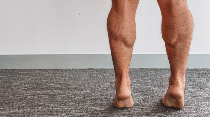
    The calf muscles, primarily responsible for plantar flexion (pointing the foot downwards), are comprised of two major muscle groups: the gastrocnemius and the soleus. These muscles work synergistically to generate powerful movements of the ankle and foot. Understanding the specific anatomical locations and functions of these muscles is essential for recognizing the potential sites of injury.

    Anatomical Description and Function of Calf Muscles

    Muscle Name Location Function Possible Injury Sites
    Gastrocnemius Located on the posterior (back) aspect of the lower leg, forming the bulk of the calf. Composed of two heads, medial and lateral. Primary plantar flexor of the foot. Also assists in knee flexion. Commonly injured at the muscle-tendon junction or within the muscle belly, especially the medial head.
    Soleus Located deep to the gastrocnemius, also on the posterior aspect of the lower leg. A larger muscle, it is situated further down the leg. Secondary plantar flexor of the foot. Crucial for maintaining posture and stability during prolonged standing. It is more active in prolonged plantar flexion. Often injured at the muscle-tendon junction, similar to the gastrocnemius. Less frequently injured than the gastrocnemius due to its deeper location.
    Plantaris A small, thin muscle located deep to the gastrocnemius. Plays a minor role in plantar flexion and knee flexion. Rarely injured on its own but can be involved in larger calf injuries.

    Types of Calf Strains

    Calf strains are typically categorized into three grades, reflecting the severity of the injury:

    • Grade 1 Strain: This involves a minor tear or stretch in the muscle fibers. Symptoms include mild pain, tenderness, and slight swelling. The affected individual may experience some difficulty with activities involving the calf muscles. The muscle might feel slightly warm to the touch. Recovery time is typically relatively short, ranging from a few days to a couple of weeks, depending on the individual’s activity level and adherence to treatment.

      This grade often responds well to conservative treatment.

    • Grade 2 Strain: This involves a partial tear in the muscle fibers. Pain, swelling, and bruising are more significant than in a Grade 1 strain. There might be a noticeable loss of strength and function in the affected leg. The individual may have difficulty bearing weight. Recovery time for a Grade 2 strain is usually longer, potentially ranging from a few weeks to several months.

      Physical therapy and rehabilitation are often necessary to regain full function. It’s important to note that the severity of symptoms can vary based on the individual’s unique anatomy and lifestyle.

    • Grade 3 Strain: This represents a complete rupture or tear of the muscle fibers. The injury is extremely painful, and significant swelling and bruising are common. The affected area may feel unstable or give way, making weight-bearing difficult or impossible. Recovery time is typically the longest, potentially exceeding several months or even requiring surgical intervention. This grade typically involves complete loss of function in the affected muscle group, with the individual unable to perform normal activities involving the calf.

      That nagging calf strain muscle spasm is a real pain, isn’t it? It can be tough to pinpoint the exact cause, but sometimes underlying conditions like high blood pressure play a role. Understanding the connection between conditions like these and calf muscle spasms is crucial. For example, if you’re curious about how diabetes might contribute to high blood pressure, check out this helpful resource on does diabetes cause high blood pressure.

      Regardless of the root cause, consistent stretching and rest are key to recovery from calf strain muscle spasm.

    Causes and Risk Factors

    Calf strains, unfortunately, are a common ailment among athletes and weekend warriors alike. Understanding the underlying causes and risk factors is crucial for prevention and effective treatment. This knowledge empowers individuals to take proactive steps to safeguard their calf muscles and avoid injury.A thorough comprehension of the mechanisms behind calf strains allows individuals to make informed decisions about their training routines and lifestyle choices.

    By identifying potential triggers, individuals can implement strategies to mitigate their risk and promote overall well-being.

    Common Causes of Calf Strains

    Various factors can contribute to the development of calf strains. Overexertion during physical activities, particularly those involving sudden increases in intensity or duration, is a frequent culprit. Rapid changes in direction or forceful movements can also strain the calf muscles. In some cases, the strain might stem from inadequate stretching or warm-up routines, which can compromise the muscle’s flexibility and resilience.

    Insufficient rest between training sessions also plays a role, as it allows the muscles insufficient time to recover and rebuild.

    Risk Factors for Calf Strains

    Several risk factors heighten the susceptibility to calf strains. Individuals with pre-existing muscle imbalances or weaknesses in the calf muscles are more prone to injury. A history of previous calf strains also significantly increases the risk of recurrence. Furthermore, inadequate warm-up routines and poor training techniques, such as improper form during exercises, can contribute to the likelihood of a calf strain.

    Underlying medical conditions, like certain neurological disorders or circulatory issues, can also influence the risk.

    Potential Contributing Factors

    Several factors can exacerbate the risk of calf strains. Poor training techniques, such as using improper form during exercises, can place excessive stress on the calf muscles, increasing the likelihood of injury. Inadequate warm-up routines fail to prepare the muscles for the demands of exercise, making them more vulnerable to strain. A lack of sufficient rest between training sessions allows insufficient time for muscle recovery and repair, increasing the risk of injury.

    Finally, certain underlying medical conditions, such as neurological disorders or circulatory issues, can also impact the muscles’ ability to handle stress, thus increasing the risk of strain.

    Comparison of Causes and Risk Factors

    Cause Description Risk Factor
    Overexertion Pushing the muscles beyond their capacity during exercise. High-intensity workouts, sudden increases in training volume.
    Inadequate Warm-up Insufficient preparation of muscles for activity. Lack of stretching, skipping dynamic movements.
    Poor Training Techniques Incorrect form during exercises, leading to uneven stress on muscles. Improper technique, lack of professional guidance.
    Previous Injury History of calf strain or other related injuries. Weakened or vulnerable muscle tissue.

    Preventative Measures for Calf Strains

    Implementing preventive measures can significantly reduce the risk of calf strains. These measures address various contributing factors, allowing individuals to actively manage their risk.

    Prevention Description Example
    Proper Warm-up Preparing the muscles for exercise through dynamic stretches and light cardio. Walking, jogging, calf raises, and static stretching.
    Gradual Progression Increasing training intensity and duration gradually. Incrementally increasing the weight lifted or running distance.
    Rest and Recovery Allowing adequate time for muscle repair and regeneration. Sufficient sleep and rest days between workouts.
    Strengthening Exercises Targeting specific muscles to enhance their strength and resilience. Exercises like calf raises, and other leg strengthening exercises.

    Symptoms and Diagnosis

    Understanding the symptoms and diagnostic methods for a calf strain is crucial for prompt and accurate treatment. A thorough assessment allows healthcare professionals to differentiate this injury from other conditions and initiate the appropriate course of action. Proper diagnosis helps avoid unnecessary delays in recovery and potential complications.

    Typical Symptoms of a Calf Strain

    Calf strains manifest with a range of symptoms, varying in intensity depending on the severity of the injury. Pain is the most prominent symptom, often localized to the affected muscle group. The pain can be sharp or dull, and it may worsen with activity or stretching. Tenderness and swelling are also common, particularly in the area of the injury.

    Muscle spasms, or involuntary contractions, might also occur as a protective mechanism. These spasms can further intensify the pain and limit movement.

    Differentiating a Calf Strain from Other Leg Injuries

    Accurate diagnosis involves distinguishing a calf strain from other leg injuries that may present with similar symptoms. A thorough physical examination, including palpation of the affected area, helps determine the precise location of pain and tenderness. Comparing the symptoms with those of other potential injuries, such as shin splints, compartment syndrome, or a hamstring strain, can be helpful in narrowing down the diagnosis.

    A detailed history of the injury, including the mechanism of injury and the type of activity performed before the onset of symptoms, can further assist in differentiating calf strains from other conditions.

    Methods Used to Diagnose a Calf Strain

    The diagnostic process for a calf strain typically involves a combination of methods. A physical examination is essential, allowing the healthcare professional to assess the range of motion, palpate the affected area for tenderness, and identify any visible signs of swelling or bruising. This examination can help determine the extent of the injury and guide further investigation. Imaging tests, such as ultrasound or MRI, might be employed to provide a more detailed view of the soft tissues in the affected area.

    These imaging techniques can reveal the extent of muscle damage, the presence of any tears, and the involvement of surrounding structures.

    Common Symptoms and Severity, Calf strain muscle spasm of the leg

    Symptom Description Severity
    Pain Localized aching or sharp pain in the calf muscle. Mild, Moderate, Severe
    Tenderness Increased sensitivity to touch in the calf muscle. Mild, Moderate, Severe
    Swelling Accumulation of fluid in the calf area. Mild, Moderate, Severe
    Muscle Spasms Involuntary contractions of the calf muscle. Mild, Moderate, Severe

    Imaging Techniques Used in Diagnosing Calf Strains

    Imaging Technique Benefits Limitations
    Ultrasound Provides real-time images of soft tissues, including muscles and tendons. Relatively inexpensive and readily available. May not show subtle tears or damage in deeper structures. Image quality can be affected by body habitus.
    MRI Detailed images of soft tissues, revealing tears, edema, and other structural abnormalities with high accuracy. Provides a comprehensive assessment of the entire calf region. More expensive than ultrasound. May not be readily available in all locations. Requires the patient to lie still for an extended period.

    Treatment and Management

    A calf strain, or muscle tear in the calf, demands prompt and appropriate treatment to minimize pain, promote healing, and prevent long-term complications. Effective management hinges on a multi-faceted approach addressing immediate pain relief, restoring function, and preventing future injuries. Early intervention significantly improves recovery outcomes.

    Immediate Treatment Strategies

    Initial care focuses on alleviating pain and preventing further damage. Crucially, the RICE protocol (Rest, Ice, Compression, Elevation) forms the cornerstone of immediate management. Following these steps promptly can significantly reduce swelling and pain, promoting faster healing.

    RICE Protocol

    • Rest: Avoid activities that aggravate the injury. This crucial step allows the injured muscles to begin the healing process without further stress. Complete rest is paramount in the initial 24-48 hours to allow the tissues to repair. Examples include avoiding running, jumping, or any activities that put stress on the affected leg.
    • Ice: Applying ice packs to the affected area for 15-20 minutes at a time, every 2-3 hours, reduces inflammation and pain. Wrap the ice pack in a thin towel to prevent direct skin contact, which can cause frostbite. The application of ice constricts blood vessels, thereby limiting swelling.
    • Compression: Using elastic bandages to compress the injured area helps control swelling. Ensure the bandage is snug but not too tight, as this could restrict blood flow. Appropriate compression reduces swelling and supports the injured tissue.
    • Elevation: Keeping the injured leg elevated above the heart helps reduce swelling by promoting the drainage of fluid away from the affected area. This position helps minimize the accumulation of blood and fluids in the injured region. Elevating the leg can be achieved by resting on pillows.

    Other Relevant Treatment Methods

    Beyond RICE, over-the-counter pain relievers like ibuprofen or naproxen can help manage pain and inflammation. In severe cases, a doctor may prescribe stronger pain medications. Proper pain management is crucial for patient comfort and to allow for adequate rest. Physical therapy, discussed further below, is essential for restoring strength and function to the injured area.

    Role of Physical Therapy

    Physical therapy plays a vital role in rehabilitation, guiding patients through exercises that progressively strengthen the calf muscles and improve flexibility. A physical therapist designs a tailored program to address the specific needs of each individual, ensuring that exercises gradually increase in intensity to promote healing and prevent re-injury. This includes targeted strengthening exercises, stretching routines, and gait training to help the patient regain full range of motion and function.

    Conservative vs. Surgical Treatment Approaches

    Conservative treatment, which includes RICE, pain medication, and physical therapy, is often the first line of treatment for most calf strains. Surgery is typically reserved for severe or complex cases, such as complete tears or those that don’t respond to conservative methods. Surgical intervention might be necessary to repair significant muscle or tendon damage. Conservative methods offer a less invasive option, but success depends on the severity and type of injury.

    Proper Warm-up Routine

    A proper warm-up routine before exercising the leg muscles helps prepare the tissues for the demands of activity, reducing the risk of injury. The warm-up should gradually increase the blood flow to the muscles and increase their flexibility. This includes activities like light cardio, such as walking or jogging, and dynamic stretches, such as leg swings, hamstring stretches, and calf raises.

    A comprehensive warm-up can significantly reduce the risk of a calf strain.

    Prevention and Rehabilitation: Calf Strain Muscle Spasm Of The Leg

    Preventing calf strains requires proactive measures to maintain muscle health and flexibility. A comprehensive approach focusing on warm-up, gradual progression, and targeted exercises is crucial for reducing the risk of injury. Rehabilitation, when a strain occurs, involves a structured program to restore strength and function, minimizing long-term impact.

    Strategies for Preventing Calf Strains

    Proper warm-up and cool-down routines, along with gradual increases in exercise intensity, significantly reduce the risk of calf strains. Maintaining flexibility and strength in the calf muscles through regular stretching and targeted exercises is essential for injury prevention. Avoiding sudden movements or overexertion, particularly when starting a new exercise routine, is also important.

    • Consistent Stretching: Regular stretching, incorporating static and dynamic stretches, improves flexibility in the calf muscles. This enhances the range of motion, reducing the risk of strains during activities that demand high levels of movement.
    • Gradual Progression: Increasing exercise intensity and duration gradually is vital. Rushing into intense workouts without proper preparation can strain the calf muscles. A gradual approach allows the muscles to adapt and strengthens them progressively.
    • Proper Footwear: Wearing appropriate footwear, especially during activities like running or jumping, provides necessary support and cushioning. Supportive shoes help maintain proper alignment and reduce the risk of injury.

    Importance of Warm-up and Cool-down Routines

    Warm-up routines prepare the muscles for physical activity, increasing blood flow and flexibility. Cool-down routines gradually reduce the heart rate and blood flow, preventing muscle stiffness and soreness. Both are crucial for preventing injuries, particularly calf strains.

    • Warm-up: Warm-up activities, such as light cardio and dynamic stretching, increase blood flow to the muscles. This prepares them for the demands of the activity, reducing the risk of strains.
    • Cool-down: Cool-down activities, such as static stretching, help reduce muscle tension and soreness. This process facilitates recovery and reduces the risk of delayed-onset muscle soreness (DOMS).

    Importance of Gradual Increases in Exercise Intensity

    Progressive overload, a principle of training, involves gradually increasing the demands placed on the muscles. This allows the body to adapt and build strength and endurance without exceeding the capacity of the muscles, which can lead to strains.

    • Avoid sudden increases: Sudden increases in exercise intensity, duration, or frequency significantly increase the risk of muscle strain. A gradual increase in workload allows the body to adapt and prevents excessive stress on the muscles.
    • Listen to your body: Pay attention to any pain or discomfort. If you experience pain, stop the activity and rest. Rest is crucial for muscle recovery and prevents further injury.

    Appropriate Exercises for Rehabilitation and Strengthening

    Rehabilitation exercises focus on restoring strength and flexibility in the affected calf muscles. A structured program of targeted exercises, tailored to the individual’s specific needs and recovery stage, is essential.

    Exercise Type Description Instructions
    Calf Raises Strengthens the gastrocnemius and soleus muscles. Stand with feet shoulder-width apart. Raise up onto the balls of your feet, then lower back down.
    Heel Drops Stretches the calf muscles. Stand with feet shoulder-width apart. Lower your heels towards the ground, maintaining a straight back.
    Toe Raises Strengthens the anterior tibialis muscle. Stand with feet shoulder-width apart. Raise up onto your toes, then lower back down.
    Seated Calf Raises Strengthens calf muscles, while offering a support system for lower back. Sit on a chair, place your feet flat on the floor, and lift your heels off the ground, slowly.
    Standing Calf Raises with Resistance Band Increases the intensity of calf raises. Use a resistance band around the balls of your feet. Perform calf raises.

    Potential Complications

    A calf strain, while often treatable with rest and proper care, can lead to more serious issues if ignored or mishandled. Understanding potential complications is crucial for prompt and effective management. Ignoring pain and continuing activity can exacerbate the injury, potentially resulting in long-term problems. Proactive management is key to minimizing these risks.Untreated or poorly managed calf strains can lead to chronic issues that affect daily activities and quality of life.

    Delayed or inadequate treatment can prolong healing time, increase the risk of re-injury, and even contribute to the development of more significant problems. Early intervention and consistent rehabilitation are essential to prevent these complications.

    Long-Term Effects of Untreated Calf Strains

    Chronic pain and stiffness are common long-term effects of untreated calf strains. This persistent discomfort can significantly impact mobility and daily tasks. The injured area may also become prone to recurring spasms or tightness. Furthermore, improper healing can lead to the development of scar tissue, which can restrict normal muscle function and potentially affect the range of motion in the affected leg.

    Recognizing Signs of Potential Chronic Problems

    It’s vital to be aware of the signs that indicate a calf strain might be developing into a chronic issue. Monitoring for changes in pain intensity, duration, and location is critical. If pain persists beyond the typical healing timeframe or if new symptoms emerge, such as swelling, discoloration, or a noticeable change in the muscle’s shape, it’s important to seek medical attention.

    Experiencing a calf strain muscle spasm? It’s super frustrating, right? While focusing on the recovery process, did you know that similar muscle tightness can sometimes stem from issues with CPAP therapy? Understanding the causes and treatment of CPAP gas buildup can be helpful for overall well-being, especially if you’re experiencing related muscle discomfort. For in-depth information on this, check out this great resource: the causes and treatment of cpap gas.

    Regardless of the cause, proper rest, stretching, and potentially professional guidance are key to getting your calf muscles back to optimal health.

    The earlier you identify these signs, the sooner you can implement appropriate treatment strategies.

    Possible Consequences of Ignoring a Calf Strain

    Ignoring a calf strain can have several negative consequences, ranging from decreased athletic performance to a greater risk of more serious injuries. Prolonged discomfort can lead to reduced mobility and a decreased ability to perform daily activities. Furthermore, ignoring the pain might lead to the injury becoming more severe, requiring extensive treatment and potentially resulting in long-term functional limitations.

    Table of Signs of Potential Chronic Problems

    Sign Description Potential Implications
    Persistent pain (beyond 6-8 weeks) Pain that continues despite appropriate rest and treatment. Possible development of chronic pain syndrome, reduced mobility, and functional limitations.
    Swelling or inflammation Continued swelling or redness in the calf area. Indicates ongoing inflammation, potentially leading to chronic issues.
    Muscle weakness or atrophy Noticeable reduction in muscle strength or size in the affected calf. Reduced ability to perform activities requiring calf muscle strength, potentially leading to re-injury.
    Changes in skin sensation (e.g., numbness, tingling) Altered sensation in the calf area. Could indicate nerve involvement and warrants immediate medical evaluation.
    Limited range of motion Difficulty flexing or extending the ankle or knee. Indicates restricted joint mobility and potential for chronic stiffness.

    Case Studies and Examples

    Calf strain muscle spasm of the leg

    Calf strains, ranging from mild discomfort to debilitating injuries, can significantly impact an individual’s daily activities. Understanding real-world examples of diagnosis, treatment, and recovery is crucial for both patients and healthcare professionals. This section delves into case studies, highlighting how various factors influence the severity and recovery process of calf strains.Case studies and examples are not intended to be a substitute for professional medical advice.

    Always consult with a qualified healthcare provider for diagnosis and treatment of any medical condition.

    Case Study: A Runner’s Calf Strain

    A 28-year-old marathon runner presented with sudden sharp pain in their right calf during a training run. The pain intensified with plantar flexion (pointing the toes downward) and was accompanied by swelling and tenderness along the calf muscle. Physical examination revealed a localized tenderness and limited range of motion in the affected area. Imaging (ultrasound or MRI) confirmed a grade II calf strain, involving partial tearing of the muscle fibers.

    Treatment included rest, ice, compression, and elevation (RICE). Physical therapy, focusing on stretching, strengthening exercises, and proprioceptive training, was initiated to restore muscle function and prevent recurrence. The runner followed the prescribed recovery plan meticulously and gradually returned to running after 6 weeks, demonstrating a successful recovery.

    Dealing with a calf strain and muscle spasms in your leg can be incredibly painful. One potential natural remedy to explore is magnesium L-threonate, which some studies suggest may help with muscle relaxation. To find the right dosage for your needs, be sure to check out this helpful resource on magnesium l threonate dosage. While magnesium might help, it’s crucial to remember that proper rest and physical therapy are still essential components of recovery from a calf strain muscle spasm.

    Factors Affecting Severity

    Several factors contribute to the varying degrees of calf strain severity. Training volume and intensity, prior injury history, and the individual’s overall fitness level play a crucial role. A runner who consistently exceeds their training limits is more susceptible to a severe strain compared to someone who maintains a moderate training schedule. Also, individuals with a history of ankle sprains or other lower leg injuries may be at higher risk due to compromised tissues and reduced flexibility.

    Sample Recovery Plan for a Calf Strain

    A typical recovery plan for a calf strain generally involves the following phases:

    • Phase 1 (Acute Phase): Rest, ice, compression, and elevation (RICE) are paramount during the first 48-72 hours to reduce inflammation and pain. Over-the-counter pain relievers may be used to manage discomfort. A physical therapist or physician can advise on the appropriate RICE protocol.
    • Phase 2 (Rehabilitation Phase): Gentle range-of-motion exercises and light stretching are introduced as tolerated. As pain subsides, strengthening exercises targeting the calf muscles and surrounding tissues are progressively incorporated. This phase aims to restore muscle function and flexibility.
    • Phase 3 (Return to Activity): Gradual return to activities like running, jumping, or other sports is crucial. The individual should start with low-impact activities and gradually increase the intensity and duration of workouts as tolerated. This phase emphasizes avoiding overexertion to prevent re-injury.

    Factors Contributing to Successful Recovery

    A successful recovery from a calf strain depends on several key factors:

    • Adherence to the prescribed treatment plan: Strict adherence to the recommended rest, ice, compression, elevation (RICE), and rehabilitation exercises is crucial for optimal healing.
    • Proper nutrition and hydration: Adequate protein intake supports muscle repair, while sufficient hydration aids in tissue recovery.
    • Early and appropriate medical intervention: Seeking timely medical advice and following a prescribed treatment plan can significantly reduce recovery time and prevent complications.
    • Progressive rehabilitation exercises: Gradual progression of exercises is essential to avoid overexertion and promote a safe and effective recovery.
    • Mental well-being: Maintaining a positive mindset and avoiding stress or anxiety is crucial for a smooth recovery process.
    • Appropriate footwear and training surfaces: Selecting appropriate footwear and training surfaces can help prevent re-injury by providing adequate support and cushioning.

    Illustrations and Visual Aids

    Visual aids play a crucial role in understanding and effectively managing calf strain. Detailed images and diagrams can clarify complex anatomical structures, proper treatment techniques, and the progression of rehabilitation. This section provides comprehensive visual representations to aid in the comprehension of calf strain, from its anatomy to management strategies.

    Cross-Section of the Lower Leg

    A detailed cross-section of the lower leg reveals the intricate arrangement of muscles, tendons, and ligaments. The image should clearly highlight the gastrocnemius and soleus muscles, which are the primary muscles of the calf. These muscles are positioned strategically in relation to the Achilles tendon, the large band of tissue connecting the calf muscles to the heel bone.

    The image should also depict the surrounding connective tissues, such as the fascia, which can be affected in cases of strain. Proper visualization of these structures aids in understanding the potential areas of injury and the mechanisms of strain.

    Proper Application of Ice Packs

    Visualizing the proper application of ice packs is essential for effective pain management. The image should depict a cold pack wrapped in a thin cloth or towel, applied directly to the affected calf area. The cold pack should cover the injured region without applying excessive pressure. The image should demonstrate the appropriate duration of application, typically 15-20 minutes at a time, with a 1-hour break between applications.

    A clear image showcasing the ice pack’s placement, covering the area of the strain, is crucial.

    Correct Technique for Stretching the Calf Muscles

    Proper stretching techniques are vital for regaining flexibility and preventing future strains. The image should show a person performing a calf stretch. The image should clearly illustrate the correct posture, emphasizing the importance of maintaining a straight back and not bending forward. The foot should be kept flat on the ground. The image should clearly demonstrate the stretch, emphasizing the range of motion required without pushing too far to avoid pain.

    The duration of the stretch should be emphasized, generally holding the stretch for 20-30 seconds, and repeating several times daily.

    Grades of Calf Strains

    Understanding the severity of a calf strain is crucial for appropriate treatment. A diagram illustrating the different grades of calf strains is essential. The diagram should visually represent the varying degrees of muscle damage, from mild (grade 1) to severe (grade 3). Grade 1 strains may show minor muscle tears, while grade 3 strains involve significant tearing of the muscle fibers.

    The diagram should use clear labeling and color coding to distinguish each grade, helping to visually differentiate the severity levels of the injury.

    Rehabilitation Program for Calf Strains

    A visual representation of a rehabilitation program for calf strains should include a series of exercises, progressively increasing in intensity and complexity. The image should depict a progression of exercises, from basic range-of-motion exercises to more advanced exercises focusing on strength and flexibility. The progression should be clearly indicated, with each exercise demonstrated to aid in understanding the steps involved.

    The diagram should emphasize the importance of gradually increasing the intensity and duration of exercises to prevent further injury.

    Concluding Remarks

    Calf strain muscle spasm of the leg

    In conclusion, calf strain muscle spasm of the leg, while often painful and disruptive, is manageable with appropriate care. By understanding the causes, symptoms, and treatment options Artikeld in this guide, you can significantly reduce your risk of injury and ensure a swift and complete recovery. Remember prevention is key, so prioritize proper warm-up routines, gradual intensity increases, and appropriate exercise techniques.

  • Hyperpathia, Hyperalgesia, Hypersensitivity A Deep Dive

    Hyperpathia, Hyperalgesia, Hypersensitivity A Deep Dive

    Hyperpathia hyperalgesia and hypersensitivity – Hyperpathia, hyperalgesia, and hypersensitivity: these terms describe heightened pain experiences, but what exactly do they mean? This exploration delves into the intricacies of these conditions, from defining each distinct pain response to understanding the underlying mechanisms and how they affect daily life. We’ll uncover the various causes, diagnostic methods, treatment options, and the profound impact on patients.

    Each condition manifests differently, impacting pain perception, intensity, and duration. Understanding these nuances is crucial for proper diagnosis and management. We’ll examine the intricate interplay of physiological and psychological factors that contribute to these conditions. This exploration will also cover the importance of addressing the psychosocial impact on patients and provide a look into current research and future directions.

    Defining Hyperpathia, Hyperalgesia, and Hypersensitivity: Hyperpathia Hyperalgesia And Hypersensitivity

    Hyperpathia hyperalgesia and hypersensitivity

    Understanding the nuances of pain perception is crucial for effective diagnosis and treatment. This exploration delves into the distinct characteristics of hyperpathia, hyperalgesia, and hypersensitivity, highlighting their physiological mechanisms and the differences in pain perception and response. These conditions, while often intertwined, present unique challenges in clinical settings.These three conditions represent heightened pain responses, characterized by amplified sensitivity to stimuli.

    However, the intensity, duration, and nature of pain differ considerably. This discussion will explore the unique features of each condition, examining the underlying mechanisms that contribute to their development.

    Defining Hyperpathia, Hyperpathia hyperalgesia and hypersensitivity

    Hyperpathia is characterized by an exaggerated response to painful stimuli, often described as a prolonged, intense, and burning sensation beyond the expected pain response. It’s not just an increase in pain intensity, but a qualitative change in the pain experience. Patients frequently report a feeling of heightened sensitivity to touch or pressure, even non-noxious stimuli, in the affected area.

    This condition often coexists with other neurological conditions, indicating a complex interplay of factors in its development.

    Defining Hyperalgesia

    Hyperalgesia signifies an increased sensitivity to painful stimuli in an area surrounding the initial injury. It’s a localized amplification of pain response, rather than a widespread alteration like hyperpathia. The pain is typically more intense than expected for the stimulus. A common example is the heightened sensitivity to touch or pressure in the area surrounding a burn.

    The underlying mechanism involves changes in the nervous system’s processing of pain signals.

    Defining Hypersensitivity

    Hypersensitivity, in the context of pain, refers to an exaggerated response to normally innocuous stimuli. This condition involves an increased perception of pain from non-noxious stimuli. This can manifest as a heightened sensitivity to light touch, temperature changes, or even air currents. The pain experienced is often described as a burning or tingling sensation. The physiological mechanisms underpinning hypersensitivity often involve altered neurotransmitter systems and inflammatory responses.

    Comparing and Contrasting Hyperpathia, Hyperalgesia, and Hypersensitivity

    Characteristic Hyperpathia Hyperalgesia Hypersensitivity
    Pain Quality Prolonged, intense, burning, heightened sensitivity to touch/pressure Increased intensity compared to the stimulus, localized Burning, tingling, heightened sensitivity to normally innocuous stimuli
    Pain Intensity Markedly increased, often disproportionate to the stimulus Increased, but often proportional to the stimulus Increased response to normally innocuous stimuli
    Pain Duration Prolonged, lasting beyond the expected recovery period Can vary depending on the stimulus and injury Can persist even after the stimulus is removed
    Stimulus Usually involves a past injury or neurological condition Localized to the site of injury or damage Normally innocuous stimuli (light touch, temperature, pressure)

    The table above provides a concise comparison of the three conditions. It highlights the distinctions in pain quality, intensity, duration, and the type of stimulus that triggers the heightened response. Further research is needed to fully elucidate the intricate mechanisms underlying these complex conditions.

    Etiology and Causes

    Understanding the root causes of hyperpathia, hyperalgesia, and hypersensitivity is crucial for developing effective treatment strategies. These conditions arise from a complex interplay of factors, ranging from peripheral nerve damage to central nervous system alterations. Delving into these etiological aspects provides a more comprehensive picture of these often debilitating conditions.Peripheral and central nervous systems are intricately connected, with signals flowing between them.

    Disruptions in this intricate network can lead to altered pain processing, resulting in the exaggerated responses seen in hyperpathia, hyperalgesia, and hypersensitivity. This heightened sensitivity to pain stems from various mechanisms, each contributing to the overall experience.

    Potential Causes of Hyperpathia, Hyperalgesia, and Hypersensitivity

    The varied etiologies of these conditions underscore the complex nature of pain processing. They encompass a wide range of factors, from direct nerve injury to systemic illnesses. It is important to understand that multiple factors often interact to produce these conditions.

    Neurological Pathways Involved

    The pathways involved in processing pain signals are intricate and multi-layered. Pain signals initiated at the periphery travel through sensory neurons to the spinal cord, where they are relayed to higher brain centers. Disruptions in any part of this pathway can contribute to the development of these conditions. For example, damage to the peripheral nerves, such as those caused by diabetes or trauma, can lead to altered signal transmission.

    Peripheral and Central Sensitization

    Peripheral sensitization involves heightened responsiveness of nociceptors (pain receptors) in the periphery. This occurs in response to damage, inflammation, or other stimuli. Central sensitization, on the other hand, involves the nervous system’s central processing centers becoming hypersensitive to pain signals. This heightened responsiveness can lead to a more prolonged and intense pain experience. Both peripheral and central sensitization are crucial in the development of chronic pain conditions.

    Genetic and Environmental Factors

    Genetic predisposition plays a role in some cases, influencing the individual’s response to pain stimuli. For example, some individuals might have a genetic susceptibility to nerve damage or inflammatory conditions that can trigger these heightened pain responses. Environmental factors such as exposure to toxins or trauma can also contribute to the development of these conditions.

    Table of Causes and Associated Conditions

    Cause Associated Conditions
    Nerve Damage (e.g., trauma, diabetes, infection) Hyperpathia, Hyperalgesia, Neuropathic pain
    Inflammation (e.g., arthritis, autoimmune diseases) Hyperalgesia, Chronic pain
    Neuropathic Pain Hyperpathia, Hyperalgesia, Hypersensitivity
    Chronic Pain Conditions Hyperpathia, Hyperalgesia, Hypersensitivity
    Systemic Illnesses (e.g., cancer, multiple sclerosis) Hyperpathia, Hyperalgesia, Hypersensitivity
    Substance Abuse Hyperalgesia, Chronic pain

    Diagnosis and Assessment

    Pinpointing hyperpathia, hyperalgesia, and hypersensitivity requires a multi-faceted approach, blending meticulous clinical evaluation with specific diagnostic tools. Accurate diagnosis hinges on identifying the unique constellation of symptoms and signs exhibited by each individual, enabling clinicians to differentiate between these overlapping conditions and pinpoint the underlying cause.A thorough understanding of the patient’s medical history, including past injuries, illnesses, and medications, is crucial.

    This information, combined with a detailed account of the current symptoms, provides valuable context for evaluating the nature and severity of the sensory disturbances.

    Clinical Evaluation Procedures

    Clinical evaluation procedures play a vital role in diagnosing and assessing the severity of hyperpathia, hyperalgesia, and hypersensitivity. These procedures involve a comprehensive examination of the patient’s neurological function, including sensory perception and motor skills. Detailed questioning about the location, intensity, quality, and duration of pain and other sensory symptoms is essential.

    Diagnostic Methods

    Various diagnostic methods are employed to ascertain the presence and extent of these conditions. These methods include a detailed neurological examination, assessing reflexes, muscle strength, and coordination. Furthermore, specific sensory testing, such as pain thresholds and pressure sensitivity, aids in identifying abnormal sensory processing.

    Pain Scales and Assessment Tools

    Pain scales are essential tools for quantifying the intensity and characteristics of pain. Visual Analog Scales (VAS) and Numerical Rating Scales (NRS) allow patients to subjectively report their pain levels. These scales provide a standardized method for assessing pain intensity and help track changes over time. Additionally, other tools like the McGill Pain Questionnaire can provide a more detailed description of the pain experience, encompassing sensory qualities, emotional aspects, and location.

    Neurological Examinations

    Neurological examinations are fundamental in assessing the integrity of the nervous system. These examinations include evaluating reflexes, muscle strength, coordination, and sensory function in various parts of the body. Abnormal findings in these areas may suggest underlying neurological conditions contributing to or exacerbating the pain and sensory issues.

    Differentiating Symptoms and Signs

    Differentiating between hyperpathia, hyperalgesia, and hypersensitivity relies on recognizing subtle yet critical distinctions in the patient’s presentation. For example, hyperpathia is characterized by exaggerated pain responses to normally innocuous stimuli, often accompanied by allodynia (pain from non-painful stimuli). Hyperalgesia, on the other hand, involves heightened pain sensitivity to painful stimuli, while hypersensitivity encompasses a broader range of abnormal sensory responses beyond pain.

    Careful observation of these specific symptoms and signs aids in accurate diagnosis and subsequent treatment planning.

    Summary Table of Diagnostic Criteria and Assessment Methods

    Condition Diagnostic Criteria Assessment Methods
    Hyperpathia Exaggerated pain response to normally innocuous stimuli; allodynia; often associated with neuropathic pain. Pain scales (VAS, NRS); neurological examination (sensory testing); patient history.
    Hyperalgesia Increased sensitivity to painful stimuli; heightened pain perception. Pain scales (VAS, NRS); quantitative sensory testing (QST); patient history.
    Hypersensitivity Abnormal sensory response to various stimuli; may include pain, temperature, touch, or pressure; can involve multiple sensory modalities. QST; neurological examination; patient history; specific sensory tests.

    Management and Treatment

    Managing hyperpathia, hyperalgesia, and hypersensitivity involves a multifaceted approach tailored to individual needs. These conditions often require a combination of pharmacologic and non-pharmacologic interventions to effectively address the underlying causes and symptoms. The goal is not just to reduce pain intensity but also to improve overall quality of life.Effective treatment strategies aim to target both the acute and chronic aspects of these conditions, acknowledging that the approach may need to evolve over time.

    This requires close collaboration between patients, healthcare providers, and other relevant specialists, such as physical therapists or psychologists.

    Pharmacologic Interventions

    Pharmacologic treatments play a crucial role in managing the pain associated with hyperpathia, hyperalgesia, and hypersensitivity. Various classes of medications are used, each with its own mechanism of action and potential side effects. Careful consideration of individual patient factors, such as comorbidities and medication history, is essential when selecting the appropriate pharmacologic intervention.

    Understanding hyperpathia, hyperalgesia, and hypersensitivity can be tricky, but it’s crucial for pinpointing the root cause of discomfort. A rash on the upper inner thigh, in a female, could be a symptom of an underlying issue. Conditions like these might be linked to heightened sensitivity in the area, which could manifest as a rash. For more information on identifying and treating a rash upper inner thigh female, check out this helpful resource: rash upper inner thigh female.

    Ultimately, though, consulting a medical professional is essential to properly diagnose and manage any such sensitivity issues.

    • Opioids: While effective for some patients, opioids carry the risk of addiction and other adverse effects, such as respiratory depression. Careful monitoring and dose titration are crucial. Examples include morphine, oxycodone, and fentanyl.
    • Nonsteroidal anti-inflammatory drugs (NSAIDs): These drugs can reduce inflammation and pain, potentially alleviating some aspects of hyperalgesia. However, they may not be as effective for hyperpathia. Examples include ibuprofen and naproxen.
    • Antidepressants: Certain antidepressants, such as tricyclic antidepressants (TCAs) and serotonin-norepinephrine reuptake inhibitors (SNRIs), can be effective in managing neuropathic pain components of these conditions. They may also help regulate mood and sleep, which can indirectly improve overall well-being.
    • Anticonvulsants: These medications can modulate neuronal activity and are often used to treat neuropathic pain associated with hyperpathia. Examples include gabapentin and pregabalin.

    Non-Pharmacologic Interventions

    Non-pharmacologic interventions are equally important in the management of hyperpathia, hyperalgesia, and hypersensitivity. These strategies can complement pharmacologic treatments and often address the broader impact of these conditions on a patient’s life.

    • Physical Therapy: Exercises, manual therapy, and other physical interventions can improve mobility, reduce muscle tension, and enhance overall physical function. This is particularly helpful in managing pain and improving function in patients with chronic conditions.
    • Cognitive Behavioral Therapy (CBT): CBT can help patients identify and modify negative thought patterns and behaviors related to pain. This can reduce the emotional distress and anxiety associated with chronic pain conditions, which can often exacerbate symptoms.
    • Mindfulness-Based Stress Reduction (MBSR): Practices like meditation and mindfulness can help regulate stress responses and promote relaxation, which can lead to a reduction in pain perception. This approach can be particularly valuable for patients experiencing heightened emotional distress related to their pain.
    • Alternative Therapies: Techniques like acupuncture, massage therapy, and spinal manipulation are sometimes used in conjunction with other treatments to reduce pain and improve overall well-being.

    Efficacy and Side Effects

    The efficacy of various treatment options varies significantly among individuals. Factors like the underlying cause of the condition, the severity of symptoms, and individual patient responses influence treatment outcomes. Close monitoring and adjustments to the treatment plan are essential to optimize efficacy and minimize potential side effects.

    Patient Population Comparisons

    The effectiveness of different treatment approaches may differ across various patient populations, taking into account factors like age, gender, and specific medical conditions. For instance, older adults may have different responses to certain medications compared to younger individuals.

    Hyperpathia, hyperalgesia, and hypersensitivity are fascinating neurological conditions, but they can be quite debilitating. Understanding these conditions is important, but so is knowing when certain eye issues might start to develop, like macular degeneration, which often appears in later life. Research suggests that age plays a significant role in the onset of macular degeneration, and for those interested in learning more, a helpful resource is available here: what age does macular degeneration start.

    While the connection between these conditions isn’t immediately apparent, a deeper understanding of both neurological and ophthalmological conditions is important for better overall health.

    Managing Pain Intensity and Quality of Life

    Management of pain intensity and improvement of quality of life involve a combination of pharmacologic and non-pharmacologic strategies. Tailoring the treatment plan to the individual patient’s needs and preferences is crucial for achieving optimal outcomes. Regular follow-up appointments with healthcare providers are essential to assess treatment effectiveness and make necessary adjustments to the management plan.

    Clinical Presentations and Manifestations

    Understanding the diverse clinical presentations of hyperpathia, hyperalgesia, and hypersensitivity is crucial for accurate diagnosis and effective management. These conditions, while sharing overlapping symptoms, can manifest in vastly different ways depending on the underlying cause. Recognizing the spectrum of presentations allows healthcare providers to tailor treatment strategies to the individual patient’s needs.These conditions are characterized by abnormal pain processing, which can range from heightened sensitivity to specific stimuli to widespread, debilitating pain.

    The specific symptoms and signs observed in patients will depend on the type of condition, its severity, and the underlying cause. Recognizing the nuances in presentation is essential for appropriate diagnostic evaluations and the selection of the most effective interventions.

    Hyperpathia, hyperalgesia, and hypersensitivity are fascinating neurological conditions, often involving heightened pain responses. Understanding these conditions requires a deep dive into the underlying mechanisms, but one crucial factor influencing pain perception is the red blood cell rbc count. Variations in this can significantly impact the body’s ability to deliver oxygen and nutrients, potentially affecting nerve function and contributing to heightened pain sensitivity.

    Ultimately, further research into the intricate interplay between the red blood cell rbc count red blood cell rbc count and the neurological pathways involved in pain processing is essential for developing effective treatments for hyperpathia, hyperalgesia, and hypersensitivity.

    Variability in Clinical Presentation

    The clinical presentations of hyperpathia, hyperalgesia, and hypersensitivity vary significantly due to the diverse etiologies. These conditions are not monolithic; rather, they represent a spectrum of abnormal pain processing. This variability in presentation can be attributed to factors such as the specific location of the affected tissue, the intensity and duration of the stimulus, and the presence of other comorbidities.

    For example, a patient with hyperalgesia resulting from nerve damage may experience extreme sensitivity to touch in the affected area, while a patient with hyperpathia due to a central nervous system disorder might experience prolonged, burning pain after a seemingly minor stimulus.

    Specific Symptoms and Signs

    Patients with these conditions may present with a wide range of symptoms and signs, which can vary in intensity and location. Common symptoms include:

    • Increased sensitivity to stimuli: Patients may report heightened pain responses to normally innocuous stimuli, such as light touch, cold, or pressure. This can range from mild discomfort to severe, debilitating pain. For example, a person with hyperalgesia may experience excruciating pain from a simple touch that would be barely noticeable to a healthy individual.
    • Allodynia: This refers to the experience of pain from a stimulus that does not normally cause pain. A patient with allodynia might experience pain from clothing touching their skin or a light breeze.
    • Hyperpathia: Characterized by a disproportionately intense and prolonged pain response to a stimulus, often accompanied by spontaneous pain episodes. A person experiencing hyperpathia may feel a sharp, intense pain following a minor injury that lasts for hours or days.
    • Spontaneous pain: This is pain that occurs without any apparent external stimulus. Patients may describe it as a burning, throbbing, or aching sensation.
    • Changes in pain perception: Patients may experience altered pain thresholds and pain qualities. This can manifest as an increased sensitivity to certain types of pain or a decreased ability to perceive other types of pain.

    Examples of Different Presentations Based on Underlying Causes

    The clinical presentation of these conditions can vary significantly depending on the underlying cause. For instance, hyperalgesia following a burn injury would likely present with localized pain and increased sensitivity to touch in the affected area. In contrast, hyperpathia associated with a spinal cord injury may manifest as widespread pain, particularly in the extremities, and be accompanied by spontaneous pain episodes.

    Table of Clinical Presentations

    Clinical Presentation Associated Symptoms Possible Causes
    Localized Hyperalgesia Increased sensitivity to touch, pressure, temperature in a specific area; allodynia Trauma, inflammation, nerve damage, burns, post-operative pain
    Widespread Hyperpathia Prolonged, intense pain, spontaneous pain episodes, altered pain perception; possible paresthesia Central nervous system disorders, spinal cord injury, neuropathies
    Diffuse Hypersensitivity Increased sensitivity to various stimuli (touch, temperature, chemicals); allodynia Chronic pain conditions, psychological factors, autoimmune diseases, fibromyalgia

    Impact on Daily Life and Quality of Life

    The chronic pain and heightened sensitivity associated with hyperpathia, hyperalgesia, and hypersensitivity profoundly affect daily life, impacting everything from simple tasks to complex social interactions. These conditions often create significant challenges, diminishing the quality of life and necessitating comprehensive management strategies.These conditions create a multifaceted impact on individuals, extending beyond the physical realm to encompass emotional, social, and vocational aspects.

    Understanding the specific ways these conditions disrupt daily routines, interpersonal relationships, and overall well-being is crucial for developing effective support systems and interventions.

    Impact on Daily Activities

    Daily tasks, often taken for granted, become significant hurdles for individuals experiencing these conditions. Simple actions like walking, dressing, or even handling everyday objects can be excruciatingly painful. This pain can lead to avoidance behaviors, resulting in reduced mobility and isolation.

    • Examples of affected activities: Walking, showering, dressing, using household appliances, or even interacting with certain fabrics or textures. For example, a person with hyperpathia might find the pressure of a bedsheet unbearable, while a person with hyperalgesia might avoid certain foods due to their perceived pain-inducing properties.
    • Reduced participation in social activities: The fear of pain and discomfort can limit participation in social events, hobbies, and recreational activities. This can lead to feelings of isolation and depression.

    Impact on Work and Education

    The fluctuating pain and sensitivity associated with these conditions can severely disrupt work and education. Chronic pain can lead to missed workdays, reduced productivity, and difficulty concentrating. Students may struggle to attend classes, complete assignments, or maintain focus.

    • Impact on work performance: Pain and discomfort can lead to absenteeism, decreased productivity, and difficulty concentrating on tasks. This can have significant financial consequences and impact career advancement.
    • Challenges in education: Students may experience difficulty concentrating in class, completing assignments, and participating in extracurricular activities. This can affect their academic performance and overall well-being.

    Impact on Relationships

    These conditions can strain interpersonal relationships due to the unpredictable nature of pain and the need for adjustments in daily routines. Family members and partners may struggle to understand the challenges faced by the affected individual.

    • Strained communication: Chronic pain and heightened sensitivity can lead to misunderstandings and conflict within relationships. Communication becomes strained as individuals may struggle to express their needs and experiences.
    • Impact on social interactions: Individuals may withdraw from social interactions due to fear of pain or discomfort. This can lead to feelings of isolation and loneliness.

    Impact on Emotional Well-being and Mental Health

    The chronic pain and limitations imposed by hyperpathia, hyperalgesia, and hypersensitivity can take a significant toll on emotional well-being. Anxiety, depression, and feelings of hopelessness are common.

    • Increased risk of depression and anxiety: The chronic pain and limitations associated with these conditions can lead to feelings of helplessness, frustration, and isolation, significantly increasing the risk of developing anxiety and depression.
    • Impact on self-esteem and confidence: The inability to perform everyday tasks and participate in social activities can lead to a decline in self-esteem and confidence.

    Importance of Psychosocial Support

    Addressing the psychosocial impact of these conditions is crucial for effective management. Support groups, counseling, and education programs can help individuals cope with the emotional and social challenges.

    • Importance of psychological support: Providing psychological support, such as counseling or therapy, is essential for managing the emotional distress associated with these conditions.
    • Role of support groups: Support groups can provide a sense of community and shared experience, allowing individuals to connect with others facing similar challenges.

    Research and Future Directions

    Hyperpathia hyperalgesia and hypersensitivity

    Unraveling the complexities of hyperpathia, hyperalgesia, and hypersensitivity requires ongoing research. Current investigations are focusing on understanding the underlying mechanisms and developing effective treatments. This exploration will hopefully lead to improved diagnostic tools, personalized therapies, and a better quality of life for those affected.

    Current Research Focuses

    Current research into these conditions encompasses a broad spectrum of investigations. Researchers are exploring the intricate interplay of neuronal pathways, neurotransmitters, and inflammatory processes. Advanced neuroimaging techniques are providing valuable insights into the structural and functional changes in the brain and nervous system related to these conditions. Genetic studies are identifying potential susceptibility genes and biomarkers for early detection.

    Ongoing Investigations into Potential Treatments

    Numerous studies are evaluating novel therapeutic approaches. These include exploring the efficacy of targeted medications, such as specific analgesics and neuromodulators. Experimental treatments like nerve stimulation techniques, including transcutaneous electrical nerve stimulation (TENS) and spinal cord stimulation, are being examined for their potential in managing symptoms. Furthermore, research into non-pharmacological interventions, such as physical therapy and cognitive behavioral therapy (CBT), is underway to determine their efficacy in alleviating symptoms and improving overall well-being.

    Promising Avenues for Future Research and Development

    Several avenues hold promise for future research and development. A deeper understanding of the specific mechanisms underlying central sensitization is crucial. Further research into the role of the immune system in these conditions is also anticipated. Developing personalized treatment strategies based on individual genetic profiles and symptom presentation is a significant goal. The exploration of novel drug targets and combination therapies is also expected to yield promising results.

    For example, combining pharmacological interventions with non-pharmacological approaches, like mindfulness-based stress reduction, may provide a more comprehensive and effective strategy.

    Need for Further Investigation into the Mechanisms

    A deeper understanding of the complex interplay of factors contributing to these conditions is essential. This includes examining the role of neuroplasticity, the mechanisms of chronic pain, and the interplay between genetic predisposition and environmental factors. Understanding the intricate mechanisms behind these conditions is crucial for developing effective and targeted therapies. For example, identifying the specific molecular pathways involved in central sensitization could pave the way for more precise and effective treatments.

    Potential Areas for Future Research and Clinical Trials

    Future research should focus on developing more sophisticated diagnostic tools, including biomarkers and advanced neuroimaging techniques. Clinical trials should investigate the efficacy of novel treatments, such as gene therapies and regenerative medicine approaches, for managing chronic pain conditions. For example, exploring the possibility of using stem cell therapies to repair damaged nerve tissue warrants further investigation. Further investigation into the role of psychological factors, such as stress and anxiety, in exacerbating symptoms is essential.

    For example, a prospective clinical trial could compare the effectiveness of a new medication with a standard therapy in a group of patients with specific genetic predispositions to hyperpathia. This would require a comprehensive evaluation of patient outcomes, including pain intensity, quality of life, and psychological well-being.

    Summary

    In conclusion, hyperpathia, hyperalgesia, and hypersensitivity are complex conditions that significantly impact patients’ quality of life. From understanding the distinct characteristics of each to exploring potential causes, diagnostic methods, and treatment approaches, this discussion highlights the importance of comprehensive care. Future research is vital to refine our understanding of these conditions and improve treatment strategies.

  • The Brains Frontal Lobe A Deep Dive

    The Brains Frontal Lobe A Deep Dive

    The brains frontal lobe – The brain’s frontal lobe sets the stage for this enthralling narrative, offering readers a glimpse into a story that is rich in detail and brimming with originality from the outset. This crucial part of the brain, located at the front of the cerebral cortex, plays a pivotal role in everything from our higher-level cognitive functions to our personality and social interactions.

    We’ll explore its intricate workings, examining its role in executive functions, language, movement, sensory integration, and the impact of various neurological conditions. We’ll also delve into its development throughout our lifespan and how it shapes who we are.

    Understanding the frontal lobe is key to comprehending the complexity of the human brain. This region is the seat of planning, decision-making, and impulse control. It’s also deeply involved in higher-level cognitive processes like working memory and attention. Furthermore, the frontal lobe has a significant impact on personality and social behavior. Throughout this exploration, we’ll uncover the fascinating relationship between structure, function, and the myriad of influences that shape the frontal lobe.

    Introduction to the Frontal Lobe

    The frontal lobe, situated at the very front of the brain, is a crucial component of the human nervous system. It’s the most evolved part of the brain, playing a vital role in higher-level cognitive functions, including planning, decision-making, and social behavior. Understanding its structure and function is essential to comprehending human cognition and behavior.The frontal lobe’s general function extends beyond simple motor control.

    It’s the seat of executive functions, a set of mental processes that allow us to control our thoughts, actions, and emotions in a way that is goal-directed and socially appropriate. This sophisticated processing allows us to adapt to our environment, plan for the future, and engage in complex problem-solving.

    Anatomical Structures of the Frontal Lobe

    The frontal lobe is not a homogenous entity. It contains several distinct areas, each contributing to specific cognitive functions. These areas, while interconnected, have unique anatomical features and specialized roles in information processing.

    Key Cognitive Functions of the Frontal Lobe

    The frontal lobe’s involvement in cognitive functions is extensive. It’s not just about motor movements, but a complex interplay of mental processes. These functions encompass planning, organizing, initiating, and inhibiting actions, as well as working memory and problem-solving.

    Historical Overview of Frontal Lobe Research

    Early understanding of the frontal lobe’s role in human behavior was gradual, evolving through meticulous observation of patients with brain damage. Phineas Gage’s famous accident in the 19th century provided early evidence of the frontal lobe’s crucial role in personality and behavior. Subsequent research, employing various neuroimaging techniques, further clarified the intricate workings of the frontal lobe and its multifaceted involvement in human cognition.

    Major Areas within the Frontal Lobe

    Understanding the different regions within the frontal lobe helps to understand the specific roles they play. Each region, while interconnected, has a unique role in the processing of information.

    Structure Location Key Function
    Prefrontal Cortex Anterior portion of the frontal lobe Higher-level cognitive functions, including planning, decision-making, working memory, and social cognition. Also involved in regulating emotions and inhibiting inappropriate responses.
    Motor Cortex Posterior portion of the frontal lobe Voluntary movement initiation. Different areas control specific body parts. Precise movements require the integration of signals from various brain areas, including the cerebellum.
    Broca’s Area Left frontal lobe, usually Language production. Damage to this area can result in difficulty in speaking or forming coherent sentences, although comprehension remains largely intact.
    Supplementary Motor Area Anterior to the primary motor cortex Planning and coordination of complex movements. It’s involved in the sequencing of actions and the control of posture.

    Specific Functions of the Frontal Lobe

    The frontal lobe, situated at the very front of the brain, is often considered the seat of higher-level cognitive functions. It plays a crucial role in planning, decision-making, and social interactions, distinguishing human behavior from that of other animals. This intricate region, responsible for complex cognitive processes, significantly impacts our daily lives.The frontal lobe’s remarkable complexity allows us to anticipate future events, strategize, and adapt to changing circumstances.

    This capacity is fundamental to navigating the intricate social landscape and making sound judgments. The frontal lobe’s influence extends beyond simple motor actions, impacting our ability to think abstractly, solve problems, and regulate our emotions.

    Executive Functions: Planning and Decision-Making

    The frontal lobe is essential for executive functions, a set of higher-order cognitive processes that enable us to plan, organize, and execute tasks. Planning involves anticipating future events and devising strategies to achieve goals. For instance, a student might plan a study schedule, anticipating the time needed for each subject and factoring in potential distractions. Decision-making relies on evaluating options, considering potential consequences, and selecting the best course of action.

    A professional choosing a career path weighs various factors, such as salary, job satisfaction, and personal aspirations.

    Executive Functions: Working Memory and Attention

    Working memory is a crucial aspect of cognitive function, enabling us to hold information in mind temporarily and manipulate it. The frontal lobe plays a vital role in this process, allowing us to retain and process information needed for immediate tasks. For example, following a complex set of instructions requires the frontal lobe to actively maintain the instructions in working memory and apply them sequentially.

    Attention, too, is significantly influenced by the frontal lobe, enabling us to focus on relevant stimuli and filter out distractions.

    Influence on Personality and Social Behavior

    The frontal lobe significantly shapes our personality and social behavior. It’s involved in regulating emotions, understanding social cues, and controlling impulses. Damage to this area can result in significant changes in personality, such as impulsivity, aggression, or apathy. This is evident in cases of traumatic brain injury affecting the frontal lobe. Understanding social norms and responding appropriately to social situations also depend on the frontal lobe’s function.

    Comparison of Frontal Lobe Functions Across Species

    While all mammals possess a frontal lobe, its size and complexity vary considerably across species. Humans, with their remarkably developed frontal lobes, exhibit a significantly higher level of executive function and cognitive flexibility compared to other animals. For example, primates, particularly chimpanzees, show sophisticated problem-solving skills, suggesting a degree of frontal lobe involvement. However, the capacity for abstract thought and complex planning appears to be more pronounced in humans.

    Comparison of Executive Function Deficits

    Characteristic Frontal Lobe Damage Other Cognitive Impairments (e.g., ADHD, Dementia)
    Planning and Organization Difficulty initiating tasks, poor organization, and lack of foresight. Varying degrees of difficulty, depending on the specific impairment. May involve challenges with planning, but also with attention and memory.
    Decision-Making Impulsive decisions, poor risk assessment, and difficulty considering consequences. Difficulties may stem from attention problems, memory lapses, or emotional dysregulation.
    Working Memory Difficulty holding and manipulating information in mind, leading to poor performance on tasks requiring short-term memory. Problems with memory encoding, storage, or retrieval, impacting working memory capacity.
    Attention Difficulty sustaining attention, easily distracted, and struggles to filter out irrelevant stimuli. Reduced attention span, distractibility, and difficulty focusing on tasks.

    This table provides a general comparison; specific deficits can vary significantly depending on the extent and location of the brain damage or the nature of the cognitive impairment.

    Frontal Lobe and Brain Development

    The frontal lobe, the brain’s command center, isn’t fully formed at birth. Its development unfolds across the lifespan, influenced by both genetic predispositions and environmental factors. Understanding this developmental trajectory is crucial for comprehending how we learn, reason, and interact with the world. This journey of maturation shapes our cognitive abilities, social behaviors, and overall well-being.The frontal lobe, responsible for executive functions like planning, decision-making, and impulse control, undergoes a complex and dynamic process of growth and refinement throughout life.

    This maturation isn’t a simple linear progression but rather a series of interconnected phases influenced by a multitude of factors. These factors are crucial in shaping the individual’s capabilities and vulnerabilities.

    Timeline of Frontal Lobe Development

    The frontal lobe’s development is a protracted process, extending well into adulthood. Early development lays the groundwork for later functions, while later refinement enhances complex cognitive skills. This process is not uniform across individuals, but follows general patterns.

    Environmental Influences on Frontal Lobe Maturation, The brains frontal lobe

    Environmental factors significantly impact frontal lobe development. Nutritious diets, stimulating environments, and supportive relationships foster healthy brain growth. Conversely, factors such as malnutrition, exposure to toxins, and chronic stress can hinder maturation. These factors highlight the importance of creating supportive environments conducive to optimal brain development.

    Impact of Early Childhood Experiences on Frontal Lobe Development

    Early childhood experiences play a pivotal role in shaping the frontal lobe’s structure and function. Positive interactions, nurturing care, and opportunities for exploration contribute to a robust and adaptable frontal lobe. Conversely, experiences of trauma, neglect, or consistent stress can disrupt the development of essential neural pathways. This highlights the crucial role of early intervention programs and support systems.

    Changes in Frontal Lobe Function with Age

    Frontal lobe function changes throughout life, adapting to new challenges and experiences. In childhood, the focus is on developing basic cognitive skills. In adolescence, the prefrontal cortex undergoes significant development, leading to improved decision-making abilities. In adulthood, the frontal lobe continues to adapt, adjusting to new information and experiences. Maintaining cognitive health throughout life involves strategies to support and stimulate frontal lobe function.

    Stages of Frontal Lobe Development

    Stage Associated Milestones Potential Challenges
    Infancy (0-2 years) Basic motor skills, social interactions, object permanence, early language development Delayed language development, difficulties with motor skills, feeding difficulties
    Early Childhood (3-5 years) Increased vocabulary, improved social skills, pre-reading and pre-writing skills, problem-solving skills Difficulties with attention, impulsivity, emotional regulation issues
    Middle Childhood (6-12 years) Improved executive functions, enhanced cognitive abilities, developing abstract thought Difficulty with organizational skills, academic struggles, social anxieties
    Adolescence (13-18 years) Improved reasoning skills, decision-making abilities, abstract thought, increased independence Risk-taking behaviors, impulsivity, emotional instability
    Adulthood (19-65 years) Maintaining cognitive function, adapting to new situations, problem-solving Cognitive decline, difficulty with multitasking, memory issues
    Older Adulthood (65+) Adjusting to changes in cognitive abilities, maintaining social connections, adapting to physical changes Increased risk of cognitive decline, loss of independence, social isolation

    Frontal Lobe and Neurological Disorders

    The frontal lobe, the brain’s executive center, plays a crucial role in higher-level cognitive functions. Damage to this area can lead to a wide array of impairments, impacting various aspects of daily life. Understanding the specific neurological conditions affecting the frontal lobe, the associated symptoms, and the challenges in diagnosis and treatment is vital for effective care and support for affected individuals.Neurological conditions can significantly disrupt the frontal lobe’s functioning, impacting cognitive abilities, personality, and behavior.

    These disruptions can manifest in a multitude of ways, often depending on the specific location and extent of the damage. Precise diagnosis and tailored treatment strategies are essential for improving outcomes.

    Common Neurological Conditions Affecting the Frontal Lobe

    Various neurological conditions can affect the frontal lobe, impacting its crucial functions. Stroke, traumatic brain injury, and dementia are prominent examples. Each condition presents unique challenges in terms of symptom presentation and treatment.

    Symptoms Associated with Frontal Lobe Damage

    Frontal lobe damage can result in a broad range of symptoms, varying depending on the extent and location of the lesion. These symptoms can impact cognitive abilities, personality, and behavior.

    • Stroke: A stroke, caused by interrupted blood flow to the brain, can lead to various frontal lobe symptoms, such as difficulties with planning, problem-solving, and decision-making. A patient might exhibit impulsivity, disinhibition, or apathy. Emotional lability, characterized by rapid shifts in mood, is also a potential symptom. The severity of symptoms correlates with the affected area and the extent of the damage.

      For example, a stroke affecting the left frontal lobe might lead to language difficulties, whereas a stroke in the right frontal lobe may manifest as neglect or spatial disorientation.

    • Traumatic Brain Injury (TBI): TBI, resulting from a blow or jolt to the head, can also cause frontal lobe damage. This damage can manifest as difficulties with executive functions, including impaired working memory, planning, and organizing. Personality changes, such as aggression or irritability, are common observations. Changes in social behavior, such as inappropriate social interactions or reduced empathy, are frequently observed.

      Ever wondered about the frontal lobe’s role in our complex brains? It’s fascinating how this part of our brain impacts everything, even seemingly unrelated things like allergic asthma triggers. For example, did you know that certain environmental factors can unexpectedly spark an asthma attack? Learning about the surprising things that trigger allergic asthma, like surprising things that trigger allergic asthma , can help us understand how our bodies respond to the world around us.

      Ultimately, understanding the frontal lobe’s role in processing information and controlling reactions is key to comprehending the intricate workings of our minds.

      Severity of symptoms depends on the nature and extent of the injury.

    • Dementia: Dementia, a progressive neurodegenerative disorder, can lead to frontal lobe dysfunction. Symptoms include difficulties with executive functions, such as planning and organizing, and personality changes. Behavioral disturbances, such as agitation, aggression, or apathy, can occur. Memory impairment is often a prominent feature, progressively worsening as the disease progresses. Specific types of dementia, like frontotemporal dementia, directly target the frontal and temporal lobes.

    Comparison of Effects of Different Frontal Lobe Lesions

    Different types of frontal lobe lesions, stemming from various conditions, can result in varying symptom presentations.

    Condition Location of Lesion Typical Symptoms
    Stroke Variable, depending on the affected artery Executive dysfunction, impulsivity, emotional lability
    TBI Variable, depending on the impact site Cognitive deficits, personality changes, disinhibition
    Frontotemporal Dementia Frontal and temporal lobes Progressive personality changes, language difficulties, behavioral disturbances

    Challenges in Diagnosing and Treating Frontal Lobe Disorders

    Diagnosing frontal lobe disorders presents significant challenges. Symptoms can overlap with other conditions, making differentiation crucial. Furthermore, the lack of specific, easily measurable biomarkers makes accurate diagnosis difficult. Treatment is also complex, focusing on managing symptoms rather than offering a cure.

    Ever wondered how your brain’s frontal lobe might be subtly influencing your sleep? It plays a significant role in regulating body temperature, which, as you might guess, is directly connected to whether you’re a hot sleeper or a cold sleeper. Understanding these sleep temperature preferences can offer insights into your overall well-being, like learning about your body’s natural temperature regulation and finding the perfect sleep environment.

    For more on the fascinating science of hot sleeper vs cold sleeper , check out this great resource. Ultimately, a better understanding of your brain’s temperature control mechanisms can lead to a more restful night’s sleep.

    Diagnostic Procedures and Tools

    Various diagnostic procedures and tools are employed to identify frontal lobe damage.

    • Neuropsychological testing: Neuropsychological evaluations assess cognitive functions, such as memory, attention, and executive functions. These tests provide valuable insights into the extent and nature of frontal lobe dysfunction.
    • Neuroimaging techniques: Neuroimaging, such as MRI and CT scans, allows visualization of brain structures and can help identify areas of damage. These techniques are crucial in confirming suspected lesions and determining their location.
    • Clinical interviews: Clinical interviews with the patient and family members provide crucial information about behavioral changes, cognitive difficulties, and medical history, contributing to a comprehensive understanding of the situation. This is essential for a thorough diagnosis.

    Frontal Lobe and Behavior

    The frontal lobe, the brain’s executive center, plays a crucial role in shaping our behavior. It’s not simply a command center, but a complex network interacting with other brain regions to orchestrate our thoughts, emotions, and actions. This intricate interplay significantly influences our personality, emotional responses, and social interactions. Understanding the frontal lobe’s involvement in behavior is essential for comprehending human actions and the impact of damage to this vital region.

    The Connection Between Frontal Lobe Activity and Personality Traits

    Personality traits, a complex combination of stable patterns of thought, feeling, and behavior, are significantly influenced by frontal lobe activity. The prefrontal cortex, a key component of the frontal lobe, is intimately involved in higher-level cognitive functions, including decision-making, planning, and working memory. These functions, in turn, contribute to the development and expression of personality characteristics. For instance, individuals with strong prefrontal cortex activity often demonstrate traits like conscientiousness, self-control, and emotional stability.

    Conversely, disruptions in this region can lead to impulsivity, lack of foresight, and difficulties in regulating emotional responses, which may manifest as personality changes.

    How Frontal Lobe Function Relates to Emotional Regulation and Impulse Control

    The frontal lobe is instrumental in regulating emotions and controlling impulses. It acts as a sort of “brake” on the more primitive emotional centers in the brain, allowing for a more nuanced and controlled response to stimuli. Effective emotional regulation involves the frontal lobe’s ability to assess situations, anticipate consequences, and choose appropriate responses. Impulse control relies on similar mechanisms, where the frontal lobe helps us inhibit immediate urges and consider long-term implications.

    Individuals with compromised frontal lobe function may experience difficulty with emotional regulation, exhibiting heightened reactivity to emotional stimuli or displaying erratic mood swings. Similarly, poor impulse control can manifest in inappropriate behaviors and difficulty delaying gratification.

    The Relationship Between Frontal Lobe Activity and Social Behavior

    Social behavior is profoundly influenced by the frontal lobe. The ability to understand social cues, empathize with others, and navigate complex social situations relies heavily on the frontal lobe’s executive functions. This includes aspects like perspective-taking, recognizing social norms, and adapting behavior to different social contexts. Individuals with intact frontal lobe function are typically better equipped to manage social interactions, displaying appropriate communication styles and understanding the impact of their actions on others.

    Conversely, damage to the frontal lobe can disrupt social cognition, leading to difficulties in understanding social cues, inappropriate social behavior, and a diminished capacity for empathy.

    Comparison and Contrast of Behavioral Effects of Frontal Lobe Damage in Different Individuals

    The behavioral effects of frontal lobe damage vary considerably among individuals. This variability stems from several factors, including the specific location and extent of the damage, the individual’s pre-existing personality traits, and the age at which the damage occurs. For example, damage in the dorsolateral prefrontal cortex may primarily affect executive functions, leading to problems with planning and decision-making, whereas damage in the orbitofrontal cortex might primarily affect emotional regulation and social behavior.

    The impact on personality, impulse control, and social interactions can be profound and varied.

    Summary Table of Behavioral Changes in Frontal Lobe Damage

    Area of Damage Specific Behavioral Changes Examples
    Dorsolateral Prefrontal Cortex Impaired planning, poor judgment, difficulty with problem-solving, lack of foresight, apathy, distractibility A patient struggling to plan a simple meal or unable to manage daily tasks, difficulty making decisions, reduced motivation to complete work.
    Orbitofrontal Cortex Emotional lability, inappropriate social behavior, impulsivity, disinhibition, poor judgment, lack of empathy A patient making inappropriate sexual advances or engaging in impulsive spending sprees, displaying significant mood swings, difficulty understanding social cues.
    Anterior Cingulate Cortex Problems with attention, emotional regulation, conflict resolution, and working memory A patient showing difficulty focusing on tasks or reacting inappropriately to frustration, trouble with maintaining consistent emotional responses.

    Frontal Lobe and Language: The Brains Frontal Lobe

    The frontal lobe, often considered the “executive” center of the brain, plays a surprisingly crucial role in language, extending far beyond simple comprehension. It’s involved in not just the production of words but also the nuances of communication, from choosing the right words to understanding context and social cues. This intricate relationship between the frontal lobe and language is essential for effective interaction and comprehension.The frontal lobe isn’t solely responsible for language; it collaborates with other brain regions, particularly those in the temporal lobe, such as Broca’s area and Wernicke’s area.

    This complex network allows for the smooth flow of thoughts into spoken or written language. The frontal lobe’s role in language is multifaceted, encompassing planning, organizing, and monitoring the language process.

    The Frontal Lobe’s Role in Language Production and Comprehension

    The frontal lobe’s contribution to language extends beyond mere articulation. It is crucial for the planning and sequencing of words, the selection of appropriate vocabulary, and the monitoring of the flow of speech. Moreover, the frontal lobe plays a critical role in understanding the nuances of language, such as tone, context, and implied meaning. This allows us to interpret the intent behind the words and react appropriately.

    Damage to this area can result in significant impairments in language production and comprehension.

    Connection Between the Frontal Lobe and Broca’s Area

    Broca’s area, located in the inferior frontal gyrus of the frontal lobe, is intimately connected to language production. This area is responsible for the motor aspects of speech, translating thoughts into the physical act of speaking. Damage to Broca’s area results in Broca’s aphasia, characterized by difficulties in producing fluent speech, although comprehension often remains relatively intact. The frontal lobe’s role in language production extends beyond Broca’s area, influencing the overall planning and execution of speech.

    Specific Areas of the Frontal Lobe Involved in Speech and Language Processing

    The frontal lobe houses multiple areas contributing to language processing. The inferior frontal gyrus, encompassing Broca’s area, is paramount for speech production. Further back, in the dorsolateral prefrontal cortex, there are areas that contribute to higher-level language functions such as planning, organizing, and monitoring the flow of language. These regions are not isolated but work in concert, facilitating complex language tasks.

    The precise contributions of each area are still being investigated.

    Language Impairments in Individuals with Frontal Lobe Damage

    Damage to different areas within the frontal lobe can lead to distinct language impairments. Damage to the inferior frontal gyrus, particularly Broca’s area, results in difficulties in producing fluent speech, known as Broca’s aphasia. Lesions in the dorsolateral prefrontal cortex can impair the ability to plan and organize language, potentially leading to disorganized speech and difficulty with abstract language use.

    These impairments can range from minor difficulties in sentence structure to complete inability to communicate.

    Anatomical Locations and Functions of Key Language-Related Areas Within the Frontal Lobe

    Area Anatomical Location Primary Function
    Broca’s Area Inferior frontal gyrus, frontal lobe Language production, speech articulation
    Dorsolateral Prefrontal Cortex Dorsolateral frontal lobe Planning, organizing, monitoring language, higher-level cognitive aspects of language
    Supplementary Motor Area (SMA) Medial frontal lobe Planning and sequencing complex movements, including speech

    This table highlights the key areas within the frontal lobe that are involved in language functions, showcasing their locations and roles in the complex process of language production and comprehension. Note that these areas often interact and overlap, contributing to a unified language system.

    Ever wondered about the amazing frontal lobe of your brain? It’s responsible for so much, from decision-making to planning. But what about the seemingly mundane act of cracking your neck? Is it actually harmful? To learn more about the potential risks, check out this informative article on is cracking your neck bad for you.

    Ultimately, a healthy brain, including a well-functioning frontal lobe, is crucial for our overall well-being.

    Frontal Lobe and Movement

    The brains frontal lobe

    The frontal lobe, often considered the “executive” center of the brain, plays a crucial role in orchestrating our voluntary movements. Beyond simply initiating actions, this intricate region is responsible for the planning, sequencing, and execution of complex motor tasks. This intricate process involves a network of connections and pathways, highlighting the frontal lobe’s vital role in our ability to interact with the world around us.The frontal lobe’s involvement in movement extends far beyond the simple act of muscle contraction.

    It’s responsible for selecting the appropriate movements, considering their consequences, and adapting them based on sensory feedback. Damage to this area can lead to significant impairments in motor control, highlighting the delicate balance required for smooth and purposeful action.

    Motor Planning and Execution

    The frontal lobe, particularly the premotor cortex and supplementary motor area, is critical in the planning and execution of movements. These areas aren’t just simple initiators; they act as sophisticated coordinators, assembling the necessary steps and strategies for a particular task. For example, preparing to reach for a cup involves not only deciding to move the arm but also anticipating the cup’s location, the necessary grip, and the overall sequence of actions.

    Connection with the Premotor Cortex

    The premotor cortex, located anterior to the primary motor cortex, acts as a crucial bridge between the frontal lobe’s higher-level planning functions and the primary motor cortex’s execution. It receives information from various brain regions, including sensory areas and association cortices, to refine the motor plan. This information helps tailor the movement to the specific context and environmental cues.

    For instance, if a person is preparing to grab a hot cup, the premotor cortex would consider the temperature and the potential burn risk. This anticipatory aspect is a key component of motor planning.

    Coordination of Voluntary Movements

    The frontal lobe coordinates voluntary movements by integrating information from different brain regions. It receives input from sensory areas, allowing the brain to assess the current environment and adjust movements accordingly. The supplementary motor area plays a critical role in coordinating the timing and sequencing of multiple muscle groups involved in complex movements. For example, walking involves coordinated movements of legs, arms, and torso, all carefully orchestrated by the frontal lobe.

    Impact of Frontal Lobe Damage on Motor Function

    Damage to the frontal lobe can significantly impact motor function. Individuals with frontal lobe injuries may experience difficulties initiating movements, maintaining smooth sequences, and adapting to changing circumstances. Apraxia, a disorder characterized by the inability to perform learned movements despite intact motor abilities, is often associated with frontal lobe damage. For example, a person might struggle to tie their shoes, even though they know how to do it.

    Similarly, deficits in executive functions, such as problem-solving and planning, can also manifest as motor impairments.

    Pathways Involved in Motor Control

    The pathways involved in motor control originating from the frontal lobe are complex and multifaceted. These pathways involve multiple cortical and subcortical structures. The corticospinal tract, originating from the primary motor cortex and passing through the brainstem and spinal cord, is crucial for controlling voluntary movements. The motor cortex sends signals to the spinal cord, which then relays them to muscles.

    Other pathways, like the corticobulbar tract, control cranial nerve-mediated movements, such as facial expressions. This intricate system allows for precise control over a vast array of movements, reflecting the complexity of motor behavior.

    Example of Motor Pathway Involvement

    Imagine reaching for a glass of water. The frontal lobe, through the premotor cortex, plans the reaching movement. Sensory input from the eyes and proprioceptors (receptors in muscles and joints) informs the brain about the glass’s location and the current position of the arm. The supplementary motor area coordinates the timing of the hand’s movement. The signal travels down the corticospinal tract, activating the necessary muscles in the arm and hand.

    This complex process, orchestrated by the frontal lobe, allows for a smooth and accurate movement.

    Frontal Lobe and Sensory Integration

    The brains frontal lobe

    The frontal lobe, often associated with higher-level cognitive functions, plays a crucial role in integrating sensory information from various parts of the brain. It doesn’t simply receive sensory data; it actively interprets and utilizes this information to guide our actions and shape our perceptions of the world around us. This integration process is essential for our ability to react appropriately to our environment and to make meaningful decisions.The frontal lobe’s role in sensory integration extends beyond mere processing.

    It acts as a central hub, coordinating input from different sensory modalities to create a comprehensive understanding of a situation. This allows us to, for example, not only see a red light but also understand its meaning as a signal to stop, or feel the warmth of the sun and perceive the pleasantness of the weather. This intricate process of integration is critical for our daily interactions.

    Sensory Modality Integration

    The frontal lobe integrates information from various sensory systems, including visual, auditory, tactile, olfactory, and gustatory. This integration is not a simple summation of individual sensations but a complex process that creates a cohesive experience. This integration enables us to make sense of our surroundings and respond appropriately. For example, hearing a siren (auditory) and seeing flashing lights (visual) simultaneously allows us to understand the urgency of the situation.

    Examples of Frontal Lobe Interpretation

    The frontal lobe interprets sensory input in many ways, often combining different sensory experiences to form a complete understanding. For instance, the smell of freshly baked bread (olfactory) combined with the sight of a warm loaf (visual) evokes a sense of comfort and anticipation. Another example is feeling the texture of a soft blanket (tactile) while listening to soothing music (auditory) creating a relaxed state.

    These experiences aren’t just isolated sensations; they’re actively interpreted and combined by the frontal lobe to create a holistic perception.

    Influence on World Perception

    The frontal lobe significantly influences our perception of the world. It allows us to assign meaning to sensory input, enabling us to react appropriately and make informed decisions. For example, the taste of a certain food (gustatory) combined with the experience of its preparation (visual and tactile) can lead to a strong positive or negative association. The frontal lobe is crucial in constructing our subjective understanding of the world around us.

    Relationship Between Frontal Lobe and Sensory Modalities

    Sensory Modality Frontal Lobe Role Example
    Visual Interprets visual information, contextualizes it with other sensory input, and influences motor responses. Seeing a stop sign (visual) and interpreting its meaning (frontal lobe) to slow down the car.
    Auditory Processes auditory information, distinguishes important sounds from background noise, and directs attention accordingly. Hearing a baby crying (auditory) and recognizing the need for assistance (frontal lobe).
    Tactile Integrates tactile sensations with other sensory information, assesses the implications of touch, and adjusts behavior accordingly. Feeling a hot stove (tactile) and quickly withdrawing your hand (motor response controlled by the frontal lobe).
    Olfactory Processes smells, connects them with memories and emotions, and influences behaviors and decisions. Smelling burnt food (olfactory) and deciding to not eat it (frontal lobe decision-making).
    Gustatory Processes taste, integrates it with other sensory experiences, and influences preferences and food choices. Tasting a delicious meal (gustatory) and enjoying it (frontal lobe influencing pleasure).

    Concluding Remarks

    In conclusion, the frontal lobe is a remarkable and intricate part of the brain, playing a multifaceted role in shaping our thoughts, actions, and overall experience. From its involvement in crucial cognitive functions to its influence on personality and behavior, this region is essential for understanding human cognition and behavior. Its vulnerability to neurological conditions highlights the importance of continued research and support for those affected.

    This exploration has just scratched the surface, but hopefully, it provides a fascinating introduction to this critical brain region.

  • CPAP vs. BiPAP Sleep Apnea Solutions

    CPAP vs. BiPAP Sleep Apnea Solutions

    What is the difference between CPAP and BiPAP? Understanding these two therapies is crucial for anyone struggling with sleep apnea. CPAP and BiPAP machines deliver pressurized air to keep your airways open during sleep, preventing interruptions to your breathing. However, they differ in how they achieve this, leading to distinct advantages and disadvantages for different patients.

    This guide delves into the specifics of CPAP and BiPAP, comparing their pressure settings, treatment applications, and user experiences. We’ll explore which type of machine might be best suited for you, based on your sleep apnea severity and individual needs. It’s important to remember that the best choice should always be determined by a healthcare professional after a thorough evaluation.

    Introduction to CPAP and BiPAP

    What is the difference between cpap and bipap

    CPAP and BiPAP are two common types of positive airway pressure (PAP) therapy used to treat sleep apnea. Sleep apnea is a sleep disorder characterized by pauses in breathing during sleep. These pauses can significantly reduce the amount of oxygen reaching the body, leading to various health problems. Both CPAP and BiPAP machines deliver pressurized air to keep the airways open during sleep, preventing these breathing interruptions.

    Understanding the differences between these therapies is crucial for patients to choose the most effective treatment for their specific needs.These therapies work by applying a gentle, constant or varying pressure to the airway, preventing collapse during sleep. This pressure keeps the airways open, allowing for uninterrupted breathing and the proper exchange of oxygen and carbon dioxide. This ultimately improves sleep quality and overall health.

    CPAP and BiPAP machines are both used to help with breathing issues, but they differ in their pressure delivery. Understanding these differences is crucial, but sometimes weight loss when sick can also affect breathing patterns, making it even more important to understand the nuances of these machines. Ultimately, consulting a doctor to determine the right machine for your specific needs is key to managing breathing problems effectively.

    CPAP Therapy

    CPAP, or Continuous Positive Airway Pressure, delivers a constant stream of pressurized air to the airway throughout the entire breathing cycle. The pressure is set by a doctor based on individual needs. CPAP is generally well-suited for individuals with mild to moderate sleep apnea.

    BiPAP Therapy

    BiPAP, or Bilevel Positive Airway Pressure, delivers two different pressures during the breathing cycle. One pressure is used during inhalation (Inspiratory Positive Airway Pressure, or IPAP), and a lower pressure is used during exhalation (Expiratory Positive Airway Pressure, or EPAP). This allows for a more natural breathing pattern and can be more comfortable for some patients, particularly those with more severe sleep apnea.

    Basic Mechanics of CPAP and BiPAP Machines

    CPAP machines generate a continuous flow of air at a pre-set pressure, which is delivered through a mask covering the nose and/or mouth. The pressure forces the airway open, preventing it from collapsing. BiPAP machines, on the other hand, deliver varying pressures during inhalation and exhalation. The higher pressure during inhalation helps to open the airways more effectively, and the lower pressure during exhalation helps to facilitate easier exhalation.

    Comparison of CPAP and BiPAP Machines

    Feature CPAP BiPAP
    Pressure Setting Single, constant pressure Two different pressures (IPAP/EPAP)
    Airflow Continuous flow Variable flow
    Suitability Mild to moderate sleep apnea Moderate to severe sleep apnea

    This table highlights the key differences in pressure and airflow between the two machines, aiding in determining which therapy is best suited for a given patient’s condition.

    Ever wondered about the difference between CPAP and BiPAP machines? They both help with breathing issues, but BiPAP offers more support, adjusting pressure for inhaling and exhaling. Knowing this can be helpful, especially if you’re researching treatment options for conditions like the various types of ovarian cancer. For example, if you’re looking for comprehensive information on different ovarian cancer types, check out this resource: types of ovarian cancer.

    Ultimately, the best choice between CPAP and BiPAP depends on individual needs and breathing patterns.

    Key Differences in Pressure Settings: What Is The Difference Between Cpap And Bipap

    CPAP and BiPAP machines both deliver pressurized air to help with breathing during sleep, but their pressure delivery systems differ significantly. This difference in pressure delivery is crucial in managing sleep apnea and achieving optimal treatment results. Understanding the varying pressure settings is essential for both patients and healthcare providers to ensure the therapy is effective and comfortable.

    Pressure Settings and Air Delivery

    The key difference between CPAP and BiPAP lies in the pressure settings and how they deliver air. CPAP, or Continuous Positive Airway Pressure, maintains a constant pressure throughout the entire breathing cycle. BiPAP, or Bilevel Positive Airway Pressure, delivers two different pressures: an inspiratory pressure (IPAP) during inhalation and an expiratory pressure (EPAP) during exhalation. This dual-pressure approach allows for a more natural breathing pattern, as it mimics the natural pressure fluctuations during respiration.

    Pressure Ranges and Sleep Apnea Severity

    The pressure settings used in CPAP and BiPAP machines are adjusted based on the severity of sleep apnea. The severity of sleep apnea is typically assessed through a sleep study. This assessment provides vital data to determine the appropriate pressure settings for each individual.

    Sleep Apnea Severity CPAP Pressure Range (cmH2O) BiPAP Pressure Range (cmH2O)
    Mild 5-10 8-12 (IPAP/EPAP)
    Moderate 10-15 10-15 (IPAP/EPAP)
    Severe 15+ 12+ (IPAP/EPAP)

    Different pressure ranges for CPAP and BiPAP machines are tailored to different sleep apnea severities. For instance, a patient with mild sleep apnea might benefit from a CPAP pressure of 7 cmH2O, while a patient with severe sleep apnea might require a CPAP pressure of 18 cmH2O. Similarly, in BiPAP therapy, adjusting IPAP and EPAP pressures based on the severity allows for a more customized approach to respiratory support.

    Impact on Therapy Effectiveness

    The pressure differences significantly impact the effectiveness of each therapy. In CPAP, a constant pressure helps to keep the airway open throughout the breathing cycle. This constant pressure is effective in preventing airway collapse, particularly during sleep. In BiPAP, the variable pressures allow for a more natural breathing pattern, which can be more comfortable for some patients.

    The tailored pressures also offer more support during inhalation and exhalation. For example, a patient with a higher inspiratory pressure requirement might experience better airflow and improved sleep quality using BiPAP compared to CPAP. Furthermore, patients with more complex breathing patterns might benefit more from BiPAP’s variable pressures to adjust to these fluctuations.

    Treatment Applications and Patient Needs

    What is the difference between cpap and bipap

    Choosing between CPAP and BiPAP depends heavily on the specific needs of the patient. Understanding the types of sleep apnea each therapy addresses, individual patient factors, and the role of physician evaluation is crucial for optimal treatment selection. This section delves into these aspects, providing examples and considerations for diverse patient profiles.

    Types of Sleep Apnea Addressed by Each Therapy

    CPAP is generally the first-line treatment for mild to moderate obstructive sleep apnea (OSA). BiPAP is often considered for more severe cases or when CPAP isn’t well-tolerated. Central sleep apnea (CSA), a condition where the brain fails to send signals to the muscles controlling breathing, can also be treated with BiPAP, as it can help regulate breathing patterns throughout the night.

    Furthermore, patients with complex medical conditions, such as heart failure or chronic obstructive pulmonary disease (COPD), may require BiPAP for improved respiratory support.

    Patient Factors Influencing Therapy Choice

    Several patient factors play a significant role in determining the most suitable therapy. These include the severity of sleep apnea, the patient’s tolerance to CPAP pressure, and any underlying medical conditions. For instance, a patient with a history of nasal congestion or other anatomical factors affecting airflow may find BiPAP more comfortable. Similarly, patients with respiratory issues beyond sleep apnea, like COPD, might benefit from BiPAP’s adjustable pressure settings.

    Additionally, a patient’s overall health status and their willingness to comply with the treatment plan are crucial considerations.

    CPAP and BiPAP machines are both used to help with breathing issues, but they differ in their delivery of air pressure. Understanding these subtle differences is key for anyone needing this kind of support. However, sometimes, more serious issues like parasitic infections of the central nervous system parasitic infections of the central nervous system can also impact breathing.

    Thankfully, with proper diagnosis and treatment, these conditions can be managed, allowing for a return to a more comfortable breathing pattern, and helping you understand the finer points of CPAP and BiPAP. Ultimately, consulting a healthcare professional is essential for determining the most suitable treatment plan for individual needs.

    Role of Physician Evaluation in Selecting the Appropriate Therapy

    A thorough sleep study, often involving overnight monitoring of breathing and brain activity, is essential for accurate diagnosis and personalized treatment recommendations. Physicians evaluate the severity of the sleep apnea, considering factors such as the apnea-hypopnea index (AHI), and the patient’s overall health. This evaluation guides the choice between CPAP and BiPAP, ensuring the most effective and well-tolerated therapy.

    Patient Scenarios Where One Therapy Might Be Preferred Over the Other

    A patient with mild OSA might find CPAP sufficient and well-tolerated, experiencing significant improvement in sleep quality and daytime alertness. Conversely, a patient with moderate to severe OSA, or a patient who experiences significant mask discomfort or pressure intolerance with CPAP, may benefit more from BiPAP’s ability to deliver varying pressures during inhalation and exhalation. Similarly, a patient with COPD might find BiPAP’s adjustable pressure settings to be more supportive of their respiratory needs.

    Considerations for Patients with Specific Medical Conditions or Anatomical Features

    Patients with nasal congestion, facial deformities, or other anatomical issues impacting airflow may find BiPAP more comfortable due to its adjustable pressure settings. Furthermore, patients with certain medical conditions, like heart failure or neuromuscular disorders, may need BiPAP’s more tailored pressure support to address their specific respiratory requirements. For instance, a patient with a history of respiratory complications might benefit from the pressure control provided by BiPAP.

    Summary of Ideal Patient Profiles

    Feature CPAP BiPAP
    Ideal Patient Type Patients with mild to moderate sleep apnea Patients with moderate to severe sleep apnea, or patients with difficulty tolerating CPAP
    Potential Challenges Mask discomfort, pressure intolerance, nasal dryness Potential for higher cost, potential for machine complexity

    Machine Functionality and User Experience

    CPAP and BiPAP machines, while both serving the crucial purpose of improving sleep apnea, differ significantly in their functionality and user experience. Understanding these differences can greatly influence patient comfort and treatment adherence. This section dives deep into the specifics of each machine’s components, user-friendliness, available masks, and crucial aspects like mask fitting.The choice between CPAP and BiPAP is often a personalized one, guided by individual needs and preferences.

    A comprehensive understanding of the machines’ characteristics empowers patients and healthcare professionals to make well-informed decisions.

    Machine Components and Functions

    CPAP machines deliver a constant stream of air pressure to keep the airways open during sleep. BiPAP machines, on the other hand, offer two different pressure levels – one for inhalation and another for exhalation. This allows for a more natural breathing pattern. CPAP machines typically consist of a compressor, a pressure regulator, a flow meter, and tubing connected to a mask.

    BiPAP machines have similar components but also include a separate pressure control mechanism to manage both inspiratory and expiratory pressures. The compressor’s efficiency directly impacts the machine’s overall performance, while the pressure regulator ensures consistent pressure delivery.

    Ease of Use and User Experience, What is the difference between cpap and bipap

    CPAP machines are generally straightforward to use. Modern machines often feature intuitive controls, clear displays, and automated settings. BiPAP machines, with their dual pressure settings, may require a bit more adjustment. Patient training is crucial for both machines to maximize effectiveness. The user experience greatly impacts adherence to the treatment plan.

    Types of Masks and Proper Fitting

    A wide array of masks are available for both CPAP and BiPAP therapy. Common types include nasal masks, full face masks, and nasal pillow masks. Proper mask fitting is paramount for successful treatment. A well-fitting mask ensures a consistent seal around the nose and mouth, preventing air leaks and ensuring the prescribed pressure is delivered effectively. Incorrect fitting can lead to discomfort, skin irritation, and reduced treatment efficacy.

    Mask Discomfort and Leaks

    Mask discomfort and leaks are common challenges for patients using CPAP or BiPAP machines. Factors like mask size, material, and facial structure can contribute to discomfort. Proper mask selection and fitting are essential for minimizing these issues. Addressing mask-related issues through adjustments, different mask types, or specialized fitting techniques can improve user experience.

    Machine Noise and Mitigation

    Some CPAP and BiPAP machines can produce noticeable noise. The noise level can vary based on the machine model and the compressor’s performance. Several strategies can help reduce noise. Using a quieter machine model or placing the machine away from the bed can significantly reduce noise disturbance. Additionally, ensuring the machine is properly maintained can minimize noise issues.

    Comparison of Mask Types

    Mask Type CPAP BiPAP
    Nasal Mask Suitable for most patients Suitable for most patients
    Full Face Mask Suitable for patients with nasal issues or who leak with nasal masks Suitable for patients with nasal issues or who leak with nasal masks, and potentially those who find full-face more comfortable
    Nasal Pillow Mask Suitable for patients with nasal issues Suitable for patients with nasal issues

    This table summarizes the suitability of different mask types for CPAP and BiPAP, considering factors like facial structure and potential leakage. Individual needs and preferences should be considered alongside these general guidelines.

    Long-Term Considerations and Maintenance

    Long-term CPAP or BiPAP therapy success hinges on consistent use and proper maintenance. This isn’t a quick fix; it’s a commitment to a healthier, more restful life. Understanding the long-term aspects, from machine upkeep to potential side effects, is crucial for maximizing the benefits and minimizing any drawbacks.Consistent use is paramount to achieving lasting positive results. Adherence to the prescribed treatment plan, coupled with proactive maintenance, significantly improves the long-term effectiveness of these therapies.

    Long-Term Effectiveness of Therapy

    CPAP and BiPAP therapies, when used consistently, offer long-term improvements in sleep quality and overall health. Patients often experience reduced daytime sleepiness, improved cognitive function, and better cardiovascular health over time. Studies have shown that sustained use leads to a noticeable reduction in sleep apnea-related complications like high blood pressure and heart problems.

    Importance of Consistent Use

    Adherence to the prescribed treatment plan is critical for long-term effectiveness. Missing treatments or using the devices inconsistently can hinder progress and potentially negate the positive effects of the therapy. Regular use allows the body to adapt to the pressure support, promoting improved breathing patterns and a better night’s sleep. This consistency is often a key factor in patient satisfaction and successful long-term management of sleep apnea.

    Factors Influencing Treatment Adherence

    Several factors can impact treatment adherence. These include the complexity of the equipment, the perceived effort required for daily use, and the potential for discomfort or side effects. Furthermore, patient education, support systems, and a strong doctor-patient relationship all contribute to successful long-term adherence.

    Maintaining CPAP and BiPAP Machines

    Regular maintenance of the CPAP or BiPAP machine is vital for optimal performance and prolonged device lifespan. This includes proper cleaning of the machine and its components.

    Maintaining CPAP and BiPAP Machines – Step-by-Step Guide

    Task Description
    Cleaning the Mask Thoroughly clean the mask according to the manufacturer’s instructions. This usually involves using mild soap and water, or a specific mask cleaner. Avoid harsh chemicals.
    Cleaning the Machine Clean the machine according to the manufacturer’s instructions. This typically involves removing the water reservoir, cleaning it, and reassembling the machine.
    Filter Replacement Replace filters as recommended by the manufacturer. Filters prevent dust and other particles from entering the machine, ensuring optimal airflow and extending the machine’s lifespan.
    Humidifier Care (if applicable) Follow the manufacturer’s instructions for cleaning and maintaining the humidifier. This usually involves regularly cleaning the humidifier reservoir and replacing water to prevent bacterial growth.

    Potential Long-Term Side Effects and Mitigation

    While CPAP and BiPAP therapies are generally safe, some patients may experience temporary side effects such as nasal dryness, mask leaks, or discomfort. Nasal dryness can be mitigated by using a humidifier, while mask leaks can often be resolved with adjustments to the mask fit. Consult your healthcare provider if any side effects persist or worsen.

    Final Summary

    In conclusion, understanding the nuances of CPAP and BiPAP is vital for navigating sleep apnea treatment effectively. Choosing the right therapy requires a careful consideration of your individual needs and sleep apnea severity, guided by a physician. This comprehensive comparison provides a starting point for making an informed decision, but always consult a medical professional for personalized advice.

  • Rheumatoid Arthritis in the Neck Understanding the Impact

    Rheumatoid Arthritis in the Neck Understanding the Impact

    Rheumatoid arthritis in the neck can significantly impact your quality of life, causing pain, stiffness, and reduced mobility. Understanding the specific ways RA affects the cervical spine, along with its symptoms and treatment options, is crucial for effective management. This guide delves into the complexities of rheumatoid arthritis in the neck, providing a comprehensive overview for anyone affected by or interested in learning more about this condition.

    This detailed exploration will cover the spectrum of RA in the neck, from its initial symptoms to potential long-term complications. We’ll examine the interplay between RA and the cervical spine, discuss diagnostic methods, and explore the range of treatment options available. We’ll also highlight the importance of lifestyle adjustments and support systems in managing RA in the neck.

    Rheumatoid Arthritis in the Neck: Understanding the Impact

    Rheumatoid arthritis (RA) is a chronic autoimmune disease that affects the body’s immune system, causing it to mistakenly attack healthy tissues and joints. This inflammation can lead to pain, stiffness, and swelling, potentially impacting various bodily functions and overall well-being. The progression of RA can vary significantly from person to person, but it generally involves a gradual worsening of symptoms over time if left untreated.Rheumatoid arthritis can affect the cervical spine, the part of the spine in the neck region.

    This can result in a range of issues, from mild discomfort to significant structural changes that can impact the alignment and mobility of the neck. The inflammatory process associated with RA can cause erosion and damage to the cartilage and bones of the cervical spine, leading to pain, stiffness, and potential complications like nerve impingement. This damage can also lead to instability in the neck.

    Common Symptoms of RA in the Neck

    Common symptoms of rheumatoid arthritis in the neck include pain, stiffness, and limited range of motion. These symptoms are often more pronounced in the morning or after periods of inactivity. Pain can radiate to other parts of the body, and individuals may experience other symptoms, like fever or fatigue.

    Dealing with rheumatoid arthritis in the neck can be tough, and managing weight is often a part of the journey. Finding the right balance of nutrients, like protein, is key. For example, understanding how much protein should I eat to lose weight can significantly impact your overall health and well-being, especially if you have RA in the neck.

    Proper nutrition is vital for supporting your body as you manage the inflammation and stiffness associated with rheumatoid arthritis in the neck. how much protein should i eat to lose weight This can help you feel better and reduce the impact of the condition on your daily life.

    Symptom Description Frequency Severity
    Pain Aching or throbbing sensation in the neck, potentially radiating to the shoulders, head, or arms. May be worse in the morning or after periods of rest. Frequent, often daily. Variable, ranging from mild discomfort to severe debilitating pain.
    Stiffness Limited ability to move the neck through its full range of motion. This stiffness may be especially noticeable in the morning or after periods of inactivity. Frequent, especially in the morning. Variable, ranging from slight limitation to significant restriction of movement.
    Limited Range of Motion Difficulty turning, bending, or tilting the head. This can impact activities like looking over your shoulder or reaching for something above your head. Variable, can worsen over time. Variable, ranging from mild restriction to complete loss of motion in extreme cases.
    Inflammation Swelling and tenderness in the neck, potentially visible or palpable. Variable, not always present. Variable, may be mild or severe, depending on the individual’s response to the disease.
    Tenderness Pain upon touch or pressure in the neck area. Frequent, especially in areas of inflammation. Variable, from slight discomfort to intense pain.

    Symptoms and Diagnosis

    Rheumatoid arthritis (RA) in the neck, while less common than RA in other joints, can significantly impact quality of life. Understanding the specific symptoms and diagnostic methods is crucial for early intervention and effective management. This knowledge empowers individuals to seek appropriate medical care and potentially alleviate suffering.Recognizing the signs and symptoms of RA in the neck is vital for prompt diagnosis.

    Early detection allows for earlier intervention, potentially minimizing long-term damage and improving overall outcomes. Accurate diagnosis relies on a combination of clinical assessments and diagnostic tests.

    Key Symptoms of RA in the Neck

    The symptoms of RA in the neck can vary in severity and presentation. Common symptoms include persistent pain, stiffness, and reduced range of motion. Pain often worsens in the morning or after periods of inactivity. Stiffness can limit the ability to turn, bend, or move the neck freely. The affected area may also feel warm to the touch.

    These symptoms, while often indicative of RA, can also be associated with other neck pain conditions. Careful consideration of the patient’s medical history and other presenting symptoms is crucial in reaching an accurate diagnosis.

    Methods for Diagnosing RA in the Neck

    Diagnosing RA in the neck involves a multi-faceted approach. A thorough physical examination by a healthcare professional is essential. This examination includes assessing the range of motion, palpating the affected areas for tenderness and inflammation, and evaluating the overall musculoskeletal system. Imaging tests, such as X-rays and MRIs, can help visualize the structures of the neck, potentially revealing joint damage or inflammation.

    Rheumatoid arthritis in the neck can be debilitating, affecting daily life significantly. While managing the pain and stiffness is crucial, exploring all treatment options is important. For example, considering the various pros and cons of IUDs pros and cons of iud might be beneficial for some, but this isn’t directly related to neck arthritis. Ultimately, the best approach to rheumatoid arthritis in the neck depends on individual needs and circumstances.

    Blood tests, particularly those measuring specific inflammatory markers, can provide valuable information about the presence and activity of RA. These tests, alongside the clinical evaluation, assist in differentiating RA from other possible causes of neck pain.

    Differentiating RA from Other Neck Pain Conditions

    Accurate diagnosis involves distinguishing RA from other possible causes of neck pain. Conditions like osteoarthritis, whiplash injuries, or infections can present with similar symptoms. A comprehensive medical history, a detailed physical examination, and appropriate diagnostic tests are essential to rule out other potential causes and arrive at a definitive diagnosis.

    Diagnostic Table

    Symptom Possible Cause (RA) Possible Cause (Other) Diagnostic Test
    Persistent neck pain, especially in the morning Inflammation of the joints in the neck Muscle strain, osteoarthritis, or other musculoskeletal issues Physical exam, blood tests (ESR, CRP), X-ray, MRI
    Stiffness and limited range of motion in the neck Inflammation and damage to the joint tissues Muscle spasms, whiplash, or other soft tissue injuries Physical exam, X-ray, MRI, blood tests
    Swelling and warmth around the affected joints Inflammation related to RA Local infections, injuries, or other inflammatory conditions Physical exam, blood tests (ESR, CRP), MRI, X-ray

    Impact on Cervical Spine: Rheumatoid Arthritis In The Neck

    Rheumatoid arthritis (RA) doesn’t just affect the joints; it can significantly impact the entire cervical spine, the portion of the spine in the neck. This impact manifests in various ways, leading to structural changes, pain, and potential functional limitations. Understanding these effects is crucial for effective management and improving quality of life for those with RA.The cervical spine, crucial for neck movement and head support, is particularly vulnerable to the destructive processes of RA.

    Inflammation, a hallmark of RA, causes the immune system to attack the lining of joints, leading to cartilage erosion and bone destruction. This progressive damage, if left unchecked, can result in instability and deformities that restrict movement and cause pain.

    Cervical Spine Structural Damage

    The inflammatory process in RA targets the cartilage and bone within the cervical spine joints. Cartilage, the smooth tissue cushioning the bones, is progressively damaged, leading to bone-on-bone contact. This friction can cause significant pain and stiffness. Bone erosion, the gradual wearing away of bone tissue, can further weaken the structure of the spine, making it more susceptible to fractures.

    These processes are particularly pronounced in individuals with more severe or longer-standing RA.

    Cervical Instability and Deformity

    The erosion and inflammation can lead to instability in the cervical spine. The weakened ligaments and joints lose their ability to maintain proper alignment, increasing the risk of dislocations or subluxations (partial dislocations). These instabilities can manifest as pain, numbness, or tingling in the arms and hands, and can severely limit movement. Over time, this instability can lead to significant deformities in the cervical spine, impacting posture and function.

    Examples include forward head posture and fusion of vertebrae.

    Role of Inflammation in Pain and Stiffness

    Inflammation plays a critical role in the pain and stiffness associated with RA in the neck. The inflammatory response releases chemicals that cause swelling, irritation, and pain in the surrounding tissues. This inflammation also leads to stiffness and reduced range of motion as the joints become inflamed and swollen. Chronic inflammation in the cervical spine can cause persistent pain, even when the individual is at rest.

    Impact on Cervical Spine: A Table

    Affected Structure Description of Damage Symptoms Imaging Findings
    Cartilage Progressive thinning and destruction, leading to bone-on-bone contact. Pain, stiffness, limited range of motion, grinding or popping sensations. Narrowing of joint spaces, loss of cartilage thickness on X-rays, MRI showing inflammation.
    Bone Erosion and thinning of bone tissue. Pain, tenderness, and possible increased susceptibility to fractures. Loss of bone density, visible bone erosions on X-rays, possible bony spurs on X-rays, MRI showing inflammation.
    Ligaments Weakening and laxity, leading to instability. Pain, clicking or popping sounds in the neck, numbness or tingling in the arms or hands. Loss of normal alignment on X-rays, instability demonstrated on stress views.
    Soft Tissues Inflammation and swelling of muscles, tendons, and ligaments. Pain, stiffness, tenderness, limited range of motion. Soft tissue swelling and inflammation visible on MRI.

    Treatment Approaches

    Managing rheumatoid arthritis (RA) in the neck requires a multifaceted approach tailored to individual needs. Effective treatment aims to reduce inflammation, alleviate pain, preserve cervical spine function, and prevent further damage. A combination of medications, physical therapy, and potentially surgery plays a crucial role in achieving these goals.Treatment strategies for RA in the neck are not simply about alleviating symptoms; they are designed to address the underlying cause of the condition.

    This includes suppressing the immune system’s attack on the joints and minimizing the inflammatory response. Different approaches offer varying degrees of effectiveness and potential side effects, necessitating careful consideration and collaboration between patients and healthcare providers.

    Medication Strategies

    Various medications are used to manage RA in the neck, each with its own mechanism of action, efficacy, and potential side effects. Nonsteroidal anti-inflammatory drugs (NSAIDs) are often the first line of defense against pain and inflammation. However, their effectiveness in controlling the underlying disease process is limited. Disease-modifying antirheumatic drugs (DMARDs) are more potent and can significantly slow the progression of RA, reducing the risk of long-term complications.

    Rheumatoid arthritis in the neck can be tricky, impacting not only the neck’s mobility but also potentially affecting other areas. Sometimes, the symptoms overlap with other conditions, like difficulties with processing sounds, which can manifest as an auditory processing disorder in adults. Auditory processing disorder in adults might be mistaken for stiffness or pain in the neck.

    However, understanding these potential connections is key to accurate diagnosis and targeted treatment for rheumatoid arthritis in the neck.

    • NSAIDs: These drugs, such as ibuprofen and naproxen, provide temporary relief from pain and inflammation. They work by inhibiting cyclooxygenase enzymes, which are involved in the production of prostaglandins. However, long-term use can lead to gastrointestinal issues, including ulcers and bleeding.
    • DMARDs: These drugs, such as methotrexate, sulfasalazine, and hydroxychloroquine, are crucial for modifying the course of RA. They work by targeting various aspects of the immune response, aiming to slow or stop joint damage. DMARDs often require regular monitoring due to potential side effects, which can vary depending on the specific drug and individual patient response. Examples include liver damage (methotrexate), blood disorders (sulfasalazine), and visual problems (hydroxychloroquine).

      These medications usually take several weeks or months to demonstrate their full effect.

    Physical Therapy

    Physical therapy plays a vital role in maintaining and improving cervical spine mobility and function. Exercises and stretches can help strengthen supporting muscles, improve range of motion, and reduce pain. Physical therapists also educate patients on proper posture and body mechanics to prevent further injury.

    • Exercises: Specific exercises tailored to the needs of each patient can improve strength, flexibility, and range of motion. These exercises often focus on strengthening the neck and shoulder muscles, which provide stability and support.
    • Manual Therapy: Techniques such as massage, mobilization, and manipulation can help alleviate pain, improve joint mobility, and restore function. This can include gentle adjustments to the neck and surrounding areas.
    • Education: Physical therapists provide education on proper posture, body mechanics, and exercises to prevent further injury and maintain long-term health.

    Surgical Interventions

    In severe cases where other treatments have failed to adequately control symptoms or prevent further damage, surgical intervention might be considered. Surgical options aim to stabilize the cervical spine, reduce pain, and improve functionality. Examples include fusion procedures, which involve joining two or more vertebrae to prevent movement and stabilize the spine.

    • Cervical Fusion: This surgical technique involves joining two or more vertebrae to stabilize the spine. It can effectively address instability and pain, but it permanently restricts movement in the affected segment.

    Multidisciplinary Approach

    A multidisciplinary approach is essential for optimal management of RA in the neck. This involves collaboration among rheumatologists, physical therapists, occupational therapists, and potentially other specialists. This coordinated effort ensures comprehensive care, addressing not only the physical symptoms but also the emotional and social impact of the disease.

    Treatment Type Mechanism of Action Potential Benefits Potential Risks
    NSAIDs Inhibit cyclooxygenase enzymes Temporary pain and inflammation relief Gastrointestinal issues, including ulcers and bleeding; limited effect on underlying disease
    DMARDs Target various aspects of the immune response Slow or stop joint damage; modify the course of RA Potential side effects (e.g., liver damage, blood disorders, visual problems); delayed onset of action
    Physical Therapy Strengthening, stretching, and education Improved mobility, reduced pain, improved function Requires patient compliance; potential for discomfort during treatment
    Surgery (Cervical Fusion) Stabilize the spine Reduced pain, improved stability Permanent loss of movement in the affected segment; risk of complications associated with surgery

    Management and Prevention

    Managing rheumatoid arthritis (RA) in the neck requires a multifaceted approach that addresses pain, inflammation, and the preservation of cervical spine health. This involves a combination of medical interventions, lifestyle adjustments, and proactive measures to prevent further damage. A personalized strategy, tailored to individual needs and disease progression, is crucial for optimal outcomes.Effective management of RA in the neck goes beyond simply treating symptoms.

    It’s about empowering individuals to actively participate in their care and make informed decisions about their health. This includes understanding the impact of RA on the cervical spine, recognizing early warning signs, and adopting strategies to minimize the long-term consequences.

    Pain Management Techniques

    Effective pain management is essential for maintaining mobility and quality of life. This encompasses a range of approaches, from over-the-counter medications to prescription drugs, physical therapies, and complementary methods. Medication selection should consider individual tolerance, potential side effects, and the severity of pain.

    • Nonsteroidal anti-inflammatory drugs (NSAIDs) can provide relief from mild to moderate pain and inflammation. Examples include ibuprofen and naproxen.
    • Disease-modifying antirheumatic drugs (DMARDs) are prescribed to slow or stop the progression of RA. They work by suppressing the immune system’s attack on the joints.
    • Corticosteroids can be used to reduce inflammation quickly, but their long-term use should be carefully considered due to potential side effects.
    • Physical therapy, including heat therapy, cold therapy, and gentle stretching exercises, can help alleviate pain and improve range of motion.
    • Complementary therapies, such as acupuncture, massage, and mindfulness practices, can also contribute to pain relief.

    Exercise Routines

    Regular exercise is crucial for maintaining muscle strength, flexibility, and joint mobility. However, exercises must be tailored to the individual’s capabilities and the specific areas affected by RA. Consult a physical therapist to create a safe and effective exercise program.

    • Gentle stretching exercises, such as neck rotations and side bends, can help maintain flexibility and prevent stiffness.
    • Strengthening exercises, targeting the neck and shoulder muscles, can improve stability and reduce pain.
    • Low-impact aerobic exercises, such as swimming or walking, can help maintain cardiovascular health and overall well-being.
    • Avoid activities that put excessive strain on the neck, such as heavy lifting or jarring movements.

    Assistive Devices

    Assistive devices can significantly improve comfort and independence for individuals with RA in the neck. These tools can help reduce strain on the neck and provide support during daily activities.

    • Cervical collars or supports can provide stability and reduce pain during movement.
    • Adaptive utensils and clothing can simplify tasks like eating and dressing, reducing strain on the neck.
    • Ergonomic chairs and workstations can help maintain proper posture and reduce stress on the neck throughout the day.
    • Weighted blankets or pillows can provide support and comfort during sleep.

    Preventive Measures

    Proactive measures to prevent further damage to the cervical spine include managing inflammation, maintaining good posture, and avoiding activities that exacerbate pain.

    • Regular monitoring of RA symptoms and prompt intervention for flares can help slow disease progression.
    • Maintaining good posture, whether sitting or standing, is crucial to reduce strain on the neck.
    • Avoiding activities that place excessive stress on the neck, such as heavy lifting or forceful movements, can prevent further injury.
    • Using assistive devices, like ergonomic tools, can help to reduce the strain on the neck.

    Lifestyle Factors

    Lifestyle factors, including diet and stress management, play a significant role in managing RA.

    • A balanced diet rich in fruits, vegetables, and whole grains can provide essential nutrients for overall health and support immune function.
    • Stress management techniques, such as meditation, yoga, and deep breathing exercises, can help reduce inflammation and improve overall well-being.
    • Adequate sleep is essential for allowing the body to repair itself and manage pain effectively.
    • Maintaining a healthy weight can reduce stress on joints.

    RA management requires a comprehensive approach encompassing medication, exercise, assistive devices, and proactive lifestyle adjustments. A balanced diet, stress reduction techniques, and adequate sleep are vital components in managing the condition and preventing further complications.

    Living with RA in the Neck

    Rheumatoid arthritis in the neck

    Living with rheumatoid arthritis (RA) in the neck can significantly impact daily life, causing pain, stiffness, and reduced mobility. Understanding the challenges and adapting to them is crucial for maintaining a good quality of life. This section explores the daily realities and available support for individuals facing RA-related neck issues.

    Impact on Daily Activities and Quality of Life

    RA in the neck can lead to a range of difficulties in performing everyday tasks. Pain and stiffness can make simple movements, like turning your head, reaching for objects, or even getting dressed, challenging and time-consuming. This can affect work productivity, social activities, and overall well-being. The chronic nature of the condition can also contribute to emotional distress and fatigue, further impacting quality of life.

    Strategies for Adapting to Challenges

    Several strategies can help individuals with RA in the neck manage their symptoms and maintain their independence. These include employing assistive devices like neck supports or ergonomic tools to minimize strain during activities. Regular exercise, tailored to individual needs and limitations, can improve range of motion and overall strength. Proper posture and body mechanics can also help alleviate pain and prevent further damage to the cervical spine.

    Learning relaxation techniques, such as deep breathing or meditation, can help manage stress and pain.

    Support Resources and Organizations

    Numerous support resources and organizations are available to help individuals with RA in the neck navigate their condition. These resources provide valuable information, emotional support, and practical guidance. Local RA support groups offer a forum for connecting with others who understand the challenges and can share coping strategies. Online communities and forums can provide a similar sense of connection and support, regardless of geographic location.

    Healthcare professionals, such as rheumatologists, physical therapists, and occupational therapists, play a vital role in providing personalized care and guidance.

    Table of Daily Activities, Impact, Adaptive Strategies, and Support Resources

    Daily Activity Impact of RA Adaptive Strategies Support Resources
    Turning head Pain, stiffness, limited range of motion Using a neck support, gentle stretching exercises, ergonomic positioning Physical therapist, online RA forums, local RA support groups
    Reaching for objects Pain, weakness, difficulty with fine motor skills Using assistive devices (e.g., long-handled utensils), adjusting workspace layout Occupational therapist, ergonomic assessments, online RA support groups
    Getting dressed Pain, stiffness, difficulty with upper body movements Wearing looser clothing, using adaptive clothing, seeking assistance when needed Physical therapist, support groups, home healthcare aids
    Working Reduced productivity, fatigue, pain Adjusting work schedule, modifying tasks, using ergonomic tools, communicating needs to employer Rheumatologist, employer support programs, employee assistance programs, online RA forums
    Social Activities Pain, fatigue, social isolation Scheduling activities in advance, pacing oneself, communicating limitations to friends and family, utilizing support networks Support groups, online communities, family and friends

    Potential Complications

    Untreated or poorly managed rheumatoid arthritis (RA) in the neck can lead to a range of serious complications, impacting the cervical spine’s structural integrity and function. These complications can significantly diminish quality of life and necessitate careful monitoring and management. Early intervention and adherence to treatment plans are crucial to minimize these risks.RA’s inflammatory processes can erode cartilage and bone in the cervical spine, potentially causing instability and deformity.

    This can lead to a variety of issues, from pain and stiffness to more severe complications like spinal cord compression and nerve damage. Understanding these potential problems is vital for proactive management and seeking appropriate medical attention.

    Spinal Cord Compression

    RA can cause inflammation and erosion of the bones and tissues surrounding the spinal cord in the neck. This narrowing of the spinal canal, known as spinal stenosis, can put pressure on the spinal cord. Symptoms can include numbness, tingling, weakness, or paralysis in the arms and legs, as well as bowel and bladder dysfunction. The severity of these symptoms can vary significantly.

    For instance, some individuals might experience mild discomfort, while others might face debilitating paralysis. Early recognition and treatment are essential to prevent permanent neurological damage.

    Nerve Damage

    The inflammatory process associated with RA can also affect the nerves that branch out from the spinal cord in the neck. This can result in pain, numbness, tingling, and weakness in the arms, hands, and shoulders. The extent of nerve damage depends on the severity of RA and the affected nerves. Some individuals may experience intermittent symptoms, while others might experience persistent discomfort and functional limitations.

    Prompt diagnosis and treatment are vital to minimize nerve damage and preserve function.

    Reduced Mobility

    Progressive inflammation and joint damage caused by RA can restrict movement in the neck. This reduced mobility can impact daily activities, such as eating, dressing, and working. The loss of flexibility can also contribute to chronic pain and discomfort. The level of mobility loss can range from mild stiffness to significant limitation in range of motion. Physical therapy and assistive devices can be beneficial in maintaining mobility and preventing further complications.

    Long-Term Implications

    The long-term consequences of untreated or inadequately managed RA in the neck can be significant. These can include chronic pain, disability, and decreased quality of life. Individuals with RA in the neck may experience persistent stiffness and pain, impacting their ability to perform routine tasks. Furthermore, the potential for spinal cord compression or nerve damage underscores the importance of early diagnosis and aggressive treatment to mitigate long-term complications.

    Potential Complications Table

    Complication Cause Symptoms Prevention Strategies
    Spinal Cord Compression Inflammation and erosion of bone and tissues surrounding the spinal cord Numbness, tingling, weakness, or paralysis in the arms and legs; bowel and bladder dysfunction Early diagnosis and treatment; regular monitoring; adherence to prescribed medication and therapies
    Nerve Damage Inflammation affecting nerves branching from the spinal cord Pain, numbness, tingling, and weakness in the arms, hands, and shoulders Early diagnosis and treatment; regular follow-up appointments; avoidance of activities that exacerbate symptoms
    Reduced Mobility Progressive inflammation and joint damage Stiffness, pain, and limitation in range of motion in the neck Physical therapy; assistive devices; maintaining a healthy lifestyle; managing RA with prescribed medication

    Illustrations and Visualizations

    Rheumatoid arthritis in the neck

    Understanding rheumatoid arthritis (RA) in the neck requires visualizing the changes it causes in the cervical spine. Visual aids help us grasp the complexities of the disease, from the initial inflammation to the long-term structural alterations. Illustrations and imaging findings offer crucial insights into the impact of RA on the neck, enabling better diagnosis, treatment, and management.The illustrations and visualizations presented here will detail the normal cervical spine, highlight the structural modifications caused by RA, and showcase how imaging techniques like X-rays and MRI reveal these changes.

    They will also depict the inflammatory mechanisms at play, demonstrating how the immune system attacks the joints and tissues in the neck. This will aid in understanding the disease’s progression and the rationale behind various treatment strategies.

    Healthy Cervical Spine, Rheumatoid arthritis in the neck

    The healthy cervical spine is a complex structure composed of seven vertebrae, intervertebral discs, ligaments, and muscles. These components work together to provide stability, flexibility, and support for the head and neck. The vertebrae are stacked atop one another, forming a curved structure that allows for a wide range of motion. The intervertebral discs act as cushions between the vertebrae, absorbing shock and facilitating movement.

    Ligaments connect the vertebrae, providing stability and preventing excessive movement. The muscles surrounding the spine provide additional support and control. A healthy cervical spine allows for smooth and controlled head and neck movements.

    Structural Changes in the Cervical Spine Due to RA

    Rheumatoid arthritis (RA) in the neck leads to a series of structural changes in the cervical spine. The immune system mistakenly attacks the synovial lining of the joints, causing inflammation and erosion of cartilage. This process can lead to joint instability, bone fusion (ankylosis), and the formation of bony spurs (osteophytes). Over time, these changes can result in significant loss of cervical spine mobility and stability.

    The cervical curve can become flattened or even reversed. Ligaments and muscles can weaken and atrophy due to disuse or inflammation. The inflammatory response can cause the surrounding soft tissues to swell and become painful.

    Imaging Findings in Patients with RA in the Neck

    Imaging plays a crucial role in diagnosing and monitoring RA in the cervical spine. X-rays and MRI scans are commonly used to assess the extent of the disease.

    • X-rays: X-rays primarily reveal the bony structures of the cervical spine. In patients with RA, X-rays often show narrowing of the joint spaces, erosion of bone, and the development of osteophytes (bony spurs). The extent of these changes can vary significantly from patient to patient.
    • MRI: Magnetic resonance imaging (MRI) provides detailed information about the soft tissues in the cervical spine, including the ligaments, muscles, and discs. MRI can detect inflammation, swelling, and damage to these structures, which are not always visible on X-rays. This is important because inflammation can be present even before significant bony changes are apparent on X-rays. Furthermore, MRI can differentiate between different types of tissues, making it helpful in identifying subtle signs of RA-related damage.

    Illustration of RA-Related Cervical Spine Inflammation

    The inflammatory process in RA-related cervical spine disease involves a complex interplay of immune cells and inflammatory mediators.

    • Immune Response: The immune system mistakenly targets the synovial lining of the joints. This leads to the recruitment of inflammatory cells, such as neutrophils and macrophages, into the affected area. These cells release inflammatory mediators, such as cytokines (e.g., TNF-α, IL-1β), which cause further damage to the joint structures.
    • Synovial Tissue Involvement: The synovial membrane, the lining of the joint capsule, becomes inflamed and thickened. This thickened synovial tissue, known as pannus, can erode cartilage and bone.
    • Cartilage Destruction: The inflammatory process leads to the breakdown of cartilage, the protective tissue covering the ends of the bones. This loss of cartilage leads to bone-on-bone contact, causing pain and further joint damage.
    • Bone Erosion: The inflammation and pannus can erode the underlying bone. This erosion can lead to instability and deformities in the cervical spine.

    Final Conclusion

    In conclusion, rheumatoid arthritis in the neck presents a multifaceted challenge requiring a multifaceted approach to treatment and management. By understanding the condition’s impact on the cervical spine, recognizing the symptoms, and exploring available treatments, you can better navigate the complexities of RA in the neck. Early diagnosis and a proactive approach to treatment, alongside supportive care, are key to managing this condition and maintaining a high quality of life.

  • Can Kidney Stones Cause UTIs?

    Can Kidney Stones Cause UTIs?

    Can kidney stones cause UTIs? This question delves into the often-overlooked connection between these two painful conditions. Kidney stones, those tiny mineral deposits forming in the kidneys, can create significant discomfort and, in some cases, lead to urinary tract infections (UTIs). Understanding the mechanisms behind this potential link is crucial for proactive healthcare.

    Kidney stones and UTIs, while distinct conditions, share some overlapping symptoms, making accurate diagnosis challenging. This comprehensive exploration examines the potential for kidney stones to trigger UTIs, highlighting the pathways, risk factors, and diagnostic considerations involved. We’ll also look at treatment options and preventive strategies.

    Kidney Stones and UTIs: A Deeper Look

    Kidney stones and urinary tract infections (UTIs) are common ailments that often cause significant discomfort and require medical attention. Kidney stones are hard deposits that form inside the kidneys, while UTIs are infections that affect any part of the urinary system. While distinct conditions, they can sometimes be linked, and understanding their relationship is crucial for proper diagnosis and treatment.

    This discussion will delve into the symptoms and potential connections between these two issues.

    Understanding Kidney Stones

    Kidney stones are mineral and salt deposits that solidify within the urinary tract. These deposits can range in size, from tiny grains to larger stones that can obstruct the flow of urine. Common causes include inadequate hydration, certain dietary habits, and underlying health conditions. The formation of kidney stones can lead to significant pain, often described as excruciating and sharp, particularly when the stones move through the urinary tract.

    Understanding Urinary Tract Infections (UTIs)

    UTIs are infections that affect any part of the urinary system, including the kidneys, bladder, and urethra. Bacterial infections are the most common cause. Symptoms can vary, but frequently include painful urination, frequent urination, and a strong urge to urinate. Some individuals may experience cloudy or foul-smelling urine, fever, and chills. The severity of symptoms can range from mild discomfort to severe illness.

    Symptoms of Kidney Stones and UTIs

    Identifying the symptoms of kidney stones and UTIs can be crucial for timely diagnosis and treatment. A thorough understanding of these symptoms can help distinguish between the two conditions and guide appropriate medical intervention.

    Kidney Stones UTIs
    Severe, sharp pain in the back, side, or lower abdomen Painful urination, frequent urination, strong urge to urinate
    Nausea and vomiting Cloudy or foul-smelling urine
    Blood in the urine Fever and chills
    Pain that comes and goes, often described as waves Feeling of pressure or fullness in the lower abdomen
    Pain radiating to the groin or genitals Discomfort in the lower back or pelvic region

    Mechanisms of Connection

    Kidney stones, while seemingly a localized problem, can disrupt the urinary tract’s delicate balance, increasing the risk of urinary tract infections (UTIs). This heightened susceptibility stems from various interconnected mechanisms, which we’ll explore in detail. Understanding these mechanisms is crucial for developing effective prevention and treatment strategies.

    Physical Obstruction

    The presence of kidney stones, particularly those lodged in the ureter, can physically obstruct the flow of urine. This obstruction creates a breeding ground for bacteria. The stagnant urine, unable to effectively flush the urinary tract, provides an ideal environment for bacterial proliferation. This blockage can lead to a buildup of urine above the obstruction, potentially causing pressure and pain, and increasing the risk of infection.

    Furthermore, the altered flow dynamics can lead to localized areas of reduced flushing action, contributing to bacterial colonization.

    While kidney stones can be incredibly painful, they aren’t typically the cause of a urinary tract infection (UTI). However, managing pain from kidney stones is crucial, and this often involves considering things to avoid with TMJ, like certain types of chewing or jaw movements. things to avoid with tmj can greatly impact overall comfort, which is important when dealing with kidney stones.

    Ultimately, it’s always best to consult a doctor for proper diagnosis and treatment of both kidney stones and UTIs.

    Favorable Environment for Bacterial Growth

    Kidney stones, by creating areas of stasis and turbulence in the urinary tract, can alter the normal flow patterns of urine. This disruption can lead to pockets of stagnant urine, which are perfect for bacterial growth. The presence of mineral deposits within the stone itself can provide a surface for bacterial adhesion, further facilitating the development of an infection.

    In addition, the mechanical irritation caused by the stone can potentially damage the lining of the urinary tract, making it more susceptible to infection. This damage can also facilitate the colonization of bacteria in the affected area.

    Table of Mechanisms

    Mechanism Explanation Illustration
    Physical Obstruction A stone lodged in the ureter, or any part of the urinary tract, physically blocks the normal flow of urine. This blockage can cause urine to accumulate above the obstruction, leading to a buildup of stagnant urine. This stagnant urine becomes a breeding ground for bacteria, increasing the risk of infection. A diagram of the urinary system showing a kidney stone lodged within the ureter, with arrows depicting the disrupted urine flow. The diagram would highlight the stagnant urine collecting above the obstruction.
    Favorable Environment for Bacterial Growth The presence of a kidney stone creates localized areas of stasis and turbulence within the urinary tract. This altered flow pattern promotes the formation of stagnant urine pools. These areas provide a favorable environment for bacterial growth and proliferation, increasing the likelihood of a UTI. The stone itself can also provide a surface for bacterial adhesion. A magnified view of a kidney stone showing bacterial colonies adhering to its surface. The diagram would illustrate the stagnant urine pools and the turbulence around the stone.
    Increased Pressure and Irritation The presence of a kidney stone can cause increased pressure and irritation within the urinary tract. This mechanical stress can lead to inflammation and damage of the urinary tract lining. Such damage compromises the natural defenses against infection, allowing bacteria to colonize more easily. A diagram of the urinary tract highlighting the pressure exerted by a kidney stone on the surrounding tissues. The diagram would also illustrate the potential for inflammation and damage to the urinary tract lining.

    Risk Factors and Predisposition

    Understanding the factors that increase a person’s susceptibility to kidney stones and urinary tract infections (UTIs) is crucial for prevention and effective management. This knowledge allows for targeted interventions and lifestyle adjustments to minimize the risk. Risk factors can overlap, creating a complex interplay that significantly impacts individual susceptibility.Kidney stones and UTIs, while distinct conditions, can share some underlying risk factors, which can influence the development and recurrence of both.

    The interplay between these factors often leads to a higher likelihood of experiencing either condition or both simultaneously. This section delves into these risk factors, examining their individual roles and the combined effects they can have.

    Risk Factors for Kidney Stones

    Dietary habits, fluid intake, and underlying health conditions can significantly increase the likelihood of developing kidney stones. A diet high in calcium-containing foods, excessive sodium consumption, and inadequate fluid intake contribute to the formation of kidney stones. Certain medical conditions, like hyperparathyroidism, can also raise the risk.

    Risk Factors for UTIs

    Several factors increase the likelihood of developing a UTI. These include poor hygiene practices, sexual activity, certain medical conditions, and anatomical factors. For instance, women are more prone to UTIs due to the shorter urethra compared to men, making bacteria easier to reach the bladder.

    Risk Factors for Both Kidney Stones and UTIs

    Certain lifestyle choices and medical conditions can elevate the risk of both kidney stones and UTIs. An insufficient intake of fluids, for example, can contribute to both. The interplay between these factors can create a complex situation where one condition increases the risk of the other. For instance, recurrent UTIs can lead to kidney damage, increasing the risk of kidney stones.

    Comparison of Risk Factors

    Risk Factor Kidney Stones UTIs Interplay
    Diet High in calcium-containing foods, excessive sodium, low fluid intake Not a primary risk factor, but poor hygiene can contribute A diet low in fluids can increase the risk of both kidney stones and UTIs, as it can lead to concentrated urine, which is a risk factor for both conditions.
    Fluid Intake Insufficient intake can lead to concentrated urine, increasing the risk of stone formation Adequate fluid intake helps flush bacteria out of the urinary tract, decreasing the risk of infection Low fluid intake increases the risk of both conditions by creating a breeding ground for bacteria and increasing the concentration of minerals that can form stones.
    Underlying Medical Conditions Hyperparathyroidism, certain metabolic disorders Diabetes, weakened immune system, pregnancy Conditions like diabetes can increase the risk of both kidney stones and UTIs by creating an environment favorable to bacterial growth and altering urine composition.
    Hygiene Less direct impact, but overall health plays a role Poor hygiene practices, especially in the genital area, can increase the risk of bacterial colonization and UTI development. While not directly connected to stone formation, poor hygiene contributes to an overall unhealthy environment, potentially increasing the risk of both conditions.
    Anatomy Not a significant direct factor, but certain conditions can increase risk Anatomical factors like a shorter urethra in women increase the risk of bacterial ascent and infection. While anatomy primarily affects UTI risk, overall health impacts both conditions.

    Diagnostic Considerations

    Pinpointing the precise cause of pain in the urinary tract can be tricky, especially when symptoms overlap. Differentiating between kidney stones and urinary tract infections (UTIs) often relies on a combination of patient history, physical examination, and diagnostic tests. Accurate diagnosis is crucial for appropriate treatment and preventing potential complications.Accurate diagnosis of kidney stones and UTIs is vital to ensure patients receive the most effective and appropriate care.

    While kidney stones can sometimes be painful, they don’t directly cause a urinary tract infection (UTI). However, it’s crucial to be mindful of your diet when managing health conditions, like those related to kidney stones. For instance, certain foods might be problematic if you’re taking metformin, like foods to avoid while taking metformin , which can indirectly affect your overall health and potentially influence kidney function.

    This is important to consider if you have a history of kidney stones or are taking medications like metformin. So, while kidney stones and UTIs aren’t directly linked, good dietary habits can support your overall health.

    Misdiagnosis can lead to delays in treatment, unnecessary interventions, or even complications. A detailed understanding of the diagnostic methods and the potential for symptom overlap is key for healthcare providers to accurately determine the underlying cause.

    Diagnostic Procedures for Kidney Stones

    Several diagnostic procedures are commonly employed to identify kidney stones. These include imaging techniques that allow visualization of the urinary tract and its contents. Non-invasive imaging plays a significant role in detecting kidney stones. These techniques utilize X-rays, ultrasound, or CT scans to produce images of the urinary tract, revealing the presence, size, and location of any stones.

    The choice of imaging modality often depends on factors such as patient accessibility to different technologies and the presence of any contraindications.

    Diagnostic Procedures for UTIs

    Diagnosing UTIs involves a combination of methods that assess the presence of infection in the urinary tract. Urinalysis, a simple and often initial test, provides crucial information about the composition of urine and the presence of bacteria. In addition, urine culture helps to identify the specific type of bacteria causing the infection, allowing for targeted antibiotic treatment.Physical examination, patient history, and laboratory tests are all critical for diagnosing UTIs.

    While kidney stones aren’t typically the cause of a urinary tract infection (UTI), it’s important to understand the complex interplay of factors affecting our overall urinary health. For example, contaminated drinking water, like water with PFAS, pfas in drinking water , can potentially create an environment more susceptible to issues like kidney stones, which could then increase the risk of UTIs.

    So, staying hydrated with clean water sources is crucial for kidney and urinary health.

    A physical examination may reveal signs of inflammation or tenderness in the lower abdomen or pelvic area. A detailed patient history regarding symptoms, such as frequency of urination, pain, and fever, provides crucial clues. Laboratory tests, such as urinalysis and urine culture, are used to confirm the presence and type of bacteria.

    Overlapping Symptoms and Challenges in Differentiation

    Kidney stones and UTIs can share similar symptoms, creating challenges in differentiating between the two based solely on symptoms. Both conditions can cause flank pain, abdominal discomfort, and frequent urination. This overlap in symptoms can lead to diagnostic difficulties and the need for further investigations to determine the underlying cause.Differentiating between kidney stones and UTIs based solely on symptoms can be challenging.

    Both conditions frequently present with similar complaints, such as pain, frequency, and urgency. This necessitates a comprehensive evaluation, including imaging studies, to establish an accurate diagnosis.

    Comparison of Diagnostic Methods

    The diagnostic methods employed for kidney stones and UTIs differ in their focus and precision. Imaging techniques are central to kidney stone diagnosis, while urine analysis and cultures are key for UTI detection. This difference reflects the distinct nature of each condition and the need for tailored diagnostic approaches.

    Diagnostic Methods Comparison Table

    Diagnostic Test Kidney Stones UTIs
    Urinalysis May reveal crystals or signs of infection if present, but not definitive for kidney stones. Crucial for detecting signs of infection, such as white blood cells or bacteria.
    Urine Culture Not usually helpful for kidney stones unless infection is present. Essential for identifying the causative bacteria and guiding antibiotic selection.
    Imaging (X-ray, Ultrasound, CT Scan) Highly accurate in detecting kidney stones. CT scans offer the most detailed view, but are more invasive than other imaging methods. Not typically used as the primary diagnostic tool for UTIs. Imaging may be considered in cases with suspected complications.
    Blood Tests May reveal elevated markers of inflammation if infection is present. May show elevated white blood cell count, indicating infection.

    Management and Treatment

    Can kidney stones cause uti

    Navigating kidney stones and UTIs requires a multifaceted approach, encompassing medical interventions, lifestyle adjustments, and preventive measures. Effective management hinges on accurate diagnosis, understanding the specific characteristics of each condition, and a commitment to long-term well-being. This section delves into the various treatment options, potential complications, and crucial preventative strategies for both conditions.

    Kidney Stone Treatments

    Kidney stones vary in size and composition, impacting treatment strategies. Conservative management, often the initial approach, involves pain management, hydration, and promoting stone passage. More invasive procedures are reserved for larger stones or those causing significant obstruction.

    Conservative Management: This usually involves pain relievers, increased fluid intake to aid stone expulsion, and dietary modifications to prevent recurrence.

    • Pain Management: Over-the-counter pain relievers (like ibuprofen or naproxen) are often sufficient for mild to moderate pain. Severe pain may necessitate stronger medications prescribed by a doctor.
    • Increased Fluid Intake: Drinking plenty of water is critical to flush out the urinary tract and help the stone pass. This is often coupled with dietary adjustments to manage specific stone types.
    • Dietary Modifications: Specific dietary restrictions can help prevent the formation of certain types of kidney stones. For example, reducing sodium intake can be crucial for calcium oxalate stones. Consulting a registered dietitian can help tailor a personalized diet.
    • Medical Expulsion Therapy: Medications can help relax the muscles in the urinary tract, making it easier for the stone to pass.

    UTI Treatments

    UTIs are typically treated with antibiotics. The specific antibiotic and duration of treatment depend on the causative bacteria and individual factors. Proper antibiotic use is crucial to prevent antibiotic resistance.

    Antibiotic Therapy: The appropriate antibiotic is determined by lab results identifying the causative bacteria. Complete treatment courses are essential to eradicate the infection and prevent complications.

    • Antibiotic Selection: The choice of antibiotic is guided by the bacterial susceptibility tests. This information is critical for effective treatment and avoiding unnecessary treatment.
    • Duration of Treatment: The prescribed duration of antibiotics is crucial to eliminate the infection. Incomplete treatment can lead to recurrent infections.
    • Follow-up Care: Follow-up appointments are vital to monitor the effectiveness of treatment and ensure the infection has cleared completely. A doctor will assess the patient’s response and make necessary adjustments.

    Complications and Their Management

    Complications of both kidney stones and UTIs can range from mild discomfort to severe medical issues. Prompt medical attention is essential for any concerning symptoms.

    • Kidney Stone Obstruction: A large stone blocking the urinary tract can cause severe pain and kidney damage. Treatment may involve surgical interventions to remove the stone.
    • Kidney Infections: Kidney stones or untreated UTIs can lead to kidney infections. Antibiotics and possibly hospitalization are needed to treat kidney infections.
    • Sepsis: Untreated infections, including severe UTIs, can progress to sepsis, a life-threatening condition requiring intensive care.

    Importance of Hydration and Dietary Modifications

    Maintaining proper hydration and making appropriate dietary adjustments are essential for preventing both kidney stones and UTIs. A balanced approach that addresses individual needs and stone types is critical.

    Hydration: Adequate fluid intake helps dilute urine, reducing the concentration of minerals that can form stones.

    Dietary Modifications: Adjusting your diet can reduce the risk of specific types of stones. Consult with a healthcare professional or registered dietitian to identify any dietary restrictions that might be appropriate.

    Prevention Strategies

    Can kidney stones cause uti

    Preventing kidney stones and UTIs involves proactive lifestyle choices. A combination of dietary adjustments, hydration, and other preventative measures can significantly reduce the risk of developing these conditions. Understanding the unique risk factors for each condition is crucial in tailoring preventative strategies.

    Dietary Recommendations, Can kidney stones cause uti

    Dietary choices play a pivotal role in both kidney stone and UTI prevention. A balanced diet rich in certain nutrients and low in others can greatly impact the likelihood of developing either condition. The specific dietary recommendations vary depending on the type of kidney stone and the individual’s predisposition to UTIs.

    • Hydration: Adequate fluid intake is paramount for both kidney stone and UTI prevention. Sufficient hydration helps dilute urine, reducing the concentration of minerals that can form stones. It also flushes out bacteria, reducing the risk of infection. Aim for a daily intake of 8-10 glasses of water, but individual needs may vary based on activity level and climate.

    • Dietary Restrictions for Kidney Stones: Individuals prone to kidney stones should limit their intake of foods high in oxalate, such as spinach, rhubarb, and chocolate. They should also limit sodium intake as high sodium levels can increase calcium excretion, potentially contributing to stone formation. In some cases, a reduction in animal protein consumption may also be recommended. Consult a healthcare professional for personalized dietary advice based on stone type and individual health status.

    • Dietary Restrictions for UTIs: Certain foods and drinks can act as irritants to the urinary tract, potentially increasing the risk of UTIs. These include acidic foods like citrus fruits and juices, as well as caffeine and alcohol. While not a complete dietary restriction, moderation in these items can be beneficial. In cases of recurring UTIs, a healthcare professional can recommend specific dietary restrictions.

    Comparison of Prevention Strategies

    Prevention strategies for kidney stones and UTIs share some commonalities, such as the importance of hydration. However, specific dietary restrictions and lifestyle adjustments differ significantly. Understanding these differences is crucial for tailored preventative measures.

    Prevention Strategy Kidney Stones UTIs
    Hydration Crucial for diluting urine and reducing mineral concentration. Essential for flushing out bacteria and preventing infection.
    Dietary Restrictions Limit oxalate-rich foods, sodium, and potentially animal protein. Potentially limit acidic foods, caffeine, and alcohol.
    Lifestyle Factors Maintain a healthy weight and engage in regular physical activity. Practice good hygiene, including proper wiping habits.
    Medical Management May involve medications to manage underlying conditions. May involve antibiotics for infections.

    Lifestyle Factors

    Beyond dietary considerations, lifestyle factors play a significant role in the prevention of both kidney stones and UTIs. Regular physical activity and maintaining a healthy weight contribute to overall well-being and can reduce the risk of both conditions.

    • Physical Activity: Regular physical activity promotes overall health, potentially reducing the risk of kidney stones and UTIs. It helps maintain a healthy weight and improves circulation, which can benefit the urinary system.
    • Weight Management: Maintaining a healthy weight is crucial for overall health and can contribute to the prevention of kidney stones and UTIs. Obesity can increase the risk of both conditions.
    • Hygiene Practices: Good hygiene practices are vital in preventing UTIs. This includes proper handwashing and maintaining genital hygiene.

    Case Studies and Examples: Can Kidney Stones Cause Uti

    Understanding the connection between kidney stones and UTIs requires looking at real-life scenarios. These case studies highlight the symptoms, diagnostic challenges, and treatment approaches often encountered in patients presenting with both conditions. They illustrate how these two seemingly distinct problems can be intertwined and the importance of a comprehensive evaluation.

    Illustrative Case Studies

    These hypothetical cases demonstrate the diverse presentations of kidney stone-UTI connections. Each patient’s journey, from initial symptoms to definitive treatment, reveals the complex interplay of these conditions.

    Patient History Symptoms Diagnoses Treatment Outcomes
    A 32-year-old female, known for recurrent UTIs, experiences severe flank pain accompanied by urinary urgency and frequency. She reports a recent episode of intense abdominal pain, followed by a sudden onset of urinary discomfort. Severe flank pain radiating to the groin, urinary urgency and frequency, cloudy urine, fever, chills. Kidney stone (likely obstructing a ureter), complicated urinary tract infection (UTI). Treatment involved intravenous antibiotics, pain management, and a surgical procedure to remove the obstructing stone. Complete resolution of symptoms and prevention of future recurrences was achieved through dietary modifications and prophylactic antibiotics.
    A 45-year-old male with a history of gout, presents with intermittent flank pain and difficulty urinating. He reports increased urinary frequency and dysuria (painful urination). Intermittent flank pain, difficulty urinating, increased urinary frequency, dysuria, cloudy urine, slight fever. Kidney stone, suspected infection in the urinary tract. Further investigation revealed a staghorn calculus, causing a complicated UTI. Initial treatment focused on pain management and hydration. Subsequently, a combination of lithotripsy (shockwave therapy) to break down the stone and intravenous antibiotics was used. The patient experienced complete resolution of pain and infection after a few weeks.
    A 60-year-old female with a history of diabetes and hypertension experiences progressively worsening lower back pain, fever, and malaise. Her urine appears cloudy and foul-smelling. Progressive lower back pain, fever, malaise, cloudy and foul-smelling urine, nausea, vomiting. Large kidney stone obstructing the ureter, complicated pyelonephritis (kidney infection), secondary to a UTI. Hospitalization was required for intravenous antibiotics, hydration, and pain management. A minimally invasive procedure was performed to remove the obstructing stone. The patient’s condition improved gradually, and she was discharged after a week with a regimen of oral antibiotics and dietary recommendations.

    Diagnostic Considerations in Complex Cases

    Accurate diagnosis is crucial in cases where kidney stones and UTIs coexist. This often involves a combination of physical examination, urinalysis, urine cultures, imaging studies (like CT scans or ultrasound), and blood tests. The symptoms of kidney stones and UTIs can overlap, making a precise diagnosis challenging.

    Final Wrap-Up

    In conclusion, while a direct causal link between kidney stones and UTIs isn’t always straightforward, the potential for stones to contribute to or exacerbate UTI risk is significant. Understanding the mechanisms, risk factors, and diagnostic challenges is key to effective management and prevention. Proper hydration, dietary adjustments, and prompt medical attention are crucial steps in mitigating the risks associated with both conditions.

  • Silicone Hydrogel Contact Lenses A Deep Dive

    Silicone Hydrogel Contact Lenses A Deep Dive

    Silicone hydrogel contact lenses have revolutionized vision correction, offering unparalleled comfort and enhanced eye health. These lenses, meticulously crafted from a blend of silicone and hydrogel, boast superior oxygen permeability compared to traditional materials. This allows for extended wear and reduces the risk of dry eyes, a common concern with other types of contact lenses. This comprehensive exploration delves into the science behind their development, the benefits they provide, and the considerations for fitting, care, and maintenance.

    The evolution of silicone hydrogel contact lenses has been remarkable. From initial designs focused on oxygen permeability to advancements incorporating advanced features and customizable designs, the technology continues to improve. This in-depth look will examine the key components, manufacturing processes, and clinical applications, providing a complete picture of this fascinating field.

    Introduction to Silicone Hydrogel Contact Lenses

    Silicone hydrogel contact lenses represent a significant advancement in the field of ophthalmic lenses. They are a type of soft contact lens that combines the desirable properties of both silicone and hydrogel materials, leading to improved comfort and vision quality for wearers. Their unique composition allows for greater oxygen permeability compared to traditional hydrogel lenses, crucial for maintaining healthy corneal tissue.These lenses have revolutionized contact lens technology, enabling extended wear and improved comfort levels, and have become the dominant type of soft contact lens available today.

    This evolution stems from the desire for more breathable and comfortable contact lenses, directly addressing the limitations of previous designs. Silicone hydrogel lenses have become a standard of care for many contact lens wearers.

    Key Components and Functionalities

    Silicone hydrogel contact lenses are composed of two primary components: a silicone component and a hydrogel component. The silicone component provides exceptional oxygen permeability, allowing more oxygen to reach the cornea. The hydrogel component, conversely, contributes to the lens’s flexibility, comfort, and ability to hold moisture. This balanced composition is crucial for maintaining corneal health during extended wear.

    Historical Development and Evolution

    The development of silicone hydrogel lenses was a gradual process, driven by advancements in polymer science and materials engineering. Early attempts to combine silicone and hydrogel materials faced challenges in achieving a balance of oxygen permeability, water content, and mechanical properties. The development of advanced polymerization techniques allowed for the creation of lenses with superior oxygen permeability and increased comfort for wearers.

    These advancements have dramatically altered the landscape of contact lens technology, with notable improvements in the safety and comfort of wear.

    Types and Variations of Silicone Hydrogel Lenses

    Silicone hydrogel lenses come in a wide variety of types, tailored to meet the specific needs of different wearers. These variations include differing base curves, diameters, and lens designs. These variations account for different prescriptions and corneal shapes, ensuring a comfortable and visually optimal fit. For example, some lenses are designed for extended wear, while others are optimized for daily wear.

    Comparison of Silicone Hydrogel Lens Types

    Lens Type Oxygen Permeability Water Content Comfort Rating
    Daily Disposable High Moderate Excellent
    Extended Wear Very High Low Good to Excellent (with proper hygiene and care)
    Multifocal High Moderate Good, varies depending on individual adaptation
    Toric High Moderate Good, depending on the specific lens design

    The table above presents a general overview. Specific values for oxygen permeability, water content, and comfort ratings can vary significantly between different brands and lens models. Individual experiences with a particular lens type can also influence comfort ratings. Careful consideration of these factors is crucial when choosing a silicone hydrogel lens.

    Benefits and Advantages

    Silicone hydrogel contact lenses have revolutionized the world of vision correction, offering a significant leap forward from traditional contact lens materials. Their unique composition provides substantial improvements in comfort, extended wear options, and overall eye health, making them a preferred choice for many contact lens wearers.The key advantages stem from the innovative blend of silicone and hydrogel in these lenses.

    This combination allows for superior oxygen permeability, crucial for healthy corneal function, and contributes to enhanced comfort and extended wear times, which are significant improvements over traditional materials. This improved comfort and extended wear translates to greater convenience and a better overall user experience for individuals.

    Improved Comfort and Extended Wear, Silicone hydrogel contact lenses

    Silicone hydrogel lenses are designed to conform more closely to the shape of the eye, resulting in a smoother and more comfortable fit. This enhanced comfort is directly linked to the lower friction and better moisture retention capabilities of the material. The reduced friction translates into less irritation and dryness, which are common complaints with traditional contact lenses.

    Extended wear options offer greater freedom and flexibility in daily activities, minimizing the need for frequent lens removal and cleaning. This allows for a more convenient lifestyle for those who prefer extended wear or have busy schedules.

    Enhanced Oxygen Permeability

    A critical advantage of silicone hydrogel lenses is their superior oxygen permeability compared to traditional materials. This higher oxygen transmission rate (OTR) allows more oxygen to reach the cornea, which is vital for maintaining healthy corneal tissue. Adequate oxygen supply prevents hypoxia, a condition where insufficient oxygen supply can damage the cornea. This improvement is crucial for extended wear, as it minimizes the risk of complications associated with reduced oxygen to the eye.

    This is particularly beneficial for individuals who wear contact lenses for extended periods or those with conditions that make traditional lens materials less suitable. For example, individuals with dry eye syndrome or certain corneal conditions may experience significant relief with the enhanced oxygen permeability of silicone hydrogels.

    Silicone hydrogel contact lenses are fantastic for extended wear, but maintaining eye health is key. Proper nutrition plays a vital role, and supplementing with the right dosage of magnesium L-threonate ( magnesium l threonate dosage ) might just be the extra boost you need. This can help support overall eye health, which is especially important when wearing lenses for extended periods.

    Ultimately, the comfort and longevity of your silicone hydrogel contact lenses depend on a holistic approach to eye care.

    Benefits for Individuals with Specific Eye Conditions

    Individuals with specific eye conditions can significantly benefit from the properties of silicone hydrogel contact lenses. For instance, those with dry eye syndrome often find that the enhanced moisture retention and improved oxygen permeability of these lenses can alleviate discomfort and dryness. Similarly, individuals with certain corneal conditions may find that the superior oxygen permeability minimizes the risk of complications.

    Furthermore, individuals with astigmatism or other refractive errors may find that the improved comfort and fit of these lenses lead to a better visual experience.

    Comparison of Silicone Hydrogel to Other Lens Materials

    Feature Silicone Hydrogel Other Lens Material
    Oxygen Permeability Higher OTR, allowing more oxygen to reach the cornea. Lower OTR, potentially leading to reduced oxygen supply to the cornea.
    Comfort Generally more comfortable due to better fit and moisture retention. May be less comfortable, leading to dryness or irritation.
    Extended Wear More options for extended wear periods. Limited extended wear options due to lower oxygen permeability.
    Potential for Eye Health Minimizes the risk of complications associated with reduced oxygen supply. May increase the risk of complications with extended wear due to lower oxygen permeability.

    Material Science and Manufacturing

    Silicone hydrogel contact lenses, a marvel of modern materials science, offer a comfortable and breathable alternative to traditional contact lenses. Their unique properties are a direct result of the meticulous material selection and intricate manufacturing processes employed. Understanding the science behind these lenses unveils the complexity and precision involved in creating these crucial vision aids.

    Material Composition

    The core of silicone hydrogel lenses lies in their polymer matrix. These polymers are meticulously engineered to combine the desirable attributes of both silicone and hydrogel materials. Silicone imparts high oxygen permeability, crucial for healthy corneal function. Hydrogel, on the other hand, provides the necessary water content for comfort and lens shape retention. The specific ratios of these components are precisely controlled during manufacturing, influencing the lens’s ultimate properties.

    The key to a comfortable and effective lens lies in the optimal balance of these materials.

    Chemical Processes

    The chemical processes involved in producing silicone hydrogel lenses are highly sophisticated. A common method involves a step-wise process of polymerization, where monomers (small molecules) combine to form long chains (polymers). This polymerization is typically initiated by catalysts and controlled by precise temperature and pressure conditions. These controlled reactions dictate the molecular structure and thus the final properties of the lens material.

    Manufacturing Techniques

    Various manufacturing techniques are used to produce silicone hydrogel lenses, each with its own advantages and limitations. One widely used approach involves a casting process where a precise mold is used to shape the liquid polymer. Subsequent treatments, like cross-linking, are applied to improve the mechanical properties of the lens. Another technique involves the use of a laser to precisely shape the lens, allowing for greater customization potential.

    The selection of a specific technique often depends on the desired characteristics of the finished product.

    Customization Strategies

    Silicone hydrogel lenses are increasingly customizable to cater to individual needs. This customization extends to various aspects like lens power, diameter, and curve. Advanced manufacturing methods, like laser ablation, enable highly precise shaping, ensuring a perfect fit and optimal comfort. Advanced designs also consider factors like the user’s tear film and eye health.

    Silicone hydrogel contact lenses are fantastic for extended wear, but did you know they can subtly affect your overall health? Properly managing your blood pressure heart rate is crucial for maintaining good eye health, and, conversely, good eye health is essential for a well-rounded lifestyle. If you’re considering these lenses, be sure to consult with an eye doctor, as they can help you understand how these lenses might interact with your existing health conditions, such as blood pressure heart rate management.

    Ultimately, silicone hydrogel lenses offer a comfortable and convenient solution for many, but proactive health checks are always key.

    Manufacturing Process Flowchart

    Manufacturing Process Flowchart

    The above image (placeholder) would depict a flowchart starting with the raw materials and showing the various steps: from polymerization, mixing, molding, cross-linking, to the final lens shaping and packaging.

    Examples of Customization

    • Toric Lenses: Designed to correct astigmatism, toric lenses have a specific curvature to compensate for the irregular shape of the cornea.
    • Multifocal Lenses: These lenses correct for both near and far vision, typically combining different lens powers in a single lens.
    • Colored Lenses: The incorporation of pigments alters the appearance of the lens, allowing for cosmetic modifications.

    The incorporation of specialized features, like toric designs for astigmatism correction, or multifocal designs for presbyopia, highlights the advancements in tailoring silicone hydrogel lenses to specific patient needs.

    Fitting, Care, and Maintenance

    Silicone hydrogel contact lenses offer significant advantages in comfort and oxygen permeability, but proper fitting, care, and maintenance are crucial for optimal performance and eye health. These lenses, while advanced, require meticulous attention to detail in handling and cleaning to prevent complications. Understanding the fitting process, care regimen, and maintenance procedures will help ensure a safe and comfortable contact lens experience.

    Fitting Process

    The fitting process for silicone hydrogel contact lenses is a precise procedure involving careful evaluation of the eye’s unique characteristics. An optometrist or ophthalmologist will meticulously measure the cornea’s curvature, determine the appropriate lens parameters, and assess the overall health of the eye. This includes taking detailed measurements, such as keratometry, and using specialized instruments to ensure a snug yet comfortable fit.

    Accurate fitting is critical for preventing dryness, irritation, and potential vision distortion. A well-fitted silicone hydrogel lens will allow for maximum oxygen permeability, promoting healthy corneal tissue and overall comfort.

    Importance of Proper Lens Care and Hygiene

    Maintaining proper lens care and hygiene is paramount to prevent eye infections and maintain optimal vision. Contamination from bacteria, fungi, or other microorganisms can lead to serious eye infections, requiring prompt medical attention. Adhering to a strict cleaning and disinfection routine, combined with proper lens storage, minimizes the risk of infection. The routine should include thorough handwashing, lens removal, and careful cleaning, to ensure that the lenses are free of debris and pathogens.

    Cleaning and Disinfection Procedures

    Proper cleaning and disinfection of silicone hydrogel lenses is essential for preventing eye infections. The process typically involves rinsing the lenses with a sterile solution, followed by disinfection in a designated cleaning solution. Different methods exist, and a thorough understanding of the recommended procedures is critical. Always follow the specific instructions provided by your eye care professional for your chosen lens and cleaning solution.

    Lens Storage

    Proper lens storage is just as important as cleaning. Storing lenses in a clean, sterile case with fresh solution is vital for preventing contamination. Cases should be thoroughly cleaned and disinfected regularly, as recommended by the manufacturer. Empty cases should be avoided. The storage solution should be replaced according to the manufacturer’s instructions.

    This will help maintain the lenses’ integrity and prevent microbial growth.

    Methods for Cleaning and Disinfecting

    Several methods can be employed for cleaning and disinfecting silicone hydrogel contact lenses. A common method involves using a multi-purpose solution, which often combines cleaning and disinfection in a single step. The multi-purpose solution is usually recommended by the lens manufacturer and will ensure proper cleaning and disinfection. Thoroughly rinsing the lenses with sterile solution after cleaning and disinfection is important.

    Always refer to the instructions provided by the lens manufacturer and eye care professional.

    Recommended Care and Maintenance Solutions

    Step Action Materials
    1 Rinse the lens Sterile contact lens solution
    2 Soak the lens in cleaning solution Contact lens cleaning solution (follow manufacturer’s instructions)
    3 Thoroughly rinse the lens with fresh sterile solution Sterile contact lens solution
    4 Place the lens in a clean, disinfected case Clean contact lens case, fresh sterile solution
    5 Replace the solution in the case daily Fresh sterile contact lens solution

    Remember to consult your eye care professional for personalized guidance on the specific care and maintenance requirements for your silicone hydrogel contact lenses. They can provide tailored recommendations based on your individual needs and eye health.

    Clinical Applications and Research

    Silicone hydrogel contact lenses

    Silicone hydrogel contact lenses have revolutionized vision correction, and ongoing research continues to refine their applications and safety profiles. This evolution is driven by a need for improved comfort, enhanced oxygen permeability, and a wider range of corrective options for diverse patient populations. Understanding the clinical applications, ongoing research, and potential risks is crucial for both practitioners and patients.This section delves into the specific clinical uses of these lenses, the advancements in material science driving these innovations, and the benefits and potential complications for different patient types.

    We’ll examine the safety and efficacy of silicone hydrogel lenses through the lens of current research findings.

    Specific Clinical Applications

    Silicone hydrogel lenses offer a wide range of applications beyond standard myopia correction. Their superior oxygen permeability makes them suitable for extended wear, allowing for prolonged comfort and reduced risk of complications. They are particularly valuable for patients with certain eye conditions that require extended lens wear or high levels of oxygen transmission. For instance, they are often preferred in cases of keratoconus or other conditions where traditional soft lenses might not provide adequate comfort or oxygenation.

    Further, the ability to tailor lens designs and materials to specific needs makes them a powerful tool in the management of various corneal conditions.

    Advancements in Material Science

    Ongoing research focuses on refining the properties of silicone hydrogel materials. Scientists are exploring ways to improve biocompatibility, further enhance oxygen permeability, and create novel lens designs for enhanced comfort and visual acuity. For example, studies are investigating the use of advanced polymers and incorporating bioactive agents into the lens material to potentially improve healing processes or reduce inflammation.

    Silicone hydrogel contact lenses are a game-changer for comfort and clarity, but proper hygiene is key. To keep your lenses sparkling clean, remember to follow the instructions for using mouthwash correctly. how to use mouthwash is a crucial step in maintaining the health of your eyes and ensuring your lenses are effectively disinfected. This will ultimately lead to a more comfortable and enjoyable experience with your silicone hydrogel contact lenses.

    This advancement leads to lenses with a greater tolerance for the individual needs of the patient. Researchers are continually striving to create a more comfortable and effective vision correction experience.

    Benefits for Different Patient Populations

    Silicone hydrogel lenses offer significant advantages for diverse patient groups. Extended wear capabilities are a major benefit for patients who require prolonged lens wear for professional or lifestyle reasons, such as athletes or medical professionals. The superior oxygen permeability reduces the risk of complications associated with prolonged wear, such as corneal hypoxia. Additionally, the material properties enable custom-fitting solutions for patients with irregular corneas or other eye conditions.

    This customization improves comfort and visual acuity in a broader spectrum of patient needs.

    Potential Complications and Risks

    While generally safe, silicone hydrogel lenses, like all contact lenses, carry potential risks. Corneal complications such as dryness, infection, and inflammation are possible, though less frequent with these lenses compared to traditional materials. Careful lens selection and proper fitting procedures minimize these risks. Proper lens care and hygiene practices are crucial in preventing infections and maintaining the health of the cornea.

    Furthermore, the possibility of allergic reactions to the lens materials must be considered, although less common with silicone hydrogel lenses.

    Summary of Research Findings

    Extensive research consistently supports the safety and efficacy of silicone hydrogel contact lenses. Studies have demonstrated a lower incidence of corneal complications compared to traditional soft lenses, particularly for extended wear. The increased oxygen permeability correlates with a reduced risk of corneal hypoxia, a key factor in long-term eye health. Research continues to explore the long-term effects of these lenses, confirming their value in improving patient outcomes and comfort.

    The results from studies, in general, are positive and point to a significant improvement in vision correction compared to previous generations of contact lenses.

    Future Trends and Innovations: Silicone Hydrogel Contact Lenses

    Silicone hydrogel contact lenses have revolutionized vision correction, and the future promises even more advanced designs and features. Continuous research and development are pushing the boundaries of comfort, performance, and functionality, leading to exciting possibilities for patients. This evolution is driven by a desire to improve lens longevity, enhance patient comfort, and potentially incorporate advanced technologies into the lenses themselves.

    Potential Advancements in Lens Design

    Improvements in lens design are critical to enhancing patient experience. Future lenses may feature optimized geometries, incorporating sophisticated curves and surfaces to provide enhanced comfort and reduce the risk of complications like dry eye. This could involve the use of 3D printing technologies to create highly customized lens shapes tailored to individual corneal topography, allowing for a more precise fit and reduced pressure points.

    Advanced computational modeling will also play a crucial role in designing lenses that provide superior optical performance and reduce the risk of astigmatism.

    Material Science Innovations

    Material science advancements are vital for achieving next-generation silicone hydrogel lenses. The pursuit of novel polymers with improved oxygen permeability and water content will lead to more comfortable and stable lenses. Researchers are exploring the integration of advanced materials like biocompatible hydrogels and nanomaterials, potentially enhancing lens longevity and bioactivity. The goal is to create lenses that adapt to the wearer’s physiology over time, further increasing comfort and reducing the need for frequent lens replacements.

    Incorporating Advanced Features

    Future silicone hydrogel lenses may incorporate advanced features beyond simple vision correction. For example, incorporating sensors to monitor tear film health, providing real-time feedback to the wearer about their ocular environment, could be a significant advancement. These sensors could also transmit data to healthcare professionals for personalized treatment plans. Furthermore, the integration of drug delivery systems into the lenses could potentially treat specific eye conditions, opening doors to novel therapies.

    Consider the possibility of lenses that adapt to changing light conditions, providing dynamic vision correction tailored to the environment.

    Potential Impact on Patient Care

    These advancements have the potential to dramatically impact patient care. Customizable lenses, fitted to individual corneal shapes, could lead to improved comfort and reduced incidence of complications. Advanced features like tear film monitoring and drug delivery systems could significantly improve treatment efficacy for various eye conditions. This could lead to fewer visits to eye care professionals and improved overall eye health.

    Possible Future Features for Silicone Hydrogel Contact Lenses

    • Customizable Lens Designs: Lenses that adjust to individual corneal shapes, using 3D printing techniques, would dramatically improve fit and comfort. This is already beginning to be utilized in the current market with personalized lens design.
    • Smart Sensors: Incorporating sensors that monitor tear film health and transmit data to healthcare providers could allow for early detection and treatment of eye conditions. The potential of early diagnosis through monitoring is a significant advancement.
    • Drug Delivery Systems: Lenses incorporating drug delivery systems could treat various eye conditions directly, leading to more effective and convenient therapies. This is a promising area with significant potential for clinical trials and future applications.
    • Adaptive Optics: Lenses capable of adapting to changing light conditions could provide dynamic vision correction tailored to the environment, particularly useful for outdoor activities or those working in variable light settings.
    • Enhanced Oxygen Permeability: Further improvements in material science could lead to lenses with even higher oxygen permeability, enabling extended wear times and reducing the risk of discomfort and complications.

    Wrap-Up

    Silicone hydrogel contact lenses

    In conclusion, silicone hydrogel contact lenses represent a significant advancement in vision correction technology. Their superior oxygen permeability, enhanced comfort, and extended wear capabilities make them a valuable option for many individuals. While specific care and maintenance are essential, these lenses have proven to be a safe and effective solution for improving visual acuity and overall eye health.

    The future of these lenses is promising, with ongoing research and development poised to introduce even more innovative features and designs.

  • What are Common Flu Shot Reactions? A Guide

    What are Common Flu Shot Reactions? A Guide

    What are common flu shot reactions? This guide dives into the possible side effects of getting a flu shot, from mild discomfort to more serious concerns. We’ll explore the various types of reactions, how long they typically last, and when you should seek medical attention. Understanding these reactions can help you feel more prepared and confident in making the best decisions for your health.

    This comprehensive overview will cover everything from local reactions at the injection site to systemic symptoms throughout the body. We’ll examine the difference between mild and severe reactions, the factors that might influence your response, and how to best manage any discomfort you may experience. The goal is to empower you with the knowledge to navigate flu shot reactions with ease and confidence.

    Introduction to Flu Shot Reactions

    Flu shots, while crucial for preventing the flu, can sometimes cause reactions in recipients. These reactions are typically mild and temporary, and understanding them can help individuals feel more prepared and informed before getting vaccinated. Knowing what to expect can alleviate anxiety and help differentiate between normal reactions and more serious conditions.Common flu shot reactions encompass a range of responses, from minor discomfort at the injection site to broader, body-wide symptoms.

    The majority of reactions are temporary and resolve without intervention. This section will Artikel the types of reactions, their typical timeframe, and why recognizing these reactions is essential.

    Types of Flu Shot Reactions, What are common flu shot reactions

    Flu shot reactions are generally categorized as either local or systemic. Understanding these distinctions can help individuals distinguish normal responses from potential complications.

    Local Reactions

    Local reactions are those that occur directly at the injection site. These are the most common type of reaction. They typically involve redness, tenderness, pain, or swelling at the injection spot. The redness and swelling often subside within a few days. Examples include a slight bruise or a hard knot forming at the injection site.

    Systemic Reactions

    Systemic reactions are those that affect the body as a whole, rather than just the injection site. These reactions are less frequent than local reactions. They may include fever, muscle aches, headache, fatigue, and chills. These symptoms are usually mild and short-lived. For example, a slight fever (up to 100.4°F) might persist for a day or two after vaccination.

    Timeframe of Reactions

    Most flu shot reactions manifest within a few days of receiving the vaccination. Local reactions typically resolve within a few days. Systemic reactions, if present, usually subside within a week. However, some individuals might experience lingering discomfort for a slightly longer period. For instance, a mild headache might linger for a couple of days.

    Importance of Understanding Reactions

    Recognizing the typical flu shot reactions is crucial for several reasons. Firstly, it helps individuals differentiate between normal, expected responses and potential medical concerns. Secondly, it enables proactive management of symptoms. Finally, understanding the timeframe for reactions can help individuals avoid unnecessary anxiety or worry. This knowledge can also be crucial for those who have pre-existing health conditions, enabling them to communicate effectively with their healthcare provider.

    Summary Table of Flu Shot Reactions

    Type of Reaction Description Typical Timeframe Example Symptoms
    Local Reactions at the injection site Usually resolves within a few days Redness, tenderness, pain, swelling, bruising
    Systemic Reactions affecting the whole body Usually resolves within a week Fever, muscle aches, headache, fatigue, chills

    Common Local Reactions

    Getting a flu shot can sometimes lead to some minor reactions at the injection site. These are usually temporary and resolve on their own without requiring medical intervention. Understanding what’s normal can help ease any anxiety you might feel.Local reactions, meaning those confined to the area where the shot was given, are a very common side effect of the flu vaccine.

    They are often mild and easily managed. Knowing what to expect can help you determine if your reaction is within the typical range.

    Most Frequent Local Reactions

    Common reactions at the injection site include pain, redness, and swelling. These reactions are generally mild and typically subside within a few days. The severity of these reactions can vary greatly between individuals. Some people experience no noticeable reaction at all, while others might experience a more pronounced response.

    Typical Symptoms at the Injection Site

    The most frequent symptoms experienced at the injection site are pain, redness, and swelling. These symptoms can manifest as a mild to moderate ache or tenderness, a noticeable pinkish or reddish discoloration of the skin, and a slight puffiness or enlargement of the area surrounding the injection point. It’s important to note that the severity of these symptoms can vary significantly from person to person.

    Examples of Local Reactions

    Examples of local reactions include a tender or painful spot at the injection site, which might feel sore to the touch. Redness around the injection area is also a common reaction. A small area of swelling or puffiness might also be present. These reactions are usually temporary and will resolve on their own.

    Duration of Local Reactions

    Most local reactions to the flu shot typically last for a few days, often resolving within 2 to 3 days. In some cases, they might persist for a slightly longer duration, up to a week. However, if the reaction worsens or doesn’t improve after a week, it’s advisable to consult a healthcare professional.

    Table of Local Reactions, Symptoms, and Duration

    Reaction Symptoms Expected Duration
    Pain Tenderness, soreness, ache at the injection site 1-3 days
    Redness Pinkish or reddish discoloration of the skin around the injection site 1-3 days
    Swelling Slight puffiness or enlargement of the area surrounding the injection point 1-3 days

    Common Systemic Reactions

    Getting a flu shot can sometimes trigger a response throughout your body, beyond just the injection site. These are known as systemic reactions, and while usually mild and temporary, understanding them can help you manage any discomfort. It’s important to remember that most people experience no or very mild reactions.Systemic reactions to flu shots are a normal part of the body’s immune response to the vaccine.

    Your body is working to build protection against the flu, and this process can sometimes lead to some temporary, but generally mild, side effects.

    Most Frequent Systemic Reactions

    A variety of systemic reactions can follow a flu shot. The most frequent include fever, headache, muscle aches, and fatigue. These reactions usually appear within a day or two of the shot and typically resolve within a few days.

    Symptoms Experienced Throughout the Body

    The symptoms of systemic reactions are felt throughout the body. Common symptoms include a general feeling of unwellness, aches and pains in muscles and joints, fatigue, and a low-grade fever. These symptoms typically manifest as a result of the immune system’s activation in response to the vaccine components.

    Examples of Systemic Reactions

    • Fever: A slight elevation in body temperature, often accompanied by chills, is a common systemic reaction. While a low-grade fever (100-101°F) is typical, more significant elevations should be reported to a healthcare provider. A fever often signals the body’s increased effort to fight off the infection or vaccine components.
    • Headache: A headache, often mild to moderate in intensity, can be a symptom of systemic reactions. The pain may be localized or diffuse and is typically not severe enough to significantly interfere with daily activities. It is often associated with other systemic symptoms.
    • Muscle Aches: Muscle soreness or aches are another common systemic reaction. The pain is typically mild to moderate and is often accompanied by fatigue. This response is part of the body’s immune response to the vaccine.
    • Fatigue: Feeling unusually tired or drained is a frequent systemic reaction. This fatigue can range from mild to significant, potentially affecting daily activities. It often subsides within a few days as the body adjusts to the vaccine.

    Duration of Systemic Reactions

    The duration of systemic reactions varies, but most symptoms typically resolve within a few days. In some cases, reactions may last for up to a week, but in the vast majority of individuals, the symptoms are temporary and generally mild. Factors influencing the duration include individual immune response, dosage, and other pre-existing health conditions.

    Contrasting Local and Systemic Reactions

    Characteristic Local Reactions Systemic Reactions
    Location Limited to the injection site Affect the entire body
    Symptoms Pain, redness, swelling, tenderness Fever, headache, muscle aches, fatigue
    Duration Usually resolves within a few days Usually resolves within a few days to a week
    Severity Generally mild Generally mild to moderate

    Mild vs. Severe Reactions

    Understanding the difference between mild and severe flu shot reactions is crucial for ensuring a safe and positive experience. While most reactions are minor and resolve quickly, knowing the signs of a severe reaction can help you seek appropriate medical attention promptly. This section will delineate the criteria for identifying severe reactions, compare and contrast symptoms of mild and severe reactions, and provide a helpful table for easy reference.Identifying the severity of a reaction is important for proper management and ensures that you can distinguish between a normal, temporary response and a potentially more serious health concern.

    Criteria for Identifying a Severe Reaction

    Severe flu shot reactions are rare but can be serious. They typically involve a rapid onset of symptoms and a more pronounced or widespread impact on the body. Key criteria for identifying a severe reaction include:

    • Sudden onset of symptoms: Symptoms appearing within minutes or hours after the shot are more likely to indicate a severe reaction, rather than a gradual response that develops over days.
    • Symptoms affecting multiple body systems: A reaction impacting multiple body systems, such as the respiratory, cardiovascular, or neurological systems, should be evaluated by a healthcare professional.
    • Rapid progression of symptoms: Symptoms that worsen rapidly and significantly over a short period of time, such as breathing difficulties or severe swelling, are concerning and require immediate medical attention.
    • Presence of serious symptoms: Symptoms like difficulty breathing, dizziness, loss of consciousness, or severe swelling (especially around the injection site) are immediate indicators of a severe reaction and should be evaluated by a medical professional immediately.

    Symptoms of Mild vs. Severe Reactions

    Mild reactions are typically localized to the injection site and resolve within a few days. Severe reactions, on the other hand, may involve more extensive symptoms and require immediate medical attention.

    • Mild Reactions: Symptoms include localized pain, redness, swelling, and tenderness at the injection site. Mild fever, fatigue, or muscle aches might also occur. These symptoms are usually mild and transient, disappearing within a day or two.
    • Severe Reactions: Symptoms may include difficulty breathing, rapid heartbeat, dizziness, lightheadedness, swelling of the face, throat, or tongue, hives or rash, or loss of consciousness. These symptoms can be more widespread and may require immediate medical intervention.

    Reaction Severity Table

    This table provides a summary of common flu shot reactions, categorized by severity, symptoms, and appropriate action steps.

    Severity Level Symptoms Action Steps
    Mild Localized pain, redness, swelling, tenderness at injection site; mild fever, fatigue, or muscle aches. Monitor symptoms; apply a cool compress; over-the-counter pain relievers (e.g., ibuprofen, acetaminophen) may help. Contact healthcare provider if symptoms worsen or persist for more than a few days.
    Severe Difficulty breathing, rapid heartbeat, dizziness, lightheadedness, swelling of face, throat, or tongue, hives or rash, or loss of consciousness. Seek immediate medical attention (call 911 or go to the nearest emergency room).

    Duration and Resolution of Reactions

    Getting a flu shot can sometimes lead to some minor side effects. Understanding how long these reactions typically last and how to manage them is crucial for a smooth recovery. Knowing when to seek medical attention is equally important. This section will detail the typical duration and resolution process for common flu shot reactions, offering practical strategies for managing them at home.

    Common flu shot reactions usually include soreness at the injection site, mild fever, and fatigue. It’s interesting to note that, while completely unrelated, recent food recalls, like those involving Lean Cuisine and Aldi Chomps, highlight the importance of checking product safety. If you’re concerned about potential side effects from a flu shot, always talk to your doctor. The information surrounding these recalls, such as food recalls lean cuisine aldi chomps , can also serve as a reminder to always be mindful of the food we consume.

    Typical Duration of Reactions

    Flu shot reactions, whether local or systemic, usually resolve within a few days. The intensity and duration of the reaction vary significantly depending on the individual and the type of reaction. Factors such as pre-existing health conditions, the individual’s immune response, and the specific flu vaccine used can influence the duration and severity of any side effects.

    Recovery Process

    The recovery process for flu shot reactions generally involves managing symptoms and allowing the body time to heal. Mild reactions often resolve on their own within 24 to 72 hours. Individuals experiencing more severe reactions may require additional time for complete recovery. Rest and hydration are key components of the recovery process.

    Strategies for Managing Mild Reactions at Home

    For mild reactions, such as soreness at the injection site or mild fatigue, several home remedies can help ease discomfort. Applying a cool compress to the injection site can help reduce swelling and soreness. Over-the-counter pain relievers, like acetaminophen or ibuprofen, can effectively manage pain and fever. Staying hydrated by drinking plenty of fluids is essential for overall recovery.

    Getting adequate rest allows the body to repair itself.

    When to Seek Medical Attention

    While most flu shot reactions are mild and resolve on their own, certain situations warrant immediate medical attention. Severe reactions, such as difficulty breathing, swelling of the face, throat, or tongue, or a high fever that persists for more than 24 hours, require immediate medical intervention. Any signs of allergic reaction, including hives, itching, or dizziness, should prompt a visit to a healthcare professional.

    Persistent or worsening symptoms should always be evaluated by a medical professional.

    Timeline of Reaction Duration and Recovery

    Reaction Type Typical Duration Recovery Process
    Local Reaction (soreness, redness, swelling at injection site) 1-3 days Rest, cool compress, over-the-counter pain relievers
    Systemic Reaction (fever, fatigue, muscle aches) 1-5 days Rest, hydration, over-the-counter pain relievers, fever reducers
    Severe Reaction (difficulty breathing, swelling, allergic symptoms) Requires immediate medical attention Immediate medical intervention

    Factors Influencing Reactions

    Flu shot reactions, while often mild, can vary significantly from person to person. Understanding the factors that influence these reactions can help individuals anticipate potential side effects and make informed decisions about receiving the vaccine. This section delves into the various elements that can impact the severity and type of response to the influenza vaccine.

    So, you’ve gotten your flu shot, and now you’re wondering about common reactions? A little soreness at the injection site, mild fever, or fatigue are pretty typical. But did you know that using the right hair oils can help with similar types of mild discomfort? For instance, the best hair oils for every hair type can soothe and moisturize your scalp, just like some soothing remedies help with post-flu shot discomfort.

    Ultimately, while those reactions are generally mild and temporary, it’s good to be prepared!

    Individual Differences and Reactions

    Individual responses to the flu shot are highly variable. Genetics, immune system function, and overall health play a crucial role in determining how an individual reacts to the vaccine. Some people may experience minimal or no symptoms, while others may have more pronounced side effects. This variability stems from the intricate interplay of individual biological factors.

    Pre-existing Medical Conditions

    Certain pre-existing medical conditions can potentially increase the risk or severity of flu shot reactions. Individuals with compromised immune systems, such as those with HIV/AIDS or undergoing chemotherapy, may experience stronger or prolonged reactions. Chronic conditions like autoimmune disorders, certain neurological conditions, or severe allergies can also influence the body’s response to the vaccine. It’s crucial for individuals with pre-existing medical conditions to discuss potential risks and benefits with their healthcare providers before receiving the flu shot.

    Other Factors

    Factors beyond pre-existing conditions and individual differences can influence flu shot reactions. The specific formulation of the vaccine, the administration technique, and the individual’s general health on the day of vaccination all contribute to the outcome. Age, for instance, can play a role; very young children and older adults might have different reactions compared to adults in their prime.

    So, common flu shot reactions? A little soreness at the injection site, maybe a mild fever or fatigue are pretty typical. While we’re on the topic of health, did you know there’s a lot of exciting research into potential type 2 diabetes cure options? But back to the flu shot, a bit of a headache or body aches can also happen.

    It’s all part of your body’s response, and usually resolves quickly.

    A person who is already ill or has recently recovered from an illness may also be more susceptible to side effects.

    Table of Factors Potentially Influencing Reactions

    Factor Potential Effect
    Pre-existing Medical Conditions (e.g., weakened immune system, autoimmune disorders, severe allergies) Increased risk of more severe or prolonged reactions, such as fever, muscle aches, or inflammation.
    Age (e.g., very young children, older adults) Potentially different reaction profiles compared to adults. Reactions may be more pronounced in some age groups.
    Overall Health Individuals who are already ill or have recently recovered from an illness may experience stronger reactions.
    Individual Immune Response Genetic factors and immune system function can significantly affect the body’s response to the vaccine.
    Vaccine Formulation Different formulations of the vaccine may elicit varying reactions in individuals.
    Administration Technique Proper injection technique minimizes the risk of adverse reactions.

    Seeking Medical Attention

    What are common flu shot reactions

    Knowing when to seek medical attention for flu shot reactions is crucial for your well-being. While most reactions are mild and resolve on their own, some can be serious and require prompt medical intervention. Understanding the signs and symptoms that warrant immediate attention can help ensure you receive appropriate care and avoid complications.

    Signs and Symptoms Requiring Immediate Medical Attention

    A variety of symptoms can signal a need for immediate medical care following a flu shot. These symptoms often indicate a more serious reaction or an underlying health condition that the flu shot has exacerbated. Prompt medical evaluation is essential in these cases.

    • Difficulty breathing or shortness of breath. This can manifest as wheezing, rapid breathing, or chest tightness. Individuals with pre-existing respiratory conditions like asthma or COPD may be particularly vulnerable.
    • Severe dizziness or fainting. These symptoms could indicate a drop in blood pressure or other systemic reaction.
    • Severe allergic reaction (anaphylaxis). Symptoms of anaphylaxis include hives, swelling of the face, lips, tongue, or throat, difficulty swallowing, nausea, vomiting, or a rapid, weak pulse. This is a life-threatening reaction and requires immediate emergency medical attention.
    • High fever (above 102°F or 39°C) that persists for more than 24 hours. This could indicate a secondary infection or a more serious reaction to the vaccine.
    • Severe pain or swelling at the injection site that spreads or worsens over time. This could be a sign of an infection or a more severe local reaction.
    • Severe headache, especially if accompanied by stiff neck, fever, or confusion. These symptoms can indicate meningitis or other serious neurological complications.
    • Severe muscle weakness or paralysis. This could indicate a rare, but serious, side effect.

    When to Contact a Doctor or Seek Emergency Care

    It is essential to understand the difference between when a reaction warrants a call to your primary care physician versus when an emergency room visit is necessary.

    • Contact your doctor if you experience any mild to moderate reactions, such as a low-grade fever, soreness at the injection site, or mild fatigue. Your doctor can provide guidance and reassurance.
    • Seek emergency care immediately if you experience any of the severe symptoms previously listed, such as difficulty breathing, severe dizziness, or a severe allergic reaction.
    • If you have any pre-existing medical conditions, such as heart disease, diabetes, or immune deficiencies, contact your doctor as soon as possible if you experience any symptoms, even if they seem minor.

    Importance of Reporting Severe Reactions

    Reporting severe reactions to healthcare providers is critical for several reasons. This crucial step allows for better understanding of rare side effects and facilitates improvements in vaccine safety.

    • Accurate reporting of adverse events allows for the identification of patterns or risk factors associated with specific reactions.
    • Data collected from reported reactions aids in the development of safer and more effective vaccines in the future.
    • It helps healthcare providers assess your individual risk factors and provide appropriate medical care.

    Questions to Ask a Doctor About Flu Shot Reactions

    Preparing a list of questions to ask your doctor can help ensure you receive the necessary information about flu shot reactions.

    • What are the common side effects of this flu shot?
    • How long do these side effects typically last?
    • What are the signs of a serious reaction that require immediate medical attention?
    • What steps can I take to manage any mild side effects, such as soreness or low-grade fever?
    • What should I do if I experience a reaction that I’m concerned about?
    • Are there any specific instructions I need to follow after receiving the flu shot?

    Preventing Reactions

    Common Lyrics, Songs, and Albums | Genius

    Flu shot reactions, while often mild, can still be bothersome. Understanding how to potentially mitigate these reactions can help ensure a smoother experience. Taking proactive steps to reduce the risk of discomfort can greatly enhance the overall experience.

    Choosing the Right Time

    Scheduling your flu shot during a time of day when you’re feeling well-rested and energized can make a difference. Avoid scheduling immediately before or after strenuous activity or a large meal. A full stomach or feeling fatigued might increase the likelihood of experiencing some mild side effects.

    Proper Preparation

    Keeping a relaxed state of mind and avoiding any unnecessary stress before the shot can be beneficial. While stress doesn’t directly cause reactions, it can impact the body’s response. A calm demeanor can reduce the potential for discomfort.

    Post-Shot Care

    After receiving the vaccine, staying hydrated by drinking plenty of fluids can help your body process the vaccine more efficiently. Resting for a short period can aid in recovery. A light snack or meal can help maintain energy levels, especially if you feel any fatigue.

    Addressing Potential Reactions

    A proactive approach to managing potential reactions involves being aware of the common side effects and recognizing the difference between mild and severe reactions. Understanding the expected timeframe for resolution will also help you stay informed.

    Specific Actions to Mitigate Reactions

    • Applying a cool compress: A cool compress can help reduce any localized pain or swelling at the injection site. Applying a cold pack for 15-20 minutes at a time can provide relief.
    • Taking over-the-counter pain relievers: Acetaminophen or ibuprofen can help manage mild aches and pains, including those potentially associated with flu shot reactions. Consult with a healthcare professional before taking any medication.
    • Elevating the affected arm: If you experience swelling or discomfort at the injection site, gently elevating the arm can help reduce swelling.
    • Staying hydrated: Drinking plenty of fluids can help your body flush out any potential toxins and aid in overall recovery. Water is generally the best choice.
    • Monitoring for severe reactions: While most reactions are mild, being vigilant for any signs of severe reactions, such as difficulty breathing or a significant rash, is crucial. Seek medical attention immediately if you experience such symptoms.

    Lifestyle Adjustments

    Maintaining a healthy lifestyle that incorporates regular exercise and a balanced diet can support your body’s immune response and overall well-being. These lifestyle choices can reduce the likelihood of experiencing flu shot reactions and boost the body’s ability to manage potential symptoms.

    Monitoring for Symptoms

    It’s important to understand what constitutes a mild reaction and what might indicate a more severe issue. While some discomfort is normal, persistent or worsening symptoms warrant medical attention. Familiarizing yourself with the typical timeline of reactions will allow you to assess the situation accurately.

    Misconceptions about Flu Shot Reactions: What Are Common Flu Shot Reactions

    Flu shots, while crucial for preventing the flu, sometimes spark misconceptions about their safety and potential reactions. Understanding these misconceptions is vital for making informed decisions about vaccination and dispelling anxieties. Many people harbor inaccurate beliefs, often leading to hesitation or distrust in the vaccination process. This section addresses common misconceptions and clarifies the scientific basis behind flu shot reactions.

    Common Misconceptions and Their Rebuttals

    Misunderstandings about flu shot reactions often stem from a lack of accurate information. These misconceptions can be easily dispelled with a better understanding of the scientific rationale behind vaccination.

    • Misconception: Flu shots cause the flu.
    • Correct Explanation: Flu shots contain inactivated or weakened influenza viruses, or components of these viruses. They cannot cause the flu. The immune response triggered by the vaccine prepares the body to fight off the actual flu virus, but it does not cause the illness itself.
    • Misconception: Flu shots are dangerous and lead to serious side effects in many people.
    • Correct Explanation: While some people may experience mild reactions, serious side effects are rare. The benefits of flu vaccination significantly outweigh the risks of mild reactions for most individuals. Extensive research and rigorous testing have established the safety of flu vaccines. Adverse events, when they occur, are typically mild and temporary.
    • Misconception: Flu shots weaken the immune system.
    • Correct Explanation: Flu shots stimulate the immune system, making it stronger and better prepared to fight off infections, including the flu. A healthy immune system is crucial for overall well-being. The vaccine acts as a training exercise, strengthening the body’s natural defenses rather than diminishing them.
    • Misconception: Flu shots contain harmful chemicals or additives.
    • Correct Explanation: Flu shots contain preservatives, stabilizers, and other components necessary for safety and effectiveness. These components are used in many medical products and have a long history of safe use. Extensive testing and regulatory approval processes ensure that these additives are safe for use in vaccines.
    • Misconception: Flu shots are only effective for a short time.
    • Correct Explanation: Flu vaccines offer varying levels of protection, depending on the specific formulation and individual factors. While the duration of protection may vary, studies show that flu shots provide substantial protection against influenza for several months, sometimes even up to a year. The effectiveness of the vaccine is influenced by factors such as the specific strain circulating and individual immune responses.

    Scientific Basis for Understanding Reactions

    The immune response to a flu shot is a complex process. The body’s immune system recognizes the components of the flu vaccine as foreign substances. It then produces antibodies to neutralize these components, creating a memory of the virus. This memory allows the immune system to respond more quickly and effectively if the body encounters the actual flu virus in the future.

    Understanding the body’s immunological response to vaccination is crucial for appreciating the safety and efficacy of the flu shot.

    Epilogue

    In conclusion, understanding common flu shot reactions is key to making informed decisions about your health. By recognizing the potential symptoms, knowing when to seek medical attention, and understanding the factors that influence reactions, you can better prepare yourself for the vaccination process. Remember, mild reactions are usually temporary and easily managed at home, while severe reactions require immediate medical attention.

    This guide provides a thorough overview, equipping you with the knowledge to confidently navigate the experience.

  • Almond Milk vs Cow Milk A Deep Dive

    Almond milk vs cow milk—a battle of the beverages! This in-depth comparison explores the nutritional, environmental, and taste factors that separate these two popular milk alternatives. We’ll examine everything from their nutritional profiles to their suitability for different diets and cooking applications, providing a comprehensive guide to help you choose the perfect milk for your needs.

    From the creamy texture of cow’s milk to the subtly nutty taste of almond milk, this exploration will highlight the differences in their production processes, environmental impact, and potential health benefits. This guide aims to empower you with the knowledge to make informed decisions about which milk best suits your lifestyle.

    Nutritional Comparison

    Almond milk and cow milk, while both popular beverage choices, differ significantly in their nutritional profiles. Understanding these differences allows individuals to make informed decisions aligned with their dietary needs and preferences. This section delves into the detailed nutritional comparison, highlighting key differences and potential health implications.

    Nutrient Composition

    A comprehensive comparison of nutrients provides valuable insights into the distinct contributions of each milk type. The table below Artikels the nutritional content of almond milk and cow milk, presenting a clear picture of their respective nutrient profiles.

    Nutrient Almond Milk (per 1 cup) Cow Milk (per 1 cup) % Daily Value
    Protein (g) 1 8 2%
    Calcium (mg) 300 300 30%
    Vitamin D (µg) 0 2 10%
    Iron (mg) 0.1 0.7 4%
    Vitamin A (µg) 0 200 20%
    Potassium (mg) 200 300 6%
    Phosphorus (mg) 10 280 18%
    Riboflavin (mg) 0.1 0.3 15%
    Vitamin B12 (µg) 0 1.5 63%

    Health Implications

    The nutritional differences between almond milk and cow milk have implications for potential health benefits and drawbacks. Cow milk, for example, is a significant source of complete protein, crucial for muscle growth and repair. It also contains essential vitamins like Vitamin D and B12, which are often lacking in plant-based diets. On the other hand, almond milk provides a source of calcium, although often fortified.

    Its lower protein content might not be suitable for athletes or individuals with high protein requirements. Moreover, while almond milk often lacks certain nutrients like Vitamin D and B12 found in cow milk, fortification can address these deficiencies.

    Specific Nutrient Differences

    The table highlights that cow milk significantly surpasses almond milk in several key nutrients. Vitamin B12 and Vitamin D are notably higher in cow milk. This difference is critical for those following plant-based diets, as these vitamins are primarily obtained from animal products. Fortified almond milk can offer comparable levels of these nutrients, but it’s crucial to check the labels for fortification.

    Similarly, cow milk is a more substantial source of protein and phosphorus.

    Ever wondered about almond milk vs. cow milk? It’s a popular debate, but the impact on sleep quality might surprise you. A recent study found that some milk alternatives could potentially affect your sleep latency, or the time it takes to fall asleep. This is often measured in a multiple sleep latency test , a crucial tool for sleep specialists.

    Ultimately, the best choice for your sleep patterns depends on individual needs and preferences. More research is always welcome on the long-term effects of various milk alternatives on sleep quality. Still, it’s clear that the connection between diet and sleep is a complex one.

    Taste and Texture

    Almond milk and cow’s milk, while both serving as liquid dairy alternatives, exhibit distinct taste and texture profiles that significantly impact consumer preferences. This difference often dictates the suitability of each milk type in various culinary applications. Understanding these nuances allows for informed choices in recipes and beverages.

    Taste Differences

    Almond milk’s flavor profile varies significantly depending on the processing and additives. Unsweetened almond milk generally possesses a subtle, slightly nutty flavor, often described as mild and neutral. This makes it a versatile base for a wide array of recipes, from smoothies to coffee creamer. Flavored almond milk varieties, such as vanilla or chocolate, introduce a more pronounced sweetness and characteristic flavor, which can influence the final taste of the dish.

    Conversely, cow’s milk typically offers a richer, creamier taste that is naturally sweet and slightly tangy. This distinct flavor profile is appreciated in many traditional recipes, especially those emphasizing dairy’s natural characteristics.

    Texture Variations

    The texture of each milk type also varies considerably. Almond milk tends to have a smoother, more watery texture compared to the richer, creamier mouthfeel of cow’s milk. This difference impacts how the milk behaves in various culinary applications. For example, almond milk may be less suitable for recipes requiring a thick, creamy consistency, while cow’s milk excels in dishes demanding such characteristics.

    The texture of almond milk can be further manipulated by processing techniques, such as adding stabilizers or thickeners.

    Recipe Preferences

    The varying taste and texture profiles dictate the preference of one milk over the other in specific recipes. Unsweetened almond milk’s neutral flavor makes it an excellent choice for smoothies, where the other ingredients take center stage. Similarly, its smooth texture is suitable for baking applications, where it contributes a moist and light texture. Cow’s milk, with its richer and creamier profile, is often preferred in recipes demanding a robust dairy flavor, such as sauces, creamy soups, and certain baked goods.

    Vanilla-flavored almond milk is a popular choice for coffee drinks or desserts, where the added flavor complements the overall taste.

    Comparison Table

    Milk Type Liquid Frothed In Smoothie
    Almond Milk (Unsweetened) Smooth, watery Forms a light, airy foam Adds a smooth, slightly watery consistency; blends well with other ingredients
    Almond Milk (Vanilla) Smooth, slightly sweet Forms a light, airy foam with vanilla flavor Adds a smooth, sweet consistency; blends well with other ingredients; vanilla flavor may be noticeable
    Cow’s Milk Rich, creamy Forms a thick, creamy foam Adds a rich, creamy consistency; blends well with other ingredients

    Production and Sustainability

    Choosing between almond milk and cow’s milk often extends beyond taste and nutrition; it involves considering the production processes and their environmental impact. Understanding the resources required and the ethical considerations associated with each type of milk is crucial for informed decision-making. This section delves into the production methods, environmental footprints, and ethical implications of both alternatives.

    Almond Milk Production

    Almond milk production involves several steps, starting with growing almonds. Almonds require significant water resources for cultivation, especially in arid regions where they are often grown. Processing almonds into milk involves soaking, grinding, and separating the liquid from the solids. This process also consumes energy, contributing to the overall environmental impact. A crucial aspect to consider is the land usage associated with almond farming.

    Expansion of almond orchards can lead to deforestation and habitat loss in certain areas.

    Cow’s Milk Production

    The production of cow’s milk involves raising cows. This requires significant land area for grazing or feed production. Modern intensive dairy farming often relies on concentrated feedlots, leading to potential environmental issues like manure management and pollution. The overall energy consumption for milking, processing, and transporting the milk also plays a role in the environmental footprint. Cows require substantial amounts of feed, leading to concerns about land use and potential deforestation.

    Environmental Impact Comparison

    The environmental impact of each milk type varies considerably. Factors like water usage, land use, and greenhouse gas emissions significantly influence the sustainability of each production method. A comparative analysis helps highlight these differences.

    Factor Almond Milk Cow Milk Impact Summary
    Water Usage Higher, particularly in arid climates due to irrigation needs. Moderate, but varies based on farming practices. Almond milk production generally requires more water than cow’s milk, especially in regions with limited water resources.
    Land Use Significant land use for almond orchards, potentially impacting natural habitats. Significant land use for grazing or feed production, potentially impacting natural habitats. Both require substantial land, but almond production may be more impactful in specific areas.
    Greenhouse Gas Emissions Generally lower than cow’s milk production, though variations exist depending on the farm practices and transportation methods. Higher due to enteric fermentation (methane production) in cows and associated transportation. Cow’s milk production typically results in higher greenhouse gas emissions.
    Ethical Considerations Potential for labor exploitation and environmental damage from unsustainable farming practices. Potential for animal welfare concerns in intensive farming, and labor issues. Both milk types can present ethical concerns; specific issues depend on the farming practices.

    Ethical Considerations

    The production of both almond milk and cow’s milk raises ethical concerns. Almond milk production may involve issues related to labor exploitation and unsustainable farming practices in some regions. The sourcing of almonds and their processing play a significant role in determining the ethical implications. Cow’s milk production can raise concerns about animal welfare, particularly in intensive farming operations where animals are often kept in confined spaces.

    Fair trade practices are crucial in ensuring ethical sourcing and treatment of workers in both industries. Transparency and traceability are key in assessing the ethical standards throughout the supply chain.

    Allergy and Dietary Considerations

    Choosing between almond milk and cow’s milk often comes down to individual needs and preferences. Beyond taste and nutritional value, allergies and dietary restrictions play a crucial role in making the right choice. Understanding these factors is key to selecting a milk alternative that supports your overall well-being.

    Common Cow’s Milk Allergies

    Cow’s milk allergy (CMA) is a significant concern, affecting a substantial portion of children and some adults. The primary culprit is the proteins in cow’s milk, particularly casein and whey. Symptoms can range from mild, such as skin rashes and hives, to more severe reactions like difficulty breathing or anaphylaxis. Early diagnosis and management are essential for individuals with CMA.

    Almond Milk as a Potential Alternative

    Almond milk, due to its entirely different composition, is often a suitable alternative for those with CMA. It’s naturally free from the milk proteins that trigger allergic reactions in individuals with cow’s milk allergy. This makes it a viable option for those seeking a dairy-free milk alternative. However, it’s crucial to recognize that almond milk isn’t a cure-all, and potential allergies or intolerances to almonds themselves should be considered.

    Potential Almond Milk Allergies and Intolerances

    While almond milk is a safe alternative for many with CMA, some individuals may experience allergies or intolerances to almonds themselves. Symptoms can range from mild skin reactions to more severe digestive issues or respiratory problems. Cross-contamination during processing or a pre-existing allergy to other nuts can also contribute to adverse reactions. Thorough labeling and understanding personal sensitivities are critical.

    Almond Milk and Dietary Needs

    Almond milk is a popular choice for various dietary needs. It’s naturally lactose-free, a crucial factor for those following a lactose-free diet. Furthermore, its vegan composition aligns with the principles of veganism, making it a widely accepted alternative for vegans. This dual-purpose characteristic significantly broadens its appeal in the market.

    Suitability for Different Dietary Needs

    Dietary Need Almond Milk Cow Milk Suitability
    Vegan Yes No Excellent
    Lactose-Free Yes No Excellent
    Cow’s Milk Allergy Likely No Excellent
    Almond Allergy No Not Applicable Poor
    Nut Allergy (general) Potentially Not Applicable Needs Caution

    Cooking and Baking Applications: Almond Milk Vs Cow Milk

    Almond milk and cow’s milk, while both used as liquid ingredients in cooking and baking, differ significantly in their behavior and impact on final products. Understanding these differences allows for better control over the outcome and selection of the ideal milk for a specific recipe. This section delves into the performance of each milk type in various applications, focusing on texture, taste, and color changes.The choice between almond milk and cow’s milk in cooking and baking depends on the desired outcome.

    Almond milk, with its lower fat content and protein profile, tends to lend a lighter texture and subtly alters the taste compared to cow’s milk. Cow’s milk, on the other hand, offers a richer flavor and a more robust structure, which is ideal for dishes requiring a more pronounced dairy flavor.

    Almond Milk in Baking

    Almond milk, with its naturally lower fat content, can affect the texture of baked goods. It often results in a slightly denser or less tender crumb compared to recipes using cow’s milk. The subtle nutty flavor of almond milk can be integrated into certain baked goods, adding an interesting dimension to the taste profile.

    • Cakes and Muffins: Almond milk can be a suitable substitute for cow’s milk in cakes and muffins, but it may result in a slightly less airy texture. Adjusting the recipe by adding a thickening agent like cornstarch can help mitigate this effect.
    • Cookies and Brownies: Almond milk can be used in cookies and brownies, but its lower fat content might make the cookies slightly crispier or less chewy. The addition of a small amount of oil can compensate for this difference.

    Cow’s Milk in Baking

    Cow’s milk, due to its higher fat and protein content, contributes to a richer, more tender texture in baked goods. The creamy consistency of cow’s milk is well-suited for recipes where a more pronounced dairy flavor is desired.

    Switching from cow’s milk to almond milk is a popular choice for many, but what about its impact on overall health? While nutritional content varies, factors like calcium intake can play a role in a woman’s health. Understanding the average height for women here and how nutrition impacts growth throughout life is also interesting. Ultimately, the best choice depends on individual needs and preferences, and consulting a doctor or nutritionist is always recommended when making such dietary changes.

    • Custards and Puddings: Cow’s milk is the traditional choice for custards and puddings due to its ability to create a smooth, creamy texture and a full dairy flavor.
    • Creamy Sauces: Cow’s milk is an excellent choice for sauces and gravies that require a rich and smooth consistency.
    • Creamy Desserts: Dishes like crème brûlée and tiramisu are typically made with cow’s milk, as its high fat content is crucial for achieving the desired smooth, rich texture.

    Comparison Table

    Dish Almond Milk Cow Milk Recommendation
    Cakes Can be used, but may result in a denser texture. Generally results in a tender, airy texture. Cow’s milk for optimal texture.
    Cookies May result in crispier cookies. Results in a more tender, chewy cookie. Cow’s milk for desired chewiness.
    Custards Can be used but may not achieve the desired smooth texture. Provides a smooth, creamy texture and full dairy flavor. Cow’s milk for optimal custard consistency.
    Sauces Can be used, but may require adjustments for desired consistency. Excellent for creamy sauces due to high fat content. Cow’s milk for creamy sauces.

    Health Implications

    Choosing between almond milk and cow’s milk extends beyond taste and convenience; it impacts our well-being. Understanding the potential health benefits and drawbacks of each is crucial for informed dietary choices. This section delves into the long-term effects, blood sugar implications, suitability for specific health conditions, and the influence on gut health.The nutritional profiles of almond milk and cow’s milk differ significantly, leading to varying health impacts.

    While both can be part of a balanced diet, understanding their unique contributions and potential downsides is key to making conscious decisions.

    Potential Health Benefits of Almond Milk

    Almond milk, often touted for its lower calorie and fat content compared to cow’s milk, can contribute to weight management strategies. Its lower saturated fat content might also be beneficial for cardiovascular health. Furthermore, the absence of lactose in almond milk can be advantageous for individuals with lactose intolerance.

    Potential Health Drawbacks of Almond Milk

    Despite its advantages, almond milk isn’t without potential drawbacks. It often lacks certain nutrients present in cow’s milk, including calcium and vitamin D. Prolonged consumption without supplementation could lead to deficiencies in these vital nutrients. Furthermore, some commercially available varieties may contain added sugars and artificial ingredients, negating some of the health benefits.

    Switching to almond milk instead of cow’s milk can be a great way to boost your health and reduce your dairy intake. But when you’re juggling health concerns like PCOS, the decision becomes more nuanced. Considering factors like your overall diet and specific health goals, it’s crucial to research whether or not you should freeze your eggs if you have PCOS.

    Learning about this can be important if you’re looking for fertility options. Ultimately, the best approach for almond milk vs cow milk depends on your individual circumstances and preferences, so make sure to consult a healthcare professional to guide your choices. This can be a sensitive issue and requires careful consideration, which is why researching should i freeze my eggs if i have pcos can be a valuable resource.

    Potential Health Benefits of Cow’s Milk

    Cow’s milk is a complete protein source, providing a balanced array of essential amino acids. It’s rich in calcium, a crucial mineral for bone health, and vitamin D, supporting calcium absorption. Furthermore, cow’s milk contains protein that promotes satiety, which can be beneficial for weight management.

    Potential Health Drawbacks of Cow’s Milk

    The high saturated fat content in cow’s milk is a potential concern for cardiovascular health. Additionally, lactose intolerance is a common issue, impacting digestion and causing discomfort for many. The potential for hormone residue in some cow’s milk varieties is another factor to consider.

    Impact on Blood Sugar Levels

    The glycemic index (GI) of almond milk is generally lower than that of cow’s milk. This means almond milk is less likely to cause a rapid spike in blood sugar levels, making it potentially more suitable for individuals managing diabetes or those aiming to control blood sugar. However, this depends heavily on the specific brand and the presence of added sugars.

    Impact on Gut Health

    Almond milk, due to the absence of lactose, might be easier to digest for individuals with lactose intolerance or those sensitive to dairy. Cow’s milk, while providing protein and nutrients, can cause digestive discomfort in those with lactose intolerance, leading to symptoms like bloating, gas, and diarrhea.

    Suitability for Specific Health Conditions

    Individuals with lactose intolerance or dairy allergies may find almond milk a suitable alternative. For those with specific nutrient requirements, such as calcium and vitamin D, supplementation might be necessary when consuming almond milk. Consult with a healthcare professional to determine the best choice for individual needs.

    Long-Term Effects of Consumption

    Long-term consumption of either milk type should be considered within a balanced diet. The potential long-term impacts of excessive saturated fat intake from cow’s milk, for instance, could lead to cardiovascular issues. Conversely, the absence of essential nutrients in almond milk over time might require supplementation to prevent deficiencies.

    Pricing and Availability

    The price and availability of almond milk and cow milk vary significantly across different regions. Factors like local farming practices, transportation costs, consumer demand, and government regulations all play a role in shaping these variations. Understanding these differences is crucial for making informed purchasing decisions and appreciating the complexities of the global food market.

    Price Comparisons Across Regions

    Almond milk prices are often influenced by the cost of almonds, which can fluctuate based on global supply and demand. Similarly, cow milk prices are affected by factors such as feed costs, the health of the cows, and the demand for dairy products in a particular region. These fluctuations often lead to varying price points between the two milk alternatives.

    Factors Contributing to Price Differences

    Several factors contribute to the discrepancies in pricing between almond milk and cow milk. These include the production processes, the availability of raw materials, and the transportation costs. For example, if almond production in a specific region experiences a drought, the cost of almonds, and consequently, almond milk, will likely increase.

    • Production Processes: Almond milk production often involves more processing steps compared to cow milk, which can contribute to higher costs. The refining and packaging processes for almond milk can influence the final price. Conversely, the relatively straightforward process of collecting and processing cow milk might keep the costs lower.
    • Raw Material Availability: The availability of almonds and cows in a particular region directly affects the cost of producing the respective milk. If almonds are scarce, the price of almond milk will likely rise, while a stable cow population in a region could result in more stable cow milk prices.
    • Transportation Costs: The distance between the production region and the consumer market significantly impacts transportation costs. Long-distance transportation for either milk type could lead to higher final prices for the consumer.
    • Government Regulations: Local government regulations, such as taxes, subsidies, and import/export tariffs, can impact the price of both almond milk and cow milk. Such regulations often vary across regions.

    Availability in Different Markets, Almond milk vs cow milk

    The availability of almond milk and cow milk varies considerably across different markets, often reflecting the local agricultural practices and consumer preferences. This availability is also a reflection of the economic and logistical realities of a region.

    Region Almond Milk Price (USD/liter) Cow Milk Price (USD/liter) Availability
    United States 2-4 1-2 High
    Europe 3-5 1.5-3 Moderate
    South America 4-6 0.5-1.5 Variable
    Asia 2-5 0.5-2 Growing

    Note: Prices and availability are approximate and can vary based on specific locations and retailers within each region.

    Concluding Remarks

    In conclusion, the choice between almond milk and cow’s milk is ultimately a personal one, influenced by your dietary needs, preferences, and values. This comparison has highlighted the distinct advantages and disadvantages of each, providing a clearer picture of their nutritional profiles, environmental impact, and suitability for various applications. Armed with this information, you can confidently select the milk that best aligns with your lifestyle and values.

  • Bone Modifying Drugs for Cancer with Bone Metastases A Comprehensive Guide

    Bone Modifying Drugs for Cancer with Bone Metastases A Comprehensive Guide

    Bone modifying drugs for cancer with bone metastases are crucial in managing the debilitating effects of this condition. These drugs work by targeting the complex interactions between cancer and bone tissue, offering a targeted approach to alleviate pain, reduce bone loss, and potentially improve patient outcomes. Understanding their mechanisms of action, various types, and potential side effects is vital for both patients and healthcare professionals.

    This in-depth exploration delves into the specifics of bone modifying drugs, examining different types of bone metastases, treatment strategies, patient selection criteria, and monitoring methods. We’ll also look at long-term effects and future research, offering a comprehensive overview of this vital area of cancer care.

    Introduction to Bone Modifying Drugs for Cancer with Bone Metastases

    Bone modifying drugs for cancer with bone metastases

    Bone-modifying drugs play a crucial role in managing cancer patients with bone metastases. These medications target the complex interplay between cancer and bone, aiming to reduce skeletal complications like pain, fractures, and the progressive weakening of bone tissue. They are an essential component of comprehensive cancer care, alongside chemotherapy, radiation, and targeted therapies, to improve the quality of life and survival for patients facing this challenging condition.These drugs work by various mechanisms, impacting bone remodeling and the interactions between cancer cells and the surrounding bone environment.

    Understanding these mechanisms is vital for optimizing treatment strategies and mitigating potential side effects. The development and use of bone-modifying drugs are grounded in extensive research into bone biology, cancer pathophysiology, and clinical trials demonstrating their efficacy and safety profiles.

    Mechanisms of Action of Different Types of Bone-Modifying Drugs

    Bone-modifying drugs target different aspects of bone metabolism and the bone-cancer interaction. Bisphosphonates, for example, work by inhibiting osteoclast activity, the cells responsible for bone resorption. This inhibition leads to a reduction in bone turnover and a stabilization or even improvement in bone density. Denosumab, on the other hand, is a monoclonal antibody that blocks RANKL, a protein crucial for osteoclast activation.

    Bone-modifying drugs are crucial in treating cancer patients with bone metastases, helping to reduce pain and prevent fractures. Learning to eat comfortably with new dentures can be a challenge, but luckily, there are helpful tips available to make this transition smoother. For example, starting with soft foods and gradually increasing the texture of your diet can be really beneficial.

    Understanding these dietary adjustments, like those in tips for eating with new dentures , can help you adapt to your new eating experience, ultimately improving your overall quality of life and supporting the effectiveness of bone-modifying drugs. These drugs work by targeting the bone cells, reducing the bone damage from the cancer, and hopefully preventing further complications.

    By neutralizing RANKL, denosumab also suppresses osteoclast activity and bone resorption. Other drugs might focus on stimulating bone formation or directly impacting the cancer cells’ ability to interact with bone.

    General Principles Behind the Development and Use of These Drugs

    The development of these drugs is rooted in a deep understanding of the pathophysiology of bone metastases. This includes how cancer cells interact with bone, leading to bone loss and pain. Clinical trials are fundamental in evaluating the efficacy and safety of these drugs in patients with bone metastases. Key considerations in their use include individual patient characteristics, such as the type of cancer, the extent of bone involvement, and the presence of other health conditions.

    Careful monitoring of patients receiving these drugs is essential to identify and manage any side effects that might arise.

    Comparison of Bone-Modifying Drugs

    Drug Class Mechanism of Action Common Side Effects Efficacy
    Bisphosphonates (e.g., zoledronic acid, pamidronate) Inhibit osteoclast activity, reducing bone resorption. Gastrointestinal upset (nausea, heartburn), musculoskeletal pain, renal problems (in high doses), osteonecrosis of the jaw. Generally effective in reducing bone pain, fractures, and skeletal complications.
    Denosumab Blocks RANKL, a protein crucial for osteoclast activation. Skin reactions, hypocalcemia, musculoskeletal pain, osteonecrosis of the jaw (less frequent than with bisphosphonates). Highly effective in preventing skeletal events and improving bone health.
    Other Agents (e.g., romosozumab) Inhibit bone resorption and stimulate bone formation. Potential for cardiovascular events, hypercalcemia, musculoskeletal pain, osteonecrosis of the jaw. Demonstrates efficacy in improving bone mineral density and reducing skeletal complications, but with potential for specific side effects.

    The table above provides a general overview. Specific side effects and efficacy can vary based on individual patient responses and the dosage and duration of treatment. Consulting with a healthcare professional is crucial for determining the most appropriate treatment plan for a given patient.

    Types of Bone Metastases and Their Impact

    Bone metastases, the spread of cancer to the bones, represent a significant challenge for cancer patients. These secondary tumors can cause debilitating pain, fractures, and a progressive decline in quality of life. Understanding the different types of bone metastases and the factors contributing to their development is crucial for effective management and treatment strategies. This section delves into the various forms of bone metastases and their impact on patients.Bone metastases are a complex and multifaceted aspect of cancer progression.

    The specific type of bone metastasis influences the symptoms, treatment options, and overall prognosis for the patient. Factors such as the primary cancer type, the extent of the metastasis, and the patient’s overall health play significant roles in the course of the disease.

    Different Types of Bone Metastases

    Bone metastases manifest in various forms, each with distinct characteristics. The most common types include osteolytic and osteoblastic metastases. Osteolytic lesions involve the breakdown of bone tissue, often resulting in pain and an increased risk of fractures. Osteoblastic lesions, on the other hand, involve the formation of new bone tissue, which can lead to bone pain, but a lesser risk of fracture.

    Mixed lesions, exhibiting both osteolytic and osteoblastic characteristics, also occur.

    Prevalence of Bone Metastases Across Cancer Types

    The frequency of bone metastases varies significantly depending on the primary cancer. Certain cancers are more prone to metastasizing to bone than others. This is often related to the biological characteristics of the cancer cells and their ability to spread to distant sites.

    Factors Contributing to Bone Metastasis

    Several factors contribute to the development of bone metastases in cancer patients. These include the inherent characteristics of the primary tumor, the presence of specific genetic mutations, the circulatory system’s role in transporting cancer cells, and the host’s immune response.

    Cancers Commonly Associated with Bone Metastases

    Certain types of cancer have a higher incidence of bone metastases than others. These include breast cancer, lung cancer, prostate cancer, and multiple myeloma. These cancers are more likely to disseminate to the bone due to their specific biological characteristics, such as cell motility and the expression of adhesion molecules.

    Frequency of Bone Metastases by Cancer Type

    Cancer Type Frequency of Bone Metastases (Approximate Percentage)
    Breast Cancer 30-70%
    Lung Cancer 20-40%
    Prostate Cancer 70-90%
    Multiple Myeloma 90-100%
    Kidney Cancer 20-30%
    Melanoma 10-20%

    Note: These percentages are approximations and can vary based on specific subtypes and individual patient factors.

    Treatment Strategies and Approaches: Bone Modifying Drugs For Cancer With Bone Metastases

    Bone metastases, a significant complication of cancer, necessitate multifaceted treatment strategies. These strategies aim to control pain, prevent further bone damage, and, importantly, improve the patient’s overall quality of life. A holistic approach, considering the specific cancer type, the extent of bone involvement, and the patient’s general health, is crucial for optimal outcomes.Effective management of bone metastases often involves a combination of approaches, including surgical interventions, radiation therapy, and the use of bone-modifying drugs.

    These strategies are not mutually exclusive and are frequently used in conjunction to maximize therapeutic benefit.

    Surgical Intervention

    Surgical procedures play a vital role in managing bone metastases, particularly when they cause significant pain, pathological fractures, or spinal cord compression. Surgical interventions can involve removing the tumor, stabilizing the affected bone, or relieving pressure on nerves or the spinal cord. The specific surgical approach depends on the location and extent of the metastases. For instance, spinal cord decompression surgery may be necessary to alleviate neurological symptoms.

    Radiation Therapy

    Radiation therapy is a powerful tool in treating bone metastases. High-energy radiation can target and destroy cancer cells within the affected bone. This approach can effectively reduce pain, slow the progression of the disease, and prevent further bone damage. External beam radiation therapy is a common method, delivering targeted radiation from an external source. Internal radiation therapy, using radioactive seeds or implants, can be used in specific situations.

    The type and dosage of radiation therapy are tailored to the individual patient.

    Role of Bone-Modifying Drugs

    Bone-modifying drugs are a critical component in the management of bone metastases. These medications primarily act by reducing bone resorption, a process where the body breaks down bone tissue, and by promoting bone formation. By modulating this process, bone-modifying drugs can help stabilize the skeleton, alleviate pain, and prevent fractures. They are often used in conjunction with other therapies to achieve the best possible outcome.

    Clinical Trials and Treatment Regimens

    Numerous clinical trials are investigating the efficacy of bone-modifying drugs in different treatment regimens for bone metastases. These trials often explore the optimal use of these drugs in combination with other therapies, such as chemotherapy or radiation therapy. One example is the use of denosumab with other targeted therapies to improve response rates and reduce skeletal-related events in patients with breast cancer bone metastases.

    These studies are essential for refining treatment protocols and improving patient care.

    Examples of Treatment Protocols

    Drug Cancer Type Treatment Regimen Clinical Trial Evidence
    Denosumab Breast Cancer Combined with chemotherapy and/or radiation Demonstrated efficacy in reducing skeletal-related events
    Bisphosphonates Prostate Cancer Often administered alongside hormone therapy Numerous studies showing efficacy in reducing bone pain and fractures
    RANKL inhibitors Various Potential for synergistic effects with other therapies Ongoing trials exploring their use in different treatment settings

    The table above highlights some examples of treatment protocols that incorporate bone-modifying drugs. Specific protocols may vary depending on the patient’s condition, the type of cancer, and the stage of the disease. Ongoing research and clinical trials are continually expanding our understanding of optimal treatment strategies.

    Patient Selection and Considerations

    Bone modifying drugs for cancer with bone metastases

    Choosing the right patients for bone-modifying drug therapy is crucial. These medications, while effective in managing bone metastases, come with potential side effects. Therefore, careful evaluation and selection are essential to maximize benefits and minimize harm. Individual patient factors, disease characteristics, and treatment goals must be meticulously considered.Thorough assessment of the patient’s overall health, including their current medical history, concomitant medications, and baseline bone mineral density, are key components of the selection process.

    This comprehensive approach helps predict potential drug interactions and adverse reactions, allowing for appropriate adjustments to the treatment plan.

    Factors Influencing Patient Selection

    Careful evaluation of various factors influences the decision to prescribe bone-modifying drugs. These include the patient’s overall health status, the extent of bone metastasis, and the presence of any pre-existing conditions that could interact with the drug.

    • General Health Assessment: Patients with pre-existing kidney or liver disease, cardiovascular issues, or other significant medical conditions may be at higher risk for adverse effects. This necessitates careful consideration of the potential benefits versus the risks for each individual patient.
    • Extent of Bone Metastasis: The severity and location of bone metastases play a significant role in treatment decisions. Patients with extensive or rapidly progressing disease might be more likely to benefit from bone-modifying therapy, while those with localized or stable lesions might be assessed for alternative approaches.
    • Disease Progression: The rate of disease progression in each patient should be considered. Patients with rapidly progressing bone metastases may be more likely to benefit from the rapid effects of bone-modifying drugs. Conversely, patients with stable disease may be suitable for other management strategies.
    • Patient Preferences and Goals: Open communication with the patient about their preferences and treatment goals is vital. Understanding the patient’s expectations regarding pain relief, mobility, and quality of life is critical to selecting the most appropriate treatment.

    Criteria for Assessing Suitability

    Several criteria are used to evaluate a patient’s suitability for bone-modifying drugs. These criteria aim to identify patients most likely to benefit from these therapies while minimizing potential risks.

    • Bone Mineral Density (BMD): Low bone mineral density is a risk factor for fractures. Monitoring BMD and implementing strategies to maintain or improve it are important.
    • Serum Calcium and Phosphate Levels: Maintaining appropriate serum calcium and phosphate levels is critical, as dysregulation can lead to various complications.
    • Renal Function: Bone-modifying drugs can impact kidney function. Therefore, regular monitoring of renal function is necessary. Patients with pre-existing kidney issues may be less suitable candidates.
    • Co-morbidities: The presence of other medical conditions, such as cardiovascular disease or liver disease, should be carefully considered to assess the potential risks and benefits of treatment.

    Potential Risks and Side Effects

    Bone-modifying drugs, while beneficial, can have side effects. Patients should be informed about these potential risks before initiating treatment.

    • Renal Impairment: Some drugs can cause renal dysfunction, requiring careful monitoring of kidney function. Patients with pre-existing kidney problems are particularly vulnerable.
    • Hypocalcemia: Bone-modifying drugs can sometimes lead to low calcium levels, potentially resulting in muscle cramps, fatigue, or numbness. This can be managed through supplementation and careful monitoring.
    • Gastrointestinal Issues: Some patients may experience nausea, vomiting, or diarrhea. Appropriate management strategies should be discussed with the patient.
    • Hypercalcemia: Conversely, in rare cases, bone-modifying drugs can lead to high calcium levels. This can result in fatigue, weakness, or even cardiac complications.
    • Fractures: While bone-modifying drugs aim to strengthen bones, they can increase the risk of atypical fractures in certain patients. Careful monitoring and patient education are crucial.

    Contraindications and Precautions

    This table summarizes important contraindications and precautions related to bone-modifying drugs.

    Contraindication/Precautions Details
    Severe Renal Impairment Patients with severely impaired kidney function may be unsuitable candidates.
    Hypocalcemia Patients with pre-existing low calcium levels should be carefully evaluated.
    Hypersensitivity to the Drug Patients with known allergies to the drug should not receive it.
    Pregnancy and Lactation The safety of these drugs during pregnancy and lactation is not fully established. Use is generally avoided.
    Active Gastrointestinal Ulcers Patients with active ulcers may be at higher risk of complications.

    Monitoring and Management of Side Effects

    Careful monitoring is crucial for patients receiving bone-modifying drugs for cancer with bone metastases. These drugs can significantly impact bone health, and potential side effects need to be proactively addressed to ensure patient well-being and treatment efficacy. The delicate balance between effective cancer treatment and minimizing adverse reactions demands a meticulous approach to patient care.Managing potential side effects requires a multifaceted strategy that integrates regular assessments, prompt intervention, and ongoing adjustments to the treatment plan.

    Early detection and management of complications can significantly improve patient outcomes and quality of life. The goal is to maintain a healthy balance that allows for continued drug effectiveness while minimizing discomfort.

    Bone-modifying drugs are crucial in treating cancer patients with bone metastases, aiming to reduce skeletal complications. However, the recent executive order to restrict gender affirming care executive order to restrict gender affirming care highlights broader societal concerns about healthcare access and patient autonomy, which can indirectly affect the long-term efficacy of treatments like bone-modifying drugs for cancer patients with bone metastases.

    This emphasizes the interconnectedness of seemingly disparate issues.

    Methods for Monitoring Patients

    Regular follow-up appointments are essential for assessing patient response to treatment and identifying any emerging side effects. These appointments should include a comprehensive review of the patient’s medical history, current symptoms, and overall well-being. Blood tests, particularly those measuring calcium, phosphorus, and alkaline phosphatase levels, provide critical insights into bone health. These blood tests offer a snapshot of the patient’s bone metabolism, and changes in these markers can signal potential problems.

    Furthermore, imaging studies like X-rays or bone scans may be necessary to evaluate bone density and identify any fractures or other structural changes.

    Examples of Potential Side Effects and Management, Bone modifying drugs for cancer with bone metastases

    A variety of side effects can arise from bone-modifying drugs. These include, but are not limited to, nausea, vomiting, fatigue, and bone pain. Gastrointestinal issues like nausea and vomiting can often be managed with antiemetics and dietary modifications. Fatigue is a common complaint, and strategies to address this could involve rest periods, adjustments to the treatment schedule, or non-pharmacological interventions.

    Bone pain, a frequent side effect, may be managed through analgesics, physical therapy, or other supportive measures. Regular communication between the patient and healthcare team is vital to address these issues promptly.

    Importance of Regular Follow-Up Appointments and Laboratory Tests

    Regular follow-up appointments and laboratory tests are paramount for detecting and managing potential side effects early. These procedures provide a crucial opportunity to monitor the efficacy of the treatment and assess any adverse reactions. Prompt identification of problems enables timely interventions, minimizing the risk of complications and improving patient outcomes. The proactive nature of these monitoring measures allows for prompt adjustments to the treatment plan if needed, ensuring the best possible patient care.

    Common Side Effects, Frequency, and Management Strategies

    Side Effect Frequency (Estimated) Management Strategies
    Nausea/Vomiting Moderate Antiemetics, dietary adjustments, smaller, more frequent meals
    Fatigue High Rest periods, adjusting treatment schedule, non-pharmacological interventions (e.g., counseling), hydration
    Bone Pain Variable Analgesics, physical therapy, supportive measures (e.g., heat/cold therapy)
    Hypocalcemia Possible Calcium supplements, vitamin D, close monitoring of calcium levels
    Hypercalcemia Possible IV fluids, diuretics, bisphosphonates, calcitonin, close monitoring of calcium levels
    Renal Impairment Low Adjusting drug dosages, monitoring kidney function, close monitoring of creatinine and other relevant markers

    Note: Frequency estimates are approximate and may vary based on individual patient characteristics and specific drug regimens. Consult with a healthcare professional for personalized management strategies.

    Bone-modifying drugs are crucial in treating cancer patients with bone metastases, aiming to reduce bone breakdown. While these drugs tackle a serious medical issue, it’s fascinating to consider the composition of other bodily substances. For example, have you ever wondered what those pesky nasal boogers are made of? What are boogers made of ? Understanding these seemingly disparate topics highlights the complexity of the human body and the intricate balance of various biological processes, even when addressing something as serious as bone metastases in cancer patients.

    Bone-modifying drugs remain vital tools in managing these conditions.

    Long-Term Effects and Outcomes

    Bone-modifying drugs are crucial for managing cancer-related bone metastases, but their long-term effects require careful consideration. These medications, while effective in alleviating pain and preventing further bone damage, can have side effects that impact a patient’s overall well-being over time. Understanding these potential complications and the importance of ongoing monitoring is vital for optimizing treatment outcomes and maintaining a good quality of life.Long-term use of bone-modifying drugs necessitates a proactive approach to monitoring and management.

    A critical aspect of this approach is recognizing the potential for complications, which often arise from the drug’s effects on bone metabolism and other bodily systems. Careful evaluation and adjustment of the treatment plan, based on individual patient response and emerging side effects, are essential for maximizing benefits and minimizing harm.

    Potential Long-Term Complications

    Bone-modifying drugs, while effective in their primary function, can cause a range of adverse effects over time. These can manifest in different ways, impacting various organ systems and overall health. Examples include renal impairment, which necessitates careful monitoring of kidney function. Gastrointestinal issues, such as nausea, vomiting, or diarrhea, may also arise and require adjustments in medication dosage or timing.

    In some cases, there may be an increased risk of osteonecrosis of the jaw (ONJ), a serious condition involving bone death in the jaw, particularly in patients receiving high doses of bisphosphonates or denosumab.

    Importance of Ongoing Monitoring

    Regular monitoring is crucial for ensuring the safety and efficacy of bone-modifying drug therapy. Blood tests to assess kidney function, calcium levels, and other relevant biomarkers are essential to detect potential complications early. Dental evaluations are critical, especially for patients receiving bisphosphonates, to identify and manage any risk factors for ONJ. Frequent communication between the patient, physician, and other healthcare providers is vital for adapting treatment strategies based on individual responses and emerging issues.

    This includes adjusting drug dosages or considering alternative therapies if needed.

    Impact on Patient Survival and Quality of Life

    The overall impact of bone-modifying drugs on patient survival is complex and depends on various factors, including the type of cancer, the extent of bone metastases, and the patient’s overall health. While these drugs cannot directly cure the underlying cancer, they can significantly improve patients’ quality of life by reducing pain, preventing further bone damage, and enabling them to participate in daily activities.

    In many cases, patients experience a considerable improvement in their ability to perform daily tasks and a noticeable reduction in pain, which translates to a better quality of life. It’s important to note that clinical trials often track survival rates and quality of life metrics to assess the effectiveness of different treatments.

    Long-Term Outcomes and Survival Rates

    Treatment Type Long-Term Outcomes Estimated Survival Rate (5-year)
    Bisphosphonates Significant pain reduction, improved bone density, potential for ONJ Variable, depending on the primary cancer type and extent of metastases. Often, patients with favorable prognoses have higher survival rates
    Denosumab Effective in preventing skeletal complications, potentially fewer gastrointestinal side effects than bisphosphonates, potential for ONJ Variable, similar to bisphosphonates. Patients with less aggressive cancers and earlier intervention often have better outcomes
    Other Bone-Modifying Agents Specific effects depend on the drug. Some agents may have advantages in terms of side effect profiles Variable, depends on individual patient characteristics and cancer stage

    Note: Survival rates are estimates and may vary based on numerous factors. This table provides a general overview and should not be interpreted as definitive data. Individual patient outcomes depend on numerous factors, including the specific cancer type, stage of disease, and other concurrent medical conditions. Consult with a healthcare professional for personalized guidance.

    Future Directions and Research

    Bone-modifying drugs for cancer with bone metastases are constantly evolving, driven by ongoing research and clinical trials. The quest for more effective treatments with fewer side effects is paramount, and researchers are actively exploring novel approaches to enhance patient outcomes. This dynamic field promises significant advancements in the years to come.The need for targeted therapies is evident, as current treatments often affect healthy bone tissue.

    Researchers are focusing on developing more precise and individualized strategies to minimize this collateral damage, ultimately improving quality of life for patients. This involves a deep dive into understanding the complex interplay between cancer cells and the bone microenvironment.

    Ongoing Research and Clinical Trials

    Numerous clinical trials are underway, evaluating new bone-modifying drugs and combinations of existing ones. These studies are designed to assess efficacy, safety profiles, and optimal dosing regimens. Trials often involve different patient populations, reflecting the diversity of cancer types and bone metastasis presentations. Results from these trials will be crucial in refining treatment protocols and personalizing care.

    Potential New Developments

    The field is exploring several promising avenues, including:

    • Targeted therapies: Researchers are developing drugs that specifically target the pathways involved in bone metastasis formation and progression. This approach could lead to more effective treatment with fewer side effects on healthy bone tissue. An example includes therapies designed to block the interaction between cancer cells and the bone matrix, thereby preventing the formation of new metastases.

    • Immunotherapy: The use of immunotherapy to stimulate the immune system’s ability to recognize and destroy cancer cells in bone metastases is a promising area of investigation. Early studies have shown promising results in some cases, particularly in combination with existing therapies.
    • Combination therapies: The combination of bone-modifying drugs with other cancer treatments, such as chemotherapy or targeted therapies, is another area of intense study. This strategy aims to enhance the effectiveness of each treatment and reduce the risk of resistance. One example is combining a bisphosphonate with a targeted therapy that inhibits cancer cell growth.
    • Personalized medicine: Tailoring treatment to individual patient characteristics, including genetic factors and specific bone metastasis features, is becoming increasingly important. Researchers are investigating biomarkers that can predict response to therapy and identify patients who are most likely to benefit from specific treatments.

    Need for Continued Research

    Improving treatment outcomes and minimizing side effects requires ongoing research. This includes exploring new drug candidates, refining existing therapies, and developing more sophisticated monitoring tools. The complex nature of bone metastasis necessitates continued investigation into the intricate mechanisms underlying the disease. Furthermore, a deeper understanding of the individual patient response to therapy is crucial for achieving optimal outcomes.

    Key Areas of Research Focus and Future Prospects

    Research Area Focus Future Prospects
    Targeted Therapies Developing drugs that specifically target bone metastasis pathways More effective and less toxic treatments, potentially personalized approaches
    Immunotherapy Stimulating the immune system to fight cancer cells in bone Improved outcomes for patients with resistant or recurrent disease
    Combination Therapies Combining bone-modifying drugs with other cancer treatments Synergistic effects leading to better response rates and reduced side effects
    Personalized Medicine Tailoring treatments based on individual patient characteristics Optimizing treatment efficacy and minimizing adverse events
    Biomarker Discovery Identifying markers to predict response to therapy and risk of metastasis Early detection and more accurate risk stratification, enabling earlier intervention

    Final Thoughts

    In conclusion, bone modifying drugs represent a significant advancement in the treatment of cancer with bone metastases. By addressing the unique challenges of bone involvement in cancer, these therapies can substantially improve patients’ quality of life and potentially extend survival. However, careful patient selection, meticulous monitoring, and a comprehensive understanding of potential side effects are paramount. Ongoing research and advancements in this field promise further progress in improving treatment outcomes.