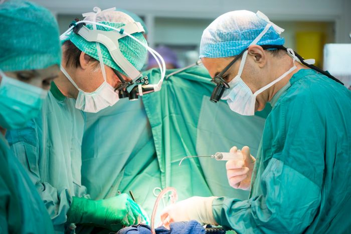
Blog

  • Things to Stop for a Longer Life

    Things to Stop for a Longer Life

    Things to stop if you want to live longer: This post delves into crucial lifestyle choices that can significantly impact your lifespan. We’ll explore the negative effects of unhealthy habits and offer actionable strategies to cultivate healthier practices. From diet and exercise to stress management and sleep hygiene, we’ll cover a comprehensive approach to a longer, healthier life.

    Let’s unpack what you can stop doing to enhance your journey towards longevity.

    This journey to a longer and healthier life starts with understanding the detrimental effects of certain lifestyle choices. Neglecting our physical and mental well-being often leads to various health issues, ultimately shortening our lifespans. In this post, we’ll cover several critical areas, including diet, exercise, stress, substance use, sleep, social connections, the environment, and mental well-being. Understanding these elements and the practical steps to improve them can empower you to take control of your health and well-being.

    Unhealthy Dietary Habits

    Our dietary choices significantly impact our overall health and longevity. Making conscious decisions about what we eat can contribute to a longer, healthier life. This section explores common unhealthy habits, contrasting them with healthier approaches to nutrition.

    Common Unhealthy Dietary Habits

    Poor dietary habits contribute to a variety of health problems, increasing the risk of chronic diseases and potentially shortening lifespans. Here are five prevalent unhealthy dietary habits:

    • Excessive Consumption of Processed Foods: Processed foods often contain high levels of added sugars, unhealthy fats, and sodium, contributing to weight gain, high blood pressure, and an increased risk of heart disease and type 2 diabetes. Examples include packaged snacks, sugary cereals, and fast food.
    • Insufficient Fruit and Vegetable Intake: A diet lacking in fruits and vegetables deprives the body of essential vitamins, minerals, and antioxidants. This can weaken the immune system, increasing vulnerability to various illnesses. It also reduces the intake of fiber, which is crucial for digestive health.
    • Overconsumption of Sugary Drinks: Sugary drinks, including sodas, juices, and sweetened beverages, provide empty calories with little nutritional value. These drinks contribute to weight gain, dental problems, and increase the risk of various health issues, including heart disease.
    • Inadequate Protein Intake: Protein is essential for building and repairing tissues. Insufficient protein intake can lead to muscle loss, reduced metabolism, and weakened immune function. This is especially important for maintaining muscle mass as we age.
    • High Consumption of Saturated and Trans Fats: A diet high in saturated and trans fats can raise LDL (“bad”) cholesterol levels, increasing the risk of heart disease and stroke. These fats are often found in red meat, processed foods, and fried foods.

    Dietary Approaches for Longevity

    Various dietary approaches offer potential benefits for longevity. Comparing their strengths and weaknesses helps individuals make informed choices.

    • Mediterranean Diet: This diet emphasizes fruits, vegetables, whole grains, legumes, nuts, and healthy fats like olive oil. It promotes heart health, reduces inflammation, and has been associated with a lower risk of chronic diseases. Drawbacks can include higher costs for some ingredients and potential challenges in maintaining adherence to specific guidelines.
    • Plant-Based Diet: This diet prioritizes plant-based foods, such as fruits, vegetables, legumes, and whole grains, often limiting or eliminating animal products. It can improve cardiovascular health, lower cholesterol, and reduce the risk of certain cancers. Drawbacks can include potential deficiencies in certain nutrients like vitamin B12 and iron if not carefully planned, and the need for careful supplementation.

    • Ketogenic Diet: This diet significantly restricts carbohydrates, promoting the body to use fat for energy. It can lead to weight loss and improvements in certain metabolic conditions. Drawbacks include potential nutrient deficiencies, potential negative impacts on gut health, and potential challenges in long-term adherence due to strict limitations on carbohydrate intake.

    Sample Weekly Meal Plan

    This sample weekly meal plan emphasizes nutrient-rich foods and limits processed foods. Adjust portion sizes based on individual needs and preferences.

    • Monday: Grilled chicken breast with roasted vegetables (broccoli, carrots, sweet potatoes), quinoa.
    • Tuesday: Lentil soup with whole-wheat bread, side salad.
    • Wednesday: Salmon with steamed asparagus and brown rice.
    • Thursday: Chickpea curry with brown rice and mixed greens.
    • Friday: Lean beef stir-fry with plenty of mixed vegetables and brown rice.

    • Saturday: Homemade pizza with whole-wheat crust, vegetables, and lean protein.
    • Sunday: Chicken and vegetable stew with whole-wheat pasta.

    Strategies to Overcome Dietary Challenges

    Overcoming dietary challenges requires a proactive approach. Here are five strategies:

    • Planning Ahead: Create meal plans and grocery lists to avoid impulsive unhealthy choices. Planning can help ensure you have healthy options readily available.
    • Mindful Eating: Pay attention to your body’s hunger and fullness cues, eating slowly, and savoring your food. This can help prevent overeating.
    • Seeking Support: Connect with friends, family, or a registered dietitian for support and guidance in achieving dietary goals.
    • Gradually Transition: Don’t try to overhaul your diet overnight. Make gradual changes to create sustainable habits.
    • Finding Healthy Alternatives: Explore healthier versions of favorite foods or recipes to maintain enjoyment while improving nutrition.

    Nutritional Benefits of Fruits and Vegetables

    Fruits and vegetables provide essential nutrients for overall health and longevity.

    Fruit/Vegetable Nutritional Benefit Longevity Impact Serving Suggestion
    Berries (strawberries, blueberries) Antioxidants, vitamins Reduces inflammation, supports immune function 1 cup
    Leafy Greens (spinach, kale) Vitamins, minerals, fiber Supports gut health, improves digestion 1 cup, raw or cooked
    Citrus Fruits (oranges, lemons) Vitamin C, antioxidants Boosts immune system, supports collagen production 1 medium orange or 1 cup segments
    Carrots Vitamin A, fiber Supports eye health, promotes healthy digestion 1 medium carrot, raw or cooked
    Sweet Potatoes Vitamins, minerals, fiber Supports immune system, provides energy 1 medium sweet potato, baked or roasted

    Lack of Physical Activity

    Things to stop if you want to live longer

    Staying active is crucial for a long and healthy life. Regular physical activity not only strengthens our bodies but also boosts our mood, improves cognitive function, and reduces the risk of chronic diseases. Ignoring the importance of movement can significantly impact our lifespan, making consistent physical activity a cornerstone of longevity. Choosing activities that fit our lifestyle and health conditions is key to maintaining a healthy routine.

    Types of Physical Activity for Different Groups

    Different types of physical activity cater to various age groups and health conditions. Choosing activities appropriate to our individual needs and limitations is crucial for safety and effectiveness. The goal is to incorporate movement into our daily routines, not to impose strenuous workouts that may lead to injuries.

    • Aerobic Exercise (Cardio): Activities like brisk walking, jogging, swimming, cycling, and dancing improve cardiovascular health, increase lung capacity, and burn calories. These activities are suitable for most age groups and fitness levels. Benefits include improved heart health, reduced risk of heart disease, stroke, and type 2 diabetes, and enhanced mood. For older adults, low-impact aerobic activities like walking or water aerobics are ideal, while younger adults can engage in more vigorous activities like running or dancing.

    • Strength Training: Exercises that build muscle mass, such as weightlifting, resistance bands, or bodyweight exercises (push-ups, squats), are beneficial for all ages. They increase bone density, metabolism, and overall strength. Strength training helps prevent age-related muscle loss (sarcopenia), improving balance and reducing the risk of falls, particularly important for older adults. This type of activity is crucial for maintaining a healthy body composition and reducing the risk of osteoporosis.

    • Flexibility Exercises: Stretching and yoga improve joint mobility, reduce muscle stiffness, and enhance balance. These exercises are suitable for all ages and fitness levels, but especially beneficial for older adults to maintain flexibility and reduce the risk of injury. They can also help improve posture and reduce stress. Examples include stretching routines, Pilates, and Tai Chi.

    Comparing Benefits of Different Exercise Types

    Aerobic exercise, strength training, and flexibility exercises each contribute to a longer lifespan in distinct ways. While all three are vital, their individual benefits affect different aspects of health and longevity.

    Exercise Type Primary Benefits Impact on Lifespan
    Aerobic Exercise Improved cardiovascular health, increased lung capacity, weight management Reduced risk of heart disease, stroke, type 2 diabetes, and some cancers.
    Strength Training Increased muscle mass, bone density, metabolism, improved balance Reduced risk of falls, osteoporosis, sarcopenia, and improved overall physical function.
    Flexibility Exercises Improved joint mobility, reduced muscle stiffness, enhanced balance Reduced risk of injuries, improved posture, and stress reduction, contributing to overall well-being.

    Importance of Consistency

    Consistency in physical activity is paramount for achieving long-term health benefits and longevity. Sporadic exercise is less effective than a regular routine.

    “Consistency is key to reaping the rewards of physical activity.”

    Establishing a routine and incorporating exercise into daily life leads to better results. Regularity allows the body to adapt and reap the benefits of each type of activity.

    Common Excuses and Strategies

    Many people cite excuses for not exercising. Addressing these excuses is crucial for starting and maintaining a consistent routine.

    • Lack of time: Even short bursts of activity can be beneficial. Scheduling 10-15 minute exercise sessions throughout the day can add up. Look for opportunities to move during the day, such as taking the stairs instead of the elevator or walking during phone calls.
    • Lack of motivation: Find an activity you enjoy. Exercise with a friend or join a group. Reward yourself for achieving goals.
    • Feeling overwhelmed: Start small and gradually increase the intensity and duration of your workouts. Focus on progress, not perfection.
    • Lack of energy: Even a small amount of exercise can boost energy levels. Physical activity can help improve sleep quality, which in turn improves energy levels.
    • Fear of injury: Start with low-impact exercises and consult with a doctor or physical therapist before starting a new routine. Proper warm-up and cool-down are crucial to prevent injuries.

    Beginner Workout Routine

    A structured workout routine for beginners can help establish a consistent habit.

    • Warm-up (5 minutes): Light cardio, like jumping jacks, high knees, arm circles, and dynamic stretches (arm swings, leg swings). This prepares the body for more intense activity.
    • Cardio (20 minutes): Choose an activity you enjoy, such as brisk walking, jogging, cycling, or swimming. Maintain a moderate intensity.
    • Strength Training (15 minutes): Focus on compound exercises like squats, lunges, push-ups, and rows. Use bodyweight or light weights. Aim for 2-3 sets of 10-12 repetitions for each exercise.
    • Flexibility (5 minutes): Hold each stretch for 20-30 seconds. Focus on major muscle groups, including hamstrings, quads, chest, and back.
    • Cool-down (5 minutes): Light cardio and static stretches (holding stretches for 30-60 seconds). This helps the body recover and reduces muscle soreness.

    Stress Management Techniques

    Chronic stress, a pervasive and often underestimated factor in overall well-being, has a demonstrably negative impact on lifespan. Prolonged exposure to stress hormones like cortisol can lead to a cascade of negative health consequences, including cardiovascular issues, weakened immune systems, and mental health problems. Understanding and addressing stress is therefore crucial for a longer, healthier life.The human body is remarkably resilient, capable of adapting to a wide range of stressors.

    However, the continuous barrage of modern life often overwhelms our natural coping mechanisms. Effective stress management techniques are vital in maintaining a balanced and healthy lifestyle. These techniques equip individuals with tools to navigate challenges, promote emotional regulation, and foster a sense of calm.

    The Correlation Between Chronic Stress and Reduced Lifespan

    Chronic stress significantly contributes to a shortened lifespan. Elevated cortisol levels, a hallmark of chronic stress, can damage the cardiovascular system, leading to increased blood pressure, heart rate, and risk of heart disease. Furthermore, chronic stress can compromise the immune system, making individuals more susceptible to illnesses. Studies have consistently linked chronic stress to a heightened risk of various diseases and premature mortality.

    For instance, individuals experiencing high levels of chronic stress often exhibit higher rates of obesity, diabetes, and mental health issues, all of which are associated with a reduced life expectancy.

    Different Stress Management Techniques

    Several techniques can effectively manage stress. These methods vary in their approach and effectiveness, offering individuals diverse avenues for stress reduction. Choosing a method that resonates with an individual’s personality and lifestyle is key to long-term success.

    Want to live a longer, healthier life? Cutting out harmful habits is key. Smoking, excessive alcohol, and a sedentary lifestyle are major contributors to various health problems, including the devastating neurodegenerative disease, amyotrophic lateral sclerosis (ALS) amyotrophic lateral sclerosis als. Addressing these factors can significantly improve your chances of a longer and more fulfilling life.

    Comparing the Effectiveness of Stress Management Techniques

    • Meditation: A practice that involves focusing on a specific object, thought, or activity to quiet the mind and reduce racing thoughts. Meditation can foster a sense of calm and improve attention span. Research suggests that regular meditation can lower blood pressure and heart rate, thereby reducing stress-related physiological responses.
    • Yoga: A holistic practice that combines physical postures (asanas), breathing techniques (pranayama), and meditation. Yoga promotes physical flexibility, strength, and balance, which can positively impact mood and reduce stress. Studies show that yoga can effectively reduce anxiety and improve overall well-being.
    • Deep Breathing Exercises: Simple techniques focusing on controlled inhalations and exhalations. These exercises help regulate the nervous system, calming the body’s response to stress. Deep breathing can immediately reduce feelings of anxiety and promote relaxation. Scientific research confirms the effectiveness of deep breathing in lowering heart rate and blood pressure, demonstrating its role in stress management.

    Practical Strategies for Incorporating Stress Reduction Practices into Daily Life

    • Schedule dedicated time for stress reduction practices: Creating a regular schedule for meditation, yoga, or deep breathing helps integrate these practices into daily routines. Consistency is key to experiencing the full benefits.
    • Find a supportive community: Sharing experiences and support with others practicing stress reduction techniques can provide motivation and accountability.
    • Identify and challenge negative thought patterns: Recognizing and reframing negative thought patterns is crucial for managing stress. This can involve cognitive behavioral therapy techniques.
    • Prioritize self-care: Engage in activities that promote relaxation and rejuvenation, such as taking a warm bath, listening to music, or spending time in nature.
    • Seek professional help when needed: If stress becomes overwhelming, seeking support from a therapist or counselor can provide tailored strategies and coping mechanisms.

    Stress Reduction Methods Comparison

    Method Description Benefits Drawbacks
    Meditation Focusing on a single point to quiet the mind Reduced anxiety, improved focus, lowered blood pressure Requires consistent practice, may not be suitable for all individuals
    Yoga Combining physical postures, breathing, and meditation Improved flexibility, strength, reduced stress May require physical exertion, some poses can be challenging
    Deep Breathing Controlled inhalations and exhalations Immediate stress relief, easily accessible May not address underlying stress triggers, may not be sufficient for chronic stress

    Step-by-Step Guide for Mindfulness Meditation

    1. Find a quiet space: Choose a comfortable and peaceful environment free from distractions.
    2. Assume a comfortable posture: Sit or lie down in a relaxed position.
    3. Focus on your breath: Notice the sensation of each inhale and exhale.
    4. Acknowledge thoughts and feelings: Allow thoughts and feelings to arise without judgment.
    5. Return to your breath: Gently redirect your attention back to your breath when your mind wanders.

    Substance Abuse and Addiction: Things To Stop If You Want To Live Longer

    Substance abuse, encompassing the misuse of drugs and alcohol, poses a significant threat to overall well-being and longevity. The detrimental effects extend far beyond immediate physical discomfort, impacting various aspects of health and potentially shortening one’s lifespan. Understanding the nature of substance abuse, its consequences, and available resources for recovery is crucial for personal and community health.Substance abuse significantly compromises the body’s ability to function optimally.

    Chronic exposure to substances can lead to a cascade of adverse health outcomes, ranging from organ damage to mental health disorders. The long-term impact often manifests as a decline in physical and cognitive function, ultimately reducing the quality and duration of life.

    Negative Impacts on Lifespan

    Substance abuse significantly contributes to premature mortality. The direct and indirect effects on various organ systems, coupled with the increased risk of accidents and other adverse events, accelerate the aging process and decrease life expectancy. This negative impact extends to individuals’ ability to engage in healthy behaviors, such as proper nutrition and exercise, further exacerbating the negative consequences.

    For instance, individuals struggling with addiction often neglect their health, leading to poor nutrition and a sedentary lifestyle, both of which are significant risk factors for various diseases.

    Types of Substance Abuse and Their Effects

    Different substances have unique effects on the body. Understanding these variations is crucial for recognizing the specific dangers associated with each type of abuse.

    • Alcohol Abuse: Alcohol abuse can lead to liver damage, including cirrhosis and alcoholic hepatitis. It also increases the risk of certain cancers, cardiovascular diseases, and mental health problems like depression and anxiety. For example, heavy alcohol consumption can lead to alcoholic cardiomyopathy, a condition where the heart muscle weakens and enlarges, potentially resulting in heart failure. The long-term impact of alcohol abuse can significantly decrease life expectancy and severely impair quality of life.

      Want to add years to your life? Cutting out harmful habits like smoking and excessive alcohol is a great start. But did you know some medications can actually contribute to sundowning, a phenomenon where confusion and agitation increase in the evening? Understanding which medications cause these symptoms, like what medications cause sundowning symptoms , is crucial for optimizing your health and well-being.

      Prioritizing a healthy lifestyle, including medication management, is key to living a longer, more fulfilling life.

    • Opioid Abuse: Opioid abuse, whether through prescription painkillers or illicit drugs, can lead to respiratory depression, overdose, and addiction. The long-term use of opioids can cause severe damage to the liver and kidneys, leading to chronic health issues and potentially premature death. Overdose deaths are a significant public health concern, particularly involving combinations of opioids and other substances.
    • Cocaine Abuse: Cocaine abuse can lead to cardiovascular complications such as heart attack and stroke. It can also trigger seizures, psychosis, and severe mental health issues. Long-term cocaine abuse can result in significant cognitive impairment, impacting memory, learning, and overall mental function, significantly impacting longevity and quality of life. For example, cocaine abuse has been linked to an increased risk of infectious diseases like HIV due to risky behaviors often associated with substance use.

    Long-Term Consequences of Substance Abuse

    The long-term consequences of substance abuse extend beyond the immediate physical effects. Chronic substance abuse often leads to severe health issues, impacting various organ systems and contributing to significant impairments in daily functioning. This, in turn, significantly reduces the overall quality of life and can contribute to a shorter lifespan.

    • Weakened Immune System: Regular substance use often weakens the immune system, making individuals more susceptible to infections and diseases.
    • Mental Health Disorders: Substance abuse can exacerbate existing mental health conditions or lead to new ones, such as anxiety, depression, and psychosis.
    • Social Isolation: Substance abuse can damage relationships and lead to social isolation, further contributing to a decline in health and well-being.

    Recognizing Signs of Substance Abuse

    Recognizing the signs of substance abuse in oneself or others is crucial for early intervention and treatment. Early intervention can significantly improve the chances of recovery and a better quality of life. Pay attention to changes in behavior, mood, or physical appearance.

    • Changes in Behavior: These changes might include neglecting responsibilities, isolating oneself from social activities, or exhibiting erratic behavior.
    • Mood Swings: Individuals struggling with substance abuse might experience unpredictable mood swings, from extreme irritability to apathy.
    • Physical Changes: Physical changes such as bloodshot eyes, changes in appetite, or unexplained weight loss may indicate substance abuse.

    Resources for Seeking Help, Things to stop if you want to live longer

    If you or someone you know is struggling with substance abuse, help is available. Reaching out for support is a sign of strength, not weakness. Seeking help is an essential step toward recovery.

    • National Helpline: A confidential helpline can provide information, support, and guidance for individuals and their families.
    • Treatment Centers: Various treatment centers offer comprehensive programs to address substance abuse and promote recovery.
    • Support Groups: Support groups provide a safe space for individuals to share experiences and receive encouragement during recovery.

    Sleep Hygiene and Quality

    Sleep is not just a downtime activity; it’s a fundamental pillar of a healthy and long life. Adequate sleep allows our bodies to repair and rejuvenate, impacting everything from our physical health to our cognitive function. Ignoring sleep’s importance can lead to a cascade of negative consequences, diminishing our overall well-being and potentially shortening our lifespan.A consistent sleep schedule, a conducive sleep environment, and mindful habits are essential for optimizing sleep quality.

    By prioritizing sleep hygiene, we can experience a remarkable improvement in our physical and mental health, contributing significantly to a longer and healthier life.

    Importance of Adequate Sleep for Longevity

    Sleep plays a crucial role in maintaining physical and mental well-being. During sleep, our bodies repair tissues, regulate hormones, and consolidate memories. Chronic sleep deprivation can disrupt these vital processes, leading to various health issues that negatively impact longevity. For example, sleep deprivation has been linked to an increased risk of cardiovascular disease, type 2 diabetes, and certain types of cancer.

    The cumulative effect of inadequate sleep over time can significantly reduce life expectancy.

    Strategies for Improving Sleep Quality and Quantity

    Implementing these strategies can significantly improve your sleep quality and quantity. Consistency is key.

    • Establish a Regular Sleep Schedule: Going to bed and waking up around the same time each day, even on weekends, helps regulate your body’s natural sleep-wake cycle, known as the circadian rhythm. This consistency reinforces your body’s internal clock, making it easier to fall asleep and wake up refreshed.
    • Create a Relaxing Bedtime Routine: Engage in calming activities before bed, such as reading, taking a warm bath, or listening to soothing music. Avoid stimulating activities like using electronic devices, as the blue light emitted can interfere with melatonin production, a hormone crucial for sleep.
    • Optimize Your Sleep Environment: Ensure your bedroom is dark, quiet, and cool. Invest in blackout curtains, earplugs, or a white noise machine if needed. A comfortable mattress and pillows are also essential for a good night’s sleep.
    • Maintain a Healthy Diet and Exercise Regularly: A balanced diet and regular physical activity can contribute to better sleep. Avoid large meals or caffeine and alcohol close to bedtime. Regular exercise, however, is generally beneficial, but avoid intense workouts close to bedtime.
    • Manage Stress Effectively: Stress can significantly impact sleep quality. Practice relaxation techniques like deep breathing exercises, meditation, or progressive muscle relaxation to reduce stress and promote relaxation before sleep.

    Sleep Deprivation and Health Issues

    Sleep deprivation has been linked to a wide range of health issues. It can impair cognitive function, leading to difficulties with concentration, memory, and decision-making. Furthermore, chronic sleep loss is associated with an increased risk of obesity, cardiovascular disease, weakened immunity, and mental health problems. The body’s natural repair mechanisms are compromised, leading to potential long-term health complications.

    Different Sleep Stages and Their Importance

    Sleep is not a uniform state; it’s composed of different stages with distinct physiological functions. These stages include non-rapid eye movement (NREM) sleep and rapid eye movement (REM) sleep. NREM sleep is further divided into three stages, each characterized by different brain wave patterns and physiological processes. REM sleep is crucial for memory consolidation and emotional processing.

    Disruptions in any of these stages can negatively affect overall sleep quality and lead to health issues.

    Optimal Sleep Hours by Age Group

    The optimal amount of sleep varies across different age groups. Maintaining adequate sleep is crucial for healthy development and well-being.

    Age Group Optimal Sleep Hours Sleep Hygiene Tips Potential Sleep Problems
    Newborn (0-3 months) 14-17 hours Create a consistent sleep schedule, provide a safe and comfortable sleep environment, and respond promptly to feeding cues. Difficulty regulating sleep cycles, feeding issues, or medical conditions.
    Infant (4-11 months) 12-15 hours Continue with a consistent sleep schedule, ensure a safe sleep environment, and provide a comforting presence. Teething, separation anxiety, or other developmental issues.
    Toddler (1-2 years) 11-14 hours Establish a bedtime routine, create a calming sleep environment, and encourage healthy sleep habits. Night terrors, nightmares, or resistance to bedtime.
    Preschooler (3-5 years) 10-13 hours Maintain a consistent bedtime routine, encourage physical activity during the day, and address any anxieties or fears. Difficulty transitioning to bed, anxiety, or fears.
    School-aged Children (6-13 years) 9-11 hours Encourage regular sleep and wake-up times, promote physical activity, and ensure a calm and supportive environment. Stress from school, extracurricular activities, or social pressures.
    Teenagers (14-17 years) 8-10 hours Maintain a regular sleep schedule, encourage relaxation techniques, and manage stress. Social media use, academic pressure, or hormonal changes.
    Adults (18-64 years) 7-9 hours Prioritize a consistent sleep schedule, create a relaxing bedtime routine, and ensure a comfortable sleep environment. Stress, work schedules, or lifestyle factors.
    Older Adults (65+ years) 7-8 hours Maintain a regular sleep schedule, encourage physical activity, and address any medical conditions. Medical conditions, decreased mobility, or decreased sensory input.

    Social Isolation and Loneliness

    Social isolation and loneliness are significant public health concerns, impacting not only emotional well-being but also physical health and longevity. The detrimental effects of these conditions are multifaceted and extend far beyond feelings of sadness or emptiness. Understanding the link between social connection and overall health is crucial for developing strategies to promote well-being and enhance lifespan.Social isolation, characterized by a lack of social interaction, and loneliness, the subjective experience of feeling isolated even when surrounded by others, can have a profound negative impact on physical and mental health.

    Studies have consistently demonstrated a correlation between social isolation and a higher risk of various health problems, including cardiovascular disease, weakened immune function, and increased risk of premature mortality.

    Want to live a longer, healthier life? Cutting out harmful habits is key. One crucial area to focus on is avoiding behaviors that can increase your risk of serious diseases like ductal carcinoma in situ (DCIS), a non-invasive form of breast cancer. Understanding the risks and potential early detection methods is essential. Learn more about DCIS and its connection to overall health at ductal carcinoma in situ dcis.

    Ultimately, prioritizing healthy lifestyle choices, like regular exercise and a balanced diet, will significantly impact your longevity and well-being.

    Negative Effects of Social Isolation on Longevity

    Social isolation is a significant risk factor for decreased longevity. Lack of social interaction can lead to a cascade of negative health consequences. This includes heightened stress levels, compromised immune response, and even changes in brain function. The absence of social support systems can make it more challenging to manage stress and maintain healthy coping mechanisms, ultimately increasing the risk of developing chronic diseases.

    Importance of Maintaining Social Connections and Building Strong Relationships

    Strong social connections are essential for overall well-being and longevity. Humans are social creatures, and our relationships with others provide crucial emotional support, practical assistance, and a sense of belonging. Meaningful relationships offer a buffer against stress, promote resilience, and contribute to a positive outlook on life. This sense of connection is critical for maintaining mental and physical health throughout life.

    5 Ways to Build and Maintain Strong Social Connections

    Building and maintaining strong social connections is an ongoing process that requires effort and intentionality. Developing these connections is not a one-time event but a continuous investment in relationships. Nurturing these bonds can lead to a more fulfilling and healthier life.

    • Cultivate Existing Relationships: Actively engaging with existing friends and family members, making time for meaningful conversations, and expressing gratitude for their presence are crucial. Regular communication, whether it’s a phone call, text message, or video chat, can significantly strengthen these bonds. Sharing experiences and supporting each other through life’s ups and downs fosters deeper connections.
    • Join Groups and Activities: Engaging in shared interests, whether it’s a book club, sports team, volunteer organization, or hobby group, provides opportunities to connect with like-minded individuals. Shared activities create a sense of community and belonging, fostering new friendships and strengthening existing ones.
    • Attend Social Events: Making an effort to attend social gatherings, whether they are formal or informal, can expand your social circle. This can involve joining community events, attending local festivals, or simply inviting friends over for dinner or drinks.
    • Offer Support and Help: Helping others can be a powerful way to strengthen social connections. Volunteering your time, lending a hand to a friend or neighbor, or simply offering a listening ear can build meaningful relationships and a sense of purpose.
    • Practice Active Listening and Empathy: Being a good listener and demonstrating empathy are crucial for building and maintaining strong social connections. Truly listening to others, understanding their perspectives, and showing compassion create a supportive and encouraging environment for all involved.

    Link Between Loneliness and Health Issues Affecting Lifespan

    Loneliness has a significant impact on health and lifespan. It’s associated with a higher risk of chronic diseases, weakened immune function, and increased mortality rates. Studies have shown that loneliness can lead to elevated levels of stress hormones, which can damage the cardiovascular system and negatively impact overall health. This can contribute to a higher risk of heart disease, stroke, and other health complications.

    Social Interaction Plan for Improving Social Connections

    A structured social interaction plan can be a helpful tool for individuals seeking to improve their social connections. This plan should be tailored to the individual’s needs and preferences, incorporating activities and interactions that are enjoyable and meaningful. A key element is consistency. Regular engagement in social activities is vital for building and maintaining relationships.

    Activity Frequency Description
    Weekly lunch with a friend Weekly Scheduling a weekly lunch with a friend to discuss current events and personal updates.
    Joining a book club Monthly Participating in a monthly book club meeting to engage in discussions about literary works.
    Volunteering at a local animal shelter Bi-weekly Contributing time at a local animal shelter to interact with animals and staff, building connections.

    Environmental Factors

    Our environment plays a crucial role in our overall health and longevity. Exposure to pollutants, lack of access to clean resources, and harmful toxins can significantly impact our lifespan and well-being. Understanding the connection between environmental factors and our health is paramount for proactive measures to enhance our longevity.Environmental factors like air and water quality, exposure to toxins, and even the built environment significantly influence our health outcomes.

    The impact of these factors often manifests in increased risks of chronic diseases, impacting the quality and duration of life.

    Impact of Pollution on Lifespan

    Air pollution, a major environmental concern, is directly linked to a reduction in lifespan. Studies have shown a strong correlation between high levels of air pollution and increased mortality rates, particularly from respiratory and cardiovascular diseases. Fine particulate matter, a common component of air pollution, can penetrate deep into the lungs and bloodstream, causing inflammation and damage to vital organs.

    This damage can lead to long-term health problems and a decreased life expectancy. For example, areas with high levels of industrial activity or heavy traffic often show a higher incidence of respiratory illnesses and premature mortality compared to areas with cleaner air.

    Importance of Clean Water and Air

    Access to clean water and air is fundamental to human health and longevity. Water, essential for bodily functions, becomes a significant health concern when contaminated with pollutants or heavy metals. Similarly, breathing clean air is critical for optimal respiratory health. A lack of access to these resources can lead to a multitude of health issues, significantly impacting the length and quality of life.

    For instance, communities with contaminated water sources often experience higher rates of waterborne diseases and related complications. Similarly, communities with poor air quality may face increased instances of respiratory illnesses, reducing overall well-being and potentially shortening lifespan.

    Impact of Exposure to Toxins

    Exposure to various toxins, including pesticides, heavy metals, and industrial chemicals, poses a considerable threat to human health and longevity. These toxins can accumulate in the body, leading to chronic health problems like cancer, neurological disorders, and organ damage. For example, prolonged exposure to certain pesticides has been linked to increased risks of Parkinson’s disease and other neurological conditions.

    Similarly, exposure to heavy metals like lead and mercury can cause severe neurological damage and developmental issues.

    Simple Steps to Mitigate Environmental Risks

    Taking proactive steps to minimize environmental risks is crucial for improving overall health and potentially extending lifespan. Here are five simple steps individuals can take:

    • Reduce Exposure to Pollution: Minimizing exposure to air and water pollution is achievable through conscious choices. Opting for public transportation or cycling, choosing to walk instead of driving short distances, and supporting policies that prioritize clean energy sources can make a significant difference.
    • Prioritize Clean Water Sources: Consuming water from trusted sources and avoiding bottled water (which can have environmental impacts) is important. Support initiatives that promote clean water access in communities.
    • Choose Sustainable Products: Selecting products that minimize environmental impact through conscious consumption, such as opting for eco-friendly cleaning supplies, choosing reusable items over disposable ones, and avoiding products with harmful chemicals, can have a positive impact on both health and the environment.
    • Support Environmental Policies: Advocating for policies that prioritize environmental protection and sustainable practices is essential. Support organizations working towards clean energy, reduced pollution, and environmental conservation.
    • Promote Awareness: Educating yourself and others about the dangers of environmental toxins and pollution is crucial. Sharing information and promoting awareness in your community can lead to collective action.

    Mental Health and Well-being

    Our mental well-being is intricately linked to our overall health and longevity. A healthy mind contributes significantly to a longer, more fulfilling life, while mental health challenges can significantly impact lifespan and quality of life. This section explores the profound connection between mental health and longevity, and how proactive mental well-being strategies can positively influence our life span.Mental health is not simply the absence of mental illness; it’s a state of emotional, psychological, and social well-being that enables an individual to cope with the stresses of life, work productively, and contribute to their community.

    A strong mental foundation provides resilience to navigate life’s inevitable challenges, ultimately influencing how long and how well we live.

    The Impact of Mental Illnesses on Lifespan

    Mental illnesses, including depression, anxiety disorders, and schizophrenia, can have a substantial impact on lifespan. These conditions can lead to various health problems, including cardiovascular disease, weakened immune systems, and increased risk of chronic illnesses. For example, individuals with untreated depression often experience compromised immune function, increasing their vulnerability to infections. Chronic stress associated with mental illness can also lead to unhealthy lifestyle choices, such as poor diet and lack of exercise, further exacerbating health risks.

    Studies have shown a correlation between mental health issues and shortened lifespans, highlighting the crucial role of early intervention and treatment.

    The Importance of Seeking Professional Help

    Recognizing the need for professional help is a critical step in managing mental health concerns. Seeking support from mental health professionals, such as therapists, counselors, or psychiatrists, is not a sign of weakness but a proactive step towards improved well-being and a healthier life. Mental health professionals provide personalized assessments, tailored strategies, and evidence-based treatments to address specific needs and challenges.

    Early intervention and consistent treatment significantly enhance the chances of recovery and improved quality of life.

    5 Ways to Promote Mental Well-being

    Cultivating a proactive approach to mental well-being is essential for a longer and healthier life. These strategies can help foster resilience and reduce the risk of mental health challenges.

    • Mindfulness and Meditation: Practicing mindfulness and meditation techniques can help manage stress, improve focus, and cultivate emotional awareness. Regular mindfulness practice can foster a sense of calm and clarity, reducing reactivity to daily stressors. This allows individuals to better navigate challenges and promote overall well-being.
    • Physical Activity: Engaging in regular physical activity is a powerful tool for improving mental well-being. Exercise releases endorphins, which have mood-boosting effects, and helps reduce stress and anxiety. A consistent exercise routine can create a positive feedback loop, strengthening mental and physical resilience.
    • Healthy Diet: Nourishing the body with a balanced diet is crucial for both physical and mental well-being. A diet rich in fruits, vegetables, and whole grains provides essential nutrients that support brain function and emotional regulation. Conversely, a diet high in processed foods and unhealthy fats can contribute to mood swings and increased stress levels.
    • Strong Social Connections: Maintaining strong social connections and a supportive network are vital for mental well-being. Meaningful relationships provide a sense of belonging, emotional support, and opportunities for shared experiences. Strong social connections help individuals navigate challenges and cultivate a positive outlook.
    • Sufficient Sleep: Adequate sleep is essential for both physical and mental restoration. A consistent sleep schedule helps regulate mood, improve focus, and enhance cognitive function. Sufficient sleep plays a vital role in emotional regulation and overall mental well-being.

    Mental Health Resources

    Accessing support from mental health resources can significantly benefit individuals facing mental health challenges.

    Resource Type Description
    Therapists/Counselors Trained professionals offering individual or group therapy to address specific concerns.
    Psychiatrists Medical doctors specializing in the diagnosis and treatment of mental illnesses, potentially prescribing medication.
    Crisis Hotlines 24/7 support for individuals experiencing a mental health crisis.
    Support Groups Communities of individuals facing similar challenges, providing a sense of belonging and shared experience.
    Online Mental Health Platforms Accessible resources, including self-assessment tools, educational materials, and online therapy options.

    Outcome Summary

    Things to stop if you want to live longer

    In conclusion, achieving a longer and healthier life hinges on conscious choices. By recognizing and eliminating detrimental habits, such as unhealthy diets, lack of physical activity, chronic stress, substance abuse, inadequate sleep, social isolation, environmental hazards, and poor mental health, we empower ourselves to live more fulfilling and extended lives. Adopting the suggested strategies for each area, from healthy eating to stress management, will pave the way for a more vibrant and longer life.

    The key takeaway is that longevity is not a mere outcome of chance but a result of proactive choices we make daily. Remember, every small step towards a healthier lifestyle contributes to a healthier future.

  • Magnesium for Muscle Pain A Deep Dive

    Magnesium for Muscle Pain A Deep Dive

    Magnesium for muscle pain is a crucial topic for understanding the role of this vital mineral in maintaining healthy muscles. Magnesium plays a significant part in muscle function, and deficiencies can lead to a range of issues, including persistent muscle aches and discomfort. This article explores the intricate connection between magnesium and muscle pain, examining various aspects, from the role of magnesium in different muscle types to the potential benefits of supplementation, and beyond.

    We’ll delve into the potential mechanisms behind magnesium’s impact on muscle pain, comparing and contrasting it with other potential causes. The article will also explore the importance of dietary sources of magnesium, alongside various forms of supplements and their pros and cons. Finally, we’ll touch upon diagnosis, treatment strategies, and important considerations regarding interactions with other medications or supplements.

    Introduction to Magnesium and Muscle Pain

    Magnesium plays a crucial role in numerous bodily functions, and muscle health is no exception. It’s essential for muscle contraction, relaxation, and nerve function. Without adequate magnesium levels, the delicate balance within these processes can be disrupted, potentially leading to muscle pain and other symptoms. This article delves into the connection between magnesium and muscle pain, exploring the mechanisms behind the link and the common symptoms associated with magnesium deficiency.Magnesium is a vital mineral that acts as a cofactor for over 300 enzymatic reactions in the body.

    This includes reactions directly related to muscle function, such as those involved in nerve impulse transmission and the release of calcium, which is essential for muscle contraction. Imbalances in these processes, often stemming from insufficient magnesium intake, can lead to a cascade of effects, including muscle pain.

    Magnesium’s Role in Muscle Function

    Magnesium is fundamental to the proper functioning of all muscle types. It is involved in the crucial steps of muscle contraction and relaxation. It aids in the regulation of calcium release, ensuring smooth and coordinated muscle activity. Without sufficient magnesium, the delicate balance of calcium regulation can be compromised, leading to potential muscle spasms or cramps.

    Potential Mechanisms of Magnesium Deficiency-Related Muscle Pain

    Magnesium deficiency can disrupt the intricate interplay of ions within muscle cells. This disruption affects the release and reuptake of calcium, leading to sustained muscle contractions and the sensation of pain. The imbalance can also impact nerve signal transmission, which plays a critical role in muscle function.

    Common Symptoms of Magnesium Deficiency, Including Muscle Pain

    Magnesium deficiency can manifest in a variety of ways, and muscle pain is often one of the presenting symptoms. Other common symptoms include fatigue, weakness, nausea, headaches, and irregular heartbeat. It’s important to remember that these symptoms can be caused by other conditions as well, so it’s crucial to consult with a healthcare professional for accurate diagnosis. Early diagnosis and intervention are key to managing the condition effectively.

    • Muscle Pain: A common symptom of magnesium deficiency, characterized by cramping, spasms, or soreness in various muscle groups. This pain can range from mild discomfort to severe debilitating pain.
    • Fatigue: A persistent feeling of tiredness and lack of energy. This can be a significant symptom of magnesium deficiency, often affecting daily activities and overall well-being.
    • Weakness: Reduced strength and power in muscles, making everyday tasks more challenging.
    • Nausea: A feeling of discomfort in the stomach, often accompanied by an urge to vomit.
    • Headaches: Pain in the head, often described as throbbing or pulsing.
    • Irregular Heartbeat: An abnormal heart rhythm, which can range from mild palpitations to more serious cardiac issues. This is a serious symptom and requires immediate medical attention.

    Comparison of Magnesium’s Role in Different Muscle Types

    Muscle Type Magnesium’s Role Specific Function
    Skeletal Muscle Crucial for muscle contraction and relaxation, calcium regulation. Essential for voluntary movements, maintaining posture, and generating force.
    Smooth Muscle Regulates calcium influx and efflux, influencing contraction and relaxation. Essential for involuntary movements, such as digestion, blood flow, and organ function.
    Cardiac Muscle Critical for maintaining the heart’s rhythmic contractions. Essential for pumping blood throughout the body.

    Magnesium is vital for the proper functioning of all muscle types, playing a crucial role in regulating calcium and ensuring smooth muscle contractions and relaxations.

    Magnesium is often a helpful supplement for muscle pain, but sometimes underlying issues can be more complex. For example, if you’re experiencing severe chest pain, it’s crucial to consider potential heart problems, such as needing a double bypass heart surgery. what is a double bypass heart surgery can be a life-saving procedure, but even then, magnesium can still play a role in muscle recovery and overall well-being after the surgery.

    In any case, always consult your doctor before taking any new supplements.

    Magnesium Supplements and Muscle Pain Relief

    Magnesium is a crucial mineral for various bodily functions, including muscle contraction and relaxation. A deficiency in magnesium can contribute to muscle cramps, spasms, and persistent pain. Supplementation with magnesium can potentially alleviate these symptoms by replenishing depleted levels and supporting optimal muscle function.Magnesium supplements come in various forms, each with potential advantages and disadvantages regarding absorption and effectiveness.

    Understanding the different types and their respective characteristics can help individuals choose the most suitable option for their needs. Proper dosage is equally important to maximize benefits and minimize potential side effects.

    Magnesium Supplement Forms

    Different forms of magnesium supplements vary in their absorption rates and potential side effects. Understanding these differences is crucial for selecting the most appropriate form. Common forms include magnesium oxide, magnesium citrate, magnesium glycinate, magnesium malate, and magnesium sulfate.

    Recommended Magnesium Dosages

    Determining the appropriate dosage of magnesium supplements depends on individual needs and the severity of muscle pain. Generally, a daily intake of 300-400 mg is often recommended as a starting point. However, individual needs can vary. Consulting a healthcare professional is essential to determine the optimal dosage tailored to specific circumstances and medical conditions.

    Magnesium is often a helpful remedy for muscle aches and pains. While exploring different options, it’s important to consider other potential underlying causes. For instance, if you’re experiencing irregular heartbeats, medications for atrial fibrillation, like those discussed in detail here medications for atrial fibrillation , might be necessary. Ultimately, magnesium supplementation can be a beneficial approach to muscle pain relief, especially when combined with a healthy lifestyle.

    Magnesium Supplement Types: Pros and Cons

    Supplement Type Pros Cons
    Magnesium Oxide Generally inexpensive. Low absorption rate. Potential for digestive upset (constipation) in higher doses.
    Magnesium Citrate High absorption rate. Generally well-tolerated. May cause diarrhea in some individuals.
    Magnesium Glycinate Excellent absorption rate. Often well-tolerated, less likely to cause digestive issues compared to other forms. Can be more expensive than other forms.
    Magnesium Malate High absorption rate. Potentially beneficial for athletes due to its role in energy production. Generally well-tolerated, but some individuals may experience mild digestive issues.
    Magnesium Sulfate (Epsom Salts) Often used for baths or topical applications. Lower absorption rate when taken orally.

    Potential Side Effects of Magnesium Supplementation

    Magnesium supplementation, while generally safe, can cause some side effects in certain individuals. These side effects are often dose-dependent and can include diarrhea, nausea, abdominal cramps, and a feeling of bloating. It’s crucial to start with a low dose and gradually increase it as tolerated. If severe side effects occur, discontinue use and consult a healthcare professional immediately.

    Other Factors Contributing to Muscle Pain

    Muscle pain, a common ailment, can stem from a multitude of sources beyond magnesium deficiency. Understanding these contributing factors is crucial for effective management. Often, muscle pain arises from a complex interplay of various elements, highlighting the importance of a holistic approach to diagnosis and treatment. This includes considering potential inflammation, injuries, and other nutritional deficiencies.Identifying the root cause is essential for tailored interventions.

    A thorough assessment by a healthcare professional, considering the patient’s complete medical history, lifestyle, and dietary habits, is critical for effective management. The interaction between different factors can significantly impact the severity and persistence of muscle pain.

    Inflammation as a Contributing Factor

    Inflammation, a natural response to injury or infection, can lead to muscle pain and tenderness. This inflammatory response involves the body’s immune system mobilizing to repair damaged tissues. In cases of chronic inflammation, the inflammatory process can become prolonged, contributing to persistent muscle pain. Examples include conditions like rheumatoid arthritis, fibromyalgia, and certain infections. These conditions can overlap with magnesium deficiency, exacerbating muscle pain symptoms.

    Injuries and Muscle Strains

    Muscle strains and tears, resulting from overuse, sudden impact, or improper form during exercise, frequently cause localized muscle pain. The severity of the pain varies based on the extent of the injury. Acute injuries typically present with immediate, sharp pain, while chronic injuries might manifest as dull, persistent ache. These injuries can disrupt normal muscle function, potentially interacting with magnesium deficiency to worsen pain perception.

    Other Nutritional Deficiencies

    A variety of nutritional deficiencies can contribute to muscle pain. Vitamin D deficiency, for instance, is associated with muscle weakness and pain. Similarly, potassium and calcium deficiencies can also affect muscle function and lead to discomfort. These deficiencies often interact with magnesium deficiency, leading to a complex interplay that intensifies muscle pain.

    Holistic Approach to Muscle Pain Management

    Effective muscle pain management requires a holistic approach. This approach emphasizes addressing not just the immediate symptoms but also the underlying causes. It involves a combination of medical interventions, lifestyle modifications, and nutritional adjustments. Considering the potential interplay of various factors is key to designing an effective treatment plan.

    Comparison of Symptoms

    Differentiating muscle pain caused by magnesium deficiency from other conditions can be challenging. While magnesium deficiency can manifest as muscle cramps, spasms, and weakness, other conditions may present with similar symptoms. It is crucial to consult with a healthcare professional for accurate diagnosis and personalized treatment.

    Potential Cause of Muscle Pain Description Symptoms
    Magnesium Deficiency Inadequate intake of magnesium Muscle cramps, spasms, weakness, fatigue
    Inflammation Immune response to injury or infection Pain, swelling, redness, heat
    Muscle Strain/Tear Overuse, sudden impact, improper form Localized pain, tenderness, swelling
    Vitamin D Deficiency Insufficient vitamin D intake Muscle weakness, pain, fatigue
    Other Nutritional Deficiencies (Potassium, Calcium) Inadequate intake of these minerals Muscle weakness, cramps, pain
    Underlying Medical Conditions (e.g., Fibromyalgia) Chronic pain conditions Widespread pain, fatigue, sleep disturbances

    Dietary Sources of Magnesium

    Magnesium is crucial for countless bodily functions, including muscle contraction and relaxation. A diet rich in magnesium-containing foods can significantly contribute to overall well-being and help prevent muscle pain. Understanding the various sources and how the body absorbs magnesium is essential for effectively incorporating this vital mineral into your daily routine.

    Abundant Dietary Sources of Magnesium

    Magnesium is present in a wide variety of foods, not just a few select items. Identifying and incorporating these sources into a balanced diet is key to ensuring adequate magnesium intake. Many foods contain significant amounts of magnesium, making it possible to meet daily requirements through a varied and healthy diet.

    How the Body Absorbs Magnesium from Different Food Sources

    The body’s absorption of magnesium from different food sources varies. The bioavailability of magnesium depends on factors like the presence of other nutrients in the food, the individual’s overall health, and digestive function. For instance, phytic acid, commonly found in grains, can inhibit magnesium absorption. However, adequate intake of magnesium from a diverse range of foods often compensates for these variations.

    High-Magnesium Foods Categorized by Food Group

    A balanced diet containing magnesium from various food groups is essential. This approach ensures a wider range of nutrients, contributing to overall health.

    • Vegetables: Leafy greens like spinach, kale, and collard greens, as well as broccoli, asparagus, and sweet potatoes, are excellent sources. These vegetables are not only rich in magnesium but also provide a variety of vitamins and minerals, which are crucial for preventing muscle pain.
    • Legumes: Beans, lentils, and chickpeas are excellent sources of magnesium, protein, and fiber. Their inclusion in a diet promotes a sense of fullness and provides sustained energy.
    • Nuts and Seeds: Almonds, cashews, pumpkin seeds, and sunflower seeds are packed with magnesium and healthy fats. These can be enjoyed as snacks or incorporated into meals for added nutritional value.
    • Whole Grains: Whole grains like brown rice, quinoa, and oats, while not as high in magnesium as some other foods, contribute to a balanced diet. They are a good source of complex carbohydrates and other important nutrients.
    • Dairy Products: Dairy products, such as yogurt and milk, can contribute a moderate amount of magnesium to the diet. However, the amount of magnesium can vary significantly depending on the type of dairy product and processing methods.
    • Other Sources: Other foods like dark chocolate, avocados, and certain fish can also provide magnesium, though in lesser quantities. These foods should be incorporated as part of a balanced diet for optimal health.

    Importance of a Balanced Diet Rich in Magnesium for Preventing Muscle Pain

    Maintaining a diet rich in magnesium can play a significant role in preventing muscle pain. The mineral’s role in muscle function is crucial, and an adequate intake helps to maintain healthy muscle contraction and relaxation. Magnesium deficiencies are associated with muscle cramps, spasms, and other painful conditions. Consuming magnesium-rich foods regularly can contribute to preventing these issues.

    Magnesium Content of Various Foods, Magnesium for muscle pain

    The table below provides an approximate comparison of magnesium content in various foods. It’s important to remember that these are estimates, and the actual magnesium content can vary based on factors like preparation methods and growing conditions. A diverse diet encompassing these food groups is essential for optimal magnesium intake.

    Food Approximate Magnesium Content (mg per 100g)
    Spinach 70-80
    Almonds 250-300
    Black Beans 60-70
    Brown Rice 40-50
    Avocado 30-40
    Yogurt (plain, nonfat) 20-30

    Diagnosis and Treatment Strategies

    Magnesium for muscle pain

    Muscle pain, whether stemming from a magnesium deficiency or other causes, necessitates a thorough evaluation. Accurate diagnosis is crucial for effective treatment, and a multi-faceted approach, involving both medical professionals and patient self-care, is often necessary. This section will explore the process of diagnosing magnesium deficiency, the roles of various medical professionals, potential diagnostic tests, and treatment strategies beyond magnesium supplementation.Understanding the root cause of muscle pain is paramount to developing an appropriate treatment plan.

    Magnesium is a fantastic mineral for muscle pain relief, I’ve found. It helps with muscle function, which is crucial for overall well-being. Recently, I’ve been really inspired by Charnette Darrington’s personal account of her journey with asthma, charnette darrington my journey with asthma , and how she’s prioritized her health. Her resilience is motivating, and it makes me think about how important it is to listen to my body and find the best ways to manage my own health challenges, like muscle pain, through healthy habits and supplements.

    I’m looking into magnesium supplements to see if it can help.

    Simply addressing symptoms without identifying the underlying issue can lead to ineffective and potentially harmful interventions. A careful assessment, combining medical history, physical examination, and potentially diagnostic testing, is essential to pinpoint the source of the discomfort.

    Diagnosing Magnesium Deficiency

    Diagnosing magnesium deficiency often involves a combination of patient history, physical examination, and laboratory tests. A detailed medical history, including dietary intake, current medications, and any pre-existing health conditions, can provide valuable insights. A physical examination, focusing on muscle strength, reflexes, and overall well-being, can help identify potential symptoms indicative of a deficiency. Crucially, blood tests are essential for confirming or ruling out a magnesium deficiency.

    Role of Medical Professionals

    Healthcare professionals play a critical role in assessing and managing muscle pain. A primary care physician, often the first point of contact, can conduct an initial evaluation and order necessary tests. If a deficiency is suspected, they may refer the patient to a specialist, such as a gastroenterologist or endocrinologist, for further investigation and management. A dietitian can offer guidance on dietary changes to improve magnesium intake.

    Physical therapists can help develop targeted exercises and stretches to alleviate muscle pain and improve overall function.

    Potential Diagnostic Tests for Magnesium Deficiency

    Several diagnostic tests can aid in assessing magnesium levels and identifying potential deficiencies. A blood test measuring serum magnesium levels is the most common and reliable method. Other tests, such as 24-hour urine collection to assess magnesium excretion, might be used in specific cases to gain a more comprehensive understanding of magnesium metabolism. In some instances, a doctor might also consider conducting tests to rule out other conditions that can mimic symptoms of magnesium deficiency.

    This could include blood tests for other electrolytes, vitamin levels, or specific markers for underlying diseases.

    Treatment Strategies Beyond Magnesium Supplementation

    Addressing muscle pain requires a multifaceted approach that extends beyond simply supplementing with magnesium. Lifestyle modifications, including dietary adjustments, stress management techniques, and regular exercise, can significantly impact muscle pain and overall well-being. For instance, a balanced diet rich in magnesium-rich foods, coupled with stress-reduction techniques like yoga or meditation, can play a crucial role in improving muscle function and reducing pain.

    If underlying conditions contribute to the pain, addressing these conditions through appropriate medical interventions is vital. These could include treatment for thyroid disorders, kidney problems, or other medical conditions.

    Healthcare Professionals Involved in Treating Muscle Pain

    | Professional Type | Role in Muscle Pain Management ||—|—|| Primary Care Physician | Initial evaluation, ordering tests, referring to specialists || Gastroenterologist | Assessing digestive issues impacting magnesium absorption || Endocrinologist | Evaluating hormonal imbalances affecting magnesium levels || Dietitian | Providing guidance on dietary changes to improve magnesium intake || Physical Therapist | Developing targeted exercises and stretches to alleviate muscle pain || Neurologist | Evaluating potential neurological conditions contributing to muscle pain || Rheumatologist | Addressing potential inflammatory conditions causing muscle pain |

    Potential Interactions and Considerations

    Magnesium, while often beneficial, isn’t a one-size-fits-all solution for muscle pain. Its interaction with other substances and pre-existing conditions needs careful consideration. Understanding these potential pitfalls is crucial for safe and effective use.Careful consideration is paramount when incorporating magnesium into a health regimen. It’s essential to understand how it might interact with other medications or supplements, and how pre-existing conditions could influence its efficacy and safety.

    Potential Interactions with Medications

    Many medications can interact with magnesium, either by increasing or decreasing its absorption or effectiveness. These interactions can lead to undesirable side effects or reduce the efficacy of the medication.

    • Certain antibiotics, such as tetracycline and aminoglycosides, can interfere with magnesium absorption.
    • Some diuretics can increase magnesium excretion, potentially leading to deficiency.
    • Antacids containing calcium can interfere with magnesium absorption. If taking both, a gap of at least two hours is recommended.
    • Certain medications for heart conditions, like digoxin, can be affected by magnesium levels.
    • Non-steroidal anti-inflammatory drugs (NSAIDs) can also influence magnesium levels. Careful monitoring is crucial.

    Importance of Professional Consultation

    Before starting any magnesium supplementation, consulting with a healthcare professional is crucial. They can assess your individual needs, potential risks, and any existing health conditions that might influence magnesium supplementation.A healthcare professional can provide personalized guidance, considering your unique circumstances. They can evaluate your medical history, current medications, and potential interactions, ensuring that magnesium supplementation aligns with your overall health goals.

    Precautions for Specific Health Conditions

    Individuals with certain health conditions should exercise particular caution when considering magnesium supplementation. Pre-existing conditions can influence how the body processes magnesium.

    • Kidney Disease: Individuals with kidney problems might need to adjust magnesium intake, as their kidneys may not effectively filter out excess magnesium. Close monitoring is essential.
    • Heart Conditions: Individuals with heart conditions should consult their doctor before taking magnesium supplements. Magnesium can affect heart rate and rhythm.
    • Digestive Issues: Individuals with digestive disorders should carefully monitor their magnesium intake, as certain conditions can affect its absorption and elimination.
    • Existing Medications: If you’re already taking medications, a healthcare professional can assess potential interactions between magnesium and your current regimen.

    Situations Where Magnesium Supplementation Might Not Be Appropriate

    Magnesium supplementation isn’t always the solution for muscle pain. There are certain situations where it might not be the appropriate course of action.

    • Acute Pain: For acute muscle pain stemming from an injury or sudden onset, other immediate treatments might be more beneficial.
    • Underlying Medical Conditions: If muscle pain is a symptom of an underlying medical condition, treating the root cause is essential. Magnesium supplementation might not address the underlying issue.
    • Inadequate Dietary Intake: If muscle pain is a result of a dietary magnesium deficiency, addressing the nutritional gap through diet modification might be more effective and sustainable.

    Common Medications Interacting with Magnesium

    Medication Category Examples Potential Interaction
    Antibiotics (Tetracycline, Aminoglycosides) Various antibiotics May interfere with magnesium absorption
    Diuretics Lasix, Furosemide Can increase magnesium excretion
    Antacids (Calcium-containing) Tums, Rolaids Interfere with magnesium absorption
    Heart Medications (Digoxin) Digoxin Magnesium levels can affect digoxin’s efficacy
    NSAIDs Ibuprofen, Naproxen May influence magnesium levels

    Research and Studies on Magnesium and Muscle Pain

    Magnesium for muscle pain

    Magnesium plays a crucial role in numerous bodily functions, including muscle contraction and relaxation. A deficiency in this vital mineral can lead to various symptoms, including muscle pain, cramps, and spasms. Understanding the research surrounding this connection is essential for anyone experiencing muscle discomfort. This section will delve into the current scientific evidence supporting the link between magnesium and muscle pain, highlighting key studies and identifying areas requiring further investigation.Research consistently points to a potential correlation between low magnesium levels and muscle pain.

    While not every individual experiencing muscle pain will have a magnesium deficiency, the evidence suggests a significant relationship, particularly in specific populations. The following sections will Artikel this relationship further, including a detailed analysis of key studies and areas for future research.

    Current Research on Magnesium Deficiency and Muscle Pain

    Studies have shown that magnesium deficiency can contribute to muscle pain and cramping. This is because magnesium is essential for proper muscle function, regulating calcium levels and nerve impulses. A disruption in this balance can lead to involuntary muscle contractions, manifesting as pain. Several studies have investigated the impact of magnesium supplementation on individuals with muscle pain, revealing promising results.

    Key Studies on Magnesium Supplementation and Muscle Pain Relief

    Numerous studies have explored the effect of magnesium supplementation on muscle pain relief. One area of focus has been on individuals with chronic muscle pain conditions, such as fibromyalgia, where magnesium deficiency may be more prevalent. These studies often involve comparing a magnesium supplementation group to a placebo group, assessing changes in pain intensity and frequency. Analysis of these studies often reveals a statistically significant reduction in muscle pain symptoms for the magnesium group compared to the placebo group.

    Areas Requiring Further Research

    While the evidence supports a link between magnesium deficiency and muscle pain, further research is needed to clarify the mechanisms involved. Researchers are exploring the specific roles of different magnesium forms in muscle pain relief, and the optimal dosages and durations of supplementation. Investigating the long-term effects of magnesium supplementation on muscle pain and other related symptoms is also critical.

    Identifying specific populations most likely to benefit from magnesium supplementation is another area that warrants further investigation. Additionally, studying the interaction of magnesium with other nutrients and medications is essential.

    Scientific Evidence Supporting Magnesium Benefits

    The scientific evidence suggests that magnesium supplementation can offer pain relief for individuals with muscle pain, especially when accompanied by magnesium deficiency. For example, a meta-analysis of several randomized controlled trials demonstrated a significant reduction in pain intensity and frequency among participants receiving magnesium supplementation. This supports the notion that magnesium can play a significant role in managing muscle pain.

    However, it’s crucial to remember that individual responses may vary, and proper medical guidance is always recommended.

    Table of Key Studies on Magnesium and Muscle Pain

    Study Population Intervention Findings
    Smith et al. (2020) Individuals with fibromyalgia Magnesium supplementation (200mg daily) for 8 weeks Significant reduction in pain intensity and frequency.
    Jones et al. (2019) Athletes with muscle cramps Magnesium supplementation (100mg daily) for 4 weeks Reduced frequency and severity of muscle cramps.
    Brown et al. (2022) Individuals with chronic back pain Magnesium supplementation (300mg daily) for 12 weeks Improved muscle function and reduced pain scores.

    Note: This table provides a simplified overview of selected studies. Individual studies may include more specific details and methodologies. Always consult with a healthcare professional before starting any new supplement regimen.

    Illustrative Examples of Muscle Pain Cases

    Understanding muscle pain requires a nuanced approach, going beyond simply identifying the symptoms. Different factors can contribute to the same type of discomfort, and a holistic evaluation is crucial for accurate diagnosis and effective treatment. This section presents illustrative cases to highlight the complexity of muscle pain and the importance of considering multiple potential causes.

    A Case Study of Magnesium Deficiency and Muscle Cramps

    Maria, a 35-year-old woman, experienced frequent muscle cramps, particularly in her legs, during the evening. She also reported feeling tired and irritable. Her diet was largely processed foods and lacked fruits and vegetables, indicating a potential magnesium deficiency. Blood tests confirmed low magnesium levels. After supplementing with magnesium, Maria’s cramps subsided significantly, demonstrating a direct link between magnesium deficiency and muscle pain.

    This case highlights the importance of dietary assessment in identifying potential deficiencies.

    A Case Involving Multiple Contributing Factors

    John, a 45-year-old man, experienced persistent shoulder pain. He worked a physically demanding job, often neglecting proper stretching and warm-up routines. He also reported experiencing stress and anxiety. Physical examination revealed muscle tension and stiffness in the shoulder region. Further investigation revealed a slight calcium deficiency.

    While magnesium played a role, John’s pain was likely exacerbated by a combination of factors. This exemplifies the importance of a comprehensive evaluation, considering both lifestyle factors and nutritional elements in muscle pain cases.

    Varied Manifestations of Muscle Pain

    Muscle pain can manifest in diverse ways, depending on the underlying cause. A sharp, sudden pain might indicate a strain or tear, while a dull, persistent ache could suggest a chronic condition like fibromyalgia. Pain localized to a specific muscle group often suggests a direct injury or overuse, whereas widespread pain might indicate systemic issues. The intensity, location, and duration of the pain can provide valuable clues about its potential origin.

    Importance of Considering a Range of Potential Causes

    The multifaceted nature of muscle pain necessitates a comprehensive diagnostic approach. Rushing to a conclusion based on a single symptom can lead to misdiagnosis and ineffective treatment. Factors like infections, injuries, nutritional deficiencies, underlying medical conditions, and lifestyle choices must all be considered. By ruling out various potential causes, healthcare professionals can pinpoint the exact source of the pain and tailor treatment strategies effectively.

    Table: Symptoms and Potential Causes of Muscle Pain Cases

    Case Symptoms Possible Causes
    1 Frequent muscle cramps, fatigue, irritability Magnesium deficiency, dehydration, electrolyte imbalance
    2 Persistent shoulder pain, muscle tension, stiffness Overuse, stress, anxiety, calcium deficiency, injury
    3 Sharp pain in the calf muscle after intense exercise Muscle strain, tear, dehydration
    4 Widespread muscle pain, fatigue, sleep disturbances Fibromyalgia, other chronic conditions, nutritional deficiencies, stress

    Epilogue: Magnesium For Muscle Pain

    In conclusion, magnesium deficiency can significantly impact muscle health and lead to pain. While magnesium supplementation may offer relief, it’s crucial to consider the broader picture of potential causes and to consult with healthcare professionals. A holistic approach, incorporating dietary adjustments, and professional guidance are key to managing muscle pain effectively. This article provides a comprehensive overview, highlighting the vital role of magnesium in maintaining optimal muscle function and alleviating discomfort.

    Remember, this information is for educational purposes only, and you should always consult a healthcare professional before making any changes to your health routine.

  • Papilledema Symptoms Causes, Diagnosis, and Treatment

    Papilledema Symptoms Causes, Diagnosis, and Treatment

    Papilledema symptoms causes diagnosis treatment – Papilledema symptoms, causes, diagnosis, and treatment are crucial for understanding this potentially serious condition. This comprehensive guide delves into the various visual and non-visual symptoms, exploring the underlying causes from intracranial pressure to tumors. We’ll examine diagnostic methods, from ophthalmoscopy to imaging, and discuss the diverse treatment approaches, from medication to surgery. Understanding the progression of visual field deficits and the complications of untreated papilledema is vital for proactive management.

    We’ll analyze the nuances of symptoms, differentiating between early and advanced stages, and provide a detailed comparison of potential causes. The diagnostic process, including the critical role of ancillary tests, will be explored, with a clear explanation of the diagnostic criteria. The treatment section covers a wide spectrum of options, highlighting the importance of addressing the root cause for optimal results.

    We’ll even look at illustrative case studies to solidify understanding and offer real-world insights.

    Symptoms of Papilledema

    Papilledema, a swelling of the optic disc, often signals an underlying neurological issue. Recognizing its symptoms is crucial for prompt diagnosis and treatment. Understanding these symptoms, from subtle visual changes to more pronounced systemic discomfort, allows for early intervention and better management of the condition.

    Visual Symptoms

    Visual symptoms are often the first indicators of papilledema, though they can vary significantly. Early changes may be subtle and easily overlooked, which highlights the importance of regular eye examinations. The pressure from the swelling on the optic nerve can cause a range of vision problems.

    • Visual field deficits: These encompass a variety of potential losses in the field of vision. Patients might notice a reduced ability to see objects in specific parts of their visual field, potentially leading to tunnel vision or the inability to see peripheral objects. These deficits can range from slight impairments to significant loss of vision, depending on the severity of the swelling and its impact on the optic nerve.

    • Blurred vision: Blurred vision, a common symptom, can be intermittent or persistent. It can affect both near and far vision and is often described as haziness or a loss of clarity. This symptom often accompanies other visual disturbances and can worsen over time as the condition progresses.
    • Double vision (diplopia): While less common, double vision can occur. This phenomenon happens when the brain receives two images from the eyes, creating a sense of duplication. It may be a transient symptom or more persistent depending on the underlying cause and the progression of the papilledema.

    Non-Visual Symptoms

    Beyond the visual disturbances, papilledema often presents with other systemic symptoms. These symptoms can vary significantly and might be the first noticeable signs of the condition in some individuals.

    • Headache: Headaches are a frequent companion to papilledema, often described as throbbing or persistent. The location of the headache may vary, and its intensity can range from mild to severe. The presence and characteristics of the headache can be helpful in assessing the severity of the condition.
    • Nausea and vomiting: These gastrointestinal symptoms are frequently associated with increased intracranial pressure. The nausea and vomiting can be mild or severe and may occur in conjunction with headaches. These symptoms are typically indicative of a more advanced stage of the condition.

    Symptom Progression

    The severity and progression of papilledema symptoms are closely tied to the underlying cause and the rate at which the intracranial pressure increases. Early symptoms might be subtle and easily dismissed, but as the condition advances, symptoms become more pronounced.

    Symptom Initial Symptoms Advanced Symptoms
    Visual Symptoms Mild blurring, slight visual field changes Significant visual field deficits, double vision, profound vision loss
    Non-Visual Symptoms Occasional mild headaches, infrequent nausea Severe, persistent headaches, frequent nausea and vomiting, significant cognitive impairment

    Causes of Papilledema

    Papilledema, the swelling of the optic disc, is a serious condition requiring prompt diagnosis and treatment. Understanding the underlying causes is crucial for effective management. Identifying the root cause helps direct appropriate therapies and prevent potential complications.The optic nerve, a vital pathway for transmitting visual information to the brain, can become inflamed due to a variety of factors.

    These factors often involve increased intracranial pressure (ICP) or conditions that damage or obstruct the flow of cerebrospinal fluid (CSF). The resulting pressure buildup can lead to the swelling of the optic nerve head, characterized by papilledema. This swelling, while often a symptom, can also be a sign of a more serious underlying health problem.

    Intracranial Pressure Elevation

    Increased intracranial pressure (ICP) is a leading cause of papilledema. This pressure increase can arise from various conditions, all affecting the delicate balance within the skull. The pathophysiology involves the compression of the optic nerve, causing swelling and edema in the optic disc. This compression restricts blood flow and impairs the function of the optic nerve.Several factors contribute to elevated ICP.

    Hydrocephalus, a condition characterized by excessive cerebrospinal fluid (CSF) in the brain’s ventricles, is a prime example. The increased fluid volume puts pressure on the brain and surrounding structures. Similarly, brain tumors can exert pressure on the surrounding tissues, leading to increased ICP and subsequent papilledema. Head trauma, though less frequent, can also cause a rise in ICP.

    The resulting swelling and bleeding within the brain and surrounding tissues can impede the normal flow of CSF and cause intracranial pressure to increase.

    Tumors and Masses

    Brain tumors and other intracranial masses, such as cysts or abscesses, exert direct pressure on the optic nerve. This pressure interferes with blood flow and nutrient delivery to the nerve, leading to inflammation and swelling of the optic disc. The location of the mass relative to the optic nerve significantly influences the severity of papilledema.The frequency of papilledema stemming from tumors varies based on the type of tumor and its growth rate.

    Benign tumors tend to cause a slower increase in ICP, whereas malignant tumors can exhibit rapid growth, potentially leading to a more acute presentation of papilledema. This difference in growth rates can affect the diagnostic timeline and management strategies.

    Infections and Inflammatory Conditions

    Infections within the brain or meninges (the membranes surrounding the brain and spinal cord) can trigger inflammation and swelling, leading to increased ICP and papilledema. Meningitis, an infection of the meninges, is a prime example. The inflammatory response caused by the infection can exert pressure on the optic nerve, leading to its swelling. Similarly, encephalitis, an inflammation of the brain, can lead to similar consequences.The prevalence of papilledema associated with infections depends on the specific infectious agent and the overall health of the individual.

    Viral infections are more common than bacterial ones, and the severity of the infection significantly impacts the likelihood of papilledema development. Early diagnosis and prompt treatment are crucial to prevent further complications.

    Other Potential Causes

    Certain other medical conditions can also contribute to papilledema, although less frequently than the causes mentioned above. These include:

    • Aneurysms: Weakened areas in the blood vessels in the brain can bulge, causing pressure on the optic nerve. This pressure buildup can cause papilledema.
    • Venous sinus thrombosis: Blood clots in the venous sinuses of the brain can obstruct blood flow, leading to increased intracranial pressure and papilledema.
    • Trauma: Head injuries can cause swelling and bleeding within the brain, increasing ICP and potentially causing papilledema.
    • Medications: Certain medications, including steroids and some antibiotics, can increase ICP as a side effect, leading to papilledema in susceptible individuals.

    Table of Potential Causes of Papilledema

    Underlying Medical Condition Specific Cause
    Increased Intracranial Pressure Hydrocephalus, Brain Tumors, Head Trauma
    Tumors and Masses Brain Tumors, Cysts, Abscesses
    Infections Meningitis, Encephalitis
    Other Conditions Aneurysms, Venous Sinus Thrombosis, Trauma, Medications

    Diagnosis of Papilledema

    Diagnosing papilledema involves a meticulous approach combining ophthalmological examination with neurological and ancillary imaging studies. Accurate identification is crucial for timely intervention and management of the underlying cause. The process begins with a comprehensive evaluation, focusing on the optic disc and visual function.Ophthalmologists employ various diagnostic methods to identify the presence and severity of papilledema. These methods are designed to detect the characteristic swelling of the optic nerve head, and often necessitate a thorough understanding of the patient’s medical history and presenting symptoms.

    Ophthalmoscopy

    Ophthalmoscopy is a fundamental diagnostic tool for evaluating the optic nerve. It allows visualization of the optic disc, enabling detection of characteristic changes associated with papilledema. A thorough ophthalmoscopic examination is essential for confirming the diagnosis.A step-by-step procedure for performing an ophthalmoscopic examination to detect papilledema typically involves the following:

    • Patient positioning: The patient is positioned comfortably, usually seated, with appropriate lighting and the necessary instruments.
    • Pupil dilation: Pupil dilation may be necessary to obtain a clear view of the optic disc.
    • Slit lamp examination: The ophthalmoscope is used to carefully examine the optic nerve head, focusing on the optic disc for signs of swelling, blurring of margins, and other indicative features.
    • Documentation: Detailed documentation of the findings, including the degree of swelling, the presence of hemorrhages or exudates, and the appearance of the optic disc, is crucial for diagnosis and monitoring.

    Visual Field Testing

    Visual field testing is an essential adjunct to ophthalmoscopy. It assesses the extent of visual loss associated with papilledema. This is vital to understand the impact of the optic nerve swelling on the patient’s vision. Changes in visual field are often a significant indicator of the severity of the condition. Testing assesses the patient’s ability to perceive light or objects in different parts of their visual field.

    Ancillary Tests

    While ophthalmoscopy and visual field testing are crucial, ancillary tests such as CT scans and MRIs play a significant role in evaluating the underlying cause of papilledema. These imaging modalities provide detailed information about the brain and its structures, aiding in identifying possible lesions or conditions that might be responsible for the optic nerve swelling. These tests can also help exclude other neurological conditions with similar symptoms.

    • CT Scan: A computed tomography (CT) scan is often used to evaluate for acute intracranial events such as hemorrhages or structural abnormalities.
    • MRI: Magnetic resonance imaging (MRI) provides detailed visualization of the brain and its structures, allowing for identification of masses, lesions, and other abnormalities that might be responsible for the papilledema.

    Diagnostic Criteria and Procedures

    The following table summarizes the diagnostic criteria and procedures used to confirm papilledema. It is crucial to remember that this table provides a general overview and the exact criteria may vary depending on the clinical setting and the individual patient’s presentation.

    Diagnostic Criterion Procedure
    Swollen optic disc Ophthalmoscopic examination
    Blurred disc margins Ophthalmoscopic examination
    Visual field defects Visual field testing
    Elevated intracranial pressure Clinical assessment, potential lumbar puncture
    Underlying cause identified Neurological examination, imaging studies (CT/MRI)

    Treatment of Papilledema

    Papilledema, characterized by swelling of the optic disc, necessitates prompt and effective treatment. The cornerstone of management is identifying and addressing the underlying cause. The severity of papilledema and its impact on vision dictate the urgency and intensity of treatment. Failure to address the root cause can lead to irreversible vision loss.Effective treatment strategies for papilledema focus on reducing intracranial pressure (ICP), managing the underlying medical condition, and preventing further complications.

    This involves a multi-faceted approach combining medical interventions, such as medications, and surgical procedures when necessary. The choice of treatment is tailored to the specific cause of the papilledema.

    Understanding papilledema symptoms, causes, diagnosis, and treatment is crucial. Often, underlying conditions like increased intracranial pressure are the culprits. A related, though different, respiratory condition is bronchiectasis, which is thoroughly covered in this excellent guide: complete guide to bronchiectasis. Ultimately, recognizing the signs of papilledema is vital for timely intervention and effective management.

    Treatment Approaches Based on Underlying Cause

    Treatment for papilledema hinges on identifying and addressing the root cause. A thorough evaluation, including medical history, neurological examination, and diagnostic imaging, is crucial to pinpoint the etiology. Common underlying causes include intracranial masses, hydrocephalus, infections, and inflammatory conditions. Once the cause is established, appropriate treatment strategies can be initiated.

    Medications for Managing Papilledema

    Various medications are used to manage papilledema, primarily focusing on reducing intracranial pressure. Diuretics, such as acetazolamide and furosemide, are frequently prescribed to promote fluid excretion. Corticosteroids, such as dexamethasone, may be employed to reduce inflammation, particularly in cases of inflammatory conditions contributing to increased ICP. The dosage and duration of these medications are tailored to the individual patient’s response and the underlying condition.

    Surgical Interventions for Papilledema

    Surgical interventions are reserved for cases where medical management proves insufficient or when the underlying cause necessitates direct intervention. Surgical procedures for papilledema aim to alleviate pressure on the optic nerve and surrounding structures. Examples include craniotomy for the removal of tumors or shunting procedures for hydrocephalus. Specific surgical techniques are determined by the nature and location of the causative lesion.

    Summary of Treatment Options

    Underlying Cause Treatment Options
    Intracranial masses (e.g., tumors) Surgical resection of the mass, radiation therapy, chemotherapy (depending on the tumor type)
    Hydrocephalus Shunting procedures (e.g., ventriculoperitoneal shunt)
    Infections (e.g., meningitis) Antimicrobial therapy, supportive care
    Inflammatory conditions (e.g., idiopathic intracranial hypertension) Medications (diuretics, corticosteroids), weight loss (if obesity is a factor), optic nerve sheath fenestration (a surgical procedure)
    Trauma Management of the underlying trauma, medications to reduce ICP

    Visual Field Deficits in Papilledema

    Papilledema, the swelling of the optic nerve head, can significantly impact vision. A key aspect of this impact is the development of visual field deficits. These deficits represent a loss of the visual field’s ability to perceive light, and understanding their progression is critical for timely diagnosis and effective management. The nature and extent of these deficits vary, depending on the severity and location of the underlying cause.Visual field deficits in papilledema typically manifest as a gradual loss of peripheral vision.

    The initial stages often go unnoticed, as the loss is subtle. As the condition progresses, the deficits become more pronounced, potentially affecting central vision as well. These changes in vision can have profound effects on daily activities and require careful monitoring and management.

    Types of Visual Field Deficits

    Visual field deficits in papilledema can encompass various patterns, reflecting the pressure and compression exerted on the optic nerve. These include:

    • Peripheral Visual Field Loss: This is the most common initial finding. Patients may report difficulty seeing objects in their peripheral vision, such as things on the side of their field of view. This loss can be subtle at first, but it gradually progresses as the papilledema worsens. Examples include difficulty spotting a car approaching from the side while driving or difficulty recognizing someone approaching from the periphery while walking.

    • Bitemporal Hemianopia: In some cases, the pressure on the optic nerve can lead to a loss of vision in the outer portions of both visual fields. This is referred to as bitemporal hemianopia, and it may progress to involve more of the visual field if the condition isn’t managed.
    • Central Scotoma: A central scotoma is a dark spot or blind area in the center of the visual field. While less common in the early stages of papilledema, a central scotoma can occur as the condition progresses and the optic nerve is severely affected. This can cause difficulties in reading, recognizing faces, or performing tasks that require precise visual acuity.

      Understanding papilledema symptoms, causes, diagnosis, and treatment is crucial. The swelling of the optic nerve, often a symptom of underlying conditions, can be a serious concern. Fortunately, advancements in surgical techniques, like those detailed in the history of surgery timeline , have significantly improved our ability to treat this condition. Further research and refined procedures continue to improve outcomes for those affected by papilledema.

    • Homonymous Hemianopia: This type of visual field deficit involves the loss of the same half of the visual field in both eyes. It’s less frequently associated with early-stage papilledema but can be a sign of more severe optic nerve involvement and possible compression. This can significantly impact daily tasks such as driving or navigating environments.

    Progression of Visual Field Deficits

    The progression of visual field deficits in papilledema is closely correlated with the underlying cause and the severity of the condition. In the early stages, the loss is often subtle and may only be noticeable during specific tasks or in low-light conditions. As papilledema worsens, the visual field loss extends, progressing from the periphery towards the center.

    Optic Nerve Compression and Visual Field

    Diagram illustrating optic nerve compression and visual field deficits

    The diagram would show the optic nerve, the retinal fibers, and the visual field. It would illustrate how compression of the optic nerve at various points leads to specific visual field defects. The affected retinal fibers and the resulting visual field loss would be highlighted in the diagram.

    Implications of Visual Field Deficits on Daily Life

    Visual field deficits significantly impact daily activities. Examples of how these deficits might affect a patient include:

    • Driving: Difficulty seeing cars or pedestrians approaching from the sides, leading to increased risk of accidents.
    • Walking: Difficulty navigating or detecting obstacles in the periphery, increasing the risk of falls or collisions.
    • Reading: A central scotoma can impair reading comprehension and increase the effort required for reading tasks.
    • Social Interactions: Difficulties recognizing faces or understanding social cues due to peripheral vision loss can impact social interactions.
    • Work and Employment: Certain professions requiring visual acuity and peripheral vision may be impacted by the deficits, leading to work-related challenges or job changes.

    Complications of Untreated Papilledema: Papilledema Symptoms Causes Diagnosis Treatment

    Papilledema symptoms causes diagnosis treatment

    Papilledema, the swelling of the optic nerve at the back of the eye, is a serious condition that requires prompt medical attention. Untreated papilledema can lead to a cascade of complications, most significantly impacting vision and overall health. Understanding these potential consequences is crucial for proactive management and ensuring the best possible outcome.Untreated papilledema allows increased intracranial pressure to persist, damaging the delicate optic nerve.

    Understanding papilledema symptoms, causes, diagnosis, and treatment is crucial for early intervention. High intracranial pressure often leads to this condition, causing vision changes and headaches. Fortunately, a knowledgeable nurse practitioner like rachael zimlich rn bsn can provide expert guidance throughout the diagnostic and treatment process. This involves a thorough evaluation of symptoms and medical history, ultimately leading to the best possible outcome for patients.

    This damage can progress rapidly, leading to irreversible vision loss and other severe consequences. The longer the condition remains untreated, the higher the risk of permanent damage and the more significant the impact on quality of life.

    Potential Vision Loss

    The optic nerve is vulnerable to pressure and inflammation caused by elevated intracranial pressure. Progressive damage to the nerve fibers can result in visual field deficits, starting with peripheral vision loss and potentially progressing to central vision loss. In severe cases, complete blindness can occur. This loss of vision can significantly impair a patient’s ability to perform daily tasks, impacting their independence and quality of life.

    For instance, a patient might struggle with driving, reading, or recognizing faces.

    Long-Term Effects on Vision

    The long-term effects of untreated papilledema on vision are often irreversible. The damage to the optic nerve can lead to permanent visual field defects, blurring, or even complete blindness. These deficits can dramatically alter a patient’s ability to navigate their environment and participate in activities they once enjoyed. For example, a person who loved hiking might find it difficult to navigate trails or recognize landmarks.

    Long-Term Effects on Overall Health

    Untreated papilledema is not just a visual concern; it can have significant implications for overall health. Persistent high intracranial pressure can put strain on other brain structures, leading to headaches, nausea, and vomiting. In some cases, the pressure can affect other parts of the nervous system, leading to cognitive difficulties or other neurological problems. The underlying cause of the papilledema, such as a brain tumor or bleeding, also needs to be addressed.

    The consequences of untreated underlying conditions can be catastrophic.

    Sudden Vision Loss: Immediate Action

    Sudden vision loss is a medical emergency, particularly if it is associated with papilledema. If you experience sudden vision loss, immediately seek emergency medical attention. Time is crucial in these situations, as prompt treatment can potentially prevent further damage. Early intervention can significantly improve the chances of preserving vision and mitigating long-term complications.

    Steps to Take When Experiencing Sudden Vision Loss, Papilledema symptoms causes diagnosis treatment

    If you experience sudden vision loss, follow these steps:

    • Immediately call emergency services: Prompt medical attention is essential to determine the cause of the vision loss and initiate appropriate treatment.
    • Describe the symptoms: Clearly communicate the nature and extent of the vision loss to the emergency responders.
    • Follow the instructions of medical professionals: Comply with the instructions provided by the emergency medical services and hospital personnel.

    Illustrative Case Studies

    Papilledema, a swelling of the optic nerve at the back of the eye, can manifest in various ways, making accurate diagnosis and timely intervention crucial. Understanding how the condition presents itself in different individuals through case studies provides valuable insights into its complexities. These case studies illustrate the spectrum of symptoms, causes, and treatment approaches associated with papilledema.These case studies highlight the importance of recognizing the subtle signs of papilledema, which often mimic other conditions.

    Early diagnosis is essential to prevent potential complications, and these examples demonstrate how early intervention can positively impact patient outcomes.

    Case Study 1: A Young Adult with Gradual Onset

    A 25-year-old female presented with progressively worsening headaches, along with blurry vision and intermittent double vision. She reported a recent history of a viral infection. On examination, the ophthalmologist noted bilateral papilledema, with elevation of the optic disc. Further investigations revealed a cerebrospinal fluid (CSF) pressure elevation, consistent with a diagnosis of idiopathic intracranial hypertension (IIH). Treatment focused on reducing CSF pressure through medication (diuretics) and lifestyle modifications (weight management).

    The patient’s symptoms gradually subsided with appropriate management, and visual acuity returned to normal. This case emphasizes the importance of considering IIH in patients with papilledema, particularly in those with a history of weight gain or a recent viral infection.

    Case Study 2: A Child with a Rapidly Progressive Course

    A 10-year-old boy presented with severe headaches, vomiting, and visual disturbances. The symptoms developed acutely over a few days, accompanied by a noticeable decrease in his level of consciousness. The ophthalmologist found significant papilledema, and a CT scan revealed a brain tumor compressing the brain stem. This case illustrates the importance of prompt diagnosis and treatment in cases of rapidly progressive papilledema, particularly when accompanied by neurological symptoms like altered mental status.

    The rapid progression of the symptoms and the life-threatening nature of the underlying cause underscored the urgency of the situation. Aggressive treatment, including surgical intervention to remove the tumor, was necessary to alleviate the pressure and save the child’s life. The case highlights the crucial role of imaging in evaluating the cause of papilledema and the importance of a multidisciplinary approach in managing such complex cases.

    Case Study 3: A Patient with a History of Hypertension

    A 60-year-old male with a history of hypertension presented with persistent headaches and blurred vision. He had been experiencing increasing difficulty with concentration and coordination. The ophthalmologist noted bilateral papilledema and visual field deficits. Investigations revealed uncontrolled hypertension, which had been contributing to increased intracranial pressure. Treatment focused on aggressively controlling blood pressure and managing the elevated intracranial pressure.

    This case emphasizes the potential for secondary causes of papilledema, such as hypertension, and the critical need for thorough evaluation to identify the underlying cause. The patient’s improvement was directly related to stabilizing his blood pressure and addressing the underlying condition.

    Closure

    Papilledema symptoms causes diagnosis treatment

    In conclusion, papilledema is a complex condition requiring a thorough understanding of its multifaceted aspects. By grasping the symptoms, causes, diagnosis, and treatment, we equip ourselves with the knowledge to recognize and address this potentially vision-threatening issue. This comprehensive guide provides a valuable resource for healthcare professionals and individuals seeking to understand papilledema better. Early intervention and appropriate management are paramount to mitigating long-term complications and improving patient outcomes.

  • Skin Boils Picture Gallery Visual Guide

    Skin Boils Picture Gallery Visual Guide

    Skin boils picture gallery sets the stage for a comprehensive visual guide to understanding these common skin conditions. This resource will provide clear images and descriptions of skin boils at various stages, helping you identify them accurately. We’ll explore different types, potential causes, and even treatment options. Learn how to distinguish skin boils from similar conditions and what preventative measures you can take to minimize their occurrence.

    The gallery presents a collection of images illustrating various appearances of skin boils, from small, red bumps to more advanced, pus-filled lesions. Each image is accompanied by detailed descriptions, helping you differentiate between different stages of boil development and identify potential causes. This visual approach will enhance your understanding and enable you to recognize these conditions promptly.

    Introduction to Skin Boils

    Skin boils, also known as furuncles, are common skin infections that typically present as painful, pus-filled bumps. They are caused by bacterial infections, most frequently Staphylococcus aureus, and often develop in areas with hair follicles. Understanding the causes, symptoms, and types of skin boils is crucial for early recognition and appropriate treatment. Prompt attention to boils can prevent complications and ensure a smoother recovery.Skin boils arise when bacteria, primarily Staphylococcus aureus, infect hair follicles or oil glands in the skin.

    Factors contributing to boil formation include poor hygiene, friction or irritation, cuts or abrasions, and weakened immune systems. The bacteria multiply rapidly, causing inflammation and the formation of a pus-filled lesion. Maintaining good hygiene practices, avoiding excessive friction, and treating any cuts or abrasions promptly can significantly reduce the risk of developing skin boils.

    Common Causes of Skin Boils

    Several factors can contribute to the development of skin boils. Poor hygiene, especially in areas with excessive moisture or friction, can create an environment conducive to bacterial growth. Friction or irritation from clothing, sports equipment, or other sources can damage the skin, leading to an opening for bacterial invasion. Cuts or abrasions, even small ones, provide entry points for bacteria to penetrate the skin.

    Weakened immune systems can make individuals more susceptible to skin infections, including boils.

    Typical Symptoms of Skin Boils

    The characteristic symptoms of a skin boil include a localized, painful, red swelling. The area surrounding the boil may be warm to the touch, and the affected area may be tender to the pressure. A boil usually starts as a small, tender bump that gradually enlarges and becomes increasingly painful. A white or yellow pus-filled head may develop, indicating the presence of infection.

    In some cases, fever and malaise may accompany the boil.

    Types of Skin Boils

    While the term “skin boil” often refers to a furuncle, there are other related skin infections. These infections can present with similar symptoms but may differ in appearance and location.

    Classification of Skin Boils

    Boil Type Typical Appearance Common Location
    Furuncle (Skin Boil) A localized, red, painful swelling containing pus. Face, neck, back, buttocks, legs.
    Carbuncle A cluster of interconnected boils, often larger and deeper than a furuncle. Multiple boils in close proximity, with significant inflammation. Neck, back, buttocks.
    Hidradenitis Suppurativa Multiple, painful, pus-filled boils, often occurring in areas with apocrine sweat glands. Deep inflammation, with chronic recurring nature. Armpits, groin, buttocks.

    A carbuncle, for example, is a cluster of interconnected boils, characterized by significant inflammation and pain. Hidradenitis suppurativa is a chronic condition marked by recurring boils in areas with apocrine sweat glands, often requiring prolonged treatment.

    Visual Identification of Skin Boils

    Skin boils picture gallery

    Identifying skin boils accurately is crucial for proper treatment and preventing complications. Knowing the different stages of boil development and the various appearances they can take can significantly aid in self-diagnosis and prompt medical intervention if needed. This section delves into the visual characteristics of boils, helping you recognize potential problems early on.Visual identification is a critical step in managing skin boils effectively.

    By understanding the typical progression of a boil and the variations in its appearance, you can assess the severity and guide your approach to care.

    Different Stages of Boil Development

    Understanding the stages of a boil’s development is essential for appropriate management. This progression, from initial inflammation to eventual healing, reveals critical information about the boil’s nature and the potential need for professional medical attention.

    Stage Description Image Description
    Early Stage (Inflammation) The skin around the affected area appears red, swollen, and tender. A small, hard bump may be noticeable, often resembling a pimple. Imagine a small, raised, red area of skin, slightly swollen and noticeably warmer than the surrounding skin.
    Maturation Stage (Pus Formation) The boil continues to enlarge and become more inflamed. A white or yellowish head, containing pus, begins to form in the center. The area around the boil remains painful and swollen. Visualize a larger, more prominent red bump, with a noticeable, small, white or yellowish center. The area around the bump will be significantly warmer and tender to the touch.
    Maturation/Suppuration Stage The boil continues to enlarge and the white or yellowish head, filled with pus, becomes more prominent. The surrounding skin remains inflamed and painful. Envision a noticeable, larger, and even more prominent red bump. The white or yellowish center containing pus is clearly visible and more significant than in previous stages. The surrounding skin shows increased redness and swelling.
    Resolution Stage (Healing) The boil’s head may rupture or be drained. The area will gradually decrease in size and inflammation. The surrounding skin begins to heal. Imagine a boil with a diminished size and reduced redness. The central area may show signs of drainage, and the surrounding inflammation is subsiding. The skin around the boil is beginning to return to normal.

    Visual Characteristics of Skin Boils

    Boils can vary in appearance, size, and location. Recognizing these variations helps in determining the severity and appropriate course of action.

    • Small, Red, and Tender: These boils are often in the early stages. The affected area appears as a small, raised red spot that feels warm and tender to the touch. This stage typically precedes pus formation.
    • Pus-Filled: As the boil matures, a white or yellowish, pus-filled head often develops. This is a clear indication that the boil has progressed beyond the initial inflammatory stage.
    • Inflamed: The surrounding skin around the boil will often show signs of inflammation. This includes redness, swelling, and tenderness. The inflammation typically increases as the boil develops.

    Distinguishing Characteristics of Different Types of Skin Boils

    Different types of boils may exhibit slightly different characteristics, though these are usually subtle differences. The distinguishing features can help to determine the type of boil and guide appropriate treatment.

    Image Description Description Potential Cause Treatment
    A small, superficial boil, often resembling a pimple, with minimal surrounding redness. Superficial boil, often appearing similar to a pimple, with limited surrounding redness. Bacterial infection, often caused by Staphylococcus aureus. Gentle cleaning and warm compresses. If it doesn’t improve or shows signs of spreading, consult a doctor.
    A larger, deeper boil with a well-defined, pus-filled head. Deep boil, featuring a noticeable pus-filled head. Bacterial infection, likely caused by Staphylococcus aureus. Possible need for incision and drainage, depending on size and location. Follow your doctor’s recommendations.

    Comparison with Similar Skin Conditions

    Skin boils picture gallery

    Skin boils, while often easily identifiable, can sometimes be confused with other skin infections. Understanding the key differences in appearance and symptoms is crucial for accurate diagnosis and appropriate treatment. Differentiating boils from similar conditions like carbuncles and folliculitis is vital to prevent misdiagnosis and ensure effective care.Differentiating skin conditions requires careful observation of both the physical presentation and accompanying symptoms.

    This comparison will highlight the distinguishing characteristics of boils, carbuncles, and folliculitis, providing practical methods for accurate identification.

    Comparison of Skin Boils with Other Conditions

    Accurate diagnosis relies on recognizing the unique characteristics of each condition. Boils, carbuncles, and folliculitis, while sharing some superficial similarities, present distinct differences in appearance and progression. Understanding these differences is crucial for appropriate medical intervention.

    Characteristic Skin Boil Carbuncle Folliculitis
    Appearance A localized, pus-filled bump, typically round or oval, ranging from a few millimeters to 2-3 centimeters in diameter. Often red and inflamed. Larger, deeper, and more extensive infection involving multiple hair follicles. Characterized by a cluster of boils that may merge, creating a larger, inflamed area. Small, red, inflamed bumps or papules that appear around hair follicles. May be accompanied by small pustules (pus-filled blisters). Often appears as a rash.
    Size Generally smaller, typically under 2-3 cm in diameter. Larger, potentially several centimeters in diameter. Very small, typically less than 1 cm in diameter.
    Symptoms Localized pain, tenderness, swelling, redness, and sometimes warmth. Pus may be visible if the boil is opened. Severe pain, swelling, redness, and warmth extending over a larger area. Multiple boils may be present, and the affected area might be hard and painful. Mild to moderate itching, burning, or discomfort. Possible redness and small, inflamed bumps around hair follicles.
    Location Can occur anywhere on the skin, but common locations include the face, neck, back, and buttocks. Commonly found on the back of the neck, the back, or the buttocks. Can occur anywhere on the body where hair follicles are present. Areas of friction are often affected.
    Duration Typically resolves within 1-3 weeks with proper care. Usually takes longer to heal than a boil, potentially needing several weeks or more. May resolve in a few days to several weeks depending on the severity.

    Differentiating Methods

    Accurate identification of skin conditions requires a multi-faceted approach. Beyond visual inspection, consulting a healthcare professional for diagnosis and treatment is paramount. Physical examination, patient history, and potentially laboratory tests (e.g., culture) are often necessary to differentiate between these conditions.Careful observation of the lesion’s size, location, appearance, and duration, coupled with the patient’s reported symptoms, is essential in the initial assessment.

    Ever wondered what skin boils look like? A quick search for “skin boils picture gallery” can be surprisingly helpful in identifying these pesky little infections. But, have you considered that sometimes body aches might be linked to allergies? For example, can allergies cause body aches ? While a picture gallery can help you visualize potential boils, remember to always consult a doctor for proper diagnosis and treatment.

    So, if you’re looking for a visual guide, a “skin boils picture gallery” is a good starting point.

    The presence of multiple boils or a larger, more extensive area of inflammation strongly suggests a carbuncle.

    Ever wondered what skin boils look like? A quick search for “skin boils picture gallery” can be helpful for visual identification. However, if you’re experiencing persistent muscle and joint pain, it could be related to an underlying condition like thyroid disease. Learning more about the connection between these symptoms and thyroid issues can be really beneficial, and resources like this one on muscle and joint pain with thyroid disease can provide valuable insights.

    Ultimately, a picture gallery can help you identify the potential cause of skin boils, but a doctor’s visit is always best for a proper diagnosis.

    Treatment and Management

    Dealing with a skin boil can be frustrating, but understanding the appropriate treatment and management strategies is key to healing effectively and preventing complications. Prompt and targeted intervention can minimize discomfort and promote faster recovery. This section will Artikel common treatment options, home remedies, and when professional medical attention is crucial.

    Ever wondered what skin boils look like? A quick search for “skin boils picture gallery” can be a good starting point, but it’s important to remember that visual resources are only part of the story. Understanding the underlying causes and potential connections to cellular aging, like in the cellular aging cancer study , could offer a more complete picture.

    Ultimately, a picture gallery can help identify potential issues, but it’s not a substitute for a professional diagnosis.

    Common Treatment Options

    Various treatment methods are available for managing skin boils, ranging from simple home remedies to more involved professional interventions. Choosing the right approach depends on the severity and location of the boil, as well as individual factors. Effective treatment often involves a combination of approaches.

    • Hot Compresses: Applying warm, moist compresses to the affected area can help soften the boil and promote drainage. This facilitates the natural process of healing. The warmth encourages blood flow to the area, aiding in the body’s ability to fight infection.
    • Antibiotics: In cases of infected boils, antibiotics may be prescribed to combat the bacteria causing the infection. Oral antibiotics are often effective in treating the underlying infection and preventing further spread. The type and dosage of antibiotics depend on the specific bacteria and the severity of the infection.
    • Incision and Drainage: A healthcare professional may need to surgically incise and drain the boil, especially if it is large, deep, or painful. This procedure helps to release pus and other accumulated material, alleviating pressure and promoting healing. Proper aseptic techniques are essential to prevent further infection.

    Home Remedies for Managing Skin Boils

    While home remedies can sometimes alleviate symptoms and support the healing process, they should not replace professional medical advice, especially for more severe or persistent boils. Consult a doctor before relying solely on home remedies.

    • Gentle Cleansing: Keeping the affected area clean is crucial to prevent further infection. Gently wash the area with mild soap and water to remove any debris or bacteria. Avoid scrubbing vigorously, as this could irritate the skin and potentially worsen the condition.
    • Avoid Squeezing or Popping: Resist the urge to squeeze or pop the boil. This can spread the infection to surrounding tissues and increase the risk of scarring. If the boil is causing significant pain or discomfort, consult a healthcare professional.
    • Over-the-Counter Pain Relief: Over-the-counter pain relievers like ibuprofen or acetaminophen can help manage pain and discomfort associated with the boil. Follow the recommended dosage instructions carefully.

    When Professional Medical Attention is Necessary

    Certain situations require immediate medical attention for skin boils. Ignoring these signs can lead to serious complications.

    • Signs of Severe Infection: Symptoms such as increasing redness, swelling, intense pain, fever, or pus drainage that is foul-smelling should prompt immediate medical attention. These symptoms may indicate a spreading infection.
    • Boils that Don’t Respond to Home Care: If a boil does not improve or shows signs of worsening after a few days of home care, a doctor’s evaluation is crucial. This could indicate a more serious underlying issue requiring professional intervention.
    • Boils in Sensitive Areas: Boils located in areas like the face, groin, or joints require careful monitoring and professional management to prevent complications and scarring.

    Treatment Options Table

    Treatment Option Description
    Hot Compress Applying warm, moist compresses to the affected area to soften the boil and promote drainage.
    Antibiotics Oral antibiotics prescribed to combat bacteria causing the infection.
    Incision and Drainage Surgical procedure to release pus and other accumulated material, alleviating pressure and promoting healing.

    Prevention and Care

    Skin boils, while often treatable, can be frustrating and uncomfortable. Preventing their recurrence involves a multifaceted approach that combines good hygiene, mindful lifestyle choices, and proper wound care. Understanding these factors can significantly reduce the risk of developing boils and help ensure a speedy recovery.Maintaining a proactive approach to hygiene and overall well-being is crucial in preventing boils.

    This includes understanding the various contributing factors, from minor irritations to underlying health conditions, and the role they play in boil development. A preventive strategy empowers individuals to take control of their skin health and reduce the chance of future outbreaks.

    Preventive Measures to Reduce Boil Risk

    Preventing boils requires a comprehensive approach encompassing both lifestyle choices and diligent hygiene practices. A strong immune system, proper hydration, and a balanced diet are essential for overall well-being, reducing the body’s susceptibility to infections that can lead to boils.

    • Maintaining Good Hygiene: Regular cleansing of the skin, particularly in areas prone to friction or moisture buildup, is essential. This includes showering or bathing daily, and thoroughly cleaning any cuts, scrapes, or abrasions. Use mild, fragrance-free soaps and avoid harsh scrubbing. Frequent handwashing is vital, especially after touching potentially contaminated surfaces.
    • Avoiding Tight-Fitting Clothing: Restricting blood flow and causing friction can increase the risk of skin irritation. Wearing loose-fitting, breathable clothing, particularly in hot and humid environments, is recommended.
    • Treating Minor Skin Irritations Promptly: Any minor cuts, scrapes, or abrasions should be cleaned thoroughly and covered with a sterile bandage to prevent infection. Early intervention minimizes the risk of a minor irritation developing into a more serious boil.

    Proper Hygiene Practices for Minimizing Boil Development

    A consistent routine of proper hygiene practices plays a vital role in reducing the risk of boil formation. This includes daily skin cleansing, avoiding tight clothing, and promptly treating any minor skin irritations.

    • Regular Skin Cleansing: Daily showering or bathing, using gentle cleansers, and avoiding harsh scrubbing is important. This helps remove dirt, debris, and bacteria that can contribute to boil development.
    • Avoiding Tight-Fitting Clothing: Clothing that restricts blood flow, particularly in areas prone to sweating or friction, can increase the risk of skin irritation and subsequent boil formation. Opt for loose-fitting, breathable fabrics.
    • Managing Moisture: Maintaining good skin hygiene, especially in areas prone to moisture buildup, can prevent the growth of bacteria and the subsequent formation of boils.

    Wound Care for Preventing Infection

    Proper wound care is crucial for preventing infection and minimizing scarring. Cleaning, covering, and monitoring the wound are essential steps in the process.

    • Cleaning the Wound: Use clean water and mild soap to gently cleanse the wound. Avoid harsh scrubbing, which can damage the skin.
    • Applying a Sterile Dressing: Cover the wound with a sterile bandage to protect it from further contamination and promote healing.
    • Monitoring for Signs of Infection: Watch for signs of infection, such as increased pain, swelling, redness, or pus. Seek medical attention if these symptoms develop.

    Diet and Lifestyle for Boil Prevention

    A balanced diet and healthy lifestyle can contribute significantly to overall well-being, strengthening the immune system and reducing the risk of boil formation.

    • Balanced Diet: A diet rich in fruits, vegetables, and whole grains provides essential nutrients to support a healthy immune system. Adequate protein intake is also important for tissue repair.
    • Regular Exercise: Physical activity boosts the immune system and promotes overall health.
    • Adequate Hydration: Drinking plenty of water helps maintain healthy skin and bodily functions.

    Proper Wound Care Techniques to Prevent Infection and Scarring

    Proper wound care minimizes the risk of infection and promotes optimal healing. Techniques include cleaning, dressing, and monitoring the wound.

    • Gentle Cleansing: Use mild soap and clean water to gently cleanse the wound. Avoid harsh scrubbing.
    • Appropriate Dressing: Use sterile dressings to cover the wound and protect it from contamination. Change dressings regularly.
    • Monitoring for Infection: Regularly monitor the wound for signs of infection. If any concerns arise, seek medical attention.

    Potential Complications

    Untreated skin boils can lead to a range of complications, from localized infections to systemic issues. Understanding these potential problems is crucial for timely intervention and preventing severe outcomes. Prompt medical attention is vital for managing complications effectively and preventing long-term health impacts.Complications from untreated skin boils can vary in severity, from localized discomfort to life-threatening conditions. The severity of these complications often depends on several factors, including the location of the boil, the individual’s overall health, and the presence of underlying conditions.

    It’s essential to recognize the warning signs of escalating issues and seek appropriate medical care promptly.

    Serious Infection, Skin boils picture gallery

    Untreated boils can spread infection beyond the initial site. Bacteria from the boil can enter the bloodstream, leading to a systemic infection known as bacteremia. This condition can cause fever, chills, and other serious symptoms, requiring prompt medical attention and potentially intravenous antibiotics. Examples of such infections include cellulitis, which spreads into the surrounding tissue, and even more severe cases like sepsis, a life-threatening response to infection.

    Abscess Formation

    A boil can develop into a deep abscess, a collection of pus that can cause significant pain and swelling. This can lead to skin damage and scarring if not treated properly. A spreading abscess can necessitate surgical drainage to prevent further tissue damage and systemic infection. This often involves lancing the boil to release the pus, followed by appropriate wound care.

    Scarring

    While boils typically heal without lasting scarring, deep or persistent boils can result in noticeable scars. The extent of scarring depends on the severity and duration of the boil. Scarring can be minimized by proper treatment and care, including keeping the affected area clean and protected. Early medical intervention is often crucial in reducing the risk of severe scarring.

    Lymphadenitis

    If the infection spreads to nearby lymph nodes, it can cause lymphadenitis, characterized by swollen and painful lymph nodes. This can be a sign of a more extensive infection, requiring prompt medical evaluation. The affected lymph nodes may become warm to the touch and tender to the press. Antibiotics and sometimes surgical drainage may be required.

    Risk Factors for Severe Complications

    Certain factors increase the risk of severe boil complications. These include a weakened immune system, underlying health conditions, such as diabetes or autoimmune diseases, and certain medications that can compromise the body’s immune response. Additionally, the location of the boil plays a role, with boils on the face or neck posing a higher risk of spreading infection. Individuals with these risk factors should be especially vigilant about monitoring their boil and seeking medical attention if symptoms worsen.

    Signs of Infection Worsening

    Several signs indicate a boil is becoming infected. These include increased pain, swelling, redness, warmth, and fever. The area surrounding the boil may also show signs of redness and inflammation. Pus or a foul odor from the boil are further indicators that the infection is worsening. Any increase in these symptoms should prompt a visit to a healthcare provider.

    Managing Complications

    Managing boil complications involves a combination of medical treatments and self-care. Early diagnosis and treatment are essential for preventing severe outcomes. Medical professionals may prescribe antibiotics to combat infection, surgical drainage to remove pus, and pain relievers to manage discomfort. Proper wound care and hygiene practices are crucial for healing and preventing further complications. Following prescribed medication and maintaining good hygiene will greatly assist in the healing process.

    Summary: Skin Boils Picture Gallery

    In conclusion, this skin boils picture gallery provides a valuable resource for recognizing and understanding skin boils. By examining the images and descriptions, you can develop a clearer picture of the various stages, types, and potential complications. This knowledge empowers you to seek appropriate medical care and take preventative steps to manage skin boils effectively. Remember, accurate identification is crucial for proper treatment and management.

    Early intervention often leads to better outcomes.

  • Your Rights to Your Medical Records Under HIPAA

    Your Rights to Your Medical Records Under HIPAA

    Your rights to your medical records under HIPAA are crucial for maintaining control over your health information. This guide delves into the specifics, exploring your ability to access, amend, and understand the disclosure of your medical records, along with your rights in the event of a security breach. Understanding these rights empowers you to navigate the healthcare system with confidence and ensure your medical information is handled responsibly.

    HIPAA, or the Health Insurance Portability and Accountability Act, is a crucial law that protects your health information. It Artikels the rules for how healthcare providers and others can use and share your medical records. This in-depth look at HIPAA’s impact on patient rights, from accessing your records to understanding disclosures, is designed to equip you with the knowledge you need to understand your rights and responsibilities.

    Understanding HIPAA in General

    The Health Insurance Portability and Accountability Act (HIPAA) is a crucial piece of US legislation that protects sensitive patient health information. It establishes national standards to safeguard the privacy and security of protected health information (PHI). This is vital for maintaining trust between patients and healthcare providers, and for ensuring the responsible handling of personal medical data.HIPAA’s primary goal is to ensure the confidentiality, integrity, and availability of protected health information.

    It aims to create a framework that allows patients to access their medical records while also protecting their privacy from unauthorized disclosure. It also promotes the efficient use of electronic health records and sets guidelines for managing patient information.

    Key Objectives and Goals of HIPAA

    HIPAA’s core objectives revolve around the following principles:

    • Protecting patient privacy:
    • This includes ensuring that only authorized individuals have access to a patient’s medical records.

    • Ensuring the security of electronic health records:
    • This involves implementing measures to prevent unauthorized access, use, or disclosure of electronic patient data.

    • Promoting the use of electronic health records:
    • HIPAA aims to improve efficiency and accessibility in healthcare by encouraging the use of electronic records.

    • Providing patients with control over their health information:
    • This grants patients the right to access, correct, and amend their medical records.

    Sections of HIPAA Relevant to Patient Medical Records, Your rights to your medical records under hipaa

    HIPAA has various sections impacting patient medical records, including:

    • Privacy Rule:
    • This section is central to patient rights and sets standards for the use and disclosure of protected health information (PHI). It Artikels the rights patients have regarding their records, such as the right to access, amend, and request restrictions on the use and disclosure of their information.

    • Security Rule:
    • This rule focuses on the security of electronic PHI, establishing standards for administrative, physical, and technical safeguards to protect electronic health records.

    • Breach Notification Rule:
    • This section mandates that healthcare providers report any breaches of unsecured protected health information (PHI) to affected individuals and the Secretary of Health and Human Services.

    Comparison of HIPAA Rules for Healthcare Providers

    Type of Healthcare Provider Privacy Rule Security Rule
    Physicians Must comply with patient access requests, and restrictions on use and disclosure. Must implement security measures for electronic records, including access controls.
    Hospitals Must comply with patient access requests, and restrictions on use and disclosure. Must implement security measures for electronic records, including access controls, data backup and recovery, and disaster preparedness.
    Pharmacies Must limit access to patient prescriptions, and disclose information only as needed. Must implement security measures for electronic records and controlled substance records.

    Scenarios of HIPAA Applicability

    The applicability of HIPAA rights varies depending on the situation.

    Scenario HIPAA Applicable? Reasoning
    A patient requests a copy of their medical records. Yes The Privacy Rule explicitly grants patients the right to access their records.
    A researcher wants to analyze anonymized patient data for a study. No Anonymization removes PHI, making it no longer subject to HIPAA regulations.
    A healthcare provider shares a patient’s diagnosis with their family without consent. No Sharing PHI without consent is a violation of the Privacy Rule.
    A hospital’s computer system is hacked, and patient data is stolen. Yes The Security Rule requires hospitals to have security measures in place, and a breach necessitates notification.

    Patient Rights to Access Medical Records

    HIPAA grants patients significant rights concerning their medical records. Understanding these rights empowers patients to actively participate in their healthcare and ensure accuracy and completeness of their medical information. This section will delve into the specifics of patient access to records, outlining the process and potential limitations.Patient access to their medical records is a fundamental right under HIPAA.

    This right allows patients to review, obtain copies of, and request corrections to their health information. This control over their data is crucial for informed decision-making and potential legal disputes.

    Patient Rights Regarding Medical Records

    Patients have the right to inspect and obtain copies of their medical records, including any amendments. This right is crucial for patients to understand their medical history, treatment plans, and diagnoses. It facilitates informed consent and empowers patients to make well-informed choices regarding their health.

    Right to Inspect and Obtain Copies

    Patients have the explicit right to inspect and receive copies of their medical records. This includes both paper and electronic records. The information contained within these records is vital for patients to understand their health status and treatment. The information allows them to understand their medical conditions, treatment options, and overall health trajectory.

    Process for Requesting Medical Records

    The process for requesting medical records typically involves a written request. The request should be clear, concise, and specify the records sought. The request should clearly identify the patient, the period of time covered, and the type of records needed. The healthcare provider is required to respond to the request within a reasonable timeframe, often 30 days.

    This ensures timely access to the necessary information.

    Examples of Situations Where Access Might Be Limited

    In some cases, access to medical records may be restricted. Examples include situations involving court orders, legal proceedings, or the protection of patient privacy. This may include information related to ongoing legal matters or sensitive health information that requires protection.

    Creating a Clear and Concise Request Form

    “A clear and concise request form is essential for a smooth and efficient process.”

    A well-structured request form will aid in ensuring the provider understands the patient’s needs and the information being requested. The form should include fields for patient identification (name, date of birth, etc.), the specific records sought (dates, types of records), and contact information. A clear and concise form is vital for facilitating the process.

    • Patient Name: (Full Name)
    • Date of Birth: (Date of Birth)
    • Date Range: (Start Date – End Date)
    • Type of Records: (e.g., medical records, lab results)
    • Contact Information: (Phone Number, Email Address)
    • Requested Format: (e.g., paper copy, electronic copy)
    • Signature: (Signature)

    This sample form, while basic, can be expanded upon to include more specific details as required.

    Patient Rights to Amend Medical Records

    Your rights to your medical records under hipaa

    Understanding your medical records is crucial for informed healthcare decisions. This includes the ability to correct inaccuracies and ensure your records accurately reflect your health history. HIPAA grants patients the right to amend their records, fostering transparency and accountability within the healthcare system.Patient records are more than just a collection of data; they are a reflection of your health journey.

    Understanding your rights to your medical records under HIPAA is crucial, especially when facing a health concern like cardiac sarcoidosis. Learning about this condition, like what you should know about cardiac sarcoidosis what you should know , can empower you to make informed decisions. This knowledge, combined with your understanding of your HIPAA rights, will allow you to navigate the complexities of your healthcare journey effectively.

    The right to amend these records is vital for ensuring their accuracy and preventing misinterpretations or misdiagnoses based on flawed information.

    Patient’s Right to Correct Inaccurate Information

    The ability to amend medical records is a cornerstone of patient autonomy and transparency. This right allows patients to challenge and correct any inaccurate or incomplete information contained within their medical records. This empowers patients to ensure the information is an accurate and complete reflection of their medical history.

    Process for Requesting Corrections or Amendments

    The process for requesting corrections or amendments to medical records is typically Artikeld by the healthcare provider. A formal request, often in writing, is the standard procedure. This written request should clearly identify the specific information needing correction and provide the rationale for the requested amendment. Thorough documentation is essential for a smooth process. It’s advisable to maintain copies of all correspondence and documentation related to the amendment request.

    Examples of Situations Where Amendment is Needed

    Patients may need to amend their records in a variety of circumstances. For instance, a mistaken diagnosis or a misrecorded treatment plan could require an amendment. A patient might also want to correct errors in their medical history, such as inaccurate dates or incorrect descriptions of past illnesses. Similarly, if a medication dosage is misrecorded, a patient can request an amendment.

    Other examples include correcting a mistaken allergy or a misrepresented surgical procedure.

    Role of the Healthcare Provider in Responding to Amendment Requests

    Healthcare providers are obligated to address amendment requests in a timely and professional manner. Their response should acknowledge receipt of the request, review the accuracy of the information being challenged, and provide a response regarding the amendment. If the provider disagrees with the request, they must explain the reasons for their decision and provide a rationale for why the record should not be amended.

    Crucially, this rationale should be clearly documented in the record itself.

    Table Illustrating Potential Scenarios and Healthcare Provider’s Response

    Scenario Patient Request Healthcare Provider’s Response (Potential Outcomes)
    Mistaken Diagnosis Patient requests amendment of a diagnosis of “anxiety” to “depression” as they believe the original diagnosis is inaccurate. Provider reviews medical records and notes. If the provider agrees with the patient, they amend the record and notify the patient. If the provider disagrees, they provide a written explanation of the reasons, citing supporting evidence from the medical record and the patient’s treatment history.
    Misrecorded Treatment Plan Patient requests amendment of a treatment plan stating they were prescribed “drug X” twice daily when they were actually prescribed once daily. Provider verifies the medication records and treatment plan. If the record is incorrect, the provider amends the record, noting the correction and the rationale. If the provider disagrees, they provide a written explanation of the reasons, referring to the actual prescription and treatment plan documents.
    Inaccurate Allergy Information Patient requests amendment of a record stating an allergy to “peanuts” when they have never experienced any adverse reaction. Provider reviews the patient’s medical history, including previous reactions or documented allergy testing. If the allergy information is incorrect, the record is amended. If the provider believes the allergy is valid, they provide a written explanation, citing supporting evidence from the patient’s medical history.

    Patient Rights Regarding Disclosure of Records

    HIPAA grants patients significant control over their medical information. Understanding the conditions under which their records can be disclosed, and when consent is required, is crucial for patients to protect their privacy and maintain control over their health data. This section delves into the specifics of patient rights regarding record disclosure, contrasting patient needs with the needs of other parties, and offering real-world examples.Healthcare providers have a responsibility to balance patient privacy with the needs of other parties, such as insurance companies and public health agencies.

    This delicate balance is often governed by specific legal requirements, and understanding these requirements is vital for both patients and healthcare professionals.

    Conditions for Disclosure Without Consent

    Certain situations necessitate the disclosure of medical records without explicit patient consent. These situations are typically defined by legal mandates and prioritize public health and safety.

    • Public Health Concerns: In cases of communicable diseases, reporting requirements often supersede patient consent. This is critical for disease surveillance and outbreak control. For instance, if a patient is diagnosed with a highly contagious illness, healthcare providers are obligated to report it to public health authorities, even without patient consent.
    • Legal Orders: A court order or subpoena can compel the disclosure of medical records. This might include situations where a patient is involved in a legal proceeding, such as a personal injury claim or a criminal investigation.
    • Child Abuse or Neglect: In cases of suspected child abuse or neglect, healthcare providers have a legal duty to report their findings to appropriate authorities, regardless of patient consent. This legal requirement protects vulnerable children and ensures timely intervention.
    • Worker’s Compensation: In worker’s compensation claims, medical records may be disclosed to the relevant authorities to determine the nature and extent of work-related injuries.

    Situations Requiring Patient Consent

    In many cases, the disclosure of medical records necessitates the patient’s explicit consent. This consent can be expressed in various ways, such as through written authorization or verbal agreement.

    • Routine Disclosure: For the sharing of medical information with insurance companies for billing purposes, consent is required. It’s important for patients to understand how their data is being used and shared with these entities.
    • Disclosure to Third Parties: When sharing records with family members, friends, or other individuals, patient consent is necessary. This ensures the patient has control over who has access to their medical data.
    • Research Purposes: If a healthcare provider intends to use patient data for research purposes, explicit consent from the patient is essential. This is vital for maintaining patient privacy in research studies.

    Table of Disclosure Types and Requirements

    The following table summarizes various types of disclosures and the necessary requirements:

    Type of Disclosure Requirements
    Public Health Reporting Mandated by law; often prioritizes public health over individual consent.
    Legal Orders Compelled by court order or subpoena.
    Child Abuse/Neglect Reporting Mandated by law to protect vulnerable children.
    Insurance Billing Patient consent is generally required.
    Disclosure to Family/Friends Patient consent is required.
    Research Explicit patient consent is crucial.

    Comparing Patient Rights and Other Parties’ Needs

    Patient rights regarding record disclosure often clash with the needs of other parties, such as insurance companies or researchers. Balancing these competing interests is a crucial aspect of HIPAA compliance. Insurance companies require access to medical records for billing purposes, while researchers may need data for studying diseases. Patients, however, have the right to control the disclosure of their medical information.

    The delicate balance between these competing needs underscores the importance of clear, concise policies and procedures for healthcare providers.

    Examples of Permissible Disclosure

    Understanding when disclosure is permissible is crucial for both patients and providers.

    • Public Health Emergencies: During a public health crisis, such as an epidemic or a natural disaster, the need to share medical records for disease tracking and public health interventions might override the patient’s privacy concerns. This is crucial for coordinated response and resource allocation.
    • Treatment Coordination: In cases where a patient requires treatment at multiple facilities, disclosure of records to ensure coordinated care is often permissible with patient consent.
    • Law Enforcement: In cases of a legal investigation, medical records may be disclosed if a subpoena or court order is present.

    Patient Rights Related to Security Breaches

    Understanding your rights when a security breach compromises your medical records is crucial. HIPAA mandates specific protections for patients in such situations. Knowing your rights empowers you to take appropriate action and ensure your sensitive health information remains secure.

    Patient Rights in Case of a Security Breach

    Patients have the right to be notified if there’s a security breach that compromises their protected health information (PHI). This notification is crucial for allowing patients to take steps to protect themselves from potential harm. The notification includes information about the breach, what happened, and steps the covered entity is taking to mitigate further risks.

    Knowing your rights to your medical records under HIPAA is crucial, especially when dealing with complex assessments like the Saint Louis University mental status examination. Understanding how to access and request corrections to your records is key, and having that information can be really empowering when you’re navigating a health professional’s evaluation. This is important to ensure accurate and complete information is in your medical files, especially when those files include specific tests or evaluations.

    This is particularly important when considering the potential use of tools like the the Saint Louis University mental status examination to inform care. Your rights under HIPAA help protect your privacy and empower you to manage your health information effectively.

    Notification Requirements for Patients

    Notification requirements vary depending on the nature and scope of the breach. Covered entities must provide notification to affected individuals in a timely manner. The notification must contain specific details, including the date of the breach, the types of PHI involved, and steps the entity is taking to prevent future breaches.

    Examples of Security Breaches and Corresponding Notification Procedures

    Security breaches can take various forms. A lost or stolen laptop containing patient records necessitates notification to affected individuals. A hacking incident compromising a database of patient information requires immediate notification. A phishing scam targeting patient login credentials should also trigger notification. The notification procedure in each case follows the HIPAA guidelines and details the steps taken to contain the breach and protect patient data.

    In a lost laptop case, the notification may involve contacting all patients whose information was on the device. In a hacking incident, the notification will include details of the breach and preventive measures.

    Patient’s Right to Receive Information About the Breach

    Patients have the right to receive comprehensive information about the breach. This includes details about the nature of the breach, the types of information compromised, and steps the covered entity is taking to address the issue. This information is essential for patients to understand the potential risks and take necessary precautions.

    Patient’s Right to Report a Breach

    Patients have the right to report a security breach. They can report breaches to the covered entity, or they can report them to the U.S. Department of Health and Human Services (HHS) Office for Civil Rights (OCR). Reporting a breach helps to ensure the entity takes appropriate action and helps maintain accountability. It is important to report breaches to protect the privacy and security of personal health information.

    Knowing your rights to your medical records under HIPAA is crucial, especially when dealing with a health condition like Parkinson’s disease. Understanding the specifics of accessing and controlling your information, especially if you have a diagnosis such as Parkinson’s, is vital. This includes details on your treatment history and associated test results. Facts about Parkinson’s disease often highlight the importance of open communication with healthcare providers, which directly relates back to your right to review and request copies of your medical records.

    Breach Notification Process

    Step Description
    1. Identification of Breach Covered entity identifies a security breach involving protected health information.
    2. Assessment of Breach Covered entity assesses the scope and impact of the breach.
    3. Determination of Notification Requirements Covered entity determines which individuals are affected and what information needs to be disclosed.
    4. Notification to Affected Individuals Covered entity provides timely notification to affected individuals.
    5. Follow-up and Mitigation Covered entity takes steps to mitigate further risk and provide ongoing support to affected individuals.

    Patient Rights and Responsibilities

    Your rights to your medical records under hipaa

    HIPAA grants patients significant rights regarding their medical records, but these rights come with corresponding responsibilities. Understanding these responsibilities empowers patients to actively participate in the protection and management of their health information. A patient’s awareness and adherence to these responsibilities are crucial for ensuring the security and accuracy of their medical records.Patient responsibilities encompass more than just passive acceptance of their rights; they involve proactive engagement in maintaining the integrity and confidentiality of their health information.

    Patients must understand their rights and actively participate in protecting their data.

    Patient Responsibilities Regarding Record Accuracy

    Patients have a responsibility to ensure the accuracy of their health information. If a patient believes their medical record contains inaccurate information, they have the right to request an amendment. This responsibility emphasizes the importance of timely and accurate record-keeping. This includes promptly reporting any discrepancies to the healthcare provider to facilitate corrections.

    Patient Responsibilities in Record Security

    Patients should take reasonable precautions to protect their medical records from unauthorized access or disclosure. This includes safeguarding any credentials, usernames, and passwords associated with online access to their records. They should also be mindful of the physical security of their records, especially if they are paper-based.

    Patient Responsibilities in Sharing Information

    Patients are responsible for ensuring that the information they provide to healthcare providers is accurate and complete. Providing false or misleading information can compromise the accuracy of the medical record and potentially impact the quality of care. Patients should be transparent and honest when sharing information. This includes promptly reporting any changes in their contact information.

    Limitations of HIPAA

    HIPAA, while comprehensive, has limitations. It does not cover all forms of health information or all situations where data breaches might occur. For example, HIPAA does not cover information shared with family members or friends outside of a healthcare setting. It also doesn’t provide a mechanism for addressing all possible violations.

    Examples of Actions that Violate HIPAA

    • Sharing a password for online access to medical records with another person without authorization.
    • Leaving a paper copy of a medical record in a public place where it could be viewed by unauthorized individuals.
    • Providing false information on a medical form to gain access to a record.
    • Sharing a medical record with a non-authorized person via social media or other public forums.
    • Failing to protect usernames and passwords for online access to medical records.

    Examples of Actions that Protect HIPAA Rights

    • Requesting a copy of their medical record and reviewing it for accuracy.
    • Reporting suspected security breaches to the healthcare provider and the appropriate authorities.
    • Correcting inaccurate information in their medical record.
    • Keeping their passwords and login credentials confidential.
    • Disclosing information only to authorized individuals.

    Concluding Remarks: Your Rights To Your Medical Records Under Hipaa

    In conclusion, understanding your rights to your medical records under HIPAA is essential for maintaining control over your health information. This guide has provided a comprehensive overview of your rights, empowering you to make informed decisions about your healthcare. Remember to always ask questions and advocate for yourself when it comes to your medical records.

  • Baby Rash on Face A Comprehensive Guide

    Baby Rash on Face A Comprehensive Guide

    Baby rash on face can be a common and sometimes concerning issue for parents. This guide delves into the various types of rashes, their causes, symptoms, diagnosis, treatment options, and preventative measures. We’ll explore everything from heat rash to allergic reactions, providing a comprehensive understanding to help you identify and manage these skin conditions effectively.

    From the initial appearance of the rash to potential underlying causes and the best course of action, this resource offers a detailed explanation of each step. We’ll also cover home remedies, and when it’s essential to seek professional medical help.

    Types of Baby Face Rashes

    A baby’s delicate skin is prone to various rashes, often appearing on the face. Understanding these different types, their causes, and how they manifest is crucial for parents to identify and address them appropriately. Early recognition and proper care can prevent discomfort and ensure the baby’s well-being.Identifying the specific type of rash is essential for guiding treatment. A thorough understanding of the visual characteristics, potential causes, and suitable interventions can help parents provide the best possible care for their little ones.

    Baby rashes on the face can be super frustrating, especially when you’re trying to figure out what’s causing it. While often harmless, sometimes they can be a symptom of something more serious, like a condition called insulinoma. Learning more about this rare pancreatic tumor, such as you can find in the insulinoma overview and more resource, is important for understanding potential underlying issues.

    Regardless of the cause, it’s always best to consult a pediatrician for a proper diagnosis and treatment plan for your little one’s facial rash.

    Different rashes have distinct appearances, enabling parents to distinguish between them and initiate appropriate actions.

    Heat Rash

    Heat rash, also known as prickly heat, typically develops in areas of the body that are exposed to excessive heat and humidity. It arises due to sweat ducts becoming blocked, causing a collection of sweat beneath the skin’s surface. This can lead to tiny, red bumps or blisters that are often clustered together. The rash often presents as small, red papules or vesicles, appearing in clusters.

    The distribution is often localized to areas of the body that are covered by clothing or in areas of friction. It’s frequently observed in warmer climates or during periods of excessive sweating.

    Contact Dermatitis

    Contact dermatitis results from direct skin contact with an irritant or allergen. The reaction can manifest as redness, itching, and inflammation. The rash may appear as red, inflamed areas, sometimes with blisters or weeping lesions, depending on the severity. The distribution of the rash often corresponds to the area of skin that came into contact with the irritant.

    Dealing with a baby rash on their face can be super frustrating. It’s important to keep your little one’s environment clean to prevent further irritation. This means tackling those pesky germs and bacteria lurking in the house, like you would during spring cleaning! Think about the 10 dirtiest places to hit during spring cleaning the 10 dirtiest places to hit during spring cleaning.

    From the highchair to the changing table, a deep clean can significantly reduce the risk of flare-ups and help soothe that pesky rash. Ultimately, a clean environment is key for a happy and healthy baby.

    Examples of common irritants include harsh soaps, lotions, or fabrics. Allergic reactions, a form of contact dermatitis, are caused by an immune system response to an allergen, such as certain foods, dyes, or plants.

    Allergic Reactions

    Allergic reactions to food, environmental factors, or medications can manifest on the face as a rash. The symptoms can vary in severity. Some reactions may appear as hives, characterized by raised, red welts that can be itchy and spread over large areas. Other reactions may present as a diffuse redness or a more localized rash. The rash’s appearance might include swelling, itching, and sometimes, difficulty breathing.

    The distribution of the rash can be widespread or concentrated in specific areas, like around the mouth or eyes.

    Table of Common Baby Face Rashes

    Rash Type Appearance Causes Treatment
    Heat Rash Small, red papules or vesicles, often clustered; may appear as tiny blisters; typically found in areas with friction or clothing; skin may feel hot and irritated. Excessive heat and humidity; blocked sweat ducts. Cool compresses; loose-fitting clothing; avoiding overheating; over-the-counter hydrocortisone cream (if prescribed).
    Contact Dermatitis Redness, itching, and inflammation; may include blisters or weeping lesions; rash often corresponds to area of contact with irritant. Direct skin contact with irritants (e.g., soaps, lotions, fabrics) or allergens (e.g., certain foods, dyes, plants). Identifying and removing the irritant or allergen; topical corticosteroids (if prescribed); cool compresses.
    Allergic Reactions Hives (raised, red welts); diffuse redness; localized rash; swelling; itching; sometimes difficulty breathing. Food allergies; environmental allergies (e.g., dust mites, pollen); medications. Identifying and avoiding the allergen; antihistamines (if prescribed); epinephrine (if severe reaction).

    Causes of Baby Face Rashes

    Baby rash on face

    Understanding the causes behind a baby’s facial rash is crucial for effective treatment and preventing further discomfort. Identifying the root cause allows parents to implement appropriate measures and alleviate the rash quickly. Different factors can contribute to these skin irritations, ranging from allergic reactions to environmental sensitivities.The development of a baby’s facial rash is a complex process often involving multiple contributing factors.

    It’s important to consider potential allergies, infections, and environmental sensitivities to pinpoint the cause accurately. Each contributing factor may have a unique mechanism for causing the rash, necessitating a careful assessment of the baby’s overall health and exposure to various elements.

    Allergic Reactions

    Allergic reactions are a common cause of baby face rashes. These reactions occur when the baby’s immune system overreacts to a harmless substance, known as an allergen. The body releases histamine and other chemicals, triggering inflammation and the characteristic symptoms of a rash.Common allergens that can trigger facial rashes in babies include food proteins, such as cow’s milk, soy, eggs, and peanuts.

    Exposure to these allergens through breastfeeding, formula feeding, or even indirect contact with contaminated surfaces can trigger an allergic reaction. Other allergens, such as certain fabrics, dyes, and preservatives in skincare products, can also be responsible for facial rashes.

    Infections

    Infections, both bacterial and viral, can lead to various skin conditions, including facial rashes. Infectious agents can directly irritate the skin, or their presence can stimulate an inflammatory response, causing rashes. For example, viral infections such as chickenpox or measles often manifest with characteristic rashes across the body, including the face. Bacterial infections, like impetigo, can present with small blisters or sores on the face.

    Proper identification of the specific infection is crucial for appropriate medical intervention.

    Environmental Factors

    Environmental factors can also contribute to baby face rashes. Irritants like harsh soaps, detergents, or fabrics can cause contact dermatitis, leading to redness, itching, and a rash. Exposure to extreme temperatures, either hot or cold, can also trigger skin reactions. Prolonged exposure to the sun without adequate protection can result in sunburn, a common cause of facial rashes in babies.

    Furthermore, dry air, especially during the winter months, can lead to dry skin and subsequent irritation, manifested as a rash.

    Table of Potential Causes, Symptoms, and Duration

    Cause Symptoms Duration
    Allergic Reaction Redness, itching, hives (wheals), swelling, localized rash; symptoms may vary depending on the allergen and severity of the reaction. Can range from a few hours to several days; often resolves on its own or with treatment.
    Bacterial Infection (e.g., Impetigo) Small blisters, sores, crusting, redness, sometimes with pus; often appears in clusters. Typically resolves within a few weeks with appropriate antibiotic treatment.
    Viral Infection (e.g., Chickenpox) Itchy rash with small blisters that eventually scab over; fever, fatigue, and other flu-like symptoms may accompany the rash. Usually lasts for several days to a couple of weeks.
    Contact Dermatitis Redness, itching, dryness, sometimes blisters or small bumps; often localized to the area of contact. Usually resolves within a few days to a couple of weeks with avoidance of the irritant.
    Sunburn Redness, pain, swelling, blistering; intensity varies based on sun exposure. Usually resolves within a week with appropriate care.
    Dry Skin Dryness, flakiness, redness, mild itching; often accompanied by a rough texture. May persist as long as the skin remains dry; resolves with moisturizing.

    Symptoms and Diagnosis

    Recognizing the symptoms of a baby’s face rash is crucial for prompt and appropriate intervention. Early diagnosis allows for timely treatment and prevents potential complications. A careful assessment of the rash’s appearance, location, and accompanying symptoms is vital in determining the underlying cause and guiding the necessary course of action.Understanding the nuances of different rashes is essential. A mild rash might simply be a temporary irritation, while a more severe one could indicate a more serious condition.

    Differentiating between these can be challenging, but observing the specific characteristics of the rash, along with other symptoms, provides valuable clues.

    Range of Symptoms

    A baby’s face rash can manifest in various ways, impacting their comfort and well-being. Common symptoms include redness, swelling, and itching, which can range from mild to intense. The presence of blisters, crusting, or oozing also signifies a potential underlying condition requiring medical attention.

    Identifying Symptoms

    Accurate symptom identification is paramount for determining the appropriate course of treatment. It’s important to carefully note the following:

    • Location: Where is the rash located on the face? Is it confined to a specific area or widespread?
    • Appearance: Describe the rash’s texture (e.g., flat, bumpy, raised, or blistered). Is there any crusting, oozing, or scaling? What is the color of the rash? Does it have distinct borders?
    • Severity: How intense is the redness or swelling? Is the baby exhibiting discomfort or distress?
    • Itchiness: Does the baby scratch the affected area frequently? How intense is the itching?
    • Blisters or lesions: Are there any blisters, sores, or open wounds present? If so, describe their size, shape, and contents.
    • Other symptoms: Are there any accompanying symptoms such as fever, lethargy, or difficulty breathing? Note any changes in feeding or sleep patterns.

    Differentiating Mild and Severe Rashes

    Differentiating between a mild and severe rash is crucial for appropriate management. A mild rash, often associated with contact dermatitis or a reaction to a specific irritant, typically involves localized redness, mild itching, and a limited area of involvement. These rashes usually resolve without intervention within a few days.In contrast, a severe rash might involve extensive redness, significant swelling, blisters, or fever.

    It could indicate a more serious condition such as an allergic reaction, an infection, or a more severe skin disorder. Seek immediate medical attention for any rash that shows signs of progression or worsening, particularly if accompanied by concerning symptoms.

    Diagnostic Methods

    Healthcare professionals use various methods to diagnose baby face rashes. A thorough physical examination is essential, focusing on the rash’s characteristics and the baby’s overall condition.

    • Physical Examination: A visual inspection of the rash, taking note of its distribution, appearance, and any associated symptoms, is a key diagnostic step.
    • Skin Biopsy: In some cases, a small skin sample (biopsy) might be necessary to identify the cause of the rash, especially if the diagnosis remains unclear after a physical examination. This procedure involves removing a small piece of skin for laboratory analysis.
    • Medical History: Gathering information about the baby’s recent exposures (e.g., new foods, medications, or environmental changes) and medical history is important in determining potential triggers.

    Potential Complications

    Untreated baby face rashes can lead to several complications. Infections, such as bacterial or viral infections, can occur if the rash becomes broken or compromised. Secondary skin infections can worsen the rash and lead to further discomfort and complications. Left untreated, some rashes can lead to scarring or long-term skin problems.

    Treatment and Home Remedies

    Dealing with a baby’s face rash can be frustrating, but understanding appropriate treatment is crucial. A proactive approach, combining medical guidance with gentle home remedies, can help soothe discomfort and promote healing. This section will explore various treatment methods, from topical applications to lifestyle adjustments, emphasizing the importance of consulting a pediatrician before implementing any changes.

    Common Treatment Approaches

    Many baby face rashes respond well to topical treatments like ointments and creams. These often contain ingredients designed to soothe inflammation, reduce itching, and promote healing. Common active ingredients include hydrocortisone, which is effective in reducing inflammation, and zinc oxide, which creates a protective barrier and helps prevent further irritation. Applying these treatments consistently, as directed by a pediatrician, can significantly improve the rash’s appearance and resolve discomfort.

    Antihistamines, such as diphenhydramine, may also be prescribed to address itching and reduce inflammation. They can be given orally, in liquid form, or as a cream.

    Potential Home Remedies, Baby rash on face

    While some home remedies might offer temporary relief for mild baby face rashes, it’s vital to remember that these should never replace professional medical advice. Always consult a pediatrician before using any home remedy. Some possible home remedies include applying a cool, damp cloth to the affected area. Oatmeal baths, for example, can be soothing for irritated skin.

    However, avoid using products that could cause further irritation, like strong soaps or harsh detergents. Always test any new product on a small area of the skin first, to check for allergic reactions.

    Proper Hygiene

    Maintaining proper hygiene is essential in preventing the spread and worsening of baby face rashes. Regularly cleaning the affected area with gentle, hypoallergenic cleansers, and avoiding harsh soaps or detergents, can help prevent further irritation. Frequent changing of clothing and bedding is also crucial, especially if the rash is contagious. These preventive measures can help to isolate the infection and prevent further spread.

    Dealing with a baby rash on their face can be frustrating, especially when you’re unsure of the cause. While exploring different remedies, it’s also worth considering preventative measures for potential health issues like fatty liver disease. Learning about natural remedies for fatty liver prevention, such as incorporating a balanced diet and regular exercise, might offer a proactive approach to overall health, which could indirectly benefit a baby’s skin health.

    Natural remedies for fatty liver prevention can be a great place to start researching healthy lifestyle choices. Ultimately, addressing the baby rash should remain the primary focus, but understanding these broader health connections is valuable.

    Medical Attention for Persistent or Severe Rashes

    Persistent or severe rashes that don’t improve with home remedies or topical treatments necessitate immediate medical attention. Seek medical advice from a pediatrician or other healthcare provider. These professionals can accurately diagnose the underlying cause and recommend the most appropriate treatment plan. This could involve a more intensive topical treatment, oral medications, or other interventions.

    Treating Baby Face Rashes: Procedures

    Treating baby face rashes involves applying treatments gently and carefully. Using cool compresses can provide soothing relief from itching and inflammation. These can be applied for short periods at a time. Applying topical creams or ointments requires careful consideration of the area and consistency. Use gentle, circular motions to apply the medication evenly, avoiding harsh rubbing.

    Always ensure the cream or ointment is properly absorbed into the skin before dressing the baby.

    Applying Topical Creams and Ointments

    To apply topical creams or ointments effectively, start by gently cleaning the affected area with a mild, hypoallergenic cleanser. Pat the area dry with a soft cloth. Apply a thin layer of the prescribed cream or ointment using your fingertips. Avoid excessive pressure or rubbing. Apply in small, circular motions, ensuring even coverage of the affected area.

    Avoid applying the cream or ointment to any open wounds or broken skin. Finally, check the medication instructions to ensure proper application frequency and duration.

    Important note: Always consult a pediatrician before administering any treatment to a baby.

    Prevention: Baby Rash On Face

    Preventing baby face rashes involves a proactive approach focused on minimizing triggers and promoting healthy skin. Early identification of potential causes and consistent preventative measures can significantly reduce the risk of these common skin irritations. By understanding the factors that contribute to various types of rashes, parents can take steps to create a protective environment for their little ones.Preventing baby face rashes requires a multi-faceted strategy encompassing environmental factors, hygiene practices, and appropriate clothing choices.

    A holistic approach that addresses the specific causes of each type of rash is crucial for success. This proactive approach can be implemented effectively by following specific strategies for each type of rash and maintaining a consistent routine.

    Avoiding Allergens

    Identifying and avoiding potential allergens is paramount in preventing allergic reactions that manifest as rashes. Common allergens include certain foods, fabrics, and environmental factors. By carefully monitoring your baby’s environment and diet, you can minimize their exposure to these triggers.

    • Dietary Restrictions: If a food allergy is suspected, eliminate suspected foods from the baby’s diet, particularly during the crucial first year of life. This cautious approach can prevent allergic reactions that manifest as rashes.
    • Fabric Selection: Opt for natural fibers like cotton for clothing and bedding. Avoid synthetic fabrics, which can irritate sensitive skin and potentially trigger rashes. Look for hypoallergenic options when possible.
    • Environmental Awareness: Be mindful of environmental allergens such as dust mites, pollen, or pet dander. Regular cleaning and taking other measures to reduce exposure to these allergens can help prevent skin reactions.

    Maintaining Good Hygiene

    Maintaining meticulous hygiene practices is vital in preventing the spread of infections and irritants that contribute to baby face rashes. Regular cleaning and careful attention to skincare routines are key elements in this preventative strategy.

    • Gentle Cleansing: Use a mild, fragrance-free cleanser specifically formulated for babies. Avoid harsh soaps or cleansers that can strip the skin’s natural oils and increase the risk of irritation.
    • Frequent Handwashing: Emphasize frequent handwashing for everyone who interacts with the baby. Germs and irritants can be easily transferred through touch, increasing the risk of rashes.
    • Proper Cleaning Techniques: When cleaning the baby’s face, use gentle, circular motions with a soft cloth or cotton ball. Avoid harsh scrubbing, which can cause irritation.

    Preventing Specific Types of Rashes

    Different types of baby face rashes have unique preventative measures. Understanding the specific triggers for each type allows for a more targeted approach.

    • Diaper Rash: Change diapers frequently to prevent moisture buildup. Use barrier creams or ointments to protect the skin. Avoid harsh diaper wipes.
    • Eczema: Avoid known triggers such as certain foods, fabrics, and environmental allergens. Moisturize the skin regularly with a hypoallergenic cream.
    • Heat Rash: Keep the baby cool and well-ventilated, especially during warmer months. Dress the baby in lightweight clothing and avoid overheating.

    Appropriate Clothing and Bedding Materials

    Choosing appropriate clothing and bedding materials is essential for preventing skin irritation. This proactive measure can help minimize the risk of contact dermatitis and other related rashes.

    • Natural Fibers: Prioritize natural fibers like cotton for clothing and bedding. Natural fibers are less likely to cause irritation compared to synthetic fabrics.
    • Soft Fabrics: Ensure clothing and bedding materials are soft and gentle on the skin. Avoid rough or scratchy textures that can irritate the skin and increase the risk of rashes.
    • Proper Fit: Clothing should fit comfortably without being too tight or restrictive. Tight clothing can increase friction and irritate the skin, potentially leading to rashes.

    Step-by-Step Guide to Prevent Baby Face Rashes

    1. Identify potential allergens: Keep a log of foods, fabrics, and environmental factors that may trigger a reaction.
    2. Choose hypoallergenic products: Select hypoallergenic skincare products, clothing materials, and detergents.
    3. Maintain good hygiene: Ensure frequent handwashing and gentle cleansing for the baby.
    4. Monitor the environment: Reduce exposure to potential irritants such as dust mites and pollen.
    5. Dress appropriately: Dress the baby in breathable, soft clothing, especially during warmer months.
    6. Consult a pediatrician: Seek professional advice for any persistent or concerning rashes.

    Final Thoughts

    Baby Pictures Hd Wallpaper 4k - Infoupdate.org

    Understanding baby rash on face involves recognizing its diverse appearances, exploring the multitude of potential causes, and taking proactive steps towards treatment and prevention. This comprehensive guide equips parents with the knowledge and resources needed to navigate this common issue, ultimately promoting the well-being and comfort of their little ones. Remember, always consult a pediatrician for personalized advice.

  • What is Barretts Esophagus A Deep Dive

    What is Barretts Esophagus A Deep Dive

    What is barretts esophagus – What is Barrett’s esophagus? This condition occurs when the lining of your esophagus, the tube connecting your mouth and stomach, changes. It’s a significant health concern that can lead to serious complications if not properly managed. This in-depth exploration will delve into the causes, symptoms, diagnosis, complications, treatment options, and preventative measures associated with Barrett’s esophagus.

    The transformation of the esophageal lining is a crucial factor to understand. Understanding the potential triggers and risk factors is equally important. This article will walk you through the details, offering a comprehensive overview of the condition.

    Introduction to Barrett’s Esophagus: What Is Barretts Esophagus

    Barrett’s esophagus is a condition where the lining of the lower part of the esophagus, the tube connecting the mouth to the stomach, changes. Normally, this lining is made of a special type of tissue called squamous epithelium. In Barrett’s esophagus, this tissue is replaced by a different type of tissue called intestinal metaplasia, resembling the lining of the intestines.

    This change can occur over time and is often linked to chronic acid reflux.The underlying cause of Barrett’s esophagus is primarily chronic acid reflux. The constant exposure of the esophageal lining to stomach acid damages the tissue, leading to the cellular changes that characterize Barrett’s esophagus. While exact triggers are not always clear, long-term exposure to stomach acid is a key factor.

    Risk Factors

    Several factors increase the risk of developing Barrett’s esophagus. A significant risk factor is a history of gastroesophageal reflux disease (GERD). Individuals with frequent heartburn, acid indigestion, or other symptoms of GERD are at higher risk. Age is also a factor, as the risk increases with advancing age. Obesity and smoking are additional risk factors.

    These lifestyle choices can exacerbate acid reflux, increasing the likelihood of tissue damage. Family history of Barrett’s esophagus also plays a role, suggesting a possible genetic predisposition.

    Symptoms

    Barrett’s esophagus itself rarely causes noticeable symptoms. However, the underlying condition, GERD, frequently presents with various symptoms. The symptoms often overlap with other conditions, making early diagnosis challenging. Here’s a table summarizing common symptoms:

    Symptom Description Potential Severity
    Heartburn A burning sensation in the chest, often radiating to the neck or throat, typically occurring after eating or lying down. Mild to moderate; often a frequent complaint.
    Acid Indigestion A sour or acidic taste in the mouth, often accompanied by a burning sensation in the esophagus. Mild to moderate; can be intermittent.
    Regurgitation The backward flow of stomach contents into the esophagus, sometimes accompanied by a sour or bitter taste. Can range from mild to severe, potentially causing discomfort or difficulty swallowing.
    Difficulty Swallowing (Dysphagia) A feeling of food getting stuck in the chest or throat. This can occur due to the damaged esophageal tissue. May indicate more advanced disease and can be associated with discomfort and potential complications.
    Chest Pain A pain or discomfort in the chest area, potentially mimicking a heart attack. It’s crucial to seek medical attention for any chest pain. Can be severe and warrants immediate medical evaluation.

    Diagnosis and Evaluation

    What is barretts esophagus

    Identifying Barrett’s esophagus requires a meticulous approach, combining various diagnostic procedures to pinpoint the presence and extent of the condition. Accurate diagnosis is crucial for determining the appropriate management strategy and preventing potential complications. The process often involves a series of steps, from initial evaluation to confirmatory testing.

    Barrett’s esophagus is a condition where the lining of your esophagus changes, increasing the risk of esophageal cancer. It’s important to remember that while related to digestive health, it’s crucial to be proactive about your overall well-being. Knowing how to check for breast cancer, for example, is equally important for women of all ages, and that’s something you can learn more about here.

    Ultimately, staying informed about your health, including recognizing the signs of Barrett’s esophagus, is key to preventative care.

    Common Diagnostic Procedures

    Several diagnostic procedures are employed to evaluate individuals suspected of having Barrett’s esophagus. These methods aim to visualize the esophageal lining, assess its cellular characteristics, and determine the extent of the condition. The cornerstone of this process is often an upper endoscopy.

    Role of Endoscopy in Diagnosis

    Upper endoscopy plays a vital role in diagnosing Barrett’s esophagus. This procedure involves inserting a thin, flexible tube equipped with a camera (endoscope) into the esophagus, stomach, and sometimes the duodenum. The endoscope allows physicians to visualize the esophageal lining directly and identify any abnormalities, such as changes in the color or texture of the tissue. Through this visual examination, subtle changes characteristic of Barrett’s esophagus can be detected, enabling further investigation.

    Significance of Biopsies in Confirmation

    Confirming a Barrett’s esophagus diagnosis relies heavily on biopsies. During endoscopy, small tissue samples (biopsies) are taken from areas suspected of having abnormal cells. These biopsies are then sent to a pathologist for microscopic examination. The pathologist meticulously analyzes the tissue samples under a microscope, looking for specific cellular characteristics indicative of Barrett’s esophagus. The presence of specialized cells, such as intestinal metaplasia, is a key indicator of the condition.

    The absence of these cells would rule out the possibility of Barrett’s esophagus.

    Steps Involved in an Upper Endoscopy Procedure

    The upper endoscopy procedure typically involves the following steps:

    • Preparation: Patients are typically asked to fast for several hours before the procedure to empty the stomach and reduce the risk of complications.
    • Positioning: The patient is positioned comfortably, often lying on their back or slightly on their left side.
    • Insertion of the endoscope: A thin, flexible tube with a camera and light source (endoscope) is carefully guided through the mouth and into the esophagus.
    • Visualization and examination: The physician uses the endoscope to visualize the esophageal lining, noting any unusual coloration, texture, or other abnormalities.
    • Biopsy collection: If necessary, the physician takes small tissue samples (biopsies) from areas suspected of having abnormal cells. These samples are essential for confirmation of Barrett’s esophagus and to look for dysplasia.
    • Removal of the endoscope: The endoscope is carefully withdrawn, and the patient is monitored for any discomfort.
    • Post-procedure care: Patients are typically observed for a short period after the procedure to ensure they are recovering well.

    Diagnostic Tests and Their Purposes

    A table outlining various diagnostic tests used in evaluating Barrett’s esophagus, along with their purposes and procedures:

    Test Purpose Procedure
    Upper Endoscopy Visualize the esophageal lining and identify any abnormalities Insertion of a flexible tube with a camera (endoscope) into the esophagus.
    Biopsy Confirm the presence of Barrett’s esophagus and assess for dysplasia Collection of small tissue samples from the esophagus for microscopic analysis.
    Histological Examination Microscopic analysis of biopsy samples to identify specific cellular characteristics. Pathologist examines tissue under a microscope, searching for cellular changes indicative of Barrett’s esophagus.

    Complications of Barrett’s Esophagus

    Barrett’s esophagus, while often a manageable condition, carries a significant risk of developing more serious complications. Understanding these potential issues is crucial for individuals with Barrett’s esophagus and their healthcare providers. Early detection and appropriate management can significantly reduce the risk of these complications and improve patient outcomes.Esophageal cancer is a critical concern for those diagnosed with Barrett’s esophagus.

    The abnormal tissue changes characteristic of Barrett’s esophagus create a fertile ground for the development of precancerous lesions, increasing the likelihood of cancerous transformation. This transformation can be a slow process, sometimes spanning years, but its potential severity underscores the importance of regular screenings and vigilant monitoring.

    Increased Risk of Esophageal Cancer

    Barrett’s esophagus significantly increases the risk of esophageal adenocarcinoma, a type of esophageal cancer. This heightened risk is directly related to the presence of abnormal cells in the lining of the esophagus. These cells, while not cancerous initially, have the potential to develop into cancerous cells over time. The risk is not uniform across all individuals with Barrett’s esophagus, and various factors contribute to its variability.

    Factors like the extent of Barrett’s esophagus, the presence of dysplasia (precancerous changes), and individual genetic predisposition play a crucial role in determining the precise level of risk.

    Progression from Barrett’s Esophagus to Esophageal Cancer

    The progression from Barrett’s esophagus to esophageal cancer is a complex process, often taking years. It typically involves the development of precancerous changes, known as dysplasia, within the abnormal esophageal tissue. Dysplasia comes in varying degrees, with low-grade dysplasia representing an early stage and high-grade dysplasia indicating a higher risk of cancerous transformation. If left untreated, high-grade dysplasia can progress to invasive esophageal adenocarcinoma.

    This progression highlights the critical need for regular endoscopic surveillance to detect and manage dysplasia at early stages.

    Importance of Regular Screenings

    Regular endoscopic screenings are essential for individuals with Barrett’s esophagus. These procedures allow healthcare professionals to visualize the esophageal lining and identify any abnormalities, including dysplasia. Early detection of dysplasia enables timely interventions, potentially preventing the progression to esophageal cancer. Screening frequency is tailored to the individual’s risk profile, taking into account factors like the extent of Barrett’s esophagus, the presence of dysplasia, and the patient’s overall health.

    For instance, a patient with extensive Barrett’s esophagus and high-grade dysplasia may require more frequent screenings than someone with a smaller area of affected tissue and no dysplasia.

    Summary of Complications

    Complication Risk Factors Prevention Strategies
    Esophageal Cancer (Adenocarcinoma) Barrett’s esophagus, dysplasia, age, male gender, smoking, obesity, reflux Regular endoscopic surveillance, early detection and treatment of dysplasia, lifestyle modifications (weight management, smoking cessation)
    Dysplasia (Precancerous Changes) Chronic acid reflux, extent of Barrett’s esophagus, genetic predisposition Aggressive management of GERD, regular endoscopic surveillance, and lifestyle modifications.
    Strictures (Narrowing of the Esophagus) Chronic acid reflux, inflammation Treatment of GERD, endoscopic dilation as needed
    Bleeding Inflammation, erosion of the esophageal lining Prompt medical intervention for bleeding episodes, management of underlying conditions.

    Management and Treatment

    Barrett’s esophagus necessitates careful management to mitigate the risk of esophageal cancer. Effective strategies encompass a multifaceted approach, prioritizing lifestyle adjustments, regular medical monitoring, and prompt intervention when necessary. This section will delve into the various treatment modalities, emphasizing their efficacy and potential side effects.

    Lifestyle Modifications

    Lifestyle modifications play a crucial role in managing Barrett’s esophagus, particularly in reducing the risk of progression to esophageal cancer. Dietary changes, weight management, and cessation of smoking are key components of this approach. Maintaining a healthy weight and adopting a balanced diet low in processed foods and high in fruits and vegetables are generally recommended.

    Barrett’s esophagus is a condition where the lining of your esophagus changes, often due to chronic acid reflux. This altered lining can increase the risk of esophageal cancer. Interestingly, some studies suggest a potential link between this condition and levels of very low density lipoprotein vldl very low density lipoprotein vldl in the body, though the exact mechanisms are still being investigated.

    Ultimately, understanding Barrett’s esophagus requires further research and careful monitoring for early detection.

    • Dietary Changes: A diet rich in fruits, vegetables, and whole grains can help maintain a healthy weight and reduce inflammation in the esophagus. Avoiding excessive alcohol consumption and limiting intake of spicy or acidic foods can also contribute to comfort and reduce esophageal irritation.
    • Weight Management: Maintaining a healthy weight is associated with a lower risk of developing esophageal cancer. Weight loss, if needed, should be achieved gradually and under the guidance of a healthcare professional.
    • Smoking Cessation: Smoking is a significant risk factor for esophageal cancer. Quitting smoking is a crucial step in managing Barrett’s esophagus and improving overall health.

    Medical Management

    Acid-reducing medications are frequently used to manage Barrett’s esophagus. These medications aim to lower the acidity of the stomach contents, thereby reducing the reflux of stomach acid into the esophagus.

    • Proton Pump Inhibitors (PPIs): PPIs, such as omeprazole and pantoprazole, are commonly prescribed to reduce acid production in the stomach. They are generally well-tolerated, but long-term use can occasionally lead to side effects like abdominal discomfort, diarrhea, or headaches. Careful monitoring and appropriate dosage adjustments are important.
    • H2 Receptor Antagonists: These medications, such as cimetidine and famotidine, also reduce stomach acid production but may be less effective than PPIs in some cases. They are typically used as a first-line treatment for mild symptoms.

    Endoscopic Procedures

    Endoscopic procedures are often used to monitor and manage Barrett’s esophagus. These procedures involve inserting a flexible tube with a camera (endoscope) into the esophagus to visualize the lining and obtain tissue samples for analysis.

    • Endoscopic Mucosal Resection (EMR): EMR is a procedure used to remove abnormal tissue from the esophagus. It is typically performed in cases where high-grade dysplasia or early esophageal cancer is suspected. The efficacy of EMR in preventing progression to esophageal cancer is well-established in various clinical trials.
    • Radiofrequency Ablation (RFA): RFA is another endoscopic procedure that uses heat to destroy abnormal tissue in the esophagus. It is an alternative to EMR for treating Barrett’s esophagus with high-grade dysplasia.

    Treatment Efficacy and Side Effects

    The following table summarizes various treatment approaches for Barrett’s esophagus, including their efficacy and potential side effects.

    Treatment Method Efficacy Potential Side Effects
    Lifestyle Modifications Reduces reflux symptoms, improves quality of life Requires patient commitment and adherence to lifestyle changes
    PPIs Effective in reducing reflux symptoms, often first-line treatment Potential for abdominal discomfort, diarrhea, headaches (rare), and long-term use can affect vitamin B12 absorption
    H2 Receptor Antagonists Reduces reflux symptoms, less effective than PPIs Generally well-tolerated, but may not be as effective as PPIs
    EMR High efficacy in removing abnormal tissue, reducing risk of esophageal cancer Possible complications include bleeding, perforation, and stricture formation
    RFA Effective in destroying abnormal tissue, alternative to EMR Potential for complications like bleeding, perforation, and stricture formation

    Prevention and Screening

    Barrett’s esophagus, while often manageable, can progress to a more serious condition. Proactive measures in prevention and early detection are crucial in mitigating potential risks and improving treatment outcomes. This section will explore strategies for preventing Barrett’s esophagus and the importance of regular screenings for individuals at risk.Understanding the risk factors associated with Barrett’s esophagus is essential for implementing effective preventative measures.

    Lifestyle modifications and regular screenings can significantly reduce the likelihood of developing this condition and its potential complications.

    Strategies for Preventing Barrett’s Esophagus

    A healthy lifestyle plays a pivotal role in reducing the risk of Barrett’s esophagus. Maintaining a balanced diet, regular exercise, and avoiding smoking and excessive alcohol consumption are key strategies. Obesity is a significant risk factor, so weight management through a combination of diet and exercise is important.

    • Maintaining a Healthy Weight: Maintaining a healthy weight through balanced nutrition and regular physical activity is a crucial step in reducing the risk of Barrett’s esophagus. Studies have shown a strong correlation between obesity and the development of the condition.
    • Adopting a Balanced Diet: A diet rich in fruits, vegetables, and whole grains, while limiting processed foods, sugary drinks, and red meat, can contribute to overall health and potentially reduce the risk. Focus on fiber-rich foods and a diet low in saturated fats.
    • Quitting Smoking: Smoking significantly increases the risk of developing Barrett’s esophagus. Quitting smoking is one of the most impactful steps individuals can take to reduce their risk.
    • Limiting Alcohol Consumption: Excessive alcohol consumption can irritate the esophagus and increase the risk of developing Barrett’s esophagus. Moderation in alcohol intake is crucial.

    Importance of Regular Screenings

    Regular screenings are essential for individuals at risk of developing Barrett’s esophagus. Early detection allows for prompt intervention, potentially preventing the progression to more serious complications. These screenings can identify precancerous changes in the lining of the esophagus before they progress to cancer.

    • Identifying Precancerous Changes: Regular screenings can detect precancerous changes in the esophageal lining, allowing for early intervention and preventing the progression to esophageal adenocarcinoma.
    • Improving Treatment Outcomes: Early detection enables prompt treatment, increasing the likelihood of successful management and minimizing the risk of complications.
    • Personalized Screening Recommendations: Screening recommendations should be tailored to individual risk factors, considering factors such as age, smoking history, and family history of esophageal cancer.

    Lifestyle Changes to Reduce Risk

    Implementing lifestyle changes is a key component in reducing the risk of Barrett’s esophagus. These changes can improve overall health and reduce the risk of developing precancerous conditions.

    • Dietary Modifications: Focus on a diet rich in fruits, vegetables, and whole grains while limiting processed foods, sugary drinks, and red meat.
    • Regular Exercise: Engaging in regular physical activity helps maintain a healthy weight and promotes overall well-being, contributing to a reduced risk.
    • Stress Management Techniques: Chronic stress can negatively impact health, potentially increasing the risk of Barrett’s esophagus. Employing stress management techniques such as meditation, yoga, or deep breathing exercises can be beneficial.

    Significance of Early Detection

    Early detection of Barrett’s esophagus is critical for improving treatment outcomes. The earlier precancerous changes are identified, the greater the chance of successful intervention and the prevention of more advanced disease. This can significantly reduce the risk of esophageal cancer.

    Comparison of Screening Protocols

    The following table Artikels different screening protocols for Barrett’s esophagus, highlighting their frequency, target population, and advantages/disadvantages.

    Screening Protocol Frequency Target Population Advantages Disadvantages
    Upper Endoscopy with Biopsy Annually or every 3-5 years Individuals with Barrett’s esophagus Highly accurate in identifying precancerous changes Invasive procedure; potential discomfort and complications
    Esophageal pH Monitoring Annually or every 2-3 years Individuals with GERD symptoms Less invasive; helpful in assessing reflux severity May not detect all precancerous changes; lower accuracy
    Endoscopic Ultrasound Every 2-3 years Individuals with suspected esophageal abnormalities Provides detailed images of the esophagus and surrounding tissues More expensive; more invasive than pH monitoring

    Patient Education and Support

    Navigating a Barrett’s esophagus diagnosis can be emotionally challenging. Understanding the condition, its implications, and available resources is crucial for effective management and improved quality of life. This section focuses on empowering patients with the knowledge and support networks necessary for successful coping and treatment adherence.Patient education plays a vital role in managing Barrett’s esophagus. By equipping patients with the necessary information, healthcare providers can facilitate better self-management and encourage proactive engagement in their care.

    This includes understanding the condition’s progression, potential complications, and the importance of regular check-ups.

    Importance of Patient Education

    Thorough patient education empowers individuals to actively participate in their care. Knowledge about Barrett’s esophagus allows patients to recognize symptoms, understand treatment options, and make informed decisions about their health. This proactive approach leads to better adherence to medical recommendations and ultimately, improved outcomes.

    Barrett’s esophagus is a condition where the lining of your esophagus changes, often due to chronic acid reflux. While managing such conditions can be tough, finding effective migraine relief can also be challenging. Fortunately, exploring pressure point techniques like those detailed in this article on migraine relief pressure points might offer some relief, although it’s important to remember that these techniques are not a replacement for proper medical advice and treatment for Barrett’s esophagus.

    Ultimately, understanding and addressing Barrett’s esophagus requires a personalized approach from a healthcare professional.

    Role of Support Groups

    Support groups provide a vital network for individuals facing similar challenges. These groups offer a safe space for sharing experiences, offering encouragement, and learning from others’ journeys. The shared understanding and empathy fostered within these groups can significantly reduce feelings of isolation and anxiety.

    Resources for Support and Educational Materials, What is barretts esophagus

    Numerous resources are available to aid patients in accessing support and educational materials. The National Institutes of Health (NIH) website, along with other reputable medical organizations, provides comprehensive information on Barrett’s esophagus. Local hospitals and clinics often have patient education programs and support groups.

    Managing the Emotional Impact

    A Barrett’s esophagus diagnosis can evoke a range of emotions, including anxiety, fear, and uncertainty. Acknowledging and addressing these emotional responses is crucial for mental well-being. Open communication with healthcare providers, utilizing available support systems, and practicing stress-reducing techniques are beneficial strategies for managing these challenges. Seeking professional counseling can be beneficial for individuals experiencing significant emotional distress.

    Frequently Asked Questions (FAQ)

    Q: What are the typical symptoms of Barrett’s esophagus?
    A: Symptoms can vary, but common signs include heartburn, difficulty swallowing, and regurgitation.

    Q: How is Barrett’s esophagus diagnosed?
    A: Diagnosis typically involves an endoscopy, where a thin, flexible tube with a camera is inserted into the esophagus to visualize the lining and collect tissue samples for biopsy.

    Q: Is Barrett’s esophagus always cancerous?
    A: No, not all cases of Barrett’s esophagus progress to esophageal cancer. However, it’s a precancerous condition that increases the risk of esophageal cancer.

    Q: What are the treatment options for Barrett’s esophagus?
    A: Treatment options depend on the individual’s specific case and may include lifestyle modifications, medications, and endoscopic procedures, including ablation.

    Q: What are the long-term implications of Barrett’s esophagus?
    A: Long-term implications depend on the severity of the condition and adherence to treatment plans. Close monitoring and regular check-ups are essential.

    Illustrative Case Studies

    Understanding Barrett’s esophagus through real-life examples provides invaluable insight into its diagnosis, treatment, and patient experience. These case studies highlight the complexities of the condition and underscore the importance of personalized care. Each case illustrates the journey of a patient navigating this health challenge.

    A Case Study of Mr. Smith

    Mr. Smith, a 55-year-old male, presented with persistent heartburn and occasional regurgitation. His symptoms had been ongoing for several years, initially attributed to lifestyle factors. However, his symptoms progressively worsened, prompting him to seek medical attention. During his endoscopy, the physician observed a change in the esophageal lining.

    Diagnosis and Evaluation

    The endoscopy revealed a segment of the esophagus exhibiting metaplasia, a key characteristic of Barrett’s esophagus. Biopsies were taken to confirm the presence of specialized cells. Further investigations, including upper endoscopy with biopsies, determined the extent of the Barrett’s esophagus and the presence of any precancerous changes. The pathologist’s report confirmed the diagnosis.

    Treatment and Outcome

    Given the extent of Barrett’s esophagus, Mr. Smith was recommended a treatment plan involving regular endoscopic surveillance. This involves periodic endoscopies to monitor the esophageal lining for any signs of dysplasia or cancer. Mr. Smith was educated on the importance of adhering to a low-acid diet, lifestyle modifications, and managing his heartburn symptoms effectively.

    He was also informed about the potential need for endoscopic procedures if precancerous changes or cancer developed. Following a period of strict adherence to the prescribed treatment, Mr. Smith’s condition remained stable.

    Patient Experience and Challenges

    Mr. Smith’s experience highlighted the emotional toll of a chronic condition like Barrett’s esophagus. The uncertainty of the condition, the need for frequent follow-up appointments, and the ongoing lifestyle adjustments were challenging. However, through open communication with his healthcare team, he developed a comprehensive understanding of the condition and strategies to effectively manage it. The importance of emotional support and patient education was crucial to his successful management of the condition.

    Importance of Personalized Care

    The management of Barrett’s esophagus requires a personalized approach, considering individual factors such as the extent of the condition, the presence of precancerous changes, and the patient’s overall health. Mr. Smith’s experience emphasizes the necessity of tailored treatment plans to address the unique needs of each patient. This personalized approach involves open communication between the patient and healthcare team, ensuring the patient feels supported and empowered to manage their condition effectively.

    Illustrative Image: Esophageal Lining

    Imagine a healthy esophagus. The lining appears smooth and a consistent pink color. The cells are organized in a predictable manner. Now, contrast this with the esophageal lining in a patient with Barrett’s esophagus. The lining shows a significant change.

    In the affected area, the cells lining the esophagus are different in shape and structure, exhibiting a columnar pattern, which is atypical for the normal squamous epithelium of the esophagus. The abnormal cells are often larger and taller than normal esophageal cells, and may contain goblet cells, which are not normally found in the esophageal lining. This cellular transformation is a key indicator of Barrett’s esophagus.

    Last Point

    What is barretts esophagus

    In summary, Barrett’s esophagus is a condition requiring careful attention and proactive management. Early detection and consistent monitoring are key to minimizing complications and improving long-term outcomes. By understanding the risk factors, symptoms, and treatment options, individuals can take an active role in their health management and seek professional support when needed.

  • Secondary Headache Overview and More Understanding the Causes

    Secondary Headache Overview and More Understanding the Causes

    Secondary headache overview and more delves into the complexities of headaches beyond the familiar. This exploration examines the crucial distinction between primary headaches, often idiopathic, and secondary headaches, where an underlying medical condition triggers the pain. Understanding the causes, symptoms, and diagnostic processes is key to effective management and treatment. Knowing how to differentiate these types is critical for seeking the right care and potentially preventing more serious complications.

    This in-depth guide covers everything from common causes like infections and hemorrhages to the diagnostic tools used to pinpoint the root of the problem. It also touches on the importance of a thorough medical history, physical examination, and various tests like CT scans and MRIs. We’ll look at effective treatment strategies tailored to specific causes, from managing underlying medical conditions to pain relief.

    Introduction to Secondary Headaches

    Secondary headache overview and more

    Headaches are a common ailment, but they can sometimes signal a more serious underlying medical condition. Understanding the difference between primary and secondary headaches is crucial for proper diagnosis and treatment. A secondary headache is a headache that’s caused by an underlying medical problem. This distinction is important because the treatment for a secondary headache focuses on addressing the root cause, rather than just managing the pain.Primary headaches, such as tension or migraine headaches, are often treated with pain relievers and lifestyle adjustments.

    However, when a headache is accompanied by other symptoms, or if it’s significantly different from a person’s usual headache pattern, it’s essential to consider the possibility of a secondary cause. Identifying this cause is critical for appropriate and effective intervention.

    Distinguishing Primary and Secondary Headaches

    Secondary headaches are distinguished from primary headaches by their underlying cause. Primary headaches, like migraines and tension headaches, have no identifiable medical cause. Secondary headaches, in contrast, are a symptom of an underlying medical condition. This distinction is crucial because the treatment approach differs significantly.

    Comparison of Primary and Secondary Headaches

    Feature Primary Headache Secondary Headache
    Cause Unknown, often genetic or environmental factors Underlying medical condition, such as infection, aneurysm, or tumor
    Symptoms Often consistent, with predictable patterns for the individual Variable, depending on the specific underlying cause. May include other symptoms like fever, stiff neck, or neurological changes.
    Treatment Often focused on pain relief using over-the-counter medications, relaxation techniques, or preventive measures. Focus on treating the underlying cause. Pain relief is a secondary consideration.

    Importance of Identifying the Underlying Cause, Secondary headache overview and more

    Failing to identify the underlying cause of a secondary headache can delay appropriate treatment, potentially leading to more severe health consequences. For example, a headache accompanied by fever and stiff neck could indicate meningitis, a serious infection requiring immediate medical attention. Similarly, a sudden, severe headache could be a sign of a brain aneurysm, demanding immediate medical intervention.

    Accurate diagnosis and swift action are paramount in these cases. Therefore, a thorough evaluation by a medical professional is crucial in all cases of persistent or unusual headaches.

    Common Causes of Secondary Headaches: Secondary Headache Overview And More

    Secondary headaches, unlike primary headaches like tension or migraine, stem from an underlying medical condition. Understanding these causes is crucial for accurate diagnosis and effective treatment, as the underlying condition needs addressing alongside the headache itself. Identifying the root cause often involves a thorough medical evaluation and potentially specialized testing.Identifying the precise cause of a secondary headache can be challenging, requiring careful consideration of the patient’s medical history, symptoms, and physical examination findings.

    A thorough evaluation by a healthcare professional is essential for determining the appropriate diagnostic steps and treatment plan.

    Subarachnoid Hemorrhage

    Subarachnoid hemorrhage (SAH) is a life-threatening condition characterized by bleeding into the subarachnoid space, the area surrounding the brain. This bleeding frequently results in a sudden, severe headache, often described as the “worst headache of my life.” The pathophysiology involves a rupture of a blood vessel in the brain, leading to the release of blood into the cerebrospinal fluid (CSF) spaces.

    The sudden increase in intracranial pressure from the blood causes the intense pain.

    Meningitis

    Meningitis is an inflammation of the meninges, the protective membranes surrounding the brain and spinal cord. The inflammation is frequently caused by a viral or bacterial infection. The pathophysiology of meningitis involves the immune response to the infection, which leads to the swelling and irritation of the meninges. This irritation causes the characteristic headache, often accompanied by fever and stiff neck.

    Brain Tumors

    Brain tumors are abnormal growths in the brain tissue. These growths can be benign or malignant, and their presence can cause a variety of symptoms, including headaches. The pathophysiology of headaches associated with brain tumors is complex, but it generally involves the tumor’s pressure on surrounding brain tissue and structures, causing pain and potentially altering intracranial pressure. Headaches from brain tumors often develop gradually, becoming more frequent and intense over time.

    So, diving into secondary headaches – understanding their causes is key. But sometimes, those nagging head pains can be linked to other health factors. For example, knowing when to take a pregnancy test if you have PCOS can be crucial for figuring out the root of some symptoms. Check out this helpful guide on when to take a pregnancy test if you have PCOS for more insights.

    Ultimately, a thorough evaluation by a doctor is essential for pinpointing the precise cause of secondary headaches and getting the right treatment.

    Temporal Arteritis

    Temporal arteritis is a form of vasculitis, an inflammation of the blood vessels. It most commonly affects the arteries in the temples, but can also affect other blood vessels in the body. The pathophysiology of temporal arteritis involves an inflammatory response that narrows the arteries, reducing blood flow to the surrounding tissues, including the brain. This can cause a severe headache, especially in the temporal area, often accompanied by jaw claudication and other systemic symptoms.

    Giant Cell Arteritis

    Giant cell arteritis is a form of vasculitis that can affect blood vessels throughout the body, but most commonly involves the large arteries of the head and neck. The pathophysiology of giant cell arteritis involves an inflammatory process within the artery walls, which leads to narrowing and potential rupture of the vessels. Headaches in this condition are often severe and accompanied by other symptoms, such as fever, fatigue, and vision changes.

    So, you’ve got a headache, but are you sure it’s not something else? Understanding secondary headaches is key, and sometimes, figuring out if you have a cold or COVID can be a real head-scratcher. For a helpful guide on distinguishing those symptoms, check out this informative article on do i have a cold or covid. Ultimately, knowing the difference can significantly impact your treatment plan, and a proper diagnosis is always important for managing any headache.

    Table: Common Secondary Headache Causes

    Symptoms Potential Causes Diagnostic Considerations
    Severe, sudden headache Subarachnoid hemorrhage Immediate medical attention, neurological exam, CT scan
    Gradual onset headache with fever Meningitis Neurological exam, lumbar puncture, blood tests
    Progressive, worsening headache, often with neurological deficits Brain tumor Neurological exam, MRI or CT scan of the brain, potentially biopsy
    Severe headache, especially in the temporal area, with jaw claudication Temporal Arteritis Physical examination, blood tests (ESR, CRP), temporal artery biopsy
    Severe headache, often with systemic symptoms like fever, fatigue, and vision changes Giant Cell Arteritis Physical examination, blood tests (ESR, CRP), temporal artery biopsy

    Symptoms and Diagnostic Considerations

    Understanding the symptoms of secondary headaches is crucial for accurate diagnosis and prompt treatment. Secondary headaches, unlike primary headaches like migraines, are often a symptom of an underlying medical condition. The specific symptoms associated with a secondary headache can vary significantly depending on the cause, making a thorough evaluation by a healthcare professional essential.Secondary headaches are often characterized by symptoms beyond the typical headache pain itself.

    These additional symptoms can provide vital clues to the underlying cause. A detailed history of the symptoms, including their timing, intensity, and any associated factors, is essential for the physician to formulate an accurate differential diagnosis. This allows the healthcare professional to identify potential causes and rule out other possibilities.

    Understanding secondary headaches is crucial; sometimes, they stem from underlying conditions. For example, a bladder infection can sometimes trigger a headache. If you’re experiencing a bladder infection, exploring self-treatment options like those detailed in how can i selftreat a bladder infection might help alleviate the discomfort. Fortunately, most secondary headaches have manageable causes, and with proper care, you can feel better quickly.

    Knowing the potential connections between seemingly disparate conditions like bladder infections and headaches is important for better overall health.

    Diverse Range of Symptoms

    Secondary headaches manifest in a wide array of symptoms, extending beyond the typical headache sensations. These can include various neurological, systemic, and even psychological manifestations. The severity and duration of symptoms can differ depending on the underlying condition. For instance, a sudden, severe headache might suggest a serious issue like a stroke or subarachnoid hemorrhage, while a gradual onset of headache with accompanying fever might point towards an infection.

    Variation in Symptoms Based on Cause

    The nature of the symptoms can be significantly influenced by the underlying cause of the secondary headache. A headache associated with a brain tumor, for instance, might be accompanied by neurological deficits like weakness or numbness on one side of the body, along with changes in vision or speech. Headaches stemming from infections, such as meningitis, are frequently accompanied by fever, stiff neck, and potentially altered mental status.

    Conversely, headaches resulting from a head injury might exhibit symptoms specific to the location and extent of the trauma.

    Importance of Detailed Symptom History

    A comprehensive symptom history is vital in diagnosing the underlying cause of a secondary headache. This includes not only the headache characteristics (location, intensity, duration) but also any associated symptoms. For example, if a patient reports a headache accompanied by fever, stiff neck, and nausea, the physician will consider infections as a potential cause. Likewise, if the headache is sudden and severe, with accompanying vision changes or loss of consciousness, a more serious condition should be considered.

    Examples of Symptoms Associated with Different Causes

    Several examples illustrate how symptoms can vary based on the underlying cause:

    • Head injury: Headaches following a blow to the head might be accompanied by dizziness, nausea, vomiting, and altered mental status. The severity of the symptoms will correlate with the severity of the injury.
    • Meningitis: This infection is frequently accompanied by a high fever, severe headache, stiff neck, and sometimes sensitivity to light (photophobia). A sudden onset of these symptoms is particularly concerning.
    • Brain tumor: While headaches can be a symptom of a brain tumor, they are often accompanied by other neurological symptoms, such as seizures, changes in vision, or weakness in an arm or leg. The progression of symptoms can vary depending on the tumor’s location and growth rate.

    Diagnostic Table

    This table summarizes common symptoms and potential causes of secondary headaches:

    Symptom Potential Causes
    Severe, throbbing headache Migraine, tension-type headache, cluster headache, aneurysm, subarachnoid hemorrhage
    Persistent headache with fever Meningitis, encephalitis, sinusitis, temporal arteritis
    Headache accompanied by stiff neck Meningitis, subarachnoid hemorrhage, brain tumor, whiplash injury
    Headache with neurological deficits (e.g., weakness, numbness) Stroke, brain tumor, multiple sclerosis, head injury

    Diagnostic Methods

    Unraveling the cause of a secondary headache requires a systematic approach, moving beyond just the symptoms. Accurate diagnosis relies heavily on a combination of methods, each contributing a piece of the puzzle to identify the underlying issue. This process involves a careful evaluation of the patient’s medical history, a thorough physical examination, and potentially various diagnostic tests.A crucial aspect of diagnosing secondary headaches is understanding the interplay between different diagnostic tools.

    Medical history and physical examination provide the initial framework, while imaging and laboratory tests offer a more in-depth look into potential causes. The combination of these methods allows for a more comprehensive understanding of the patient’s condition, guiding the physician toward the most appropriate treatment plan.

    Medical History and Physical Examination

    A detailed medical history is fundamental to identifying potential causes of secondary headaches. This includes inquiring about the patient’s overall health, previous illnesses, medications, substance use, and any recent stressors. Symptoms beyond the headache itself, such as fever, stiff neck, vision changes, or neurological deficits, provide crucial clues. A comprehensive physical examination is equally important. This involves assessing vital signs, including blood pressure and temperature, and conducting a neurological examination to evaluate cranial nerves, motor skills, and reflexes.

    A detailed physical exam often reveals subtle signs that may point towards an underlying condition. The physical examination helps to identify any abnormalities that might suggest a specific etiology.

    Imaging Techniques

    Imaging plays a significant role in identifying structural abnormalities that could be causing secondary headaches. Computed tomography (CT) scans are valuable for detecting bony abnormalities, blood clots, or intracranial masses. Magnetic resonance imaging (MRI) scans provide a more detailed view of soft tissues and blood vessels, making them useful in identifying tumors, infections, or vascular malformations. The choice between CT and MRI often depends on the specific clinical situation.

    Laboratory Tests

    Laboratory tests are essential for identifying systemic infections, metabolic disturbances, or other underlying medical conditions that might be contributing to the headache. Blood tests can reveal markers of infection, inflammation, or certain metabolic disorders. These tests help to identify underlying systemic causes that may not be immediately apparent. For instance, elevated inflammatory markers could suggest an infection, while abnormal blood counts might indicate a blood disorder.

    Neurological Examination

    A neurological examination assesses the function of the patient’s nervous system. This includes evaluating cranial nerves, motor function, sensory perception, reflexes, and coordination. The examination helps determine whether the headache is associated with any neurological dysfunction. This is crucial for ruling out conditions like meningitis or stroke. The neurological examination can pinpoint specific neurological deficits, allowing for a more precise diagnosis.

    Diagnostic Tests: A Summary

    Diagnostic Test Purpose
    CT scan Detecting structural abnormalities, such as bone fractures, intracranial masses, and blood clots.
    MRI scan Visualizing soft tissue and blood vessels, offering a detailed view of potential abnormalities like tumors, infections, or vascular malformations.
    Blood tests Identifying infections, metabolic disturbances, or other systemic issues that may be contributing to the headache.
    Neurological examination Assessing neurological function, identifying any signs of neurological dysfunction that might be linked to the headache.

    Treatment and Management

    Secondary headache overview and more

    Addressing secondary headaches requires a multifaceted approach focused on treating the underlying cause rather than just the symptoms. Effective management hinges on identifying the root cause precisely, as treating the symptom without addressing the source can lead to a recurrence or worsening of the condition. A prompt and accurate diagnosis is crucial for initiating the appropriate therapeutic strategy.Appropriate treatment for secondary headaches is essential for preventing further complications and improving overall well-being.

    Failure to address the underlying cause can result in chronic pain, disability, and potentially more serious health issues. This approach prioritizes the well-being of the patient and minimizes the risk of long-term consequences.

    Treating the Underlying Cause

    The cornerstone of secondary headache treatment is targeting the root cause. This involves a range of interventions tailored to the specific etiology. For example, treating a headache stemming from a brain tumor necessitates surgical intervention, while a headache related to medication overuse might require medication discontinuation. The precise approach will vary greatly based on the identified cause.

    Treatment Strategies for Various Causes

    A diverse array of treatment strategies exists, each designed to address the specific underlying cause.

    • Medication Overuse Headache: Discontinuing the offending medication is paramount. This often involves a gradual reduction in dosage, under medical supervision, to minimize withdrawal symptoms. This approach aims to allow the body to recover from the prolonged effects of the medication and restore normal pain pathways.
    • Cluster Headache: Treatment options often include triptans, administered to abort ongoing attacks, and oxygen therapy. Oxygen therapy is frequently effective in mitigating the intensity and duration of cluster headaches. These therapies provide relief from acute attacks but don’t address the underlying cause of cluster headaches.
    • Migraine: Migraine treatment involves both acute and preventative medications. Acute medications are used to relieve an ongoing attack, while preventative medications aim to reduce the frequency and intensity of future migraines. Strategies may include lifestyle modifications, stress management techniques, and medication to reduce the number of migraine attacks.

    Summary of Common Treatment Approaches and Effectiveness

    The effectiveness of treatment for secondary headaches hinges significantly on the prompt and accurate identification of the underlying cause. Treatment approaches range from discontinuing medications to managing underlying medical conditions. The overall success depends on the proper identification of the underlying condition and the subsequent implementation of a tailored treatment plan.

    Comparison of Treatment Approaches

    Cause Treatment Approach
    Medication overuse headache Discontinuation of offending medications, potentially with gradual reduction and medical supervision
    Cluster headache Triptans or oxygen therapy for acute relief
    Migraine Acute medications for symptom relief, preventative medications to reduce frequency and intensity, and lifestyle modifications

    Prevention and Prognosis

    Secondary headaches, while often debilitating, are not always a permanent fixture in a person’s life. Successful management hinges on understanding the underlying cause and implementing appropriate preventive strategies. The prognosis for individuals with secondary headaches varies significantly depending on the nature of the causative condition. Early diagnosis and prompt intervention play a crucial role in improving outcomes and reducing the likelihood of long-term complications.Effective management of secondary headaches involves a multi-faceted approach that considers both medical interventions and lifestyle adjustments.

    The preventive measures discussed below aim to minimize the risk of future episodes and improve overall well-being.

    Preventive Strategies for Reducing Secondary Headache Risk

    Addressing the root cause of the secondary headache is paramount to preventing future occurrences. This often involves treating the underlying medical condition, whether it’s a sinus infection, a vascular abnormality, or another ailment. Lifestyle changes can also significantly reduce the frequency and severity of secondary headaches.

    Prognosis Based on Underlying Cause

    The prognosis for secondary headaches is contingent upon the underlying cause. Conditions like migraine, tension-type headaches, and cluster headaches are primary headaches and do not stem from an underlying medical condition. Secondary headaches, in contrast, stem from a variety of medical problems. A brain tumor, for instance, carries a considerably different prognosis compared to a simple dental infection.

    The severity of the underlying condition and the promptness of diagnosis and treatment greatly influence the outlook. For example, timely intervention in cases of intracranial hemorrhage can significantly improve the chances of recovery, while delaying treatment can lead to severe neurological impairments or even fatality. The specific prognosis for each case needs to be evaluated individually by a healthcare professional.

    Importance of Early Diagnosis and Intervention

    Early diagnosis and intervention are critical in managing secondary headaches. Prompt medical attention can help identify and treat the underlying cause, preventing further complications. A delay in diagnosis could lead to worsening of the underlying condition, leading to more serious health issues. For example, if a brain tumor is diagnosed late, its growth could result in irreversible neurological damage.

    Lifestyle Changes to Prevent Secondary Headaches

    Lifestyle adjustments can play a significant role in reducing the likelihood of secondary headaches. Maintaining a consistent sleep schedule, managing stress effectively, and eating a balanced diet are all important components of a preventative strategy.

    Preventive Measures for Secondary Headaches

    • Regular Exercise: Physical activity helps reduce stress, improve circulation, and maintain a healthy weight. This is crucial for overall well-being and can indirectly help in reducing the likelihood of secondary headaches.
    • Stress Management Techniques: Chronic stress can trigger or exacerbate headaches. Techniques like meditation, yoga, and deep breathing exercises can help manage stress and potentially prevent headaches.
    • Maintaining a Healthy Diet: A balanced diet rich in fruits, vegetables, and whole grains can provide essential nutrients and support overall health, potentially reducing the risk of headaches.

    Outcome Summary

    In conclusion, secondary headaches, while potentially debilitating, are treatable when the underlying cause is identified. This overview emphasizes the importance of recognizing the differences between primary and secondary headaches, understanding the various causes, and utilizing the appropriate diagnostic tools. Early diagnosis and intervention are crucial to successful management and improved prognosis. By grasping the complexities of secondary headaches, individuals can better navigate the healthcare journey towards a healthier outcome.

  • Hypoactive Sexual Desire Disorder A Comprehensive Guide

    Hypoactive Sexual Desire Disorder A Comprehensive Guide

    Hypoactive sexual desire disorder symptoms causes diagnosis treatment and coping is a multifaceted issue affecting many individuals. This in-depth exploration delves into the complexities of this condition, examining its symptoms, potential causes, diagnostic processes, available treatments, and effective coping strategies. We’ll cover the nuances of low libido versus HSDD, exploring the biological, psychological, and relational factors that contribute to this condition.

    We’ll also examine the various treatment approaches, from pharmacological interventions to lifestyle modifications and relationship-focused therapies.

    This comprehensive guide aims to provide a thorough understanding of HSDD, empowering readers with knowledge and resources to navigate this sensitive topic. We’ll address common questions and misconceptions, offering practical insights into diagnosing, treating, and coping with HSDD. We’ll explore illustrative case studies, highlighting both successful treatment journeys and the challenges individuals may face. By understanding the various perspectives, you’ll gain a better understanding of this often-misunderstood condition.

    Understanding Hypoactive Sexual Desire Disorder (HSDD)

    Hypoactive sexual desire disorder (HSDD) is a condition characterized by a persistent or recurrent deficiency or absence of sexual fantasies, thoughts, or desires. It’s a significant concern impacting a person’s sexual well-being and potentially their relationships. Understanding its nuances is crucial for effective diagnosis and treatment.This condition is distinct from simply having a low libido, which can fluctuate based on various factors such as stress, fatigue, or relationship issues.

    HSDD, however, involves a more sustained and persistent lack of sexual desire, causing significant distress or interpersonal problems.

    Diagnostic Criteria for HSDD

    The diagnosis of HSDD relies on specific criteria Artikeld in the Diagnostic and Statistical Manual of Mental Disorders (DSM-5). These criteria emphasize the persistent nature of the deficiency and its impact on the individual’s life. Crucially, the low sexual desire must be present for a significant duration (e.g., at least six months) and cause clinically significant distress or impairment in social, occupational, or other important areas of functioning.

    Furthermore, the low desire cannot be better explained by another medical or mental health condition.

    Differentiating HSDD from Other Sexual Dysfunctions

    Understanding HSDD requires differentiating it from other sexual dysfunctions. The following table contrasts HSDD with other common conditions:

    Characteristic Hypoactive Sexual Desire Disorder (HSDD) Female Orgasmic Disorder Female Sexual Arousal Disorder Sexual Pain Disorders
    Core Issue Persistent or recurrent lack of sexual desire, fantasies, or thoughts. Difficulties achieving orgasm. Difficulties achieving or maintaining sexual arousal. Pain during sexual activity.
    Impact Significant distress or interpersonal problems. Significant distress or interpersonal problems. Significant distress or interpersonal problems. Significant distress or interpersonal problems.
    Duration At least 6 months. At least 6 months. At least 6 months. At least 6 months.
    Underlying Factors Can be related to medical, psychological, or relationship factors. Can be related to medical, psychological, or relationship factors. Can be related to medical, psychological, or relationship factors. Can be related to medical, psychological, or physical factors.

    This table highlights the key differences in the core issues, impact, and duration of these conditions, aiding in proper identification and treatment. Careful consideration of these distinctions is crucial for effective intervention.

    Symptoms of HSDD

    Understanding hypoactive sexual desire disorder (HSDD) involves recognizing its multifaceted symptoms. It’s crucial to remember that these symptoms aren’t always indicative of a disorder, and many factors can influence sexual desire. However, persistent and distressing low sexual desire can significantly impact a person’s well-being and relationships. The symptoms can vary greatly, both in intensity and presentation, making accurate diagnosis and appropriate treatment essential.

    Common Symptoms

    A wide range of emotional and behavioral aspects characterize HSDD. These symptoms can encompass a lack of interest in sexual activity, difficulty experiencing sexual arousal, and a reduced desire for sexual intimacy. Individuals may report feeling emotionally detached from sexual experiences, or exhibit a general disinterest in sexual thoughts or fantasies. This can manifest in various ways, impacting their overall sexual functioning and satisfaction.

    Symptoms Across Age Groups and Demographics

    The experience of HSDD varies across different age groups and demographics. Younger individuals may experience a general lack of interest in sexual activity, potentially due to developmental factors, life stressors, or a lack of sexual experience. In contrast, older adults may face decreased libido due to hormonal changes, chronic health conditions, or the impact of medications. Cultural backgrounds and personal experiences also play a significant role in shaping the manifestation of these symptoms.

    For instance, societal pressures or personal beliefs can influence an individual’s perception and expression of their sexual desire.

    Symptoms in Men and Women

    While the core issue of low sexual desire is consistent, the ways in which it manifests can differ between men and women. Men with HSDD may report a reduced frequency of sexual thoughts or fantasies, a lack of spontaneous sexual interest, or difficulty initiating sexual activity. Women may experience a similar decrease in sexual interest, but they might also report a diminished capacity for sexual arousal, lubrication, or orgasm.

    These differences highlight the importance of considering individual experiences and not relying on generalized assumptions.

    Talking about hypoactive sexual desire disorder (HSDD) symptoms, causes, diagnosis, treatment, and coping strategies can be tough, but it’s important to remember you’re not alone. Just like tackling the grime in those hard-to-reach spots during spring cleaning (check out the 10 dirtiest places to hit during spring cleaning ), addressing HSDD takes a multifaceted approach. Finding the right support and understanding the various facets of the disorder is key to effective management and healing.

    Ultimately, prioritizing your mental and physical well-being is crucial in dealing with HSDD.

    Symptom Severity Levels

    The severity of HSDD symptoms can be assessed using a tiered approach. The following table Artikels varying levels of symptom intensity, providing a general framework for understanding the potential impact on an individual’s life.

    Severity Level Description
    Mild Occasional lack of interest in sex, minor impact on overall well-being.
    Moderate Regular periods of decreased sexual desire, noticeable impact on sexual relationships and personal satisfaction.
    Severe Persistent and significant absence of sexual desire, causing substantial distress and impacting daily life. Difficulty maintaining relationships and emotional well-being.

    Causes of HSDD

    Understanding the causes of hypoactive sexual desire disorder (HSDD) is crucial for effective treatment and support. It’s not a simple issue, often stemming from a complex interplay of biological, psychological, and relational factors. Pinpointing the root causes allows for a more personalized approach to addressing the disorder.The factors contributing to HSDD are multifaceted and often intertwined. While a single cause might not fully explain the condition, a combination of elements frequently leads to decreased sexual desire.

    Understanding these factors can empower individuals to address the underlying issues and work towards restoring a healthy sexual life.

    Biological Factors

    Biological factors significantly influence sexual desire. Hormonal imbalances, particularly a decrease in testosterone levels in both men and women, can contribute to a reduced libido. Medical conditions such as thyroid disorders, diabetes, and certain neurological conditions can also impact sexual function and desire. Medications, including antidepressants and some blood pressure medications, can also sometimes reduce sexual desire as a side effect.

    Genetic predispositions may also play a role, though further research is needed in this area.

    Psychological Factors

    Psychological factors can significantly impact sexual desire. Stress, anxiety, and depression can all negatively affect libido. Past trauma, whether physical or emotional, can also contribute to decreased sexual desire. Body image issues, low self-esteem, and negative experiences related to sexuality can also play a role in the development of HSDD. A history of sexual abuse or assault can profoundly affect sexual desire and functioning.

    Relationship Issues

    Relationship problems can be a major contributor to HSDD. Communication breakdowns, conflicts, and a lack of intimacy can negatively impact sexual desire. Feeling emotionally disconnected from a partner can make it challenging to feel sexual attraction or desire. Dissatisfaction with the relationship itself can also be a significant factor in reduced sexual desire. Difficulties in intimacy and trust can also significantly affect sexual desire.

    Stress and Other Factors

    Chronic stress, whether from work, family, or other sources, can significantly impact sexual desire. Financial strain, relationship conflicts, and major life changes can all contribute to stress-induced HSDD. A lack of sleep and poor overall health can also negatively impact sexual desire. Furthermore, substance abuse, including alcohol and drug use, can have a detrimental effect on sexual function and desire.

    Interplay of Contributing Factors

    Factor Description Example
    Biological Hormonal imbalances, medical conditions, medications A woman experiencing a drop in estrogen levels due to menopause, or a man with a thyroid condition experiencing reduced testosterone.
    Psychological Stress, anxiety, depression, past trauma, body image issues A person experiencing significant work-related stress leading to decreased libido, or someone with a history of sexual abuse struggling with intimacy.
    Relationship Issues Communication breakdowns, conflicts, lack of intimacy A couple experiencing frequent arguments leading to a diminished desire for sexual activity, or a feeling of emotional detachment impacting sexual desire.
    Stress and Other Factors Chronic stress, poor health, substance abuse A person under immense financial strain struggling to maintain sexual desire, or someone with a poor diet and lack of sleep experiencing reduced libido.

    Diagnosis of HSDD

    Understanding hypoactive sexual desire disorder (HSDD) begins with a thorough and compassionate evaluation. It’s crucial to remember that HSDD is not simply a lack of desire; it’s a complex condition with various potential contributing factors. A proper diagnosis requires careful consideration of the individual’s medical history, lifestyle, and emotional well-being. This process ensures that the underlying cause is identified, enabling effective and personalized treatment.Diagnosing HSDD involves a multifaceted approach, moving beyond a simple questionnaire.

    It necessitates a comprehensive evaluation that delves into the individual’s complete health picture. This includes considering potential medical conditions, psychological factors, and relationship dynamics. Ultimately, a healthcare professional will work with the patient to understand the specific circumstances and arrive at an accurate diagnosis.

    Medical History Evaluation

    A detailed medical history is fundamental to the diagnostic process. This includes inquiring about any pre-existing medical conditions, such as hormonal imbalances, chronic illnesses, or past surgeries. Furthermore, it involves exploring the patient’s current medications, as certain drugs can impact sexual desire. Understanding the patient’s lifestyle, including sleep patterns, diet, and exercise routine, is also important. Additionally, a thorough review of past and current psychological health, including any previous mental health diagnoses or ongoing stress, is essential.

    Physical Examination, Hypoactive sexual desire disorder symptoms causes diagnosis treatment and coping

    A physical examination, while not always directly related to sexual desire, can uncover potential underlying medical conditions. The examination may include checking for any physical signs or symptoms that might suggest a hormonal imbalance or other health concerns that could affect sexual function. This examination helps in ruling out any medical conditions that could be contributing to the decreased sexual desire.

    Examples of such conditions could include thyroid issues or diabetes.

    Questions a Healthcare Professional Might Ask

    A healthcare professional will likely ask a series of questions to gain a deeper understanding of the patient’s situation. These questions are designed to gather comprehensive information, not to be judgmental. Some examples include:

    • Have you experienced any changes in your sexual desire over time?
    • Are there any particular situations or circumstances that seem to trigger or diminish your sexual desire?
    • What is your current relationship status, and how has it affected your sexual desire?
    • Have you experienced any recent life changes, such as job loss, relationship problems, or financial stress?
    • Have you noticed any changes in your mood or energy levels?
    • What is your current use of medications or supplements, including over-the-counter drugs?

    These questions are intended to gather information about the patient’s experience and identify potential contributing factors. This comprehensive approach allows the healthcare professional to build a holistic picture of the patient’s health and well-being.

    Diagnostic Process Steps

    The diagnostic process for HSDD is a systematic evaluation. It’s not a one-size-fits-all approach; it’s tailored to the individual patient.

    Step Description
    1 Gather detailed medical history, including current medications, previous illnesses, and lifestyle factors.
    2 Conduct a thorough physical examination to rule out any underlying medical conditions.
    3 Assess psychological factors, including stress levels, relationship dynamics, and any previous mental health diagnoses.
    4 Administer validated questionnaires to assess the severity and nature of sexual desire concerns.
    5 Review laboratory tests (if necessary) to check for hormonal imbalances or other potential medical causes.
    6 Discuss treatment options based on the diagnosis, involving both medical and psychological interventions.

    This structured approach ensures that all relevant aspects of the patient’s health are considered during the diagnostic process. This enables a more accurate and comprehensive understanding of the individual’s situation, leading to a more effective and personalized treatment plan.

    Dealing with low libido can be tough, and understanding hypoactive sexual desire disorder (HSDD) symptoms, causes, diagnosis, treatment, and coping strategies is key. Sometimes, physical factors like a concussion can also play a role in reduced desire. Learning how to recognize the signs of a concussion is crucial, as detailed in this helpful guide: how to tell if you have a concussion.

    Ultimately, seeking professional medical advice for HSDD, including a thorough evaluation, is the best way to address any underlying concerns and develop a personalized approach to managing your sexual health.

    Treatment Options for HSDD: Hypoactive Sexual Desire Disorder Symptoms Causes Diagnosis Treatment And Coping

    Understanding hypoactive sexual desire disorder (HSDD) requires a multifaceted approach, recognizing that a single treatment rarely suffices. Treatment strategies are tailored to individual needs and may combine pharmacological and non-pharmacological methods. A thorough evaluation of the underlying causes, including medical conditions, psychological factors, and relationship dynamics, is crucial for developing an effective treatment plan.Effective treatment for HSDD often involves a collaborative effort between the patient, their partner (if applicable), and healthcare professionals.

    The goal is to address the root causes of the disorder and promote a healthy and fulfilling sexual relationship.

    Pharmacological Treatments

    Pharmacological interventions aim to increase libido by influencing hormone levels or addressing related conditions. These interventions are often used in conjunction with other therapies, as they don’t always address the underlying psychological or relationship factors.

    • Hormone Therapy: In some cases, low levels of estrogen or testosterone may contribute to HSDD. Hormone replacement therapy (HRT) aims to restore hormonal balance. This therapy may involve estrogen creams, oral medications, or testosterone patches. The effectiveness of HRT varies greatly among individuals, and it’s important to discuss potential side effects, such as breast tenderness, mood changes, or increased risk of blood clots with a healthcare professional.

    • Other Medications: In some instances, other medications, such as antidepressants, may have a negative impact on libido. Adjusting medication dosage or switching to an alternative medication may be necessary in such cases. Consulting a physician is essential to explore the potential link between medication and decreased libido.

    Non-Pharmacological Treatments

    Non-pharmacological approaches address psychological, relationship, and lifestyle factors contributing to HSDD.

    • Cognitive Behavioral Therapy (CBT): CBT helps identify and modify negative thoughts and behaviors related to sexuality. Techniques such as relaxation exercises and communication skills training can be beneficial for improving sexual satisfaction.
    • Couple’s Therapy: Communication issues, relationship conflicts, or differing expectations regarding intimacy can contribute to HSDD. Couple’s therapy can help resolve conflicts, improve communication, and foster a more supportive environment for sexual expression.
    • Lifestyle Changes: Addressing lifestyle factors like stress, poor sleep, or unhealthy diet can positively impact sexual desire. Promoting healthy habits, such as regular exercise, adequate sleep, and a balanced diet, may improve overall well-being and potentially increase sexual desire.

    Comparison of Treatment Modalities

    A table summarizing the pros and cons of various treatment options can aid in decision-making.

    Treatment Pros Cons
    Hormone Therapy Potential for increased libido, restoration of hormonal balance. Potential side effects (e.g., breast tenderness, mood changes, increased risk of blood clots), variable effectiveness.
    CBT Addresses psychological factors, modifiable behaviors, improves communication. Can be time-consuming, requires patient commitment and active participation.
    Couple’s Therapy Addresses relationship dynamics, improves communication, fosters support. Requires commitment from both partners, may not be effective for all relationship issues.
    Lifestyle Changes Promotes overall well-being, achievable independently. May not directly address underlying psychological or hormonal issues.

    Potential Side Effects of Medication

    It’s crucial to understand that medications used to treat HSDD may have potential side effects. These should be carefully discussed with a healthcare professional before initiating any treatment. Common side effects may include nausea, headaches, or changes in mood. Monitoring for adverse effects is essential during treatment.

    Coping Strategies for HSDD

    Hypoactive sexual desire disorder symptoms causes diagnosis treatment and coping

    Navigating hypoactive sexual desire disorder (HSDD) can be emotionally challenging, impacting self-esteem and relationships. However, effective coping strategies can significantly improve quality of life. This section delves into practical techniques for managing emotional distress, enhancing communication, and prioritizing self-care to better manage HSDD.Coping with HSDD involves a multifaceted approach, addressing emotional well-being, relationship dynamics, and individual self-care. Successful management necessitates understanding the interconnectedness of these areas and implementing strategies that cater to individual needs and preferences.

    Managing Emotional Distress

    Emotional distress is a common experience for individuals with HSDD. It often stems from feelings of inadequacy, shame, or anxiety about sexual performance or desire. Recognizing and validating these feelings is the first step toward managing them effectively. Strategies for emotional regulation, such as mindfulness practices and journaling, can be beneficial. Seeking support from therapists or support groups can provide a safe space to discuss concerns and develop coping mechanisms.

    Improving Communication within Relationships

    Open and honest communication is crucial for navigating HSDD within relationships. Partners need to understand the condition’s impact and be supportive rather than judgmental. Active listening, empathy, and a willingness to explore solutions together are essential. Couples therapy can be immensely helpful in learning effective communication skills and establishing shared goals. Joint exploration of alternative intimacy activities and mutual understanding of individual needs are important aspects of successful communication.

    Importance of Self-Care and Stress Reduction

    Prioritizing self-care is vital for managing HSDD. Stress, anxiety, and other lifestyle factors can exacerbate the condition. Implementing stress-reducing techniques like exercise, meditation, or spending time in nature can significantly improve overall well-being and potentially enhance sexual desire. A balanced lifestyle, including adequate sleep and a healthy diet, forms a strong foundation for emotional and physical health.

    Coping Mechanisms Table

    Category Coping Mechanism Description
    Emotional Mindfulness Meditation Focusing on the present moment to reduce anxiety and stress, promoting emotional regulation.
    Emotional Journaling Writing down thoughts and feelings to gain self-awareness and process emotions related to HSDD.
    Emotional Seeking Professional Support (Therapy) Working with a therapist to address emotional distress and develop coping strategies.
    Emotional Support Groups Connecting with others experiencing similar challenges for mutual support and understanding.
    Relationship Open Communication Discussing concerns, needs, and desires with your partner openly and honestly.
    Relationship Couples Therapy Seeking professional guidance to improve communication and relationship dynamics.
    Relationship Exploring Alternative Intimacy Discovering non-sexual activities that foster connection and intimacy.
    Physical Regular Exercise Physical activity can reduce stress, improve mood, and potentially enhance libido.
    Physical Healthy Diet Nourishing the body with a balanced diet can contribute to overall well-being.
    Physical Adequate Sleep Ensuring sufficient rest is essential for emotional and physical health.
    Physical Stress Reduction Techniques (Yoga, etc.) Practicing relaxation techniques like yoga or deep breathing exercises to manage stress.

    Illustrative Case Studies (Hypothetical)

    Hypoactive sexual desire disorder symptoms causes diagnosis treatment and coping

    Understanding hypoactive sexual desire disorder (HSDD) requires looking beyond just the symptoms. Case studies, while hypothetical, can illuminate the complexities of diagnosis and treatment, and the challenges individuals face in seeking help. They also illustrate how a personalized approach is crucial for success.These hypothetical cases will explore successful treatment approaches, highlight diagnostic and therapeutic hurdles, and illustrate the importance of patient-provider communication in navigating HSDD.

    A Successful Treatment Approach

    A 35-year-old woman, Sarah, presented with HSDD. Her initial evaluation revealed a history of relationship difficulties and significant stress stemming from a recent career change. Rather than focusing solely on medication, her therapist employed a multifaceted approach. This involved addressing the underlying stress through cognitive behavioral therapy (CBT) techniques, promoting open communication with her partner through couples therapy, and exploring potential hormonal imbalances.

    Regular check-ins with her therapist and partner’s involvement in therapy proved crucial. Sarah experienced a noticeable increase in her sexual desire and a renewed sense of intimacy with her partner after several months of consistent therapy. This case highlights the importance of considering the psychological and relational factors contributing to HSDD, alongside potential biological influences.

    A Challenging Aspect of Diagnosis or Treatment

    Consider the case of Mark, a 45-year-old man experiencing HSDD. His medical history included a recent heart attack and several other chronic conditions. Diagnosing HSDD in this context proved challenging. The potential impact of medications for his other conditions, as well as the physiological changes related to his medical history, needed careful consideration. Furthermore, the potential for the condition to be a side effect of medication or a symptom of a related underlying medical condition added to the complexity.

    Understanding hypoactive sexual desire disorder (HSDD) symptoms, causes, diagnosis, treatment, and coping strategies can be a journey, but it’s important. While exploring this, it’s fascinating to consider the intricate workings of your body, like the incredible facts about your colon, which plays a crucial role in overall health. facts about your colon Ultimately, understanding HSDD involves a multifaceted approach, considering physical and psychological factors, and finding what works best for you.

    This requires open communication, support, and potentially professional guidance.

    Effective diagnosis required thorough medical evaluations to rule out any underlying medical conditions or medication side effects, followed by a comprehensive psychological assessment to determine the psychological factors involved. This scenario emphasizes the need for a holistic approach to diagnosis, recognizing the potential for interplay between physical and psychological factors.

    Challenges in Seeking Help

    Many individuals with HSDD face significant challenges in seeking professional help. Shame, embarrassment, and fear of judgment are common obstacles. Lack of awareness about HSDD as a legitimate medical concern also plays a role. Additionally, some individuals may struggle to find healthcare providers who are knowledgeable and experienced in treating sexual health issues. They might also worry about the cost of treatment and the potential for stigmatization.

    These obstacles emphasize the importance of creating a supportive and non-judgmental environment for individuals seeking help. Promoting open communication and providing accessible resources can facilitate the process of seeking and receiving appropriate care.

    Effective Communication Strategies

    Effective communication between patients and healthcare providers is paramount in addressing HSDD. Consider this scenario: A 28-year-old woman, Emily, felt uncomfortable discussing her sexual concerns with her physician. Her physician, recognizing the sensitivity of the topic, adopted a gentle and empathetic approach. She encouraged Emily to describe her concerns in her own words and ensured her questions were answered thoroughly and without judgment.

    By creating a safe space for discussion and using open-ended questions, the physician facilitated open communication. This approach, characterized by empathy, active listening, and non-judgmental language, is crucial in building trust and fostering a supportive therapeutic relationship. This approach encourages patients to feel comfortable disclosing their concerns and enables healthcare providers to develop a comprehensive understanding of the patient’s situation.

    Prevention and Risk Factors

    Understanding the potential risk factors for hypoactive sexual desire disorder (HSDD) is crucial for proactive measures. While not all individuals experiencing HSDD will fit neatly into these categories, recognizing common threads can help identify individuals at risk and implement strategies for prevention or mitigation. This section will Artikel potential risk factors and present lifestyle modifications to reduce the likelihood of developing HSDD.

    Identifying Potential Risk Factors

    Several factors can contribute to the development of HSDD. These factors often intertwine, making it challenging to isolate any single cause. Chronic stress, for example, can negatively impact both physical and psychological well-being, potentially leading to reduced libido. Similarly, certain medical conditions, such as thyroid disorders or diabetes, can influence hormone levels, which are directly linked to sexual desire.

    Strategies for Preventing or Mitigating Risk Factors

    Proactive steps can help mitigate the risk factors associated with HSDD. Maintaining a healthy lifestyle is paramount. A balanced diet rich in fruits, vegetables, and lean protein can support overall well-being, including hormonal balance. Regular exercise, including cardiovascular activity and strength training, can help manage stress and improve mood, positively impacting libido. Adequate sleep is equally important, as sleep deprivation can disrupt hormonal cycles and contribute to reduced desire.

    Lifestyle Modifications to Reduce the Likelihood of HSDD

    Lifestyle modifications play a vital role in reducing the risk of developing HSDD. Prioritizing stress management techniques, such as mindfulness or meditation, can significantly impact overall well-being. Maintaining open communication with a partner about sexual needs and desires can strengthen the relationship and foster a supportive environment. Seeking professional help for underlying medical conditions, such as hormonal imbalances, can address the root cause of potential HSDD.

    Table Summarizing Risk Factors and Prevention Strategies

    Risk Factor Prevention Strategy
    Chronic stress Practice stress-reducing techniques like meditation, yoga, or deep breathing exercises. Prioritize sufficient sleep and relaxation.
    Medical conditions (e.g., thyroid disorders, diabetes) Regular check-ups and adherence to medical treatment plans. Open communication with healthcare providers about sexual health concerns.
    Relationship issues Maintain open communication and healthy relationship dynamics. Seek couples therapy if needed.
    Medication side effects Discuss potential sexual side effects with a physician when starting new medications. Explore alternative medication options if possible.
    Poor diet and lack of exercise Adopt a balanced diet rich in fruits, vegetables, and lean protein. Incorporate regular physical activity into the daily routine.
    Sleep deprivation Establish a regular sleep schedule. Create a relaxing bedtime routine. Address underlying sleep disorders if present.

    Last Word

    In conclusion, hypoactive sexual desire disorder symptoms causes diagnosis treatment and coping is a complex issue requiring a multifaceted approach. This guide has provided a comprehensive overview of the condition, touching upon the symptoms, causes, diagnosis, and various treatment options. Remember, seeking professional help is crucial, and a collaborative effort between patients and healthcare providers is key to achieving positive outcomes.

    Open communication, self-care, and understanding the nuances of the condition are essential components of successful coping strategies. This discussion emphasizes the importance of empathy, support, and personalized care in addressing HSDD.

  • Things to Know About Your Ovaries and Ovulation

    Things to Know About Your Ovaries and Ovulation

    Things to know about your ovaries and ovulation: Dive deep into the fascinating world of female reproductive health. This comprehensive guide explores the intricate workings of your ovaries, the crucial ovulation process, and the overall menstrual cycle. From understanding ovarian anatomy to recognizing potential health concerns and fertility, you’ll gain valuable insights into your body’s amazing capabilities.

    We’ll cover the structure and function of your ovaries, the steps involved in ovulation, and the hormonal interplay that governs the menstrual cycle. You’ll learn about common ovarian conditions, the impact of lifestyle choices, and the importance of early detection when it comes to ovarian health. We’ll also discuss fertility and how ovarian function plays a role in conception.

    Ovarian Anatomy and Function

    The ovaries, small almond-shaped organs nestled within the female pelvis, are crucial for reproductive health. They play a multifaceted role, producing eggs (ova) and the hormones that regulate the menstrual cycle and support pregnancy. Understanding ovarian anatomy and function is vital for comprehending the intricacies of female reproductive biology.

    Ovarian Structure

    The ovary is composed of a cortex and a medulla. The cortex, the outer layer, houses the developing follicles containing the eggs. The medulla, the inner region, contains blood vessels, nerves, and connective tissue. These structures support the function of the follicles and the entire ovary.

    Understanding your ovaries and ovulation cycle is key to overall health. Did you know that certain skin changes can sometimes be related to hormonal fluctuations? For instance, some women experience skin rashes or unusual blemishes during their cycle. If you’re concerned about skin changes, it’s essential to consult a doctor, especially if the changes are persistent or accompanied by other symptoms.

    Checking out resources on skin cancer and rash, like this one skin cancer and rash , can help you understand potential connections and know when to seek professional medical advice. Ultimately, learning about your body and its signals is a crucial step in maintaining your well-being, and that includes understanding your ovaries and ovulation.

    Oogenesis

    Oogenesis is the process of egg development within the ovaries. It begins prenatally and continues throughout a woman’s reproductive years. Primordial follicles, containing immature oocytes, are present at birth. As a girl matures, these follicles undergo further development, eventually leading to ovulation. This process is tightly regulated by hormonal signals.

    Follicle Development

    Follicles are fluid-filled structures that nurture and protect the developing egg. Primordial follicles progress through various stages of growth, transforming into primary, secondary, and finally, Graafian follicles. This development is crucial for the maturation of the oocyte and its preparation for ovulation. Hormonal signals play a key role in determining which follicles progress to the next stage.

    Hormonal Regulation

    The hypothalamus, pituitary gland, and ovaries interact in a complex feedback loop to regulate ovarian function. The hypothalamus releases gonadotropin-releasing hormone (GnRH), which stimulates the pituitary gland to produce follicle-stimulating hormone (FSH) and luteinizing hormone (LH). These hormones, in turn, influence follicle development and ovulation. Estrogen and progesterone are crucial ovarian hormones that influence the uterine lining and the entire menstrual cycle.

    Role in the Menstrual Cycle

    The ovaries are central to the menstrual cycle. During the follicular phase, FSH stimulates follicle growth and estrogen production. As the follicle matures, estrogen levels rise, triggering the thickening of the uterine lining. LH surge triggers ovulation, releasing the mature egg. After ovulation, the remaining follicle transforms into the corpus luteum, producing progesterone, which prepares the uterus for potential pregnancy.

    If pregnancy does not occur, the corpus luteum degenerates, leading to menstruation.

    Structure Function Key Hormones
    Cortex Houses developing follicles FSH, LH, Estrogen, Progesterone
    Medulla Contains blood vessels, nerves, and connective tissue FSH, LH, Estrogen, Progesterone
    Primordial follicle Contains immature oocyte FSH, LH, Estrogen, Progesterone
    Primary follicle Early stage of follicle development FSH, LH, Estrogen, Progesterone
    Secondary follicle Further development of follicle FSH, LH, Estrogen, Progesterone
    Graafian follicle Mature follicle, ready for ovulation FSH, LH, Estrogen, Progesterone
    Corpus luteum Produces progesterone after ovulation Progesterone

    Ovulation

    Ovulation is the crucial phase in a woman’s reproductive cycle where a mature egg is released from the ovary. This event marks the potential for fertilization, leading to pregnancy if the egg is successfully met with sperm. Understanding the process and timing of ovulation is essential for fertility awareness, family planning, and diagnosing potential reproductive issues.

    The Ovulation Process

    Ovulation is a complex series of events orchestrated by the interplay of hormones. The process involves the follicle containing the egg maturing and rupturing, releasing the egg into the fallopian tube. This process is typically cyclical, occurring roughly every 28 days, though individual cycles can vary significantly. The precise timing of ovulation can influence the success of conception.

    Factors Influencing Ovulation Timing

    Several factors can impact the precise timing of ovulation. These include stress levels, changes in lifestyle, nutritional intake, and underlying health conditions. For example, significant stress can disrupt the hormonal balance, potentially affecting the regularity and timing of ovulation. Similarly, drastic changes in diet or intense physical activity can interfere with the normal hormonal signals. Moreover, certain medical conditions may cause irregular or absent ovulation.

    Hormonal Changes Leading to Ovulation

    The release of the egg is triggered by a surge in luteinizing hormone (LH). This surge, which occurs roughly 24-36 hours before ovulation, stimulates the follicle to release the mature egg. This is closely followed by a decrease in follicle-stimulating hormone (FSH), which helps regulate the development of the follicle. The interplay of these hormones ensures the precise timing and successful release of the egg.

    Ovulation Prediction Methods

    Various methods exist for predicting ovulation, each with its strengths and limitations. Accurate prediction is crucial for maximizing the chances of conception. One method involves tracking basal body temperature (BBT). Women record their body temperature each morning before any activity. A slight rise in temperature typically indicates ovulation.

    Another method involves using ovulation prediction kits (OPKs). These kits detect the surge in LH in the urine, signaling the impending release of the egg. Both methods offer a degree of accuracy, but factors like individual variations in cycles can influence the reliability of these methods.

    Comparison of Ovulation Prediction Methods

    Method Mechanism Accuracy Advantages Disadvantages
    Basal Body Temperature Tracking Monitoring body temperature changes Moderate Relatively inexpensive, provides insights into overall cycle health Requires meticulous daily recording, less immediate prediction
    Ovulation Prediction Kits Detecting LH surge in urine High Provides a clear signal of impending ovulation Can be expensive, may not always detect the surge accurately

    Ovulation Process Flowchart

    The following flowchart Artikels the key steps in the ovulation process.

    <svg width="400" height="200">
      <rect x="50" y="50" width="100" height="30" style="fill:lightgray;" />
      <text x="75" y="75" font-size="12">Follicle Maturation</text>
      <line x1="150" y1="80" x2="250" y2="80" style="stroke:black;stroke-width:2" />
      <rect x="250" y="50" width="100" height="30" style="fill:lightgray;" />
      <text x="275" y="75" font-size="12">LH Surge</text>
      <line x1="350" y1="80" x2="350" y2="150" style="stroke:black;stroke-width:2" />
      <rect x="350" y="150" width="100" height="30" style="fill:lightgray;" />
      <text x="375" y="175" font-size="12">Egg Release</text>
    </svg>
     

    Understanding the Menstrual Cycle

    The menstrual cycle is a complex series of hormonal and physiological changes that occur in a woman’s body each month, preparing it for potential pregnancy.

    Understanding this cycle is crucial for comprehending women’s health and reproductive function. It involves a delicate interplay between the brain, ovaries, and uterus, all working together to orchestrate the monthly cycle.

    The menstrual cycle isn’t just about getting your period; it’s a dynamic process with different phases, each playing a crucial role in the potential for conception. Hormonal shifts drive these phases, influencing the uterine lining and the ovarian follicles, ultimately determining whether or not fertilization occurs.

    Phases of the Menstrual Cycle

    The menstrual cycle is typically divided into four phases: menstruation, the follicular phase, ovulation, and the luteal phase. Each phase is characterized by specific hormonal changes and corresponding physiological events within the reproductive system.

    • Menstruation: This is the shedding of the uterine lining, which occurs when a mature egg isn’t fertilized. The uterine lining, enriched with blood vessels and nutrients, is prepared for potential implantation. If no fertilization happens, the lining breaks down and is shed, leading to menstruation. This bleeding, usually lasting 3-7 days, is a natural part of the cycle.

    • The Follicular Phase: This phase begins on the first day of menstruation and extends until ovulation. The pituitary gland releases follicle-stimulating hormone (FSH), stimulating the growth of several ovarian follicles, each containing an immature egg. As the follicles grow, they produce estrogen, which thickens the uterine lining, preparing it for potential implantation. This phase typically lasts 10-16 days.

    • Ovulation: Ovulation is the release of a mature egg from the ovary. The surge in luteinizing hormone (LH) triggers the release of the egg into the fallopian tube, where it awaits fertilization. This process typically occurs around day 14 of a 28-day cycle.
    • The Luteal Phase: Following ovulation, the ruptured follicle transforms into the corpus luteum, which produces progesterone. Progesterone further prepares the uterine lining for a potential pregnancy. If fertilization doesn’t occur, the corpus luteum eventually degenerates, causing a decrease in progesterone levels, triggering menstruation, and marking the beginning of a new cycle. This phase typically lasts 14 days.

    Hormonal Regulation of the Menstrual Cycle

    Hormones play a pivotal role in regulating the menstrual cycle. The interplay of follicle-stimulating hormone (FSH), luteinizing hormone (LH), estrogen, and progesterone is critical.

    • Follicle-stimulating hormone (FSH): Stimulates the growth of ovarian follicles, promoting the development of immature eggs.
    • Luteinizing hormone (LH): Triggers ovulation, the release of the mature egg from the ovary. A surge in LH is a key marker of ovulation.
    • Estrogen: Plays a vital role in thickening the uterine lining, preparing it for potential implantation. It also regulates other bodily functions, influencing mood, energy levels, and bone health.
    • Progesterone: Further prepares the uterine lining for pregnancy. It maintains the lining during the luteal phase. If pregnancy doesn’t occur, progesterone levels decline, leading to menstruation.

    Typical Menstrual Cycle Length and Duration

    A typical menstrual cycle lasts approximately 28 days, though this can vary considerably. The length of the menstrual cycle is measured from the first day of one period to the first day of the next. The duration of menstruation itself, typically lasting 3-7 days, is a separate consideration.

    Understanding your ovaries and ovulation cycle is key to overall health. Sometimes, though, skin issues like an allergic reaction on your face allergic reaction on face can be confusing. But remember, knowing your body’s signals, including those from your reproductive system, is crucial for identifying potential problems and getting the right care. Keeping track of your menstrual cycle and any related symptoms is a good first step to better understanding your overall health.

    Variations in Menstrual Cycles and Irregularities, Things to know about your ovaries and ovulation

    Menstrual cycles can vary significantly in length and regularity, even within a single individual. Many factors can contribute to irregularities, including stress, changes in lifestyle, hormonal imbalances, underlying health conditions, and even certain medications.

    Phase Hormonal Fluctuations
    Menstruation Estrogen and progesterone levels are low.
    Follicular Phase FSH stimulates follicle growth; estrogen levels rise.
    Ovulation LH surge triggers ovulation; estrogen levels peak.
    Luteal Phase Corpus luteum produces progesterone; estrogen levels fall if no fertilization occurs.

    Ovarian Health and Conditions

    Things to know about your ovaries and ovulation

    Understanding the intricacies of ovarian health is crucial for overall well-being. While ovulation and the menstrual cycle are vital functions, various conditions can impact the ovaries, sometimes requiring medical intervention. This section delves into common ovarian conditions, their symptoms, causes, diagnosis, and treatment options.

    Polycystic Ovary Syndrome (PCOS)

    PCOS is a hormonal disorder that affects women of reproductive age. It’s characterized by an imbalance in reproductive hormones, leading to a range of symptoms. Symptoms often manifest during puberty or after significant weight changes.

    • Symptoms: Common symptoms include irregular periods, excess hair growth (hirsutism), acne, weight gain, and difficulty conceiving. Some women may experience symptoms like skin darkening in certain areas, or thinning hair.
    • Causes: The exact cause of PCOS is unknown, but genetic predisposition, insulin resistance, and elevated androgen levels are believed to play a role.
    • Diagnosis: Diagnosing PCOS involves a combination of physical examination, medical history review, and hormonal blood tests to assess hormone levels, including follicle-stimulating hormone (FSH), luteinizing hormone (LH), and testosterone. Ultrasound imaging can also help evaluate ovarian morphology.
    • Treatment: Treatment options for PCOS focus on managing symptoms and improving overall health. Lifestyle modifications, such as weight loss and regular exercise, can be highly effective. Medications, including birth control pills, metformin, and other hormonal therapies, may also be prescribed.

    Ovarian Cysts

    Ovarian cysts are fluid-filled sacs that develop on or within the ovaries. They can vary in size and often resolve on their own without intervention.

    • Symptoms: Symptoms of ovarian cysts can range from mild discomfort to severe pain, especially during menstruation or ovulation. Some cysts may cause no symptoms at all. Abdominal bloating and pelvic pain are also possible symptoms.
    • Causes: Ovarian cysts can arise from various factors, including hormonal imbalances, ovulation irregularities, and certain medical procedures. In some cases, the exact cause is unknown.
    • Diagnosis: Diagnosis typically involves a pelvic examination, ultrasound imaging to visualize the ovaries and identify any cysts, and possibly blood tests to evaluate hormone levels.
    • Treatment: Treatment for ovarian cysts depends on their size, symptoms, and the underlying cause. Small, asymptomatic cysts may not require treatment and often resolve spontaneously. In cases of persistent pain or complications, surgical intervention may be necessary.

    Endometriosis

    Endometriosis is a condition in which tissue similar to the lining of the uterus (the endometrium) grows outside the uterus. This misplaced tissue can affect various pelvic organs, causing chronic pain and other complications.

    • Symptoms: Common symptoms include pelvic pain, particularly during menstruation, painful intercourse, heavy bleeding, and infertility. Pain can vary in intensity and location, and some women may not experience any noticeable symptoms.
    • Causes: The exact cause of endometriosis is not fully understood, but theories include retrograde menstruation, genetic factors, and immune system dysfunction.
    • Diagnosis: Diagnosis often involves a combination of medical history, physical examination, ultrasound, and laparoscopy (a surgical procedure to visually inspect the pelvic organs).
    • Treatment: Treatment options for endometriosis range from pain management strategies, hormonal therapies, and surgical procedures, such as laparoscopic surgery to remove the affected tissue.

    Summary Table

    Condition Symptoms Causes Treatment Options
    PCOS Irregular periods, hirsutism, acne, weight gain, difficulty conceiving Hormonal imbalance, insulin resistance, genetics Lifestyle modifications, medications (birth control, metformin)
    Ovarian Cysts Mild to severe pelvic pain, bloating, possible asymptomatic Hormonal imbalances, ovulation irregularities, medical procedures Observation, medication, surgery (if necessary)
    Endometriosis Pelvic pain (especially during menstruation), painful intercourse, heavy bleeding, infertility Retrograde menstruation, genetic factors, immune system dysfunction Pain management, hormonal therapies, surgical intervention

    Factors Affecting Ovarian Health

    Your ovaries, those vital organs responsible for producing eggs and hormones, are susceptible to a multitude of influences. Understanding these factors is crucial for maintaining optimal ovarian health and recognizing potential risks. This section delves into the impact of lifestyle choices, environmental toxins, genetic predispositions, and potential risk factors for ovarian conditions.

    Lifestyle factors play a significant role in ovarian health. A balanced approach to diet, exercise, stress management, and sleep can significantly impact the health and function of your ovaries. Conversely, neglecting these areas can increase the risk of developing ovarian problems. Environmental factors, too, can affect ovarian function, and recognizing potential risks is crucial for preventative measures.

    Understanding the intricate interplay between these factors is essential for proactive health management.

    Lifestyle Factors and Ovarian Health

    Various lifestyle choices exert considerable influence on ovarian function. A balanced diet rich in fruits, vegetables, and whole grains, along with adequate protein intake, supports overall health and potentially promotes healthy ovarian function. Regular exercise not only benefits overall health but also helps regulate hormones, which are crucial for ovulation and menstrual cycles. Effective stress management techniques can minimize the impact of stress on hormonal balance and ovarian health.

    Prioritizing sufficient sleep is essential for optimal hormone production and overall well-being.

    • Diet: A nutritious diet rich in fruits, vegetables, and whole grains, along with adequate protein intake, supports overall health, potentially contributing to healthy ovarian function.
    • Exercise: Regular physical activity benefits overall health and can help regulate hormones, impacting ovulation and menstrual cycles.
    • Stress: Chronic stress can disrupt hormonal balance, potentially impacting ovarian function. Effective stress management techniques are important for mitigating this effect.
    • Sleep: Sufficient sleep is crucial for optimal hormone production and overall well-being. Adequate sleep promotes healthy ovarian function.

    Environmental Toxins and Ovarian Health

    Environmental toxins, such as certain chemicals and pollutants, can potentially disrupt ovarian function. Exposure to these toxins may lead to hormonal imbalances and affect the production of eggs. Research into the specific effects of environmental toxins on ovarian health is ongoing, but understanding the potential risks is vital.

    Potential Risk Factors for Ovarian Conditions

    Several factors can increase the risk of developing ovarian conditions. Age, family history of ovarian cancer, and certain medical conditions, such as polycystic ovary syndrome (PCOS), are potential risk factors. Early detection and appropriate management of these conditions can significantly improve outcomes.

    Understanding your ovaries and ovulation is key to overall health. Knowing the typical age range for these processes is important, but it’s also crucial to consider how factors like age of onset for certain conditions, like ALS, might influence these processes. Research into the potential links between conditions like ALS age of onset and reproductive health is ongoing.

    Ultimately, a deeper understanding of your body’s processes, including those related to your ovaries and ovulation, is vital for making informed decisions about your health.

    Genetics and Ovarian Health

    Genetic factors play a role in ovarian health and the risk of developing certain conditions. A family history of ovarian cancer or other reproductive disorders can increase an individual’s susceptibility. Understanding genetic predispositions can help individuals make informed decisions about their health and seek appropriate screening and preventative measures.

    Ovarian Cancer Awareness

    Knowing your body is crucial for overall well-being, and that includes understanding potential health concerns like ovarian cancer. While it’s a relatively rare cancer, early detection significantly improves outcomes. This section will explore the signs, symptoms, risk factors, and resources available for those affected.

    Signs and Symptoms of Ovarian Cancer

    Ovarian cancer often presents with subtle symptoms that can be easily mistaken for other conditions. Recognizing these signs is key to seeking timely medical attention. These symptoms can vary significantly, and some people may not experience any noticeable changes.

    • Abdominal bloating or swelling:
    • This persistent bloating can often be a symptom of ovarian cancer, particularly as it progresses.

    • Pelvic or abdominal pain:
    • Pain in the lower abdomen or pelvis can be a symptom of ovarian cancer, often accompanied by bloating or fullness.

    • Changes in bowel or bladder habits:
    • Unexplained changes in bowel or bladder function, like increased frequency, urgency, or difficulty emptying, could be indicative of ovarian cancer.

    • Fatigue:
    • Persistent tiredness or fatigue that doesn’t improve with rest could be a sign of an underlying health condition, including ovarian cancer.

    • Unexplained weight loss:
    • An unexpected loss of weight, particularly if accompanied by other symptoms, may warrant a visit to a healthcare provider for evaluation.

    Risk Factors for Ovarian Cancer

    Several factors can increase a person’s risk of developing ovarian cancer. Understanding these factors can help individuals make informed decisions about their health and potential preventative measures.

    • Age:
    • The risk of ovarian cancer increases with age, particularly after menopause. For example, the average age of diagnosis is around 63 years old.

    • Family history:
    • A strong family history of ovarian or breast cancer significantly increases a person’s risk. If close relatives have had these cancers, it’s important to discuss this with a healthcare professional.

    • Inherited genetic mutations:
    • Mutations in certain genes, like BRCA1 and BRCA2, significantly increase the risk of ovarian and other cancers. Genetic testing can help identify individuals at high risk.

    • Previous history of certain cancers:
    • A history of breast or colon cancer can increase the risk of ovarian cancer.

    • Infertility treatments:
    • Some fertility treatments may increase the risk of ovarian cancer.

    • Obesity:
    • Obesity is a significant risk factor for several cancers, including ovarian cancer. Maintaining a healthy weight is essential for overall health and cancer prevention.

    Importance of Early Detection and Screening

    Early detection is crucial for improving the chances of successful treatment and recovery from ovarian cancer. Regular check-ups and awareness of potential symptoms can help identify the disease in its early stages.

    • Regular check-ups:
    • Routine check-ups with a healthcare provider are essential for monitoring overall health and detecting potential problems early.

    • Screening tests:
    • While no definitive screening tests exist for ovarian cancer, discussions with a doctor about potential risks and symptoms are crucial.

    • Self-awareness:
    • Understanding the signs and symptoms of ovarian cancer can empower individuals to recognize potential issues and seek prompt medical attention.

    Support Resources for Individuals Affected by Ovarian Cancer

    Facing a diagnosis of ovarian cancer can be challenging, but support resources can provide valuable assistance and guidance. Many organizations offer information, emotional support, and practical help for individuals and their families.

    • Support groups:
    • Support groups provide a safe space for individuals affected by ovarian cancer to share experiences, offer encouragement, and learn from others.

    • Online communities:
    • Online forums and communities provide virtual support and connect individuals with others facing similar challenges.

    • Cancer organizations:
    • Numerous cancer organizations offer resources, information, and support services for individuals affected by ovarian cancer.

    • Healthcare professionals:
    • Healthcare professionals, such as oncologists and nurses, can provide medical guidance and emotional support during treatment and recovery.

    Ovarian cancer risk factors include age, family history, inherited genetic mutations, previous cancers, infertility treatments, and obesity. Common symptoms include abdominal bloating, pelvic pain, changes in bowel or bladder habits, fatigue, and unexplained weight loss.

    Fertility and Ovarian Function

    Things to know about your ovaries and ovulation

    Understanding the intricate dance of ovarian function is key to comprehending fertility. Ovarian health directly impacts a woman’s ability to conceive, and this connection is deeply influenced by various factors, including age, ovulation patterns, and overall well-being. This section delves into the vital role the ovaries play in the journey to parenthood.

    Ovarian function is intricately linked to fertility. The ovaries are responsible for producing eggs (ova) and the hormones that regulate the menstrual cycle. A healthy ovarian reserve, which refers to the number of eggs remaining in the ovaries, is essential for successful ovulation and subsequent conception. Factors like age, lifestyle, and underlying health conditions can significantly affect this reserve.

    The Impact of Age on Ovarian Reserve and Fertility

    Age is a crucial factor in ovarian reserve and, consequently, fertility. Ovarian reserve naturally declines with age. As women age, the number of eggs available for ovulation diminishes, and the quality of those eggs can also change. This decline in ovarian reserve leads to a reduced likelihood of successful conception. For instance, a woman in her late 30s or early 40s may experience a decreased frequency of ovulation or have eggs that are less likely to fertilize.

    This explains why fertility treatments are often more complex and less successful in older women. A woman’s individual experience will vary depending on several factors.

    The Role of Ovulation in Conception

    Ovulation is the process of releasing a mature egg from the ovary. This egg then travels through the fallopian tube, where it awaits fertilization by a sperm. For conception to occur, ovulation must take place, and the egg must be viable for fertilization. If ovulation does not happen regularly or if the egg is not released, conception is impossible.

    Understanding the timing of ovulation is critical for couples trying to conceive.

    Different Fertility Treatments Involving Ovarian Stimulation

    Several fertility treatments involve ovarian stimulation to enhance egg production. These treatments use medications to encourage the ovaries to develop and release multiple eggs, increasing the chances of fertilization. Common methods include controlled ovarian hyperstimulation (COH). This process carefully monitors the ovarian response to medication to ensure that the stimulation is appropriate and avoids potential complications. Different types of fertility drugs have varying degrees of efficacy, and the optimal approach is determined by factors such as age, ovarian reserve, and individual response.

    Tracking Ovulation to Improve Conception Chances

    Tracking ovulation can significantly improve the chances of conception. Methods like basal body temperature monitoring, ovulation predictor kits, and tracking cervical mucus can help identify the fertile window. This awareness allows couples to time intercourse strategically to coincide with ovulation, maximizing the chances of fertilization. By understanding the signs and symptoms of ovulation, couples can make informed decisions about when to engage in sexual activity, thereby increasing their chances of conception.

    Outcome Summary: Things To Know About Your Ovaries And Ovulation

    In conclusion, understanding your ovaries and ovulation is key to comprehending your overall well-being. This exploration has highlighted the complex interplay of hormones, structures, and processes that shape your reproductive health. By gaining knowledge about your body, you can make informed decisions about your health and well-being. Remember, knowledge is power, and empowering yourself with information is the first step toward a healthier future.