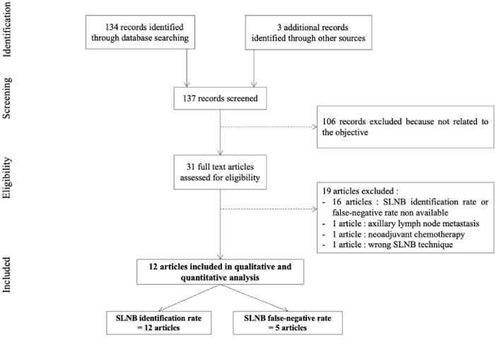
Lymphoscintigraphy technique sentinel node biopsy provides a non-invasive way to identify the sentinel lymph nodes, crucial in cancer staging and treatment planning. This technique uses a radioactive tracer to map the lymphatic drainage pathway, highlighting the sentinel lymph nodes, which are the first lymph nodes to receive drainage from a tumor. Understanding the procedure, from preoperative preparation to post-procedure management, is key to successful cancer treatment.
By pinpointing the sentinel nodes, surgeons can accurately target the region with the highest likelihood of cancer spread, reducing unnecessary procedures and improving treatment outcomes.
The process begins with meticulous preoperative preparation, which includes administering a radiotracer to the tumor site. Specialized imaging equipment then tracks the tracer’s movement through the lymphatic system, highlighting the sentinel lymph nodes. Accurate interpretation of the images is critical, as the appearance of the sentinel lymph nodes can vary depending on the presence or absence of cancer.
This intricate procedure provides invaluable information for surgeons, enabling precise staging and tailoring of treatment plans.
Introduction to Lymphoscintigraphy and Sentinel Node Biopsy: Lymphoscintigraphy Technique Sentinel Node Biopsy
Lymphoscintigraphy and sentinel node biopsy are crucial techniques in the management of cancer, particularly in breast, melanoma, and other malignancies. These minimally invasive procedures aim to identify and assess the spread of cancer to lymph nodes, enabling more precise staging and tailored treatment plans. Understanding the underlying principles and applications of these techniques is essential for healthcare professionals involved in cancer diagnosis and treatment.Lymphoscintigraphy is a specialized imaging technique that utilizes radioactive tracers to visualize the lymphatic drainage pathways.
This allows clinicians to pinpoint the sentinel lymph nodes, which are the first lymph nodes to receive lymphatic drainage from the primary tumor site. The process is non-invasive and plays a critical role in cancer staging.
Lymphoscintigraphy: Visualizing Lymphatic Drainage
Lymphoscintigraphy involves the injection of a radioactive tracer into the area surrounding the suspected tumor. This tracer, often technetium-99m sulfur colloid or similar radiopharmaceutical, travels along the lymphatic vessels to the draining lymph nodes. Specialized cameras detect the emitted radiation, producing images that reveal the lymphatic pathways and identify the sentinel lymph node(s). The process is typically performed on an outpatient basis, with the patient undergoing a simple injection procedure.
Sentinel Node Biopsy: Minimally Invasive Staging
Sentinel node biopsy is a surgical procedure that focuses on the removal and pathological examination of the sentinel lymph node(s) identified by lymphoscintigraphy. The principle behind this technique is that if cancer has spread to the lymph nodes, it will most likely do so first to the sentinel nodes. By examining these nodes, oncologists can determine the extent of the disease’s spread and assess the prognosis.
Role of Lymphoscintigraphy in Sentinel Node Identification
Lymphoscintigraphy plays a pivotal role in the identification of sentinel lymph nodes. It provides a roadmap, guiding the surgeon to the precise location of the nodes. This precise identification minimizes unnecessary dissection of the surrounding lymph nodes, leading to reduced morbidity and improved patient outcomes. The use of lymphoscintigraphy improves accuracy compared to traditional methods.
Significance in Cancer Staging and Treatment Planning
The information gleaned from sentinel node biopsy is critical in cancer staging. Knowing if the sentinel nodes are involved (positive) or not (negative) allows for more accurate staging of the cancer. This accurate staging helps determine the appropriate treatment approach. For instance, if the sentinel node is positive, more aggressive treatment, such as additional surgery or radiation therapy, might be necessary.
Conversely, if the sentinel node is negative, less extensive treatment may suffice.
Types of Lymphoscintigraphy Techniques
This table Artikels different lymphoscintigraphy techniques and their applications.
| Technique | Tracer Material | Procedure | Indications |
|---|---|---|---|
| Technetium-99m sulfur colloid | Technetium-99m sulfur colloid | Injection of tracer into the area of the tumor | Breast cancer, melanoma, and other cancers |
| Technetium-99m nanocolloid | Technetium-99m nanocolloid | Injection of tracer into the area of the tumor | Breast cancer, melanoma, and other cancers; potentially improved visualization |
| Lymphoscintigraphy with fluorescence | Fluorescence-based tracer | Injection of tracer followed by imaging with fluorescent light | Potential for enhanced visualization, particularly in superficial tumors |
| Combined lymphoscintigraphy with ultrasound | Radioactive tracer | Combined use of radioactive tracer injection and ultrasound imaging | Potential for improved visualization, particularly in areas with overlying anatomy |
Preoperative Preparation and Procedure
Preparing for lymphoscintigraphy and sentinel node biopsy involves meticulous planning and careful execution. This crucial step ensures accurate identification of the sentinel lymph node, maximizing the effectiveness of the procedure. Understanding the preoperative preparation, the procedure itself, and the role of radiopharmaceuticals is vital for both the patient and the medical team.
Lymphoscintigraphy, a technique used in sentinel node biopsy, helps pinpoint the first lymph node draining a cancer. This is crucial in various cancers, including those in the lung. For instance, in lung cancer with an egfr mutation, lung cancer with an egfr mutation often spreads to lymph nodes, making accurate identification vital for staging and treatment planning.
Understanding the spread patterns is key for successful lymphoscintigraphy and tailoring the biopsy procedure.
Preoperative Preparation
Thorough preoperative preparation is paramount for successful lymphoscintigraphy. Patients are typically instructed to fast for a period of time before the procedure, and any medications that might interfere with the scan, such as certain diuretics, should be discussed with the physician. The patient’s medical history is reviewed, and any allergies or contraindications to the radiopharmaceutical are carefully assessed.
Detailed instructions on the procedure and expected sensations are provided to alleviate patient anxiety and facilitate cooperation. The area of injection is marked, often with a skin pen, to allow for precise injection placement during the procedure.
Procedure Steps
The lymphoscintigraphy procedure involves several key steps. First, a radiopharmaceutical, typically a technetium-99m-labeled colloid, is injected into the surgical site, usually near the tumor. This injection site may vary based on the primary tumor location. The radiopharmaceutical travels through lymphatic vessels, accumulating in the sentinel lymph node(s). Following injection, the patient is positioned to allow for optimal imaging acquisition.
The exact positioning depends on the location of the surgical site and the lymphatic drainage pattern. Post-injection, a series of images are acquired using a gamma camera, capturing the movement of the tracer as it reaches the sentinel lymph node(s). The acquired images are then analyzed by a trained radiologist, who identifies the sentinel node(s) based on their uptake of the radiopharmaceutical.
Patient Positioning and Immobilization
Proper patient positioning and immobilization during lymphoscintigraphy are critical for obtaining high-quality images. The patient’s body must be maintained in a stable position to avoid motion artifacts that can obscure the visualization of the sentinel lymph node. This typically involves using a supportive surface, such as a padded table, and sometimes specialized positioning devices. Immobilization techniques may involve tape or straps, ensuring the patient remains still throughout the imaging process.
Lymphoscintigraphy, a technique used in sentinel node biopsy, helps pinpoint the first lymph node draining a potential cancer site. While this procedure is highly effective, it’s important to consider the broader picture of lung health, especially given the ongoing debate about vaping and its potential link to lung cancer. Researching if vaping is a contributing factor to lung cancer is crucial for patients, and a good resource for understanding this is does vaping cause lung cancer.
Ultimately, lymphoscintigraphy remains a valuable tool for early cancer detection and staging, particularly in cases of suspicious lymph nodes.
This careful positioning and immobilization helps ensure clear and accurate identification of the sentinel lymph node(s).
Radiopharmaceuticals in Lymphoscintigraphy
Radiopharmaceuticals play a crucial role in lymphoscintigraphy, serving as tracers to guide the identification of the sentinel lymph node. These radioisotopes are carefully selected for their ability to be effectively transported by the lymphatic system, while minimizing potential side effects. The choice of radiopharmaceutical will depend on the individual patient’s condition, the characteristics of the tumor, and the preferences of the medical team.
A gamma camera detects the emitted radiation, allowing visualization of the radiopharmaceutical’s path and the location of the sentinel lymph node.
Comparison of Radiotracers
| Tracer | Half-Life | Detection Sensitivity | Side Effects |
|---|---|---|---|
| Technetium-99m sulfur colloid | 6 hours | High | Generally well-tolerated, minimal side effects |
| Technetium-99m antimony colloid | 6 hours | High | Generally well-tolerated, minimal side effects |
| Indium-111 labeled monoclonal antibodies | 2.8 days | High | Potential for allergic reactions in some individuals |
| Gallium-67 citrate | 78.3 hours | Moderate | Generally well-tolerated, some patients may experience mild fatigue or nausea |
The table above provides a concise overview of different radiotracers used in lymphoscintigraphy. Each tracer possesses unique characteristics, impacting the procedure’s efficacy and safety. The selection of the appropriate tracer is contingent on various factors, including the clinical scenario and the expertise of the medical team.
Imaging Interpretation and Findings
Interpreting lymphoscintigraphy images is crucial for accurately identifying sentinel lymph nodes (SLNs) and guiding surgical procedures. This process involves careful analysis of the radiotracer distribution within the lymphatic system, allowing clinicians to pinpoint the SLNs that are most likely to contain cancer cells. Accurate interpretation is paramount to ensure appropriate treatment planning and patient outcomes.
Identification of Sentinel Lymph Nodes
The identification of sentinel lymph nodes relies on visualizing the initial uptake and distribution of the radiotracer injected into the surgical site. The tracer, typically a radioactive colloid, travels along lymphatic vessels, accumulating in the sentinel lymph nodes. These nodes are the first lymph nodes draining the area of suspected malignancy. Careful attention to the patterns of tracer flow and the location of initial uptake is essential for precise identification.
Radiographic imaging techniques, like gamma cameras, are used to detect and localize these nodes.
Characteristics of Sentinel Lymph Nodes on Lymphoscintigraphy
Sentinel lymph nodes on lymphoscintigraphy images typically exhibit early and intense radiotracer uptake, appearing as hot spots on the images. This intense uptake differentiates them from surrounding, non-sentinel lymph nodes. The size and shape of the sentinel lymph nodes can vary depending on the individual and the location of the primary tumor. Furthermore, the intensity of uptake can vary.
However, the characteristic feature remains the early and intense accumulation of the radiotracer.
Potential Pitfalls in Interpreting Lymphoscintigraphy Images
Several factors can complicate the interpretation of lymphoscintigraphy images. Superficial skin activity, overlapping anatomical structures, and the presence of inflammatory conditions can create false positive or false negative results. Experienced radiologists and surgeons must carefully consider these potential pitfalls during interpretation. Moreover, variations in the injection site and technique can lead to unexpected tracer patterns, requiring careful evaluation of the entire image set.
Appearance of Normal and Abnormal Sentinel Lymph Nodes
Normal sentinel lymph nodes typically demonstrate a clear, well-defined area of radiotracer accumulation. The intensity of the signal should be relatively uniform throughout the node. Abnormal sentinel lymph nodes, on the other hand, may show heterogeneous or intense uptake, indicative of metastatic disease. They might also display irregular borders, or demonstrate increased size. The radiologist and surgeon must carefully consider the morphology and signal intensity of each suspected sentinel lymph node to make an informed assessment.
Table of Possible Lymphoscintigraphy Findings
| Finding | Description | Significance | Management |
|---|---|---|---|
| Normal Sentinel Lymph Node | Clear, well-defined area of radiotracer accumulation, uniform intensity. | Indicates absence of tumor spread to the regional lymph nodes. | Usually no further intervention needed; surgical excision is not necessary. |
| Abnormal Sentinel Lymph Node (Hot Spot) | Heterogeneous or intense uptake; irregular borders, increased size. | Suggests presence of metastatic cancer cells in the sentinel lymph node. | Surgical excision and pathological examination of the sentinel lymph node are necessary. |
| Absent Sentinel Lymph Node | No radiotracer uptake in the expected drainage area. | May indicate tumor spread beyond the sentinel lymph node chain or technical issues. | Further imaging and/or alternative evaluation strategies may be necessary to assess the extent of the disease. |
| Multiple Sentinel Lymph Nodes | More than one lymph node exhibits intense radiotracer uptake. | May indicate widespread disease or involvement of multiple lymphatic channels. | Surgical excision and pathological examination of all identified sentinel lymph nodes are crucial. |
Applications and Indications

Lymphoscintigraphy and sentinel node biopsy represent a significant advancement in the management of various cancers. These techniques offer a precise and less invasive approach to identifying and assessing the spread of malignancy, allowing for targeted treatment and improved patient outcomes. This section delves into the specific applications of this methodology across different cancer types, comparing its effectiveness with other imaging methods, and highlighting the advantages and disadvantages of this procedure.
Cancer Types Where Used
Lymphoscintigraphy and sentinel node biopsy are particularly valuable in cancers where lymphatic spread is a primary concern. These include breast cancer, melanoma, and various other malignancies. The effectiveness of the technique hinges on the specific characteristics of the cancer and its tendency to metastasize via the lymphatic system. For instance, breast cancer often spreads to axillary lymph nodes, making sentinel node biopsy a crucial tool in determining the extent of the disease.
Effectiveness Compared to Other Imaging Techniques
Compared to other imaging techniques like CT scans or MRI, lymphoscintigraphy offers a more targeted approach to identifying sentinel lymph nodes. While CT and MRI can reveal the presence of enlarged lymph nodes, they may not always pinpoint thesentinel* node, the first lymph node to receive drainage from the primary tumor. Lymphoscintigraphy, using radiotracers, provides a direct visualization of the lymphatic pathway, precisely identifying the sentinel node.
This targeted approach minimizes unnecessary procedures and ensures the surgeon directly examines the most relevant node.
Advantages and Disadvantages
- Advantages: Lymphoscintigraphy and sentinel node biopsy offer several advantages, including a lower invasiveness compared to traditional methods of lymph node assessment. The technique is minimally invasive, preserving healthy tissue and potentially reducing the need for extensive surgery. It also provides crucial information regarding the extent of the disease, enabling more targeted treatment strategies. The procedure’s accuracy and precision contribute to improved patient outcomes and reduced morbidity.
- Disadvantages: While generally safe, lymphoscintigraphy and sentinel node biopsy are not without limitations. False-positive or false-negative results are possible, although rare. Careful interpretation of the images and meticulous surgical technique are paramount in mitigating these risks. Furthermore, the procedure’s efficacy depends on the precise location and size of the primary tumor, as well as the individual patient’s lymphatic drainage pattern.
In some cases, the sentinel node may not be identified, requiring additional procedures.
Potential Complications, Lymphoscintigraphy technique sentinel node biopsy
Potential complications associated with lymphoscintigraphy and sentinel node biopsy are generally minimal. Possible complications include allergic reactions to the radiotracer, bleeding, infection at the injection site, and, rarely, damage to surrounding tissues. The risk of complications is generally low, but patients should be fully informed about the potential risks before undergoing the procedure.
Detailed Explanation of Use in Specific Cancers
- Breast Cancer: In breast cancer, lymphoscintigraphy and sentinel node biopsy are frequently employed to determine if the cancer has spread to the axillary lymph nodes. The procedure helps assess the extent of the disease and guide treatment decisions, whether it be lumpectomy or mastectomy. By precisely identifying the sentinel node, surgeons can remove it for analysis, reducing the need for a full axillary lymph node dissection.
- Melanoma: Melanoma is another malignancy where lymphoscintigraphy is crucial. The procedure helps pinpoint the sentinel lymph node in the regional drainage area of the melanoma lesion. This enables targeted removal and analysis, potentially preventing unnecessary and extensive lymph node dissections.
- Other Malignancies: Lymphoscintigraphy and sentinel node biopsy can be utilized in other malignancies, such as head and neck cancers, skin cancers, and gastrointestinal cancers. The procedure’s application varies based on the tumor’s location and lymphatic drainage pattern.
Benefits Over Traditional Methods
The traditional method of assessing lymph node involvement involves a complete dissection of lymph nodes, a significantly more invasive procedure. Sentinel node biopsy offers a substantial advantage by identifying and removing only the sentinel node(s) that are most likely to contain cancerous cells. This approach minimizes the extent of the surgery, reducing potential complications, recovery time, and the likelihood of lymphedema.
Comparison with Other Techniques
Lymphoscintigraphy, while a valuable tool, isn’t the only method for identifying sentinel lymph nodes. Understanding its strengths and weaknesses in comparison to other imaging modalities is crucial for optimal patient care. This section delves into the comparative analysis of lymphoscintigraphy with ultrasound and CT scans, highlighting their respective advantages and disadvantages in the context of sentinel node biopsy.
Comparison with Ultrasound
Ultrasound, a non-invasive technique employing high-frequency sound waves, offers a real-time visualization of the lymphatic system. However, its utility in sentinel node biopsy is limited. While useful for evaluating superficial structures, ultrasound struggles to visualize deep-seated lymph nodes. It is often used in conjunction with other imaging methods. The major advantage of ultrasound is its immediate feedback and low cost.
Its accuracy in identifying sentinel nodes, however, is generally lower than that of lymphoscintigraphy, especially in complex anatomical regions.
Comparison with CT Scans
Computed Tomography (CT) scans utilize X-rays to generate cross-sectional images of the body. CT scans can visualize a wide range of structures, including lymph nodes, but they are not specifically designed for sentinel node detection. Their primary use is for detecting anatomical abnormalities and evaluating the extent of disease. The accuracy of CT in identifying sentinel nodes is often lower than lymphoscintigraphy.
The primary advantage of CT scans is their comprehensive imaging capabilities. Their ability to provide a detailed overview of the lymphatic network is less precise than lymphoscintigraphy. The use of ionizing radiation is a significant consideration.
Lymphoscintigraphy, a technique used in sentinel node biopsy, helps pinpoint the lymph nodes that drain an area of concern. While it’s a crucial diagnostic tool for various medical conditions, it’s important to remember that sometimes, even during pregnancy, a patient might experience heart palpitations. Understanding these can be just as important as the initial lymphatic mapping, especially if those palpitations are coupled with other symptoms or are new or more frequent.
For more on heart palpitations during pregnancy, check out this resource: heart palpitations during pregnancy. Ultimately, though, the lymphoscintigraphy technique is a valuable tool for accurate diagnosis and treatment planning in many different cases.
Cost-Effectiveness
The cost-effectiveness of lymphoscintigraphy compared to other techniques depends on several factors. Lymphoscintigraphy, while requiring specialized equipment and radioisotope, often yields quicker and more accurate results, potentially reducing the need for further investigations. Ultrasound is generally the least expensive option. CT scans are often more expensive than both ultrasound and lymphoscintigraphy, due to their complexity.
Summary Table
| Technique | Accuracy | Invasiveness | Cost |
|---|---|---|---|
| Lymphoscintigraphy | Generally high, particularly in detecting sentinel nodes. | Non-invasive, except for the injection of the radioisotope. | Moderate, influenced by the cost of radioisotope and specialized equipment. |
| Ultrasound | Lower accuracy compared to lymphoscintigraphy, especially for deep-seated nodes. | Non-invasive. | Lowest cost among the three techniques. |
| CT Scan | Lower accuracy compared to lymphoscintigraphy, requires ionizing radiation. | Non-invasive. | Highest cost among the three techniques. |
Post-Procedure Management and Follow-up

After undergoing lymphoscintigraphy and sentinel node biopsy, patients require careful post-procedure management and follow-up to ensure proper healing and detect any potential complications. This crucial phase involves monitoring for signs of infection, bleeding, or other adverse effects, while also providing patients with essential instructions for their recovery. A well-structured follow-up plan is key to minimizing risks and maximizing positive outcomes.Post-procedure care focuses on managing potential discomfort, ensuring proper wound healing, and promptly addressing any complications.
The specifics of the post-operative plan will vary based on individual patient factors and the extent of the procedure. This section will detail the key aspects of post-procedure management, potential complications, and the significance of follow-up.
Post-Procedure Care Instructions
Patients are usually discharged with clear instructions on managing pain, swelling, and potential complications. This includes instructions on activity levels, wound care, and medications. Adherence to these instructions is crucial for a smooth recovery. For instance, patients may be advised to avoid strenuous activities for a specified period, apply ice packs to the incision site, and take prescribed pain relievers as needed.
Detailed instructions should also include when to seek medical attention for any concerning symptoms.
Potential Complications, Lymphoscintigraphy technique sentinel node biopsy
Several complications can potentially arise after lymphoscintigraphy and sentinel node biopsy, although they are generally infrequent. These range from minor to more serious issues, and prompt recognition and management are essential.
- Hematoma Formation: A hematoma, or collection of blood, can occur at the incision site. This is usually managed with rest, ice application, and potentially compression dressings. In rare cases, drainage may be necessary. Severity varies from mild bruising to significant swelling, and size is not always predictive of symptoms. A small hematoma might be barely noticeable, while a larger one could cause significant discomfort.
- Infection: Infection at the incision site is a possibility. Symptoms may include redness, swelling, warmth, and purulent drainage. Prompt antibiotic treatment and wound care are usually sufficient to resolve the infection. The risk of infection is influenced by factors like the patient’s immune status and the surgical technique. Monitoring the wound for signs of infection is critical.
- Seroma Formation: A seroma is a collection of serous fluid at the surgical site. It typically resolves spontaneously but may require aspiration if significant. Management usually involves observation and, if necessary, drainage. Symptoms can include swelling and tenderness around the incision. Frequency of seroma formation can vary greatly depending on individual surgical technique.
- Lymphedema: Lymphedema, or swelling due to lymphatic system impairment, is a rare but potential long-term complication. This is typically observed and managed with compression therapy and/or physical therapy, if needed. Careful monitoring of the affected limb is essential.
- Numbness or Tingling: Temporary numbness or tingling around the incision site is a possible but often transient side effect. It typically resolves on its own. Careful assessment of the nerves in the area is important.
Importance of Follow-up Care
Regular follow-up appointments are essential to monitor the healing process, assess for any complications, and address any concerns the patient may have. These appointments provide a crucial opportunity for the healthcare team to detect and manage issues early, improving the chances of a successful outcome. Follow-up schedules are usually tailored to individual patients and their specific circumstances. Early detection of complications is crucial.
- Regular Check-ups: Post-operative follow-up appointments are essential to assess the wound’s healing, identify potential complications, and address patient concerns.
- Imaging Studies: Imaging, such as ultrasound or X-rays, might be used to monitor the site for abnormalities if needed. Frequency and type of imaging vary depending on the patient’s condition.
- Communication: Open communication between the patient and healthcare provider is vital. This enables prompt reporting of any unusual symptoms.
Last Point
In summary, lymphoscintigraphy technique sentinel node biopsy offers a valuable tool for cancer diagnosis and treatment. The technique provides a clear pathway for early detection and precise treatment, potentially improving patient outcomes. While lymphoscintigraphy is not without its limitations, the ability to identify sentinel lymph nodes allows for more targeted and less invasive approaches to cancer management, ultimately leading to better patient care.

