Category: Skin Conditions

  • Papule Definition of an Acne Papule A Comprehensive Guide

    Papule Definition of an Acne Papule A Comprehensive Guide

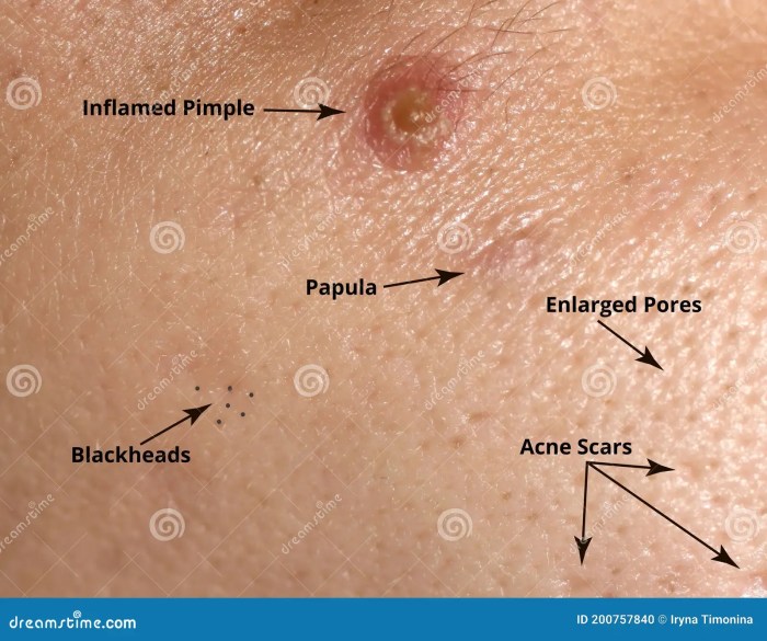
    Papule definition of an acne papule delves into the specifics of this skin condition. Understanding what constitutes an acne papule is key to effective management and treatment. This in-depth exploration covers everything from defining the papule itself to examining its causes, symptoms, and available treatments.

    Acne papules are small, solid bumps on the skin. They’re often inflamed and reddish, and their formation is intricately linked to clogged pores, bacteria, and hormonal changes. This guide will explore the characteristics of papules, their connection to other acne types, and the importance of proper skincare and medical advice for effective management.

    Definition and Characteristics

    Acne papules are a common skin condition characterized by inflamed, raised bumps. These lesions are typically a result of blocked hair follicles and the subsequent inflammatory response within the skin. Understanding their characteristics is crucial for proper diagnosis and treatment.Understanding the differences between various types of acne lesions is essential for effective management. Different types of acne lesions, like papules, pustules, and nodules, have unique appearances and causes, influencing the treatment approach.

    Differentiating these lesions helps determine the severity and appropriate course of action.

    Papule Definition

    A papule is a small, solid, raised skin lesion that doesn’t contain pus. It arises from inflammation beneath the skin’s surface. The defining feature of a papule is its solid, non-pustular nature.

    Key Characteristics Distinguishing Papules

    Papules are distinguished from other skin lesions by their specific characteristics. These characteristics include a firm, raised texture, and a limited depth. These characteristics help differentiate them from other skin conditions. For example, they differ from flat spots or lesions filled with pus.

    Typical Appearance of a Papule

    Papules typically present as small, round or slightly irregular bumps. They range in size from a few millimeters to about 1 centimeter in diameter. The color of a papule can vary from pinkish-red to reddish-brown, depending on the severity of inflammation. The appearance reflects the underlying inflammatory process.

    Comparing Papules with Pustules and Nodules

    Papules, pustules, and nodules are all types of acne lesions, but they differ in appearance and cause. Understanding these differences aids in effective treatment. The key distinctions lie in the presence or absence of pus and the depth of the lesion.

    Table: Key Differences between Papules, Pustules, and Nodules

    Characteristic Papule Pustule Nodule
    Appearance Small, solid, raised bump; pinkish-red to reddish-brown Small, pus-filled bump; white or yellow Large, solid, deep, often painful bump; reddish-pink or purplish
    Size Usually a few millimeters to 1 centimeter Usually a few millimeters to 1 centimeter Often larger than 1 centimeter; can extend deep into the skin
    Texture Firm, often slightly tender Soft, pus-filled, often tender Firm, deep, often painful
    Cause Blocked hair follicles and inflammation Blocked hair follicles, inflammation, and pus formation Blocked hair follicles, inflammation, and deep tissue involvement

    Causes of Acne Papules

    Papule definition of an acne papule

    Acne papules, those pesky little bumps that often plague our skin, aren’t just a matter of bad luck. They develop due to a complex interplay of factors, and understanding these causes is key to effective management. This deeper dive will explore the critical roles of bacteria, inflammation, clogged pores, hormonal fluctuations, and sebum production in the formation of these skin imperfections.The formation of acne papules is a multifaceted process involving the interaction of several factors.

    From the microscopic level of bacterial activity to the broader impact of hormonal changes, each element plays a crucial role in the development of these skin lesions. Understanding these factors is essential for developing personalized strategies to combat acne and promote healthier skin.

    Role of Bacteria

    Acne papules are often associated with the presence ofP. acnes*, a bacterium naturally found on the skin. When pores become clogged, this bacterium multiplies rapidly, leading to inflammation and the formation of papules. The inflammatory response triggered by the bacteria further contributes to the swelling and redness characteristic of acne. This is a critical link in the chain of events that causes acne, highlighting the importance of maintaining skin hygiene to prevent bacterial overgrowth.

    So, you’re trying to understand acne papules? A papule is basically a small, raised bump on the skin. It’s often a sign of inflammation, like a pimple, but learning about these skin conditions can be a good starting point for understanding your body. Sometimes, understanding your body’s signals can help you focus on your overall health and well-being, like when you’re trying to get pregnant fast.

    How to get pregnant fast can be a complex topic, and you’ll want to talk to your doctor for personalized advice. No matter what, understanding your body is key, and acne papules are just one small piece of that puzzle.

    Role of Inflammation

    Inflammation is a natural response to injury or infection, playing a significant role in the development of papules. When clogged pores become infected with bacteria, the immune system triggers an inflammatory response, leading to redness, swelling, and pain. This inflammatory reaction is the key to the visible characteristics of the papule, highlighting the importance of managing inflammation to reduce discomfort and prevent scarring.

    Role of Clogged Pores

    Clogged pores are the foundation of acne development. When dead skin cells, sebum, and bacteria accumulate in the pore, it creates a breeding ground forP. acnes*. This blockage leads to increased pressure within the follicle, contributing to inflammation and the development of papules. Effective skincare routines that focus on exfoliation and pore cleansing can help prevent clogged pores and thus reduce the risk of acne.

    Influence of Hormonal Changes

    Hormonal fluctuations, particularly during puberty and menstruation, significantly impact sebum production, a key factor in acne development. Increased androgen levels, for example, stimulate sebaceous glands to produce more sebum. This heightened sebum production, combined with other factors like bacterial activity and clogged pores, can lead to the formation of acne papules. Understanding these hormonal influences is crucial for tailoring acne management strategies to individual hormonal cycles.

    So, you’ve got a pesky acne papule? A papule, basically, is a small, raised, inflamed bump on the skin. While figuring out your sleeping position might not directly cure your acne, it could reveal some interesting things about your personality. For example, did you know that certain sleeping positions could reflect your personality traits? Check out this fascinating article on what your sleeping position says about you to see if your nighttime posture aligns with your known characteristics.

    Regardless of your sleeping style, understanding the papule definition is key to effective acne management.

    Sebum Production

    Sebum, an oily substance produced by sebaceous glands, is essential for skin health. However, excessive sebum production can lead to clogged pores and subsequent inflammation. When sebum production is elevated, it can mix with dead skin cells and bacteria, creating a perfect environment forP. acnes* proliferation. This process is critical in the formation of acne papules.

    Maintaining a balance in sebum production is vital for preventing acne.

    Potential Triggers

    Various factors can trigger or exacerbate acne papules. These include:

    • Stress: Stress can disrupt hormonal balance, leading to increased sebum production and a higher risk of acne development. For example, students experiencing exam stress might see a rise in acne breakouts.
    • Diet: Certain foods, such as high-glycemic index carbohydrates, may influence hormone levels and sebum production, potentially increasing acne risk. A diet high in processed foods, for example, could be a trigger.
    • Certain Medications: Some medications, such as corticosteroids or certain birth control pills, can affect sebum production and increase the likelihood of acne development.
    • Cosmetics: Certain cosmetics, particularly those with pore-clogging ingredients, can contribute to acne development. Heavy makeup, for instance, can trap sebum and exacerbate the problem.

    Symptoms and Associated Conditions

    Acne papules, while often a cosmetic concern, can sometimes present with additional symptoms that extend beyond the visible lesion. Understanding these associated symptoms is crucial for proper management and recognizing potential complications. This section will delve into the broader picture of papules, exploring their relationship with other acne types and potential complications.Beyond the noticeable bump, acne papules can present with various accompanying symptoms.

    These range from mild tenderness and slight swelling to more significant discomfort and inflammation. The intensity of these symptoms can fluctuate depending on the individual and the severity of the acne.

    Common Symptoms Associated with Acne Papules

    Acne papules, though often small, can trigger a localized inflammatory response. This leads to symptoms like redness, warmth, and tenderness to the touch. In some cases, the affected area may feel slightly swollen, and the papule may become slightly elevated. Sometimes, there’s also a slight tingling or burning sensation. The severity of these symptoms varies, and not all individuals will experience them all.

    Relationship Between Papules and Other Acne Lesions

    Acne papules are often closely linked to other types of acne lesions. Comedones, for example, can progress to papules if the trapped sebum and dead skin cells become inflamed. This inflammatory response is a key factor in the development of papules from comedones. The progression from comedones to papules is a common stage in acne development.

    So, you’re looking for the definition of an acne papule? It’s basically a small, red, solid bump on the skin. Knowing the best time to check your blood sugar can be crucial for managing health conditions, and understanding the details of papules is important for identifying and treating skin issues. For a deeper dive into optimal blood sugar testing times, check out this helpful resource: best time to check blood sugar.

    Ultimately, understanding papules is key to properly diagnosing and treating acne.

    Potential Associated Conditions or Complications

    While generally not life-threatening, acne papules can lead to potential complications. Scarring is a significant concern, particularly with persistent or severe inflammation. Scarring can range from mild, barely noticeable marks to deep, noticeable pits. In rare cases, acne can lead to psychological distress, impacting self-esteem and confidence.

    Importance of Hygiene and Skincare Practices

    Maintaining good hygiene and implementing proper skincare routines are essential for managing acne papules. Regular cleansing with a gentle, non-comedogenic cleanser helps remove excess oil and debris. Gentle exfoliation can also help prevent the accumulation of dead skin cells that can exacerbate inflammation. Avoiding harsh scrubbing, however, is crucial to prevent irritation. Choosing products specifically formulated for acne-prone skin can significantly aid in managing symptoms.

    Importance of Seeking Medical Advice

    Persistent or severe acne papules, particularly those that don’t respond to at-home remedies, require professional medical attention. A dermatologist can accurately diagnose the condition, recommend appropriate treatment options, and potentially identify underlying causes. Early intervention and proper treatment are key to preventing further complications, such as scarring. A dermatologist can also advise on the best skincare practices tailored to individual needs.

    Diagnosis and Treatment Options: Papule Definition Of An Acne Papule

    Pinpointing the cause and severity of acne papules is crucial for effective treatment. A dermatologist can accurately diagnose acne papules through a visual examination and a detailed patient history. This assessment helps determine the type and extent of the acne, enabling the development of a personalized treatment plan.Understanding the underlying factors contributing to acne papules is vital for choosing the right treatment strategy.

    Factors like hormonal fluctuations, genetics, and lifestyle choices play a significant role in the development and progression of acne.

    Diagnostic Process

    A dermatologist meticulously examines the affected skin area, looking for characteristics like the size, shape, and distribution of papules. They will also inquire about the patient’s medical history, including any previous acne treatments, medications, and lifestyle habits. This detailed evaluation allows the dermatologist to rule out other skin conditions with similar appearances.

    Common Treatment Methods, Papule definition of an acne papule

    Various methods are employed to treat acne papules, ranging from topical applications to oral medications. The chosen treatment depends on the severity and extent of the acne.

    Topical Treatments

    Topical treatments directly target the affected skin, providing localized relief and addressing the acne at its source. These treatments often contain active ingredients like benzoyl peroxide, salicylic acid, or retinoids. Benzoyl peroxide works by killing bacteria and reducing inflammation, while salicylic acid exfoliates the skin and unclogs pores. Retinoids stimulate cell turnover, preventing future breakouts.

    • Benzoyl peroxide: This common ingredient is often a first-line treatment for acne papules due to its effectiveness in reducing inflammation and killing bacteria. It can be quite effective for mild to moderate cases, but can sometimes cause skin irritation, redness, and dryness in some individuals.
    • Salicylic acid: Salicylic acid is another effective topical treatment for acne. It has keratolytic properties, meaning it helps to exfoliate the skin and unclog pores. This can be particularly beneficial for those with comedonal acne (blackheads and whiteheads) that often accompany papules.
    • Retinoids: Prescription retinoids, like tretinoin, are potent topical treatments that work by regulating skin cell turnover. This helps prevent future breakouts and reduces the appearance of existing acne scars. Retinoids can take several weeks to show significant results, and some individuals may experience mild skin irritation initially.

    Oral Medications

    Oral medications are often reserved for more severe or persistent cases of acne papules. These medications address the underlying hormonal imbalances or inflammatory processes that contribute to the condition.

    • Antibiotics: Oral antibiotics, such as tetracycline or erythromycin, can help control bacterial overgrowth that plays a significant role in acne development. They are often prescribed in conjunction with topical treatments to maximize effectiveness.
    • Hormonal Contraceptives: In some cases, particularly for women experiencing hormonal acne, oral contraceptives can help regulate hormone levels, reducing the frequency and severity of breakouts. This is a common treatment for hormonal imbalances and acne.
    • Isotretinoin: Isotretinoin, an oral retinoid, is a powerful medication often used for severe cases of acne that don’t respond to other treatments. However, it carries significant side effects and is only prescribed by dermatologists in specific cases, due to potential serious side effects.

    Effectiveness Comparison

    The effectiveness of different treatment approaches varies depending on individual factors. For example, benzoyl peroxide is often effective for mild to moderate acne, while retinoids are more suitable for persistent cases. Oral antibiotics can be helpful in controlling bacterial overgrowth, while isotretinoin is reserved for the most severe cases.

    Prevention Strategies

    Preventing acne papules requires a multi-pronged approach targeting lifestyle factors, diet, skincare routines, and stress management. A proactive approach to these elements can significantly reduce the likelihood of breakouts and improve overall skin health. This is crucial for maintaining a clear complexion and boosting self-esteem.A holistic approach to preventing acne papules goes beyond simply treating existing breakouts. By understanding the contributing factors and implementing preventative measures, individuals can actively manage their skin health and minimize the occurrence of blemishes.

    Lifestyle Changes for Acne Prevention

    Maintaining a healthy lifestyle plays a vital role in controlling acne. Regular habits contribute to overall well-being, which in turn positively impacts skin health. Incorporating these changes into your routine can significantly reduce the risk of developing acne papules.

    • Regular Exercise: Physical activity boosts blood circulation, which helps deliver essential nutrients to the skin cells and remove toxins. Aim for at least 30 minutes of moderate-intensity exercise most days of the week. For instance, a brisk walk, jogging, or swimming can improve circulation and overall skin health. Exercise also helps regulate hormones, which can influence acne development.

    • Adequate Sleep: Sufficient sleep allows the body to repair and regenerate cells, including skin cells. Aim for 7-9 hours of quality sleep per night to promote optimal skin health. Lack of sleep can disrupt hormone balance, contributing to acne breakouts.
    • Stress Management: Chronic stress can trigger hormonal imbalances that lead to acne. Incorporating stress-reducing activities, such as meditation, yoga, or deep breathing exercises, can significantly help manage stress and reduce acne risk. Identifying and managing stress triggers is also an important component.

    Dietary Recommendations for Acne Management

    A balanced diet rich in fruits, vegetables, and lean proteins is essential for overall health and can contribute to clearer skin. Nutrients like vitamins, minerals, and antioxidants play a crucial role in maintaining healthy skin function. Conversely, certain foods and drinks can exacerbate acne.

    • Balanced Diet: Prioritize a diet rich in fruits, vegetables, and whole grains. These foods provide essential vitamins, minerals, and antioxidants that support healthy skin function. Limit processed foods, sugary drinks, and excessive consumption of dairy products, which may trigger acne in some individuals.
    • Hydration: Drinking plenty of water is crucial for overall health and supports skin hydration. Dehydration can lead to dry skin, which can contribute to acne breakouts. Ensure you drink sufficient water throughout the day.

    Effective Skincare Routines for Papule Prevention

    Consistent and appropriate skincare practices are crucial in maintaining healthy skin and preventing acne papules. A personalized routine adapted to your skin type is essential for preventing breakouts.

    • Gentle Cleansing: Use a gentle, non-comedogenic cleanser twice daily to remove dirt, oil, and makeup. Avoid harsh scrubbing, as this can irritate the skin and worsen acne. Opt for a cleanser suitable for your skin type.
    • Exfoliation: Regular, gentle exfoliation can help remove dead skin cells and prevent clogged pores. Choose a mild exfoliant, such as a chemical exfoliant, and avoid over-exfoliating, which can damage the skin barrier.
    • Moisturization: Moisturizing is crucial, even for oily skin. Using a non-comedogenic moisturizer can hydrate the skin without clogging pores. This helps maintain the skin’s natural barrier.

    Stress Management Techniques for Acne Reduction

    Stress can significantly impact hormone levels and contribute to acne breakouts. By incorporating stress-reducing techniques, individuals can positively influence their skin health.

    • Mindfulness Practices: Mindfulness techniques, such as meditation and deep breathing exercises, can help manage stress levels and reduce its impact on acne. Consistent practice can significantly lower stress and improve skin health.
    • Physical Activity: Regular exercise can help alleviate stress and improve overall well-being, which can positively affect acne. Physical activity promotes relaxation and releases endorphins, further reducing stress.
    • Adequate Sleep: Sufficient sleep is crucial for the body’s ability to manage stress and maintain overall health, which also positively impacts acne. A consistent sleep schedule is essential for stress reduction and improved skin health.

    Illustrations and Visual Aids

    Papule definition of an acne papule

    Visual aids are crucial for understanding complex concepts like acne papules. Illustrations and diagrams help visualize the characteristics, development, and relationship of papules to other acne lesions and skin structures. This section provides detailed descriptions of various visual representations that can aid in comprehension.

    Papule Visual Representation

    A papule is a small, solid, raised skin lesion. A visual representation would depict a slightly elevated area on the skin, typically round or oval, with a well-defined border. Its color might vary, from skin-toned to reddish or pinkish, depending on the inflammation level. The size would typically be less than 1 centimeter in diameter. The illustration should clearly distinguish the papule from surrounding skin.

    Diagram of a Typical Papule Anatomy

    A diagram illustrating a typical papule’s anatomy should show a cross-section of the skin. The diagram should highlight the key elements: the epidermis, dermis, and the papule itself. The papule should be depicted as an elevated area within the dermis, potentially indicating inflammation or a buildup of material. The diagram should clearly show the boundary between the papule and the surrounding skin.

    It would also potentially illustrate the inflammatory cells that might be present in the dermis around the papule.

    Comparison of Acne Lesions

    A visual aid comparing and contrasting different acne lesions, including papules, should use a table format. This table would categorize various acne lesions (e.g., papules, pustules, nodules, cysts) and illustrate them using simple shapes. Each lesion would be clearly labeled. Important characteristics, such as size, color, and texture, would be noted for each lesion type. For example, a papule would be shown as a small, solid elevation, while a pustule would be a similar elevation containing pus.

    This visual aid would help differentiate between different types of acne lesions.

    Lesion Type Description Appearance
    Papule Small, solid elevation Small, round or oval, slightly raised area, skin-colored to reddish
    Pustule Papule containing pus Small, pus-filled elevation, white or yellow
    Nodule Deep, solid lesion Larger, firm, and painful, often inflamed
    Cyst Deep, pus-filled lesion Larger, inflamed, painful, pus-filled, and often tender

    Cross-section of a Skin Pore with a Papule

    A labeled illustration of a cross-section of a skin pore with a papule should clearly show the pore structure. The pore should be shown as a canal in the epidermis and dermis. A papule should be shown as an accumulation of material, possibly sebum, within or adjacent to the pore, leading to inflammation and swelling. The illustration should highlight the relationship between the pore, the papule, and the surrounding skin tissue.

    Labels should indicate the epidermis, dermis, hair follicle, sebaceous gland, and the location of the papule.

    Stages of Acne Papule Development

    A graphic representation of the stages of acne papule development would use a series of sequential images. The first image could depict a normal skin pore. Subsequent images would show the buildup of sebum and/or bacteria, progressing to inflammation and finally to the formation of a papule. This visual aid should clearly illustrate the progression of events, highlighting the contributing factors at each stage.

    It would effectively show how a simple buildup can lead to an acne papule.

    Final Review

    In conclusion, understanding acne papules, from their definition to their management, empowers individuals to take control of their skin health. This comprehensive guide provides a thorough understanding of this common skin condition. By recognizing the causes, symptoms, and treatment options, individuals can proactively address acne papules and work towards clearer, healthier skin.

  • Ringworm vs Eczema Symptoms A Deep Dive

    Ringworm vs Eczema Symptoms A Deep Dive

    Ringworm vs eczema symptoms: Understanding the differences between these skin conditions is crucial for proper diagnosis and treatment. Both can cause frustrating itching, redness, and scaling, making it challenging to distinguish them at first glance. This comprehensive guide will explore the overlapping symptoms, unique characteristics, and diagnostic considerations for each condition, helping you gain a clearer picture of the underlying causes and effective treatments.

    Ringworm, a fungal infection, often presents as a circular rash with raised borders and central clearing. Eczema, on the other hand, can manifest in various ways, from dry, cracked skin to inflamed, oozing patches. This exploration delves into the nuances of each condition, highlighting the key differences and similarities to aid in proper identification.

    Introduction to Ringworm and Eczema: Ringworm Vs Eczema Symptoms

    Ringworm and eczema are both common skin conditions that can cause discomfort and itching. While they share some overlapping symptoms, they have distinct causes and characteristics. Understanding the differences is crucial for proper diagnosis and treatment.Ringworm, despite its name, isn’t caused by a worm at all. It’s a fungal infection that appears as itchy, red, and often ring-shaped rashes on the skin.

    Eczema, on the other hand, is a chronic inflammatory skin condition that can manifest in various forms and affect different areas of the body.

    Defining Ringworm and Eczema

    Ringworm is a contagious fungal infection that affects the skin, hair, or nails. The characteristic circular rash often has a raised border and clear center. These fungal infections are typically caused by dermatophytes, a type of fungus that thrives in warm, moist environments. The infection is spread through direct contact with infected individuals or animals, or indirectly through contaminated surfaces.

    Eczema, also known as atopic dermatitis, is a chronic skin condition characterized by inflammation, dryness, and itching. It encompasses a range of types, including atopic dermatitis, contact dermatitis, and dyshidrotic eczema. The exact cause of eczema isn’t fully understood, but a combination of genetic predisposition, environmental factors, and immune system dysfunction are thought to play a role.

    Common Affected Areas

    Both ringworm and eczema can affect various areas of the body. Knowing the typical locations can aid in distinguishing between the two conditions.

    Comparison Table: Ringworm vs Eczema Locations

    Body Part Ringworm Eczema Notes
    Scalp Yes, often presenting as patchy, scaly areas. Less common, though possible, especially in children. Ringworm on the scalp can be easily mistaken for dandruff.
    Groin (Jock Itch) Yes, particularly in moist areas. Possible, but often characterized by more widespread inflammation. Jock itch is a common form of ringworm.
    Feet (Athlete’s Foot) Yes, often affecting spaces between toes. Possible, but typically involves more dryness and cracking. Athlete’s foot is a type of ringworm infection.
    Face Possible, but less common than other locations. Common, particularly in infants and children. Facial eczema can manifest as redness, dryness, and scaling.
    Hands Possible, but often limited to areas with moisture. Very common, often causing dryness, cracking, and intense itching. Hand eczema can be triggered by various irritants.
    Body Possible, often in a circular pattern. Possible, can manifest as patches of dry, inflamed skin. Generalized body eczema can be difficult to pinpoint.

    Overlapping Symptoms

    Ringworm and eczema, while distinct conditions, can sometimes share similar symptoms, leading to confusion in diagnosis. This overlap can make it challenging to differentiate between the two without a proper examination by a healthcare professional. Understanding these shared characteristics is crucial for accurate identification and appropriate treatment.Identifying the subtle differences between these skin conditions is not always straightforward. Many people experience similar itching, redness, and scaling, making self-diagnosis unreliable.

    This section delves into the common symptoms shared by ringworm and eczema, highlighting potential misconceptions and emphasizing the importance of professional medical advice.

    Common Misconceptions

    A common misconception is that the presence of itching, redness, and scaling automatically points towards one specific condition. In reality, these symptoms can manifest in both ringworm and eczema, making visual differentiation difficult. It’s crucial to remember that visual assessments alone are not sufficient for accurate diagnosis. Accurate diagnosis necessitates a detailed evaluation of the patient’s medical history, physical examination, and potentially, laboratory tests.

    Shared Symptoms, Ringworm vs eczema symptoms

    Itching, redness, and scaling are common to both ringworm and eczema. The location, pattern, and overall presentation of these symptoms, however, can offer clues, but not definitive ones. For example, ringworm often presents with a ring-like shape, while eczema can appear as dry, inflamed patches. Nevertheless, these overlapping symptoms necessitate professional medical evaluation for a precise diagnosis.

    Symptom Summary Table

    Symptom Ringworm Eczema Notes
    Itching Often intense, especially during the night Variable intensity, often worse at night or when skin is dry Both conditions can cause significant discomfort due to itching.
    Redness May appear as a well-defined red ring Skin can appear red, inflamed, and sometimes blistered Redness is a common sign in both, but the pattern may differ.
    Scaling Can appear dry, flaky, and sometimes with a yellowish tint Scaling can be present, ranging from fine to thick, dry patches Both conditions can present with scaling, but the texture and appearance can differ.
    Pattern Often circular or ring-shaped Can be patchy or widespread, sometimes affecting specific body areas While both can be localized, ringworm’s distinctive ring shape is a helpful, though not foolproof, indicator.

    Distinguishing Features

    Ringworm, a common fungal infection, often presents with telltale signs that help differentiate it from other skin conditions, including eczema. Understanding these distinctive characteristics is crucial for accurate diagnosis and effective treatment. While some symptoms can overlap, specific visual patterns and characteristics help pinpoint ringworm.

    Circular or Ring-Like Shape

    Ringworm infections typically manifest as a circular or ring-like rash. The affected area expands outward, giving the characteristic ring shape. The edges of the ring often appear raised and inflamed, creating a border around the central area. This expanding ring pattern is a key visual indicator of ringworm.

    Raised Borders and Central Clearing

    A significant characteristic of many ringworm infections is the presence of raised borders around the ring-like lesion. These raised borders are often scaly and slightly red or inflamed. The center of the ring often appears clear or less inflamed compared to the surrounding border, creating a distinctive ring-like appearance. This combination of raised borders and central clearing is frequently observed in ringworm.

    Potential for Spread

    Ringworm is contagious and can spread to other parts of the body if not treated properly. The fungus can easily spread through contact with infected skin or contaminated surfaces. Therefore, prompt diagnosis and treatment are crucial to prevent further spread. Carefully following treatment instructions and avoiding scratching the affected area is important to prevent the infection from spreading.

    Trying to tell ringworm from eczema can be tricky, right? Different rashes have similar symptoms, making diagnosis a bit of a puzzle. Fortunately, there’s a memory trick that can help you remember the key differences – like using keyword mnemonics to help you remember the key features of each condition. Check out memory tip 1 keyword mnemonics for a great way to organize these details in your mind.

    This can really make a difference in distinguishing between the two skin conditions and getting the right treatment.

    Identifying Ringworm Patterns

    The characteristic patterns created by ringworm on the skin can aid in identification. The ring shape, with the raised border and central clearing, is a common visual cue. Furthermore, the ringworm rash often appears as a series of rings or patches, with one ring growing larger than another. Observing these patterns can help distinguish ringworm from other skin conditions.

    Visual Characteristics of Ringworm

    Feature Description Illustration Notes
    Shape Circular or ring-like, with an expanding border. Imagine a circle on the skin, with the edges of the circle being slightly raised and inflamed, and the center being relatively clear or less inflamed. The ring may appear as a single ring or multiple rings, with one ring growing larger than another.
    Borders Raised, scaly, and often inflamed. Visualize the edges of the ring being elevated and having a slightly rough texture. The raised borders often appear reddish or pinkish.
    Central Area Clear or less inflamed compared to the surrounding border. Imagine the center of the ring being less red and irritated than the edges. This central clearing is a distinguishing feature.
    Spread May spread to other areas of the body if not treated promptly. Visualize the ring expanding outwards, creating multiple rings or patches. Follow proper hygiene and treatment instructions to prevent further spread.

    Distinguishing Features

    Ringworm vs eczema symptoms

    Identifying ringworm and eczema can be tricky, especially when symptoms overlap. Understanding the unique characteristics of each condition helps in accurate diagnosis. This section focuses on the distinguishing features of eczema, delving into its various presentations, inflammatory role, and different types.Eczema, a chronic inflammatory skin condition, manifests in diverse ways. The appearance of eczema can vary greatly, depending on the specific type and individual factors.

    Dryness, cracking, and oozing are common features, but the specific presentation can provide crucial clues for diagnosis.

    Eczema Appearances

    Eczema’s diverse presentations are often characterized by inflammation, dryness, and potential for cracking and oozing. These symptoms can vary depending on the type of eczema and individual factors. Understanding these varied presentations is crucial for differentiating eczema from other skin conditions.

    • Dryness: A hallmark of many eczema types, dryness often presents as rough, scaly skin, particularly on areas like the elbows, knees, and behind the knees. This dryness can be accompanied by itching and discomfort. In severe cases, the skin can become very tight and painful. This is often seen in atopic dermatitis, a common form of eczema.

    • Cracking: The skin’s dryness can lead to cracking, especially in areas subjected to friction or pressure. These cracks can become fissures and may bleed, increasing the risk of infection. The severity of cracking can vary greatly, from minor surface cracks to deeper, more pronounced fissures.
    • Oozing: Some eczema types, particularly in acute stages, may exhibit oozing. This often presents as a weeping or leaking of fluid from the affected area. The fluid can be clear or yellowish, and it can lead to crusting and scaling of the skin. This is frequently seen in atopic dermatitis flares.

    Role of Inflammation in Eczema

    Inflammation is a crucial component in eczema. It’s the body’s immune response to an irritant, allergen, or unknown trigger. This response leads to the characteristic symptoms. The inflammatory process plays a significant role in the development and progression of eczema.

    Inflammation triggers the release of chemicals that can cause redness, swelling, itching, and dryness.

    The inflammation in eczema can manifest in various ways, influencing the skin’s appearance and function. Chronic inflammation can lead to thickening and changes in the skin’s structure, making it more susceptible to damage and infection.

    Types of Eczema

    Eczema isn’t a single entity; it encompasses several types, each with unique characteristics. Understanding these different types helps in targeted treatment and management. The underlying causes and specific presentations vary between types.

    Eczema Type Description Typical Appearance Notes
    Atopic Dermatitis A chronic, relapsing skin condition often associated with a family history of allergies, asthma, or hay fever. Dry, itchy, inflamed skin; often appears on the folds of the elbows and knees, face, and neck. Can be characterized by oozing, cracking, and scaling. The most common type of eczema.
    Contact Dermatitis Inflammation of the skin triggered by direct contact with an irritant or allergen. Redness, itching, and inflammation at the site of contact. Can also exhibit blistering or oozing. Reaction to specific substances, like chemicals or plants.
    Dyshidrotic Eczema Characterized by small, fluid-filled blisters on the palms, soles, and sides of the fingers and toes. Itchy, small blisters, often clustered. Often triggered by stress or sweating.
    Nummular Eczema Rounded, coin-shaped patches of inflamed, itchy skin. Round or oval patches, dry, scaly, and often thickened. Can be persistent and difficult to treat.

    Diagnostic Considerations

    Ringworm vs eczema symptoms

    Accurate diagnosis of ringworm and eczema is crucial for effective treatment. Incorrect diagnoses can lead to ineffective treatments, prolonged discomfort, and potentially unnecessary use of strong medications. A healthcare professional’s expertise is essential in differentiating these conditions, given the potential for overlapping symptoms. A proper diagnosis hinges on a thorough evaluation of the patient’s medical history, physical examination, and sometimes, further testing.The diagnostic process involves more than just identifying the visible signs.

    A doctor must consider various factors, including the location, appearance, and duration of the rash, alongside the patient’s overall health and any potential underlying conditions. This multifaceted approach ensures the most accurate and appropriate treatment plan.

    Importance of a Medical Professional’s Diagnosis

    A healthcare professional possesses the knowledge and expertise to accurately interpret the complex interplay of symptoms and factors associated with ringworm and eczema. They can differentiate between similar-appearing skin conditions and identify any underlying conditions that may be contributing to the symptoms. Their experience in assessing various skin lesions, combined with the patient’s medical history, provides the best chance for an accurate diagnosis.

    Diagnostic Methods

    A comprehensive diagnostic approach often includes several methods. Skin scraping is a common technique, involving the removal of a small sample of skin for microscopic examination. This allows for the visualization of fungal elements, which are indicative of ringworm. The potassium hydroxide (KOH) test is another valuable tool. A skin sample is treated with KOH, which helps to break down skin cells, making fungal hyphae (the thread-like structures of fungi) more visible under a microscope.A doctor may also perform a physical examination to assess the skin’s texture, look, and location of the rash.

    Figuring out if it’s ringworm or eczema can be tricky, as their symptoms can overlap. Both can cause itchy, red patches, but the specific location and appearance can sometimes offer clues. Interestingly, while we’re on the topic of skin conditions, did you know there’s a fascinating connection between caffeine consumption and breast health? Research suggests a potential link, but more study is needed.

    For more insights into this topic, check out this article on the link between caffeine breast health. Regardless of the underlying cause, it’s always best to consult a doctor to get an accurate diagnosis and appropriate treatment for skin conditions like ringworm or eczema.

    They may ask questions about the duration of the rash, any recent exposures to allergens or irritants, and any other relevant symptoms. This multifaceted approach provides a more comprehensive understanding of the underlying cause of the skin condition.

    Potential for Misdiagnosis

    Misdiagnosis is a possibility in any medical field. With conditions like ringworm and eczema, misdiagnosis can occur due to the similarity in some symptoms. For example, certain types of eczema can exhibit ring-like patterns, mimicking ringworm. Conversely, ringworm can sometimes present with symptoms that overlap with eczema. Therefore, relying solely on visual cues can lead to errors.

    A thorough evaluation by a dermatologist or other qualified healthcare professional is essential.

    Significance of Medical History and Patient Reports

    The patient’s medical history and detailed description of symptoms are vital components of the diagnostic process. A history of recent illnesses, medications, or exposures to potential irritants or allergens can offer crucial insights into the underlying cause of the skin condition. Patient reports, such as the duration of the rash, its progression, and any associated symptoms (itching, pain, or discomfort), provide valuable context for the healthcare professional.

    Trying to figure out if it’s ringworm or eczema can be tricky, as their symptoms can overlap. Both often present with itchy, red patches on the skin. However, understanding the common symptoms of low back pain, like stiffness and radiating pain, can help differentiate these skin conditions from musculoskeletal issues. For a deeper dive into the various presentations of low back pain, check out this helpful resource: common symptoms of low back pain.

    Ultimately, accurate diagnosis of ringworm or eczema still requires a proper medical evaluation, so don’t hesitate to consult a doctor if you’re unsure.

    These details help rule out or support various possibilities.

    Comparison of Diagnostic Methods

    Method Description Ringworm Applicability Eczema Applicability
    Skin Scraping Removal of a small skin sample for microscopic examination. Helpful in identifying fungal elements. Can help rule out fungal infection, but not specific to eczema.
    KOH Test Treating a skin sample with KOH to reveal fungal hyphae. Highly effective in detecting fungal elements. May be used to rule out fungal infection, but other tests might be needed to confirm eczema.
    Physical Examination Visual assessment of skin texture, appearance, and location of the rash. Useful for identifying characteristic ring-shaped lesions. Essential for observing skin inflammation, dryness, and other eczema-related characteristics.
    Medical History and Patient Reports Gathering information about the patient’s past illnesses, medications, and reported symptoms. Can help determine potential exposure to fungal sources. Provides insight into potential triggers and patterns associated with eczema.

    Treatment Approaches

    Understanding the appropriate treatment for ringworm and eczema is crucial for effective management. Choosing the right approach depends on several factors, including the severity, type, and location of the condition. Different treatments can have varying degrees of effectiveness and potential side effects, making personalized care essential.Effective treatment often involves a combination of strategies, tailored to address the specific needs of the individual.

    For instance, mild cases might respond well to topical treatments, while more severe cases may require oral medications or a combination of therapies. A healthcare professional can guide you in developing a personalized treatment plan.

    Ringworm Treatment

    Ringworm, caused by fungal infections, typically responds well to antifungal medications. Topical antifungal creams are frequently the first line of treatment for mild to moderate cases. These creams contain ingredients like clotrimazole, miconazole, or terbinafine, which directly target and eliminate the fungi. The duration of treatment often depends on the severity and location of the infection, and usually lasts from a few weeks to several months.In more severe cases or when topical treatments are insufficient, oral antifungal medications may be prescribed.

    These medications, taken by mouth, provide a broader reach and are more effective in treating deeper or more widespread infections. Examples include terbinafine and griseofulvin. Oral medications typically have a longer treatment duration compared to topical creams. Important considerations include potential side effects, drug interactions, and patient compliance.

    Eczema Treatment

    Eczema, a chronic inflammatory skin condition, often requires a multifaceted approach to treatment. The goal is to manage symptoms and prevent flare-ups. Moisturizers play a vital role in maintaining skin hydration and preventing dryness, a common trigger for eczema. They help create a protective barrier and reduce the itching and discomfort associated with the condition.Topical corticosteroids are often used to reduce inflammation and itching.

    Their strength and frequency of use depend on the severity of the eczema. Mild to moderate cases may respond well to over-the-counter or low-potency corticosteroids, while more severe cases may require prescription-strength medications. It is important to follow the prescribed dosage and application instructions carefully. In some cases, other topical therapies like calcineurin inhibitors or immunomodulators might be considered.

    These medications can help reduce the immune response that contributes to the inflammation.

    Treatment Comparison

    Different treatment approaches for ringworm and eczema have unique characteristics and effectiveness. While antifungal creams and oral medications are effective for ringworm, topical corticosteroids and moisturizers are frequently used for eczema. The effectiveness of each treatment can vary based on the individual’s response, the severity of the condition, and the type of fungus or eczema involved.

    Treatment Variations by Severity

    The intensity of treatment often depends on the severity of the condition. Mild cases of ringworm or eczema might respond well to topical therapies, whereas severe cases may necessitate oral medications or a combination of topical and systemic treatments. A healthcare provider can evaluate the severity of the condition and recommend the most appropriate course of action.

    Table of Common Treatment Options

    Condition Treatment Description Notes
    Ringworm Topical antifungal creams Creams containing antifungal agents like clotrimazole or miconazole applied to the affected area. Effective for mild to moderate cases; treatment duration varies.
    Ringworm Oral antifungal medications Oral medications such as terbinafine or griseofulvin taken by mouth. More effective for severe or widespread infections; longer treatment duration; potential side effects.
    Eczema Moisturizers Hydrating creams or lotions applied regularly to maintain skin moisture and prevent dryness. Essential for managing eczema; helps create a protective barrier.
    Eczema Topical corticosteroids Creams or ointments containing corticosteroids to reduce inflammation and itching. Available in varying strengths; follow prescribed dosage and application instructions.

    Prevention and Management

    Ringworm and eczema, while distinct conditions, share some overlapping symptoms and can be challenging to manage. Effective prevention and ongoing management strategies are crucial for minimizing flare-ups and improving quality of life for those affected. Understanding the specific preventative measures and consistent management approaches for each condition can significantly reduce the impact of these skin issues.Preventing and managing these conditions involves a multifaceted approach, focusing on both external factors and internal well-being.

    A proactive approach, combining hygiene practices, avoidance of triggers, and consistent monitoring, is essential for long-term control.

    Ringworm Prevention

    Preventing ringworm often involves minimizing contact with potential sources of infection. Maintaining good hygiene practices, particularly handwashing, is paramount. Avoid sharing personal items like towels, clothing, or hats with others, especially those known to have ringworm. Prompt treatment of infected individuals is vital to prevent further spread within communities or families. Identifying and isolating infected individuals, combined with proper cleaning of potentially contaminated surfaces, plays a key role in preventing the spread of the fungus.

    Eczema Prevention

    Eczema management heavily relies on identifying and avoiding triggers. Common triggers include harsh soaps, detergents, and certain fabrics. Maintaining consistent skin hydration is also crucial. Applying moisturizing creams or ointments regularly can help prevent dryness and cracking, reducing the likelihood of flare-ups. Identifying and eliminating specific allergens or irritants that trigger eczema can drastically reduce the frequency and severity of outbreaks.

    Stress management techniques can also play a role in preventing eczema flare-ups in some individuals.

    Ongoing Management Strategies

    Effective management of both ringworm and eczema requires ongoing monitoring and proactive intervention. Regular check-ups with a dermatologist or healthcare professional are essential for both conditions. Prompt treatment of any new or worsening symptoms is critical to prevent complications. Identifying patterns of flare-ups and understanding triggers can aid in developing personalized management strategies. Early intervention and consistent follow-up care can significantly improve outcomes.

    Symptom Management and Flare-Up Prevention

    Managing symptoms and preventing flare-ups involves a combination of strategies. For ringworm, adhering to prescribed antifungal medications and avoiding scratching are crucial. For eczema, maintaining a consistent skincare routine, using appropriate moisturizers, and managing stress are key components. Regular application of prescribed topical treatments, along with avoiding triggers, can help manage both conditions effectively. A personalized management plan, tailored to individual needs and triggers, can optimize treatment outcomes and minimize flare-ups.

    Prevention and Management Summary Table

    Condition Prevention Tip Description Example
    Ringworm Avoid contact with infected individuals Minimizing exposure to the fungus that causes ringworm is essential to prevent infection. Do not share personal items like towels or hats with others who have ringworm.
    Ringworm Practice good hygiene Maintaining cleanliness, particularly handwashing, is crucial in preventing the spread of ringworm. Wash hands thoroughly after touching potentially contaminated surfaces.
    Eczema Avoid irritants Identifying and eliminating substances that trigger eczema flare-ups is key. Switch to fragrance-free soaps and detergents.
    Eczema Maintain skin hydration Regular moisturizing helps maintain skin barrier function, reducing dryness and irritation. Apply moisturizer several times a day, especially after bathing.
    Both Monitor symptoms Regularly checking for signs of worsening or new symptoms is vital. Report any changes in rash appearance or intensity to a healthcare professional.
    Both Follow prescribed treatment Adherence to prescribed medications and therapies is essential for effective management. Take antifungal medications as directed for ringworm.

    Concluding Remarks

    In conclusion, while both ringworm and eczema can produce similar symptoms, crucial differences exist in their appearance, cause, and treatment. A proper diagnosis from a healthcare professional is essential for effective management. By understanding the unique characteristics of each condition, individuals can work towards effective treatment and prevent future outbreaks. This detailed comparison provides a valuable resource for anyone seeking to understand the nuances of these common skin conditions.

  • Sebaceous Hyperplasia Causes, Symptoms, and Treatments

    Sebaceous Hyperplasia Causes, Symptoms, and Treatments

    Sebaceous hyperplasia causes symptoms and treatments are explored in detail. This comprehensive guide dives into the causes, symptoms, and various treatment options for sebaceous hyperplasia, a common skin condition. We’ll examine the underlying reasons for its development, the visible signs it presents, and the different approaches to managing it. Understanding this condition empowers individuals to make informed decisions about their health and well-being.

    Sebaceous hyperplasia, often appearing as small, yellowish bumps on the face, neck, or upper chest, is frequently misunderstood. This condition, often benign and not requiring immediate medical attention, can be easily confused with other skin issues. However, a thorough understanding of its characteristics, causes, and potential treatments is crucial for appropriate management.

    Introduction to Sebaceous Hyperplasia

    Sebaceous hyperplasia is a common, benign skin condition characterized by the overgrowth of sebaceous glands. These glands, responsible for producing sebum (oil), become enlarged and form small, often flesh-colored or yellowish bumps on the skin. Understanding this condition is crucial for differentiating it from other skin issues with similar appearances and for managing patient concerns.

    Definition of Sebaceous Hyperplasia

    Sebaceous hyperplasia is a benign (non-cancerous) proliferation of sebaceous glands, typically occurring in middle-aged and older adults. It’s a common finding, often appearing as small, raised bumps on the skin.

    Locations of Sebaceous Hyperplasia

    Sebaceous hyperplasia commonly appears on areas of the body with a high concentration of sebaceous glands. These locations include the face (especially the nose, cheeks, and forehead), chest, back, and shoulders. The distribution often mirrors areas with higher sebum production.

    Appearance of Sebaceous Hyperplasia

    Sebaceous hyperplasia typically presents as small, smooth, flesh-colored or yellowish papules (small, solid bumps). They are usually less than 5 millimeters in diameter and may appear slightly raised above the skin surface. The bumps often have a pearly or waxy texture. Their appearance can vary from subtle to more noticeable, depending on the individual and the affected area.

    Comparison to Other Skin Conditions

    Condition Typical Appearance Location Key Differences
    Sebaceous Hyperplasia Small, smooth, flesh-colored or yellowish papules, less than 5 mm in diameter Face, chest, back, shoulders Benign, often multiple, typically asymptomatic.
    Basal Cell Carcinoma Small, pearly, translucent, or reddish papules or nodules; may have a slightly crusted or ulcerated surface Face, ears, neck, scalp Can be cancerous; often has a non-healing or bleeding characteristic; may have a rolled or pearly border
    Moles (Melanocytic Nevi) Various colors, shapes, and sizes; typically dark brown or black Anywhere on the skin May have irregular borders; can be raised or flat; a change in size, shape, or color can be a warning sign.
    Acne Red, inflamed papules, pustules, or nodules; often accompanied by blackheads or whiteheads Face, back, chest Often accompanied by inflammation and pain; commonly seen in adolescents and young adults

    Risk Factors for Sebaceous Hyperplasia

    Risk Factor Description Potential Impact Supporting Evidence
    Age Increased incidence with advancing age Higher probability of developing sebaceous hyperplasia as one ages. Numerous studies show a correlation between age and prevalence.
    Genetics Family history of sebaceous hyperplasia Genetic predisposition may increase the risk of developing the condition. Family history studies suggest a potential genetic component.
    Hormonal changes Hormonal fluctuations (e.g., during puberty or pregnancy) Possible influence on sebaceous gland activity. Limited direct evidence, but hormonal fluctuations are known to affect skin conditions.
    Sun exposure Prolonged or intense exposure to sunlight No direct evidence linking sun exposure to the development of sebaceous hyperplasia. Other skin conditions can be affected by sun exposure. Data is inconclusive. The condition itself is not directly sun-related.

    Causes of Sebaceous Hyperplasia: Sebaceous Hyperplasia Causes Symptoms And Treatments

    Sebaceous hyperplasia causes symptoms and treatments

    Sebaceous hyperplasia, benign skin growths, often appear as small, yellowish bumps, particularly on the face, chest, and back. While their exact cause remains somewhat elusive, researchers are uncovering crucial insights into the factors that contribute to their development. Understanding these factors is vital for both diagnosis and management strategies.The underlying causes of sebaceous hyperplasia are complex and not fully understood.

    Current research suggests a combination of genetic predispositions and environmental influences. This interplay is likely the driving force behind the formation of these common skin lesions.

    Genetic Predisposition

    Genetic factors play a significant role in the development of sebaceous hyperplasia. Family history often reveals a pattern of these lesions. Inherited variations in genes related to skin cell growth and function might increase an individual’s susceptibility to developing these bumps. Studies on large families with a high prevalence of sebaceous hyperplasia have identified potential candidate genes, although definitive links are still being investigated.

    The exact mechanisms through which these genetic variations influence sebaceous hyperplasia remain a subject of ongoing research.

    Environmental Factors

    Environmental factors may also contribute to the development or exacerbation of sebaceous hyperplasia. Exposure to certain irritants or chronic inflammation could potentially trigger the growth of these benign lesions. Hormonal fluctuations, especially in individuals with hormonal imbalances, could also influence the proliferation of sebaceous glands. For instance, individuals experiencing significant hormonal shifts during puberty or pregnancy may see an increase in sebaceous hyperplasia lesions.

    Comparison of Theories

    Different theories exist regarding the precise cause of sebaceous hyperplasia. One prominent theory emphasizes the role of genetic predisposition, suggesting that individuals with a specific genetic makeup are more prone to developing these lesions, irrespective of environmental factors. Another theory highlights the importance of environmental factors, suggesting that prolonged exposure to specific irritants or chronic inflammation could trigger or exacerbate sebaceous hyperplasia in genetically susceptible individuals.

    These theories aren’t mutually exclusive, and a more accurate explanation likely involves an interplay between genetic predisposition and environmental triggers.

    Triggers for Exacerbation

    While the exact causes of sebaceous hyperplasia are not fully understood, several factors could potentially exacerbate the growth or appearance of existing lesions. For example, irritation from frequent rubbing or pressure on the affected area might lead to inflammation and a visible increase in lesion size. Likewise, individuals with sensitive skin might experience flare-ups when exposed to harsh skincare products.

    The effects of prolonged sun exposure on sebaceous hyperplasia lesions are also a potential factor, although further research is needed to determine the extent of this effect.

    Hypotheses on Triggers

    Hypothesis Description Supporting Evidence Limitations
    Genetic Predisposition Individuals inherit a predisposition to develop sebaceous hyperplasia due to specific gene variations. Family history studies show clustering of cases in families. Candidate genes have been identified, although definitive links remain to be established. Doesn’t explain all cases; environmental factors likely play a role. The specific genes and their interactions are still under investigation.
    Chronic Inflammation Chronic skin irritation or inflammation may trigger or exacerbate the development of sebaceous hyperplasia. Some studies suggest a correlation between chronic inflammation and sebaceous hyperplasia. Irritants like certain cosmetics or friction could contribute. Difficult to isolate inflammation as the sole cause; genetic predisposition likely plays a significant role. Studies on the specific inflammatory pathways are limited.
    Hormonal Fluctuations Changes in hormone levels, especially in puberty or pregnancy, could influence sebaceous gland activity and hyperplasia. Observations of increased lesions during hormonal shifts in some individuals suggest a potential link. Correlation does not equal causation; other factors may contribute. More rigorous studies are needed to assess the precise hormonal impact.
    Environmental Irritants Prolonged exposure to specific irritants or chemicals could contribute to the development or exacerbation of sebaceous hyperplasia. Certain cosmetics or products may trigger inflammation. Friction or pressure on the affected area could also exacerbate the condition. Identifying specific triggers and quantifying their impact on sebaceous hyperplasia remains challenging. Individual responses to irritants vary significantly.

    Symptoms of Sebaceous Hyperplasia

    Sebaceous hyperplasia, a common benign skin condition, often presents with subtle changes that can be easily overlooked. Understanding the typical symptoms is crucial for distinguishing it from other skin issues and for appropriate management. While generally harmless, it’s important to recognize any unusual developments.

    Common Symptoms

    Sebaceous hyperplasia is typically characterized by small, raised, and yellowish or flesh-colored bumps. These lesions are usually smooth and round, and they vary in size, often ranging from a few millimeters to a few millimeters in diameter. They are frequently found on the face, particularly on the cheeks, nose, and forehead, but can also appear on the chest, back, and shoulders.

    These lesions are typically painless and do not cause any discomfort. Their appearance can be subtle, with the bumps sometimes being so small they may be barely noticeable.

    Distinguishing Features

    Several key features help differentiate sebaceous hyperplasia from other skin conditions. Unlike skin cancers, sebaceous hyperplasia lesions are usually well-defined and do not have irregular borders. They tend to appear in clusters, rather than scattered individually. They are often associated with areas of increased sebum production, a factor that can be noted in some cases. It is important to note that sebaceous hyperplasia typically does not cause itching, bleeding, or pain.

    Potential Complications

    While sebaceous hyperplasia is generally benign, potential complications are rare. The following table Artikels some possibilities and their associated symptoms.

    Complication Description Symptoms Severity
    Inflammation Localized redness, swelling, and tenderness around the lesion. Pain, warmth, redness, potential swelling, discomfort. Mild to moderate. Often resolves with time.
    Secondary Infection Bacterial or fungal infection of the lesion. Pus-filled sores, increased redness, pain, fever. Moderate to severe. Requires medical intervention.
    Cosmetic Concerns Patient dissatisfaction with the appearance of lesions. Psychological distress, desire for treatment, concerns about social interaction. Mild to moderate. Management varies based on patient preference.
    Accidental Trauma Injury or damage to the lesion from external factors. Bleeding, broken skin, pain. Mild to severe depending on the extent of injury.

    Variations in Symptoms

    The appearance and extent of symptoms can vary depending on the location and extent of the sebaceous hyperplasia. Lesions on sun-exposed areas may be slightly darker in color. Larger clusters of lesions may cause a noticeable thickening of the skin in the affected area. In cases where the lesions are numerous and widespread, the appearance may be more noticeable.

    Individual variations in skin tone and texture can also affect the overall visual impression of the lesions.

    Sebaceous hyperplasia, a common skin condition, often presents with small, yellowish bumps, typically on the face and chest. While usually harmless, understanding its causes, symptoms, and treatments is important. Interestingly, some research suggests a potential link between certain medications, like NSAIDs (non-steroidal anti-inflammatory drugs), and inflammatory bowel disease (IBD), which can sometimes complicate the picture. For a deeper dive into the relationship between NSAIDs and IBD, check out this informative article: nsaids non steroidal anti inflammatories and ibd.

    Fortunately, various treatment options exist for sebaceous hyperplasia, ranging from topical creams to surgical removal, depending on the severity and location of the lesions.

    Treatments for Sebaceous Hyperplasia

    Sebaceous hyperplasia, while typically harmless, can be cosmetically bothersome for some individuals. Fortunately, various treatment options are available to address the lesions and improve appearance. The best approach depends on factors such as the size, number, and location of the lesions, as well as individual preferences and potential risks.Effective treatment strategies aim to reduce the size, flatten the surface, or remove the sebaceous hyperplasia lesions, restoring a more even skin tone.

    The decision of whether or not to pursue treatment is a personal one and should be made in consultation with a dermatologist.

    Treatment Options

    Several treatment options exist for sebaceous hyperplasia, each with varying degrees of effectiveness and potential side effects. The choice of treatment often depends on the severity and location of the lesions, as well as the patient’s individual preferences.

    Surgical Removal

    Surgical removal is a definitive treatment option for sebaceous hyperplasia. This procedure involves excising the lesions under local anesthesia. Surgical removal is generally effective in eliminating the lesions and can provide a very satisfactory aesthetic outcome. However, it carries a risk of scarring, which can be more noticeable on certain areas of the body. The extent of scarring depends on the size of the lesion, the surgical technique, and the individual’s skin characteristics.

    This treatment is often considered for larger or more numerous lesions, particularly those on areas of the body where scarring might be less noticeable, such as the back or chest.

    Cryotherapy

    Cryotherapy involves freezing the sebaceous hyperplasia lesion using liquid nitrogen. This method is generally effective in reducing the size and appearance of the lesions. It is often a less invasive approach compared to surgical removal, with a lower risk of scarring. However, it may not be as effective for larger lesions and may take multiple treatments for optimal results.

    Side effects include temporary skin discoloration and discomfort, and, in rare cases, blistering. This treatment is well-suited for smaller lesions on less sensitive skin areas.

    Laser Therapy

    Laser therapy uses focused light beams to target and destroy the sebaceous hyperplasia lesions. Different types of lasers can be used, each with varying effectiveness and potential side effects. Laser therapy can be highly effective in treating sebaceous hyperplasia, leading to a significant reduction in lesion size and improved skin texture. Potential side effects include redness, swelling, and temporary skin discoloration.

    The choice of laser therapy will depend on the type of laser and the individual patient’s skin characteristics. It is often a viable option for treating multiple lesions, especially those on visible areas, and can offer a more precise approach compared to cryotherapy.

    Chemical Peels

    Chemical peels can be used to treat sebaceous hyperplasia, although their effectiveness may vary depending on the concentration and type of chemical used. The treatment involves applying a chemical solution to the skin to remove the top layers, potentially causing the sebaceous hyperplasia lesions to become less noticeable. Chemical peels can be effective in treating sebaceous hyperplasia, particularly when used in conjunction with other treatments.

    Side effects can include temporary redness, swelling, and skin sensitivity. This treatment is often considered for less severe cases or in combination with other methods.

    Comparison Table

    Treatment Description Effectiveness Potential Side Effects
    Surgical Removal Excision of lesions under local anesthesia Generally very effective Scarring possible, depending on location and technique
    Cryotherapy Freezing lesions with liquid nitrogen Effective for smaller lesions, multiple treatments may be needed Temporary discoloration, discomfort, possible blistering
    Laser Therapy Using focused light beams to target lesions Highly effective, often precise Redness, swelling, temporary discoloration
    Chemical Peels Applying chemical solution to remove skin layers Variable effectiveness, often used in combination Temporary redness, swelling, skin sensitivity

    Spontaneous Resolution

    In some cases, sebaceous hyperplasia lesions may spontaneously resolve without any treatment. However, this is not a reliable outcome and the time it takes for resolution is unpredictable. The likelihood of spontaneous resolution varies depending on factors such as the size, location, and individual characteristics of the lesions.

    Diagnosis of Sebaceous Hyperplasia

    Sebaceous hyperplasia causes symptoms and treatments

    Sebaceous hyperplasia, while often benign and easily recognizable, requires a proper diagnosis to rule out similar skin conditions. Accurate identification is crucial for appropriate management and to ensure patients receive the right treatment. This process typically involves a combination of visual assessment and, sometimes, additional diagnostic tools.Proper diagnosis of sebaceous hyperplasia is vital to differentiate it from other skin lesions that may mimic its appearance.

    This distinction helps guide treatment and avoid unnecessary interventions. The process often begins with a detailed physical examination, focusing on the characteristics of the lesions.

    Sebaceous hyperplasia, those little bumps on your skin, are usually harmless and often caused by aging or hormones. While some remedies suggest turmeric for potential benefits, it’s important to understand how much turmeric is too much, as with any supplement. This page will give you more details on the safe dosage of turmeric. Ultimately, proper medical advice from a dermatologist remains the best approach for addressing sebaceous hyperplasia causes, symptoms, and effective treatments.

    Physical Examination, Sebaceous hyperplasia causes symptoms and treatments

    A thorough physical examination plays a key role in diagnosing sebaceous hyperplasia. Clinicians visually inspect the skin lesions, paying close attention to their size, shape, color, and distribution. The presence of multiple, well-defined, yellowish or flesh-colored papules or plaques, often clustered together, is a strong indicator. The location of the lesions, typically on the face, neck, chest, or back, can also provide valuable clues.

    Careful observation of the surrounding skin and the absence of any alarming symptoms like inflammation or ulceration further support the diagnosis.

    Differential Diagnosis

    Differentiating sebaceous hyperplasia from other skin conditions is crucial. Similar-appearing lesions, such as basal cell carcinoma, seborrheic keratosis, or other benign growths, can require further investigation. The clinician must carefully assess the characteristics of the lesion to rule out these potential alternatives. Features like irregular borders, ulceration, rapid growth, or bleeding strongly suggest the need for additional tests.

    Diagnostic Criteria

    Careful evaluation of diagnostic criteria is critical to confirm a diagnosis of sebaceous hyperplasia. This systematic approach helps clinicians determine if the lesion meets the criteria for sebaceous hyperplasia.

    Criterion Description Diagnostic Method Significance
    Appearance Well-defined, yellowish or flesh-colored papules or plaques, often clustered together. Visual inspection during physical examination. Suggests the presence of sebaceous hyperplasia.
    Location Typically found on the face, neck, chest, or back. Physical examination. Provides additional context and supports the diagnosis.
    Size Usually small, ranging from a few millimeters to a few centimeters in diameter. Physical examination. Helps differentiate from larger, potentially concerning lesions.
    Consistency Soft and slightly raised; often with a smooth surface. Palpation during physical examination. Assists in distinguishing from other lesions with different consistencies.

    Imaging Techniques

    While not always necessary, imaging techniques can aid in the diagnosis, particularly when differential diagnoses are suspected. Dermoscopy, a non-invasive technique that uses a specialized microscope to examine the skin’s surface, can provide detailed images of the lesion’s structure and characteristics. This can be useful in differentiating sebaceous hyperplasia from other skin conditions with similar appearances. In cases of doubt, a biopsy may be performed to obtain a tissue sample for microscopic examination.

    This is the definitive diagnostic method, confirming the presence and nature of the lesion.

    Sebaceous hyperplasia, those little bumps on your skin, often have simple causes like genetics or aging. Understanding the different types of carbs, like good vs bad carbs , might not directly impact the condition, but a balanced diet can certainly contribute to overall health and potentially influence skin health. Treatments range from topical creams to laser procedures, depending on the severity and location of the hyperplasia.

    Prevention of Sebaceous Hyperplasia

    Sebaceous hyperplasia, while typically harmless, can be bothersome due to its appearance. Unfortunately, there are no definitive preventive measures currently available to stop the development of these benign skin growths. Understanding the factors that might increase the risk, however, can help in managing potential triggers.Currently, the primary focus in managing sebaceous hyperplasia is on symptom management and treatment when necessary, rather than on prevention.

    This is because the exact causes of the condition are not fully understood. Research continues, but for now, there’s no proven method to prevent their formation.

    Lifestyle Factors Influencing Development

    Several lifestyle factors could potentially influence the likelihood of developing sebaceous hyperplasia. These factors, however, are not definitive causes, and further research is needed to establish clear links. It’s important to note that this is not an exhaustive list and more research is necessary to determine the complete picture.

    • Genetics: A family history of sebaceous hyperplasia might increase a person’s predisposition to developing the condition. However, this doesn’t guarantee development, and many individuals with a family history do not experience it.
    • Age: Sebaceous hyperplasia typically appears in middle-aged and older adults. This suggests an association with aging, but it doesn’t mean younger individuals are immune. The exact mechanisms behind this correlation are still under investigation.
    • Hormonal Changes: While not fully understood, hormonal fluctuations might play a role. However, there’s no conclusive evidence to support this theory, and further studies are needed to establish a definite link.

    Managing Potential Risk Factors

    While definitive prevention is not yet possible, managing potential risk factors can help in reducing the likelihood of developing sebaceous hyperplasia. These approaches focus on general health and well-being rather than on targeting the specific causes.

    • Healthy Diet: A balanced diet rich in fruits, vegetables, and whole grains may contribute to overall health and well-being. This may indirectly influence the body’s response to factors that might contribute to sebaceous hyperplasia.
    • Regular Exercise: Physical activity can contribute to a healthier lifestyle, which might help in managing potential risk factors. The exact impact on sebaceous hyperplasia development is not yet fully understood.
    • Stress Management: Managing stress effectively through techniques like yoga, meditation, or spending time in nature can have positive effects on overall health. There is some evidence linking stress to skin conditions, but further research is needed to explore its relationship with sebaceous hyperplasia.

    Potential Effectiveness of Preventive Measures

    The effectiveness of these preventive measures is difficult to assess due to the lack of definitive research on the causes of sebaceous hyperplasia. While adopting a healthy lifestyle can improve overall health, there’s no guarantee it will prevent the development of sebaceous hyperplasia. The current understanding is that these strategies contribute to general well-being, which might indirectly influence the development of various skin conditions.

    However, further research is crucial to determine a definitive link between lifestyle choices and the prevention of sebaceous hyperplasia.

    Illustrations and Visual Aids

    Visual aids are crucial for understanding and recognizing sebaceous hyperplasia. They allow for a clear comparison with other skin conditions and facilitate the identification of various stages and locations of the lesions. This section provides detailed visual representations of sebaceous hyperplasia in different contexts.Visual representations are a valuable tool for learning about the nuances of sebaceous hyperplasia. By visualizing the different stages of development, locations, microscopic structures, and comparisons with other skin conditions, one can gain a deeper understanding of this benign skin lesion.

    Detailed Visual Representation of Sebaceous Hyperplasia in Various Stages

    Sebaceous hyperplasia typically presents as small, yellowish or flesh-colored bumps. Early-stage lesions appear as slightly raised, smooth papules. As the lesions mature, they can become more prominent, slightly larger, and potentially develop a slightly rough or slightly pearly texture. The color might vary slightly, but generally remains in the yellowish or flesh-colored range. Advanced stages may show slightly more noticeable enlargement and a slight increase in the number of lesions.

    These visual representations help differentiate between benign and malignant lesions, particularly important for medical professionals.

    Graphic Illustration of Common Locations

    Sebaceous hyperplasia frequently appears on the face, particularly on the cheeks, nose, forehead, and chin. It can also appear on the back, chest, and shoulders. A visual representation of these common locations would show the distribution patterns on different areas of the body. The illustrations would highlight the clustering tendency of these lesions in certain areas, providing context for potential misdiagnosis.

    For example, a cluster of lesions on the nose might be mistaken for another skin condition.

    Microscopic Structure of Sebaceous Hyperplasia

    A microscopic view of sebaceous hyperplasia reveals an accumulation of sebaceous glands. These glands are often enlarged and appear as clusters of cells, with a slightly irregular arrangement. The cells within the glands might show some degree of atypia but generally maintain a benign appearance. The surrounding skin tissue usually appears normal, lacking significant inflammation or other cellular abnormalities.

    A clear visual comparison with a normal sebaceous gland would be valuable for educational purposes.

    Visual Comparison with Other Skin Conditions

    Differentiating sebaceous hyperplasia from other skin conditions, like basal cell carcinoma or acne, can be crucial. A visual comparison chart, including images of each condition, would highlight the key differences. Sebaceous hyperplasia often presents as a more rounded, slightly raised bump, while basal cell carcinoma might have a slightly ulcerated or pearly appearance. Acne lesions, in contrast, might be inflamed and have a pustular or papular appearance.

    Accurate visual representations help medical professionals make informed diagnoses.

    Recognizing Sebaceous Hyperplasia: A Detailed Illustration

    A detailed illustration should guide the viewer through the process of identifying sebaceous hyperplasia. The illustration could begin with a general overview of the appearance of the lesion, progressing to close-up views showing the characteristic features. Specific details, such as the size, shape, color, and consistency of the lesions, should be highlighted. This visual representation could be combined with a key identifying the key characteristics to aid in diagnosis.

    Concluding Remarks

    In conclusion, sebaceous hyperplasia, while generally harmless, warrants careful consideration. Understanding its causes, symptoms, and treatment options is key to managing this condition effectively. This discussion highlights the importance of accurate diagnosis and the various approaches available to address sebaceous hyperplasia, from watchful waiting to more interventionist treatments. Remember, consulting a dermatologist is crucial for personalized guidance and effective management.

  • Skin Pain in Psoriasis A Deep Dive

    Skin Pain in Psoriasis A Deep Dive

    Skin pain in psoriasis is a significant concern for many sufferers, impacting their daily lives and overall well-being. This detailed exploration delves into the various types of pain, their mechanisms, and the factors that exacerbate them. We’ll also examine the profound impact on daily routines, mental health, and social interactions. Further, we’ll discuss diagnosis, management strategies, patient experiences, potential underlying causes, and successful case studies.

    Finally, we’ll present practical pain relief strategies.

    Psoriasis-related skin pain can manifest in different ways, from mild discomfort to severe agony. Understanding these nuances is crucial for effective management. This exploration will cover everything from the different types of psoriasis and their pain characteristics to the psychological and social implications. The following sections will also cover the diagnostic process, treatment options, patient perspectives, potential underlying causes, case studies, and strategies for pain relief.

    Understanding Psoriasis-Related Skin Pain

    Skin pain in psoriasis

    Psoriasis, a chronic autoimmune disease, is often accompanied by skin pain, ranging from mild discomfort to severe agony. This pain can significantly impact a person’s quality of life, affecting their daily activities and emotional well-being. Understanding the different types of pain, the underlying mechanisms, and exacerbating factors is crucial for effective management and treatment strategies.Psoriasis-related skin pain arises from a complex interplay of inflammatory processes, nerve involvement, and individual factors.

    The inflammation characteristic of psoriasis can directly irritate nerve endings in the skin, leading to pain signals. Additionally, the skin lesions themselves, such as plaques and scales, can be tender and susceptible to injury, further contributing to pain.

    Types of Psoriasis-Related Skin Pain

    Psoriasis skin pain manifests in various forms, each with its unique characteristics. The pain can range from a dull ache to sharp, stabbing sensations. Some patients experience burning or itching, while others describe a tingling or prickling sensation. The intensity of the pain can fluctuate depending on factors like the severity of the psoriasis flare-ups, the location of the affected skin, and the individual’s pain tolerance.

    Mechanisms of Psoriasis-Related Skin Pain

    Several mechanisms contribute to the development of skin pain in psoriasis. Inflammation plays a pivotal role, triggering the release of inflammatory mediators that can irritate nerve endings and increase pain sensitivity. The presence of skin lesions, including plaques and scales, can directly compress or irritate nerve fibers, leading to pain signals. Furthermore, the altered skin barrier function in psoriasis can make the skin more susceptible to injury and external stimuli, exacerbating pain.

    The heightened sensitivity of the nervous system in psoriasis may also contribute to the experience of pain.

    Factors Exacerbating Psoriasis Skin Pain

    Several factors can exacerbate skin pain in psoriasis patients. Environmental triggers, such as exposure to extreme temperatures, certain fabrics, or irritants, can aggravate existing skin lesions and increase pain. Stress and emotional distress can also play a significant role, as stress hormones can worsen inflammation and increase pain perception. Certain medications, such as some topical treatments or systemic therapies, may also contribute to skin irritation and pain.

    Furthermore, infections, such as skin infections, can increase the inflammatory response and lead to increased pain.

    Comparison of Psoriasis Types and Pain Characteristics

    Psoriasis Type Pain Description Location Severity
    Plaque Psoriasis Dull ache, burning, or itching; can be sharp or stabbing. Usually on elbows, knees, scalp, and lower back. Moderate to severe, often fluctuating.
    Pustular Psoriasis Burning, throbbing, or sharp pain; often accompanied by tenderness. Can occur anywhere on the body; often affecting hands and feet. High; can be debilitating and require hospitalization.
    Inverse Psoriasis Burning, itching, or stinging pain; may be accompanied by redness and inflammation. Folds of the skin, such as the groin, armpits, and under breasts. Moderate, often accompanied by intense itching.
    Erythrodermic Psoriasis Intense burning, itching, and pain; can be widespread and debilitating. Widespread over the entire body. Extremely severe; life-threatening in some cases.

    Impact of Skin Pain on Daily Life

    Living with psoriasis often involves more than just the visible skin condition. The persistent pain associated with psoriasis can significantly impact various aspects of daily life, from simple tasks to emotional well-being and social interactions. Understanding these effects is crucial for developing effective management strategies and support systems for those affected.The chronic nature of psoriasis-related skin pain can lead to a cascade of challenges that extend far beyond the physical discomfort.

    This pain can disrupt daily routines, impacting work, social life, and overall quality of life. The psychological toll of managing this condition can be substantial, and the potential for social isolation should not be overlooked. This discussion delves into the multifaceted impact of psoriasis-related pain on individuals’ lives.

    Psoriasis skin pain can be tough, especially when accompanied by other skin issues. Sometimes, that pain is connected to dry patches on the face, which can be incredibly irritating. Learning more about how dry patches on the face affect the skin might actually offer some clues about the root causes of your psoriasis pain. Ultimately, understanding these different factors can help you better manage your psoriasis symptoms.

    Impact on Daily Activities and Routines

    Psoriasis pain can manifest in various ways, ranging from mild discomfort to intense throbbing or burning sensations. This pain can make simple tasks, such as dressing, showering, or even sleeping, difficult and uncomfortable. Individuals may experience limitations in their mobility and range of motion, affecting their ability to perform everyday activities. For example, someone with severe psoriasis affecting their hands might find it challenging to hold utensils or perform tasks at work.

    Dealing with the itchy, burning skin pain of psoriasis can be tough. Finding the right products can make a big difference, and for some, that might include exploring options like skyn non latex condoms for potential relief from irritation. While there’s no magic bullet, choosing the right materials for intimate contact can help minimize friction and further skin discomfort associated with psoriasis flare-ups.

    These limitations can lead to decreased productivity and a reduced sense of independence.

    Psychological Effects of Chronic Skin Pain, Skin pain in psoriasis

    Chronic pain, like that associated with psoriasis, can have profound psychological effects. The constant discomfort can lead to feelings of anxiety, depression, and frustration. The fear of flare-ups, the emotional toll of managing the condition, and the perceived stigma can contribute to feelings of isolation and low self-esteem. The emotional distress associated with psoriasis pain can affect relationships and overall mental well-being.

    Individuals may experience decreased self-confidence and social withdrawal due to the pain and discomfort.

    Social Implications of Skin Pain

    The social implications of psoriasis-related skin pain are significant. The visible nature of the condition, combined with the pain and discomfort, can lead to social isolation and discrimination. Individuals may avoid social situations, fearing judgment or negative reactions from others. This can result in feelings of shame, embarrassment, and a reduced sense of belonging. Furthermore, individuals might struggle to maintain relationships, as their partners or family members may not fully understand the impact of the pain and its associated challenges.

    Summary Table of Effects

    Area of Life Effect of Pain
    Work Reduced productivity, missed workdays, difficulty concentrating, limitations in performing job tasks. This can lead to financial strain and feelings of inadequacy.
    Social Interactions Avoidance of social gatherings, reduced participation in activities, feelings of isolation and embarrassment, potential for discrimination and prejudice.
    Personal Care Difficulty with showering, dressing, and other self-care tasks. This can affect hygiene and overall well-being.
    Sleep Disrupted sleep patterns due to discomfort and pain. This further impacts mood and overall health.
    Emotional Well-being Increased anxiety, depression, frustration, and low self-esteem. This can negatively impact relationships and daily functioning.

    Diagnosis and Management of Skin Pain in Psoriasis

    Understanding the skin pain associated with psoriasis is crucial for effective management. This pain can range from mild discomfort to debilitating agony, significantly impacting a person’s quality of life. Effective diagnosis and treatment plans require a multifaceted approach, considering both the physical manifestation of the condition and the individual patient’s experience.Accurate diagnosis and tailored treatment strategies are essential for managing psoriasis-related skin pain.

    This involves recognizing the unique characteristics of the pain, considering potential underlying factors, and employing a range of interventions to alleviate symptoms and improve overall well-being.

    Diagnostic Process for Skin Pain

    The diagnostic process for evaluating skin pain in psoriasis patients typically begins with a thorough clinical evaluation by a dermatologist. This involves a detailed patient history, including the location, duration, intensity, and character of the pain. Physical examination focuses on the affected skin areas, looking for signs of psoriasis plaques, inflammation, and any other potential contributing factors.Furthermore, the dermatologist will assess the patient’s medical history, including any pre-existing conditions that could be contributing to the pain or interacting with psoriasis treatments.

    This comprehensive evaluation helps determine the cause of the pain and rule out other potential diagnoses. In some cases, additional diagnostic tests, such as skin biopsies, may be necessary to confirm the diagnosis and rule out other conditions.

    Treatment Options for Skin Pain

    A variety of treatment options are available to manage skin pain in psoriasis patients. These options are categorized to facilitate a clear understanding of the various approaches.

    • Topical Medications: Topical medications, such as corticosteroids, vitamin D analogs, and calcineurin inhibitors, are often the first line of treatment for managing mild to moderate psoriasis-related skin pain. These medications can help reduce inflammation and irritation in the affected areas, thereby alleviating pain. Examples of topical corticosteroids include hydrocortisone and betamethasone valerate, while vitamin D analogs like calcipotriene and tacalcitol, and calcineurin inhibitors like tacrolimus and pimecrolimus, are also effective in reducing inflammation and pain.

    • Systemic Therapies: For patients with severe psoriasis and significant skin pain, systemic therapies may be necessary. These therapies, such as methotrexate, cyclosporine, and biologics, target the underlying immune response driving the inflammatory process, leading to a reduction in pain and improvement in overall psoriasis symptoms. These medications often have more significant side effects compared to topical options and are typically prescribed under the guidance of a rheumatologist or dermatologist.

    • Alternative Therapies: Alternative therapies, such as acupuncture, massage therapy, and dietary modifications, can also play a supportive role in managing psoriasis-related skin pain. These therapies aim to promote relaxation, reduce stress, and improve overall well-being. However, it is essential to note that alternative therapies should be considered complementary to, not replacements for, conventional medical treatments.

    Role of Healthcare Professionals

    Healthcare professionals, including dermatologists, rheumatologists, and pain management specialists, play a critical role in managing psoriasis-related skin pain. They work collaboratively to develop a comprehensive treatment plan that addresses both the psoriasis and the associated pain. Dermatologists are often the primary point of contact, managing the skin condition and prescribing topical medications. Rheumatologists may be involved for systemic treatments, and pain management specialists can help manage chronic pain, offering additional strategies for pain relief.

    Organizing Treatment Options

    A clear and concise organization of treatment options can be beneficial for patients and healthcare providers. A table format can effectively present the different approaches:

    Treatment Category Examples Mechanism of Action Considerations
    Topical Medications Corticosteroids, Vitamin D analogs, Calcineurin inhibitors Reduce inflammation and irritation Effective for mild-to-moderate pain, potential for skin thinning with prolonged use
    Systemic Therapies Methotrexate, Cyclosporine, Biologics Target underlying immune response More potent, greater potential side effects, require close monitoring
    Alternative Therapies Acupuncture, Massage, Dietary modifications Promote relaxation, stress reduction Complementary to conventional treatments

    This structured approach allows for easy comparison and selection of appropriate treatment strategies based on the individual patient’s needs and severity of pain.

    Patient Experiences with Skin Pain: Skin Pain In Psoriasis

    Skin pain in psoriasis

    Living with psoriasis can be incredibly challenging, and one of the most distressing aspects for many patients is the persistent skin pain. This pain can range from mild discomfort to excruciating agony, significantly impacting daily life and overall well-being. Understanding the diverse experiences of psoriasis sufferers is crucial for developing effective management strategies and providing compassionate care.The experience of skin pain in psoriasis is highly individualistic, varying in intensity, location, and type.

    While the underlying cause is often related to inflammation, the subjective experience is shaped by psychological factors, pre-existing conditions, and individual pain thresholds. This section delves into the realities of psoriasis-related skin pain, offering firsthand accounts that illuminate the impact on patients’ lives.

    Patient Descriptions of Skin Pain

    Patients describe psoriasis-related skin pain using a wide range of terms. Some common descriptions include:

    • Burning: Patients often report a sensation of intense heat or burning in affected areas.
    • Itching: The relentless itch associated with psoriasis can be incredibly bothersome, often leading to scratching and further skin damage.
    • Sharp or stabbing pain: In some cases, psoriasis can cause sharp or stabbing pain, particularly during flares or when skin lesions are inflamed.
    • Aching or throbbing pain: This type of pain may be more persistent and diffuse, impacting a larger area of the body.
    • Tenderness or pressure sensitivity: Certain areas of affected skin may be unusually sensitive to touch or pressure, causing pain upon minimal contact.

    Impact on Daily Life

    The chronic nature of psoriasis and the accompanying skin pain can significantly impact various aspects of daily life. These impacts range from physical limitations to emotional distress.

    • Sleep disturbances: Severe skin pain can disrupt sleep patterns, leading to fatigue and decreased energy levels.
    • Social isolation: Some patients feel self-conscious about their condition and avoid social situations due to the pain and visible lesions. This can lead to feelings of isolation and loneliness.
    • Reduced physical activity: The discomfort and pain can limit mobility and participation in physical activities.
    • Work productivity: Chronic pain can negatively impact work productivity, potentially leading to missed workdays or reduced job performance.
    • Emotional well-being: Living with chronic skin pain can take a toll on emotional well-being, leading to anxiety, depression, and feelings of hopelessness.

    Importance of Patient-Centered Care

    Effective management of psoriasis-related skin pain necessitates a patient-centered approach. This involves actively listening to patients’ experiences, understanding their individual needs, and tailoring treatment plans accordingly.

    • Empathetic communication: Healthcare providers must actively listen to patients’ descriptions of their pain and validate their experiences.
    • Personalized treatment plans: Treatment strategies must be tailored to individual needs and preferences, considering factors such as the severity of pain, location of lesions, and overall health.
    • Collaboration between patient and provider: Open communication and collaboration are essential for successful pain management. Patients should feel empowered to actively participate in decisions about their care.
    • Multidisciplinary approach: Involving other healthcare professionals, such as psychologists or social workers, can further enhance the patient’s support network.

    Patient Testimonials

    The following quotes highlight the diverse experiences of psoriasis patients:

    “My skin pain was so severe that I couldn’t sleep, and the constant itching drove me to distraction.”

    “I felt isolated and ashamed of my condition. The pain made it hard to participate in social activities.”

    “The pain affected my work performance. I missed many days due to the discomfort and lack of concentration.”

    “The emotional toll of living with psoriasis was just as significant as the physical pain.”

    “My doctor’s empathy and willingness to listen to my concerns made a huge difference in my ability to manage my pain.”

    Exploring Potential Causes

    Psoriasis skin pain isn’t always solely due to inflammation. Other factors can contribute, potentially making the experience more complex and challenging to manage. Understanding these underlying causes is crucial for developing a comprehensive treatment plan.Beyond the well-known inflammatory process, various contributing factors can intensify the discomfort associated with psoriasis. These include nerve involvement, infections, and even the presence of other medical conditions.

    Dealing with the constant skin pain of psoriasis can be incredibly isolating. It’s easy to feel like you’re carrying a heavy burden, and that can sometimes lead to feelings of loneliness and fear of being alone. This can be a surprisingly common side effect, and understanding those anxieties is key to managing the condition. The emotional toll of psoriasis can be significant, and it’s important to seek support and resources like those found at fear of being alone to help navigate these difficult feelings.

    Ultimately, acknowledging and addressing these feelings is crucial to finding relief and better managing the skin pain associated with psoriasis.

    A thorough medical evaluation is essential to pinpoint the specific cause and tailor the most effective approach to pain management.

    Underlying Medical Conditions

    Many medical conditions can coexist with psoriasis, potentially influencing or exacerbating skin pain. Conditions like diabetes, hypothyroidism, and certain autoimmune diseases can impact nerve function, leading to chronic pain. Proper diagnosis of such co-morbidities is crucial to provide targeted treatment strategies. A careful review of a patient’s medical history and current medications is vital.

    Nerve Involvement and Neuropathic Pain

    Psoriasis can sometimes affect nerve endings, leading to neuropathic pain. This type of pain often differs from the typical inflammatory pain associated with psoriasis, presenting as burning, shooting, or tingling sensations. The exact mechanism linking psoriasis and nerve involvement isn’t fully understood, but studies suggest inflammatory mediators released during psoriasis flares may contribute to nerve damage. Careful neurological assessments can help differentiate between inflammatory and neuropathic pain components.

    Infections and Skin Conditions

    Skin infections, such as bacterial or fungal infections, can coexist with psoriasis, causing additional pain and discomfort. Superimposed infections can make the skin more susceptible to pain and inflammation. Careful examination of the skin lesions, including checking for signs of infection (e.g., redness, swelling, pus), is necessary.

    Mechanical Factors

    Certain mechanical factors can contribute to skin pain in psoriasis. For example, scratching or picking at the lesions can lead to further irritation and pain. Tight clothing or jewelry rubbing against affected areas can also increase discomfort. Patients should be educated on proper skin care techniques, including avoiding harsh scrubbing and choosing appropriate clothing.

    Importance of a Comprehensive Medical Evaluation

    A thorough medical evaluation is essential to rule out other conditions and determine the precise cause of the skin pain. This process involves a detailed medical history, physical examination, and potentially diagnostic tests.

    Steps Involved in a Comprehensive Medical Evaluation

    • Detailed Medical History: Gathering information about the patient’s medical history, including any pre-existing conditions, medications, and allergies, is the first step. This includes a detailed history of psoriasis, including its duration, severity, and location.
    • Physical Examination: A physical examination of the skin, focusing on the affected areas, is crucial. The examination should include evaluating the characteristics of the lesions (e.g., size, shape, color), as well as assessing for any signs of infection or other conditions.
    • Diagnostic Tests: Depending on the suspected underlying causes, diagnostic tests might be necessary. These tests can include blood tests to check for infections, autoimmune markers, or other medical conditions, and nerve conduction studies to evaluate nerve function.

    Illustrative Cases of Skin Pain Management

    Managing skin pain associated with psoriasis requires a multifaceted approach, tailoring treatment to the individual patient’s needs and the specific characteristics of their condition. Different factors influence the effectiveness of interventions, including the severity of psoriasis, the location of the lesions, and the presence of comorbidities. This section presents a case study illustrating a successful approach to skin pain management in a psoriasis patient.Successful skin pain management hinges on a proactive and collaborative approach between the patient and healthcare professionals.

    This involves careful assessment of the patient’s pain, understanding the triggers and contributing factors, and then developing a comprehensive treatment plan that addresses both the psoriasis and the associated pain.

    Case Study: A Successful Management Strategy

    A 35-year-old female presented with moderate plaque psoriasis affecting her elbows, knees, and scalp. She experienced significant pain, particularly in the affected areas, which significantly impacted her daily activities. Her pain was described as a persistent, throbbing ache, aggravated by temperature changes and friction.

    Initial Assessment and Treatment Plan

    The initial assessment included a thorough examination of the skin lesions, a detailed evaluation of the patient’s pain, and a review of her medical history. The patient reported that stress and changes in weather patterns often exacerbated her pain. The treatment plan initially focused on topical corticosteroids to manage the psoriasis and reduce inflammation. Additionally, a topical analgesic cream was prescribed to provide immediate relief from the pain.

    Combination Therapy and Monitoring

    Recognizing the importance of addressing both the psoriasis and the pain, a combination therapy approach was implemented. This included topical corticosteroids for the psoriasis, along with a low-dose oral anti-inflammatory medication for systemic inflammation and pain management. Regular follow-up appointments were scheduled to monitor the response to treatment.

    Factors Contributing to Positive Outcome

    Several factors contributed to the positive outcome:

    • Patient Compliance: The patient diligently followed the prescribed treatment regimen, including applying topical medications as directed and attending follow-up appointments. This adherence to the plan was critical for achieving optimal results.
    • Regular Monitoring: Regular monitoring of the skin lesions and pain levels allowed for adjustments to the treatment plan. This iterative approach ensured the treatment remained effective and addressed any emerging issues.
    • Addressing Lifestyle Factors: Recognizing the impact of stress and weather changes, the patient was encouraged to incorporate stress-reducing techniques, such as meditation and yoga, and to adjust her clothing to minimize friction on the affected areas.
    • Combination Therapy: The combination of topical and oral medications addressed both the inflammatory component of the psoriasis and the pain sensation. This multi-pronged approach was crucial for long-term relief.

    Significance of Monitoring and Adjustments

    Monitoring and adjusting the treatment plan is essential for optimizing outcomes in psoriasis-related skin pain. Regular evaluations allow healthcare professionals to assess the effectiveness of the chosen therapies and make necessary modifications to improve treatment efficacy and minimize potential adverse effects. This iterative process ensures the treatment remains targeted and relevant to the individual patient’s evolving needs.

    Pain Relief Strategies for Psoriasis

    Psoriasis, a chronic autoimmune condition, often presents with painful skin inflammation. Effective pain relief strategies are crucial for managing discomfort and improving quality of life for those living with this condition. These strategies go beyond simply masking the symptoms; they address the underlying causes and triggers of the pain, leading to long-term relief and better overall well-being.Effective pain relief for psoriasis involves a multifaceted approach, combining topical treatments, lifestyle modifications, and potentially, medical interventions.

    Understanding how these strategies interact and addressing individual needs are key to achieving lasting results.

    Topical Treatments for Skin Pain

    Topical treatments are often the first line of defense against psoriasis-related skin pain. These therapies directly target the affected skin, reducing inflammation and providing localized relief. Various options exist, each with unique mechanisms of action and potential side effects.

    • Corticosteroids: These potent anti-inflammatory agents can significantly reduce inflammation and associated pain. However, prolonged use can lead to skin thinning and other side effects. Prescription strength is often required.
    • Calcineurin Inhibitors: These topical medications work by suppressing the immune response, thereby reducing inflammation and pain. They are often prescribed for less severe cases or when corticosteroids are not suitable.
    • Vitamin D Analogues: These topical treatments can help modulate the immune response and reduce inflammation, often effectively managing pain. They are a valuable option for long-term use.
    • Moisturizers: Maintaining skin hydration is crucial. Moisturizers help prevent dryness and cracking, which can exacerbate pain and discomfort.

    Lifestyle Modifications for Pain Management

    Managing psoriasis-related skin pain extends beyond topical treatments. Lifestyle choices can significantly impact pain levels and overall well-being.

    • Stress Management: Stress can exacerbate psoriasis symptoms, including pain. Techniques like meditation, yoga, or spending time in nature can help manage stress levels effectively.
    • Diet and Nutrition: A balanced diet rich in fruits, vegetables, and lean protein may contribute to overall health and reduce inflammation. Avoiding trigger foods, such as processed foods or those high in sugar, may also help.
    • Hydration: Drinking plenty of water helps maintain skin hydration, which can be beneficial in reducing itching and dryness associated with psoriasis.
    • Sun Exposure (with Caution): Controlled sun exposure can sometimes help reduce inflammation. However, prolonged or unprotected sun exposure can be harmful. Consult a dermatologist for guidance.

    Managing Flare-Ups and Pain Triggers

    Identifying and managing triggers can significantly reduce the frequency and intensity of psoriasis flare-ups, which often come with increased pain.

    • Identifying Triggers: Keeping a journal can help pinpoint specific triggers, such as certain foods, stress levels, or environmental factors.
    • Avoiding Triggers: Once triggers are identified, avoiding them is crucial for managing pain and preventing future flare-ups.
    • Stress Reduction Techniques: Practicing stress-reducing techniques can help minimize the impact of stress on psoriasis symptoms.

    Practical Strategies for Pain Reduction

    This table offers a practical guide to managing pain in different situations.

    Situation Strategy
    Dry, itchy skin Use a fragrance-free, hypoallergenic moisturizer regularly.
    Skin cracking and bleeding Apply a barrier cream to protect the skin and prevent further irritation.
    Severe flare-up Consult a dermatologist for stronger topical treatments or systemic medications.
    Stress-related pain Practice relaxation techniques, such as deep breathing or meditation.
    Pain from clothing friction Wear loose-fitting, soft clothing to minimize irritation.

    Wrap-Up

    In conclusion, skin pain in psoriasis is a multifaceted issue that requires a comprehensive understanding. We’ve explored the various types of pain, their causes, impact on daily life, diagnostic approaches, treatment options, and patient experiences. We’ve also delved into potential underlying causes and successful management strategies. Ultimately, effective pain management requires a holistic approach that considers the physical, emotional, and social aspects of the patient’s experience.

  • Atopic Dermatitis vs Eczema A Deep Dive

    Atopic Dermatitis vs Eczema A Deep Dive

    Atopic dermatitis vs eczema – are they the same thing? This exploration delves into the similarities and differences between these skin conditions, highlighting their often-overlapping nature. We’ll uncover the historical understanding, examine the underlying causes, and explore the nuances of diagnosis and management. Understanding these distinctions is key to providing the best possible care for those affected.

    Atopic dermatitis and eczema often appear similar, making differentiation crucial. This detailed comparison will uncover the subtle but significant distinctions, paving the way for a clearer understanding of these common skin conditions.

    Understanding Atopic Dermatitis and Eczema

    Atopic dermatitis vs eczema

    Atopic dermatitis, often referred to as eczema, is a chronic inflammatory skin condition characterized by dry, itchy, and inflamed skin. These conditions are closely related, with eczema being the broader term encompassing various types of skin inflammation, while atopic dermatitis is a specific subtype often associated with other allergic conditions like asthma and hay fever. Understanding their relationship and historical context helps in effectively managing and treating these common skin issues.The historical understanding of atopic dermatitis and eczema has evolved significantly.

    Early descriptions focused on the symptoms, with treatments often relying on soothing topical remedies. Modern research has revealed the complex interplay of genetic, environmental, and immunological factors in the development and progression of these conditions. This deeper understanding allows for more targeted and effective treatment strategies.

    While atopic dermatitis and eczema are often used interchangeably, there are subtle differences. Understanding these nuances is key, but sometimes a different health concern like a sinus infection can throw things off. If you’re experiencing sinus pressure, it’s crucial to consult a doctor about the right treatment plan, like antibiotics for sinus infection , as inappropriate treatments can complicate skin conditions like atopic dermatitis.

    Ultimately, a proper diagnosis for atopic dermatitis vs. eczema hinges on careful evaluation by a healthcare professional.

    Distinguishing Atopic Dermatitis and Eczema

    The terms atopic dermatitis and eczema are often used interchangeably, but differentiating them is crucial for accurate diagnosis and treatment. Eczema is a general term for various skin conditions characterized by inflammation, while atopic dermatitis is a specific type of eczema associated with a genetic predisposition to allergies. This predisposition can manifest as other allergic conditions like asthma or hay fever, highlighting the interconnected nature of these diseases.

    Common Features of Atopic Dermatitis and Eczema

    A comprehensive understanding of these conditions involves recognizing their shared and distinct characteristics. The table below Artikels some common features.

    Symptom Description Affected Areas Typical Triggers
    Dry, itchy skin; redness; inflammation; possible weeping or crusting; scaling; skin thickening; and sometimes small bumps or blisters. Flexural areas (like the inner elbows and knees); face, especially in infants; scalp; hands; and feet. Symptoms can vary depending on the age and individual. Allergens (dust mites, pollen, pet dander); irritants (soaps, detergents, harsh chemicals); stress; temperature extremes; infections; and certain foods. Exposure to allergens can cause a severe inflammatory response in individuals with a genetic predisposition.

    The symptoms described in the table often vary in intensity and presentation, highlighting the complexity of these conditions. Individual experiences can differ greatly, emphasizing the importance of personalized approaches to diagnosis and management.

    Similarities: Atopic Dermatitis Vs Eczema

    Atopic dermatitis (AD) and eczema are often used interchangeably, reflecting their significant overlap. While distinct conditions can sometimes exist, the shared characteristics frequently blur the lines between them, making precise diagnosis and management challenging. This section will delve into the similarities between AD and eczema, focusing on their underlying causes, clinical presentations, and risk factors.Underlying causes and mechanisms of AD and eczema share a common thread: a complex interplay of genetic predisposition and environmental factors.

    Both conditions involve an aberrant immune response, leading to inflammation and skin barrier dysfunction. Genetic variations predisposing individuals to both AD and eczema often involve genes controlling the skin’s barrier function and the immune system’s response to allergens and irritants. This intricate interplay results in an increased susceptibility to both skin conditions.

    Shared Clinical Presentations and Diagnostic Challenges

    The clinical presentations of AD and eczema can be remarkably similar, often presenting as itchy, inflamed skin lesions. This similarity frequently poses diagnostic challenges, requiring a thorough assessment by a dermatologist to distinguish between the two. Different patterns of skin involvement, including the distribution of rashes and the presence of other symptoms, can provide clues. A detailed patient history and physical examination are essential for accurate diagnosis.

    Overlapping Symptoms and Manifestations

    Both atopic dermatitis and eczema often exhibit similar symptoms. Dry, itchy skin is a common feature, often accompanied by redness, scaling, and inflammation. The location of the lesions can vary, though some areas, such as the folds of the elbows and knees, are frequently affected. In children, facial involvement is also common. These overlapping symptoms highlight the difficulty in distinguishing between AD and eczema without a deeper analysis of the individual case.

    Shared Risk Factors and Predisposing Genetic Conditions

    A shared constellation of risk factors contributes to the development of both AD and eczema. A family history of allergic diseases, such as asthma or hay fever, is a significant risk factor for both conditions. Exposure to environmental allergens, irritants, and infections also plays a critical role in triggering or exacerbating both AD and eczema. Certain genetic predispositions, such as mutations in genes related to the skin barrier, increase the susceptibility to both conditions.

    It is important to note that these factors are not mutually exclusive, and their interplay can significantly influence the severity and presentation of both diseases.

    Table of Overlapping Diagnostic Criteria and Symptoms

    Characteristic Atopic Dermatitis Eczema Ambiguity
    Skin Lesions Erythematous, itchy, and often excoriated plaques, typically in flexural areas Variable, including erythema, papules, vesicles, and lichenification, possibly in flexural areas Both conditions can manifest similarly, making visual differentiation difficult
    Pruritus (Itching) Intense, often nocturnal itching Itching is a prominent feature, though intensity can vary Severity of itching doesn’t always provide a clear distinction
    Age of Onset Often begins in infancy or early childhood Can manifest at any age, though often diagnosed in infants or children Age alone is not a definitive diagnostic criterion
    Family History Strong association with family history of atopy (allergies, asthma) Family history of atopy is often a risk factor Shared genetic predisposition creates overlap
    Associated Symptoms Possible associated symptoms include asthma, allergic rhinitis, and food allergies May show similar associated symptoms The presence of associated symptoms can aid but not always guarantee distinction

    The table above highlights the overlapping diagnostic criteria and symptoms associated with AD and eczema. Note the areas of ambiguity, emphasizing the need for a comprehensive assessment by a healthcare professional for accurate diagnosis and appropriate management.

    Differences

    Atopic dermatitis vs eczema

    While atopic dermatitis and eczema are closely related, subtle yet significant differences exist. Understanding these distinctions is crucial for accurate diagnosis and effective treatment. These variations impact the severity, chronicity, and response to treatment, as well as the associated health complications.These differences stem from the complex interplay of genetic predisposition, environmental triggers, and immune system responses. While both conditions manifest as skin inflammation, the specific characteristics and underlying mechanisms can vary.

    Distinct Features

    Atopic dermatitis, a chronic inflammatory skin condition, is characterized by specific features like intense itching, dry skin, and recurring flares. Eczema, a broader term encompassing various skin conditions, may also exhibit these symptoms, but the underlying causes and characteristics can differ. For example, contact dermatitis, a type of eczema, arises from direct skin contact with allergens, while atopic dermatitis has a more complex, multi-factorial origin.

    A key difference lies in the presence of a strong familial tendency for atopic dermatitis, often linked to other allergic conditions like asthma and hay fever.

    Severity and Chronicity

    Atopic dermatitis typically presents with a more severe and chronic course compared to some forms of eczema. The intensity of itching and inflammation can fluctuate, leading to periods of remission and exacerbation. While eczema can be chronic in some cases, it might not always involve the same degree of severity or persistent itchiness. The severity and chronicity of both conditions can vary significantly from person to person, impacting their quality of life.

    Treatment Responses

    The response to treatment can also differ between atopic dermatitis and various types of eczema. Atopic dermatitis often requires a multifaceted approach, encompassing moisturizers, topical corticosteroids, and potentially immunomodulatory therapies, to manage symptoms effectively. The efficacy of these treatments can vary depending on the individual and the specific type of eczema. Some forms of eczema may respond well to topical treatments like emollients and avoidance of triggers, while others might necessitate more intensive interventions.

    Associated Comorbidities

    Atopic dermatitis is frequently associated with other allergic conditions, such as asthma and allergic rhinitis. These comorbidities often appear in a pattern, highlighting the interconnectedness of immune responses. While eczema can sometimes be linked to allergies, the specific comorbidities associated with different types of eczema can differ.

    Comparison Table

    Feature Atopic Dermatitis Eczema (Various Types)
    Severity Generally more severe, with frequent and intense flares. Severity varies depending on the specific type of eczema.
    Duration Chronic, often persistent, with periods of remission and exacerbation. Can be chronic or acute, depending on the specific type and trigger.
    Treatment Approaches Multifaceted, often involving moisturizers, topical corticosteroids, and potential immunomodulators. May vary depending on the type, with options ranging from topical emollients and avoidance of triggers to more intensive therapies.

    Diagnosis and Management

    Diagnosing atopic dermatitis (AD) and eczema often involves a combination of clinical evaluation and sometimes additional testing. A thorough medical history, including a family history of allergies or AD, is crucial. The physical examination plays a vital role in identifying characteristic skin lesions, such as erythema (redness), scaling, and pruritus (itching).The diagnostic process for both conditions typically begins with a detailed patient history and a comprehensive physical examination.

    A healthcare professional will assess the location, duration, and severity of the skin rash, and will look for other symptoms, such as dryness, cracking, and oozing. The clinician will also consider the patient’s age and family history to identify potential risk factors.

    Diagnostic Process

    The diagnostic process aims to differentiate AD from other skin conditions. This involves a careful evaluation of the patient’s symptoms, medical history, and physical examination findings. Skin biopsies and allergy testing may be necessary to confirm the diagnosis and rule out other potential causes.

    Skin Biopsies

    Skin biopsies are sometimes performed to provide a definitive diagnosis when the clinical presentation is unclear or atypical. A small skin sample is taken and examined under a microscope. This allows the dermatologist to assess the skin’s cellular structure and identify any inflammatory changes that may indicate AD or another skin condition. The results of a skin biopsy can help confirm the diagnosis and rule out other possible causes of the skin rash.

    Allergy Testing

    Allergy testing can be valuable in identifying potential allergens that may trigger or worsen AD symptoms. Different types of allergy tests, such as skin prick tests or blood tests, can identify specific allergens. Identifying these allergens is crucial for developing a personalized treatment plan and minimizing exposure to triggers. In some cases, patients may need to undergo both skin prick and blood tests to obtain a complete picture of their allergic profile.

    Treatment Approaches for Atopic Dermatitis

    Managing AD involves a multi-faceted approach that addresses both the acute and chronic aspects of the condition. Treatment focuses on reducing inflammation, controlling itching, and preventing flare-ups.

    Management Strategies for Eczema

    Managing eczema, like AD, requires a personalized approach that addresses the specific needs of each patient. A combination of topical treatments, medications, and lifestyle modifications is often used. Treatment aims to alleviate symptoms, reduce inflammation, and improve skin barrier function.

    Topical Treatments

    Topical treatments are often the first line of defense in managing AD and eczema. These treatments are applied directly to the affected skin and aim to reduce inflammation, relieve itching, and moisturize the skin.

    Personalized Treatment Plans

    Developing personalized treatment plans is essential for managing AD and eczema effectively. The treatment approach should be tailored to the individual patient’s specific needs, symptoms, and triggers. This may include different topical treatments, medications, or lifestyle modifications. Regular follow-up appointments are crucial to monitor the effectiveness of the treatment and adjust the plan as needed.

    Comparison of Topical Treatments

    Treatment Active Ingredients Application Method Potential Side Effects
    Hydrocortisone Hydrocortisone Apply thinly to affected areas, as directed Skin thinning, skin irritation, or acne
    Topical Calcineurin Inhibitors (e.g., tacrolimus, pimecrolimus) Tacrolimus or pimecrolimus Apply thinly to affected areas, as directed Skin irritation, burning, or rash
    Moisturizers (e.g., ceramides, emollients) Ceramides, emollients Apply liberally to damp skin after bathing or showering Generally well-tolerated, but some individuals may experience mild skin irritation

    Note: This table provides a general overview and is not exhaustive. Always consult with a dermatologist or healthcare professional for personalized recommendations and guidance on appropriate treatment options. The information provided here is not a substitute for professional medical advice.

    Impact on Quality of Life

    Atopic dermatitis and eczema, while often viewed as primarily skin conditions, significantly impact the overall quality of life for individuals and families. The chronic nature of these conditions, coupled with the potential for discomfort, social stigma, and emotional distress, can profoundly affect daily routines, relationships, and well-being. Understanding these multifaceted impacts is crucial for developing effective management strategies.The constant itch, inflammation, and potential for secondary infections associated with atopic dermatitis and eczema can disrupt sleep, concentration, and overall mood.

    This, in turn, affects various aspects of life, including academic performance, professional success, and social interactions. The challenges faced by individuals and families often extend beyond the physical symptoms, touching upon emotional and psychological well-being.

    Daily Life Disruptions

    The persistent itch and discomfort associated with atopic dermatitis and eczema can lead to significant disruptions in daily life. Sleep is frequently interrupted by the need to scratch or manage flare-ups. Concentration and focus during school or work can be impaired due to the physical discomfort and emotional distress. Simple tasks like dressing, bathing, or participating in physical activities can become challenging and frustrating.

    These challenges can cascade into broader issues, affecting participation in social activities and overall well-being.

    Social and Emotional Impacts

    The visible nature of atopic dermatitis and eczema can lead to social challenges. Children and adults may experience teasing, bullying, or feelings of isolation due to their skin condition. This can impact self-esteem and body image, leading to emotional distress. Social withdrawal, anxiety, and depression are potential consequences of the chronic nature of these conditions. The emotional toll on families is also substantial.

    Caregivers often face increased stress and responsibilities, while the emotional well-being of affected individuals can be profoundly impacted.

    Educational and Occupational Impacts

    Atopic dermatitis and eczema can significantly impact educational performance. Children may experience difficulty concentrating in class due to itchiness and discomfort. Missed school days due to flare-ups or secondary infections can lead to falling behind in their studies. Similarly, adults with atopic dermatitis or eczema may face challenges in maintaining focus and productivity at work. Missed workdays, impaired concentration, and the potential for social stigma can all contribute to career setbacks.

    Impact on Relationships

    The constant discomfort and emotional distress associated with atopic dermatitis and eczema can strain relationships. Individuals may become irritable or withdrawn, impacting their interactions with family members and friends. Understanding and empathy from those around them are vital for maintaining healthy relationships and fostering emotional well-being. The stress placed on families due to the ongoing management of the condition can also create tensions.

    Importance of Support Systems

    Strong support systems are essential for individuals and families managing atopic dermatitis and eczema. These systems can provide emotional support, practical assistance, and access to resources and information. Family members, friends, healthcare professionals, support groups, and online communities can all play crucial roles in mitigating the impact of these conditions. These systems provide a network of understanding and encouragement, promoting a more positive outlook and better management of the condition.

    Strategies for Coping

    Developing coping strategies is essential for managing the impact on quality of life. These strategies can include stress management techniques, such as relaxation exercises and mindfulness practices. Building a support network and actively seeking out resources and information are also crucial. Educating oneself and others about the condition is key to reducing stigma and fostering understanding.

    Illustrations

    A crucial aspect of understanding atopic dermatitis and eczema lies in recognizing the various skin manifestations. Visual cues are often invaluable in diagnosis and management, enabling both patients and healthcare providers to identify the condition and tailor appropriate treatment strategies. Visual identification of the lesions can be significantly helpful in distinguishing between these conditions.The appearance of skin lesions in atopic dermatitis and eczema, while often overlapping, can present with distinct features.

    While atopic dermatitis and eczema are often used interchangeably, there are subtle differences. Understanding these nuances can be key to effective management. Fortunately, exploring creative approaches to skin conditions like psoriasis can offer valuable insights. For instance, checking out ask an expert creative ways to manage psoriasis might reveal techniques applicable to atopic dermatitis and eczema, too.

    Ultimately, finding the right solutions for managing these skin conditions requires a personalized approach, considering individual needs and triggers.

    Understanding these differences is essential for accurate diagnosis and personalized treatment plans.

    Ever wondered about the difference between atopic dermatitis and eczema? While often used interchangeably, they’re essentially the same thing. Finding ways to manage the itch and discomfort is key, and incorporating activities like walking with a weighted vest walking with a weighted vest might be a helpful strategy. These activities can help build strength and potentially reduce inflammation, which can contribute to atopic dermatitis/eczema management.

    Typical Skin Lesions in Atopic Dermatitis

    Atopic dermatitis, often referred to as eczema, typically presents with intensely itchy, inflamed skin patches. The lesions are frequently erythematous, meaning they appear red or pink due to inflammation. The affected skin can also become dry, rough, and cracked, often with visible scaling. In some cases, small vesicles (blister-like bumps filled with fluid) or oozing might be observed, especially in acute phases.

    The affected areas are commonly located in skin folds, such as the inside of the elbows, behind the knees, and the neck. These areas are more prone to moisture loss and friction, contributing to the development and exacerbation of lesions.

    Characteristic Appearance of Eczema Lesions

    Eczema, a broad term encompassing several types of skin conditions, can present with a range of appearances. The most common form, atopic dermatitis, has been previously discussed. However, other types of eczema exist, each with unique characteristics. Contact dermatitis, for instance, manifests as localized reactions to specific allergens or irritants, often appearing as red, itchy rashes at the site of contact.

    Dyshidrotic eczema presents with small, fluid-filled blisters, typically on the palms of the hands and soles of the feet. Nummular eczema is characterized by coin-shaped, itchy lesions. Seborrheic dermatitis, often seen in infants, is typically characterized by yellow, greasy scales on the scalp, face, and body folds.

    Visible Differences between Atopic Dermatitis and Eczema, Atopic dermatitis vs eczema

    While atopic dermatitis is a type of eczema, the differences in appearance, particularly in chronic cases, can be significant. Atopic dermatitis is frequently associated with intense itching and a characteristic dry, rough texture. Other types of eczema might manifest with different patterns, such as localized rashes or blister formations.

    Lesions in Children and Adults

    The appearance of skin lesions in atopic dermatitis and eczema can vary based on age. In children, the lesions often appear as red, itchy, and scaly patches, frequently located on the face, scalp, and extensor surfaces of the limbs. As children grow into adulthood, the lesions may shift to different locations, such as the hands and feet, and the presentation might become less acute, characterized by dry, thickened skin.

    Comparison Table of Eczema and Atopic Dermatitis Types

    Type Affected Areas Symptoms Description
    Atopic Dermatitis (Eczema) Flexural areas (elbows, knees), face, neck, hands Itching, redness, dryness, scaling, possible oozing Chronic, relapsing inflammatory skin condition.
    Contact Dermatitis Area of contact with allergen or irritant Redness, itching, burning, blistering Localized reaction to allergens or irritants.
    Dyshidrotic Eczema Palms, soles Small blisters, intense itching Characterized by small, fluid-filled blisters.
    Nummular Eczema Various locations Coin-shaped lesions, intense itching Lesions resemble coins in shape.
    Seborrheic Dermatitis Scalp, face, body folds Yellow, greasy scales, redness Common in infants, often characterized by greasy scales.

    Prevention and Prognosis

    Atopic dermatitis and eczema, while often frustrating and impacting quality of life, are manageable conditions. Understanding the factors contributing to their development and progression is crucial for effective prevention and long-term well-being. This section delves into strategies for reducing the risk of these conditions, the importance of early intervention, and the potential long-term outlook.Early intervention and consistent management are key to minimizing the severity and frequency of flare-ups.

    This proactive approach significantly impacts the long-term prognosis, potentially reducing the risk of complications and improving the overall quality of life for those affected.

    Strategies to Reduce the Risk of Developing Atopic Dermatitis

    Several factors can increase the likelihood of developing atopic dermatitis. Identifying and mitigating these factors can significantly reduce the risk. Avoiding known triggers and maintaining healthy skin practices are essential preventive measures.

    • Genetic Predisposition: A family history of atopic conditions, such as asthma, hay fever, or eczema, increases the risk. While genetics are a significant factor, it’s not a guarantee of developing the condition. Environmental factors play a crucial role in determining whether or not a person with a genetic predisposition will develop atopic dermatitis.
    • Environmental Factors: Exposure to irritants, allergens, and environmental factors like dry air, can exacerbate existing conditions or trigger flare-ups. Understanding personal triggers is essential for preventative measures.
    • Early Introduction to Allergens: Delayed exposure to common allergens might reduce the risk of developing allergies and atopic dermatitis. However, this is not always the case, and proper medical guidance is essential for determining the best approach for each individual.

    Importance of Early Intervention and Management

    Prompt diagnosis and appropriate management are crucial for mitigating the severity of atopic dermatitis and eczema. Early intervention can prevent complications and improve long-term outcomes.

    • Preventing Skin Damage: Early intervention and consistent management reduce the risk of chronic skin damage and complications. Addressing flare-ups promptly can prevent long-term skin problems.
    • Improving Quality of Life: Early and effective management can significantly improve the quality of life for individuals with atopic dermatitis and eczema. This includes reduced discomfort, improved sleep, and enhanced self-esteem.
    • Minimizing Future Complications: Conditions such as bacterial skin infections, chronic inflammation, and psychological distress are more likely to develop if atopic dermatitis and eczema are left untreated or inadequately managed. Early intervention significantly minimizes these risks.

    Long-Term Prognosis and Potential Complications

    The long-term prognosis for atopic dermatitis and eczema varies greatly depending on factors such as the severity of the condition, the individual’s response to treatment, and the consistency of management strategies. However, consistent management strategies can often lead to a manageable condition.

    • Chronic Condition: Atopic dermatitis and eczema are often chronic conditions, meaning they may recur or worsen over time. Consistent management and preventative measures are crucial to maintain the condition under control.
    • Potential Complications: Complications can include skin infections, psychological distress, and reduced quality of life. These complications are more likely in individuals who do not receive early and consistent treatment or who experience frequent flare-ups.
    • Remission and Recurrence: Periods of remission, where symptoms are minimal or absent, are possible. However, the condition can recur. Identifying triggers and consistently managing the condition are crucial to maintain remission and prevent recurrence.

    Methods for Preventing Flare-Ups

    Identifying and avoiding triggers is crucial for preventing flare-ups in atopic dermatitis and eczema.

    • Identifying Triggers: Identifying specific triggers is essential for preventative measures. This could include certain foods, fabrics, or environmental factors. Keeping a detailed log can help pinpoint patterns.
    • Avoiding Irritants: Avoiding harsh soaps, detergents, and other irritants is crucial. Opting for gentle cleansers and moisturizers can help maintain skin barrier function.
    • Maintaining Skin Moisture: Maintaining skin hydration is a critical preventative measure. Regular moisturizing can help prevent dryness and cracking, which can lead to flare-ups.

    Preventative Measures for Atopic Dermatitis and Eczema

    A comprehensive approach to prevention involves a combination of strategies. Consistency is key to long-term management.

    Category Specific Measures
    Lifestyle Modifications Avoiding known triggers, stress management, maintaining a healthy diet, and adequate sleep
    Skin Care Routine Using gentle cleansers, regular moisturizing, avoiding harsh fabrics, and applying emollients
    Medical Management Following prescribed treatments, attending follow-up appointments, and managing co-occurring conditions

    Last Word

    In conclusion, while atopic dermatitis and eczema share some overlapping features, crucial distinctions exist in their presentation, severity, and management. This in-depth look has highlighted the complexities of these conditions, emphasizing the importance of accurate diagnosis and personalized treatment plans. By understanding the nuances of atopic dermatitis and eczema, we can work towards better support and improved quality of life for those affected.

  • How to Diagnose Hidradenitis Suppurativa

    How to Diagnose Hidradenitis Suppurativa

    How to diagnose hidradenitis suppurativa is a crucial step in receiving appropriate treatment. This comprehensive guide delves into the various methods used by healthcare professionals to identify this chronic skin condition. From initial symptoms and physical examinations to diagnostic imaging and histological evaluations, we’ll explore the key aspects of accurate diagnosis.

    Understanding the diverse presentations of hidradenitis suppurativa, its variability across individuals, and its potential to mimic other skin conditions is paramount. This guide provides a detailed overview of the diagnostic process, helping readers gain a deeper understanding of the condition and the steps involved in reaching an accurate diagnosis.

    Initial Symptoms and Presentation: How To Diagnose Hidradenitis Suppurativa

    Understanding the initial symptoms of hidradenitis suppurativa (HS) is crucial for early diagnosis and treatment. Early recognition allows for prompt intervention, potentially minimizing the severity and impact of the disease. This section delves into the common presenting signs, highlighting the variability in symptoms across individuals and stages.Early symptoms of HS can often be subtle and easily overlooked, sometimes mimicking other skin conditions.

    This makes accurate self-diagnosis challenging. This section will detail common presenting symptoms and help distinguish them from similar skin issues.

    Common Initial Symptoms

    The initial presentation of hidradenitis suppurativa is often characterized by the development of painful, inflamed nodules or abscesses. These lesions typically appear in areas of the body where skin folds and friction are common. The pain associated with these lesions can range from mild discomfort to severe throbbing or aching sensations.

    • Skin changes: Initial symptoms frequently involve skin changes like redness, swelling, and warmth around the affected area. These changes can be subtle in the early stages, but progressively worsen with the development of the disease.
    • Pain and discomfort: Pain is a significant symptom of HS. It often presents as a throbbing or aching sensation that can worsen with movement or pressure on the affected area. The intensity of the pain can vary greatly, even within the same individual, depending on the severity of the lesion and inflammation.
    • Other physical sensations: In addition to skin changes and pain, individuals may experience other physical sensations, including itching, tenderness, or a feeling of tightness in the skin around the affected area.

    Variability of Symptoms

    The symptoms of HS vary significantly between individuals and even across different stages of the disease. Some individuals may experience only mild discomfort and a few small lesions, while others may have severe, chronic inflammation with multiple, large, and painful lesions. This variability makes diagnosis challenging, as the disease can present in a range of ways.

    Comparison with Other Skin Conditions

    | Feature | Hidradenitis Suppurativa | Acne | Folliculitis ||——————-|————————–|—————–|——————–|| Location | Axillae, groin, buttocks | Face, back, chest | Hair follicles || Lesion Type | Nodules, abscesses, sinuses | Comedones, papules | Small pustules, boils || Pain | Often significant, throbbing | Mild to moderate | Can be painful but not typically severe, throbbing || Recurrence | Often recurrent | Can be recurrent | Often recurrent || Progression | Can progress to chronic inflammation | Can progress to scarring | Can progress to scarring |

    Potential Locations for Initial Lesions

    The initial lesions of HS frequently develop in areas of the body where skin folds and friction are common. This is because these areas are prone to bacterial colonization and inflammation.

    Body Location Description
    Axillae (armpits) A common site due to friction and moisture.
    Groin Skin folds and friction are common.
    Buttocks Skin folds and friction, especially in areas with increased pressure.
    Inner thighs Skin folds and friction.
    Under breasts Skin folds and friction.
    Perianal area Skin folds and friction.

    Physical Examination Techniques

    A crucial aspect of diagnosing hidradenitis suppurativa (HS) involves a thorough physical examination. This process helps differentiate HS from other skin conditions and assess the extent and severity of the disease. Accurate assessment is vital for developing an appropriate treatment plan.Physical examination of the affected skin areas is paramount in diagnosing HS. It involves careful observation of the lesions, their characteristics, and their location.

    This allows healthcare professionals to determine the depth, size, and nature of the inflammatory process. By understanding these details, the severity of HS can be gauged, and the most suitable treatment approach can be decided.

    Assessment of Lesions

    Careful inspection of the affected skin areas is essential for a proper diagnosis. The examiner must evaluate the characteristics of the lesions, including their size, shape, and color. Identifying the presence of draining sinuses, fistulas, or scars is critical. Paying attention to the presence of surrounding erythema (redness) and induration (hardening) is also vital. Accurate assessment of the lesions helps differentiate HS from other conditions and determines the appropriate course of action.

    Differentiating HS from Other Skin Conditions

    Physical examination plays a critical role in distinguishing hidradenitis suppurativa from other skin conditions that may exhibit similar symptoms. Key differences include the location of the lesions (primarily in apocrine gland-rich areas), the presence of recurrent abscesses, and the presence of draining sinuses. For instance, acne vulgaris typically presents with comedones (blackheads or whiteheads) and inflammatory papules or pustules, primarily on the face, back, and chest.

    This distinct presentation helps differentiate it from HS. Similarly, folliculitis, while potentially presenting with similar symptoms in certain cases, typically affects hair follicles, whereas HS targets apocrine glands.

    Key Physical Findings Supporting a Diagnosis of HS

    Several key physical findings support a diagnosis of hidradenitis suppurativa. These include the presence of inflamed, painful nodules, abscesses, or draining sinuses, often located in areas with apocrine glands. The presence of recurrent lesions and chronic inflammatory changes in these areas are also characteristic of HS. The location of the lesions and the presence of scarring are critical to distinguishing HS from other skin conditions.

    Table of Physical Examination Findings for Various Stages of HS

    Stage Description Key Physical Findings
    Early Characterized by small, tender, and painful nodules. Localized inflammatory papules or pustules; erythema and induration; possible tenderness and pain.
    Moderate Presence of larger abscesses and draining sinuses. Multiple, deeper nodules; fluctuant or draining lesions; formation of fistulas; significant erythema and induration.
    Severe Extensive involvement with extensive scarring and fibrosis. Large, interconnected abscesses and sinuses; widespread scarring and fibrosis; significant pain and discomfort; possible deformity.

    This table provides a general overview. The specific physical findings can vary from person to person, and a thorough examination by a healthcare professional is always necessary.

    Diagnostic Imaging and Lab Tests

    Diagnosing hidradenitis suppurativa (HS) often involves a combination of clinical evaluation and supportive investigations. While a thorough history and physical examination are crucial, imaging studies and laboratory tests can provide additional information to confirm the diagnosis and rule out other conditions. This section will explore the types of imaging and laboratory tests used, their rationale, and how their results contribute to the overall diagnostic picture.

    Imaging Studies

    Imaging plays a vital role in evaluating the extent and characteristics of HS lesions. It can help distinguish HS from other skin conditions and identify potential complications.

    • Ultrasound (US): Ultrasound is a non-invasive imaging technique that uses sound waves to create images of internal structures. In HS, ultrasound can help visualize the extent of inflammation and abscess formation, particularly in deeper tissues. It can assess the size and characteristics of lesions, aiding in differentiating between abscesses and other inflammatory processes. For example, a deep, complex lesion with irregular borders might suggest HS, while a well-defined, rounded lesion could point towards a different diagnosis.

    • Computed Tomography (CT) Scan: A CT scan provides detailed cross-sectional images of the body, offering a more comprehensive view of the affected areas. CT scans are particularly useful in assessing the extent of inflammation and the presence of deep-seated abscesses or fistulas, which are common complications of HS. The detailed anatomical information obtained from a CT scan can assist in surgical planning and management of complex cases.

    Laboratory Tests

    Laboratory tests are used to rule out other conditions that may present with similar symptoms. These tests can help assess the patient’s overall health and identify potential systemic complications.

    • Complete Blood Count (CBC): A CBC provides information about the number and types of blood cells. A CBC can help assess for anemia, infection, or other inflammatory conditions that could mimic HS. Elevated inflammatory markers such as erythrocyte sedimentation rate (ESR) and C-reactive protein (CRP) may be observed in cases of active HS.
    • Blood Cultures: Blood cultures are performed to detect the presence of bacteria in the bloodstream. This is crucial if the patient presents with fever or other signs of systemic infection. The presence of bacteria in blood cultures could indicate a secondary infection related to HS.
    • Biochemical Profile: A biochemical profile measures various substances in the blood, such as electrolytes, glucose, and liver enzymes. This helps assess the patient’s overall metabolic health and rule out underlying medical conditions that might be contributing to the symptoms.

    Interpretation of Results

    The interpretation of imaging and laboratory results should be considered in conjunction with the patient’s clinical presentation. For instance, the presence of multiple, inflamed nodules and sinus tracts on imaging, coupled with a positive inflammatory marker profile, strongly suggests HS. However, negative results do not definitively exclude HS, and further evaluation may be necessary.

    Typical Imaging Findings in HS

    Imaging Modality Typical Findings
    Ultrasound Focal or diffuse areas of increased echogenicity, possible presence of fluid collections, irregular borders of lesions
    CT Scan Soft tissue swelling, abscess formation, fistulas, deep-seated inflammation, and involvement of surrounding structures

    Differential Diagnosis

    Accurately diagnosing hidradenitis suppurativa (HS) hinges on distinguishing it from other skin conditions that might mimic its symptoms. Failing to properly differentiate HS from similar entities can lead to delayed or inappropriate treatment, impacting the patient’s quality of life. This crucial step involves a thorough understanding of the shared characteristics and key differences between HS and various other dermatological issues.

    Importance of Differentiation

    Differentiating HS from other conditions is vital for appropriate management. Misdiagnosis can lead to unnecessary treatments, delay in proper HS management, and potential exacerbation of the condition. Early and accurate identification allows for timely initiation of effective therapies, minimizing complications and improving patient outcomes. Understanding the nuances between HS and other conditions allows healthcare providers to formulate the most appropriate treatment plan and maximize patient well-being.

    Conditions Sharing Similar Symptoms

    Several skin conditions can present with symptoms similar to HS, making accurate diagnosis challenging. These include acne, folliculitis, furunculosis, pilonidal sinus, abscesses, and various inflammatory skin conditions. Careful consideration of the clinical presentation, location of lesions, and patient history is essential in distinguishing these conditions. The presence of specific characteristics, such as chronic recurrent abscesses, skin tags, and specific distribution patterns, aids in differentiating HS from these conditions.

    Key Characteristics Comparison

    Characteristic Hidradenitis Suppurativa Acne Folliculitis Furunculosis Pilonidal Sinus
    Location of Lesions Axillae, groin, buttocks, inframammary folds, and perineum Face, back, chest, and upper arms Hair follicles Hair follicles (typically deeper than folliculitis) Sacral area, near the tailbone
    Lesion Appearance Nodules, abscesses, sinus tracts, and scarring Comedones, papules, pustules, nodules Small pustules, erythema, and sometimes deeper infection Large, painful, erythematous nodules or abscesses Abscess or draining sinus tract, often painful
    Duration and Recurrence Chronic, recurrent, and progressive Variable, often cyclical with hormonal changes Acute, often resolving with appropriate treatment Acute, often resolves with treatment but can recur Chronic, with periods of exacerbation and remission
    Presence of Scarring Extensive and characteristic Scarring less common and usually mild Scarring uncommon Scarring uncommon unless severe Scarring common, often noticeable

    Ruling Out Other Diagnoses

    A step-by-step approach to ruling out other diagnoses is crucial for accurate HS diagnosis.

    1. Detailed Patient History: Collecting a comprehensive patient history, including details about the location, duration, and frequency of lesions, is essential. A detailed history helps determine if the symptoms are consistent with HS or another condition.
    2. Physical Examination: A thorough physical examination focusing on the location and characteristics of the lesions is critical. Assessing the presence of sinus tracts, scarring, and other associated findings aids in distinguishing HS from other conditions.
    3. Diagnostic Imaging (if needed): Imaging studies, such as ultrasound or CT scans, may be necessary in cases of suspected deep-seated infection or complex presentations to rule out other conditions.
    4. Laboratory Tests (if needed): Blood tests may be performed to rule out underlying systemic conditions, especially in cases of suspected systemic involvement. However, laboratory tests alone are often insufficient to confirm or exclude HS.
    5. Exclusion Criteria: A crucial step is ruling out conditions with similar presentations. This includes comparing the duration of symptoms, the distribution of lesions, and the presence of scarring. Conditions like acne, folliculitis, and furunculosis are often ruled out based on the history, physical examination, and lesion characteristics.

    Histological Examination

    A skin biopsy plays a crucial role in confirming a diagnosis of hidradenitis suppurativa (HS). It allows pathologists to examine tissue samples microscopically, revealing characteristic inflammatory patterns that are not always evident in clinical presentations. This detailed examination can distinguish HS from other skin conditions with similar symptoms, leading to accurate treatment plans.Histological analysis is essential for a definitive diagnosis, particularly in cases where clinical presentation is ambiguous or suggestive of multiple possibilities.

    Figuring out hidradenitis suppurativa (HS) often involves a combination of physical exams and tests. Doctors typically start with a thorough examination of the affected skin areas, looking for characteristic lesions and patterns. To pinpoint the specific cause and rule out other conditions, a range of diagnostic medical equipment, such as imaging tools like ultrasounds and X-rays, can be crucial in the diagnostic process.

    Common types of diagnostic medical equipment play a vital role in determining the best course of treatment for HS. Ultimately, a proper diagnosis of HS requires a collaborative effort between the patient and healthcare professional.

    The microscopic examination reveals specific inflammatory patterns and tissue changes that are highly suggestive of HS.

    Role of Biopsy in Diagnosing Hidradenitis Suppurativa

    The skin biopsy is a crucial diagnostic tool, enabling pathologists to identify the unique inflammatory patterns and tissue changes characteristic of HS. It’s particularly valuable when clinical presentation is unclear or in cases of suspected HS, but other conditions may also have similar symptoms. The biopsy is also essential in differentiating HS from other skin conditions that can mimic its appearance.

    Histological Features of Hidradenitis Suppurativa

    The histological hallmark of HS is the presence of chronic, suppurative inflammation within the hair follicles and apocrine glands. This inflammation is typically characterized by a mixed infiltrate of inflammatory cells, including neutrophils, lymphocytes, and plasma cells. The inflammatory infiltrate extends into the surrounding dermis, often accompanied by fibrosis and scarring.

    Microscopic Findings in Hidradenitis Suppurativa

    Microscopic examination of a skin biopsy in HS reveals characteristic features. The presence of numerous neutrophils, often forming microabscesses or collections of pus within the dermis, is a significant finding. Lymphocytes and plasma cells are also commonly seen in the inflammatory infiltrate. The inflammation frequently extends into the surrounding dermis, and often shows evidence of fibrosis and scarring.

    These findings are key in distinguishing HS from other skin conditions.

    Steps Involved in Performing a Skin Biopsy, How to diagnose hidradenitis suppurativa

    The process of obtaining a skin biopsy typically involves the following steps:

    • Site Selection: The biopsy site is chosen based on the location and extent of the skin lesions. A site with active inflammation and/or a visible lesion is ideal.
    • Anesthesia: Local anesthesia is typically administered to numb the area. This can be topical or injected, depending on the patient’s preference and the biopsy site.
    • Incision: A small incision is made in the skin to expose the area of interest. The incision size depends on the amount of tissue to be collected. The size of the incision can vary based on the tissue needed for the analysis.
    • Tissue Collection: A small sample of tissue is removed using specialized instruments, such as a punch biopsy instrument. The collected tissue is carefully handled to avoid damage. The technique is selected depending on the location of the lesion, and the tissue depth to be sampled.
    • Closure: The incision is closed with sutures or surgical glue, depending on the biopsy type and the physician’s preference.
    • Post-Biopsy Care: Post-biopsy care instructions are provided to the patient to ensure proper healing and to prevent complications.

    Key Histological Features Distinguishing Hidradenitis Suppurativa from Other Conditions

    Condition Histological Features
    Hidradenitis Suppurativa Chronic, suppurative inflammation in apocrine glands and hair follicles; mixed infiltrate of neutrophils, lymphocytes, and plasma cells; often with fibrosis and scarring.
    Acne Vulgaris Comedones (blackheads/whiteheads), inflammatory papules, pustules, nodules; often without the chronic, suppurative inflammation and extensive fibrosis seen in HS.
    Folliculitis Inflammation primarily confined to hair follicles; may have pustules but often without the extensive chronic inflammation and tissue destruction seen in HS.
    Pyoderma Gangrenosum Ulcerative lesions with necrotic tissue and neutrophils; distinct from the suppurative inflammation and chronic nature of HS.

    Clinical Evaluation and Patient History

    Unraveling the enigma of hidradenitis suppurativa (HS) often begins with a detailed patient history. A thorough understanding of the patient’s symptoms, their progression, and associated factors significantly aids in accurate diagnosis and effective management. This crucial step helps distinguish HS from other skin conditions with overlapping symptoms, guiding the diagnostic journey and ultimately leading to personalized treatment plans.A comprehensive patient history provides invaluable insights into the nature and characteristics of the disease, allowing clinicians to formulate a more accurate and targeted diagnostic approach.

    It encompasses not only the physical symptoms but also the patient’s lifestyle, medical history, and family history, all contributing pieces to the diagnostic puzzle.

    Diagnosing hidradenitis suppurativa (HS) can be tricky, often involving a combination of physical exams and medical history. Sometimes, persistent pain under the left rib cage, for example, pain under left rib cage , can be a symptom. A dermatologist or other healthcare professional will assess the affected area, look for characteristic lesions, and consider potential contributing factors.

    Ultimately, a proper diagnosis requires a thorough evaluation to rule out other conditions.

    Significance of a Thorough Patient History

    A comprehensive patient history is fundamental in diagnosing HS because it helps identify patterns, triggers, and potential comorbidities. This information can distinguish HS from other conditions with similar presentations, allowing for a more precise diagnosis and appropriate management strategies. A well-documented history provides critical context for the physical examination and subsequent diagnostic tests, leading to a more holistic understanding of the patient’s condition.

    Key Questions for Gathering Relevant Information

    Gathering relevant information is crucial for a comprehensive understanding of the patient’s experience with hidradenitis suppurativa. Specific inquiries provide a detailed picture of the condition’s evolution, influencing the subsequent diagnostic steps.

    • Detailed descriptions of the location, type, and frequency of skin lesions are essential. This includes documenting the evolution of lesions, from their initial appearance to their current state, including any changes in size, shape, or discharge characteristics.
    • Identifying potential triggers, such as friction, clothing, or specific activities, is crucial. Understanding these triggers can help tailor preventive measures and lifestyle modifications. For example, a patient reporting worsening symptoms after intense exercise could suggest a link between physical activity and lesion exacerbation.
    • A comprehensive medical history, including any allergies, current medications, and previous diagnoses, is essential. This ensures that any coexisting conditions are considered and that potential drug interactions or contraindications are recognized. A history of diabetes, for example, might suggest a possible link to increased inflammatory response in the patient’s skin.
    • Information about family history of inflammatory skin conditions or other relevant medical conditions can provide insights into potential genetic or environmental predispositions. Knowing that a family member has a history of HS, for example, strengthens the suspicion of HS in the patient being evaluated.

    Elements Distinguishing HS from Other Conditions

    Patient history plays a vital role in distinguishing HS from other skin conditions with similar presentations. These differences in history can significantly guide the diagnostic process.

    • The characteristic location of lesions, often in areas with friction or occlusion, is a key distinguishing factor. For example, lesions confined to the armpits, groin, or buttocks strongly suggest HS, whereas lesions confined to the face might point to another condition.
    • The presence of recurrent inflammatory skin lesions, accompanied by pain, swelling, and pus formation, is a hallmark of HS. The duration and severity of these symptoms can provide further insight into the condition’s characteristics. For instance, a patient reporting chronic, recurring painful lesions in the armpits, with a history spanning several years, suggests a higher likelihood of HS compared to a case with acute, isolated lesions.

      Diagnosing hidradenitis suppurativa (HS) can be tricky, often involving a combination of physical exams and medical history reviews. While there’s no single definitive test, a doctor will likely consider your symptoms, like painful skin lesions, and rule out other conditions. It’s also important to remember that a diet rich in ultraprocessed food will increase your chance of an early death, potentially impacting overall health and making HS diagnosis more complex.

      Ultimately, a proper diagnosis relies on a thorough evaluation by a healthcare professional.

    • The patient’s response to previous treatments, if any, provides valuable information. Understanding how past interventions impacted the condition helps tailor future treatment strategies. A patient reporting minimal or no improvement with topical treatments, for example, might suggest the need for more aggressive therapeutic approaches.

    Impact of Patient History on Diagnostic Approach

    The patient’s history influences the diagnostic approach by directing the subsequent steps. It determines the focus of the physical examination, the necessity of additional diagnostic tests, and the selection of appropriate treatment options.

    Question Information Gained
    Describe the location and appearance of your skin lesions. Identifying the affected areas and characteristics of the lesions.
    When did the lesions first appear? How have they changed over time? Understanding the onset and progression of the condition.
    What factors seem to worsen or improve your symptoms? Identifying potential triggers and patterns.
    Have you experienced any pain, swelling, or discharge from the lesions? Evaluating the severity and type of symptoms.
    Have you had any other skin conditions in the past? Assessing potential predispositions and prior treatments.
    Do you have any family history of skin conditions? Identifying potential genetic factors.

    Staging and Severity Assessment

    How to diagnose hidradenitis suppurativa

    Understanding the severity of hidradenitis suppurativa (HS) is crucial for tailoring effective treatment plans. Different staging systems provide a framework for categorizing the disease’s extent and impact on a patient’s life, enabling clinicians to make informed decisions about the most appropriate interventions. This assessment helps anticipate potential complications and predict treatment responses.Accurate staging and severity assessment are essential for guiding treatment decisions.

    It enables healthcare providers to determine the most suitable course of action, from topical treatments to surgical interventions, and to monitor the effectiveness of these interventions over time. This approach ensures that patients receive personalized care, maximizing the likelihood of successful outcomes.

    Staging Systems for Hidradenitis Suppurativa

    Various staging systems exist for HS, each with its own criteria. These systems help classify the disease based on the extent and severity of involvement. The choice of system can influence treatment recommendations. No single system is universally accepted, but they all aim to categorize the disease’s impact and guide treatment.

    Criteria for Assessing Severity

    Severity assessment of HS goes beyond simply counting lesions. It considers factors such as the number, size, and location of affected areas, the presence of complications like scarring, and the impact on a patient’s quality of life. A thorough evaluation is key to accurately determining the severity level.

    Impact of Staging on Treatment Decisions

    The stage of HS significantly influences the treatment approach. Mild cases may respond well to topical therapies, while more advanced stages often require more aggressive interventions, including surgical procedures. Treatment decisions are tailored to the individual patient’s needs and disease characteristics.

    Examples of Staging Categories and Characteristics

    Different staging systems categorize HS into various stages. For instance, one system might use a numerical scale, while another might employ descriptive terms. These categories generally reflect the extent and severity of disease involvement. It is crucial to remember that these categories are not absolute and can vary across different systems. The key is that these classifications help determine the appropriate level of intervention.

    Summary Table of Staging Criteria

    Stage Description Characteristics Treatment Approach
    Stage I (Mild) Limited involvement with few lesions, minimal impact on daily life. Small, localized abscesses; few affected areas; minimal scarring. Topical treatments, antibiotics, and lifestyle modifications.
    Stage II (Moderate) More widespread involvement with increased lesion numbers and size. Multiple lesions, some scarring; noticeable impact on daily activities. Combination therapy including topical medications, antibiotics, and possible surgical interventions (e.g., incision and drainage).
    Stage III (Severe) Extensive involvement with significant impact on daily life and significant scarring. Large, deep, and/or recurrent abscesses; significant scarring and deformity; profound impact on quality of life. Surgical procedures (e.g., skin grafting, excision), anti-inflammatory medications, and potential systemic therapies.

    Concluding Remarks

    How to diagnose hidradenitis suppurativa

    In conclusion, diagnosing hidradenitis suppurativa involves a multifaceted approach, encompassing patient history, physical examination, imaging studies, laboratory tests, and potentially a biopsy. Recognizing the early symptoms, utilizing appropriate diagnostic tools, and understanding the nuances of differential diagnosis are all crucial for timely and accurate identification. This process empowers patients and healthcare providers to embark on a path towards effective management and treatment.

  • Psoriasis Treatments That Are Fast

    Psoriasis Treatments That Are Fast

    Psoriasis treatments that are fast are a top priority for those seeking relief from this chronic skin condition. This guide delves into various treatment approaches, from topical creams to systemic medications and phototherapy, highlighting methods known for quicker results. We’ll explore the mechanisms of action, potential side effects, and typical timelines for each, ultimately empowering you with the knowledge to discuss effective options with your dermatologist.

    Understanding the diverse spectrum of psoriasis treatments is crucial. Different types of psoriasis respond differently to various treatments. This comprehensive overview will help you understand the nuances of each method, allowing for informed decision-making in partnership with your healthcare provider.

    Overview of Psoriasis Treatments

    Psoriasis treatments that are fast

    Psoriasis is a chronic autoimmune skin condition that causes skin cells to multiply rapidly, leading to red, inflamed, and often itchy plaques. While there’s no cure, various treatment options can effectively manage the condition and improve quality of life. Understanding the different approaches to psoriasis treatment is crucial for making informed decisions about care.Effective psoriasis management involves a multifaceted approach, often tailored to the individual’s specific needs and the severity of their condition.

    The goal is to reduce inflammation, slow skin cell growth, and alleviate symptoms like itching and pain. Different treatment modalities target different aspects of the disease process, offering a range of possibilities for managing psoriasis effectively.

    Types of Psoriasis

    Psoriasis presents in various forms, each with its own characteristics. Plaque psoriasis, the most common type, manifests as raised, red patches covered with silvery scales. Other forms include guttate psoriasis (small, red spots), inverse psoriasis (smooth, red patches in skin folds), pustular psoriasis (pus-filled bumps), and erythrodermic psoriasis (a widespread, severe form affecting most of the body). Understanding the specific type of psoriasis can help tailor treatment strategies.

    General Approaches to Psoriasis Treatment

    Psoriasis treatment strategies are categorized broadly into topical, systemic, and phototherapy approaches. Each method has its strengths and limitations, and the optimal strategy often involves a combination of these approaches. For example, a mild case might be effectively managed with topical treatments alone, while a more severe case might require a combination of topical and systemic medications.

    Comparison of Treatment Methods

    Treatment Category Description Typical Timeline for Effectiveness Potential Side Effects
    Topical Treatments These involve creams, ointments, and lotions applied directly to the affected skin. Common ingredients include corticosteroids, vitamin D analogs, and salicylic acid. Results can vary, but improvements are often noticeable within a few weeks to a few months. Consistency is key. Skin irritation, dryness, and, in some cases, thinning of the skin, especially with prolonged use of high-potency corticosteroids.
    Systemic Treatments These medications are taken orally or intravenously and target the body’s immune system to reduce inflammation. Examples include methotrexate, cyclosporine, and biologics. Systemic treatments generally take longer to show results, often requiring several weeks or months to achieve significant improvements. The specific timeline depends on the medication and the individual’s response. Potential side effects can vary significantly depending on the medication, ranging from mild gastrointestinal upset to more serious complications. Careful monitoring is essential.
    Phototherapy This involves exposing the affected skin to specific types of light, such as ultraviolet B (UVB) or narrowband UVB. This can be done in a clinic or with home devices. Results can be observed within a few weeks, with significant improvements often seen within a few months. Treatment frequency and duration depend on individual needs. Sunburn, skin aging, and a slight risk of skin cancer, though this risk is minimized with appropriate safety measures.

    A crucial aspect of psoriasis treatment is the individualized approach. What works for one person may not work for another. Open communication with a dermatologist is essential for developing a personalized treatment plan.

    Topical Treatments for Fast Results

    Psoriasis can manifest with varying degrees of severity, and finding a treatment that works quickly and effectively is crucial for managing the condition. Topical treatments often offer a direct approach, delivering medications directly to the affected skin areas. This can lead to faster results compared to systemic treatments, which take time to be fully absorbed into the body.

    Understanding the different types of topical medications, their mechanisms of action, and potential side effects is key to making informed choices about psoriasis treatment.

    Examples of Topical Treatments for Faster Results, Psoriasis treatments that are fast

    Various topical treatments are available for psoriasis, each with its own approach to managing the inflammation and scaling associated with the condition. Some examples known for potentially faster results include corticosteroids, vitamin D analogs, and calcineurin inhibitors. These medications work in different ways, targeting various inflammatory pathways and skin cell growth processes.

    Mechanisms of Action of Topical Medications

    The mechanisms of action for topical psoriasis treatments vary significantly. Corticosteroids, for instance, reduce inflammation by suppressing the immune response in the affected area. Vitamin D analogs, like calcipotriol, work by regulating cell growth and differentiation, thus promoting healthier skin cell turnover. Calcineurin inhibitors, such as tacrolimus, target specific inflammatory pathways in the skin, thereby minimizing inflammation and redness.

    Potential Side Effects of Topical Treatments

    While topical treatments are generally well-tolerated, potential side effects can occur. Corticosteroids, while effective, can lead to skin thinning, striae (stretch marks), and local skin atrophy if used excessively or over prolonged periods. Vitamin D analogs might cause skin irritation, redness, or dryness. Calcineurin inhibitors, though effective for many, can sometimes lead to skin infections or contact dermatitis.

    Application Methods and Frequency

    The optimal application method and frequency depend on the specific topical medication and the individual’s condition. A crucial factor to consider is the severity of the psoriasis and the location of the affected skin.

    Medication Typical Application Method Typical Frequency
    Corticosteroids Thin layer applied directly to the affected area, typically once or twice daily. Once or twice daily, as prescribed by a dermatologist. Frequency should be determined by the severity and location of the affected skin.
    Vitamin D Analogs Thin layer applied directly to the affected area, typically once or twice daily. Once or twice daily, as prescribed by a dermatologist. Frequency should be determined by the severity and location of the affected skin.
    Calcineurin Inhibitors Thin layer applied directly to the affected area, typically once or twice daily. Once or twice daily, as prescribed by a dermatologist. Frequency should be determined by the severity and location of the affected skin.

    It’s important to note that this table provides a general guideline. The specific instructions for application and frequency will be determined by a healthcare professional based on individual needs. Always follow the prescribed instructions carefully to maximize treatment effectiveness and minimize potential side effects.

    Systemic Medications for Rapid Improvement

    Psoriasis treatments that are fast

    Psoriasis, a chronic autoimmune condition, can manifest with varying degrees of severity. While topical treatments often provide relief for mild cases, more aggressive therapies, like systemic medications, are crucial for severe or widespread psoriasis. These medications act on the body’s immune system to reduce inflammation and slow skin cell growth, leading to faster improvement. Understanding these therapies is essential for informed discussions with healthcare professionals about treatment options.Systemic medications for psoriasis are potent therapies targeting the underlying immune response driving the disease.

    These drugs can significantly reduce inflammation and skin cell turnover, leading to rapid improvements in psoriasis symptoms. However, they come with potential risks and side effects that need careful consideration. Choosing the right approach requires a thorough evaluation by a dermatologist.

    Mechanisms of Action of Oral and Injectable Systemic Drugs

    Different systemic medications work through distinct mechanisms to combat psoriasis. Understanding these mechanisms provides insight into how they effectively reduce inflammation and promote skin healing.

    • Methotrexate, a widely used oral medication, inhibits cell growth and inflammation. It interferes with the production of certain proteins crucial for cell division and immune response. This, in turn, reduces the rapid proliferation of skin cells, a hallmark of psoriasis.
    • Cyclosporine, an oral immunosuppressant, significantly suppresses the immune system’s activity. By targeting immune cells and reducing their inflammatory responses, cyclosporine slows the rapid skin cell turnover associated with psoriasis.
    • TNF inhibitors, administered intravenously or subcutaneously, are biological agents targeting specific inflammatory pathways. They block tumor necrosis factor-alpha (TNF-α), a protein implicated in the inflammatory cascade of psoriasis. This targeted approach can lead to rapid and noticeable improvements in skin lesions.
    • Other biological agents, such as interleukin-12/23 inhibitors and apremilast, act on different inflammatory targets within the immune system. They aim to reduce the immune response that drives the inflammatory processes associated with psoriasis, allowing for a reduction in the rate of skin cell production. Examples include ustekinumab, a monoclonal antibody targeting interleukin-12 and interleukin-23, and apremilast, a phosphodiesterase-4 inhibitor that reduces inflammation in the skin.

    Potential Risks and Side Effects of Systemic Treatments

    Systemic treatments, while effective, carry potential risks and side effects. Patients must weigh the benefits against the potential downsides when considering these options.

    • Methotrexate can cause gastrointestinal issues, liver damage, and potentially affect bone marrow function. Regular monitoring is crucial.
    • Cyclosporine can lead to kidney problems, high blood pressure, and increased susceptibility to infections. Regular blood tests and close monitoring are essential.
    • TNF inhibitors, while generally safe, can potentially increase the risk of infections and certain cancers. Regular monitoring and careful consideration of individual risk factors are paramount.
    • Other biological agents can also have side effects, such as infections, allergic reactions, and injection site reactions. Regular monitoring and open communication with healthcare providers are vital.

    Typical Timeframes for Seeing Results with Systemic Treatments

    The timeframe for seeing results varies depending on the specific medication, the severity of the psoriasis, and the individual patient’s response.

    • Significant improvement in psoriasis symptoms is often noticeable within a few weeks to a few months of starting systemic treatments. For instance, a patient taking methotrexate might see a reduction in plaque size and inflammation within a couple of months. The pace of improvement can vary greatly, depending on the individual’s response and the type of medication.

    • For biological agents, such as TNF inhibitors, the onset of improvement can be more rapid, potentially showing improvement within a few weeks. Real-world examples of patients experiencing a noticeable reduction in psoriasis symptoms and inflammation within weeks of starting treatment with TNF inhibitors are documented.

    Phototherapy for Quick Relief

    Phototherapy, a cornerstone of psoriasis treatment, utilizes light therapy to target and reduce inflammation in affected skin. This approach offers a potential pathway to faster relief compared to some other methods, particularly when combined with topical treatments. It’s crucial to understand the various types, mechanisms, and potential risks associated with this treatment to make informed decisions.Phototherapy leverages specific wavelengths of light to modulate immune responses and reduce the overproduction of skin cells that characterize psoriasis.

    Different types of phototherapy target psoriasis with varying degrees of effectiveness and side effects. Understanding these nuances allows for personalized treatment plans tailored to individual needs.

    Types of Phototherapy

    Phototherapy encompasses several methods, each employing distinct light sources. Ultraviolet (UV) light, specifically UVB and UVA, plays a central role. UVA is often used in combination with a photosensitizing drug, psoralen, for PUVA therapy. Understanding the specific types allows for tailored treatment selection.

    Mechanisms of Action

    Phototherapy’s effectiveness stems from its ability to trigger a cascade of biological responses in the skin. The UV light absorbed by the skin initiates cellular processes that suppress inflammation and modulate immune responses. These effects lead to a decrease in the rapid cell turnover characteristic of psoriasis, reducing skin thickening and redness. The specific mechanisms vary depending on the type of phototherapy used.

    For instance, UVB light directly inhibits the proliferation of keratinocytes, the cells responsible for skin formation. In PUVA therapy, psoralen increases the skin’s sensitivity to UVA, amplifying the therapeutic effects.

    Common Side Effects and Potential Risks

    Phototherapy, while effective, can come with potential side effects. Careful monitoring and management are vital.

    Type of Phototherapy Common Side Effects Potential Risks
    UVB Skin redness, mild sunburn, itching, and temporary skin darkening Increased risk of skin cancer (especially with long-term exposure), premature skin aging, cataracts
    PUVA Skin redness, blistering, itching, and skin pigmentation changes (especially with long-term exposure). Increased risk of skin cancer (especially with long-term exposure), premature skin aging, cataracts, potential for severe allergic reactions.

    Treatment Schedules

    The optimal treatment schedule varies based on the type of phototherapy and individual patient response. Consistency is key to achieving the best results. The treatment schedule typically involves frequent sessions, initially over several weeks, and may be adjusted based on progress.

    • UVB Therapy: Treatment sessions usually take place 2-3 times a week, with the frequency and duration adjusted based on the individual’s skin response.
    • PUVA Therapy: This approach often requires a combination of oral psoralen and UVA exposure. Treatment sessions are typically scheduled 2-3 times a week, with the dosage of psoralen and duration of UVA exposure adjusted by a healthcare professional.

    Combination Therapies for Accelerated Outcomes

    Combining different psoriasis treatments can often lead to faster and more effective results than using a single approach. This strategy leverages the unique mechanisms of action of various therapies to target the disease from multiple angles, potentially minimizing side effects and maximizing improvement. Careful consideration of individual patient needs and potential interactions is crucial for successful combination therapy.Combining therapies for psoriasis isn’t simply adding treatments together.

    Instead, it’s a calculated approach that considers the interplay of different mechanisms. By addressing the inflammation, immune response, and skin barrier function simultaneously, combination therapies aim to achieve a more profound and sustained reduction in psoriasis symptoms. This approach is often more effective than relying on a single treatment alone.

    Rationale Behind Combining Treatment Modalities

    Different psoriasis treatments target various aspects of the disease process, including inflammation, immune response, and skin barrier function. Combining therapies can leverage these different mechanisms to achieve a more comprehensive and effective outcome. For instance, topical treatments can address the localized skin inflammation, while systemic medications can control the systemic immune response. Phototherapy can reduce inflammation and stimulate the skin’s natural healing process.

    Finding quick psoriasis treatments can be a real struggle. While there’s no magic bullet, some approaches show promise faster than others. Using a face mask with a solution like salt water for the face, for example, can potentially help with some of the symptoms. Salt water for the face might offer a soothing, temporary relief from irritation, which could be a helpful component of a comprehensive psoriasis treatment plan.

    Ultimately, consistent, effective psoriasis treatments often take time, but understanding potential fast-acting solutions is important.

    Examples of Effective Combination Therapies

    Numerous effective combination therapies exist, often tailored to individual patient needs and disease severity.

    • Topical corticosteroids combined with vitamin D analogs: Topical corticosteroids reduce inflammation locally, while vitamin D analogs can modulate the immune response. This combination can offer significant improvement in plaque psoriasis, especially in mild to moderate cases. The topical corticosteroids provide immediate relief, while the vitamin D analogs work to maintain long-term control.
    • Systemic methotrexate with phototherapy: Methotrexate is a systemic immunosuppressant that helps control the immune response contributing to psoriasis. Phototherapy, such as UVB or PUVA, can reduce inflammation and promote skin healing. This combination is often used in moderate to severe cases where a more comprehensive approach is needed. The systemic effect of methotrexate, combined with the localized effect of phototherapy, creates a synergistic approach.

    • Biologics with topical calcineurin inhibitors: Biologics, like TNF inhibitors, target specific immune pathways involved in psoriasis. Topical calcineurin inhibitors, such as tacrolimus or pimecrolimus, reduce inflammation locally. This combination may be suitable for patients with severe or recalcitrant psoriasis, especially when other therapies have failed. The systemic effect of the biologic and the targeted localized effect of the calcineurin inhibitor creates a powerful synergy in controlling the disease.

    How Different Treatments Work Together

    The effectiveness of combination therapies stems from the complementary actions of different treatments. For example, topical treatments address localized inflammation, while systemic medications manage the systemic immune response. Phototherapy can help reduce inflammation and promote skin repair. This combined approach can lead to faster and more sustained improvements in psoriasis symptoms.

    Finding quick psoriasis treatments can be a real game-changer, right? While there’s no magic bullet, exploring different options like topical creams and light therapy can sometimes lead to faster results. For those dealing with the added challenges of fibromyalgia, finding the right mobility aids with fibromyalgia can significantly impact daily life. Mobility aids with fibromyalgia can help maintain independence and improve overall well-being, allowing for more time and energy to focus on psoriasis treatments that are fast-acting.

    Ultimately, the key is finding a combination of strategies that work best for you.

    Comparison of Efficacy and Side Effects

    Combination Therapy Efficacy (Estimated Improvement %) Common Side Effects
    Topical corticosteroids + Vitamin D analogs 70-85% Skin irritation, dryness, thinning of the skin
    Methotrexate + Phototherapy 80-90% Nausea, liver damage, increased risk of infections
    Biologics + Topical calcineurin inhibitors 85-95% Increased risk of infections, allergic reactions, injection site reactions

    Note: Efficacy and side effect profiles can vary significantly depending on individual patient factors, such as age, pre-existing health conditions, and the specific medications used. Always consult a dermatologist for personalized recommendations.

    Lifestyle Modifications for Quicker Improvements

    Psoriasis isn’t just a skin condition; it significantly impacts overall well-being. Effective management often involves more than just topical creams or oral medications. Lifestyle modifications can play a crucial role in reducing flare-ups and improving quality of life. These changes, when incorporated alongside prescribed treatments, can lead to quicker improvements and better long-term control of the condition.Lifestyle factors like stress, diet, and exercise habits can directly influence the body’s response to psoriasis.

    Maintaining healthy habits, including hydration and a proper skin care routine, can significantly ease discomfort and promote healing. Understanding these lifestyle considerations is key to achieving optimal management of psoriasis.

    Stress Management

    Chronic stress can exacerbate psoriasis symptoms. Identifying and managing stress is essential for minimizing flare-ups. Techniques like mindfulness meditation, deep breathing exercises, yoga, and spending time in nature can help regulate stress levels. Regular relaxation techniques can create a calmer internal environment, potentially reducing the inflammatory responses that contribute to psoriasis. Finding activities that bring joy and relaxation is key to managing stress effectively.

    Diet

    A balanced diet rich in fruits, vegetables, and whole grains can contribute to overall health and potentially mitigate psoriasis symptoms. Some studies suggest that certain foods may trigger or worsen inflammation. Focusing on anti-inflammatory foods and limiting processed foods, sugary drinks, and excessive alcohol consumption may positively impact psoriasis. Nutritional deficiencies can also influence skin health, so a well-rounded diet is vital for managing the condition.

    Exercise

    Regular physical activity can improve overall health and potentially reduce psoriasis symptoms. Exercise promotes blood circulation, which can improve skin health. Moderate-intensity exercise, such as brisk walking, swimming, or cycling, can be beneficial. It’s important to consult with a healthcare professional to determine an appropriate exercise regimen. Consistency in exercise routines is key to achieving and maintaining positive results.

    Hydration and Skin Care

    Adequate hydration is crucial for maintaining healthy skin. Drinking plenty of water helps keep the skin moisturized from within. Maintaining a consistent skin care routine is also important. Gentle cleansers and moisturizers that are specifically formulated for sensitive skin can help prevent irritation and dryness. Regular application of moisturizers can provide a protective barrier and reduce discomfort.

    Choosing gentle products can minimize irritation, which can be crucial for reducing flare-ups.

    Tips for Maintaining a Healthy Lifestyle

    Maintaining a healthy lifestyle is essential to minimize psoriasis flare-ups. Here are some practical tips:

    • Prioritize stress management through relaxation techniques, like meditation or yoga.
    • Adopt a balanced diet that includes plenty of fruits, vegetables, and whole grains, while limiting processed foods and excessive sugar.
    • Incorporate regular exercise into your daily routine for improved circulation and overall well-being.
    • Ensure adequate hydration by drinking plenty of water.
    • Use gentle, fragrance-free cleansers and moisturizers to keep skin healthy and moisturized.
    • Establish a consistent sleep schedule to support the body’s natural healing processes.
    • Seek support from friends, family, or support groups to address the emotional impact of psoriasis.
    • Avoid harsh environmental triggers, such as extreme temperatures and prolonged sun exposure.

    These lifestyle modifications, when combined with appropriate medical treatments, can significantly improve psoriasis management and quality of life. They are integral to achieving optimal control of the condition and reducing flare-ups.

    Patient Experiences and Success Stories

    Navigating psoriasis can be a challenging journey, but understanding patient experiences can empower individuals and healthcare providers alike. This section delves into the diverse ways psoriasis treatments impact patients, offering real-life examples to illustrate the effectiveness and potential timeframe for different approaches. It highlights not just the positive outcomes, but also the nuances of the treatment process.Patient experiences with psoriasis treatments are highly variable.

    Individual responses to medications and therapies depend on factors like the severity of the condition, the specific type of psoriasis, overall health, and the individual’s body’s reaction to the treatment. Some patients find rapid relief, while others experience a more gradual improvement. This variability underscores the importance of personalized treatment plans.

    Different Ways Patients Experience Treatments

    Patient experiences with psoriasis treatments encompass a wide range of sensations and observations. Some patients report a noticeable reduction in inflammation and scaling within days of initiating topical treatments. Others experience a more gradual but sustained improvement over several weeks. Systemic medications, while potentially more effective, may take longer to show significant results, often requiring several weeks or months of consistent use.

    Phototherapy, while effective for many, can also involve a series of treatments before noticeable improvement is observed.

    Sample Patient Success Stories

    These stories represent a glimpse into the diverse patient journeys. They highlight the variability in response times and the positive impact of various treatment approaches.

    Topical Treatments

    • Sarah, a 30-year-old woman with mild plaque psoriasis, experienced significant improvement after two weeks of using a topical steroid cream. She reported a reduction in redness and itching, and a noticeable decrease in scaling within a week.
    • Mark, a 45-year-old man with moderate psoriasis, noticed a gradual improvement in his condition after consistently using a topical vitamin D analogue cream for six weeks. He described a reduction in the thickness and redness of his plaques.

    Systemic Medications

    • Emily, a 28-year-old woman with severe psoriasis, experienced a dramatic improvement after initiating a course of oral methotrexate. After three months of treatment, she reported a significant reduction in plaque size and thickness. She also experienced improvement in overall joint pain.
    • David, a 50-year-old man with moderate psoriasis, noticed a steady improvement in his condition after starting a biologic medication. He observed a marked decrease in the size and inflammation of his plaques over six months.

    Phototherapy

    • Chloe, a 32-year-old woman with moderate psoriasis, found that a combination of narrowband UVB phototherapy resulted in a significant reduction in the severity of her psoriasis plaques within eight weeks of treatment. She reported improved quality of life.

    Typical Timeframes for Patient Experiences

    Treatment Type Typical Timeframe for Noticeable Improvement
    Topical Treatments 1-8 weeks
    Systemic Medications (e.g., Oral, Biologics) 4-12 weeks (or longer)
    Phototherapy 4-12 weeks (or longer, depending on frequency)

    Note: These are estimated timeframes and individual experiences may vary.

    Addressing Potential Concerns

    While fast-acting psoriasis treatments can significantly improve skin condition, understanding potential concerns is crucial for informed decision-making. It’s important to weigh the benefits against potential risks and discuss them thoroughly with your dermatologist. Choosing the right treatment path requires a careful evaluation of your individual needs and circumstances.Addressing potential concerns about fast psoriasis treatments is essential for patient well-being and treatment success.

    This involves acknowledging the potential risks and side effects associated with different treatment approaches, and equipping patients with the knowledge to discuss these concerns with their healthcare provider.

    Common Concerns about Fast Treatments

    Many individuals experiencing psoriasis may harbor concerns about the speed of treatment, potential side effects, and the overall impact on their well-being. Concerns may stem from the desire for quick relief, coupled with anxieties about the potential consequences of aggressive treatments.

    Potential Risks and Side Effects

    Different treatment approaches carry varying degrees of risk. Topical treatments, while generally safer, can cause skin irritation or allergic reactions in some individuals. Systemic medications, designed to affect the entire body, may have broader side effects, ranging from mild gastrointestinal issues to more serious complications. Phototherapy, involving controlled exposure to ultraviolet light, can increase the risk of skin damage, including sunburn and long-term skin aging.

    Combination therapies, integrating multiple treatment modalities, may heighten the risk of side effects due to the cumulative effect of different drugs or therapies.

    Finding quick psoriasis treatments can be a real lifesaver. While there’s no magic bullet, some topical creams and light therapies can show results relatively fast. But beyond external remedies, did you know that a healthy diet might play a role? For example, exploring foods like strawberries, and whether they’re truly beneficial for your overall health, could be a fascinating area to investigate.

    Are strawberries good for you is a question worth pondering if you’re interested in holistic approaches to psoriasis management. Ultimately, the best course of action is always to consult with a dermatologist to find the most effective psoriasis treatments for your individual needs.

    How to Address Concerns with a Healthcare Provider

    Open communication with your dermatologist is paramount. Actively engage in discussions about your concerns, ask clarifying questions, and express your preferences. Honest communication ensures that the treatment plan aligns with your individual needs and risk tolerance. Discuss the potential benefits and drawbacks of each treatment option to make an informed decision. Documenting your concerns and questions beforehand can help you stay organized and focused during the consultation.

    Be prepared to provide your medical history and any existing allergies or sensitivities.

    Important Considerations for Systemic Medications

    Systemic medications, impacting the entire body, necessitate careful monitoring. Regular check-ups and blood tests are essential to assess the medication’s impact on your overall health. Discuss potential long-term effects and the need for ongoing monitoring with your dermatologist.

    Importance of Consulting a Dermatologist

    “A dermatologist is uniquely qualified to assess your specific needs and tailor a treatment plan that balances the speed of improvement with your overall health and well-being.”

    Consulting a dermatologist is crucial for personalized treatment. Dermatologists have the expertise to evaluate your psoriasis, assess your individual risk factors, and recommend the most appropriate treatment plan. They can accurately assess the potential risks and side effects of each treatment approach and develop a strategy to mitigate them. They can also provide guidance on lifestyle modifications to support your treatment and overall health.

    Understanding Treatment Duration

    Psoriasis treatment isn’t a one-and-done affair. The journey to managing this chronic condition often involves a series of interventions, each with its own timeline and potential for long-term benefits. Understanding the expected duration of different treatments is crucial for patients to set realistic expectations and maintain a consistent approach to their care.Effective psoriasis management is a multifaceted process that requires ongoing effort and adaptation to individual needs.

    Different treatment modalities have varying durations of effectiveness, and some require long-term commitment. This understanding empowers patients to work collaboratively with their healthcare providers to design a strategy that addresses their specific condition and lifestyle.

    Expected Treatment Durations for Various Methods

    Various psoriasis treatments have varying durations of effectiveness, impacting the expected length of treatment. Factors such as the severity of the condition, individual response, and the chosen treatment approach influence the overall treatment timeline.

    Treatment Method Typical Treatment Duration Explanation
    Topical Treatments Weeks to months Creams, ointments, and lotions are often used initially and can show results within weeks. However, consistent use and adherence to the prescribed regimen are essential for sustained improvement.
    Systemic Medications Months to years Oral or injectable medications can be potent in controlling psoriasis, but the duration of treatment often depends on the patient’s response and the severity of their condition. Some patients may require long-term maintenance.
    Phototherapy Weeks to months Light therapy, such as UVB or PUVA, is frequently used for short-term relief, but may require multiple sessions over several weeks to achieve significant improvement. Maintenance treatments may be needed for sustained effects.
    Combination Therapies Variable Combining topical treatments with systemic medications or phototherapy can yield faster results, but the duration depends on the individual’s response and the complexity of the combination.

    Importance of Ongoing Management and Maintenance

    Effective psoriasis management often requires ongoing maintenance to prevent flare-ups and maintain improvements. This includes continued use of prescribed treatments, lifestyle modifications, and regular follow-up appointments with healthcare professionals.

    Factors Influencing Treatment Duration

    Several factors can influence the duration of psoriasis treatment.

    • Severity of Psoriasis:
    • The severity of psoriasis plaques plays a significant role. More extensive or severe cases might require longer treatment periods to achieve optimal results.

    • Individual Response:
    • Individual responses to treatment vary considerably. Some individuals experience rapid improvement, while others might need more time to see results.

    • Type of Treatment Used:
    • Different treatments have different durations of effectiveness. Topical treatments often require longer use than systemic medications.

    • Adherence to Treatment Plan:
    • Consistent adherence to the prescribed treatment plan is critical. Missing doses or discontinuing treatment prematurely can hinder progress and prolong the treatment duration.

    • Underlying Health Conditions:
    • Other underlying health conditions can impact treatment response and duration.

    Final Thoughts: Psoriasis Treatments That Are Fast

    In conclusion, achieving faster results with psoriasis treatment often involves a multifaceted approach. While fast-acting topical solutions can provide initial relief, systemic medications and phototherapy can offer more substantial and long-lasting improvements. Combination therapies further enhance efficacy, and a supportive lifestyle plays a crucial role. Remember, open communication with your dermatologist is paramount for tailoring a treatment plan that best suits your individual needs and goals.

  • What is Acral Peeling Skin Syndrome? A Deep Dive

    What is Acral Peeling Skin Syndrome? A Deep Dive

    What is acral peeling skin syndrome? This condition, characterized by skin peeling primarily on the extremities, presents a fascinating interplay of genetics, environment, and potential causes. Understanding its characteristics, causes, diagnosis, management, and prognosis is crucial for those affected and healthcare professionals alike. This comprehensive exploration delves into the complexities of this often-misunderstood syndrome, offering insights into its various facets.

    Acral peeling skin syndrome typically involves peeling skin on the hands and feet, often in a symmetrical pattern. Different types, if any, might present with varying degrees of severity and associated symptoms. Early recognition and appropriate management are essential for mitigating potential complications and improving quality of life.

    Definition and Overview

    Acral peeling skin syndrome (APSS) is a rare, chronic skin condition characterized by recurrent, often painful, peeling of the skin on the extremities. This peeling typically affects the hands and feet, but can sometimes extend to other areas. Understanding the various presentations and types of APSS is crucial for accurate diagnosis and effective management.

    Key Characteristics and Symptoms, What is acral peeling skin syndrome

    The hallmark of APSS is the recurrent, superficial peeling of the skin, often in a symmetrical pattern. This peeling can be quite painful and may be accompanied by erythema (redness) and pruritus (itching) in the affected areas. The peeling itself is usually flaking or scaling, and the skin underneath is often moist or slightly macerated. In some cases, the skin may show signs of inflammation or slight blistering.

    Typical Presentation

    The typical presentation of APSS involves the palms, soles, and/or the backs of the hands and feet. The peeling can manifest as discrete flakes or more extensive areas of desquamation (shedding). The affected skin may be dry and rough, and there might be some tenderness to the touch. The peeling episodes often recur, sometimes triggered by environmental factors like temperature changes or increased humidity.

    Types or Subtypes

    While acral peeling skin syndrome is generally considered a single entity, there are variations in the severity and pattern of skin involvement that can influence treatment strategies. The diverse presentations might be associated with different underlying causes.

    Classification of Acral Peeling Skin Syndromes

    Syndrome Name Description Affected Body Parts
    Acral Peeling Skin Syndrome (APSS) Recurrent, superficial peeling of the skin on the extremities, typically symmetrical. Palms, soles, backs of hands and feet

    The table above summarizes the key characteristics of APSS. It’s important to remember that this is a simplified representation; a comprehensive evaluation by a dermatologist is necessary for accurate diagnosis. Individual cases can exhibit varying degrees of severity and presentation. Additional investigations might be required to identify underlying conditions that could contribute to the skin condition.

    Causes and Etiology

    Unraveling the mysteries behind acral peeling skin syndrome (APSS) is a complex journey, requiring a deep dive into potential triggers and underlying mechanisms. While the exact cause remains elusive in many cases, current research suggests a combination of genetic predispositions and environmental factors play crucial roles. This exploration delves into the potential causes, examining the genetic landscape, environmental influences, and contrasting theories about the syndrome’s origins.Understanding the causes of APSS is vital for developing targeted treatments and preventative strategies.

    It also allows for a better comprehension of the diverse presentations and experiences of individuals affected by this condition. By examining the intricate interplay of genetic and environmental factors, we can gain a more holistic perspective on this enigmatic skin disorder.

    Potential Genetic Factors

    Genetic factors are strongly implicated in the development of acral peeling skin syndrome. Inherited mutations in specific genes could lead to abnormal protein production or function, disrupting cellular processes and contributing to the characteristic skin peeling. Studies have identified certain genetic variations potentially associated with increased susceptibility to the condition. These genetic predispositions may influence an individual’s response to environmental triggers, ultimately impacting the severity and manifestation of APSS.

    Environmental Triggers and Risk Factors

    Environmental triggers can exacerbate or even initiate acral peeling skin syndrome in genetically predisposed individuals. Certain irritants, such as harsh chemicals or excessive exposure to certain allergens, might trigger or worsen the peeling process. Furthermore, extremes of temperature, particularly prolonged exposure to cold or heat, can potentially contribute to the skin’s dryness and subsequent peeling. Variations in humidity levels might also play a role in exacerbating the symptoms.

    Comparison of Theories on Underlying Mechanisms

    Various theories attempt to explain the underlying mechanisms driving acral peeling skin syndrome. One theory proposes that faulty keratinocyte function, possibly stemming from genetic mutations, leads to an inability to properly maintain skin barrier integrity. This deficiency in maintaining the skin’s protective layers could leave the skin vulnerable to environmental stressors, resulting in the characteristic peeling. Another theory suggests that an autoimmune response plays a role, where the body’s immune system mistakenly targets healthy skin cells, triggering inflammation and subsequent peeling.

    Further research is needed to definitively determine the precise mechanisms behind this complex disorder.

    Potential Causes and Associated Symptoms

    Potential Cause Associated Symptoms
    Genetic mutations Varying degrees of skin peeling, often localized to acral areas (hands, feet, and sometimes other areas); potential for other associated symptoms, such as dryness, discomfort, and itching.
    Exposure to harsh chemicals Acute skin peeling, often localized to the areas exposed to the chemical; burning, stinging, and redness.
    Prolonged exposure to extreme temperatures Dry, flaky skin; peeling that is more pronounced in the exposed areas; discomfort and potential for secondary infections.
    Allergic reactions Localized skin peeling at the site of contact with the allergen; potential for itching, hives, and other allergic reactions.

    Diagnosis and Assessment

    What is acral peeling skin syndrome

    Pinpointing acral peeling skin syndrome (APSS) requires a meticulous approach, combining clinical observations with specific diagnostic tests. Accurate diagnosis is crucial for implementing the appropriate management strategy and potentially preventing complications. Early identification allows for timely intervention and improved patient outcomes.

    Diagnostic Process Overview

    The diagnostic process for APSS typically involves a comprehensive evaluation that begins with a thorough patient history and physical examination. This initial assessment provides critical clues to the potential cause of the peeling skin, which is a crucial starting point in the diagnosis process. Subsequent investigations, such as skin biopsies, may be necessary to confirm the diagnosis and rule out other conditions with similar presentations.

    Medical History and Physical Examination

    A detailed medical history plays a pivotal role in the diagnostic process. The doctor will inquire about the onset, duration, and location of the skin peeling. Information regarding potential triggers, such as exposure to irritants or medications, will also be crucial in the diagnosis. A thorough physical examination is equally important. The dermatologist will carefully inspect the affected areas for characteristics like the shape and size of the lesions, the degree of peeling, and the presence of any associated symptoms.

    This detailed evaluation helps in identifying potential underlying conditions that might be contributing to the acral peeling.

    Diagnostic Tests and Procedures

    Several tests and procedures may be employed to confirm the diagnosis of APSS and differentiate it from other similar conditions. Skin biopsies are frequently performed. A small skin sample is taken from the affected area and examined under a microscope to evaluate the skin’s cellular structure and identify any abnormalities. Additionally, dermatological tests can be performed to identify the specific cause of the peeling skin, such as patch tests for allergies or immunologic tests for underlying diseases.

    Diagnostic Criteria and Supporting Tests

    A table summarizing the key diagnostic criteria and supporting tests for APSS is presented below. This table helps in organizing the essential information required for accurate diagnosis. It facilitates a structured approach to assessing patients with potential APSS.

    Diagnostic Criterion Supporting Test(s)
    Presence of recurrent, localized skin peeling affecting the extremities (hands, feet, etc.) Detailed patient history, physical examination, photographs of affected areas
    Absence of other skin conditions or diseases that might mimic APSS Skin biopsy, patch testing, immunologic tests (if indicated)
    Characteristic histological findings (if skin biopsy is performed) Microscopic examination of skin biopsy specimen
    Exclusion of other causes of skin peeling, such as allergic reactions, infections, or other dermatological disorders Allergy testing, culture and sensitivity testing (if infection suspected), additional dermatological evaluations

    Management and Treatment

    Acral peeling skin syndrome, while often not life-threatening, significantly impacts a person’s quality of life due to the chronic nature of the skin lesions and potential discomfort. Effective management focuses on minimizing symptoms, preventing complications, and improving the patient’s overall well-being. This involves a multi-faceted approach, encompassing various treatment modalities and supportive care.

    Acral peeling skin syndrome, a condition causing skin peeling, often on the hands and feet, can sometimes be a sign of underlying health issues. While it’s not immediately linked to the warning signs of a dying liver, warning signs of a dying liver can sometimes include unusual skin changes. It’s crucial to consult a doctor for proper diagnosis and treatment of acral peeling skin syndrome, as it could be a symptom of a more serious condition.

    Current Management Strategies

    Management strategies for acral peeling skin syndrome revolve around symptom control and minimizing the risk of complications. A crucial element is patient education, empowering individuals to recognize triggers and adopt preventative measures. Prompt medical intervention is essential for early detection and management of potential complications. Ongoing monitoring is necessary to track the progression of the condition and adjust treatment strategies as needed.

    Available Treatment Options

    A range of treatment options is available, but a definitive cure for acral peeling skin syndrome is not currently known. Treatments are often aimed at managing symptoms and preventing exacerbations.

    • Topical Medications: Moisturizers, emollients, and topical corticosteroids can help soothe the affected skin and reduce inflammation. These medications are often used in conjunction with other therapies.
    • Systemic Medications: In some cases, systemic medications like oral corticosteroids may be considered for severe or widespread inflammation. However, long-term use of systemic steroids carries potential side effects, so careful consideration and monitoring are crucial.
    • Other Therapies: Phototherapy, using ultraviolet light, may be helpful in reducing inflammation and promoting skin healing. Other supportive therapies, such as physical therapy or occupational therapy, might be needed to address associated conditions or limitations.

    Potential Complications

    Several complications can arise from acral peeling skin syndrome. These include secondary infections, which can be triggered by compromised skin integrity. Chronic skin lesions can lead to pain and discomfort, impacting daily activities. Scarring is another potential concern, especially if the condition is severe and untreated. Psychological impact should not be overlooked; the chronic nature of the condition can affect self-esteem and mental well-being.

    Role of Supportive Care

    Supportive care plays a vital role in managing acral peeling skin syndrome. This encompasses providing emotional support, addressing psychological concerns, and educating patients and their families about the condition. Access to resources for managing skin care, such as advice on appropriate skincare products and techniques, can significantly enhance the patient’s quality of life.

    Treatment Options Summary

    Treatment Option Potential Effectiveness Considerations
    Topical Medications (moisturizers, emollients, corticosteroids) Generally effective in managing mild to moderate symptoms; often used as a first-line approach. May not be sufficient for severe cases; potential for side effects with prolonged use.
    Systemic Medications (oral corticosteroids) Potentially effective in reducing inflammation in severe cases; but carries significant side effects. Requires careful monitoring; not suitable for long-term use.
    Phototherapy (UV light) May be beneficial in reducing inflammation and promoting healing. Requires careful monitoring and appropriate precautions to avoid adverse effects.
    Supportive Care (emotional support, education) Crucial in managing the psychological impact of the condition. Essential component of comprehensive care.

    Prognosis and Long-Term Outcomes: What Is Acral Peeling Skin Syndrome

    Acral peeling skin syndrome, while not typically life-threatening, can significantly impact a person’s quality of life. Understanding the potential long-term implications and factors influencing prognosis is crucial for both patients and healthcare providers. This section explores the expected outcomes, potential challenges, and strategies for successful management.

    Expected Prognosis

    The prognosis for acral peeling skin syndrome varies depending on the severity of the condition and individual responses to treatment. Generally, the syndrome is chronic, meaning it persists over a long period. While there’s no cure, effective management strategies can significantly improve the quality of life for affected individuals. Many individuals with milder forms experience relatively minimal long-term consequences.

    Long-Term Implications on Quality of Life

    The long-term implications of acral peeling skin syndrome can encompass physical and psychosocial aspects. The chronic nature of the condition can lead to discomfort, emotional distress, and potential social isolation. Repeated episodes of peeling and skin inflammation can cause pain, itching, and difficulty with daily activities, particularly if the affected areas are on the hands and feet. The condition may also lead to self-consciousness and psychological distress, affecting a person’s self-image and social interactions.

    Factors Influencing Prognosis

    Several factors contribute to the prognosis of acral peeling skin syndrome. The severity of the initial presentation, the frequency and severity of peeling episodes, and the individual’s response to treatment all play a significant role. Early diagnosis and prompt, appropriate management are critical for achieving favorable outcomes. Genetic factors also potentially influence the course of the disease.

    The patient’s overall health, including their immune system and pre-existing conditions, can also influence the prognosis.

    Successful Management Strategies and Positive Outcomes

    Implementing effective management strategies is key to achieving positive outcomes. Successful strategies often involve a multidisciplinary approach, including dermatological care, regular monitoring, and support from a psychological perspective. Examples of positive outcomes include individuals with mild forms of the syndrome who maintain a high quality of life with diligent adherence to prescribed treatments. Successful management often involves minimizing triggers, applying emollients, and maintaining good hygiene.

    Organizing Prognosis Information: A Flow Chart

    A flow chart can visually represent the prognosis and factors influencing outcomes in acral peeling skin syndrome. It can be a helpful tool for patients, healthcare professionals, and researchers.

    A hypothetical example of a flow chart is provided below.

    Factor Description Impact on Prognosis
    Severity of Initial Presentation Mild, moderate, or severe symptoms Mild: Generally better prognosis. Severe: Potentially more challenging management and long-term consequences.
    Frequency of Peeling Episodes Occasional, frequent, or persistent Occasional: Potentially less impact. Frequent: May require more frequent interventions and support.
    Response to Treatment Excellent, good, fair, or poor Excellent: Often leads to improved quality of life. Poor: May necessitate alternative treatment approaches.
    Individual’s Overall Health Healthy or with pre-existing conditions Healthy: Typically better prognosis. Pre-existing conditions: May impact treatment response and overall well-being.

    Note: This is a simplified example, and a comprehensive flow chart would need to incorporate more factors and variables specific to the individual.

    Research and Future Directions

    Unraveling the mysteries of acral peeling skin syndrome (APSS) relies heavily on ongoing research. Current studies are focused on identifying the underlying genetic mechanisms, improving diagnostic accuracy, and developing effective treatment strategies. Understanding the complex interplay of factors contributing to this rare condition is crucial for providing better care and improving long-term outcomes.

    Current State of Research

    Research into APSS is still in its nascent stages, particularly compared to more prevalent dermatological conditions. While significant strides have been made in understanding the genetic basis of the syndrome, further investigation is needed to fully elucidate the complex molecular pathways involved. This knowledge gap hinders the development of targeted therapies. Researchers are actively exploring the role of specific genes and proteins in the pathogenesis of APSS, aiming to identify potential drug targets for future interventions.

    Ongoing Research Efforts and Clinical Trials

    Several research groups are actively pursuing investigations into APSS. These efforts span various approaches, including genetic studies, biochemical analyses, and pre-clinical animal models. However, large-scale clinical trials specifically focused on APSS are still limited due to the rarity of the condition. This lack of robust clinical data hinders the development of evidence-based treatment protocols.

    Key Areas for Further Research

    A primary area of focus should be the development of more precise diagnostic tools. Currently, diagnosis often relies on clinical presentation, which can be challenging in milder cases. Developing a comprehensive diagnostic panel incorporating genetic testing, biochemical markers, and advanced imaging techniques could lead to earlier and more accurate diagnoses. Additionally, investigating the correlation between genetic mutations and disease severity is critical.

    Understanding this relationship will pave the way for personalized treatment approaches.

    Potential Breakthroughs in Understanding and Treatment

    Potential breakthroughs could involve the identification of novel therapeutic targets stemming from a deeper understanding of the molecular mechanisms. This could lead to the development of targeted therapies tailored to specific genetic mutations, potentially reducing the severity of the condition and improving patient quality of life. Researchers are exploring potential avenues for gene therapy and stem cell-based approaches, although these are still in early stages of investigation.

    Acral peeling skin syndrome, a fascinating but often perplexing skin condition, involves the peeling of skin on the hands and feet. While the exact causes of this peeling are still being researched, it’s sometimes linked to underlying health issues, much like how a sour taste in the mouth could be a sign of something else going on, for example, digestive problems or what causes sour taste in the mouth.

    Ultimately, proper diagnosis and treatment are crucial for managing this condition effectively.

    Improved understanding of the skin’s regenerative processes in APSS could pave the way for innovative treatments focused on repairing damaged tissues.

    Table of Ongoing Research Projects

    Research Project Objectives
    Genetic Association Study of APSS Identify novel genes associated with APSS and investigate their functional roles in skin biology.
    Biochemical Profiling of APSS Patients Develop a biochemical profile of APSS patients to identify potential biomarkers for diagnosis and monitoring disease progression.
    Animal Model Development for APSS Establish a reliable animal model to study the disease pathogenesis and evaluate potential therapies before clinical trials.
    Investigation of Skin Regeneration Mechanisms Investigate the skin’s regenerative mechanisms in APSS and identify potential therapeutic targets to promote skin repair.

    Case Studies and Examples

    What is acral peeling skin syndrome

    Understanding acral peeling skin syndrome (APSS) often benefits from examining real-life cases. Case studies provide invaluable insights into the diverse presentations, management approaches, and long-term outcomes associated with this condition. They highlight the variability in symptom severity and response to treatment, which is crucial for developing personalized strategies for each patient. Analyzing these individual experiences helps refine our understanding of the syndrome and guide future research.Case studies of APSS are particularly important because they allow researchers to explore the complex interplay of genetic, environmental, and immunological factors that contribute to the development and progression of the condition.

    By studying the specific features of each case, we can better understand the spectrum of clinical manifestations and identify potential risk factors or triggers for the syndrome’s onset. This, in turn, can pave the way for developing more targeted and effective diagnostic and therapeutic approaches.

    Illustrative Case Studies

    Case studies are presented below as a series of blockquotes to showcase diverse clinical presentations and management approaches.

    Case 1: A 30-year-old male presented with recurrent, circumscribed areas of erythema and desquamation predominantly affecting the palms and soles. The peeling was characterized by a distinctive, lamellar pattern. Histological analysis revealed epidermal thinning and acantholysis, consistent with APSS. Management involved topical emollients and meticulous wound care to prevent secondary infections. The patient’s condition remained stable with periodic exacerbations, demonstrating the unpredictable nature of APSS.

    Case 2: A 12-year-old female presented with a history of chronic, itchy, and scaly lesions on the extremities. The peeling was less pronounced compared to the previous case, but the lesions were widespread. Genetic testing revealed a mutation in the desmoglein 3 gene, a known factor in APSS. Treatment involved a combination of topical corticosteroids and immunosuppressants. The patient’s response to treatment was variable, with periods of remission and relapse.

    Case 3: A 65-year-old female presented with a gradual onset of fine scaling and peeling on the dorsal aspects of her hands and feet. The peeling was associated with mild pruritus. Her medical history included a history of psoriasis, which has been reported as an associated condition with APSS in some instances. Treatment involved a combination of topical corticosteroids and topical calcineurin inhibitors.

    Acral peeling skin syndrome is a fascinating, albeit often frustrating, condition that causes the skin on the hands and feet to peel. While the exact cause is still being researched, it’s thought that certain underlying conditions can play a role. For example, understanding the differences between testosterone cypionate vs enanthate here might be helpful in some cases, although this is not a direct link to acral peeling.

    Ultimately, the best approach is to consult a dermatologist for proper diagnosis and treatment, regardless of potential underlying causes. Hopefully, more research will help unlock the secrets of this unusual skin condition.

    The patient experienced a significant reduction in symptoms with sustained therapy.

    Importance of Case Studies

    Case studies play a crucial role in advancing our understanding of APSS. They allow for a deeper exploration of individual experiences and provide valuable data for researchers. The data gathered can lead to improvements in diagnosis, treatment strategies, and prognosis. By compiling a detailed record of patients’ characteristics, medical history, symptoms, and response to interventions, researchers can identify patterns and develop more personalized approaches to management.

    Further, case studies contribute to a better understanding of the underlying mechanisms of the condition.

    Presenting Case Studies Method

    A structured method for presenting case studies as a series of blockquotes ensures clarity and conciseness. Each blockquote should include essential information such as patient demographics, clinical presentation, diagnostic findings, treatment interventions, and outcomes. This organized format facilitates easy comparison and analysis across different cases. By presenting case studies in a consistent format, we can create a more accessible and comprehensive resource for healthcare professionals and researchers.

    Illustrative Images and Visual Aids

    Understanding the visual presentation of acral peeling skin syndrome is crucial for both diagnosis and monitoring. The appearance of the affected skin provides valuable clues to the nature and progression of the condition. Visual aids, such as detailed descriptions, can be vital for healthcare professionals and individuals with the condition to effectively communicate about their experience and facilitate accurate diagnoses.Visual characteristics of the skin lesions are key elements in identifying acral peeling skin syndrome.

    Recognizing the specific textures, colors, and patterns allows for a more accurate and timely diagnosis. The visual presentation also helps track the disease’s progression, making it easier to monitor treatment effectiveness.

    Skin Lesion Descriptions

    The visual characteristics of acral peeling skin syndrome lesions vary depending on the stage of the condition. A crucial aspect of accurate diagnosis is understanding the differences in the appearance of lesions at different stages. This aids healthcare professionals in making informed decisions regarding the most appropriate course of treatment.

    • Early Stage Lesions: Initial lesions often appear as small, slightly raised, or flat areas of skin. The affected skin might exhibit a subtle redness or erythema, and there could be a mild scaling or flaking. The texture may feel slightly rough or dry. The color of the affected skin may appear similar to the surrounding skin, or may be slightly more erythematous (reddened) than the surrounding skin.

      This initial presentation can sometimes be subtle and easily missed.

    • Intermediate Stage Lesions: As the condition progresses, the lesions may expand in size, and the scaling or flaking becomes more pronounced. The affected skin might exhibit a more pronounced redness or erythema. The texture of the skin may become more thickened or rough, and the scaling may take on a characteristic appearance. The color of the lesions may progress to a more pronounced reddish-pink tone, or in some cases, the affected area might appear as a patchy, dry area with a slight scaling.

      The peeling might become more evident at this stage, with layers of skin coming off. There may be noticeable cracking or fissures in the skin.

    • Late Stage Lesions: In advanced stages, the peeling of the skin becomes more extensive and profound. The affected skin may exhibit significant redness, erythema, or even inflammation. The texture of the skin becomes very dry, rough, and cracked. The affected skin may appear deeply red or even develop a brownish or grayish discoloration in some areas. Deep fissures or erosions can develop in the affected areas.

      Significant discomfort or pain may be associated with the late-stage lesions.

    Visual Aid Representation

    A table format can effectively illustrate the varying characteristics of acral peeling skin syndrome across different stages. This table provides a structured overview, aiding in the recognition of subtle changes in the condition’s presentation.

    Stage Appearance Texture Color Pattern
    Early Small, slightly raised or flat Slightly rough, dry Subtle redness (erythema) or similar to surrounding skin May have mild scaling or flaking
    Intermediate Expanded lesions Thickened, rough, more pronounced scaling More pronounced redness (erythema), reddish-pink Increased peeling, possible cracking/fissures
    Late Extensive peeling Very dry, rough, cracked Deep redness, brownish/grayish discoloration Deep fissures, erosions

    Last Point

    In conclusion, acral peeling skin syndrome, while potentially challenging, is a treatable condition with a spectrum of potential outcomes. By understanding its multifaceted nature, from its definition and causes to diagnosis, management, and prognosis, we can better support those affected and pave the way for future research and improved treatment strategies. Further research and continued exploration into the underlying mechanisms and potential treatments are crucial for advancing our knowledge and care for individuals with this syndrome.