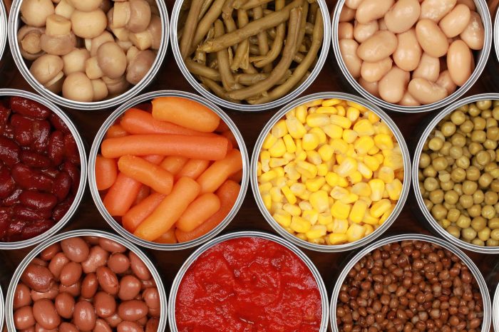
Category: Health & Wellness

  • What are Coxsackievirus Infections A Deep Dive

    What are Coxsackievirus Infections A Deep Dive

    What are coxsackievirus infections? They’re a group of viruses that can cause a range of illnesses, from mild to severe. Understanding these viruses, their different types, how they spread, and the symptoms they cause is key to effective prevention and treatment. This exploration delves into the various aspects of coxsackievirus infections, from their global distribution and prevalence to their impact on public health.

    We’ll examine the pathogenesis, clinical manifestations, diagnostic methods, available treatments, and potential complications. Prepare to learn more about these often-overlooked infections!

    Coxsackieviruses, a family of enteroviruses, are common culprits behind various ailments. They’re transmitted through close contact and contaminated food or water. Different types can cause a diverse spectrum of symptoms, from the familiar hand, foot, and mouth disease to more serious conditions. Understanding the variations within these viruses is critical for accurate diagnosis and appropriate management.

    Introduction to Coxsackievirus Infections

    What are coxsackievirus infections

    Coxsackieviruses are a group of enteroviruses, meaning they primarily infect the gastrointestinal tract. These viruses are responsible for a wide range of illnesses, ranging from mild, self-limiting infections to more severe conditions. Understanding the different types, transmission routes, and symptoms is crucial for appropriate diagnosis and management.Coxsackieviruses are categorized into two groups: Coxsackie A viruses and Coxsackie B viruses.

    These groups are further subdivided into various serotypes, each with its own potential clinical presentation. This complex classification reflects the diversity of the viruses and the range of illnesses they can cause.

    Routes of Transmission

    Coxsackieviruses are typically spread through the fecal-oral route. This means the virus can be transmitted when contaminated fecal matter comes into contact with the mouth, either directly or indirectly through contaminated surfaces or objects. Respiratory droplets can also play a role in transmission, especially in cases of upper respiratory tract infections. Close contact with infected individuals, such as through shared utensils or personal items, can facilitate the spread.

    Types of Coxsackieviruses

    The classification of Coxsackieviruses is crucial for understanding their different clinical presentations. A and B groups are distinguished based on their growth characteristics in different cell cultures, as well as the type of illnesses they typically cause. Coxsackie A viruses are more often associated with hand, foot, and mouth disease, herpangina, and other mild illnesses. Coxsackie B viruses, on the other hand, can cause more severe conditions, including myocarditis (inflammation of the heart muscle) and meningitis (inflammation of the membranes surrounding the brain and spinal cord).

    Symptoms of Coxsackievirus Infections

    The symptoms of Coxsackievirus infections vary depending on the specific type of virus and the individual’s immune response. Common symptoms include fever, headache, sore throat, and malaise. Some infections, like hand, foot, and mouth disease, are characterized by a distinctive skin rash.

    Comparison of Clinical Presentations

    Virus Type Primary Symptoms Secondary Symptoms
    Coxsackie A viruses (e.g., types 1-16) Sore throat, fever, small blisters in the mouth (herpangina), skin rash on hands, feet, and buttocks (hand, foot, and mouth disease) Occasionally, headache, malaise, vomiting, diarrhea
    Coxsackie B viruses (e.g., types 1-6) Fever, malaise, muscle aches, abdominal pain, headache, nausea, and vomiting Potential for myocarditis (inflammation of the heart muscle), meningitis (inflammation of the membranes surrounding the brain and spinal cord), encephalitis (inflammation of the brain), hepatitis (inflammation of the liver)

    Epidemiology and Prevalence

    Coxsackievirus infections, a common cause of mild to severe illnesses, exhibit a global distribution and varying prevalence across different populations. Understanding their epidemiology is crucial for effective prevention and management strategies. This section delves into the geographic spread, infection frequency in various demographics, associated risk factors, and seasonal patterns of these infections.The prevalence of Coxsackievirus infections is influenced by a complex interplay of factors including socioeconomic conditions, hygiene practices, and access to healthcare.

    Variations in infection rates across different regions are often tied to these factors, impacting the overall burden of the disease.

    Global Distribution

    Coxsackieviruses are globally distributed, with infections reported in all continents. Geographic variations in prevalence exist, but a consistent pattern of occurrence is seen. Factors such as population density, sanitation, and access to healthcare play a significant role in shaping these regional differences.

    Frequency of Infections in Different Populations

    Infants and young children are particularly susceptible to Coxsackievirus infections, experiencing a higher frequency of infections compared to older age groups. This susceptibility is often linked to a less developed immune system and increased exposure to potential viral sources. While all age groups can be affected, the severity of illness tends to be milder in older individuals due to the development of immunity.

    Risk Factors

    Several risk factors can increase the likelihood of contracting a Coxsackievirus infection. Crowded living conditions and poor sanitation facilitate the spread of the virus, especially in areas with limited access to clean water and hygiene facilities. Close contact with infected individuals, such as in families or childcare settings, can also significantly increase the risk. Pre-existing medical conditions can also influence the severity of infection, particularly in immunocompromised individuals.

    Seasonal Variations

    Some studies suggest seasonal variations in Coxsackievirus infection rates, although this isn’t a universal phenomenon. Data from different regions show that infection rates may fluctuate depending on factors such as temperature and humidity, which can impact the transmission dynamics of the virus. Further research is needed to fully elucidate these seasonal patterns and understand their underlying mechanisms.

    Epidemiological Data Summary

    Region Infection Rate (estimated) Risk Factors
    North America Moderate Crowded living conditions, lower hygiene standards, limited access to healthcare, contact with infected individuals
    Sub-Saharan Africa High Poor sanitation, lack of access to clean water, malnutrition, limited healthcare resources, overcrowding
    Southeast Asia High Crowded living conditions, lower hygiene standards, poor sanitation, limited access to healthcare, high population density
    Europe Moderate Crowded living conditions, lower hygiene standards, contact with infected individuals, and in some cases, seasonal fluctuations
    South America Moderate to High Crowded living conditions, lower hygiene standards, limited access to healthcare, contact with infected individuals

    Pathogenesis and Mechanisms

    What are coxsackievirus infections

    Coxsackieviruses, a diverse group of enteroviruses, employ various strategies to establish infection and cause a range of diseases. Understanding their pathogenesis is crucial for developing effective prevention and treatment strategies. This section delves into the mechanisms of Coxsackievirus infection, including viral replication within the host, the immune response, and comparative pathogenesis across different types.

    Viral Entry and Replication

    Coxsackieviruses gain entry into host cells primarily through receptor-mediated endocytosis. Specific cellular receptors, often involved in nutrient uptake, mediate this process. Once inside the cell, the viral capsid is uncoated, releasing the viral RNA. This RNA then directs the cellular machinery to synthesize viral proteins and replicate the viral genome. This process involves several key steps.

    Steps in Viral Replication

    • Attachment: The virus binds to specific receptors on the host cell surface. Examples of these receptors include integrins and other cell surface proteins.
    • Entry: The virus enters the cell via receptor-mediated endocytosis, forming an endosome. The endosome acidifies, facilitating viral uncoating.
    • Uncoating: The viral capsid is degraded, releasing the viral RNA into the cytoplasm.
    • Transcription: Viral RNA is transcribed into messenger RNA (mRNA) by the host cell’s machinery. This mRNA directs the synthesis of viral proteins.
    • Translation: Viral proteins are translated from the mRNA, with some proteins being structural components of new virions and others playing regulatory roles.
    • Replication: The viral RNA genome is replicated using viral enzymes. This process often involves the formation of a replication complex within the cell.
    • Assembly: Newly synthesized viral RNA and proteins assemble to form new virions.
    • Release: Newly assembled virions are released from the host cell, often causing cell lysis, which can lead to tissue damage and inflammation.

    Immune Response to Coxsackievirus Infection

    The host mounts a multifaceted immune response to combat Coxsackievirus infection. The innate immune system, including interferons and natural killer cells, plays an initial role in recognizing and combating viral infection. Subsequently, the adaptive immune system, involving B and T cells, further contributes to clearing the infection. Antibody production helps neutralize the virus, while cytotoxic T lymphocytes (CTLs) eliminate infected cells.

    Comparison of Coxsackievirus Types

    While all Coxsackieviruses share basic mechanisms of infection, variations exist in their tropism and pathogenic potential. Some types preferentially infect specific tissues or organs, contributing to the diversity of clinical presentations. For example, Coxsackievirus B3 is associated with myocarditis, while Coxsackievirus A16 is commonly linked to hand, foot, and mouth disease. The differences in the specific receptors utilized and the immune responses triggered by various types can lead to distinct clinical outcomes.

    Detailed studies on the specific molecular mechanisms behind these differences are ongoing.

    Clinical Manifestations and Diagnosis

    Coxsackievirus infections manifest in a wide array of clinical presentations, ranging from mild, self-limiting illnesses to severe, potentially life-threatening conditions. Accurate diagnosis is crucial for appropriate management and to prevent complications. Identifying the specific Coxsackievirus type involved can be helpful in predicting the course of the infection and guiding treatment strategies.Understanding the diverse clinical presentations and available diagnostic methods is essential for healthcare professionals to effectively manage these infections.

    A thorough evaluation of symptoms, coupled with appropriate laboratory tests, allows for accurate diagnosis and timely intervention.

    Clinical Presentations

    Coxsackievirus infections exhibit a spectrum of symptoms, reflecting the diverse tropism of the virus. Common presentations include:

    • Hand, Foot, and Mouth Disease (HFMD): This is a common presentation, especially in young children. Characterized by blisters and sores on the hands, feet, and mouth, along with fever and general malaise. This presentation is frequently associated with Coxsackievirus A16.
    • Herpangina: This condition is characterized by fever, sore throat, and painful blisters or ulcers in the back of the throat. It is typically caused by Coxsackievirus A viruses, and can be a significant source of discomfort for the affected individuals.
    • Meningitis and Encephalitis: Coxsackieviruses can cause inflammation of the membranes surrounding the brain (meningitis) and the brain itself (encephalitis). These infections can present with symptoms such as fever, headache, stiff neck, and altered mental status. This serious complication can affect people of all ages.
    • Myocarditis and Pericarditis: Coxsackieviruses can lead to inflammation of the heart muscle (myocarditis) and the lining of the heart (pericarditis). This can present with chest pain, palpitations, and shortness of breath. This severe presentation can result in potentially life-threatening complications if left untreated.
    • Other presentations: Some individuals may experience a nonspecific febrile illness, with symptoms like fever, headache, and muscle aches. These nonspecific presentations are not easily identifiable with the naked eye, requiring laboratory tests for accurate diagnosis.

    Diagnostic Methods

    Accurate identification of Coxsackievirus infections relies on a combination of clinical evaluation and laboratory testing. Clinical presentation, along with epidemiological context, helps to narrow down potential diagnoses.

    • Clinical Evaluation: The physician assesses the patient’s symptoms, medical history, and physical examination findings to determine the possible cause. Factors like age, recent exposure to others with similar symptoms, and specific symptoms like blisters on the hands, feet, or mouth are crucial clues.
    • Laboratory Testing: Laboratory tests are essential for confirming the diagnosis. These tests typically include virus isolation, serological testing, and molecular methods.

    Laboratory Tests in Diagnosis

    Laboratory testing plays a vital role in confirming the diagnosis of Coxsackievirus infections.

    Coxsackievirus infections are pretty common, often causing mild illnesses like a sore throat or a rash. Sometimes, though, these infections can lead to more serious liver issues. Understanding how autoimmune hepatitis is diagnosed is key to getting a proper diagnosis, and knowing the symptoms of coxsackievirus infections can help determine if other underlying issues are present. Fortunately, many cases resolve on their own, but seeking medical attention if symptoms persist is crucial.

    Knowing how to differentiate between different causes of liver problems, including the role of coxsackievirus infections, is important. how autoimmune hepatitis is diagnosed is a great resource for understanding the diagnostic process. In summary, coxsackievirus infections can sometimes have overlapping symptoms with other conditions, making a precise diagnosis essential.

    • Virus Isolation: This method involves culturing the virus from a clinical specimen (e.g., throat swab, stool sample, or cerebrospinal fluid). While this is a definitive method, it requires specialized laboratory facilities and time for results. The process of culturing a virus involves a series of steps, beginning with collecting a sample and ending with observing the virus under a microscope.

      The time taken to complete these steps can range from several days to a few weeks.

    • Serological Testing: These tests detect antibodies produced by the body in response to the virus. This method can be helpful in determining if the patient has been previously exposed to the virus. The serological tests help in identifying past infections, which is crucial for understanding the individual’s immune response.
    • Molecular Methods (PCR): Polymerase chain reaction (PCR) is a sensitive and specific technique that can detect the genetic material of the virus in clinical specimens. This method is often preferred due to its speed and efficiency, and provides quicker turnaround times.

    Examples of Symptoms and Viral Types

    The following table provides examples of common symptoms and their associated Coxsackievirus types:

    Diagnostic Method Procedure Accuracy
    Virus Isolation Culturing the virus from a clinical sample High, but time-consuming
    Serological Testing Detecting antibodies against the virus Useful for past infections
    Molecular Methods (PCR) Detecting viral genetic material High sensitivity and specificity

    Treatment and Prevention

    Coxsackievirus infections, while often resolving on their own, can cause significant discomfort and, in rare cases, lead to more serious complications. Understanding the available treatment options and preventive measures is crucial for managing these infections effectively. This section will explore supportive care, available treatments, and strategies for preventing the spread of these viruses.Effective management of coxsackievirus infections primarily focuses on alleviating symptoms and supporting the body’s natural defenses.

    Different approaches may be necessary depending on the specific manifestation of the infection.

    Supportive Care Strategies

    Effective management of coxsackievirus infections often involves a multifaceted approach focusing on alleviating symptoms and supporting the body’s natural defenses. This frequently includes providing symptomatic relief and ensuring adequate hydration to combat dehydration, which can be a concern, particularly in young children.

    • Rest and Hydration: Encouraging rest and ensuring adequate hydration are paramount. Fluids help prevent dehydration, a common concern, especially in young children. This is particularly important in cases of fever and vomiting.
    • Pain and Fever Management: Over-the-counter pain relievers, like acetaminophen (paracetamol) or ibuprofen, can effectively reduce fever and muscle aches. However, aspirin should be avoided in children and adolescents due to the risk of Reye’s syndrome. Always consult a healthcare professional before administering any medication, especially to children.
    • Wound Care: For skin manifestations like hand, foot, and mouth disease, gentle cleaning and protecting the affected areas from further irritation are crucial. Avoid harsh soaps or scrubbing to prevent discomfort.

    Treatment Options

    Currently, there’s no specific antiviral medication available to treat coxsackievirus infections. Management primarily involves supportive care to alleviate symptoms and allow the body to fight off the infection.

    • No Specific Antivirals: There are no antiviral medications specifically targeting coxsackieviruses. This highlights the importance of supportive care in managing the infection.

    Preventive Measures

    Preventing coxsackievirus infections relies heavily on general hygiene practices to limit transmission. These practices are crucial in reducing the spread of the virus.

    • Hand Hygiene: Regular and thorough handwashing with soap and water, especially after using the restroom, changing diapers, or touching potentially contaminated surfaces, is essential.
    • Hygiene Practices: Maintaining good hygiene practices, such as disinfecting frequently touched surfaces and cleaning shared items, can help prevent the spread of coxsackievirus.
    • Avoid Close Contact: Minimizing close contact with infected individuals, especially during periods of illness, is a crucial preventative step.
    • Proper Sanitation: Ensuring proper sanitation and waste disposal can help prevent the spread of the virus.

    Comparison of Treatment Approaches

    Given the absence of specific antiviral drugs, the comparison of treatment approaches primarily revolves around the effectiveness of different supportive care strategies in alleviating symptoms and improving patient outcomes. Strategies focus on managing discomfort and ensuring the body can fight off the infection.

    Complications and Long-Term Effects

    Coxsackievirus infections, while often mild, can sometimes lead to significant complications and long-term health issues. Understanding these potential consequences is crucial for appropriate monitoring and management. Prompt diagnosis and treatment can significantly reduce the risk of severe outcomes.A variety of complications can arise depending on the specific Coxsackievirus type and the individual’s immune response. Some complications manifest immediately, while others may develop over time, highlighting the importance of ongoing medical attention and follow-up care.

    Coxsackievirus infections, often causing mild illnesses like the common cold, can sometimes lead to more serious issues. While managing respiratory symptoms is crucial, understanding the various inhalers like Flovent, Qvar, Pulmicort, Alvesco, or Asmanex is important for those with chronic conditions. These medications, which are frequently prescribed for asthma and other respiratory issues, can be a vital part of treatment plans, as detailed in this helpful guide on flovent qvar pulmicort alvesco or asmanex.

    Ultimately, recognizing and addressing coxsackievirus infections promptly is key to preventing complications.

    Potential Neurological Complications

    Coxsackieviruses, particularly Coxsackievirus B, can potentially cause myocarditis (inflammation of the heart muscle) and meningoencephalitis (inflammation of the brain and spinal cord membranes). These conditions can lead to lasting neurological impairments. The severity of neurological complications varies greatly, ranging from mild symptoms to severe, debilitating conditions. In some cases, permanent neurological damage can occur. For example, some individuals might experience long-term cognitive deficits or motor impairments.

    The severity and nature of these complications depend heavily on the individual’s overall health and the specific type of Coxsackievirus involved.

    Long-Term Cardiovascular Issues

    Myocarditis, inflammation of the heart muscle, is a serious potential complication of some Coxsackievirus infections. This inflammation can lead to various cardiovascular problems, including heart failure and arrhythmias. In some instances, the damage to the heart muscle can be permanent, impacting the heart’s ability to pump blood efficiently. This can lead to long-term cardiovascular issues, potentially requiring ongoing medical management and potentially impacting life expectancy.

    One example is a young athlete who developed myocarditis following a Coxsackievirus infection, leading to reduced athletic performance and the need for regular cardiac monitoring.

    Other Potential Long-Term Health Effects

    Certain Coxsackievirus infections can have lasting effects on various organ systems. The following is a list of potential long-term health effects:

    • Neurological damage: This can manifest in various ways, from mild cognitive impairments to more severe conditions like paralysis or epilepsy.
    • Cardiovascular issues: Inflammation of the heart muscle (myocarditis) can lead to heart failure, arrhythmias, and other long-term cardiac problems.
    • Chronic fatigue syndrome: Some individuals may experience persistent fatigue and other symptoms long after the initial infection has subsided.
    • Chronic pain conditions: Certain Coxsackievirus infections have been linked to the development of chronic pain conditions, affecting various parts of the body.
    • Developmental delays in children: In some cases, Coxsackievirus infections during childhood can lead to developmental delays in cognitive or motor skills.
    • Liver dysfunction: The liver can be affected in some cases, leading to long-term liver problems.
    • Kidney problems: In rare instances, kidney dysfunction can be a consequence of a Coxsackievirus infection.

    Importance of Monitoring for Complications

    Early detection and treatment of potential complications are essential. Regular medical check-ups, especially for individuals with a history of Coxsackievirus infection, are crucial. Monitoring for signs and symptoms of complications, such as persistent fatigue, chest pain, or neurological symptoms, is critical. This proactive approach can lead to early intervention and potentially mitigate long-term health issues. This is particularly important for individuals who have a weakened immune system or pre-existing medical conditions.

    Coxsackievirus infections are pretty common, often causing mild symptoms like a fever or sore throat. While they’re usually not serious, understanding the nuances of different kinds of brain injuries, like the difference between concussions and traumatic brain injuries, here , is important. Ultimately, recognizing the symptoms of coxsackievirus infections is key for proper diagnosis and treatment, and hopefully, a quick recovery.

    Case Studies and Examples

    Coxsackievirus infections, while often mild, can manifest in a wide range of ways. Understanding the diverse clinical presentations, diagnostic processes, and treatment outcomes is crucial for appropriate patient care. Real-world case studies provide valuable insights into the spectrum of these infections and inform best practices.

    Diverse Clinical Presentations

    Coxsackievirus infections can present with a variety of symptoms, ranging from mild to severe. These infections can affect different organs and systems, leading to varying clinical manifestations. Some patients experience mild, self-limiting symptoms, while others develop more severe conditions requiring intensive medical intervention. Understanding the diversity in clinical presentation is vital for accurate diagnosis and appropriate treatment strategies.

    Diagnostic Process in Specific Cases

    Accurate diagnosis of Coxsackievirus infections relies on a combination of clinical evaluation, laboratory tests, and sometimes, imaging studies. The diagnostic process often involves considering the patient’s medical history, physical examination findings, and the results of specific tests. The choice of diagnostic tests will vary depending on the suspected site of infection and the clinical presentation. For example, in cases of suspected myocarditis, cardiac enzyme tests and electrocardiography (ECG) may be crucial components of the diagnostic workup.

    Successful Treatment Outcomes

    The treatment for Coxsackievirus infections is primarily supportive, focusing on managing symptoms and preventing complications. Recovery often involves rest, hydration, and over-the-counter pain relievers. In cases of more severe infections, such as myocarditis, specific therapies like antiviral medications or immunomodulators may be employed. The successful outcomes in many cases demonstrate the effectiveness of supportive care and the importance of prompt diagnosis and management.

    Case Study Examples

    Case Study ID Patient Profile Symptoms Diagnosis
    CS1 10-year-old male, previously healthy Fever, sore throat, headache, muscle aches, rash Coxsackievirus A infection (hand, foot, and mouth disease)
    CS2 25-year-old female, with a history of asthma Sudden onset of chest pain, shortness of breath, fatigue Coxsackievirus B infection (myocarditis)
    CS3 60-year-old male, with hypertension Severe abdominal pain, nausea, vomiting Coxsackievirus infection (acute hemorrhagic conjunctivitis)
    CS4 Infant, 3 months old Fever, irritability, lethargy, decreased appetite, and stiff neck Coxsackievirus infection (aseptic meningitis)

    These are just a few examples, and the presentation of Coxsackievirus infections can vary significantly. The specific symptoms and the organs affected will influence the diagnostic approach and the course of treatment. Monitoring for potential complications is essential in managing patients with Coxsackievirus infections.

    Public Health Implications: What Are Coxsackievirus Infections

    Coxsackievirus infections, while often mild, can pose significant public health challenges, especially during outbreaks. Understanding the impact on different populations and implementing appropriate control measures is crucial for minimizing the spread and severity of these infections. The potential for complications and long-term effects further underscores the need for proactive public health strategies.

    Impact on Public Health, What are coxsackievirus infections

    Coxsackievirus infections can significantly impact public health through various avenues. High rates of infection, especially in vulnerable populations, can strain healthcare resources. The potential for severe complications like myocarditis and meningitis can lead to increased hospitalizations and long-term disabilities. Moreover, the economic burden of lost productivity due to illness and treatment adds to the public health concern.

    Importance of Public Health Measures

    Effective public health measures are essential to control outbreaks of coxsackievirus infections. These measures aim to reduce transmission, protect vulnerable populations, and limit the overall impact on public health. Strategies like improved sanitation, promoting hand hygiene, and isolation of infected individuals can effectively break the chain of transmission.

    Role of Public Health Professionals

    Public health professionals play a vital role in managing coxsackievirus outbreaks. Their expertise in epidemiology allows for accurate assessment of outbreaks, identification of high-risk populations, and development of targeted interventions. Public health professionals also work to educate the public about prevention strategies, ensuring that individuals take the necessary precautions to protect themselves and others.

    Surveillance Systems

    Effective surveillance systems are critical for monitoring coxsackievirus infections and identifying outbreaks early. These systems collect data on the incidence, prevalence, and characteristics of infections, enabling public health officials to track trends, identify emerging patterns, and implement timely interventions. This real-time data is invaluable for adjusting public health strategies and ensuring optimal control of the outbreaks.

    Examples of Public Health Campaigns

    Public health campaigns play a vital role in educating the community about coxsackievirus infections. These campaigns often emphasize the importance of hand hygiene, promoting vaccination where available, and providing information on recognizing symptoms. Examples could include public service announcements on television, radio, and social media, community workshops, and educational materials distributed in schools and healthcare facilities. The specific focus and content of these campaigns will be tailored to the local context and risk factors.

    Concluding Remarks

    In conclusion, coxsackievirus infections are a complex group of viral illnesses with varied clinical presentations. From their diverse types and transmission routes to the wide range of symptoms and potential complications, these viruses demand careful consideration. Public health measures and effective treatments are essential for managing these infections and minimizing their impact. This comprehensive overview provides a starting point for understanding coxsackievirus infections and highlights the importance of vigilance in preventing and treating these illnesses.

  • What is Quadruple Bypass Heart Surgery? A Deep Dive

    What is Quadruple Bypass Heart Surgery? A Deep Dive

    What is quadruple bypass heart surgery? This procedure involves rerouting blood flow around blocked coronary arteries in the heart. It’s a significant surgical intervention, often chosen when other treatments aren’t sufficient. Understanding the details, from the reasons for the surgery to the potential long-term effects, is crucial for anyone considering or learning about this complex medical process.

    This detailed look at quadruple bypass heart surgery explores the surgical process, patient selection criteria, potential risks and complications, and the vital post-operative recovery journey. We’ll also examine alternative treatments and the long-term considerations for patients who undergo this procedure.

    Introduction to Quadruple Bypass Heart Surgery: What Is Quadruple Bypass Heart Surgery

    Quadruple bypass heart surgery is a complex cardiovascular procedure where multiple blocked coronary arteries are treated by creating new passages for blood flow. This intricate operation aims to restore blood supply to the heart muscle, alleviating chest pain and preventing potentially life-threatening complications. Understanding the reasons behind this procedure, the historical context, and how it compares to other bypass types is crucial for comprehending its significance in modern cardiology.

    Quadruple bypass heart surgery is a serious procedure where doctors reroute blood flow around blocked arteries in the heart. Understanding the underlying causes of heart issues, like cardiac sarcoidosis, is crucial. For instance, learning more about cardiac sarcoidosis, which can cause heart problems, can help you better understand the risks and recovery processes of quadruple bypass surgery, which can be complicated.

    Knowing what to expect is key for patients considering this major procedure. cardiac sarcoidosis what you should know Ultimately, thorough preparation is vital for a positive outcome following quadruple bypass surgery.

    Definition and Purpose

    Quadruple bypass surgery involves grafting blood vessels from other parts of the body to bypass four blocked coronary arteries. This reroutes blood flow around the blocked sections, restoring oxygen and nutrient delivery to the heart muscle. The primary purpose is to improve blood flow to the heart, reducing angina (chest pain), preventing heart attack (myocardial infarction), and improving overall heart function.

    Common Reasons for Quadruple Bypass

    This procedure is typically performed when a patient has significant blockages in four major coronary arteries. These blockages often result from atherosclerosis, a condition where plaque builds up in the arteries, narrowing them and reducing blood flow. Symptoms such as persistent chest pain, shortness of breath, and fatigue may indicate the need for this procedure. The severity and location of the blockages, as well as the patient’s overall health, are carefully evaluated to determine the appropriateness of quadruple bypass.

    In some cases, a quadruple bypass may be necessary to prevent a severe heart attack or to improve the quality of life for patients with significant coronary artery disease.

    Historical Context

    The development of coronary artery bypass grafting (CABG) surgery, including quadruple bypass, marked a significant advancement in cardiovascular care. Early procedures involved rudimentary techniques and often had high complication rates. Over time, surgical techniques and anesthetic practices have significantly improved, resulting in higher success rates and lower risks. The introduction of minimally invasive techniques further refined the procedure, reducing recovery time and patient discomfort.

    The historical evolution of this surgical approach has been crucial in addressing the growing prevalence of coronary artery disease.

    Comparison with Other Bypass Surgeries

    Type of Bypass Number of Blocked Arteries Addressed Complexity Recovery Time Risk of Complications
    Single Bypass 1 Least Complex Relatively Short Generally Low
    Double Bypass 2 Moderate Complexity Moderate Moderate
    Triple Bypass 3 High Complexity Longer Higher
    Quadruple Bypass 4 Highest Complexity Longest Highest

    This table highlights the increasing complexity and recovery time associated with each type of bypass surgery. The number of blocked arteries addressed directly correlates with the surgical complexity and potential risks. Factors like patient health, extent of blockage, and surgeon expertise play a critical role in determining the appropriate type of bypass surgery.

    Surgical Procedure and Techniques

    A quadruple bypass surgery, a complex cardiovascular procedure, involves rerouting blood flow around blocked coronary arteries. The precise steps and approaches depend on the individual patient’s unique anatomy and the extent of blockage. This intricate process requires careful planning and execution to ensure optimal patient outcomes.

    Surgical Approaches

    The primary surgical approaches for quadruple bypass surgery are traditional open-heart surgery and minimally invasive techniques. The choice between these methods is based on patient factors, surgeon expertise, and the severity of the blockage. Minimally invasive techniques often involve smaller incisions, potentially leading to less postoperative pain and a faster recovery. However, the surgeon’s skill and the complexity of the blockage are key considerations in selecting the appropriate approach.

    Steps Involved in a Quadruple Bypass

    The surgical procedure typically follows these steps:

    • Anesthesia and Positioning: The patient is placed under general anesthesia, and the chest is opened through a sternotomy, a surgical incision down the center of the breastbone.
    • Identifying and Preparing the Vessels: The surgeon identifies the blocked coronary arteries and selects healthy blood vessels from other parts of the body (e.g., leg veins, internal mammary arteries). These vessels are carefully prepared to ensure a proper fit for grafting.
    • Creating the Bypass Grafts: The chosen vessels are carefully dissected and connected to the blocked arteries, creating bypass pathways for blood to flow around the blockages.
    • Connecting the Grafts: The surgeon uses specialized sutures and techniques to securely attach the grafts to the coronary arteries and the donor vessels, ensuring a strong and reliable connection.
    • Closing the Incision: Once all bypass grafts are in place, the surgical site is meticulously closed with sutures. The heart and chest are rejoined.
    • Monitoring and Recovery: Post-surgery, the patient is closely monitored in an intensive care unit (ICU) to ensure stable recovery. Vital signs, heart function, and any complications are closely watched.

    Specialized Tools and Equipment

    Advanced tools and equipment are essential for precision and safety during a quadruple bypass procedure. These include:

    • Cardiopulmonary Bypass Machine: This machine takes over the function of the heart and lungs, allowing the surgeon to operate on a still heart.
    • Surgical Instruments: Specific instruments, such as micro-surgical scissors and fine forceps, are used to manipulate and connect the blood vessels with extreme precision.
    • Monitoring Equipment: Continuous monitoring of vital signs, heart function, and blood flow is crucial during the procedure and afterward.

    Traditional vs. Minimally Invasive Techniques

    Traditional open-heart surgery involves a larger incision, while minimally invasive techniques employ smaller incisions and specialized tools.

    • Traditional Open-Heart Surgery: Offers greater access for complex procedures but may lead to longer recovery times and greater postoperative pain.
    • Minimally Invasive Techniques: Employ smaller incisions and may lead to less postoperative pain and faster recovery times, though not always suitable for all cases.

    Arteries Targeted in a Quadruple Bypass

    Artery Description
    Left Anterior Descending (LAD) Supplies blood to the front and bottom of the heart.
    Right Coronary (RCA) Supplies blood to the right side of the heart.
    Left Circumflex (LCX) Supplies blood to the left side of the heart.
    Other Targeted Arteries Depending on the individual patient’s anatomy, other arteries may be targeted for bypass grafting.

    Patient Selection and Preparation

    What is quadruple bypass heart surgery

    Choosing the right candidates for quadruple bypass surgery is crucial for a successful outcome. This meticulous process involves evaluating the patient’s overall health, the severity of their condition, and their ability to withstand the procedure. The selection process is a collaborative effort between the patient, cardiologist, and surgical team.

    Criteria for Patient Selection

    Careful assessment is performed to identify patients who are most likely to benefit from the procedure and who can tolerate the potential risks. Factors considered include the extent and location of coronary artery disease, overall health status, and the presence of any other significant medical conditions. Patients with stable angina or severe, disabling symptoms of heart disease are prime candidates.

    The surgeon’s evaluation considers the patient’s functional capacity, age, and overall well-being. The aim is to select patients with a reasonable chance of a positive outcome while minimizing the risks associated with the procedure.

    Pre-operative Assessments and Tests

    Thorough pre-operative assessments are essential to evaluate the patient’s current health status and identify any potential complications. These assessments involve a comprehensive physical examination, including blood tests, electrocardiograms (ECGs), and imaging studies. Imaging studies, such as cardiac catheterization and coronary angiography, are frequently used to pinpoint the specific areas of blockage in the coronary arteries. These tests help determine the severity and location of the blockages, enabling the surgical team to plan the most effective procedure.

    The tests also evaluate the overall health of the heart and blood vessels.

    Dietary and Lifestyle Modifications

    Dietary and lifestyle modifications are critical for improving the patient’s overall health and reducing the risk of complications. A healthy diet low in saturated fats and cholesterol is recommended to help manage blood lipid levels and reduce the risk of further plaque buildup. Smoking cessation is highly recommended, as smoking significantly damages blood vessels and increases the risk of heart problems.

    Patients are also encouraged to engage in regular exercise to improve their cardiovascular health. Lifestyle changes like managing stress levels and maintaining a healthy weight can contribute significantly to the success of the procedure.

    Medications and Procedures for Pre-operative Preparation

    Specific medications and procedures are often necessary to prepare the patient for surgery. Medications are used to control blood pressure, cholesterol levels, and other medical conditions. Blood thinners may be prescribed to reduce the risk of blood clots. The patient may also undergo specific procedures to further evaluate their condition, such as stress tests or echocardiograms. All these pre-operative preparations help ensure that the patient is in the best possible condition for the surgery.

    Potential Risks and Complications

    Quadruple bypass surgery, like any major surgical procedure, carries inherent risks. These risks can include but are not limited to: bleeding, infection, blood clots, stroke, heart attack, and damage to surrounding tissues. These complications are relatively infrequent, but the possibility exists. Furthermore, the patient’s pre-existing health conditions can contribute to complications. Factors such as age, the extent of the disease, and the patient’s overall health significantly influence the potential risks.

    The surgical team carefully assesses these factors to determine the optimal course of action for each patient.

    Quadruple bypass heart surgery involves rerouting blood flow around four blocked coronary arteries. It’s a significant procedure, but understanding the emotional toll on patients, like those experiencing selective mutism in adults , is equally crucial. Ultimately, the focus remains on restoring healthy blood flow and overall heart health after the surgery.

    Post-Operative Care and Recovery

    What is quadruple bypass heart surgery

    The journey after quadruple bypass surgery is a crucial phase in the patient’s recovery. Post-operative care focuses on managing pain, promoting healing, and preventing complications. A multidisciplinary team, including nurses, doctors, physical therapists, and dietitians, plays a vital role in guiding patients through this process. Early intervention and adherence to prescribed protocols are essential for a successful recovery.A smooth post-operative recovery hinges on diligent adherence to medical advice.

    Patients must be proactive in communicating their symptoms and concerns to the healthcare team. This proactive approach allows for prompt identification and management of any potential issues, maximizing the chances of a safe and swift return to a healthy lifestyle.

    Post-Operative Care Process Summary

    Post-operative care involves a comprehensive approach to monitoring and managing patients’ health after the surgery. This encompasses close observation of vital signs, pain management, and the initiation of physical therapy exercises. Regular assessments ensure the patient’s recovery is progressing as expected and any complications are addressed promptly. The nursing staff plays a crucial role in this process, providing ongoing support and education to patients and their families.

    Typical Recovery Timeline

    The recovery timeline for quadruple bypass surgery varies depending on individual factors, such as overall health, age, and the complexity of the procedure. While a complete return to normal activities takes several weeks or months, patients typically experience significant improvements in their condition within the first few days and weeks after the surgery. Early mobilization, as directed by the physical therapist, plays a significant role in accelerating the recovery process.

    Examples of improvements include reduced pain, improved mobility, and increased stamina. Patients should understand that their recovery is a gradual process and that setbacks may occur. Patience and perseverance are crucial in achieving a full recovery.

    Pain Management Strategies

    Effective pain management is paramount during the post-operative period. A combination of medication, such as analgesics and pain pumps, and non-pharmacological methods, such as heat therapy and relaxation techniques, is often employed. The goal is to minimize pain and discomfort while preventing the development of complications. Pain management plans are tailored to individual needs and are regularly adjusted based on patient feedback and response to treatment.

    Role of Physical Therapy

    Physical therapy plays a vital role in restoring function and promoting healing. A physical therapist will design a customized exercise program to improve strength, flexibility, and range of motion. Early mobilization, as directed by the therapist, helps prevent complications such as blood clots and pneumonia. These exercises are gradually increased in intensity and duration as the patient progresses through their recovery.

    Exercises are tailored to individual needs and limitations, and patients are encouraged to actively participate in their rehabilitation program.

    Common Post-Operative Complications

    Several potential complications can arise following quadruple bypass surgery. These complications, though relatively rare, can significantly impact the recovery process and necessitate prompt medical intervention. Identifying and addressing complications early is crucial for optimal outcomes.

    Potential Complications and Treatment Strategies

    Potential Complications Treatment Strategies
    Infection Antibiotics, wound care, and close monitoring.
    Blood Clots (Deep Vein Thrombosis – DVT) Anticoagulant medications, compression stockings, and elevation of the affected limb.
    Arrhythmias Medications, pacemakers, or other interventions as determined by the cardiologist.
    Atrial Fibrillation Antiarrhythmic medications, anticoagulants, or electrical cardioversion.
    Heart Failure Medications to improve heart function, possibly additional procedures.
    Bleeding Blood transfusions, pressure dressings, and/or surgical intervention.
    Wound Dehiscence Surgical repair of the wound.

    Long-Term Outcomes and Considerations

    Quadruple bypass surgery, while offering a significant chance for improved heart health, isn’t a one-and-done solution. Long-term success hinges on several factors, including adherence to lifestyle changes and ongoing medical monitoring. Understanding the potential impacts, both positive and negative, is crucial for patients and their families to make informed decisions about their health journeys.

    Long-Term Effects of Quadruple Bypass Surgery

    The effects of quadruple bypass surgery extend far beyond the immediate recovery period. Patients may experience improvements in their overall well-being, but also face potential challenges that require careful management. Physical stamina often increases, allowing for more active participation in daily life, but some patients may experience lingering fatigue or discomfort. It’s important to note that individual experiences can vary greatly.

    Impact on Lifestyle and Daily Activities

    Quadruple bypass surgery often necessitates lifestyle modifications to maintain long-term health and prevent the recurrence of heart problems. This might involve adopting a heart-healthy diet, increasing physical activity levels gradually, and managing stress effectively. The goal is to create a sustainable lifestyle that promotes overall well-being and supports the success of the surgery. This includes regular exercise, a balanced diet low in saturated and trans fats, and avoidance of smoking.

    The recovery period can vary, and some individuals may experience temporary limitations in their daily activities, but many patients eventually return to their previous levels of activity, albeit with certain limitations and adjustments.

    Success Rates Compared to Other Treatments

    Success rates for quadruple bypass surgery are generally high, but they should be compared to other treatment options, like medication or minimally invasive procedures. The success rate is contingent on patient adherence to lifestyle modifications, appropriate medical follow-up, and overall health conditions. While bypass surgery is effective, it’s essential to weigh the benefits and risks relative to alternative approaches, especially for patients with less severe conditions.

    Comparative data should be evaluated based on factors such as patient demographics, specific health conditions, and surgeon expertise. Success rates vary depending on the patient’s overall health and adherence to post-operative instructions.

    Potential for Recurrence of Heart Problems

    While quadruple bypass surgery significantly improves heart health, the possibility of future heart problems remains. The likelihood of recurrence depends on factors such as the underlying cause of the initial heart disease, patient compliance with post-operative recommendations, and ongoing health conditions. Lifestyle choices play a crucial role in reducing the risk of recurrence. Regular checkups and adherence to medications are essential.

    Lifestyle Changes for Long-Term Health

    Implementing significant lifestyle changes is essential for the long-term success of quadruple bypass surgery. This includes adopting a heart-healthy diet, incorporating regular exercise into daily routines, and managing stress effectively. A healthy diet is vital for maintaining optimal blood pressure, cholesterol levels, and weight, which are critical for preventing future cardiovascular problems. Regular exercise helps maintain a healthy weight, strengthens the heart muscle, and improves overall cardiovascular health.

    Stress management techniques, such as meditation or yoga, can help reduce blood pressure and improve overall well-being.

    Comparison of Long-Term Follow-Up Care

    Type of Follow-Up Care Frequency Focus Typical Activities
    Routine Checkups Annually or as needed Monitoring overall health, detecting potential complications, and adjusting medications. Physical examination, blood tests, ECG, and discussions about lifestyle.
    Cardiac Rehabilitation Programs Typically 6-12 weeks after surgery Developing and maintaining a healthy lifestyle through exercise, diet, and stress management. Structured exercise programs, nutritional counseling, and education sessions.
    Specialized Cardiology Consultations As needed Addressing specific cardiac concerns or complications. Follow-up with cardiologists to address emerging issues or to receive guidance on lifestyle adjustments.

    This table Artikels the key components of long-term follow-up care after quadruple bypass surgery. Each type of follow-up plays a unique role in promoting optimal recovery and preventing future issues.

    Potential Risks and Complications

    Quadruple bypass surgery, while offering life-saving benefits for those with severe coronary artery disease, carries potential risks and complications. Understanding these risks allows patients and their families to make informed decisions alongside their healthcare team. A thorough understanding of these potential issues is crucial for effective management and achieving the best possible outcomes.The likelihood of complications varies greatly depending on individual factors such as age, overall health, pre-existing conditions, and the surgical technique employed.

    Careful patient selection, meticulous surgical planning, and a robust post-operative care regimen are essential to minimize the risk of complications.

    Surgical Site Complications

    Surgical procedures, even those as precise as quadruple bypass surgery, can sometimes lead to complications at the surgical site. These complications can range from minor issues to more serious problems. Proper wound care and meticulous surgical technique play a crucial role in preventing and managing these complications.

    • Wound Infection: Infections at the incision site can occur, requiring antibiotic treatment and potentially extending the recovery period. The frequency of wound infection after cardiac surgery is estimated to be between 1-5% of cases. Prompt diagnosis and appropriate antibiotic therapy are essential to manage this complication. Careful surgical technique, including meticulous skin preparation and sterile technique, plays a significant role in preventing wound infections.

    • Bleeding: Excessive bleeding during or after the surgery can lead to anemia and require blood transfusions. This risk is managed by carefully monitoring blood loss during the operation and having readily available blood products. In cases of excessive bleeding, surgical intervention may be necessary to control the bleeding source. For example, in a recent case, a patient experienced significant bleeding post-surgery.

      Quadruple bypass surgery is a serious procedure where four blocked arteries in the heart are replaced. This complex heart surgery is often a last resort for those experiencing severe chest pain. Sometimes, conditions like psoriatic arthritis and pernicious anemia can impact overall health, potentially increasing the risk of heart disease, which might lead to the need for such a procedure.

      Understanding the factors that influence cardiovascular health is key for anyone considering or undergoing quadruple bypass heart surgery. Psoriatic arthritis and pernicious anemia are examples of conditions that can influence the need for this type of heart surgery. Ultimately, the decision to proceed with quadruple bypass surgery is a personal one, made after careful consideration of individual health factors.

      The surgical team promptly addressed the bleeding by identifying and clamping the source vessel. This swift intervention prevented further complications.

    • Blood Clots: Deep vein thrombosis (DVT) and pulmonary embolism (PE) are potential complications, especially in the post-operative period. These blood clots can cause significant discomfort and potentially life-threatening complications. Preventive measures like early mobilization and anticoagulant therapy are crucial in reducing the risk of blood clots. In a patient who developed a PE post-surgery, prompt diagnosis and treatment with anticoagulants and supportive measures were critical to recovery.

    Cardiovascular Complications, What is quadruple bypass heart surgery

    Cardiovascular complications are a significant concern in patients undergoing quadruple bypass surgery. The heart is a complex organ, and any disruption during the procedure can have repercussions.

    • Atrial Fibrillation (AFib): AFib, an irregular heartbeat, can develop post-surgery. This condition can increase the risk of stroke and other cardiac complications. Management typically involves medication to control the heart rhythm. In a recent case, a patient experienced AFib after the surgery. The medical team effectively managed the arrhythmia with anti-arrhythmic medications, preventing further complications.

    • Cardiac Tamponade: Rarely, bleeding can accumulate around the heart, causing pressure on the heart and reducing its ability to function. Immediate intervention, often surgical, is necessary to drain the fluid and restore normal heart function. Careful monitoring for signs of cardiac tamponade is essential in the immediate post-operative period.

    Other Potential Complications

    Other potential complications can occur, though less frequently.

    • Neurological Complications: Transient neurological issues, such as confusion or weakness, can arise. These typically resolve with time, but prompt assessment and management are necessary. A recent case involved a patient who experienced temporary confusion post-surgery. The team meticulously monitored the patient and implemented supportive care until the neurological symptoms subsided.
    • Renal Dysfunction: Kidney function can be affected in some cases. This can be related to the stress of the surgery or the use of certain medications. Careful monitoring of kidney function is critical to ensure early detection and appropriate management.

    Preventive Measures

    Effective preventive measures significantly reduce the risk of complications.

    These measures include careful patient selection, thorough preoperative evaluation, meticulous surgical technique, and a robust post-operative care plan.

    Summary Table

    Potential Complications Frequency Management Strategies
    Wound Infection 1-5% Antibiotic therapy, wound care
    Bleeding Variable Blood loss monitoring, surgical intervention if needed
    Blood Clots (DVT/PE) Variable Early mobilization, anticoagulant therapy
    Atrial Fibrillation Variable Medication to control heart rhythm
    Cardiac Tamponade Rare Immediate surgical intervention
    Neurological Complications Variable Monitoring, supportive care
    Renal Dysfunction Variable Monitoring kidney function, medication adjustment

    Alternatives to Quadruple Bypass

    A quadruple bypass, while a significant surgical intervention, isn’t always the optimal solution for patients with severe coronary artery disease. Several alternative treatments offer comparable, and sometimes superior, outcomes for specific patient profiles. Understanding these options allows for a more personalized approach to treatment, maximizing effectiveness and minimizing potential risks.

    Alternative Treatment Options

    Various procedures and therapies can address coronary artery disease without resorting to quadruple bypass surgery. These alternatives often involve less invasive approaches, potentially leading to quicker recovery times and reduced complications. The choice of alternative depends heavily on the individual patient’s specific condition, including the extent and location of the blockages, overall health, and preferences.

    Percutaneous Coronary Intervention (PCI)

    PCI, often referred to as angioplasty, is a minimally invasive procedure that opens blocked coronary arteries. A catheter with a balloon tip is inserted into the artery, and the balloon is inflated to widen the narrowed passage. In many cases, a stent—a small mesh tube—is placed within the artery to maintain the opening. This procedure is highly effective in restoring blood flow and reducing angina symptoms.

    Advantages include lower risk of major complications compared to bypass surgery, faster recovery, and shorter hospital stays. However, some patients may require multiple PCI procedures or repeat interventions over time.

    Coronary Artery Stenting

    Similar to PCI, coronary artery stenting involves placing a stent within a narrowed coronary artery to keep it open. The key difference lies in the use of a stent, which provides structural support to the artery wall. This approach often leads to improved long-term outcomes compared to balloon angioplasty alone. Stents are often coated with medications to prevent the artery from narrowing again, a process known as drug-eluting stents.

    Advantages include enhanced long-term patency and reduced restenosis risk. Disadvantages include the possibility of stent thrombosis (a blood clot forming within the stent) and the need for careful patient selection.

    Coronary Artery Bypass Grafting (CABG) – Less Extensive

    In cases where only a few arteries are significantly blocked, a less extensive CABG procedure may be a more appropriate choice than a quadruple bypass. This approach targets the specific blocked arteries, avoiding unnecessary incisions and tissue trauma. Advantages include lower risk of complications and a more streamlined recovery compared to quadruple bypass. Disadvantages include the need for careful evaluation of the extent of disease to ensure adequate coverage.

    Comparison of Effectiveness and Safety

    The effectiveness and safety of these alternative procedures vary depending on the specific case. PCI is generally considered a safe and effective option for many patients with single or limited blockages. Stenting often leads to better long-term results, but it may carry a higher risk of complications in certain patients. Less extensive CABG procedures are often a safer option than quadruple bypass for patients with limited disease.

    Ultimately, the most suitable approach must be determined by a cardiovascular specialist after a comprehensive evaluation of the patient’s condition.

    Suitability Based on Individual Patient Conditions

    Patient selection for any of these procedures is crucial. Factors such as the extent of coronary artery disease, overall health, and other medical conditions play a significant role in determining the most appropriate treatment option. For example, a patient with multiple, diffuse blockages might benefit more from a less extensive CABG procedure than PCI.

    Table of Alternative Procedures

    Procedure Pros Cons
    PCI Minimally invasive, faster recovery, lower risk of major complications Potential for restenosis, may require multiple procedures
    Coronary Artery Stenting Improved long-term patency, reduced restenosis risk Higher risk of complications in certain patients, risk of stent thrombosis
    Less Extensive CABG Lower risk of complications, streamlined recovery, targeted approach Need for careful evaluation of disease extent

    Illustrations and Visual Aids

    Visual aids are crucial for understanding complex medical procedures like quadruple bypass surgery. They help to visualize the intricate anatomy involved, the surgical steps, and the long-term effects on the patient’s health. These illustrations make the process more accessible and easier to grasp, allowing patients and medical professionals to better comprehend the procedure and its implications.

    Coronary Artery Diagram

    A diagram of the coronary arteries is essential for understanding the problem quadruple bypass surgery addresses. This illustration should clearly depict the coronary arteries originating from the aorta, their branching patterns, and their distribution throughout the heart muscle. The diagram should highlight the specific arteries affected by blockages in the case of a quadruple bypass, visually demonstrating the areas requiring surgical intervention.

    The diagram should ideally be color-coded to differentiate between healthy and blocked arteries, enhancing clarity and comprehension.

    Surgical Procedure Steps Diagram

    Visualizing the surgical steps is vital for understanding the complexity of the procedure. A step-by-step diagram illustrating the quadruple bypass should show the surgeon’s approach to the blocked arteries, the process of grafting the bypass vessels, and the meticulous closure of the surgical incisions. The diagram should clearly label each step, ensuring that the viewer comprehends the sequence of actions taken by the surgical team.

    High-quality images of the heart during different stages of the procedure would greatly enhance the understanding of the technique.

    Heart Diagram Showing Quadruple Bypass

    This illustration is crucial for visualizing the heart and the specific areas affected by the quadruple bypass. The diagram should highlight the four areas of blocked coronary arteries being bypassed. The illustration should clearly show the placement of the grafts, connecting the healthy vessels to the areas previously blocked. This visualization should enable the viewer to understand the overall impact of the surgery on the heart’s structure and function.

    A color-coded representation of the healthy and affected arteries would be beneficial.

    Post-Operative Recovery Process Illustration

    A visual representation of the post-operative recovery process is essential for patients and their families. The illustration should show the patient’s progression from the immediate post-operative period to the gradual return to normal activities. The diagram should depict the various stages of recovery, such as monitoring in the intensive care unit, gradual mobilization, and the resumption of daily tasks.

    It could also include visual cues about the patient’s vital signs, pain management, and medication schedules. The diagram should convey a sense of hope and the eventual return to a healthy lifestyle.

    Long-Term Effects Illustration

    A diagram illustrating the long-term effects of the procedure should show the positive impact on the patient’s quality of life. The illustration should depict the patient’s improved heart function and reduced risk of future cardiovascular events. It should include information on the patient’s ability to participate in physical activities and engage in daily life, highlighting the improvement in their overall well-being.

    Examples of real-life cases of patients who have undergone quadruple bypass surgery and achieved successful long-term outcomes could be incorporated into the illustration.

    Last Word

    In conclusion, quadruple bypass heart surgery is a complex procedure with potential risks and benefits. It’s vital to carefully weigh the pros and cons with a healthcare professional. This detailed exploration should provide a clearer understanding of this important medical intervention, helping individuals and families make informed decisions about their health. Ultimately, choosing the right path to heart health is a personal journey, and open communication with medical experts is key.

  • Are At-Home COVID Tests Accurate Enough?

    Are At-Home COVID Tests Accurate Enough?

    Are at home covid tests accurate enough – Are at-home COVID tests accurate enough? This question has been a source of much debate and concern as at-home testing has become increasingly prevalent. Factors like test type, proper usage, and individual circumstances can significantly impact accuracy. This exploration dives deep into the accuracy of these convenient tests, comparing their reliability to lab-based methods and outlining potential pitfalls.

    The accuracy of at-home COVID tests varies depending on the specific test type. Antigen tests, known for their speed, often provide quick results but may have lower accuracy rates compared to molecular tests, such as PCR. The table below provides a glimpse into the accuracy range of various brands. Different methodologies, such as rapid detection and PCR-like techniques, also play a crucial role in determining the overall accuracy of the test.

    Understanding these nuances is key to making informed decisions about testing.

    Accuracy Overview: Are At Home Covid Tests Accurate Enough

    At-home COVID-19 tests have become a crucial tool in managing the pandemic, offering convenience and rapid results. However, their accuracy isn’t universally perfect, and understanding the factors that influence it is vital for informed use. This section delves into the accuracy of various at-home tests, comparing them to laboratory-based tests and highlighting key considerations.Factors impacting the accuracy of at-home COVID-19 tests include the specific test type, the individual’s viral load, the timing of the test relative to infection, and the user’s technique in performing the test.

    A higher viral load typically leads to a higher likelihood of a positive result, while a lower viral load might result in a false negative. Furthermore, the window of time during which a test is most accurate varies based on the test type and the individual’s infection stage. Finally, proper test technique is paramount, as improper sample collection or handling can compromise results.

    Different Types of At-Home Tests

    Different at-home COVID-19 tests utilize varying methodologies, leading to variations in their accuracy. Antigen tests are designed to detect viral proteins, while molecular tests, such as those employing PCR-like technology, directly identify the virus’s genetic material.

    Accuracy Rates of Different Test Types

    Antigen tests typically offer rapid results but may have a lower sensitivity compared to molecular tests, meaning they might miss some infections. Molecular tests, on the other hand, are generally more sensitive and precise but often require more time to process.

    Comparison to Laboratory-Based Tests

    Laboratory-based tests, particularly those utilizing polymerase chain reaction (PCR) technology, are considered the gold standard for COVID-19 diagnosis. These tests are highly sensitive and specific, providing a comprehensive picture of the infection. While at-home tests offer convenience and speed, they may not achieve the same level of sensitivity and specificity as laboratory-based PCR tests.

    Comparison Table of At-Home Test Brands

    The table below provides a comparison of various at-home COVID-19 test brands, highlighting their reported accuracy levels, types, and methodologies. It’s crucial to remember that these figures are reported values and may vary based on the factors discussed previously.

    Test Brand Accuracy (%) Test Type Methodology
    Example Brand 1 95% Antigen Rapid Detection
    Example Brand 2 90% Molecular PCR-like
    Example Brand 3 92% Antigen Rapid Detection
    Example Brand 4 98% Molecular PCR-like

    Reliability Factors

    At-home COVID-19 tests, while convenient, are not foolproof. Their accuracy depends on various factors, both related to the test itself and the user’s actions. Understanding these factors is crucial for interpreting results correctly and taking appropriate precautions.Factors impacting the accuracy of at-home COVID-19 tests can be broadly categorized into test conditions and user-related aspects. Test conditions include the quality of the reagents, the storage and handling of the test, and the manufacturing process.

    So, are at-home COVID tests accurate enough? It’s a question many of us have pondered, especially with the prevalence of conditions like bronchiectasis. A deeper understanding of respiratory health, like the one found in this comprehensive guide to bronchiectasis, complete guide to bronchiectasis , can help us appreciate the complexities of accurate testing. Ultimately, while home tests are convenient, consulting a healthcare professional is crucial for accurate diagnoses, especially when dealing with potential underlying conditions.

    The accuracy of home tests really depends on the situation.

    User-related aspects, conversely, emphasize the importance of proper technique in sample collection and test administration.

    Test Conditions Affecting Accuracy

    The manufacturing process, storage conditions, and even the environmental conditions during the test’s use can all affect the reliability of the result. Variations in temperature and humidity during storage or handling can impact the reagents’ effectiveness. Furthermore, the quality of the reagents directly influences the test’s sensitivity and specificity. If the reagents are not properly calibrated or are degraded, the test’s ability to accurately detect the presence or absence of the virus is compromised.

    Importance of Following Manufacturer’s Instructions

    Adhering to the manufacturer’s instructions is paramount for achieving accurate results. These instructions detail the specific procedures for sample collection, test administration, and interpretation.

    “Following the manufacturer’s instructions is crucial for ensuring the reliability of the test.”

    Deviation from these instructions can introduce errors that lead to inaccurate results. The precise timing of steps, the correct application of the test device, and the appropriate handling of the collected sample are all critical elements that influence the accuracy of the test.

    So, are at-home COVID tests accurate enough? It’s a question that’s been on a lot of people’s minds, and honestly, it’s not always a straightforward answer. While they’re generally reliable, their accuracy can vary. It’s a bit like trying to keep teen acne to a minimum – a combination of factors, like proper hygiene and good skincare habits, plays a big role in the outcome.

    Similarly, the accuracy of these tests depends on proper use and potentially the individual’s specific health condition, much like acne treatment. Ultimately, it’s best to follow the manufacturer’s instructions carefully to get the most reliable results, just like following a skincare routine to keep teen acne to a minimum. keep teen acne to a minimum for more tips on managing skin concerns.

    So, while they’re a helpful tool, remember to approach the results with a degree of caution, just like you would with any medical test. Ultimately, at-home COVID tests are still a useful tool in managing the pandemic.

    Potential Human Error During Test Administration

    Human error during the test administration process is a significant factor affecting the accuracy of the results. This includes mistakes in sample collection, inadequate sample volume, or incorrect handling of the test device. Inaccurate or incomplete instructions given by the test provider can also contribute to errors. The user’s understanding of the instructions is vital to the accuracy of the test results.

    For instance, incorrect placement of the sample on the test strip or insufficient contact with the reagent can result in an inaccurate reading.

    Common Mistakes Leading to Inaccurate Results

    Mistake Impact on Result Mitigation Strategy
    Incorrect sample collection (e.g., inadequate nasal swabbing, insufficient sample volume) False negative or false positive Adhere strictly to manufacturer’s instructions for sample collection, ensuring adequate sample volume and proper technique.
    Insufficient test sample False negative Follow the sample collection guide provided by the manufacturer, ensuring the correct amount of sample is collected.
    Improper test device handling (e.g., bending the test strip, touching the test area) False negative or false positive Handle the test device carefully, avoiding touching the test area or bending the strip. Maintain cleanliness throughout the test procedure.
    Incorrect interpretation of results (e.g., misreading the test lines, comparing results to images not provided by the manufacturer) Misinterpretation of results, leading to unnecessary anxiety or delayed isolation Carefully review the provided instructions and the test result interpretation guide. Refer to the manufacturer’s example results for proper comparison.

    Interpretation and Use

    Are at home covid tests accurate enough

    At-home COVID-19 tests are a valuable tool for self-assessment, enabling individuals to quickly determine if they might have the virus. However, understanding how to interpret the results correctly is crucial for appropriate actions and to prevent unnecessary alarm or delays in seeking medical attention. Proper interpretation is key to avoiding false positives or negatives and ensuring responsible use.Accurate interpretation of at-home COVID-19 test results involves careful consideration of the test’s instructions and understanding the potential for both true and false results.

    This guide provides a clear process for interpreting your test results and what actions to take next.

    Interpreting Positive Results

    Positive results on an at-home COVID-19 test indicate the presence of the virus. This doesn’t automatically mean you are experiencing severe symptoms. While a positive result generally suggests active infection, it’s essential to consider the test’s limitations.

    • A positive result warrants immediate isolation. Contacting your healthcare provider is also highly recommended to discuss next steps, especially if you are experiencing severe symptoms, or are part of a high-risk group.
    • Adherence to public health guidelines, such as self-isolation and minimizing contact with others, is paramount to prevent further transmission. This includes isolating yourself from household members if possible. If this is not feasible, ensure thorough hand hygiene and mask-wearing to reduce exposure.
    • Follow the specific isolation guidelines provided by your local health authorities, as they may vary based on local conditions and regulations.

    Interpreting Negative Results, Are at home covid tests accurate enough

    A negative result suggests you do not have detectable levels of the virus at the time of the test. However, a negative result does not definitively rule out infection.

    • A negative result doesn’t guarantee immunity and doesn’t preclude the possibility of future infection.
    • If you are experiencing COVID-19 symptoms despite a negative test, it’s essential to contact your healthcare provider for further evaluation and guidance.
    • Consider repeating the test if symptoms persist or if you believe you may have been exposed to someone with COVID-19. Testing within a few days of exposure can be beneficial. If you experience symptoms and have been exposed, retesting after a few days may be helpful, as viral load can fluctuate.

    Distinguishing True Positives from False Positives

    False positives can occur due to various factors, including the presence of other viruses, or a poorly performed test. True positives, on the other hand, definitively indicate the presence of the virus.

    • A false positive result is a test indicating a positive result when the individual is not actually infected. These can arise from various factors, such as cross-reactivity with other viral antigens or contamination during testing. Carefully reviewing the test instructions and performing the test according to the guidelines is essential.
    • A true positive result definitively indicates the presence of the virus. It’s crucial to follow the appropriate guidelines for isolation and contact tracing.

    Flowchart for Result Interpretation

    
    Start --> Sample Collection --> Test Application --> Result Interpretation --> Positive? --> Isolate/Consult --> Negative? --> No Action
    

    This flowchart provides a simple guide for interpreting your test results. Follow the steps carefully, and seek guidance from a healthcare professional if necessary. Remember, this is not a substitute for medical advice. A healthcare professional can provide tailored recommendations based on your individual circumstances.

    Limitations and Considerations

    At-home COVID tests offer a convenient way to screen for the virus, but they aren’t foolproof. Understanding their limitations is crucial for accurate self-assessment and appropriate action. Factors like the timing of the test relative to infection, the quality of the sample, and individual health conditions can all impact test reliability.

    Knowing when a home test might not be reliable allows for informed decision-making and, when necessary, the pursuit of further testing or medical advice. This understanding helps to avoid unnecessary anxiety or delays in seeking appropriate care if infection is suspected.

    Limitations in Specific Situations

    Home tests are generally effective for self-screening, but their accuracy can vary depending on the circumstances. Factors like the timing of the test relative to infection and the quality of the sample collection significantly influence results.

    • Early Infection: A person might test negative even if infected in the early stages of infection, before the virus has reached detectable levels in the body. This is because the test may not be sensitive enough to detect the low levels of viral particles present during the very beginning of the illness. Early testing is less likely to be reliable.

      It is crucial to remember that early symptoms might be very mild, or even absent, making early diagnosis challenging. Symptoms often appear 2-14 days after exposure.

    • Late Infection: Conversely, someone in the later stages of infection, especially as they are recovering, might test negative if the virus has cleared from the body or reduced significantly. This is due to a decline in viral load. In such cases, the test may not accurately reflect the infection status.
    • Poor Sample Quality: A crucial aspect of reliable testing is proper sample collection. If the sample isn’t collected correctly, or if the sample is contaminated, the results may be inaccurate. Factors such as not following the manufacturer’s instructions, inadequate nasal swab collection, or contamination from the environment or other substances can influence the reliability of the test.

    Additional Testing Recommendations

    Certain situations necessitate additional testing beyond a home test. This includes instances where the initial test raises concerns, symptoms persist, or there are significant health concerns.

    • Inconclusive Results: If a home test yields an inconclusive result, a follow-up test or a professional medical evaluation is recommended. This helps to provide a more definitive answer, especially in cases where the individual experiences persistent symptoms or a high suspicion of infection.
    • Persistent Symptoms: If symptoms persist despite a negative home test result, further testing, including a professional medical evaluation, is warranted. A physician can assess the situation, consider other potential health concerns, and order appropriate diagnostic tests.
    • High-Risk Exposure: Individuals exposed to someone with confirmed COVID-19, especially if they had prolonged close contact, may benefit from additional testing. A physician can provide personalized advice on testing schedules and monitoring procedures.

    Impact on Specific Populations

    The reliability of at-home tests can vary among different demographics. Factors such as age, underlying health conditions, and other factors can affect the accuracy of the test results.

    • Children: Children, particularly young children, might have different sensitivities to the virus compared to adults. They may also experience different symptom presentations, and their nasal passages might make proper sample collection more challenging. This can affect the accuracy of at-home tests in children, potentially leading to false negatives or inaccurate results. In such cases, healthcare professionals are crucial for guiding appropriate testing and management strategies.

      Consulting a pediatrician is essential to determine the best course of action.

    • Pregnant Women: Pregnant women may experience specific physiological changes that could impact the test’s accuracy. These changes can affect the virus’s presence in the body and potentially influence test outcomes. A doctor can assess the specific circumstances and recommend the most appropriate testing strategy.

    Accuracy and Diagnostic Value

    Are at home covid tests accurate enough

    At-home COVID-19 tests have revolutionized self-diagnosis, but their accuracy plays a critical role in their overall diagnostic value. Understanding how accurate these tests are, and how that accuracy translates into reliable results, is crucial for both individual health decisions and public health strategies. This section delves into the interplay between test accuracy, public health implications, and the evolving value of at-home tests during different stages of a pandemic.

    Relationship Between Test Accuracy and Diagnostic Value

    The accuracy of a diagnostic test is a key factor in its diagnostic value. A highly accurate test correctly identifies individuals with the condition (true positives) and those without (true negatives) more often than not. This translates into a higher likelihood of correct diagnoses, which is essential for appropriate treatment and isolation measures. Lower accuracy, on the other hand, leads to a higher risk of false positives or false negatives, impacting the reliability of the test results and potentially leading to unnecessary quarantines or missed diagnoses.

    Impact on Public Health Decisions

    Test accuracy directly influences public health decisions. High-accuracy tests allow for more reliable tracking of disease spread, enabling health authorities to implement targeted interventions more effectively. More precise identification of infected individuals leads to more efficient contact tracing and isolation measures, potentially slowing the spread of the virus. Conversely, lower accuracy can lead to inaccurate estimations of the prevalence of the virus, hindering effective public health responses.

    This could result in unnecessary restrictions or a delayed response, both of which can have economic and social consequences.

    At-Home Test Value in Different Stages of an Outbreak

    The value of at-home tests varies depending on the stage of the outbreak. During the initial stages of an outbreak, when cases are emerging, rapid and accurate at-home tests can play a crucial role in identifying individuals who may be carrying the virus and potentially spreading it. This early detection can be vital in containing the spread and implementing preventative measures quickly.

    In later stages, as prevalence stabilizes, the diagnostic value of at-home tests shifts to supporting ongoing surveillance and symptom management, aiding individuals in self-monitoring and making informed decisions about their health. The emphasis on accuracy in these phases ensures reliable data for tracking and assessing the evolving situation.

    Home COVID tests are definitely convenient, but are they accurate enough? It’s a valid question, and sometimes, factors like underlying health conditions can impact test results. For example, someone with diabetes might experience skin problems related to their condition, which could potentially affect the accuracy of a home COVID test. Learning more about how health conditions like diabetes and skin problems can affect test results is important for understanding the full picture.

    diabetes and skin problems can certainly complicate things, and it’s crucial to remember that home tests aren’t always a foolproof method. So, while handy, remember to be cautious and consider potential confounding factors when interpreting the results.

    Influence of Accuracy and Limitations on Clinical Interpretation

    The interpretation of at-home test results in a clinical setting must consider the test’s limitations. While at-home tests can provide valuable preliminary information, a definitive diagnosis often requires a clinical evaluation, including a medical history and physical examination. A positive at-home test result may warrant further testing or consultation with a healthcare professional. A negative result, especially in the presence of suggestive symptoms, may not always exclude the possibility of COVID-19 infection.

    Clinicians must interpret test results within the context of the individual’s overall clinical picture and the current epidemiological situation. Accuracy and limitations influence how healthcare professionals evaluate and manage patient care. This is especially critical in situations where a single test result might not provide a complete or reliable picture of the patient’s condition.

    Impact on Public Health

    At-home COVID-19 tests have significantly altered public health strategies and individual responses to the virus. Their widespread availability and ease of use have profoundly impacted how we approach managing outbreaks and preventing transmission. This shift necessitates a careful evaluation of their impact on overall public health measures.

    The accessibility of at-home tests empowers individuals to take proactive steps to manage the virus. This self-monitoring capability, coupled with the ability to quickly identify positive cases, can lead to quicker isolation and reduced potential for community spread. However, the effectiveness of these tests relies heavily on accurate interpretation and responsible action, which are not always guaranteed.

    Influence on Public Health Measures

    The widespread adoption of at-home tests has influenced public health strategies by shifting the focus from relying solely on healthcare systems for testing to incorporating individual responsibility. This shift has implications for contact tracing, quarantine protocols, and the overall approach to managing outbreaks. Public health departments need to adapt their strategies to leverage the information from at-home tests and integrate it into their response mechanisms.

    Role in Managing COVID-19 Spread

    At-home tests play a crucial role in managing COVID-19 spread by allowing individuals to identify infection early. This early detection allows for prompt self-isolation, preventing potential further transmission to vulnerable populations. However, the reliability of self-reported isolation and the potential for underreporting of positive cases are crucial factors to consider. Public health messaging must emphasize the importance of responsible isolation to mitigate the spread.

    Impact on Individual and Healthcare Provider Decision-Making

    At-home tests have significantly impacted the decision-making processes of both individuals and healthcare providers. Individuals can now make informed decisions about their social interactions and potential exposure risks based on their test results. Healthcare providers are also adapting to this new landscape, incorporating at-home test results into patient assessments and treatment plans. For instance, a positive at-home test result might prompt a telehealth consultation rather than an in-person visit, thereby reducing potential exposure risks.

    Influence of Testing Availability on Public Health Measures

    The availability of at-home tests has undeniably influenced public health measures. Increased accessibility allows for broader testing, enabling a more comprehensive understanding of infection rates within a community. This data can inform public health interventions and targeted public health campaigns, leading to more effective measures for mitigating outbreaks. However, disparities in access to at-home tests can exacerbate existing health inequities, potentially leaving vulnerable populations with limited access to critical information about their infection status.

    Strategies to ensure equitable access are vital to maximizing the positive impact of at-home testing on public health.

    Technological Advancements

    At-home COVID-19 tests have rapidly evolved from simple antigen tests to more sophisticated, accurate, and convenient options. These advancements in technology are continually pushing the boundaries of what’s possible in rapid diagnostic testing, leading to improvements in both speed and accuracy of results.

    Technological breakthroughs have significantly impacted the accessibility and reliability of COVID-19 testing. This is crucial in public health initiatives, enabling faster diagnoses, more targeted interventions, and ultimately, better management of the pandemic.

    Advancements in Test Methodology

    Innovations in test methodology are driving the development of more accurate and reliable at-home COVID-19 tests. These advancements focus on improving the sensitivity and specificity of the tests, enabling them to detect the virus with greater precision.

    • Enhanced Antigen Detection: Current antigen tests often rely on detecting viral proteins. Future advancements involve incorporating more sophisticated detection methods, such as improved antibodies or advanced molecular techniques. This enhances the test’s ability to identify the virus more precisely at earlier stages of infection, potentially leading to quicker and more reliable results.
    • Improved Sample Collection: The ease and accuracy of sample collection are critical factors in at-home testing. Ongoing research focuses on developing more user-friendly and efficient sample collection methods, such as saliva-based tests, to minimize errors and increase patient comfort. These advancements make testing more accessible to a wider population, especially those with limited access to healthcare facilities.

    New Technologies for Faster Results

    The speed of results is a critical aspect of at-home COVID-19 testing. Rapid advancements in molecular diagnostic technology, including isothermal amplification techniques and CRISPR-based methods, are shortening the time it takes to obtain results.

    • Isothermal Amplification: Isothermal amplification methods enable rapid detection of viral nucleic acids without the need for complex temperature cycling, unlike traditional polymerase chain reaction (PCR). This approach can significantly reduce the turnaround time for results, making it suitable for point-of-care testing. For instance, a test utilizing isothermal amplification could provide results within 15-30 minutes, as opposed to the several hours that PCR-based tests often take.

    • CRISPR-Cas Technology: CRISPR-Cas technology holds significant promise for rapid and accurate COVID-19 detection. These technologies can potentially provide results in a matter of minutes. The speed and potential for high throughput of this technology could be revolutionary in widespread testing scenarios, allowing for more efficient monitoring and control of outbreaks.

    Potential Future Advancements

    The future of at-home COVID-19 testing is likely to involve even more innovative technologies and methodologies.

    • Integration with Mobile Devices: Future tests may integrate directly with smartphones or other mobile devices, enabling real-time data transmission and analysis of results. This integration could provide personalized recommendations and facilitate seamless communication with healthcare providers, especially for remote communities or individuals with limited access to healthcare facilities.
    • Multiplexing for Multiple Pathogens: Future tests might be capable of simultaneously detecting multiple pathogens, including COVID-19, influenza, or other respiratory viruses. This capability would be invaluable in diagnosing and differentiating between various infections, which could aid in better treatment strategies and prevent misdiagnosis.

    Last Word

    In conclusion, while at-home COVID tests offer a valuable tool for self-diagnosis and monitoring, their accuracy isn’t universally guaranteed. The crucial aspect of proper usage and understanding limitations cannot be overstated. Accuracy varies significantly based on test type and user adherence to instructions. This comprehensive analysis highlights the importance of considering these factors, including potential human error and specific circumstances, to ensure responsible interpretation of results.

    Ultimately, at-home testing should be seen as a complement, not a replacement, for professional medical advice, especially in ambiguous or critical situations.

  • What Medications Cause Sundowning Symptoms?

    What Medications Cause Sundowning Symptoms?

    What medications cause sundowning symptoms? This exploration delves into the often-overlooked connection between prescription drugs and the distressing phenomenon of sundowning. Sundowning, characterized by a decline in cognitive function and behavioral changes, typically occurring in the late afternoon or evening, can significantly impact individuals and their caregivers. While various factors contribute to sundowning, understanding the role of medications is crucial for effective management.

    We’ll uncover common culprits, examine potential interactions, and discuss strategies for monitoring and mitigating the impact on patients.

    Sundowning symptoms, often associated with conditions like dementia and Alzheimer’s, can manifest as agitation, confusion, and disorientation. Medications can sometimes exacerbate these symptoms, making it essential to explore the link between specific drugs and this distressing pattern. This article aims to provide a comprehensive overview of the potential triggers, enabling caregivers and medical professionals to make informed decisions about medication management and overall care.

    Introduction to Sundowning Symptoms

    Sundowning symptoms, a common phenomenon in aging and cognitive decline, manifest as a worsening of behavioral and cognitive function in the late afternoon or evening hours. These symptoms can range from subtle changes in mood to significant disruptions in daily life, impacting both the individual experiencing them and those who care for them. Understanding the nature of sundowning, its potential causes, and the impact it has is crucial for providing effective support and care.

    Definition of Sundowning Symptoms

    Sundowning symptoms are characterized by a decline in cognitive function and behavioral changes that become more pronounced during the late afternoon and evening hours. This includes increased confusion, agitation, disorientation, and difficulty with communication. Sundowning is a broad term encompassing a spectrum of potential symptoms, from mild mood swings to severe behavioral disturbances.

    Typical Characteristics and Behaviors

    Sundowning symptoms often include increased restlessness, irritability, confusion, and difficulty with tasks requiring concentration. Individuals may exhibit wandering, aggression, or hallucinations. These symptoms can vary in intensity, but they typically emerge in the late afternoon or evening and subside or lessen with the onset of sleep.

    Common Causes of Sundowning (Excluding Medications)

    Sundowning is not a single condition, but rather a constellation of symptoms potentially stemming from several factors. Common causes include: changes in the body’s natural circadian rhythms, sleep disturbances, sensory overload, and environmental factors such as noise or lack of familiarity in a setting. Cognitive decline, particularly in conditions like Alzheimer’s disease and dementia, can exacerbate these symptoms.

    Impact of Sundowning on Individuals and Caregivers

    Sundowning symptoms significantly impact the quality of life for individuals experiencing them, often leading to feelings of isolation, frustration, and distress. For caregivers, it can be challenging to manage the increased agitation and confusion, often resulting in emotional exhaustion and stress. The unpredictability of sundowning can lead to significant difficulties in daily routines and create a constant sense of worry for both the individual and the caregiver.

    Stages of Sundowning Severity

    Stage Description Impact on Daily Life
    Mild Occasional confusion, mood swings, and slight restlessness in the late afternoon or evening. Changes are noticeable but do not significantly disrupt daily activities. Minor adjustments to schedules or routines might be necessary.
    Moderate Increased confusion, agitation, and disorientation in the late afternoon and evening. Tasks requiring concentration become challenging. Wandering or disruptive behaviors may appear. Significant disruptions to daily routines, requiring increased supervision and support.
    Severe Extreme confusion, aggression, hallucinations, and severe disorientation in the late afternoon and evening. Individuals may exhibit significant safety concerns. Requires constant supervision and specialized care. The safety and well-being of the individual and those around them are at risk.

    Medications Potentially Linked to Sundowning

    Sundowning, a phenomenon characterized by increased agitation, confusion, and disorientation in the late afternoon and evening, can be influenced by a variety of factors, including medications. Understanding which medications might contribute to these symptoms is crucial for personalized care plans and symptom management. This section explores the link between specific medications and sundowning, highlighting potential mechanisms and providing examples.Medications can disrupt the delicate balance of neurochemicals in the brain, potentially exacerbating pre-existing cognitive impairments or introducing new symptoms.

    The effects of certain drugs, particularly those with sedative or stimulating properties, can manifest as sundowning. Dosage, duration of use, and individual patient responses all play a role in the relationship between medications and sundowning.

    Common Medications Potentially Contributing to Sundowning

    Many medications, regardless of their intended purpose, can influence the brain’s chemistry, leading to sundowning-like symptoms. Understanding these effects is vital for healthcare professionals and caregivers.

    • Anticholinergics: These medications block the action of acetylcholine, a neurotransmitter crucial for cognitive function and alertness. Anticholinergics can cause confusion, memory problems, and disorientation, symptoms often seen in sundowning. Examples include some antihistamines, tricyclic antidepressants, and certain Parkinson’s medications. High doses or prolonged use are often associated with these symptoms.
    • Sedatives and Hypnotics: Medications used to induce sleep or reduce anxiety can lead to daytime fatigue and nighttime restlessness, creating a cycle that mirrors sundowning. This is particularly relevant for individuals with pre-existing sleep disorders. Benzodiazepines and some over-the-counter sleep aids fall into this category. Extended use or high doses can exacerbate these effects.
    • Antipsychotics: While often used to treat psychosis, certain antipsychotic medications can cause confusion and agitation, contributing to sundowning. The specific mechanisms are complex, but include disruptions in neurotransmitter balance. The duration of use and individual tolerance play a critical role in assessing potential impacts.
    • Pain Medications: Opioids and some nonsteroidal anti-inflammatory drugs (NSAIDs) can affect cognitive function and sleep patterns, potentially contributing to sundowning. The effects can be more pronounced in individuals with pre-existing cognitive impairment or in those taking multiple medications. Dosages and durations are crucial factors to consider in relation to sundowning.

    Dosage and Duration Considerations, What medications cause sundowning symptoms

    The relationship between medication dosage, duration of use, and the potential for sundowning is complex. A higher dosage or longer duration of use can intensify the impact of certain medications on cognitive function and sleep patterns.

    • High Doses: Higher-than-recommended dosages of medications can more significantly affect cognitive functions and sleep cycles, increasing the likelihood of sundowning. This is particularly relevant for medications known to have a narrow therapeutic index.
    • Prolonged Use: Prolonged use of medications can lead to tolerance and the development of compensatory mechanisms. However, in some cases, this can lead to a cumulative effect that contributes to sundowning. Long-term use of certain medications can also lead to the development of cognitive impairment.

    Medication Type and Sundowning Impact Comparison

    This table summarizes the potential impact of different medication types on sundowning symptoms. It is important to remember that individual responses vary, and this is not an exhaustive list.

    Medication Type Potential Impact on Sundowning Examples
    Anticholinergics Increased confusion, disorientation, and memory problems Some antihistamines, tricyclic antidepressants, Parkinson’s medications
    Sedatives/Hypnotics Daytime fatigue, nighttime restlessness, sleep disturbances Benzodiazepines, some over-the-counter sleep aids
    Antipsychotics Confusion, agitation, and sleep disturbances Various antipsychotic medications
    Pain Medications (e.g., Opioids, NSAIDs) Cognitive impairment, sleep disturbances Opioids, some NSAIDs

    Potential Interactions and Combinations

    Sundowning, characterized by a decline in cognitive function and behavioral changes in the late afternoon and evening, can be significantly influenced by medication interactions. Understanding these interactions is crucial for effective management and improving the quality of life for individuals experiencing sundowning. A comprehensive approach to medication review is vital, especially when dealing with complex medical conditions that often involve multiple medications.Medication interactions can exacerbate sundowning symptoms by altering the effectiveness or side effects of individual drugs.

    This is particularly true when several medications are taken simultaneously. Polypharmacy, the practice of taking multiple medications, is common, especially for older adults with complex health issues. However, this increased use of medications can also lead to unwanted and potentially dangerous interactions.

    Certain medications can unfortunately trigger sundowning symptoms, making it harder to sleep and more likely to experience confusion. Sometimes, these symptoms can manifest as a tingling sensation in the head scalp, which can be a bit disconcerting. If you’re experiencing this, it’s crucial to consult your doctor to determine if there’s a connection to the medications you’re taking or explore other underlying causes, like those related to tingling sensation in head scalp.

    Understanding the potential links between medications and sundowning is key to managing these effects and improving overall well-being.

    Potential Medication Combinations Increasing Sundowning Risk

    Certain combinations of medications can increase the risk of sundowning symptoms. For instance, combining sedative medications with other drugs that also have sedative properties can lead to excessive drowsiness and confusion, mirroring the characteristics of sundowning. Similarly, medications affecting the central nervous system, including those used for anxiety or depression, can, when combined, potentially worsen sundowning symptoms. Careful consideration of drug interactions is essential in managing sundowning.

    Importance of Comprehensive Medication Reviews

    A thorough medication review is critical for individuals experiencing sundowning. This review should consider all medications, including prescription drugs, over-the-counter medications, supplements, and herbal remedies. The review should meticulously analyze the potential for interactions between medications and their potential to contribute to sundowning symptoms. By identifying these interactions, healthcare providers can adjust medication regimens to minimize or eliminate the risk of sundowning.

    This approach is essential to create personalized strategies for optimal health and well-being.

    Examples of Medications Potentially Contributing to Sundowning

    Numerous medications have the potential to contribute to sundowning. For instance, certain anticholinergics, commonly used to treat urinary issues, can cause cognitive impairment and behavioral changes, sometimes mimicking sundowning symptoms. Furthermore, some medications used to treat hypertension or other cardiovascular conditions can potentially exacerbate cognitive decline. Similarly, some antidepressants and antipsychotics can have side effects that contribute to sundowning symptoms.

    Table of Potential Drug Interactions and Sundowning

    Medication Class Example Medications Potential Interaction Potential Impact on Sundowning
    Anticholinergics Benztropine, scopolamine Increased central nervous system effects when combined with other sedatives or anticholinergics Increased drowsiness, confusion, and cognitive impairment, potentially worsening sundowning.
    Sedatives Benzodiazepines, some antihistamines Additive sedative effects when combined with other sedatives or central nervous system depressants. Exacerbation of drowsiness, confusion, and cognitive impairment, resembling sundowning patterns.
    Antipsychotics Haloperidol, risperidone Potential for increased central nervous system effects when combined with other drugs affecting the nervous system. Increased cognitive impairment, potentially mimicking sundowning.
    Antihypertensives Some beta-blockers Potential for interactions with other medications affecting blood pressure or the nervous system. Possible cognitive impairment or mood changes that might manifest as sundowning symptoms.

    Non-Pharmacological Factors

    Sundowning, the worsening of symptoms in the late afternoon or evening, isn’t solely a medication-related issue. Various non-pharmacological factors often play a crucial role in its manifestation and severity. Understanding these factors is key to developing a comprehensive management strategy.Understanding the interplay between medication side effects and non-pharmacological triggers is crucial. For example, a medication might slightly impair sleep, and this sleep disruption could exacerbate sundowning symptoms, making it difficult to isolate the cause.

    Sleep Disturbances

    Sleep disturbances are frequently implicated in sundowning. These disturbances can manifest in various ways, including difficulty falling asleep, frequent awakenings during the night, and early morning awakenings. These disruptions can lead to fatigue, irritability, and cognitive impairment, all of which can contribute to the progression of sundowning symptoms. Ensuring a consistent sleep schedule and a calming bedtime routine can often help mitigate these issues.

    Environmental Changes

    Environmental changes can also significantly impact sundowning symptoms. Sudden shifts in lighting, noise levels, or temperature can be especially disruptive to individuals with cognitive impairment. A familiar and predictable environment, with consistent lighting and sounds, can often provide a calming and supportive setting.

    Sensory Impairments

    Sensory impairments, such as vision or hearing problems, can also contribute to sundowning. If an individual is unable to fully perceive their surroundings, it can create confusion and disorientation, leading to increased agitation and sundowning. Appropriate accommodations, such as assistive devices or modifications to the environment, can often significantly improve the situation.

    Non-Medication Interventions

    Various non-medication interventions can help reduce sundowning symptoms. These interventions are often crucial in addressing the underlying factors contributing to the symptoms. A holistic approach is often most effective, combining different strategies.

    Certain medications can unfortunately trigger sundowning symptoms, a confusing mix of disorientation and agitation in the late afternoon and evening. While exploring the various causes, it’s interesting to consider how conditions like myasthenia gravis and ALS can also impact a person’s well-being, particularly as they relate to the sundowning phenomenon. For a deeper dive into the differences between myasthenia gravis and ALS, check out this helpful resource: myasthenia gravis vs als.

    Ultimately, understanding the interplay between medications and sundowning is key to better management and support for those experiencing these symptoms.

    • Consistent Routine: Establishing a regular daily schedule, including mealtimes, activities, and bedtime, can provide structure and predictability, reducing confusion and anxiety. A predictable schedule helps maintain a sense of normalcy and security. For instance, a person who typically enjoys a quiet evening reading before bed may find that maintaining this routine significantly mitigates evening agitation.
    • Environmental Modifications: Adjusting the environment to create a calming and supportive atmosphere can be very effective. Dim lighting, soft music, and familiar objects can create a sense of comfort and security. Using visual cues, such as clocks and calendars, can also help maintain a sense of time and orientation. Examples include reducing glare, improving lighting, and ensuring that walkways are free of obstacles.

    • Sensory Stimulation: Providing appropriate sensory stimulation, such as gentle touch, soft music, or calming aromatherapy, can help reduce anxiety and promote relaxation. Avoid overstimulation, which can worsen agitation. For example, a gentle massage or soft music played in the background can be soothing for someone experiencing sundowning.
    • Cognitive Stimulation: Engaging in calming and appropriate cognitive activities, such as puzzles, games, or memory exercises, can help maintain cognitive function and reduce agitation. Activities should be tailored to the individual’s abilities and preferences, avoiding anything too challenging or stimulating. For instance, a simple memory game, like matching pictures, can be engaging and calming.

    Importance of a Multifaceted Approach

    Managing sundowning effectively often requires a multi-faceted approach that addresses both pharmacological and non-pharmacological factors. This holistic strategy aims to mitigate the impact of both medications and environmental factors.

    Non-Pharmacological Intervention Potential Effectiveness
    Consistent Routine High
    Environmental Modifications High
    Sensory Stimulation Moderate
    Cognitive Stimulation Moderate

    Specific Populations and Sundowning: What Medications Cause Sundowning Symptoms

    What medications cause sundowning symptoms

    Sundowning, the worsening of symptoms in the late afternoon and evening, isn’t a uniform experience. Its manifestation varies significantly across different populations, influenced by underlying conditions and individual factors. Understanding these variations is crucial for tailoring appropriate interventions and support.Sundowning in individuals with cognitive impairments is not simply a matter of worsening symptoms. It’s a complex interplay of factors, including the progression of the underlying condition, medication effects, and environmental stressors.

    Certain medications can unfortunately trigger sundowning, a confusing symptom characterized by increased confusion and agitation in the late afternoon and evening. Learning how to manage these symptoms is crucial, and sometimes, that involves adjusting the timing of medications. For example, if you’re dealing with an ostomy appliance, ensuring proper care and timely changes, like in how to change an ostomy appliance , can reduce stress and improve overall well-being, which can also lessen the impact of sundowning-related symptoms.

    Ultimately, identifying and managing any underlying medication factors is key to effectively combating sundowning.

    For example, someone with mild cognitive impairment might experience subtle changes in mood and behavior, while someone with advanced Alzheimer’s disease might exhibit more pronounced agitation and disorientation. The same medication can affect different individuals in diverse ways.

    Dementia-Related Sundowning Variations

    Individuals with various types of dementia, such as Alzheimer’s disease, vascular dementia, and Lewy body dementia, can experience different forms of sundowning. The severity and presentation often correlate with the stage of dementia progression. Early-stage dementia may show subtle behavioral changes, while later stages may involve more pronounced confusion, anxiety, and agitation.

    Alzheimer’s Disease-Specific Sundowning

    In Alzheimer’s disease, sundowning often presents with increased anxiety, wandering, and aggression. These behaviors can be challenging for caregivers, and the intensity of sundowning may fluctuate based on the individual’s specific cognitive and emotional state. The presence of hallucinations or delusions can further complicate the picture.

    Influence of Age and Pre-existing Conditions

    Age and pre-existing health conditions can influence the impact of medications and environmental factors on sundowning. Older adults may be more susceptible to adverse drug reactions, and co-occurring conditions, such as arthritis or heart disease, can exacerbate sundowning symptoms. For instance, a 70-year-old with Alzheimer’s and arthritis might experience more pronounced sundowning symptoms compared to a 65-year-old with Alzheimer’s alone, especially if they are taking multiple medications.

    Comparison of Sundowning Presentations Across Demographics

    Demographic Typical Sundowning Presentation Possible Contributing Factors
    Individuals with Mild Cognitive Impairment (MCI) Subtle changes in mood, increased irritability, and disorientation, often in the late afternoon and evening. Progression of cognitive decline, medication side effects, and environmental factors.
    Individuals with Moderate Alzheimer’s Disease Increased agitation, confusion, wandering, and verbal outbursts. May exhibit more pronounced disorientation. Progression of cognitive decline, medication side effects, and environmental factors, such as unfamiliar surroundings.
    Individuals with Severe Alzheimer’s Disease Extreme agitation, hallucinations, delusions, and aggressive behavior, usually in the late afternoon and evening. Advanced cognitive decline, medication side effects, sensory overload, and environmental factors.
    Individuals with Vascular Dementia Fluctuating levels of confusion, disorientation, and agitation. May present with more pronounced mood swings. Vascular damage affecting the brain, medication side effects, and environmental factors.

    Monitoring and Management Strategies

    What medications cause sundowning symptoms

    Sundowning, a phenomenon characterized by worsening cognitive and behavioral symptoms in the late afternoon or evening, can significantly impact the quality of life for individuals experiencing it. Effective management requires a multi-faceted approach that considers the complex interplay of medications, lifestyle factors, and environmental influences. Monitoring and adapting strategies to address sundowning are crucial for improving well-being and reducing distress.Understanding the intricate relationship between medications and sundowning symptoms is essential for successful management.

    Often, adjusting medication regimens, even slightly, can dramatically improve outcomes. Furthermore, lifestyle adjustments and environmental modifications can complement these changes, creating a supportive environment for the individual. Careful monitoring and proactive intervention by healthcare professionals are vital to achieving optimal results.

    Importance of Medication Side Effect Monitoring

    Medication side effects can contribute significantly to sundowning symptoms. Careful monitoring of current medications is essential. This involves reviewing the individual’s medication list, paying close attention to potential side effects listed by the manufacturer, and noting any reported changes in behavior, mood, or cognition that might coincide with medication administration. This proactive approach allows for early identification of medication-related issues and enables timely adjustments.

    Strategies for Managing Sundowning Symptoms

    A comprehensive approach to managing sundowning requires a blend of adjustments to medication regimens, lifestyle changes, and environmental modifications. These strategies should be implemented in collaboration with healthcare professionals.

    • Medication Adjustments: Adjusting medication dosages or switching to alternative medications may be necessary. For instance, if a particular medication is suspected to be contributing to sundowning symptoms, reducing the dosage or exploring an alternative with a similar therapeutic effect might be beneficial. Healthcare professionals should always be consulted before making any changes to medication regimens. Examples include reducing the dose of a stimulant or switching from a sedative to a less sedating alternative if possible.

      It is critical to avoid abrupt changes and to work closely with the prescribing physician.

    • Lifestyle Changes: Regular sleep schedules, physical activity, and social engagement can positively influence sundowning symptoms. Consistent sleep patterns are paramount. Encouraging physical activity during the day can also help regulate the individual’s internal clock and contribute to a more restful evening. Meaningful social interactions can combat feelings of isolation and loneliness, both of which can contribute to sundowning symptoms.

      Creating a routine and predictable schedule can help to mitigate anxiety and confusion.

    • Environmental Modifications: The environment plays a crucial role in managing sundowning. A well-lit, calming environment with clear pathways and minimized clutter can reduce confusion and disorientation. Providing visual cues, such as clocks and calendars, can also assist with maintaining orientation. Reducing noise and distractions can create a more peaceful atmosphere, particularly in the late afternoon and evening.

      Minimizing environmental changes that occur at night is important.

    Role of Healthcare Professionals

    Healthcare professionals, including physicians, nurses, and therapists, play a critical role in assessing and managing sundowning. Their expertise in evaluating the patient’s overall health, medication history, and cognitive function is essential for developing a tailored management plan. This includes performing thorough assessments to identify underlying causes and potential interactions between medications and other factors.

    • Assessment and Evaluation: Healthcare professionals should meticulously assess the patient’s medical history, including medications, past diagnoses, and any relevant family history. A thorough cognitive assessment, including memory and attention tests, can help determine the severity of sundowning and identify contributing factors. Careful consideration of the patient’s individual needs and preferences should be prioritized.
    • Collaboration and Communication: Open communication among the healthcare team, family members, and caregivers is vital. Sharing information and coordinating care ensures a holistic approach to managing sundowning symptoms. This collaborative effort can lead to a more effective management plan.

    Monitoring and Management Strategies Table

    Strategy Description Potential Benefits
    Medication Adjustments Adjusting dosage or switching medications Reduced sundowning symptoms, improved overall well-being
    Lifestyle Changes Regular sleep schedule, physical activity, social engagement Improved sleep quality, reduced anxiety, increased social interaction
    Environmental Modifications Well-lit, calming environment, clear pathways, minimized clutter Reduced confusion, improved safety, increased sense of security
    Healthcare Professional Involvement Comprehensive assessment, medication review, care coordination Accurate diagnosis, tailored management plan, improved patient outcomes

    Closure

    In conclusion, understanding what medications cause sundowning symptoms is vital for effective care. While sundowning isn’t solely caused by medication, the potential impact of specific drugs shouldn’t be underestimated. This discussion highlights the importance of a holistic approach to managing sundowning, encompassing both pharmacological and non-pharmacological interventions. Comprehensive medication reviews, coupled with lifestyle adjustments and environmental modifications, can often significantly improve the quality of life for individuals experiencing sundowning.

  • How Autoimmune Hepatitis Is Diagnosed A Comprehensive Guide

    How Autoimmune Hepatitis Is Diagnosed A Comprehensive Guide

    How autoimmune hepatitis is diagnosed is a crucial step in ensuring timely and effective treatment. This intricate process involves a multifaceted approach, combining various tests and assessments to pinpoint the presence and severity of this condition. Understanding the diagnostic journey is vital for both patients and healthcare professionals alike, and this guide delves into the key aspects of the process, from initial evaluations to confirming the diagnosis.

    This detailed look at the diagnostic pathway will explore the importance of early diagnosis and its impact on patient outcomes, highlighting the different steps involved in determining the presence of autoimmune hepatitis. We’ll also cover the significance of various tests and assessments, from blood work and imaging techniques to the crucial role of liver biopsies. This guide provides a clear and concise explanation of the process, making it accessible for those seeking information about autoimmune hepatitis diagnosis.

    Introduction to Autoimmune Hepatitis Diagnosis

    Autoimmune hepatitis (AIH) is a chronic liver disease where the body’s immune system mistakenly attacks and damages the liver cells. This leads to inflammation and scarring, potentially causing serious liver complications if left untreated. Understanding the diagnostic process is crucial for timely intervention and improved patient outcomes.The general approach to diagnosing AIH involves a multifaceted evaluation. This process begins with a thorough medical history and physical examination, moving on to specific blood tests, imaging, and possibly a liver biopsy.

    Each step provides crucial information that helps clinicians piece together the puzzle of the disease. Early diagnosis is paramount, as it allows for prompt treatment and management, preventing severe liver damage and the need for more invasive procedures. Early intervention often translates to better long-term outcomes for individuals with AIH.

    Common Symptoms of Autoimmune Hepatitis

    Identifying the symptoms of AIH is vital for early diagnosis. Recognizing these signs can prompt individuals to seek medical attention and facilitate a quicker diagnosis. The following table summarizes the most common symptoms experienced by patients with AIH.

    Symptom Description
    Fatigue Persistent tiredness and lack of energy.
    Loss of Appetite Reduced desire to eat, often accompanied by nausea.
    Jaundice Yellowing of the skin and whites of the eyes due to elevated bilirubin levels.
    Abdominal Pain Discomfort or pain in the upper right quadrant of the abdomen.
    Nausea and Vomiting Feeling unwell in the stomach, often accompanied by the urge to vomit.
    Dark Urine Urine appears darker than usual.
    Clay-Colored Stools Stools are pale or light-colored.
    Muscle and Joint Pain Pain in muscles and joints, sometimes accompanied by stiffness.
    Fever Elevated body temperature.

    Diagnostic Tests for Autoimmune Hepatitis

    Several diagnostic tests are used to confirm or rule out a diagnosis of AIH. These tests help determine the presence and extent of liver inflammation and damage.Blood tests play a crucial role in evaluating liver function and identifying specific markers associated with AIH. These tests measure levels of liver enzymes, bilirubin, and other proteins. Furthermore, blood tests can detect autoantibodies, which are proteins produced by the immune system that attack the body’s own tissues, a hallmark of autoimmune diseases.

    Diagnosing autoimmune hepatitis involves a series of tests, including blood tests to check for liver enzymes and autoantibodies. Knowing the role of prescription and OTC drugs prescription and otc drugs in potential liver issues is crucial, as certain medications can trigger or exacerbate autoimmune conditions. Further investigations like a liver biopsy might be necessary to confirm the diagnosis.

    An example includes detecting antinuclear antibodies (ANAs).Imaging techniques, such as ultrasound or CT scans, can help visualize the liver and identify any abnormalities in its structure or size. Liver biopsies are often performed to examine liver tissue under a microscope. This procedure provides a definitive diagnosis and assesses the extent of liver damage. A liver biopsy allows for direct visualization of inflammatory cells and other features characteristic of AIH.

    It is important to note that the decision to perform a liver biopsy is made on a case-by-case basis and is often guided by clinical findings and the results of other tests.

    Initial Assessment and Blood Tests

    How autoimmune hepatitis is diagnosed

    Suspecting autoimmune hepatitis (AIH) often begins with a careful evaluation of the patient’s symptoms and medical history. A thorough understanding of potential contributing factors, including family history of autoimmune diseases, is crucial. This initial assessment, combined with relevant blood tests, helps pinpoint potential causes and guide the diagnostic process.The initial evaluation for AIH involves a combination of medical history review, physical examination, and a battery of blood tests.

    This multi-faceted approach aims to differentiate AIH from other liver diseases and to identify any associated complications.

    Diagnosing autoimmune hepatitis often involves a series of tests, including blood tests to check for specific antibodies and liver function markers. Understanding the various causes of liver inflammation is crucial, and this often overlaps with conditions like overview of fatty liver disease , where fat buildup in the liver can mimic some symptoms. Ultimately, a liver biopsy is sometimes necessary to confirm the diagnosis and rule out other potential causes of liver damage.

    Initial Evaluation Process

    The process starts with a comprehensive medical history, encompassing details about the patient’s symptoms, their duration, and any potential triggers. A thorough review of medications, including over-the-counter supplements, is essential as certain drugs can induce liver damage. This history also includes a detailed family history, focusing on autoimmune disorders, to determine genetic predispositions. A physical examination helps identify any physical manifestations of liver disease, such as jaundice (yellowing of the skin and eyes) or abdominal distension.

    Common Laboratory Tests

    A crucial aspect of diagnosing AIH involves a series of laboratory tests. These tests help assess liver function and identify specific markers associated with autoimmune conditions. The most common tests include complete blood count (CBC), liver function tests (LFTs), and specific blood markers for autoimmune reactions.

    Liver Function Tests (LFTs)

    Liver function tests (LFTs) are vital in evaluating liver health. They measure the levels of various enzymes and substances produced by the liver. Elevated levels of these substances often indicate liver damage or dysfunction. LFTs provide valuable insights into the severity and nature of liver disease.

    Test Normal Value Range Value Indicative of Liver Damage
    Alanine aminotransferase (ALT) 0-40 IU/L >40 IU/L
    Aspartate aminotransferase (AST) 0-40 IU/L >40 IU/L
    Alkaline phosphatase (ALP) 40-120 IU/L >120 IU/L
    Total bilirubin 0.2-1.2 mg/dL >1.2 mg/dL
    Albumin 3.5-5.0 g/dL <3.5 g/dL

    Specific Blood Markers for AIH

    Several blood markers are frequently associated with autoimmune hepatitis. These markers indicate the presence of an immune response against the liver. The most common markers include antinuclear antibodies (ANA), smooth muscle antibodies (SMA), and anti-liver-kidney microsomal antibodies (LKM). The presence and titre (concentration) of these antibodies play a significant role in the diagnostic process.

    Differential Diagnoses

    Creating a comprehensive list of potential differential diagnoses is crucial. This list includes other liver diseases that share similar symptoms or laboratory findings. Examples include viral hepatitis, alcoholic liver disease, non-alcoholic fatty liver disease, and other autoimmune conditions. A thorough consideration of all possibilities is vital for an accurate diagnosis and tailored treatment plan. Careful consideration of the patient’s medical history and other diagnostic tests helps to differentiate AIH from these other conditions.

    Imaging Techniques

    While blood tests are crucial for initial AIH assessment, imaging studies can provide valuable supplementary information. These techniques allow physicians to visualize the liver and potentially identify structural abnormalities that might suggest AIH or other conditions. Understanding the role and limitations of imaging is essential for accurate diagnosis and management.Imaging studies are not typically the primary diagnostic tool for autoimmune hepatitis (AIH).

    Their role is often secondary to blood test results and liver biopsy, but they can be useful in certain situations. For instance, if the blood tests suggest AIH, but the liver biopsy results are inconclusive, imaging might help reveal specific features that point towards the diagnosis. Conversely, if the blood tests are negative for AIH but imaging shows liver abnormalities, further investigation is needed to determine the underlying cause.

    Role of Imaging Studies in AIH Evaluation

    Imaging techniques, such as ultrasound, computed tomography (CT) scan, and magnetic resonance imaging (MRI), are used to visualize the liver and identify potential abnormalities. Ultrasound is often the first-line imaging modality due to its accessibility, affordability, and lack of ionizing radiation. CT scans offer detailed cross-sectional views of the liver, helping to identify structural abnormalities like masses or cysts.

    MRI provides excellent soft-tissue contrast, allowing for the visualization of inflammation and edema within the liver. The choice of imaging technique depends on the specific clinical question and available resources.

    Limitations of Imaging in AIH Diagnosis

    It’s important to acknowledge the limitations of imaging in diagnosing AIH. While these techniques can reveal structural abnormalities, they cannot directly diagnose AIH. Imaging studies may show features suggestive of liver inflammation or damage, but these features can also be present in other liver conditions. Therefore, imaging results should be interpreted in conjunction with other diagnostic data, particularly blood test results and liver biopsy.

    A high degree of clinical suspicion for AIH is required before imaging is used as a primary diagnostic tool.

    Comparison of Imaging Modalities

    Imaging Modality Description Advantages Disadvantages Role in AIH Diagnosis
    Ultrasound Uses sound waves to create images of the liver. Non-invasive, readily available, affordable, and does not use ionizing radiation. Limited resolution compared to other modalities; may not detect subtle abnormalities. Useful for initial screening and assessing the general appearance of the liver, but not definitive for AIH.
    CT Scan Uses X-rays to create cross-sectional images of the liver. Provides detailed anatomical information, including identifying masses or cysts. Involves ionizing radiation; can be less sensitive to subtle inflammatory changes. May show structural abnormalities suggestive of AIH, but not specific for AIH.
    MRI Uses strong magnetic fields and radio waves to create detailed images of the liver. Excellent soft-tissue contrast, enabling the visualization of inflammation and edema. Can be time-consuming and more expensive than other modalities. May show features of liver inflammation, but not definitive for AIH.

    Liver Biopsy

    A liver biopsy is a crucial diagnostic procedure for autoimmune hepatitis (AIH). It involves obtaining a small tissue sample from the liver to examine under a microscope for cellular abnormalities. This allows pathologists to assess the extent of inflammation, fibrosis, and other potential features indicative of AIH. The results help refine the diagnosis and guide treatment strategies.The procedure is generally safe when performed by experienced medical professionals.

    However, potential complications are always considered, and patients should discuss these risks with their healthcare providers. The information gathered from a liver biopsy is vital for confirming the diagnosis and tailoring treatment plans for optimal outcomes.

    Procedure for Performing a Liver Biopsy, How autoimmune hepatitis is diagnosed

    A liver biopsy is typically performed under local anesthesia, minimizing discomfort for the patient. A thin needle, guided by imaging techniques like ultrasound or CT scans, is inserted into the liver to extract a small tissue sample. The procedure is usually quick, and the patient is monitored closely afterward to ensure there are no complications.

    Steps Involved in Obtaining Liver Tissue Samples

    The steps involved in obtaining liver tissue samples are carefully orchestrated to ensure patient safety and the quality of the sample. Firstly, the patient is prepped and positioned. Then, using imaging guidance, the needle is inserted into the liver. Next, the tissue sample is extracted, and the site is carefully closed. Finally, the tissue is sent to a pathologist for microscopic examination.

    Diagnosing autoimmune hepatitis often involves a combination of blood tests, liver biopsies, and ruling out other potential causes. It’s crucial to consider factors like lifestyle and environmental exposures, and recent news about EPA’s new PFAS limits for drinking water ( epa pfas limits drinking water ) highlights the importance of comprehensive assessments. Ultimately, a team of specialists, including gastroenterologists and hepatologists, work together to confirm the diagnosis and develop an appropriate treatment plan.

    Importance of Liver Biopsy in Confirming AIH Diagnosis

    A liver biopsy plays a vital role in confirming a diagnosis of AIH. While blood tests can suggest the possibility of AIH, only a biopsy can definitively identify the specific cellular and histological features that are indicative of the condition. This allows physicians to accurately determine the stage and severity of the disease, influencing the choice of treatment.

    Potential Complications Associated with Liver Biopsies

    While generally safe, liver biopsies carry potential complications, though they are rare. These may include bleeding, infection, pain, and, in very rare cases, a bile leak. The risk of these complications is minimized when performed by skilled physicians using appropriate techniques and close patient monitoring. The benefits of a liver biopsy in diagnosing and managing AIH must be weighed against these potential risks.

    Histological Findings Typically Observed in AIH

    Histological examination of the liver tissue reveals key features in AIH. These include portal inflammation (inflammation in the spaces between liver lobules), piecemeal necrosis (death of liver cells), and the presence of lymphocytes and plasma cells in the liver tissue. The extent and pattern of these findings can help distinguish AIH from other liver diseases.

    Comparison of Different Types of Liver Biopsies

    Type of Biopsy Description Advantages Disadvantages
    Percutaneous Liver Biopsy The most common type, involving a needle inserted through the skin. Relatively safe and readily available. Potential for bleeding, pain, or infection.
    Transjugular Liver Biopsy Performed through a vein in the neck, offering better visualization. Potentially less painful, and better visualization of the liver. Higher risk of complications related to the procedure.

    This table summarizes the different types of liver biopsies, highlighting their characteristics and potential drawbacks. The choice of biopsy type depends on several factors, including the patient’s condition, the expertise of the medical team, and the specific needs of the diagnostic evaluation.

    Autoimmune Markers and Antibody Testing

    Autoimmune hepatitis (AIH) is a chronic liver disease characterized by an immune system attack on the liver. Diagnosis relies heavily on identifying specific markers indicative of this immune response. Antibody testing plays a crucial role in this process, often revealing the presence of specific autoantibodies in the patient’s blood. These antibodies, directed against liver-specific antigens, provide valuable insights into the underlying autoimmune process.Identifying and interpreting these markers is a critical step in the diagnostic journey, allowing healthcare professionals to differentiate AIH from other liver diseases and tailor treatment strategies.

    The accuracy and reliability of these tests contribute significantly to the overall diagnostic process, enabling appropriate management of the disease.

    Specific Autoimmune Markers Tested for AIH

    Various autoimmune markers are commonly screened for in the diagnosis of AIH. These markers are proteins produced by the immune system in response to an identified target. A positive result often indicates the presence of an immune response, which could point towards AIH. The absence of these markers does not necessarily rule out AIH, but it reduces the likelihood of it.

    • Anti-smooth muscle antibodies (ASMA): These antibodies target proteins in the smooth muscle cells of the liver and other organs. Positive results for ASMA are a frequent finding in AIH.
    • Anti-liver-kidney microsomal type 1 antibodies (LKM-1): These antibodies are directed against specific proteins within the liver cells. LKM-1 positivity is less common than ASMA but highly suggestive of AIH.
    • Anti-nuclear antibodies (ANA): While not liver-specific, ANA can be present in AIH. Their presence may indicate a broader autoimmune condition. The presence of ANA is not diagnostic for AIH on its own.
    • Anti-soluble liver antigen (SLA): These antibodies target a protein found in liver cells. Positive SLA results can support an AIH diagnosis, particularly when combined with other markers.

    Significance of Positive and Negative Results

    A positive result for any of the above markers strengthens the suspicion of AIH. However, a positive result alone is not sufficient for diagnosis. It is crucial to consider other clinical findings, liver function tests, and the overall patient presentation. A negative result for all these markers makes AIH less likely but doesn’t rule it out entirely.

    Further investigation, including a liver biopsy, might be necessary to confirm or exclude the diagnosis.

    Comparison of Antibody Tests

    The accuracy of antibody tests varies depending on the specific marker and the testing method. ASMA is generally considered a more common and sensitive marker, with a higher likelihood of being positive in AIH cases. LKM-1, while less frequent, often indicates a more severe form of the disease. ANA, while not liver-specific, may provide additional information about the broader autoimmune profile.

    The presence of multiple positive markers further supports an AIH diagnosis.

    Designing a Table for Antibody Correlation

    A table illustrating the correlation between antibody presence and AIH would need columns for the antibody type (ASMA, LKM-1, ANA, SLA), a column indicating the presence or absence of the antibody, and a column describing the likelihood of AIH. A positive result for ASMA and LKM-1, for example, would have a high likelihood of AIH. The table should also highlight the sensitivity and specificity of each test.

    Antibody Presence Likelihood of AIH
    ASMA Positive High
    ASMA Negative Low
    LKM-1 Positive Very High
    LKM-1 Negative Low
    ANA Positive Moderate
    SLA Positive Moderate to High

    Limitations of Antibody Tests in AIH Diagnosis

    Antibody tests are valuable tools but have limitations. False-positive results can occur due to other conditions or medications. Conversely, false-negative results can occur in cases of early or mild AIH, or if the immune response is not strong enough to produce detectable antibodies. These factors can potentially affect the accuracy of the test results. The presence or absence of antibodies should be considered alongside other diagnostic criteria for a more complete assessment.

    Detailed Comparison of Common Autoimmune Markers

    A detailed comparison would include sensitivity, specificity, and positive predictive values for each marker, as well as their prevalence in AIH. A comprehensive analysis should also discuss the potential for overlap and cross-reactivity among different markers. Understanding these aspects can help healthcare professionals make informed decisions about the best diagnostic approach. Such a detailed comparison would also include a discussion of the prevalence of each marker in various populations, considering the variations based on demographics or other risk factors.

    Differential Diagnosis

    Navigating the diagnosis of autoimmune hepatitis (AIH) requires careful consideration of other conditions that might mimic its presentation. This is crucial because the treatment for AIH differs significantly from other liver diseases. Misdiagnosis can lead to inappropriate therapies and potentially harm the patient. Therefore, a thorough differential diagnosis process is essential to ensure accurate identification and management of AIH.

    Importance of Considering Mimicking Conditions

    AIH can present with symptoms and laboratory findings that overlap with other liver diseases. This overlap necessitates a comprehensive approach that considers various possibilities. Failure to recognize these conditions can lead to delayed or incorrect treatment, impacting patient outcomes.

    Common Differential Diagnoses

    Several conditions can mimic the symptoms and lab results of AIH. These include chronic viral hepatitis (hepatitis B and C), alcoholic liver disease, non-alcoholic fatty liver disease (NAFLD), primary biliary cholangitis (PBC), and granulomatous liver diseases. Understanding the characteristics of these conditions allows for a more accurate comparison with AIH.

    Differentiating AIH from Other Liver Diseases

    The process of differentiating AIH from other liver diseases involves a multifaceted approach. It combines careful analysis of patient history, physical examination findings, laboratory results, imaging studies, and potentially, a liver biopsy. A key element is identifying specific markers and patterns that support or refute a diagnosis of AIH.

    Key Features Distinguishing AIH from Other Conditions

    The following table highlights key features that can assist in differentiating AIH from other liver diseases:

    Feature Autoimmune Hepatitis (AIH) Chronic Viral Hepatitis (e.g., Hepatitis C) Alcoholic Liver Disease Non-Alcoholic Fatty Liver Disease (NAFLD) Primary Biliary Cholangitis (PBC) Granulomatous Liver Diseases
    Symptoms Fatigue, jaundice, abdominal pain, elevated liver enzymes Fatigue, jaundice, abdominal pain, fever, flu-like symptoms Fatigue, abdominal pain, nausea, vomiting, hepatomegaly, ascites Often asymptomatic, but may present with fatigue, mild abdominal discomfort, elevated liver enzymes Fatigue, itching, jaundice, elevated liver enzymes Fever, weight loss, malaise, hepatomegaly, skin rashes
    Laboratory Findings Elevated liver enzymes, presence of autoantibodies (e.g., ANA, SMA, anti-LKM-1) Elevated liver enzymes, detection of viral markers Elevated liver enzymes, AST/ALT ratio >2, steatosis Elevated liver enzymes, fatty infiltration on imaging Elevated liver enzymes, elevated alkaline phosphatase, presence of antimitochondrial antibodies (AMA) Elevated liver enzymes, granulomas on biopsy
    Imaging May show liver enlargement or inflammation in advanced stages May show liver enlargement or inflammation May show liver enlargement, steatosis, fibrosis May show fatty infiltration of the liver May show bile duct damage May show granulomatous inflammation
    Biopsy Characteristic lymphocytic infiltration, interface hepatitis Viral inclusions in hepatocytes Steatosis, ballooning degeneration Fatty infiltration Bile duct damage, cholestasis Granulomas

    Creating a Detailed Comparison

    A comprehensive comparison involves evaluating each potential diagnosis based on the patient’s individual characteristics. This includes factors such as age, gender, family history, alcohol consumption, and exposure to potential viral infections. Detailed clinical history, laboratory results, and imaging findings are integrated into a comprehensive assessment to establish the most probable diagnosis. This approach helps rule in or rule out specific conditions.

    Diagnostic Criteria and Guidelines

    How autoimmune hepatitis is diagnosed

    Navigating the complexities of autoimmune hepatitis (AIH) diagnosis often requires careful consideration of various factors. Accurate classification is crucial for appropriate treatment and monitoring. Established diagnostic criteria, often summarized in guidelines, provide a structured approach for healthcare professionals. These criteria help ensure consistency and reliability in diagnosing AIH, leading to better patient outcomes.Diagnostic guidelines serve as standardized frameworks for clinicians, providing a clear set of criteria and parameters for evaluating patients suspected of having AIH.

    They are developed through extensive research and collaboration among experts, reflecting the current best understanding of the disease. Adherence to these guidelines is essential for ensuring accurate diagnoses and appropriate management strategies.

    Diagnostic Criteria for Autoimmune Hepatitis

    Diagnostic criteria for AIH are multifaceted, encompassing various clinical and laboratory findings. A combination of these factors, rather than a single test, is essential for a definitive diagnosis. The importance of adhering to established criteria lies in maintaining diagnostic consistency and ensuring appropriate patient care.

    Specific Criteria in Different Guidelines

    Different diagnostic guidelines emphasize various aspects of AIH. These variations exist because of ongoing research and refinement of understanding the disease. For example, some guidelines may place greater emphasis on the presence of specific autoantibodies, while others may give more weight to the histological findings of a liver biopsy. Understanding the nuances of different criteria is essential for clinicians to apply the most suitable guidelines to their patient population.

    Importance of Adhering to Established Guidelines

    Adherence to established diagnostic guidelines is paramount for several reasons. Firstly, it ensures consistent application of diagnostic criteria across different healthcare settings. This consistency leads to more reliable and accurate diagnoses. Secondly, it facilitates the comparison of diagnostic results across different studies and institutions. This comparability is critical for understanding the prevalence and characteristics of AIH in diverse populations.

    Finally, adherence to guidelines promotes the development of evidence-based treatment strategies and helps clinicians make informed decisions regarding patient management.

    Organizing a Scoring System for Diagnostic Criteria

    A structured scoring system can effectively summarize the diagnostic criteria for AIH. This system allows for a quantitative assessment of the evidence supporting the diagnosis, facilitating a more objective evaluation. The scoring system should be clearly defined, with specific weights assigned to different criteria. For instance, a higher score might indicate a stronger likelihood of AIH, while a lower score might suggest a need for further investigation or consideration of alternative diagnoses.

    Summary of Diagnostic Criteria

    Criterion Description Scoring (Example)
    Presence of autoantibodies (e.g., anti-smooth muscle, anti-liver-kidney microsomal type 1) Presence of specific antibodies in the blood. 2 points
    Elevated liver enzymes (ALT, AST) High levels of liver enzymes in the blood. 1 point
    Liver biopsy showing characteristic inflammatory changes Histological examination revealing specific inflammatory patterns in the liver tissue. 3 points
    Exclusion of other liver diseases Rule out alternative diagnoses that might mimic AIH. 1 point

    This table provides a simplified example of a scoring system. The specific criteria and assigned scores might vary depending on the particular diagnostic guideline being used.

    Last Point: How Autoimmune Hepatitis Is Diagnosed

    In conclusion, diagnosing autoimmune hepatitis requires a systematic approach that considers various factors, including initial assessments, blood tests, imaging techniques, liver biopsies, and autoimmune marker testing. The accuracy of the diagnosis depends heavily on the careful interpretation of these results and a comprehensive understanding of the potential differential diagnoses. Ultimately, a correct diagnosis allows for appropriate treatment and improved patient outcomes.

    This guide serves as a helpful resource for those navigating this complex diagnostic journey.

  • Why Do We Get Hiccups? Explained

    Why Do We Get Hiccups? Explained

    Why do we get hiccups? This seemingly simple question delves into a fascinating interplay of nerves, muscles, and even historical beliefs. From the intricate pathways in our brainstem to the involuntary contractions of our diaphragm, understanding hiccups reveals a surprising complexity beneath this common, often annoying, experience. We’ll explore the neurological basis, the physiological mechanisms, and even the cultural interpretations surrounding this involuntary twitch.

    This exploration will unravel the science behind these sudden, spasmodic breaths, touching on possible triggers, durations, and even home remedies. We’ll look at the causes, from stress and certain foods to underlying medical conditions, and examine the various ways people have sought relief throughout history. Finally, we’ll consider the potential medical interventions for persistent hiccups and discuss the intriguing cultural significance of this seemingly universal phenomenon.

    The Neurological Basis of Hiccups

    Hiccups, those involuntary spasms of the diaphragm, are a common and often perplexing bodily phenomenon. While seemingly simple, their underlying neurological mechanisms are intricate and fascinating. Understanding the nerve pathways and brain regions involved provides a clearer picture of this seemingly straightforward yet surprising reflex.The hiccup reflex is a complex interplay of nerves, originating in the brainstem and affecting the diaphragm, the primary muscle of respiration.

    The initiation and control of this reflex are tightly regulated, ensuring the rapid and coordinated contraction and relaxation of the diaphragm, causing the characteristic hiccup sound.

    Ever wondered why we get those pesky hiccups? It’s a fascinating involuntary spasm, usually caused by irritation of the diaphragm. Surprisingly, some neurological conditions like amyotrophic lateral sclerosis ALS, a progressive neurodegenerative disease affecting nerve cells in the brain and spinal cord, can also contribute to hiccups, making them more frequent and persistent. Thankfully, most hiccups are harmless and resolve on their own, but understanding the underlying mechanisms can be crucial for diagnosing and treating more serious issues.

    amyotrophic lateral sclerosis ALS can have a range of symptoms and is best discussed with a medical professional.

    Nerve Pathways in Hiccup Initiation

    The intricate network of nerve pathways orchestrates the hiccup reflex, starting with the initiation signal in the brainstem. These signals travel through specific neural pathways to reach the diaphragm, triggering the involuntary contraction that characterizes a hiccup. The precise location and structure of these pathways are critical to understanding the hiccup’s mechanism.

    Role of the Phrenic Nerve

    The phrenic nerve is a crucial component in the hiccup reflex. Originating in the brainstem, this nerve innervates the diaphragm, the primary muscle responsible for breathing. The phrenic nerve carries signals from the brain to the diaphragm, controlling its contractions. Damage or stimulation to the phrenic nerve can directly affect the hiccup reflex.

    Brainstem Involvement in Hiccup Regulation

    The brainstem acts as a vital control center for the hiccup reflex. It contains specific areas, like the medulla oblongata, responsible for initiating and regulating the signals that ultimately result in the hiccup. These areas are sensitive to various stimuli, both internal and external. Disruptions in this area can lead to hiccup disorders.

    Anatomical Structures in the Hiccup Reflex Arc

    Several anatomical structures play crucial roles in the hiccup reflex arc. The medulla oblongata, a part of the brainstem, is the primary controller of the reflex. This region houses the neural circuits responsible for the hiccup signal. The phrenic nerve, which connects the brainstem to the diaphragm, is essential for transmitting the signal to the diaphragm, causing the contraction.

    The diaphragm itself, the primary muscle for respiration, receives the signal from the phrenic nerve, executing the hiccup.

    Comparison of Neurological Components of the Hiccup Reflex

    Component Location Function
    Brainstem (Medulla Oblongata) Lower brain Initiates and regulates hiccup signals
    Phrenic Nerve Connects brainstem to diaphragm Transmits signals to the diaphragm
    Diaphragm Chest cavity Contracts and relaxes, causing the hiccup

    The Physiology of Hiccup Production: Why Do We Get Hiccups

    Hiccups, those involuntary spasms, are more than just an annoying interruption. Understanding the physiological mechanisms behind them reveals a fascinating interplay of nerves and muscles. This intricate process, while seemingly simple, showcases the complex communication within our bodies. The precise sequence of events leading to a hiccup involves the coordinated actions of various components of the nervous and muscular systems.

    The Hiccup Sequence

    The initiation of a hiccup involves a complex interaction between the brain stem, the phrenic nerve, and the diaphragm. The brain stem, the control center for many involuntary actions, plays a critical role in initiating the hiccup reflex. The phrenic nerve, a crucial component of the nervous system, transmits signals from the brain stem to the diaphragm.

    Ever wondered why those pesky hiccups strike? It’s a fascinating involuntary spasm of the diaphragm, often triggered by something as simple as eating too fast or swallowing air. However, certain medical conditions like cardiac sarcoidosis, a condition where abnormal immune cells build up in the heart, can also cause hiccups. Learning more about cardiac sarcoidosis what you should know could shed light on some unusual hiccup triggers.

    Even though most hiccups are harmless, persistent or severe hiccups should be checked by a doctor. So, next time those hiccups hit, remember the complexity behind this seemingly simple body function!

    Diaphragm and Intercostal Muscle Involvement

    The diaphragm, a dome-shaped muscle situated beneath the lungs, is the primary muscle responsible for inhalation and exhalation. During normal breathing, the diaphragm contracts, flattening, and increasing the volume of the chest cavity. This allows air to rush in. Conversely, during exhalation, the diaphragm relaxes, increasing the pressure in the chest cavity, forcing air out. Intercostal muscles, located between the ribs, assist the diaphragm in expanding and contracting the chest cavity.

    The coordinated action of these muscles ensures the smooth flow of air during respiration.

    Contraction Differences: Normal Breathing vs. Hiccups

    The muscular contractions during normal breathing are smooth and rhythmic, maintaining a steady flow of air in and out of the lungs. In contrast, a hiccup involves a sudden, involuntary contraction of the diaphragm. This contraction is significantly faster and more forceful than the contractions involved in normal breathing. The intercostal muscles also participate in the hiccup reflex, although the role of the intercostal muscles in hiccup generation is not as prominent as the diaphragm’s role.

    Pressure Changes in the Chest Cavity

    During a hiccup, the sudden contraction of the diaphragm creates a rapid and significant increase in pressure within the chest cavity. This pressure change is responsible for the characteristic “hic” sound, as the air is abruptly forced through the closed glottis. This pressure surge also momentarily disrupts the normal breathing cycle, leading to the brief pause before the next breath.

    Step-by-Step Procedure of Hiccup Production

    1. The brain stem initiates the hiccup reflex.
    2. The phrenic nerve transmits signals to the diaphragm.
    3. The diaphragm contracts suddenly and forcefully.
    4. The intercostal muscles also contract, although less forcefully than the diaphragm.
    5. The glottis closes to prevent air from rushing out.
    6. The pressure within the chest cavity increases rapidly.
    7. The air is forced through the closed glottis, creating the “hic” sound.
    8. The diaphragm relaxes, and the glottis opens, allowing for a normal inhalation.

    Possible Causes and Triggers of Hiccups

    Hiccups, those involuntary spasms of the diaphragm, are a common, albeit often frustrating, experience. While usually harmless, understanding their potential triggers can help in managing or preventing them. This section delves into the various factors that can induce hiccups, from everyday dietary choices to more significant physiological factors.Hiccups are a complex phenomenon, and the exact mechanisms behind their triggering are still not fully understood.

    However, research has identified a range of factors that can increase the likelihood of experiencing these involuntary contractions.

    Dietary Factors

    Certain foods and beverages can irritate the diaphragm and trigger hiccups. Spicy foods, carbonated drinks, and large meals are frequently implicated. The stretching and irritation of the diaphragm muscles, often associated with these factors, can initiate the hiccup reflex.

    • Spicy foods, particularly those containing capsaicin, can stimulate nerve endings in the mouth and throat, potentially triggering hiccup episodes. Chili peppers and other spicy dishes are common culprits.
    • Carbonated drinks, with their rapid release of carbon dioxide, can cause bloating and pressure changes within the abdomen, potentially irritating the diaphragm.
    • Large meals, especially those consumed rapidly, can overload the digestive system. The increased pressure and stretching of the stomach and intestines can trigger the hiccup reflex.

    Stress and Emotional Factors

    Stress and anxiety are frequently linked to hiccup episodes. The body’s physiological response to stress, which includes increased heart rate and breathing, may also affect the diaphragm and trigger the hiccup reflex.

    Ever wondered why those pesky hiccups pop up out of nowhere? It’s a fascinating involuntary muscle spasm, often triggered by irritation in the diaphragm. But if you’re trying to conceive and have PCOS, figuring out how long it might take to get pregnant is a different kind of mystery. For more info on that, check out this resource on how long will it take to get pregnant if i have pcos.

    Regardless, hiccups are just a quirky reminder of the complex ways our bodies work, even when we’re not consciously thinking about them.

    • Emotional distress, such as fear, anxiety, or anger, can lead to heightened stress responses. These heightened responses can impact the nervous system and cause hiccups.
    • Sudden emotional shifts, both positive and negative, can also potentially contribute to hiccup episodes.

    Age-Related Frequency

    The frequency of hiccups varies across different age groups. Infants and young children often experience hiccups more frequently than adults, largely due to the immaturity of their nervous systems.

    • Infants and young children often experience hiccups more frequently than adults. This is partly due to the immaturity of their nervous systems and the greater sensitivity of their diaphragms.
    • The frequency of hiccups tends to decrease with age as the nervous system matures and becomes more stable.

    Possible Causes and Triggers Table

    Possible Cause/Trigger Description Relative Frequency
    Spicy Foods Irritate nerve endings in the mouth and throat. Moderate
    Carbonated Drinks Cause bloating and pressure changes in the abdomen. Moderate
    Large Meals Overload the digestive system, increasing pressure on the diaphragm. High
    Stress/Anxiety Trigger heightened physiological responses, affecting the diaphragm. High
    Sudden Emotional Shifts Potential impact on the nervous system, triggering hiccups. Moderate
    Infancy/Childhood Immature nervous systems, leading to increased frequency. High (in this age group)

    Hiccup Duration and Severity

    Why do we get hiccups

    Hiccups, those involuntary spasms of the diaphragm, are a common and often harmless experience. However, the duration and intensity can vary significantly, from a fleeting annoyance to a prolonged and concerning medical issue. Understanding the typical range of hiccup durations, the potential implications of prolonged episodes, and the variations in severity across different individuals is crucial for recognizing when medical attention is warranted.

    Typical Hiccup Durations

    Hiccups typically last from a few seconds to a few minutes. Most episodes resolve spontaneously without any intervention. This self-limiting nature is a key characteristic of the majority of hiccup experiences. However, the unpredictability of hiccups makes it difficult to precisely define the typical duration for all cases.

    Prolonged Hiccup Episodes and Their Implications

    Prolonged hiccup episodes, lasting more than 48 hours, are considered a medical concern and require prompt evaluation. Such extended periods can lead to significant discomfort, reduced quality of life, and potential complications. For example, a patient experiencing severe and persistent hiccups might lose their appetite, have trouble sleeping, or even suffer from dehydration. In some cases, prolonged hiccups can be a symptom of a more serious underlying condition, necessitating further diagnostic investigation.

    Varying Severity Across Individuals

    The severity of hiccups is subjective and varies considerably between individuals. While some people experience only mild, barely noticeable hiccups, others might experience intense, disruptive spasms that significantly impact their daily activities. This variation is largely due to individual physiological factors and the underlying causes of the hiccups.

    Comparison of Hiccup Duration and Severity Across Populations

    There isn’t extensive research comparing hiccup duration and severity across different populations (e.g., age groups, ethnicities). While anecdotal evidence suggests variations, robust data is limited. Future studies are needed to provide a more complete understanding of these potential correlations.

    Hiccup Duration and Severity Indicators

    Duration (Hours) Potential Severity Indicators
    Less than 1 Mild, transient; typically self-resolving
    1-24 Moderate; may cause slight discomfort or inconvenience; often resolves spontaneously
    24-48 Significant; may impact sleep, appetite, and daily activities; warrants medical evaluation
    >48 Severe; substantial impact on quality of life; urgent medical evaluation is necessary; potential underlying medical condition

    This table provides a general guideline. Individual experiences can deviate significantly. Consult a healthcare professional for personalized advice and diagnosis.

    Home Remedies and Treatments for Hiccups

    Hiccups, those involuntary spasms of the diaphragm, can be incredibly frustrating. While often harmless, persistent hiccups can disrupt daily life. Fortunately, many simple home remedies can help quell these irritating contractions. This section explores common home remedies, evaluating their effectiveness and safety.Understanding the underlying mechanisms behind these remedies is crucial for assessing their potential impact. Many traditional approaches rely on stimulating the vagus nerve, a crucial part of the autonomic nervous system that plays a role in regulating involuntary functions.

    These remedies often work by triggering a response that momentarily interrupts the hiccup reflex.

    Common Home Remedies for Hiccups

    Several home remedies have been used for centuries to combat hiccups. Their effectiveness varies, and their safety profiles differ, making a thorough understanding essential.

    • Drinking Water: A widely practiced remedy, drinking a glass of water quickly, sometimes in a gulp, is believed to stimulate the vagus nerve and stop the hiccup reflex. This is often successful for short-lived hiccups.
    • Holding Your Breath: This technique also aims to stimulate the vagus nerve by altering the body’s natural physiological responses. Holding your breath for a short period and then exhaling slowly can sometimes interrupt the hiccup cycle.
    • Eating a spoonful of sugar: The sweet sensation can potentially stimulate the vagus nerve, offering a different approach compared to other methods. However, the evidence for its effectiveness is less conclusive than for water or breath-holding.
    • Lemon Juice: A sour flavor can also sometimes interrupt the hiccup reflex. Squeezing a few drops of lemon juice into the mouth or having a small sip of lemon juice may trigger the vagus nerve, but its effectiveness is not as consistently reported as other methods.
    • Ice Water: The cold temperature of ice water can potentially have a calming effect on the body. A quick sip of ice water might momentarily interrupt the hiccup reflex.
    • Sugar or Mint: These sweet or minty sensations may trigger a reflex response that helps calm the hiccuping sensation.
    • Suction: Sucking on a hard candy or a piece of ice may stimulate the vagus nerve, offering a different way to interrupt the hiccup cycle.
    • Pressure on the Vagus Nerve: Applying gentle pressure to the vagus nerve, located in the neck, may trigger a response that helps stop hiccups. This often involves gentle pressure or a slight massage, but it can be less effective than other remedies.

    Effectiveness and Safety of Home Remedies

    The effectiveness of home remedies for hiccups varies significantly from person to person. Factors like the underlying cause of the hiccups, the duration of the hiccups, and the individual’s physiology play a role. While these remedies often offer temporary relief, they are generally safe for most individuals when used appropriately.

    Rationale Behind Home Remedies

    The rationale behind most home remedies for hiccups centers on stimulating the vagus nerve, a crucial component of the autonomic nervous system that regulates various involuntary bodily functions. These techniques aim to interrupt the involuntary signal that causes the diaphragm spasms.

    Comparison of Home Remedies

    Comparing the effectiveness of various home remedies is challenging due to the lack of rigorous scientific studies. Anecdotal evidence suggests that drinking water is frequently effective for short-lived hiccups, while holding your breath might offer more sustained relief for some individuals. The effectiveness of other methods often depends on the individual’s response and the cause of the hiccups.

    More research is needed to establish definitive comparisons.

    Medical Interventions for Persistent Hiccups

    Why do we get hiccups

    Persistent hiccups, while often a temporary nuisance, can become a debilitating medical concern when they last for days or even weeks. These prolonged episodes, known as chronic hiccups, can significantly impact quality of life, leading to exhaustion, malnutrition, and anxiety. Understanding the medical interventions available for these cases is crucial for appropriate management and relief.

    Medical Conditions Associated with Persistent Hiccups, Why do we get hiccups

    Persistent hiccups can be a symptom of an underlying medical condition. These conditions range from relatively simple issues to more complex and serious ones. Identifying the root cause is essential for effective treatment. Some conditions that may be linked to chronic hiccups include neurological disorders like stroke, brain tumors, or multiple sclerosis; digestive problems like gastroesophageal reflux disease (GERD); and even certain medications.

    It is crucial to consult a medical professional for a proper diagnosis.

    Medical Interventions for Chronic Hiccups

    Medical interventions for persistent hiccups vary in their approach and effectiveness. These interventions often involve addressing the underlying cause or directly targeting the hiccup reflex itself. Some of the medical interventions are relatively simple, such as medication adjustments, while others may involve more invasive procedures.

    Examples of Medical Interventions

    A variety of medical interventions are employed for chronic hiccups, ranging from dietary adjustments to more complex procedures. These interventions often focus on managing potential triggers, reducing nerve stimulation, or addressing the underlying medical condition. Examples include anti-emetics, anticholinergics, and benzodiazepines, used to suppress the hiccup reflex. In some cases, nerve blocks or even surgical procedures may be considered.

    Procedures for Treating Persistent Hiccups

    The procedures for treating persistent hiccups are tailored to the individual patient and the underlying cause. A comprehensive medical evaluation is essential to determine the most appropriate intervention. This evaluation might include blood tests, imaging scans, and neurological assessments. Based on the diagnosis, a treatment plan will be developed. It is important to follow the prescribed treatment plan meticulously to achieve the desired outcome.

    Effectiveness and Potential Risks of Different Medical Treatments

    The effectiveness of different medical treatments for persistent hiccups varies considerably. Some treatments may provide significant relief, while others may only offer temporary or minimal improvement. The potential risks associated with these treatments also need careful consideration. It is crucial to weigh the potential benefits against the possible risks and discuss them thoroughly with a healthcare professional.

    There is no one-size-fits-all approach to treating chronic hiccups.

    Table of Medical Interventions

    Intervention Efficacy Potential Side Effects
    Dietary Adjustments May alleviate symptoms in some cases, especially those linked to GERD Minimal, primarily discomfort from diet changes
    Medication (e.g., anti-emetics, anticholinergics) Can effectively suppress the hiccup reflex in some patients Drowsiness, dry mouth, blurred vision (varies by medication)
    Nerve Blocks Potentially effective in severe cases; temporary relief Pain at injection site, potential nerve damage (rare)
    Surgical Procedures (e.g., vagal nerve stimulation) Reserved for the most persistent cases that haven’t responded to other interventions Significant risk of complications, including infection, bleeding, and nerve damage

    Historical and Cultural Perspectives on Hiccups

    Hiccups, those involuntary spasms of the diaphragm, have intrigued and perplexed people across cultures and throughout history. Beyond their physiological basis, hiccups have often been imbued with symbolic meaning, leading to a variety of beliefs and practices surrounding them. This exploration delves into the rich tapestry of historical and cultural interpretations of this common yet mysterious phenomenon.From ancient superstitions to modern medical understanding, hiccups have been interpreted through a lens shaped by cultural contexts.

    Different societies have assigned varying significance to hiccups, ranging from simple annoyances to portents of the future. This exploration will trace the historical timeline of these beliefs, highlight cultural variations, and compare how societies across the globe have perceived and addressed this seemingly universal experience.

    Historical Beliefs and Explanations

    Early societies often lacked a scientific understanding of the human body. Consequently, hiccups were frequently attributed to supernatural forces or external influences. In some ancient cultures, hiccups were believed to be caused by trapped air or spirits, or even the result of an angered deity. These beliefs often led to rituals or practices aimed at expelling the perceived cause.

    Cultural Variations in Interpreting Hiccups

    The interpretation of hiccups varied widely across cultures. In some societies, hiccups were considered a sign of good luck or a blessing. Conversely, in others, they were viewed as an omen of misfortune or a sign of impending danger. The specific meaning often depended on the context, the individual’s social status, and the circumstances surrounding the hiccups.

    Significance of Hiccups in Various Cultures

    Hiccups played a significant role in various cultural traditions. In some cultures, hiccups were associated with courtship rituals, or even used as a form of divination. In others, they were considered a source of amusement or a playful distraction. The societal response to hiccups often mirrored the broader cultural values and beliefs of the time.

    Comparison and Contrast of Interpretations Across Societies

    A comparison across different societies reveals a fascinating diversity of interpretations. For instance, while some cultures viewed hiccups as a sign of good fortune, others believed they indicated a lack of respect or a divine punishment. This disparity highlights the complex interplay between culture, belief systems, and the human experience of hiccups.

    Historical Timeline of Hiccup-Related Beliefs and Practices

    1. Ancient Greece (circa 500 BCE): Early Greek philosophers attributed hiccups to a disturbance of the air within the body, possibly linked to indigestion or fright. Some practitioners attempted to treat them with herbal remedies.
    2. Medieval Europe (circa 1000-1500 CE): Hiccups were often viewed as a manifestation of demonic influence or a divine judgment. Prayers, incantations, and other rituals were used to appease the perceived spirits. Some medieval healers suggested swallowing a silver coin to break the spell.
    3. 18th Century Enlightenment (circa 1700-1800 CE): The rise of scientific inquiry led to more rational explanations for hiccups, albeit not fully understood in the modern sense. Physicians began to investigate the physiological mechanisms involved in the involuntary spasm.
    4. Modern Era (circa 1900-present): Advances in medical technology and scientific understanding have enabled a more precise understanding of the neurological pathways responsible for hiccups. Modern treatments have become more effective, moving beyond superstition and towards scientific solutions.

    Final Thoughts

    In conclusion, hiccups, while often frustrating, offer a fascinating glimpse into the intricate workings of the human body. From the nerve impulses that initiate the reflex to the cultural interpretations surrounding them, this exploration highlights the interconnectedness of our physiology, psychology, and history. Whether a fleeting annoyance or a persistent medical concern, understanding the “why” behind hiccups provides a richer appreciation for the human experience.

  • MGUS Undetermined Significance, Unveiled

    MGUS Undetermined Significance, Unveiled

    Monoclonal gammopathy of undetermined significance (MGUS) is a condition where a type of protein called a monoclonal immunoglobulin is produced in excess in the blood. While often asymptomatic, MGUS can sometimes progress into more serious conditions like multiple myeloma. Understanding the characteristics, diagnostics, and management strategies for MGUS is crucial for both patients and healthcare professionals.

    This blog post dives into the intricacies of MGUS, exploring its definition, diagnostic criteria, risk factors, management strategies, and potential complications. We’ll also touch on the ongoing research in this field and what the future might hold for individuals diagnosed with MGUS.

    Definition and Overview

    Monoclonal gammopathy of undetermined significance (MGUS) is a condition characterized by the presence of a monoclonal protein (M protein) in the blood. This M protein is produced by a clone of plasma cells, a type of white blood cell. While the presence of this M protein signifies a potential for a more serious condition, the significance remains uncertain, hence the “undetermined” designation.

    It’s crucial to distinguish MGUS from other related conditions like multiple myeloma, Waldenström macroglobulinemia, and amyloidosis, as these have different clinical courses and treatments.

    Monoclonal gammopathy of undetermined significance (MGUS) is a condition where your body produces too much of a specific type of antibody. Sometimes, these excess antibodies can be linked to various prescription and otc drugs prescription and otc drugs , potentially contributing to the abnormal protein levels. While the exact causes of MGUS are still being researched, it’s important to be aware of the potential interactions between medications and this condition.

    Distinguishing Characteristics of MGUS

    MGUS is distinct from other plasma cell dyscrasias due to several key factors. The levels of monoclonal protein are typically lower, and the bone marrow involvement is minimal. Furthermore, patients with MGUS usually exhibit no apparent symptoms or signs of disease, which can sometimes lead to delayed diagnosis. This lack of obvious symptoms is a significant distinguishing factor from more aggressive conditions.

    Typical Presentation of MGUS

    Patients with MGUS often present with no noticeable symptoms. This asymptomatic nature is common and often leads to the condition being discovered incidentally during routine blood tests or screenings for other conditions. Sometimes, patients may report nonspecific symptoms like fatigue or bone pain, but these are often mild and can be easily attributed to other causes.

    Prevalence and Incidence of MGUS

    MGUS is relatively common, affecting approximately 3% of adults over the age of 50 in the United States. The prevalence increases with age, indicating a higher risk of developing MGUS in older individuals. The incidence rates are also higher in males compared to females. This higher prevalence underscores the importance of regular health screenings for individuals at risk.

    Monoclonal gammopathy of undetermined significance (MGUS) is something I’ve been researching lately. It’s often a silent condition, but it can sometimes lead to complications like skin problems, which are frequently associated with other health issues. For instance, if you have diabetes, certain skin conditions are more common, such as those related to diabetes and skin problems.

    Thankfully, MGUS is often managed without significant intervention, but it’s important to be aware of the potential connections to other health concerns.

    Potential Clinical Significance of MGUS

    While initially asymptomatic, MGUS carries a potential for progression to more aggressive plasma cell dyscrasias, such as multiple myeloma. This progression is slow and not all cases progress. Careful monitoring and regular follow-up are crucial to detect any signs of progression. The risk of progression is influenced by factors such as the level of monoclonal protein, the type of immunoglobulin involved, and the patient’s age.

    Key Differences Between MGUS and Multiple Myeloma

    Characteristic MGUS Multiple Myeloma
    Monoclonal Protein Level Low (typically less than 3 g/dL) High (typically greater than 3 g/dL)
    Bone Marrow Involvement Minimal Significant
    Symptoms Usually asymptomatic Often symptomatic (bone pain, fatigue, etc.)
    Progression to Myeloma Potential, but slow High risk of progression
    Life Expectancy Generally normal with careful monitoring Reduced life expectancy if not treated

    The table above summarizes the critical distinctions between MGUS and multiple myeloma. These differences are essential for appropriate diagnosis and management strategies. It’s vital to remember that these are general guidelines, and individual cases may vary. Consultation with a healthcare professional is essential for accurate diagnosis and personalized management.

    Diagnostic Criteria and Procedures

    Monoclonal gammopathy of undetermined significance (MGUS) is a condition characterized by the presence of a monoclonal protein (M-protein) in the blood, but without the signs or symptoms of multiple myeloma or other related disorders. Accurate diagnosis is crucial for appropriate management and to differentiate MGUS from more serious conditions. Early detection and monitoring are essential to identify potential progression to a more aggressive form of the disease.

    Diagnostic Criteria for MGUS, Monoclonal gammopathy of undetermined significance

    MGUS is diagnosed based on specific criteria established by expert consensus. These criteria help clinicians distinguish MGUS from other conditions that might present with similar laboratory findings. These criteria focus on the levels and characteristics of the monoclonal protein in the blood and urine, along with the absence of organ damage. Failure to meet these criteria would indicate further investigation is needed.

    Laboratory Tests for MGUS Diagnosis

    Several laboratory tests play a crucial role in diagnosing MGUS. These tests are essential to detect the presence of monoclonal proteins, measure their concentration, and assess their characteristics.

    • Serum Protein Electrophoresis (SPEP): This is a fundamental test that separates serum proteins based on their electrical charge. An abnormal peak, indicative of an M-protein, is a key indicator. This test helps identify the presence and approximate amount of the monoclonal protein.
    • Immunofixation Electrophoresis (IFE): This technique builds on SPEP by further characterizing the M-protein. It identifies the specific type of immunoglobulin (e.g., IgG, IgA, IgM) and the light chain involved (kappa or lambda). This crucial information aids in distinguishing MGUS from other conditions with similar findings.
    • Serum and Urine Protein Quantification: Measuring the total amount of protein in the serum and urine helps assess the concentration of the M-protein. Specific quantification of the M-protein is important for meeting diagnostic criteria.
    • Complete Blood Count (CBC): This test assesses overall blood cell counts, which can provide insights into the patient’s general health and detect any potential complications related to the condition.

    Imaging Techniques in Assessing MGUS

    Imaging techniques are typically not the first-line diagnostic tools for MGUS. Their role is primarily in evaluating for complications or organ involvement, and are typically not required for diagnosis. However, these tests may be used to assess the extent of any potential complications.

    • Bone X-rays: These are used to evaluate for lytic bone lesions, a hallmark of multiple myeloma. The absence of these lesions in patients suspected of having MGUS is a supportive finding.
    • Bone Scans: These scans can detect subtle areas of bone damage not visible on X-rays. Their use is often reserved for cases where there’s a suspicion of bone involvement.
    • MRI or CT Scans: These imaging techniques are used to evaluate the soft tissues and organs for involvement. In cases of MGUS, the presence or absence of organ involvement helps distinguish it from other related conditions.

    Steps in the Diagnostic Workup for MGUS

    The diagnostic workup for a suspected case of MGUS typically involves a systematic approach.

    1. Detailed Patient History and Physical Examination: A comprehensive history, including symptoms, medical history, and family history, is crucial for understanding the patient’s overall health status and ruling out other possible conditions.
    2. Laboratory Tests: This step involves performing SPEP and IFE to detect the presence of M-protein. Serum and urine protein quantification are also conducted to determine the exact concentration of the monoclonal protein. Other relevant blood tests, such as a CBC, are also part of the initial assessment.
    3. Imaging Studies (if indicated): If there are concerns about organ involvement or bone lesions, imaging studies like bone X-rays, bone scans, or CT scans may be performed to evaluate for any abnormalities.
    4. Bone Marrow Biopsy (if indicated): In some cases, a bone marrow biopsy might be necessary to rule out other conditions or to assess the extent of any underlying disease.
    Diagnostic Criterion Description
    Monoclonal protein (M-protein) in serum Presence of an abnormal protein peak on serum protein electrophoresis (SPEP).
    Absence of myeloma-defining events No evidence of organ damage or other features indicative of multiple myeloma.
    No evidence of other related plasma cell disorders No signs of other conditions that might explain the presence of the monoclonal protein.

    Risk Factors and Associated Conditions

    Monoclonal gammopathy of undetermined significance (MGUS) is a perplexing condition, and while its precise causes remain elusive, certain factors increase the likelihood of its development. Understanding these risk factors is crucial for both prevention and early detection. This section delves into potential predisposing elements, connections to other hematological conditions, and the observed variations in prevalence across different populations.

    Potential Risk Factors

    Several factors may elevate an individual’s susceptibility to MGUS. Age is a significant contributor, with the risk increasing substantially as individuals age. A strong genetic component is also recognized, suggesting a familial predisposition. Environmental exposures, while not definitively proven, might play a role. Certain lifestyle factors, such as smoking history, could also influence the risk, although more research is needed to fully understand these relationships.

    Monoclonal gammopathy of undetermined significance (MGUS) is a condition where your body produces too many of one type of antibody. While often harmless, it’s important to be aware of the potential for it to progress to something more serious. Learning more about similar conditions, like the various facts about parkinsons disease , can be fascinating and shed light on the complexities of the human body.

    Understanding these connections helps in managing the potential health concerns related to MGUS.

    Link Between MGUS and Hematological Malignancies

    A crucial aspect of MGUS is its potential progression to more serious hematological malignancies, including multiple myeloma. While MGUS itself is not a malignant condition, a substantial proportion of individuals with MGUS eventually develop multiple myeloma. This progression isn’t uniform; some individuals with MGUS remain stable, while others may experience a transition to a more aggressive form of the disease.

    The risk of progression is often assessed based on the characteristics of the monoclonal protein and the overall clinical presentation.

    Prevalence in Different Populations

    The prevalence of MGUS varies across different populations. Studies have shown that certain ethnic groups may exhibit higher rates compared to others. This variation could be attributed to a combination of genetic predispositions and environmental factors. For instance, a study might reveal a higher prevalence of MGUS in individuals of African descent compared to those of European descent.

    However, it’s important to note that these findings are still under investigation, and further research is needed to establish definitive correlations.

    Genetic Predisposition

    Genetic factors are believed to contribute to the development of MGUS. Family history of MGUS or other related conditions may suggest a hereditary component. Further research into specific genetic markers is ongoing, aiming to identify potential susceptibility genes and potentially offer early diagnostic tools. For example, if a parent has MGUS, their children might have an increased likelihood of developing the condition.

    Risk Factor Impact on MGUS Progression

    Risk Factor Potential Impact on MGUS Progression
    Age Increased risk of progression to multiple myeloma.
    Elevated monoclonal protein levels Higher risk of progression to multiple myeloma.
    Presence of specific genetic markers Potentially increased risk of progression to multiple myeloma.
    Family history of MGUS or multiple myeloma Increased risk of developing MGUS or progression to multiple myeloma.
    Smoking history Potential but not definitively proven increased risk of progression.

    Management and Prognosis: Monoclonal Gammopathy Of Undetermined Significance

    Monoclonal gammopathy of undetermined significance (MGUS) is often a condition that requires careful monitoring rather than aggressive treatment. Understanding the management strategies and the potential for progression is crucial for patients and their healthcare providers. The key is to watch for any signs of progression to a more serious condition like multiple myeloma, while maintaining a good quality of life.

    Common Management Strategies for MGUS

    MGUS is typically managed by a watchful waiting approach. This involves monitoring the disease and intervening only if it progresses. Regular blood tests and urine tests are vital for detecting any changes in the levels of monoclonal protein. These tests help track the disease’s stability or potential progression. Treatment is not usually initiated unless the condition advances to a more serious form, such as multiple myeloma.

    However, certain risk factors, like high levels of monoclonal protein, can influence the management plan.

    Monitoring Protocols for MGUS Patients

    Regular monitoring is essential for managing MGUS effectively. Monitoring protocols aim to detect early signs of progression to multiple myeloma. The protocols typically involve a combination of blood tests, urine tests, and sometimes, imaging studies. The frequency of these tests depends on the individual patient’s risk factors and the initial levels of monoclonal protein. The goal is to identify any significant changes that may signal progression.

    For instance, a significant increase in monoclonal protein levels might necessitate a more intensive monitoring schedule.

    Prognosis for Individuals with MGUS

    The prognosis for individuals with MGUS is generally good. Most people with MGUS do not develop multiple myeloma. However, the risk of progression does exist, and it’s crucial to understand the factors influencing this progression. The prognosis depends largely on factors such as the level of monoclonal protein and the rate of increase in its level. For example, a patient with a low level of monoclonal protein and stable levels over time has a more favorable prognosis than someone with high levels and increasing protein.

    Factors Influencing the Progression of MGUS

    Several factors can influence the progression of MGUS to multiple myeloma. High levels of monoclonal protein, rapid increases in protein levels, and the presence of other related conditions can all contribute to a higher risk of progression. Moreover, age and genetic factors play a role, though the exact nature of these influences is still under investigation. Understanding these factors allows for personalized monitoring strategies and better risk assessment.

    Summary of Monitoring Intervals for MGUS

    Risk Factor Initial Monitoring Interval Monitoring Interval if Protein Levels Increase Monitoring Interval if Symptoms Appear
    Low level monoclonal protein, stable levels Annually Every 6 months Monthly
    Moderate level monoclonal protein, slow increase Every 6 months Quarterly Bi-monthly
    High level monoclonal protein, rapid increase Quarterly Monthly Weekly

    Note: This table is a general guideline and individual monitoring intervals may vary based on specific circumstances. Consult with your physician for personalized recommendations.

    Treatment Options and Approaches

    Monoclonal gammopathy of undetermined significance

    Monoclonal Gammopathy of Undetermined Significance (MGUS) is often a benign condition, but in some cases, it can progress to more serious diseases like multiple myeloma. Understanding treatment approaches for potential progression is crucial for managing patient care effectively. The key is early detection and proactive management to minimize complications and improve outcomes.Treatment for MGUS, particularly in cases of progression, focuses on slowing or preventing the disease from advancing.

    The rationale behind various strategies stems from the understanding of the underlying mechanisms driving the disease’s progression. Crucially, treatment decisions must consider the individual patient’s characteristics, including the stage of disease, overall health, and preferences. This personalized approach ensures the most effective and tolerable treatment plan.

    Treatment Approaches for MGUS Progression

    The primary goal in treating MGUS progression is to slow the disease’s progression and prevent the development of more aggressive forms of plasma cell disorders. This is achieved through various strategies, each with its own rationale and potential side effects. Crucially, treatment decisions should always be individualized and carefully weighed against the potential benefits and risks.

    Rationale Behind Treatment Strategies

    Treatment strategies for MGUS progression are driven by the goal of reducing the risk of malignant transformation. Several factors influence the choice of therapy. These include the extent of disease progression, the patient’s overall health status, and the potential side effects of the treatment. The aim is to maximize efficacy while minimizing toxicity. For example, in cases where there is a significant risk of progression, more aggressive treatment may be warranted, even if the potential side effects are greater.

    Comparison of Treatment Options Based on Patient Characteristics

    Treatment selection for MGUS progression is not a one-size-fits-all approach. The choice of therapy depends on a number of factors, including the patient’s age, overall health, the extent of the disease, and the presence of any other co-morbidities. For example, younger patients with less severe disease may be suitable for less intensive interventions, whereas older patients with more advanced disease may benefit from a more conservative approach.

    Potential Side Effects of Treatment Approaches

    Any treatment for MGUS progression, especially those targeting plasma cells, can have potential side effects. These may range from mild, temporary discomfort to more serious, long-term complications. Common side effects can include fatigue, nausea, hair loss, and increased risk of infection. Moreover, some treatments can impact bone marrow function, potentially leading to anemia or thrombocytopenia.

    Efficacy and Safety Comparison of Treatment Modalities

    Treatment Modality Efficacy Safety Profile Considerations
    Immunomodulatory drugs (IMiDs) Demonstrated effectiveness in reducing plasma cell burden and slowing disease progression in some cases. Potential for significant side effects, including fatigue, nausea, and peripheral neuropathy. Generally reserved for cases of significant disease progression.
    Bortezomib (Velcade) Showed promising results in reducing plasma cell counts in clinical trials. May cause peripheral neuropathy, renal impairment, and gastrointestinal issues. Often considered a more aggressive option, suitable for patients with significant disease burden and a high risk of progression.
    Thalidomide Potentially effective in slowing disease progression. Significant side effects, including peripheral neuropathy and teratogenicity. Should be used cautiously, and only in specific circumstances.
    Supportive care Focuses on managing symptoms and maintaining quality of life. Generally well-tolerated, with minimal side effects. Often the primary approach for patients with less aggressive disease or who are not suitable for aggressive therapies.

    Complications and Long-Term Outcomes

    Monoclonal Gammopathy of Undetermined Significance (MGUS) is often a benign condition, but it can sometimes lead to complications. Understanding these potential issues and their impact on quality of life is crucial for effective management and patient support. Knowing the possible progression to more serious conditions is key to navigating this situation.While MGUS typically doesn’t cause immediate symptoms, it carries a risk of progression to more serious plasma cell disorders, such as multiple myeloma.

    Careful monitoring and understanding of potential complications are vital for individuals diagnosed with MGUS.

    Potential Complications of MGUS

    MGUS, though initially asymptomatic, can progress to more serious conditions. A crucial aspect of management is recognizing these potential complications and understanding the risk factors associated with their development. This allows for early intervention and potentially better outcomes.

    • Progression to Multiple Myeloma: The most significant risk associated with MGUS is its potential progression to multiple myeloma. This malignancy is characterized by the uncontrolled proliferation of plasma cells, leading to bone damage, organ dysfunction, and a range of other complications. While not all individuals with MGUS will develop multiple myeloma, the risk exists and requires careful monitoring.

    • Amyloidosis: In some cases, MGUS can lead to amyloidosis, a condition where abnormal proteins called amyloid fibrils accumulate in various organs, causing organ damage. The specific organs affected and the severity of the condition can vary greatly, but amyloidosis can be a serious complication. A real-life example of this complication is a case where a patient with MGUS developed amyloid deposits in their heart, causing significant cardiac dysfunction.

    • Bone Disease: The presence of abnormal plasma cells can lead to bone lesions and fractures. The accumulation of abnormal cells in the bone marrow can lead to a variety of complications, ranging from bone pain to pathologic fractures. This can significantly impact mobility and overall quality of life.
    • Peripheral Neuropathy: In some instances, the abnormal proteins associated with MGUS can affect the nerves, leading to peripheral neuropathy. This can manifest as numbness, tingling, or pain in the extremities and can significantly impact daily activities.

    Long-Term Outcomes for Individuals with MGUS

    The long-term outcomes for individuals with MGUS vary greatly depending on several factors. The presence of risk factors, the individual’s overall health, and the aggressiveness of monitoring play a key role in determining the trajectory of the disease.

    • Variable Progression: It’s crucial to understand that the progression from MGUS to a more serious condition is not predictable. Some individuals with MGUS may remain stable for years, while others may experience progression more rapidly. This highlights the importance of regular monitoring and personalized management strategies.
    • Impact on Quality of Life: The impact of MGUS on quality of life can be significant, particularly as the condition progresses. Factors such as fatigue, pain, and functional limitations can affect a patient’s ability to participate in daily activities and maintain a healthy lifestyle.

    Strategies for Mitigating Complications in MGUS

    Early detection and regular monitoring are crucial for managing MGUS effectively and mitigating the risk of complications. A proactive approach to management can significantly improve the long-term outcomes.

    • Regular Monitoring: Regular blood tests and monitoring of the abnormal protein levels are essential for detecting any changes that might indicate progression to a more serious condition. This allows for early intervention and treatment if needed.
    • Personalized Management: Individualized management plans are vital for optimizing care. These plans should take into account the patient’s risk factors, overall health, and preferences. The patient and their healthcare provider should work together to develop a comprehensive plan that balances the need for monitoring with the patient’s lifestyle.

    Frequency and Severity of Complications in MGUS

    The frequency and severity of complications in MGUS vary widely. It’s crucial to remember that not everyone with MGUS will develop complications. The risk is influenced by multiple factors.

    Complication Frequency (estimated) Severity
    Progression to Multiple Myeloma 1-10% per year High, potentially life-threatening
    Amyloidosis Rare Potentially life-threatening
    Bone Disease Variable Moderate to severe
    Peripheral Neuropathy Rare Variable

    Research and Future Directions

    Monoclonal Gammopathy of Undetermined Significance (MGUS) research is actively exploring the intricate mechanisms driving its development and progression. Understanding these mechanisms is crucial for developing targeted therapies and potentially preventing the progression to more serious conditions like multiple myeloma. This exploration is leading to promising avenues of investigation, which are likely to shape future management strategies for MGUS.

    Current Research on MGUS Mechanisms

    Current research into the underlying mechanisms of MGUS is focused on identifying specific genetic and environmental factors that contribute to the development and progression of the disease. Researchers are investigating the role of immune dysregulation, genetic mutations, and inflammation in the abnormal proliferation of plasma cells that characterizes MGUS. Studies are also exploring the influence of epigenetic modifications on the expression of genes involved in plasma cell differentiation and survival.

    Promising Areas of Research

    Several promising research areas hold significant potential for advancing our understanding of MGUS and improving its management. These include:

    • Targeting specific molecular pathways: Identifying key molecular pathways involved in the development and progression of MGUS is crucial for the development of targeted therapies. Researchers are investigating the roles of signaling pathways, such as the NF-κB pathway, and growth factors, such as IL-6, in the abnormal proliferation of plasma cells.
    • Developing diagnostic tools: More accurate and early diagnostic tools are needed to identify individuals at risk of developing MGUS and monitor disease progression. Researchers are exploring new biomarkers, such as specific protein profiles in blood and urine, that can potentially aid in earlier diagnosis and risk stratification.
    • Understanding the role of the microenvironment: The interactions between plasma cells and the surrounding cells in the bone marrow (the microenvironment) play a crucial role in MGUS pathogenesis. Further research into this microenvironment could reveal novel therapeutic targets and improve the prediction of disease progression.

    Areas Needing Further Research

    Despite progress, several areas require further investigation to fully understand MGUS and develop effective interventions. These include:

    • Longitudinal studies: Large-scale longitudinal studies are needed to better understand the natural history of MGUS, including the factors that influence progression to myeloma or other diseases. These studies will help refine risk stratification models and identify individuals who are at higher risk for progression.
    • Identifying preventative measures: Research on lifestyle factors, environmental exposures, and other potential preventative measures could potentially contribute to delaying or preventing the development of MGUS.
    • Developing effective preventive strategies: Research is needed to explore the possibility of interventions to prevent the progression of MGUS to myeloma, such as immunomodulatory therapies or targeted therapies. This could potentially lead to a paradigm shift in MGUS management.

    Potential Future Directions in MGUS Management

    Future directions in MGUS management may involve personalized medicine approaches, focusing on identifying individuals at high risk of progression and tailoring interventions accordingly. This personalized approach could include using genetic and molecular profiling to identify specific risk factors and predict the likelihood of progression in each individual patient. This precision medicine approach could also lead to the development of more effective targeted therapies.

    Key Areas of MGUS Research

    Research Area Description Potential Impact
    Molecular Mechanisms Identifying key molecular pathways and genetic alterations driving MGUS development and progression. Development of targeted therapies and biomarkers for early diagnosis.
    Diagnostic Tools Development of more sensitive and specific diagnostic tools for early detection and risk stratification. Improved patient selection for appropriate interventions and better monitoring of disease progression.
    Microenvironment Interactions Understanding the interactions between plasma cells and the bone marrow microenvironment. Identification of novel therapeutic targets and potential preventive strategies.
    Longitudinal Studies Large-scale studies to assess the natural history of MGUS and identify factors associated with progression. Improved risk stratification models and understanding of disease progression.

    Final Wrap-Up

    Monoclonal gammopathy of undetermined significance

    In conclusion, monoclonal gammopathy of undetermined significance (MGUS) presents a complex medical landscape. While often initially benign, its potential to progress necessitates careful monitoring and management. This post has provided a comprehensive overview of the condition, from its definition to potential complications and future research. Understanding the nuances of MGUS is vital for patients, healthcare providers, and anyone interested in this fascinating area of hematology.

  • Can Allergies Cause Body Aches?

    Can Allergies Cause Body Aches?

    Can allergies cause body aches? This question delves into the fascinating connection between allergic reactions and the often-misunderstood discomfort of body aches. Understanding the intricate mechanisms behind this link is key to recognizing and managing these symptoms effectively. From the initial triggers to the physiological responses, this exploration unravels the mysteries surrounding allergic body aches, offering insights into diagnosis, treatment, and prevention.

    Allergies, essentially an overreaction of the immune system to harmless substances, can manifest in various ways, including body aches. This post will cover different types of allergic reactions, common allergens, and how to differentiate allergic body aches from other conditions. We’ll also discuss treatment strategies, prevention tips, and the importance of seeking professional medical advice.

    Introduction to Allergies and Body Aches

    Allergies are an overreaction of the immune system to a harmless substance, called an allergen. This reaction triggers the release of histamine and other chemicals, leading to a range of symptoms. Understanding the mechanisms behind allergic reactions is crucial for recognizing and managing potential health issues. These reactions can manifest in various ways, impacting different parts of the body, and sometimes causing discomforting body aches.Allergic reactions are broadly categorized into different types, each with its own set of characteristics.

    The body’s response to an allergen can vary from mild discomfort to severe, life-threatening conditions. Some types of allergic reactions are more likely to cause body aches than others, and recognizing these patterns can aid in accurate diagnosis and effective treatment.

    Different Types of Allergic Reactions

    Allergic reactions are classified based on the immune system’s response and the time it takes for symptoms to develop. Immediate hypersensitivity reactions, often involving immunoglobulin E (IgE), are characterized by rapid onset of symptoms, while delayed hypersensitivity reactions involve other immune cells and manifest hours or even days later. Both types can potentially cause body aches, though the mechanisms and contributing factors may differ.

    Common Allergens Associated with Body Aches

    Several substances can trigger allergic reactions that manifest as body aches. Foods like peanuts, shellfish, and dairy products are frequent culprits. Certain medications, including penicillin and aspirin, can also induce allergic reactions, sometimes leading to widespread body discomfort. Environmental allergens, such as pollen, mold spores, and dust mites, are also capable of triggering reactions that include body aches, though these are often accompanied by other symptoms like respiratory issues.

    Symptoms Comparison of Different Allergy Types

    Allergy Type Common Symptoms Body Aches (Frequency and Description)
    Immediate Hypersensitivity (e.g., food allergies) Hives, itching, swelling, difficulty breathing, vomiting, diarrhea Possible, but less frequent than other symptoms. May occur as a part of a more widespread inflammatory response. Aches may be diffuse and mild to moderate.
    Delayed Hypersensitivity (e.g., contact dermatitis) Skin rash, itching, redness, swelling at the site of contact, fatigue More likely to include localized muscle soreness or aches at the site of contact or as a result of inflammation spreading to other areas.
    Respiratory Allergies (e.g., pollen allergies) Runny nose, sneezing, congestion, coughing, shortness of breath Possible, though often accompanied by other symptoms like fatigue or general malaise. Aches are usually not the primary symptom.

    This table provides a general overview. The specific symptoms and their severity can vary greatly depending on the individual and the allergen involved. Consult a medical professional for accurate diagnosis and treatment.

    Symptoms and Characteristics of Body Aches Related to Allergies

    Body aches are a common complaint among allergy sufferers, often mistaken for other conditions. Understanding the specific characteristics of these aches can aid in proper diagnosis and management. This section will delve into the various ways allergies manifest as body aches, examining their intensity, duration, and how they might differ from other pain types.

    Different Manifestations of Allergic Body Aches

    Allergic body aches can present in various forms, from mild discomfort to significant pain. They are often accompanied by other allergy symptoms, such as runny nose, sneezing, or itchy eyes. The specific type of ache can vary depending on the allergen and the individual’s sensitivity. For instance, some individuals might experience a dull, throbbing pain, while others might describe a sharp, localized pain.

    Intensity and Duration of Allergic Body Aches

    The intensity and duration of allergic body aches can vary considerably. Mild aches might last for a few hours, while more severe cases can persist for days. The intensity often correlates with the severity of the allergic reaction. For example, a severe reaction to bee sting might result in intense, persistent muscle aches throughout the affected areas.

    Generally, these aches are temporary and subside as the allergic reaction resolves.

    Differentiating Allergic Body Aches from Other Pain Types

    Distinguishing allergic body aches from other types of pain, such as those related to musculoskeletal issues, can be challenging. However, a key differentiating factor is the presence of other allergy symptoms. If body aches are accompanied by a runny nose, sneezing, or skin rash, it’s more likely that the aches are related to an allergic reaction. Additionally, the location and pattern of the aches can sometimes offer clues.

    So, can allergies actually cause body aches? Sometimes, yes, but it’s often a bit more complicated than that. It’s important to remember that sometimes symptoms like aches and pains can be a sign of something more serious, like stroke level blood pressure. If you’re experiencing persistent body aches along with other allergy symptoms, it’s crucial to see a doctor to rule out underlying conditions and get the right diagnosis and treatment for your specific situation.

    Ultimately, figuring out the root cause of body aches, especially if accompanied by other symptoms, is key.

    For example, if the aches are localized to the areas where hives appear, it strongly suggests an allergic origin.

    Common Locations of Allergic Body Aches

    The table below highlights common locations of body aches associated with allergies. Note that this is not an exhaustive list, and the exact location can vary significantly.

    Body Part Description
    Muscles (especially in the limbs) Dull, aching, or throbbing sensations in the muscles, often described as a general discomfort.
    Joints Stiffness, soreness, or tenderness in joints, possibly mimicking arthritis symptoms.
    Head Headaches, often described as dull or throbbing, can be accompanied by other allergy symptoms.
    Throat Sore throat, often a symptom of upper respiratory allergies.
    Back Aching or stiffness in the back, potentially due to muscle tension or inflammatory responses.

    Underlying Mechanisms and Physiology

    Allergic reactions, while often characterized by itchy skin or runny noses, can sometimes manifest as body aches. Understanding the underlying physiological processes is crucial for comprehending why this happens. These aches are not simply a byproduct of discomfort; they’re a direct result of the body’s complex immune response to perceived threats.The body’s response to allergens is a multi-layered process involving various cells and signaling pathways.

    This intricate dance of the immune system can trigger inflammation, which in turn leads to pain and discomfort, sometimes presenting as body aches. Different types of allergies can elicit different intensities and locations of these aches, influenced by the specific allergen and the individual’s immune system.

    Inflammation and the Immune Response

    The body’s immune system is a remarkable network designed to protect against foreign invaders. When an allergen enters the body, it triggers an immune response, often resulting in inflammation. This response involves the release of various chemicals, including histamine and other inflammatory mediators, from specialized immune cells like mast cells.

    “Inflammation is a complex biological response to harmful stimuli, such as pathogens, damaged cells, or irritants.”

    These inflammatory mediators cause blood vessels to dilate and become more permeable, allowing immune cells and fluids to reach the affected area. This increased blood flow and fluid accumulation are part of the body’s attempt to eliminate the allergen and repair any damage. However, this process can also lead to pain and discomfort. The pain signals, interpreted by the nervous system, can result in the sensation of body aches.

    Types of Allergic Reactions and Inflammation, Can allergies cause body aches

    Allergic reactions vary significantly in their manifestations. While some reactions, like hay fever, are characterized by localized symptoms, others, like anaphylaxis, involve a more widespread and potentially life-threatening response. The intensity and location of body aches can vary based on the type of allergic reaction.

    • Food allergies often manifest with a range of symptoms, including hives, swelling, and digestive issues. In some cases, these allergies can lead to muscle aches and pain throughout the body. For example, a severe peanut allergy might cause widespread inflammation, leading to aches in the muscles and joints.
    • Environmental allergies, such as those triggered by pollen or dust mites, typically result in localized inflammation in the respiratory system, leading to symptoms like sneezing and coughing. However, in some individuals, these reactions can trigger more generalized aches and pains, although less severe than with food allergies.
    • Drug allergies can cause a variety of symptoms, from mild skin rashes to severe reactions like anaphylaxis. In cases of drug allergies, body aches can be a component of the overall reaction, reflecting the widespread inflammatory response triggered by the offending drug.

    Immune System Response to Allergens

    The immune system’s response to allergens involves a complex cascade of events. When an allergen is encountered, specialized immune cells, such as B cells and T cells, recognize it as foreign. These cells then trigger the production of antibodies, which bind to the allergen and mark it for destruction. This interaction often leads to the release of inflammatory mediators.

    • The production of antibodies is a crucial part of the immune response to allergens. Different types of antibodies (e.g., IgE) are associated with different allergic reactions.
    • The release of histamine and other inflammatory mediators is a hallmark of allergic reactions. These mediators cause the characteristic symptoms of allergy, including itching, swelling, and pain.

    Distinguishing Body Aches from Other Conditions

    Can allergies cause body aches

    Body aches, a common complaint, can stem from various sources. While allergies can certainly cause discomfort, it’s crucial to differentiate them from other potential underlying conditions. Misdiagnosis can lead to delayed treatment and worsen the overall health experience. This section will explore other causes of body aches and provide a framework for distinguishing allergic aches from those related to infections or musculoskeletal issues.

    Potential Causes of Body Aches

    Body aches are a common symptom with diverse origins. Beyond allergies, several other factors can contribute to this discomfort. Infections, such as viral or bacterial illnesses, frequently manifest with widespread aches. Musculoskeletal issues, including strains, sprains, and arthritis, also produce pain in the affected areas. Even certain medications can cause body aches as a side effect.

    Accurately identifying the root cause is paramount for effective treatment.

    Differentiating Allergic Body Aches from Other Pains

    Distinguishing allergic body aches from other types of pain often relies on a combination of symptom patterns and medical history. Allergic reactions typically involve a broader range of symptoms, beyond just aches. For example, allergic reactions can cause hives, itching, and difficulty breathing, alongside the aches. Infections, on the other hand, often present with fever, chills, and localized inflammation, whereas musculoskeletal issues might be accompanied by stiffness or limited range of motion.

    Understanding the entire clinical picture is vital for accurate diagnosis.

    Symptoms to Consider

    Identifying the source of body aches requires careful consideration of the accompanying symptoms. This table Artikels common symptoms that help differentiate allergic aches from other conditions:

    Symptom Allergic Body Aches Infections (e.g., Viral/Bacterial) Musculoskeletal Issues (e.g., Strains/Sprains)
    Pain Location Often generalized, not limited to a specific area May be localized or generalized, often with localized tenderness Specific to the affected joint or muscle group
    Severity Can range from mild to moderate Can range from mild to severe, potentially escalating Severity varies depending on the extent of the injury
    Other Symptoms Hives, itching, runny nose, sneezing, difficulty breathing Fever, chills, fatigue, nausea, vomiting, cough, localized swelling Stiffness, limited range of motion, swelling, redness in the affected area
    Duration Can be acute or chronic, depending on the trigger Usually acute, with symptoms typically resolving within a few days to several weeks Can be acute (immediate) or chronic (persisting) depending on the injury’s severity and treatment response

    Importance of Proper Diagnosis and Medical Evaluation

    Accurate diagnosis is critical for effective treatment. A healthcare professional can evaluate the patient’s symptoms, medical history, and conduct necessary tests to determine the underlying cause of body aches. Ignoring or misinterpreting symptoms can lead to delayed treatment and potential complications. A proper diagnosis ensures that the appropriate treatment is initiated to alleviate the discomfort and address the root cause.

    Potential Severity and Complications

    Can allergies cause body aches

    While body aches are often a mild symptom of an allergic reaction, they can sometimes indicate a more serious underlying issue. Understanding the potential severity and the circumstances under which they warrant concern is crucial for appropriate management. Ignoring persistent or worsening aches could delay necessary treatment and potentially lead to complications.Allergic reactions vary in intensity, and the associated body aches can range from a mild, temporary discomfort to a more pronounced and prolonged pain.

    This variability underscores the importance of careful monitoring and appropriate medical attention when symptoms become concerning.

    Potential Severity of Allergic Body Aches

    Allergic body aches, while typically resolving with treatment, can escalate in severity depending on the trigger and the individual’s sensitivity. Mild aches might resolve quickly with over-the-counter pain relievers and avoidance of the allergen. However, more severe reactions can cause widespread muscle pain, accompanied by other systemic symptoms like fever, swelling, and difficulty breathing. These symptoms necessitate immediate medical intervention.

    Circumstances Warranting Concern

    Certain situations demand immediate medical attention, regardless of the perceived severity of the body aches. These situations include:

    • Sudden onset of intense, widespread body aches, especially if accompanied by difficulty breathing, dizziness, or loss of consciousness. This could indicate a severe allergic reaction, known as anaphylaxis, requiring emergency medical care.
    • Body aches persisting for several days despite treatment. This prolonged duration could suggest an underlying infection or other medical condition that requires further investigation.
    • Body aches associated with other concerning symptoms, such as hives, swelling in the face or throat, or difficulty swallowing. These additional symptoms often indicate a more serious allergic response.

    Potential Complications from Untreated Allergic Body Aches

    Untreated allergic reactions with body aches can lead to several complications, ranging from discomfort to potentially life-threatening consequences.

    • Delayed Diagnosis and Treatment of Underlying Conditions: Persistent body aches, if not properly investigated, can mask the symptoms of more serious conditions, such as infections or autoimmune disorders. Prolonged delays in diagnosis and treatment can lead to further complications and potential long-term health consequences.
    • Anaphylaxis: A severe allergic reaction, anaphylaxis, can occur rapidly and without warning. This life-threatening condition is characterized by a sudden drop in blood pressure, difficulty breathing, and swelling of the throat and airways. If untreated, anaphylaxis can lead to loss of consciousness, cardiac arrest, and even death.
    • Chronic Pain Conditions: While less common, some individuals may experience lingering muscle pain and stiffness following severe allergic reactions. In such cases, persistent body aches could contribute to the development of chronic pain conditions, requiring ongoing management and support.

    Progression of Allergic Reactions Causing Body Aches (Illustrative Table)

    Stage Symptoms Characteristics
    Initial Mild body aches, localized Typically mild and short-lived, often resolving with avoidance of the allergen.
    Intermediate Widespread body aches, increasing intensity Symptoms become more pronounced, affecting larger muscle groups. Other symptoms like fever or hives may emerge.
    Advanced Severe body aches, systemic symptoms Characterized by intense pain, difficulty breathing, swelling, and potentially loss of consciousness. This stage requires immediate medical attention.

    Management and Treatment Strategies

    Managing allergic reactions and accompanying body aches requires a multifaceted approach. Effective strategies involve a combination of medication, lifestyle adjustments, and professional guidance. Understanding the limitations of over-the-counter remedies and the crucial role of healthcare consultation is key to successful management. Ultimately, a personalized approach tailored to individual needs and sensitivities is essential for optimal results.Effective allergy management often involves a combination of strategies.

    A crucial element is recognizing the limits of over-the-counter medications and the importance of professional medical advice. This allows for a personalized approach to managing allergic reactions and their associated symptoms, such as body aches. Combining various methods, such as medication, lifestyle changes, and home remedies, can provide comprehensive relief and long-term well-being.

    So, can allergies actually cause body aches? It’s a tricky question, and sometimes it’s hard to pinpoint the exact cause. Learning how to properly use a glucometer can help you distinguish between allergy symptoms and potential diabetes-related issues, as some symptoms can overlap. For a comprehensive guide on how to use a glucometer, check out this helpful resource: how to use a glucometer.

    Ultimately, if you’re experiencing persistent body aches, it’s always best to consult a doctor to get a proper diagnosis.

    Over-the-Counter Medications

    Over-the-counter (OTC) medications can provide temporary relief from allergy symptoms, including body aches. However, they have limitations and may not be suitable for everyone. OTC antihistamines can help alleviate itching and some discomfort, but they may not address all aspects of an allergic reaction. Analgesics like ibuprofen or acetaminophen can help manage muscle aches, but they do not address the underlying allergic response.

    It is important to carefully review the package insert and consult with a pharmacist or healthcare professional before taking any OTC medication, especially if you have other health conditions or are taking other medications. OTC medications are often not suitable for severe allergic reactions.

    Medical Consultation

    Consulting with a healthcare professional is essential for accurate diagnosis and personalized treatment. A healthcare professional can identify the specific allergen causing the reaction, determine the severity of the allergy, and develop a tailored treatment plan. This personalized approach is vital because allergies can manifest differently in various individuals. A doctor can recommend appropriate medications, including prescription-strength antihistamines or other therapies, as needed.

    This is crucial for managing potential complications and ensuring effective long-term allergy control. They can also rule out other potential causes of body aches that might mimic allergic symptoms.

    So, can allergies actually cause body aches? Sometimes, the symptoms can overlap, making it tricky to pinpoint the exact cause. While allergies themselves don’t typically cause severe muscle pain, other underlying health conditions, like some herpes viruses, can present with similar symptoms. Understanding the risk factors for these conditions, like herpes causes risk factors , is key to getting a proper diagnosis.

    Ultimately, if you’re experiencing unexplained body aches, it’s always best to consult a doctor to determine the root cause.

    Home Remedies and Lifestyle Adjustments

    Implementing certain home remedies and lifestyle adjustments can significantly complement other treatment strategies. These methods can help mitigate the severity of allergic reactions and reduce the associated body aches. Avoiding known allergens is a fundamental step in managing allergies. This may involve specific dietary changes or avoiding particular environments or situations where allergens are prevalent. Staying hydrated and maintaining a healthy diet are also important.

    Keeping the environment clean and minimizing dust and mold can help prevent allergic reactions.

    Examples of Home Remedies and Lifestyle Adjustments

    • Avoiding Allergens: Identifying and avoiding specific allergens is paramount. This includes food allergens, environmental triggers like pollen, and even certain fabrics. A detailed allergy diary can help track potential triggers and tailor lifestyle choices.
    • Environmental Control: Reducing exposure to allergens in the home and workplace can significantly minimize allergic reactions. This includes regularly cleaning, using air purifiers, and keeping windows closed during high pollen seasons.
    • Dietary Modifications: Some dietary changes can help manage allergies. Avoiding known food allergens or introducing anti-inflammatory foods into the diet can help reduce the body’s inflammatory response and lessen symptoms.
    • Stress Management: Chronic stress can exacerbate allergic symptoms. Incorporating stress-reducing activities, such as yoga, meditation, or spending time in nature, can contribute to better overall health and allergy management.

    Prevention and Avoidance Strategies

    Knowing your triggers and actively avoiding them is key to managing allergy-related body aches. This proactive approach allows you to significantly reduce the frequency and intensity of these uncomfortable symptoms. By understanding the common allergens and implementing effective avoidance strategies, you can dramatically improve your overall well-being.

    Identifying and Avoiding Common Allergens

    Identifying specific allergens is crucial for preventing body aches. Common culprits include pollen, dust mites, mold spores, pet dander, certain foods, and even certain medications. Keep a detailed log of your symptoms and the activities or exposures that precede them. This meticulous record-keeping can help pinpoint potential triggers. Visiting an allergist for testing can definitively identify specific allergens, providing personalized guidance on avoidance strategies.

    Minimizing Exposure to Triggers

    Minimizing exposure to identified triggers is a cornerstone of allergy management. This involves controlling indoor environments, selecting appropriate clothing, and making informed lifestyle choices. For example, regularly cleaning your home, using air purifiers, and keeping pets out of bedrooms can help reduce exposure to dust mites and pet dander. Seasonal allergies can be managed by staying indoors during high-pollen count periods and using appropriate filters for your air conditioning.

    Maintaining a Healthy Lifestyle

    A healthy lifestyle plays a vital role in bolstering the immune system’s response to allergens. Regular exercise, a balanced diet rich in fruits, vegetables, and whole grains, and adequate sleep are crucial. These lifestyle choices contribute to overall well-being, supporting the body’s natural defenses against allergens. Maintaining a healthy weight is also important as it can have a positive impact on the body’s response to allergies.

    Furthermore, managing stress levels through relaxation techniques can help reduce inflammation, potentially mitigating allergy symptoms.

    Common Allergy Triggers and Preventive Measures

    Common Allergy Triggers Preventive Measures
    Pollen (seasonal allergies) Stay indoors during peak pollen seasons, use air conditioning with a HEPA filter, and wear a mask when outdoors.
    Dust mites Regularly vacuum and dust, use allergen-proof bedding, and wash bedding frequently in hot water.
    Mold Maintain proper ventilation in your home, address any moisture issues promptly, and avoid areas with known mold growth.
    Pet dander Keep pets out of bedrooms, regularly bathe pets (if possible), and consider hypoallergenic breeds.
    Certain foods (e.g., peanuts, dairy) Avoid known food allergens, carefully read food labels, and practice food allergy awareness.
    Medications Be mindful of any medication side effects, consult your doctor if you suspect a medication allergy, and thoroughly research medications.

    Illustrative Case Studies or Examples

    Understanding how allergies manifest as body aches requires examining real-world examples. These case studies illustrate the diverse presentations of allergic reactions and the diagnostic and treatment pathways involved. Analyzing these scenarios helps us better grasp the complexities of allergic myalgias.

    Hypothetical Case Study: Sarah’s Experience

    Sarah, a 30-year-old woman, experienced recurring episodes of muscle pain and fatigue. These symptoms appeared consistently after exposure to pollen during spring. She reported that the pain often concentrated in her neck, shoulders, and lower back, and was accompanied by headaches and general malaise. Initial assessments focused on musculoskeletal conditions, but these tests did not yield conclusive results.

    Diagnostic Steps

    The diagnostic process began with a thorough medical history, including a detailed account of Sarah’s symptoms and potential triggers. A physical examination, including checking for any visible skin reactions or other abnormalities, followed. Subsequently, allergy testing was crucial. This involved skin prick tests to identify specific allergens. Blood tests further confirmed the presence of allergic antibodies against pollen.

    Treatment and Outcome

    Based on the results, Sarah was prescribed antihistamines to manage her allergic reactions. These medications effectively reduced the intensity and frequency of her body aches. She was also advised on avoidance strategies, such as staying indoors during peak pollen seasons and using air purifiers. Following these interventions, Sarah’s symptoms significantly improved, and she experienced a considerable reduction in muscle pain and fatigue.

    She reported a return to her normal activities.

    Summary Table of Case Studies

    Case Study Allergen Symptoms Diagnosis Treatment Outcome
    Sarah Pollen Muscle pain (neck, shoulders, back), fatigue, headache Medical history, physical exam, skin prick test, blood test Antihistamines, avoidance strategies Significant improvement in symptoms
    David Pet dander Generalized body aches, joint pain, swollen lymph nodes Medical history, physical exam, allergy testing Allergen avoidance, antihistamines, corticosteroids Improved symptoms, no further complications
    Emily Food (peanuts) Severe muscle pain, swelling, difficulty breathing Medical history, physical exam, allergy testing Emergency treatment (epinephrine), hospitalization, medications Recovered with continued monitoring

    Ultimate Conclusion: Can Allergies Cause Body Aches

    In conclusion, while allergies can indeed cause body aches, it’s crucial to understand that these aches aren’t always a clear sign of an allergy. Differentiating allergic body aches from other conditions requires careful consideration of symptoms, triggers, and medical evaluation. This comprehensive guide provides valuable information to help you understand the connection between allergies and body aches, empowering you to take proactive steps towards managing your health and well-being.

  • Three Things to Do with Expired Condoms

    Three Things to Do with Expired Condoms

    Three things to do with expired condoms: Proper disposal, understanding the risks of use, and exploring safe alternatives. This post dives into the crucial aspects of handling expired condoms, from safe disposal methods to the potential risks and ethical considerations. We’ll also explore alternative contraceptive options and the environmental impact of improper disposal.

    Expired condoms, while seemingly innocuous, pose several issues. Understanding the potential risks associated with using them, and learning safe disposal practices, is key to protecting your health and the environment. This guide will cover the science behind condom degradation, different disposal methods, and provide reliable resources for further information on contraception.

    Safe Disposal Methods

    Proper disposal of expired condoms is crucial for both personal health and environmental protection. Improper disposal can lead to contamination of water sources and soil, posing risks to both human and animal health. Understanding the best methods for discarding these items is vital for minimizing potential harm.

    Disposal Methods Comparison

    Different methods for disposing of expired condoms offer varying levels of effectiveness and environmental impact. Careful consideration of these factors is essential for making responsible choices.

    Method Effectiveness Environmental Impact Health Risks
    Trash Generally effective if the container is secure and prevents leakage. Can contribute to landfill waste, potentially releasing harmful substances over time. Landfills often lack proper waste management systems to prevent contamination of soil and water. Minimal health risk if disposal is done properly, avoiding contamination of water or soil.
    Special Bins Highly effective as they are designed to prevent leakage and contamination. Reduces landfill burden, promoting a more sustainable disposal process. Properly managed special bins minimize environmental impact. Minimal health risk, as specialized bins prevent cross-contamination and leakage.
    Other Options (e.g., Household Waste Disposal) Effectiveness varies based on the specific method used. Careful handling and sealing are necessary to avoid leakage and contamination. Environmental impact depends on the chosen method. Some methods may be more environmentally friendly than others, considering factors like landfill usage. Health risks are minimal if precautions are taken to avoid leakage and cross-contamination.

    Detailed Steps for Safe Disposal

    Proper disposal of expired condoms involves a few simple steps. These steps are crucial to ensure effective and safe removal of the items.

    • Secure the condom: Carefully wrap the condom in a sturdy paper or plastic bag to prevent leakage and maintain its integrity. Avoid using fragile materials that might tear or allow the contents to leak. This will minimize potential contamination.
    • Disposal in the Trash: Place the sealed bag in the regular trash bin. Make sure to use a liner bag if available, adding another layer of protection against leakage.
    • Using Special Bins: If available, dedicated bins for hazardous or medical waste are the most effective way to dispose of expired condoms. These bins are specifically designed to prevent leakage and contamination. Ensure that the bins are sealed and properly managed.
    • Avoiding improper disposal: Never flush expired condoms down the toilet or drain. This can lead to blockage and potential contamination of water sources.

    Importance of Proper Disposal

    Proper disposal of expired condoms is essential to prevent environmental contamination. Improper disposal can introduce pathogens into the environment and create health risks for both humans and animals.

    Proper waste management practices minimize the potential harm caused by improper disposal of expired condoms.

    So, you’ve got a stash of expired condoms? No worries! There are definitely three safe and responsible things to do with them. First, dispose of them properly. Second, consider donating them to a local organization if you are concerned about the spread of disease, like bird flu, which is a serious concern, especially given how it spreads to humans.

    Third, check online resources for more detailed information on proper disposal and responsible handling. How does bird flu spread to humans ? Understanding the transmission routes helps us all to be more cautious about potential health risks. Regardless, always prioritize safe sex practices to prevent the spread of disease.

    Proper disposal practices are essential for maintaining a clean and healthy environment. By following these guidelines, we can all contribute to a healthier planet.

    Potential Risks of Using Expired Condoms

    Using expired condoms significantly compromises their ability to protect against sexually transmitted infections (STIs) and unintended pregnancies. This decline in effectiveness stems from the gradual degradation of the latex or polyurethane material, making the barrier less reliable and potentially increasing the risk of breakage. Understanding the factors that influence this degradation is crucial for informed sexual health decisions.Expired condoms may no longer provide adequate protection due to a variety of factors.

    The material’s structural integrity can weaken over time, making it more prone to tears or punctures during use. This compromised barrier can lead to the transmission of STIs, including HIV, gonorrhea, chlamydia, and syphilis, and potentially unwanted pregnancies. Furthermore, the potential for leakage of seminal fluid or vaginal fluids increases with decreased material strength.

    Material Degradation and Effects

    The primary material in condoms, latex, is susceptible to deterioration over time. Factors like exposure to heat, light, and air can accelerate this process. Polyurethane condoms, while more resistant to degradation, are still susceptible to deterioration under certain conditions. The degradation can manifest in various ways, from a noticeable change in texture to a loss of elasticity and strength.

    This diminished strength can compromise the barrier’s integrity, potentially leading to breakage during use. A compromised barrier exposes individuals to STIs and the risk of pregnancy.

    Reduced Effectiveness on Preventing STIs

    Expired condoms offer significantly reduced protection against STIs compared to their optimally effective counterparts. The degradation of the condom material compromises its ability to prevent the transmission of bodily fluids, potentially containing pathogens, between partners. This reduced effectiveness translates into a higher risk of contracting STIs for both partners. The risk is directly correlated to the extent of material degradation.

    Factors Influencing Degradation Rate

    Various factors influence the rate at which condoms degrade. Exposure to high temperatures, direct sunlight, and humidity can significantly accelerate the breakdown of the material. Storing condoms in areas with fluctuating temperatures or in direct sunlight should be avoided. The quality of the manufacturing process and the specific material composition also play a role.

    Durability of Different Condom Materials

    Latex condoms are the most common type, known for their sensitivity to degradation. Polyurethane condoms, on the other hand, are generally more resistant to degradation and are suitable for those with latex allergies. However, both materials degrade over time, and their longevity depends on storage conditions.

    Correlation Between Storage Time and Condom Effectiveness

    Storage Time (Months) Effectiveness (%)
    0 98
    3 95
    6 90
    12 80

    The table above illustrates the estimated effectiveness of condoms based on storage time. Note that these are estimations and actual effectiveness can vary depending on the specific storage conditions. For optimal protection, using condoms within their expiration date is recommended.

    Alternatives to Using Expired Condoms

    Expired condoms are unsafe and should never be used. Choosing reliable and effective contraceptive methods is crucial for preventing unintended pregnancies and sexually transmitted infections (STIs). This section explores various alternatives, highlighting their effectiveness, safety, and importance of proper usage.Using an expired condom is a significant risk. There are many safe and effective methods available for birth control and STI prevention.

    Understanding these options and using them correctly is vital for reproductive health.

    Contraceptive Options

    A wide range of effective and safe contraceptive methods exist. These methods vary in their mechanisms of action, effectiveness rates, and potential side effects. Choosing the right method depends on individual circumstances, preferences, and health considerations.

    • Oral Contraceptives (Birth Control Pills): These pills contain hormones that prevent ovulation, making it harder for sperm to fertilize an egg. They require daily use and should be taken at the same time each day for optimal effectiveness. Proper usage includes consistent daily intake and addressing any potential side effects.
    • Condoms (For Men and Women): Condoms act as a physical barrier to prevent sperm from reaching the egg. Consistent and correct use is critical for preventing pregnancy and STIs. There are different types of condoms available, including male and female condoms, and they should be used correctly and consistently for maximum effectiveness.
    • Intrauterine Devices (IUDs): These small devices are placed inside the uterus to prevent pregnancy. IUDs are highly effective and provide long-term birth control. They come in hormonal and non-hormonal forms. Consistent use and proper care are essential.
    • Injectable Contraceptives: These hormonal injections are administered at regular intervals (e.g., every three months). They prevent ovulation and thicken cervical mucus, making it difficult for sperm to reach the egg. Consistent follow-up appointments are important.
    • Implants: Small rods containing hormones are implanted under the skin. These implants prevent pregnancy for several years and are a long-term birth control option. Consistent follow-up appointments are essential for proper care and maintenance.
    • Diaphragms and Cervical Caps: These devices are placed inside the vagina to cover the cervix, preventing sperm from entering the uterus. They require fitting and usage instructions from a healthcare provider. Proper usage is crucial.
    • Natural Family Planning: This method involves tracking menstrual cycles and avoiding intercourse during the fertile period. Its effectiveness varies depending on the accuracy of cycle tracking and adherence to the method. Careful tracking and adherence to instructions are essential for its effectiveness.

    Effectiveness and Safety Comparisons

    Contraceptive methods vary in their effectiveness and potential side effects. A healthcare provider can help individuals determine the best method based on their specific needs and health conditions. Effectiveness is often measured by the percentage of women who do not get pregnant during a year of use.

    Method Typical Use Failure Rate (%) Perfect Use Failure Rate (%) Potential Side Effects
    Oral Contraceptives 9% 0.3% Nausea, weight changes, headaches, mood swings
    Condoms (Male) 15% 2% Allergic reactions to latex
    IUDs 0.5% 0.1% Spotting, cramping, pain
    Injectable Contraceptives 6% 0.3% Weight gain, headaches, mood changes

    Importance of Consistent and Correct Use

    Consistent and correct use of any chosen contraceptive method is essential for its effectiveness. Skipping doses, incorrect placement, or improper technique can significantly reduce the method’s effectiveness, increasing the risk of unintended pregnancy or STIs. This emphasizes the importance of thorough understanding and adherence to instructions from a healthcare provider.

    Resources for Further Information

    Numerous resources provide comprehensive information about contraception. These include:

    • Planned Parenthood: A reputable organization offering information and services related to sexual and reproductive health.
    • Your Local Health Clinic: These clinics often provide free or low-cost contraception and counseling.
    • Your Doctor or Gynecologist: A healthcare provider can provide personalized advice and recommendations.
    • Online Health Resources: Websites like the CDC and WHO offer reliable information about contraception.

    Finding Local Health Clinics or Resources for Free Contraception

    Many local health clinics and organizations offer free or low-cost contraception services. Finding these resources often involves contacting your local health department, Planned Parenthood, or searching online for clinics in your area. These services are invaluable for individuals facing financial barriers to accessing contraception.

    Impact on the Environment

    Three things to do with expired condoms

    Improper disposal of expired condoms poses a significant threat to the environment, particularly aquatic ecosystems. The consequences extend beyond simply littering; these discarded items can have lasting and detrimental effects on the delicate balance of nature. This section delves into the environmental impact of improper condom disposal, exploring its effects on aquatic life, and providing practical methods for minimizing this impact.Expired condoms, like any other plastic waste, can take hundreds of years to decompose.

    Their presence in the environment, especially in waterways, creates a serious pollution problem. This plastic debris can become entangled in marine life, leading to injury, suffocation, and death. Furthermore, the chemicals used in condom manufacturing, when improperly disposed of, can leach into the soil and water, potentially contaminating the food chain.

    Environmental Impact of Improper Disposal

    Improper disposal of expired condoms directly impacts the health of aquatic ecosystems. These discarded items often end up in landfills, rivers, and oceans, where they can cause various problems. For example, entanglement in marine life, like turtles, seals, and fish, can lead to severe injuries and even death. The slow decomposition of these materials means that they remain a source of pollution for extended periods.

    Furthermore, the chemical components of condoms can contaminate water sources, affecting both aquatic and terrestrial life.

    Effects on Aquatic Life and Ecosystems

    The impact on aquatic life is profound. Condoms, often discarded improperly, can become entangled in the gills, fins, or other body parts of marine animals. This entanglement can lead to suffocation, infections, and even death. Further, the gradual degradation of condoms releases harmful chemicals into the surrounding water, potentially harming aquatic organisms and disrupting the delicate balance of the ecosystem.

    Ever wondered what to do with expired condoms? Well, besides proper disposal, there are a few other options. First, you could donate them to a local sex ed program. Second, consider using them for crafting, like making unique decorations or repurposing them into other fun projects. Finally, it’s crucial to remember that while ghee might be a healthy cooking oil, its effect on heart health is complex and depends on several factors.

    For more info on does ghee hurt heart health , check out this resource. Ultimately, responsible disposal is key, and these options help you make the most of any expired condoms.

    For instance, studies have shown that plastic debris, including discarded condoms, can accumulate in the food chain, with potential consequences for human health.

    Ever wondered what to do with expired condoms? Well, it’s important to prioritize safe sex practices, and that includes checking expiry dates. Three things to remember are proper disposal, donation (if applicable), and safe storage. Speaking of safe practices, did you know that a rash can sometimes be a symptom of COVID-19? COVID rash in adults can manifest in various ways, so it’s crucial to consult a doctor if you’re experiencing any skin issues.

    Ultimately, remember to always handle expired condoms responsibly and safely, and don’t hesitate to get professional advice if you have any questions or concerns.

    Methods for Reducing Environmental Impact, Three things to do with expired condoms

    Several measures can be taken to reduce the environmental impact of condom disposal. First and foremost, proper disposal in designated recycling bins, if available, is crucial. Furthermore, promoting responsible consumption and disposal practices within communities is vital. Education and awareness campaigns are essential tools to shift attitudes and behaviors towards more environmentally friendly practices. Additionally, supporting initiatives aimed at recycling or repurposing plastic waste, including condoms, can contribute significantly to minimizing environmental damage.

    Finally, utilizing biodegradable or compostable alternatives to conventional condoms whenever possible can significantly reduce the environmental footprint.

    Visual Representation of the Condom Lifecycle

    Imagine a lifecycle chart, starting with the raw materials—latex, chemicals, packaging—being processed. This stage demonstrates the resource consumption and energy expenditure required for manufacturing. The chart then depicts the use phase, where the product is used for its intended purpose. Finally, the chart illustrates the improper disposal, showing how the discarded condom ends up in the environment, potentially polluting waterways and harming wildlife.

    The chart should clearly contrast the proper disposal method, highlighting the positive environmental impact.

    Detailed Description of Environmental Consequences

    Improper disposal of expired condoms can have severe environmental consequences, including water contamination, marine life entanglement, and ecosystem disruption. The chemicals used in manufacturing can leach into water bodies, affecting aquatic organisms and potentially entering the food chain. Furthermore, the slow decomposition rate of condoms contributes to long-term pollution. As a result, the presence of these discarded items in the environment negatively impacts biodiversity and ecosystem health.

    The visual representation of this lifecycle demonstrates the extent of the impact from manufacturing to disposal.

    Environmental Organizations Involved in Condom Waste Management

    Unfortunately, there aren’t specific organizations solely dedicated to condom waste management. However, organizations focused on plastic pollution, waste reduction, and marine conservation are actively involved in addressing this issue. Their efforts often include promoting responsible disposal practices, advocating for policies that support waste reduction, and conducting research to understand the environmental impact of various materials. These groups play a critical role in raising awareness and driving change towards more sustainable practices.

    Misconceptions about Expired Condoms: Three Things To Do With Expired Condoms

    Three things to do with expired condoms

    Expired condoms are a sensitive topic, often shrouded in misinformation. Many people harbor misconceptions about their safety and effectiveness, leading to potentially risky sexual encounters. Understanding these misconceptions is crucial for making informed decisions about safe sex practices.Unfortunately, inaccurate information can easily spread online and through word-of-mouth, creating confusion and anxiety. This article aims to clarify common myths and provide evidence-based insights to ensure accurate understanding of expired condom use.

    Common Misconceptions

    Many believe that expired condoms are automatically unsafe, regardless of their apparent condition. This is a significant misconception. Expired condoms are not inherently unsafe, but their effectiveness can be compromised over time.

    Expiration Date Accuracy and Reliability

    The expiration date on a condom is a manufacturer’s estimate of when the latex or polyurethane material may start to degrade. Factors such as storage conditions and handling practices can affect the accuracy of this date. While the date is a general guideline, it does not always precisely predict the exact point of material degradation.

    Strategies to Combat Misinformation

    Reliable sources such as the Centers for Disease Control and Prevention (CDC) and the World Health Organization (WHO) offer crucial information on condom use. It’s essential to rely on these reputable organizations to avoid misinformation and dangerous misconceptions.

    Factors Influencing Condom Expiration Dates

    Several factors influence the expiration date of a condom. These include the manufacturing process, storage conditions, and the material’s inherent properties. Storage in excessive heat or direct sunlight can hasten degradation. Proper storage in a cool, dry place is essential to extend the usability of the condom.

    Misleading Information and Inaccurate Claims

    Some sources may claim that expired condoms are always dangerous. This is not necessarily true. Visual inspection and tactile examination of the condom’s condition are crucial for determining its usability. An expired condom might still be safe if its integrity is maintained.

    Importance of Consulting Reliable Sources

    Consulting trustworthy sources is paramount for accurate information. Seeking guidance from healthcare professionals, educational materials from reputable organizations, and verified online resources can help dispel myths and ensure safer practices. It’s crucial to rely on evidence-based knowledge rather than anecdotal evidence or unsubstantiated claims.

    Ethical Considerations

    Using expired condoms raises significant ethical concerns, impacting individual well-being and societal health. The responsibility for safe sexual practices falls squarely on individuals, yet factors like cultural norms and product lifespan standards play a crucial role in the ethical landscape of condom use. Manufacturers also have a moral obligation to ensure their products maintain effectiveness for their intended purpose.Understanding the ethical implications necessitates a nuanced approach, acknowledging the complex interplay between individual choices, societal norms, and the responsibility of manufacturers.

    This includes examining different cultural perspectives, the potential for harm, and the moral imperative for safe sex practices.

    Individual Responsibility for Sexual Health and Safety

    Safe sexual practices are paramount to maintaining individual well-being. Individuals bear the primary responsibility for their sexual health and safety, which encompasses a thorough understanding of safe sex practices, including the proper use and disposal of condoms. This includes recognizing the limitations of expired condoms and making informed choices to avoid potential risks. Prioritizing sexual health and safety demonstrates a commitment to one’s own well-being and the well-being of potential partners.

    Moral Considerations Related to Safe Sex Practices

    Safe sex practices are not merely a matter of physical health; they also have significant moral implications. The ethical considerations encompass the respect for one’s own body, the responsibility to protect potential partners from sexually transmitted infections, and the broader societal impact of promoting responsible sexual behavior. Promoting safe sex practices embodies a commitment to ethical considerations within the context of sexual relationships.

    Cultural Perspectives on Condom Usage and Disposal

    Different cultures have varying perspectives on condom usage and disposal. In some cultures, open discussion of sexual health and the use of condoms may be taboo, leading to a lack of awareness about the importance of safe sex practices. Conversely, other cultures may place a greater emphasis on sexual health and the importance of preventative measures. Recognizing these cultural nuances is essential to promoting effective and ethical sexual health education and support globally.

    Ethical Responsibilities of Manufacturers Regarding Condom Lifespan

    Manufacturers have a significant ethical responsibility to ensure their products maintain their effectiveness for the intended lifespan. This involves rigorous quality control measures throughout the manufacturing process, transparent labeling regarding expiration dates, and clear communication to consumers about the potential risks of using expired condoms. Ethical manufacturers prioritize consumer safety and well-being by producing reliable products.

    Examples of Organizations Advocating for Safe Sexual Practices

    Numerous organizations actively promote safe sexual practices and advocate for comprehensive sexual health education. These organizations often work to raise awareness, provide resources, and support individuals in making informed decisions about their sexual health. Examples include Planned Parenthood, Scarleteen, and the World Health Organization. Their efforts highlight the importance of safe sex practices and demonstrate a commitment to public health and well-being.

    Outcome Summary

    In conclusion, handling expired condoms responsibly involves a multi-faceted approach. Prioritizing safe disposal, understanding the risks of use, and exploring reliable alternatives are crucial steps in maintaining sexual health and minimizing environmental impact. Remember, your well-being and the well-being of the planet are interconnected. By making informed choices, you can contribute to a healthier and safer environment for everyone.

  • Early Stage Toenail Fungus A Guide

    Early Stage Toenail Fungus A Guide

    Early stage toenail fungus is a common condition that often goes unnoticed in its initial stages. This guide dives into the specifics, from recognizing the early symptoms and causes to understanding preventative measures, diagnostic methods, and treatment options, including home remedies. We’ll explore everything from visual comparisons of healthy and infected nails to detailed tables outlining different infection stages.

    Knowing the early signs of toenail fungus is crucial for effective treatment and preventing potential complications. This comprehensive guide provides a detailed understanding of the condition, empowering you with the knowledge to address it promptly and effectively.

    Introduction to Early Stage Toenail Fungus

    Early-stage toenail fungus, also known as onychomycosis, is a common fungal infection affecting the toenails. It typically begins subtly and progresses over time if left untreated. Understanding the early signs and causes can help in early detection and prompt treatment, potentially preventing more severe complications. Recognizing the differences between healthy nails and those showing early signs of infection is crucial for prompt action.Early-stage toenail fungus often presents with subtle changes that can be easily overlooked.

    Dealing with early stage toenail fungus can be a real drag, but it’s not the only health concern that can pop up. Sometimes, issues like fatty liver disease can be a factor, and understanding its connection to overall health is crucial. For a detailed look at overview of fatty liver disease , remember that even seemingly unrelated health problems can intertwine, so keeping a watchful eye on your body’s signals is key.

    This all highlights the importance of addressing early stage toenail fungus effectively and promptly.

    These early signs can be mistaken for other conditions, leading to delayed diagnosis and treatment. This makes understanding the initial symptoms and the progression of the infection critical for effective management.

    Early Symptoms of Toenail Fungus

    The initial symptoms of toenail fungus are often subtle and can easily be missed. Common early symptoms include discoloration, changes in texture, and mild thickening of the nail plate. These early symptoms may be mistaken for other nail conditions, or simply ignored. It’s important to be aware of these subtle changes to seek medical attention promptly.

    Causes of Early Toenail Fungus

    Several factors can contribute to the development of early-stage toenail fungus. These include exposure to fungi in damp, humid environments, such as public pools, locker rooms, or poorly ventilated showers. Sharing personal items, such as nail clippers or towels, with others who have the infection can also increase the risk. Underlying health conditions that weaken the immune system may also play a role.

    It is crucial to maintain good hygiene to prevent fungal infections.

    Healthy vs. Infected Toenail Appearance

    Healthy toenails typically exhibit a smooth, consistent surface and a natural, translucent or pinkish-white appearance. The nail plate is firm and well-formed. In contrast, early-stage toenail fungus can manifest with visible changes in the nail’s appearance. These include discoloration, ranging from yellowish-brown to dark brown or even black, along with a rough or uneven surface. The infected nail plate may become brittle or thick, and may separate from the nail bed.

    The infection may start in one or two toenails.

    Comparison of Mild, Moderate, and Severe Toenail Fungus

    Stage Symptoms Nail Appearance Potential Complications
    Mild Slight discoloration (e.g., yellowish or brownish spots), slight thickening, or minor changes in the nail’s surface texture. May be asymptomatic. Discoloration in a small area, mild thickening, and slight changes in the nail surface. The infected nail may not show significant separation from the nail bed. Discomfort or pain may be minimal or absent. The infection may spread to other toenails or fingernails if not treated.
    Moderate Increased discoloration (e.g., significant discoloration spreading across the nail), substantial thickening, and crumbling or splitting of the nail. Some discomfort may be present. Significant discoloration covering a larger portion of the nail, noticeable thickening, and more significant separation from the nail bed. The nail may show crumbling or splitting. Increased discomfort or pain, potential spreading to other toenails or fingernails, and potential complications with mobility or hygiene if left untreated.
    Severe Extensive discoloration (e.g., complete discoloration of the nail plate), significant thickening and distortion of the nail, severe pain and discomfort, and possible separation from the nail bed. The nail may become extremely brittle and deformed. Complete discoloration, significant thickening and distortion of the nail plate, complete separation from the nail bed, and potential complete destruction of the nail plate. Severe pain and discomfort, difficulty with mobility and hygiene, increased risk of spreading to other parts of the body, and potential long-term effects on the nail’s structure and appearance.

    Risk Factors and Prevention

    Early stage toenail fungus

    Toenail fungus, while often appearing in early stages as subtle discoloration, can quickly progress if left untreated. Understanding the risk factors and implementing preventative measures are crucial in halting the spread and maintaining healthy toenails. This section will explore potential causes, Artikel effective preventive strategies, and detail essential foot hygiene practices.Proper foot hygiene and awareness of potential risk factors are key to preventing toenail fungus.

    By understanding what increases your likelihood of infection and implementing preventative steps, you can significantly reduce the risk of developing this common condition.

    Potential Risk Factors

    Several factors can increase the likelihood of contracting toenail fungus. These include compromised immune systems, which may not effectively fight off infections. Underlying medical conditions such as diabetes, which can affect circulation and wound healing, are also contributing factors. Additionally, individuals with weakened immune systems or compromised circulation are more susceptible to infection.

    Preventative Measures

    Implementing preventative measures can significantly reduce the risk of toenail fungus. Maintaining good foot hygiene is paramount. This includes regular washing and drying of feet, particularly between the toes. Wearing appropriate footwear is also critical, ensuring that shoes provide adequate ventilation and are made of breathable materials.

    Practical Tips for Healthy Toenails

    Maintaining healthy toenails is crucial in preventing fungus. Regularly trimming toenails straight across and avoiding excessively tight or poorly fitting shoes are vital. Inspecting your toenails regularly for any signs of discoloration or changes in texture is important. Additionally, consider using antifungal foot powders or sprays to create a protective barrier. Avoid sharing personal items like towels and shoes, as this can spread fungal infections.

    Proper Foot Hygiene Practices

    Thorough foot hygiene is a cornerstone of preventing toenail fungus. Washing your feet daily with warm, soapy water is essential. Pay particular attention to cleaning between the toes, a common area for fungal growth. After washing, dry your feet thoroughly, especially between the toes, to eliminate moisture, a breeding ground for fungi.

    Method Description Effectiveness Potential Side Effects
    Wearing breathable footwear Choosing shoes made of breathable materials like leather or mesh allows for proper air circulation, reducing moisture buildup. High May require a larger investment in shoes.
    Regular nail trimming Trimming toenails straight across, avoiding ingrown nails, and maintaining a short length. Moderate Potential for ingrown nails if not done correctly.
    Avoiding walking barefoot in public areas Public spaces often have high concentrations of fungal spores. Protecting your feet minimizes exposure. High May require extra effort when outdoors.
    Using antifungal powders Applying antifungal powders between toes and on feet can help prevent fungal growth. Moderate May cause mild skin irritation in some individuals.

    Diagnosis and Assessment

    Early stage toenail fungus

    Early detection of toenail fungus is crucial for effective treatment and preventing its spread. Proper diagnosis ensures the right course of action, avoiding unnecessary or ineffective therapies. A misdiagnosis can delay treatment, potentially leading to more severe fungal infections and complications. This section will delve into the methods used to diagnose early-stage toenail fungus, emphasizing the role of healthcare professionals in accurate assessment.

    Methods for Diagnosing Early-Stage Toenail Fungus

    Accurate diagnosis is essential for effective treatment of early-stage toenail fungus. Various methods help determine the presence and extent of the infection. Visual inspection and physical examination play a significant role in identifying the early signs.

    Dealing with early stage toenail fungus can be a real drag, but it’s totally manageable. One thing that often gets overlooked is how to increase stamina, which can be surprisingly helpful. If you’re finding yourself constantly tired, and that’s affecting your ability to keep up with your daily routine, check out this guide on how to increase stamina.

    Building your stamina can indirectly help with the early stages of toenail fungus by improving your overall well-being and enabling you to take better care of your feet. Keeping your feet clean and dry is crucial in preventing and treating early stage toenail fungus.

    Importance of Proper Diagnosis

    A precise diagnosis is critical in managing early-stage toenail fungus. It ensures the appropriate treatment is initiated, minimizing the risk of the infection worsening and spreading to other nails or skin areas. Misdiagnosis can lead to inappropriate treatment, delaying the healing process and potentially increasing the risk of complications. Early intervention with the correct treatment plan greatly increases the likelihood of successful resolution.

    Role of a Healthcare Professional in Diagnosis

    A healthcare professional, typically a dermatologist or podiatrist, plays a vital role in diagnosing toenail fungus. Their expertise allows for a comprehensive evaluation, including visual inspection, physical examination, and potentially, additional diagnostic tests. They can differentiate between toenail fungus and other conditions that might mimic its symptoms. Their knowledge and experience are crucial in ensuring an accurate diagnosis and developing an appropriate treatment plan.

    Common Diagnostic Tests for Toenail Fungus

    Careful evaluation and consideration of various diagnostic tests are essential to properly assess and treat toenail fungus.

    Test Type Procedure Accuracy Limitations
    Visual Inspection The healthcare professional visually examines the affected toenail, looking for characteristic signs such as discoloration, thickening, crumbling, or separation from the nail bed. Generally good for initial screening, especially for obvious cases. May not be sufficient to definitively diagnose early-stage infections; requires additional testing for confirmation.
    Microscopic Examination A small sample of the affected toenail is scraped and examined under a microscope. This allows for the identification of fungal hyphae, which are characteristic of fungal infections. Highly accurate for confirming the presence of fungi. Requires a skilled technician and may not detect all types of fungi. The sample may not be representative of the entire nail surface.
    Culture A sample of the affected toenail is placed in a growth medium to allow fungi to grow. The type of fungus can then be identified. Highly accurate for identifying the specific fungal species causing the infection. Can take several weeks to produce results; not always readily available.
    Wood’s Lamp Examination The affected toenail is examined under a Wood’s lamp, which emits ultraviolet light. Certain types of fungi fluoresce under this light, making them visible. Helpful for detecting some types of fungal infections. Not suitable for all types of fungal infections; may not detect early-stage infections; not always conclusive.

    Examining a Toenail for Early Signs of Fungus

    Regular self-examination can help identify early signs of toenail fungus. Pay close attention to any changes in the appearance of your toenails, such as discoloration (yellowing, browning, or darkening), thickening, crumbling, or separation from the nail bed. If you notice any of these signs, consult a healthcare professional for a proper diagnosis and treatment plan. Early detection is key to successful management of toenail fungus.

    Treatment Options

    Dealing with toenail fungus can be frustrating, but early intervention significantly improves treatment success. Effective treatment often involves a multifaceted approach tailored to the individual’s specific situation and the severity of the infection. Different treatment options have varying degrees of effectiveness, especially in the early stages.Early-stage toenail fungus is often treatable with topical antifungal medications. Systemic treatments, like oral medications, might be necessary for more advanced cases, but topical options are a primary line of defense for mild infections.

    Choosing the right treatment depends on factors such as the extent of the infection, individual health conditions, and personal preferences.

    Over-the-Counter Treatments

    Over-the-counter (OTC) antifungal creams and lacquers are frequently the first line of defense for early-stage toenail fungus. These products contain antifungal agents that work to inhibit the growth of the fungus. While effective for mild infections, OTC treatments often require consistent and prolonged use for significant improvement.

    Comparison of Treatment Options

    Treatment Type Description Effectiveness (early stage) Potential Side Effects
    Topical Antifungal Creams/Lacquer Applied directly to the affected toenail. Creams penetrate the nail plate, while lacquers provide a protective layer. Generally effective for early-stage infections if used diligently. Results may take several weeks or months. Possible skin irritation, allergic reactions, or discomfort at the application site. May not be effective in all cases.
    Oral Antifungal Medications Taken orally, these medications target the fungus throughout the body. Highly effective but typically reserved for more severe or widespread infections. Potential for side effects such as nausea, vomiting, or liver problems. Requires close monitoring by a physician.
    Laser Therapy Uses focused light energy to target and destroy the fungus. Effectiveness in early stages is still being researched, but it has shown promise in treating some cases. Potential for mild discomfort or skin reactions. Usually more expensive than other treatments.

    Proper Application of Topical Antifungal Medications, Early stage toenail fungus

    Proper application is crucial for maximizing the effectiveness of topical antifungal treatments. Cleanliness is key. First, thoroughly clean the affected toenail and surrounding skin with soap and water. Then, carefully dry the area. Apply the medication to the affected toenail, ensuring complete coverage, following the product instructions.

    Use a cotton swab or applicator to spread the medication evenly. It is essential to repeat the application daily or as directed by the product instructions for optimal results. Regular monitoring and adjustment of the treatment plan might be needed depending on the progress of the infection.

    Home Remedies and Self-Care

    Dealing with toenail fungus can be frustrating, and the urge to find quick, home-based solutions is understandable. While some remedies might offer temporary relief or minor improvements, it’s crucial to understand their limitations. Many home remedies haven’t been rigorously studied and aren’t guaranteed to eliminate the fungus completely. Always consult a dermatologist or podiatrist for a proper diagnosis and treatment plan, especially for persistent or worsening symptoms.While some home remedies might seem promising, it’s important to remember that they are often not scientifically proven to effectively treat toenail fungus.

    Their use should be approached with caution and should not replace professional medical advice. A holistic approach that combines home remedies with professional guidance can be more effective.

    Potential Home Remedies

    Many people turn to natural remedies to address early-stage toenail fungus. However, the effectiveness of these remedies varies widely, and they should be considered as complementary, not as a primary treatment. It’s essential to approach these remedies with realistic expectations.

    • Tea Tree Oil: Tea tree oil is often touted for its antiseptic properties. It may help reduce inflammation and inhibit the growth of some fungi. However, it can also cause irritation and skin reactions in some individuals. Application should be done carefully, diluted with a carrier oil like coconut oil, and monitored for any adverse effects.
    • Vinegar (Apple Cider or White): Vinegar, particularly apple cider vinegar, is another popular home remedy. Its acidic nature may help to create an environment less favorable for fungal growth. However, vinegar can be quite irritating to the skin and may not be suitable for everyone.
    • Garlic: Garlic has been used traditionally for its antibacterial properties. It may help to combat fungal infections. However, the effectiveness of garlic as a topical treatment for toenail fungus is limited, and it may cause skin irritation.
    • Coconut Oil: Coconut oil contains lauric acid, which has antimicrobial properties. Applying coconut oil topically might help to soothe the affected area. However, it may not directly eliminate the fungus itself.

    Applying Essential Oils or Other Home Remedies

    Proper application is crucial to maximize any potential benefits of home remedies and minimize discomfort. Always dilute essential oils with a carrier oil (like coconut or olive oil) before applying them to the affected toenail. This dilution prevents potential skin irritation.

    1. Clean the Affected Area: Clean the affected toenail and surrounding skin thoroughly with mild soap and water to remove dirt and debris. This helps create a clean surface for the application.
    2. Apply the Remedy: Apply a small amount of the diluted essential oil or home remedy directly to the affected toenail. Avoid applying it to healthy skin.
    3. Cover the Area: If desired, you can cover the affected toenail with a bandage or a breathable material to help the remedy stay in place and provide moisture.
    4. Monitor for Reactions: Pay close attention to any reactions, such as redness, irritation, or pain. If you experience any discomfort, stop using the remedy and consult a healthcare professional.

    Managing Discomfort and Preventing Spread

    It’s essential to take steps to manage discomfort associated with toenail fungus and to prevent its spread. Keeping the affected area clean and dry is critical.

    • Keep Nails Dry: Avoid moisture buildup by ensuring the affected foot and nails are dry. Avoid wearing tight shoes that trap moisture.
    • Proper Hygiene: Practice good foot hygiene by washing your feet regularly with mild soap and water, and ensuring they are completely dry after washing.
    • Separate Shoes and Socks: Use separate shoes and socks for the affected foot to prevent the spread of fungus to other areas.
    • Use Separate Tools: Use separate nail clippers, files, and other tools for the affected foot to avoid spreading the infection to other nails or areas.

    Common Ingredients in Home Remedies

    • Tea Tree Oil
    • Apple Cider Vinegar
    • White Vinegar
    • Garlic
    • Coconut Oil
    • Olive Oil

    Long-Term Management and Prevention

    Preventing toenail fungus recurrence requires a proactive and sustained approach. While treatment can effectively address the infection, preventing future outbreaks hinges on understanding the factors that contribute to its development and consistently implementing preventative measures. This phase of care is crucial for long-term well-being and avoiding the potential for the infection to return and potentially affect other nails.Long-term management isn’t just about stopping the current infection; it’s about fostering a healthy environment for your toenails to thrive and resist future fungal incursions.

    This proactive approach includes regular monitoring, appropriate lifestyle adjustments, and diligent foot hygiene practices. Consistent attention to these details significantly reduces the likelihood of recurrence.

    Strategies for Preventing Toenail Fungus Recurrence

    To effectively prevent recurrence, a multi-faceted approach is essential. This involves addressing both the immediate environment and the underlying factors that contribute to the infection.

    Early stage toenail fungus can be tricky to spot, often starting with subtle discoloration. It can sometimes mimic other skin conditions, making diagnosis a bit of a puzzle. Similar to the way a Covid rash in adults might appear , early signs can be easily overlooked. Fortunately, early intervention for toenail fungus is key to preventing further spread and discomfort.

    • Maintaining Good Foot Hygiene: Regular foot hygiene is paramount. This includes washing your feet daily with mild soap and warm water, paying special attention to the spaces between toes. Drying thoroughly, particularly between the toes, is crucial to prevent moisture buildup, a prime breeding ground for fungus. Use clean, dry towels and avoid sharing towels with others.
    • Choosing Appropriate Footwear: Opt for well-ventilated shoes and socks made of breathable materials like cotton. Avoid tight-fitting or poorly ventilated shoes, as they can trap moisture and create an ideal environment for fungal growth. Change socks daily, and consider using antifungal foot powders or sprays.
    • Regular Nail Trimming and Care: Keeping toenails trimmed short and straight helps to prevent ingrown nails and allows for better airflow around the nail. Use clean, sharp clippers and avoid sharing tools. Consider using a nail file to smooth any rough edges, which can reduce the risk of injury and subsequent infection.
    • Monitoring for Recurrence: It’s essential to consistently monitor your toenails for any signs of infection, even after successful treatment. Look for changes in color, texture, or thickness. Prompt identification of recurrence allows for early intervention and treatment, potentially preventing the spread to other nails.

    Importance of Long-Term Management

    Ignoring long-term management strategies can lead to a higher risk of the infection returning. By understanding the contributing factors and implementing preventative measures, individuals can actively participate in maintaining healthy toenails and avoiding a recurring fungal infection. A proactive approach is more cost-effective and less stressful in the long run.

    • Preventing Recurrence: Consistent preventative measures significantly reduce the likelihood of the infection returning. This includes maintaining proper foot hygiene, choosing appropriate footwear, and monitoring for any signs of reinfection.
    • Minimizing Impact on Quality of Life: The discomfort and potential embarrassment associated with toenail fungus can negatively impact daily life. Long-term management helps to prevent recurrence, thus minimizing the potential impact on quality of life.
    • Avoiding Secondary Infections: If left untreated, toenail fungus can sometimes spread to other parts of the body or cause secondary infections. Long-term management strategies, including appropriate foot hygiene, can help prevent these complications.

    Lifestyle Changes for Healthy Toenails

    Adopting specific lifestyle changes can significantly contribute to the long-term health of your toenails and reduce the risk of recurrence.

    • Maintaining a Healthy Diet: A balanced diet rich in vitamins and minerals can support overall health, including nail health. Include foods rich in zinc, biotin, and vitamin B, as these nutrients are vital for healthy nail growth and structure.
    • Managing Underlying Conditions: Certain medical conditions, such as diabetes, can increase the risk of toenail fungus. Managing underlying conditions effectively is crucial in maintaining overall health and minimizing the risk of recurrence.
    • Avoiding Overuse and Trauma: Overuse of your feet or trauma to the nails can create an opening for infection. Protecting your feet and minimizing stress on your toenails through appropriate footwear and supportive practices is key.

    Significance of Maintaining Good Foot Hygiene

    Good foot hygiene is a fundamental aspect of long-term toenail fungus prevention. It plays a crucial role in reducing moisture and preventing the ideal environment for fungal growth. Proper hygiene extends beyond just washing and drying; it includes consistent attention to the areas between toes and the use of clean, dry towels.

    • Preventing Moisture Buildup: Moisture trapped between toes provides an ideal environment for fungal growth. Regular washing and drying thoroughly are essential to maintain dry, clean feet.
    • Reducing Fungal Growth: By keeping feet clean and dry, individuals significantly reduce the likelihood of fungal growth, a key factor in preventing toenail fungus.
    • Promoting Overall Foot Health: Consistent good foot hygiene practices contribute to the overall health and well-being of your feet, reducing the risk of various foot-related problems, including toenail fungus.

    When to Seek Professional Help

    Dealing with toenail fungus can be frustrating, but it’s important to know when to seek professional help. Ignoring early symptoms can lead to more serious complications, potentially requiring more extensive and complex treatment. This section Artikels the warning signs and situations where immediate medical intervention is crucial for effective management and preventing long-term damage.

    Signs of Toenail Fungus Complications

    Ignoring toenail fungus can sometimes lead to serious complications. These complications may require more aggressive treatment approaches and, in some cases, might lead to permanent damage to the nail. Understanding the symptoms associated with complications is crucial for timely intervention and minimizing potential damage.

    Symptoms Requiring Medical Intervention

    Certain symptoms indicate a need for professional evaluation and treatment. These symptoms may suggest the infection has progressed beyond the early stages or has developed complications. If you notice any of these, it’s important to schedule a consultation with a podiatrist or dermatologist.

    Symptom Severity Duration Recommendation
    Severe pain or throbbing Moderate to severe More than a few days Consult a doctor immediately. Severe pain may indicate an infection spreading to the surrounding tissues.
    Increased redness and swelling around the affected toenail Moderate to severe More than a few days Seek medical attention. Increased inflammation could signal a systemic infection or a more aggressive fungal growth.
    Thickening and crumbling of the toenail Moderate to severe More than a few months Schedule an appointment with a medical professional. This could indicate a more advanced stage of infection.
    Pus or drainage from the affected area Severe Any duration Seek immediate medical attention. Pus indicates a possible bacterial infection, which often requires antibiotics.
    Difficulty walking or wearing shoes due to severe pain or discomfort Severe More than a few days Urgent medical evaluation. Significant discomfort may indicate complications or the need for pain management.
    Visible discoloration or discoloration spreading to other nails Moderate to severe More than a few weeks Consult a medical professional. Spread to other nails indicates a potential systemic issue or an advanced stage of the infection.
    Fever or chills High Any duration Immediate medical attention. These symptoms may indicate a systemic infection, potentially requiring systemic antifungal medication.

    Final Summary

    In conclusion, early stage toenail fungus, while often treatable, requires proactive measures. By understanding the early symptoms, risk factors, and various treatment options, you can effectively manage this condition. Remember that proper foot hygiene, preventative measures, and timely consultation with a healthcare professional are essential in preventing further complications and achieving long-term management. This guide provides a solid foundation for navigating the challenges of early stage toenail fungus.