Tag: skin lesions

  • Papule Definition of an Acne Papule A Comprehensive Guide

    Papule Definition of an Acne Papule A Comprehensive Guide

    Papule definition of an acne papule delves into the specifics of this skin condition. Understanding what constitutes an acne papule is key to effective management and treatment. This in-depth exploration covers everything from defining the papule itself to examining its causes, symptoms, and available treatments.

    Acne papules are small, solid bumps on the skin. They’re often inflamed and reddish, and their formation is intricately linked to clogged pores, bacteria, and hormonal changes. This guide will explore the characteristics of papules, their connection to other acne types, and the importance of proper skincare and medical advice for effective management.

    Definition and Characteristics

    Acne papules are a common skin condition characterized by inflamed, raised bumps. These lesions are typically a result of blocked hair follicles and the subsequent inflammatory response within the skin. Understanding their characteristics is crucial for proper diagnosis and treatment.Understanding the differences between various types of acne lesions is essential for effective management. Different types of acne lesions, like papules, pustules, and nodules, have unique appearances and causes, influencing the treatment approach.

    Differentiating these lesions helps determine the severity and appropriate course of action.

    Papule Definition

    A papule is a small, solid, raised skin lesion that doesn’t contain pus. It arises from inflammation beneath the skin’s surface. The defining feature of a papule is its solid, non-pustular nature.

    Key Characteristics Distinguishing Papules

    Papules are distinguished from other skin lesions by their specific characteristics. These characteristics include a firm, raised texture, and a limited depth. These characteristics help differentiate them from other skin conditions. For example, they differ from flat spots or lesions filled with pus.

    Typical Appearance of a Papule

    Papules typically present as small, round or slightly irregular bumps. They range in size from a few millimeters to about 1 centimeter in diameter. The color of a papule can vary from pinkish-red to reddish-brown, depending on the severity of inflammation. The appearance reflects the underlying inflammatory process.

    Comparing Papules with Pustules and Nodules

    Papules, pustules, and nodules are all types of acne lesions, but they differ in appearance and cause. Understanding these differences aids in effective treatment. The key distinctions lie in the presence or absence of pus and the depth of the lesion.

    Table: Key Differences between Papules, Pustules, and Nodules

    Characteristic Papule Pustule Nodule
    Appearance Small, solid, raised bump; pinkish-red to reddish-brown Small, pus-filled bump; white or yellow Large, solid, deep, often painful bump; reddish-pink or purplish
    Size Usually a few millimeters to 1 centimeter Usually a few millimeters to 1 centimeter Often larger than 1 centimeter; can extend deep into the skin
    Texture Firm, often slightly tender Soft, pus-filled, often tender Firm, deep, often painful
    Cause Blocked hair follicles and inflammation Blocked hair follicles, inflammation, and pus formation Blocked hair follicles, inflammation, and deep tissue involvement

    Causes of Acne Papules

    Papule definition of an acne papule

    Acne papules, those pesky little bumps that often plague our skin, aren’t just a matter of bad luck. They develop due to a complex interplay of factors, and understanding these causes is key to effective management. This deeper dive will explore the critical roles of bacteria, inflammation, clogged pores, hormonal fluctuations, and sebum production in the formation of these skin imperfections.The formation of acne papules is a multifaceted process involving the interaction of several factors.

    From the microscopic level of bacterial activity to the broader impact of hormonal changes, each element plays a crucial role in the development of these skin lesions. Understanding these factors is essential for developing personalized strategies to combat acne and promote healthier skin.

    Role of Bacteria

    Acne papules are often associated with the presence ofP. acnes*, a bacterium naturally found on the skin. When pores become clogged, this bacterium multiplies rapidly, leading to inflammation and the formation of papules. The inflammatory response triggered by the bacteria further contributes to the swelling and redness characteristic of acne. This is a critical link in the chain of events that causes acne, highlighting the importance of maintaining skin hygiene to prevent bacterial overgrowth.

    So, you’re trying to understand acne papules? A papule is basically a small, raised bump on the skin. It’s often a sign of inflammation, like a pimple, but learning about these skin conditions can be a good starting point for understanding your body. Sometimes, understanding your body’s signals can help you focus on your overall health and well-being, like when you’re trying to get pregnant fast.

    How to get pregnant fast can be a complex topic, and you’ll want to talk to your doctor for personalized advice. No matter what, understanding your body is key, and acne papules are just one small piece of that puzzle.

    Role of Inflammation

    Inflammation is a natural response to injury or infection, playing a significant role in the development of papules. When clogged pores become infected with bacteria, the immune system triggers an inflammatory response, leading to redness, swelling, and pain. This inflammatory reaction is the key to the visible characteristics of the papule, highlighting the importance of managing inflammation to reduce discomfort and prevent scarring.

    Role of Clogged Pores

    Clogged pores are the foundation of acne development. When dead skin cells, sebum, and bacteria accumulate in the pore, it creates a breeding ground forP. acnes*. This blockage leads to increased pressure within the follicle, contributing to inflammation and the development of papules. Effective skincare routines that focus on exfoliation and pore cleansing can help prevent clogged pores and thus reduce the risk of acne.

    Influence of Hormonal Changes

    Hormonal fluctuations, particularly during puberty and menstruation, significantly impact sebum production, a key factor in acne development. Increased androgen levels, for example, stimulate sebaceous glands to produce more sebum. This heightened sebum production, combined with other factors like bacterial activity and clogged pores, can lead to the formation of acne papules. Understanding these hormonal influences is crucial for tailoring acne management strategies to individual hormonal cycles.

    So, you’ve got a pesky acne papule? A papule, basically, is a small, raised, inflamed bump on the skin. While figuring out your sleeping position might not directly cure your acne, it could reveal some interesting things about your personality. For example, did you know that certain sleeping positions could reflect your personality traits? Check out this fascinating article on what your sleeping position says about you to see if your nighttime posture aligns with your known characteristics.

    Regardless of your sleeping style, understanding the papule definition is key to effective acne management.

    Sebum Production

    Sebum, an oily substance produced by sebaceous glands, is essential for skin health. However, excessive sebum production can lead to clogged pores and subsequent inflammation. When sebum production is elevated, it can mix with dead skin cells and bacteria, creating a perfect environment forP. acnes* proliferation. This process is critical in the formation of acne papules.

    Maintaining a balance in sebum production is vital for preventing acne.

    Potential Triggers

    Various factors can trigger or exacerbate acne papules. These include:

    • Stress: Stress can disrupt hormonal balance, leading to increased sebum production and a higher risk of acne development. For example, students experiencing exam stress might see a rise in acne breakouts.
    • Diet: Certain foods, such as high-glycemic index carbohydrates, may influence hormone levels and sebum production, potentially increasing acne risk. A diet high in processed foods, for example, could be a trigger.
    • Certain Medications: Some medications, such as corticosteroids or certain birth control pills, can affect sebum production and increase the likelihood of acne development.
    • Cosmetics: Certain cosmetics, particularly those with pore-clogging ingredients, can contribute to acne development. Heavy makeup, for instance, can trap sebum and exacerbate the problem.

    Symptoms and Associated Conditions

    Acne papules, while often a cosmetic concern, can sometimes present with additional symptoms that extend beyond the visible lesion. Understanding these associated symptoms is crucial for proper management and recognizing potential complications. This section will delve into the broader picture of papules, exploring their relationship with other acne types and potential complications.Beyond the noticeable bump, acne papules can present with various accompanying symptoms.

    These range from mild tenderness and slight swelling to more significant discomfort and inflammation. The intensity of these symptoms can fluctuate depending on the individual and the severity of the acne.

    Common Symptoms Associated with Acne Papules

    Acne papules, though often small, can trigger a localized inflammatory response. This leads to symptoms like redness, warmth, and tenderness to the touch. In some cases, the affected area may feel slightly swollen, and the papule may become slightly elevated. Sometimes, there’s also a slight tingling or burning sensation. The severity of these symptoms varies, and not all individuals will experience them all.

    Relationship Between Papules and Other Acne Lesions

    Acne papules are often closely linked to other types of acne lesions. Comedones, for example, can progress to papules if the trapped sebum and dead skin cells become inflamed. This inflammatory response is a key factor in the development of papules from comedones. The progression from comedones to papules is a common stage in acne development.

    So, you’re looking for the definition of an acne papule? It’s basically a small, red, solid bump on the skin. Knowing the best time to check your blood sugar can be crucial for managing health conditions, and understanding the details of papules is important for identifying and treating skin issues. For a deeper dive into optimal blood sugar testing times, check out this helpful resource: best time to check blood sugar.

    Ultimately, understanding papules is key to properly diagnosing and treating acne.

    Potential Associated Conditions or Complications

    While generally not life-threatening, acne papules can lead to potential complications. Scarring is a significant concern, particularly with persistent or severe inflammation. Scarring can range from mild, barely noticeable marks to deep, noticeable pits. In rare cases, acne can lead to psychological distress, impacting self-esteem and confidence.

    Importance of Hygiene and Skincare Practices

    Maintaining good hygiene and implementing proper skincare routines are essential for managing acne papules. Regular cleansing with a gentle, non-comedogenic cleanser helps remove excess oil and debris. Gentle exfoliation can also help prevent the accumulation of dead skin cells that can exacerbate inflammation. Avoiding harsh scrubbing, however, is crucial to prevent irritation. Choosing products specifically formulated for acne-prone skin can significantly aid in managing symptoms.

    Importance of Seeking Medical Advice

    Persistent or severe acne papules, particularly those that don’t respond to at-home remedies, require professional medical attention. A dermatologist can accurately diagnose the condition, recommend appropriate treatment options, and potentially identify underlying causes. Early intervention and proper treatment are key to preventing further complications, such as scarring. A dermatologist can also advise on the best skincare practices tailored to individual needs.

    Diagnosis and Treatment Options: Papule Definition Of An Acne Papule

    Pinpointing the cause and severity of acne papules is crucial for effective treatment. A dermatologist can accurately diagnose acne papules through a visual examination and a detailed patient history. This assessment helps determine the type and extent of the acne, enabling the development of a personalized treatment plan.Understanding the underlying factors contributing to acne papules is vital for choosing the right treatment strategy.

    Factors like hormonal fluctuations, genetics, and lifestyle choices play a significant role in the development and progression of acne.

    Diagnostic Process

    A dermatologist meticulously examines the affected skin area, looking for characteristics like the size, shape, and distribution of papules. They will also inquire about the patient’s medical history, including any previous acne treatments, medications, and lifestyle habits. This detailed evaluation allows the dermatologist to rule out other skin conditions with similar appearances.

    Common Treatment Methods, Papule definition of an acne papule

    Various methods are employed to treat acne papules, ranging from topical applications to oral medications. The chosen treatment depends on the severity and extent of the acne.

    Topical Treatments

    Topical treatments directly target the affected skin, providing localized relief and addressing the acne at its source. These treatments often contain active ingredients like benzoyl peroxide, salicylic acid, or retinoids. Benzoyl peroxide works by killing bacteria and reducing inflammation, while salicylic acid exfoliates the skin and unclogs pores. Retinoids stimulate cell turnover, preventing future breakouts.

    • Benzoyl peroxide: This common ingredient is often a first-line treatment for acne papules due to its effectiveness in reducing inflammation and killing bacteria. It can be quite effective for mild to moderate cases, but can sometimes cause skin irritation, redness, and dryness in some individuals.
    • Salicylic acid: Salicylic acid is another effective topical treatment for acne. It has keratolytic properties, meaning it helps to exfoliate the skin and unclog pores. This can be particularly beneficial for those with comedonal acne (blackheads and whiteheads) that often accompany papules.
    • Retinoids: Prescription retinoids, like tretinoin, are potent topical treatments that work by regulating skin cell turnover. This helps prevent future breakouts and reduces the appearance of existing acne scars. Retinoids can take several weeks to show significant results, and some individuals may experience mild skin irritation initially.

    Oral Medications

    Oral medications are often reserved for more severe or persistent cases of acne papules. These medications address the underlying hormonal imbalances or inflammatory processes that contribute to the condition.

    • Antibiotics: Oral antibiotics, such as tetracycline or erythromycin, can help control bacterial overgrowth that plays a significant role in acne development. They are often prescribed in conjunction with topical treatments to maximize effectiveness.
    • Hormonal Contraceptives: In some cases, particularly for women experiencing hormonal acne, oral contraceptives can help regulate hormone levels, reducing the frequency and severity of breakouts. This is a common treatment for hormonal imbalances and acne.
    • Isotretinoin: Isotretinoin, an oral retinoid, is a powerful medication often used for severe cases of acne that don’t respond to other treatments. However, it carries significant side effects and is only prescribed by dermatologists in specific cases, due to potential serious side effects.

    Effectiveness Comparison

    The effectiveness of different treatment approaches varies depending on individual factors. For example, benzoyl peroxide is often effective for mild to moderate acne, while retinoids are more suitable for persistent cases. Oral antibiotics can be helpful in controlling bacterial overgrowth, while isotretinoin is reserved for the most severe cases.

    Prevention Strategies

    Preventing acne papules requires a multi-pronged approach targeting lifestyle factors, diet, skincare routines, and stress management. A proactive approach to these elements can significantly reduce the likelihood of breakouts and improve overall skin health. This is crucial for maintaining a clear complexion and boosting self-esteem.A holistic approach to preventing acne papules goes beyond simply treating existing breakouts. By understanding the contributing factors and implementing preventative measures, individuals can actively manage their skin health and minimize the occurrence of blemishes.

    Lifestyle Changes for Acne Prevention

    Maintaining a healthy lifestyle plays a vital role in controlling acne. Regular habits contribute to overall well-being, which in turn positively impacts skin health. Incorporating these changes into your routine can significantly reduce the risk of developing acne papules.

    • Regular Exercise: Physical activity boosts blood circulation, which helps deliver essential nutrients to the skin cells and remove toxins. Aim for at least 30 minutes of moderate-intensity exercise most days of the week. For instance, a brisk walk, jogging, or swimming can improve circulation and overall skin health. Exercise also helps regulate hormones, which can influence acne development.

    • Adequate Sleep: Sufficient sleep allows the body to repair and regenerate cells, including skin cells. Aim for 7-9 hours of quality sleep per night to promote optimal skin health. Lack of sleep can disrupt hormone balance, contributing to acne breakouts.
    • Stress Management: Chronic stress can trigger hormonal imbalances that lead to acne. Incorporating stress-reducing activities, such as meditation, yoga, or deep breathing exercises, can significantly help manage stress and reduce acne risk. Identifying and managing stress triggers is also an important component.

    Dietary Recommendations for Acne Management

    A balanced diet rich in fruits, vegetables, and lean proteins is essential for overall health and can contribute to clearer skin. Nutrients like vitamins, minerals, and antioxidants play a crucial role in maintaining healthy skin function. Conversely, certain foods and drinks can exacerbate acne.

    • Balanced Diet: Prioritize a diet rich in fruits, vegetables, and whole grains. These foods provide essential vitamins, minerals, and antioxidants that support healthy skin function. Limit processed foods, sugary drinks, and excessive consumption of dairy products, which may trigger acne in some individuals.
    • Hydration: Drinking plenty of water is crucial for overall health and supports skin hydration. Dehydration can lead to dry skin, which can contribute to acne breakouts. Ensure you drink sufficient water throughout the day.

    Effective Skincare Routines for Papule Prevention

    Consistent and appropriate skincare practices are crucial in maintaining healthy skin and preventing acne papules. A personalized routine adapted to your skin type is essential for preventing breakouts.

    • Gentle Cleansing: Use a gentle, non-comedogenic cleanser twice daily to remove dirt, oil, and makeup. Avoid harsh scrubbing, as this can irritate the skin and worsen acne. Opt for a cleanser suitable for your skin type.
    • Exfoliation: Regular, gentle exfoliation can help remove dead skin cells and prevent clogged pores. Choose a mild exfoliant, such as a chemical exfoliant, and avoid over-exfoliating, which can damage the skin barrier.
    • Moisturization: Moisturizing is crucial, even for oily skin. Using a non-comedogenic moisturizer can hydrate the skin without clogging pores. This helps maintain the skin’s natural barrier.

    Stress Management Techniques for Acne Reduction

    Stress can significantly impact hormone levels and contribute to acne breakouts. By incorporating stress-reducing techniques, individuals can positively influence their skin health.

    • Mindfulness Practices: Mindfulness techniques, such as meditation and deep breathing exercises, can help manage stress levels and reduce its impact on acne. Consistent practice can significantly lower stress and improve skin health.
    • Physical Activity: Regular exercise can help alleviate stress and improve overall well-being, which can positively affect acne. Physical activity promotes relaxation and releases endorphins, further reducing stress.
    • Adequate Sleep: Sufficient sleep is crucial for the body’s ability to manage stress and maintain overall health, which also positively impacts acne. A consistent sleep schedule is essential for stress reduction and improved skin health.

    Illustrations and Visual Aids

    Papule definition of an acne papule

    Visual aids are crucial for understanding complex concepts like acne papules. Illustrations and diagrams help visualize the characteristics, development, and relationship of papules to other acne lesions and skin structures. This section provides detailed descriptions of various visual representations that can aid in comprehension.

    Papule Visual Representation

    A papule is a small, solid, raised skin lesion. A visual representation would depict a slightly elevated area on the skin, typically round or oval, with a well-defined border. Its color might vary, from skin-toned to reddish or pinkish, depending on the inflammation level. The size would typically be less than 1 centimeter in diameter. The illustration should clearly distinguish the papule from surrounding skin.

    Diagram of a Typical Papule Anatomy

    A diagram illustrating a typical papule’s anatomy should show a cross-section of the skin. The diagram should highlight the key elements: the epidermis, dermis, and the papule itself. The papule should be depicted as an elevated area within the dermis, potentially indicating inflammation or a buildup of material. The diagram should clearly show the boundary between the papule and the surrounding skin.

    It would also potentially illustrate the inflammatory cells that might be present in the dermis around the papule.

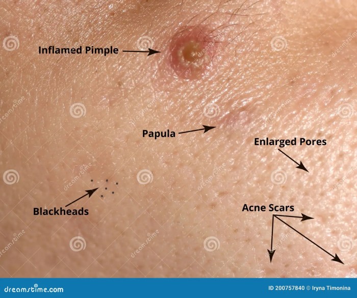
    Comparison of Acne Lesions

    A visual aid comparing and contrasting different acne lesions, including papules, should use a table format. This table would categorize various acne lesions (e.g., papules, pustules, nodules, cysts) and illustrate them using simple shapes. Each lesion would be clearly labeled. Important characteristics, such as size, color, and texture, would be noted for each lesion type. For example, a papule would be shown as a small, solid elevation, while a pustule would be a similar elevation containing pus.

    This visual aid would help differentiate between different types of acne lesions.

    Lesion Type Description Appearance
    Papule Small, solid elevation Small, round or oval, slightly raised area, skin-colored to reddish
    Pustule Papule containing pus Small, pus-filled elevation, white or yellow
    Nodule Deep, solid lesion Larger, firm, and painful, often inflamed
    Cyst Deep, pus-filled lesion Larger, inflamed, painful, pus-filled, and often tender

    Cross-section of a Skin Pore with a Papule

    A labeled illustration of a cross-section of a skin pore with a papule should clearly show the pore structure. The pore should be shown as a canal in the epidermis and dermis. A papule should be shown as an accumulation of material, possibly sebum, within or adjacent to the pore, leading to inflammation and swelling. The illustration should highlight the relationship between the pore, the papule, and the surrounding skin tissue.

    Labels should indicate the epidermis, dermis, hair follicle, sebaceous gland, and the location of the papule.

    Stages of Acne Papule Development

    A graphic representation of the stages of acne papule development would use a series of sequential images. The first image could depict a normal skin pore. Subsequent images would show the buildup of sebum and/or bacteria, progressing to inflammation and finally to the formation of a papule. This visual aid should clearly illustrate the progression of events, highlighting the contributing factors at each stage.

    It would effectively show how a simple buildup can lead to an acne papule.

    Final Review

    In conclusion, understanding acne papules, from their definition to their management, empowers individuals to take control of their skin health. This comprehensive guide provides a thorough understanding of this common skin condition. By recognizing the causes, symptoms, and treatment options, individuals can proactively address acne papules and work towards clearer, healthier skin.

  • Skin Lesions Types, Pictures, Causes & Treatment

    Skin Lesions Types, Pictures, Causes & Treatment

    Types of skin lesion pictures causes and treatment is a comprehensive guide to understanding skin abnormalities. From identifying various lesion types and their characteristics, to exploring potential causes and effective treatments, this article provides a thorough overview. We’ll delve into the specifics of primary and secondary lesions, offering visual aids and descriptions to aid in recognition. This will cover the importance of proper diagnosis and treatment options for different types of skin lesions.

    Learning about skin lesions is crucial for recognizing potential health concerns. Knowing the characteristics of different types of lesions, their potential causes, and appropriate treatment options empowers individuals to take proactive steps toward their well-being.

    Introduction to Skin Lesions

    Types of skin lesion pictures causes and treatment

    Skin lesions are any noticeable changes in the appearance or texture of the skin. These changes can range from minor imperfections to serious medical conditions. Understanding skin lesions is crucial for both individuals and healthcare professionals, as early detection and diagnosis can lead to prompt treatment and improved outcomes. Recognizing the different types of lesions is essential for determining potential causes and appropriate management strategies.Skin lesions are broadly classified into primary and secondary lesions.

    Primary lesions are the initial changes in the skin’s structure, while secondary lesions are the changes that occur as a result of the primary lesion or due to other factors like scratching or infection. Accurate identification of these lesions is fundamental to differentiating between benign and malignant conditions. This differentiation is vital for initiating appropriate treatment plans.

    Primary Skin Lesions

    Primary skin lesions represent the initial or intrinsic alterations in the skin’s structure. They are crucial for initial assessment and diagnosis. Understanding their characteristics helps in differentiating various conditions.

    • Macule: A flat, discolored area on the skin, varying in size. Examples include freckles, birthmarks, and flat moles. Macules are typically less than 1 cm in diameter.
    • Papule: A small, solid elevation on the skin, typically less than 1 cm in diameter. Examples include warts, skin tags, and some types of skin cancer.
    • Plaque: A flat-topped, raised area of skin, larger than a papule. Examples include psoriasis and eczema lesions.
    • Nodule: A solid, elevated lesion that extends deeper into the skin than a papule, typically larger than 1 cm in diameter. Examples include lipomas and some types of skin cancer.
    • Tumor: A large, solid mass that can extend deep into the skin. Examples include melanomas and other cancerous tumors.
    • Wheal: A raised, itchy area of skin, often appearing as a hive or welt. Examples include allergic reactions and insect bites.
    • Vesicle: A small, fluid-filled blister on the skin, typically less than 1 cm in diameter. Examples include herpes simplex and chickenpox.
    • Bulla: A large, fluid-filled blister on the skin, larger than 1 cm in diameter. Examples include severe burns and some allergic reactions.
    • Pustule: A pus-filled blister on the skin. Examples include acne and impetigo.
    • Cyst: A sac-like lesion containing fluid or semi-solid material. Examples include sebaceous cysts and epidermal cysts.

    Secondary Skin Lesions

    Secondary skin lesions are alterations to the primary lesion, or they develop as a result of other factors like scratching, infection, or healing. Analyzing secondary lesions can provide insights into the lesion’s evolution and the underlying condition.

    Type Description Examples
    Primary Initial changes in the skin’s structure. Macule, Papule, Plaque, Nodule, Tumor, Wheal, Vesicle, Bulla, Pustule, Cyst
    Secondary Changes resulting from the primary lesion or other factors. Erosion, Excoriation, Fissure, Ulcer, Scar, Crust, Scale, Atrophy
    • Erosion: A superficial loss of the epidermis, leaving a shallow depression in the skin. Examples include skin irritation from scratching.
    • Excoriation: A scratch mark or abrasion on the skin. Examples include intense itching or scratching.
    • Fissure: A linear crack or split in the skin. Examples include athlete’s foot or cracked lips.
    • Ulcer: A deep loss of skin tissue, extending into the dermis. Examples include pressure sores and some skin infections.
    • Scar: A fibrous tissue that forms after the healing of a wound. Examples include surgical scars or acne scars.
    • Crust: A dried, scab-like accumulation of blood, serum, or pus on the skin. Examples include impetigo and skin infections.
    • Scale: Flaky or dry skin, often associated with inflammatory conditions. Examples include psoriasis and eczema.
    • Atrophy: Thinning of the skin, resulting in a depressed appearance. Examples include prolonged sun exposure and some skin conditions.

    Types of Skin Lesions

    Types of skin lesion pictures causes and treatment

    Skin lesions are changes in the skin’s appearance, texture, or color that can be caused by various factors. Understanding different types of skin lesions is crucial for accurate diagnosis and appropriate treatment. This section delves into the characteristics of various skin lesions, providing a visual guide for identification.

    Classifying Skin Lesions by Morphology, Types of skin lesion pictures causes and treatment

    Skin lesions are categorized based on their visual characteristics, including size, shape, color, and elevation from the surrounding skin. These morphological features are key in differentiating between various conditions and aid in proper diagnosis.

    Lesion Type Description Size Range (mm) Color Elevation
    Macule A flat, discolored area on the skin. It does not protrude above or below the surface. Less than 10 Various (e.g., red, brown, black, white, pink) Flat
    Papule A small, solid, elevated lesion. It is typically less than 1 centimeter in diameter. 1-10 Various (e.g., red, brown, pink, dark-red) Elevated
    Plaque A flat-topped, elevated lesion, often larger than a papule. A plaque can be slightly raised or flat-topped, and is typically more than 1 centimeter in diameter. Greater than 10 Various Elevated, but not as prominent as a nodule
    Nodule A solid, elevated lesion that is deeper and firmer than a papule or plaque. It can extend into the dermis and may be palpable. Greater than 10 Various (e.g., red, brown, skin-colored) Elevated and palpable
    Vesicle A small, fluid-filled blister, less than 0.5 centimeters in diameter. Less than 5 Usually translucent or slightly pink Elevated and filled with fluid
    Bullae A large, fluid-filled blister, greater than 0.5 centimeters in diameter. Similar to vesicles, but significantly larger. Greater than 5 Usually translucent or slightly pink Elevated and filled with fluid
    Pustule A small, pus-filled blister. Less than 10 Yellowish or whitish Elevated and filled with pus
    Ulcer A break in the skin’s surface that extends into the dermis. It may be shallow or deep, and may be filled with exudate. Variable Red, yellow, or brown Depressed below the skin surface

    Lesion Shapes

    The shape of a skin lesion can also provide important clues about its potential cause. Different shapes may indicate different types of underlying conditions.

    Shape Description Example
    Round Circular or nearly circular. A mole that is perfectly round
    Oval Elliptical or egg-shaped. A birthmark that follows an oval pattern
    Irregular Not round or oval; often with uneven borders. A skin lesion with a complex, non-symmetrical shape
    Annular Ring-shaped. A ring-like rash
    Linear Long and narrow. A rash that follows a line on the skin

    Pictures of Skin Lesions

    A crucial aspect of understanding skin lesions is visual recognition. Detailed descriptions of skin lesions, highlighting key characteristics like size, shape, color, and texture, are essential for accurate diagnosis. These descriptions help healthcare professionals differentiate between benign and potentially serious conditions. Visual aids, although not directly presented here, can be invaluable tools in confirming the diagnosis.Visual recognition plays a vital role in identifying skin lesions.

    Detailed descriptions of characteristics, including size, shape, color, and texture, are critical for accurate diagnosis. By understanding these visual cues, we can distinguish between benign and potentially concerning conditions.

    Examples of Skin Lesions

    Accurate diagnosis often hinges on the detailed characteristics of a skin lesion. Careful observation of size, shape, color, and texture is crucial. These features contribute significantly to distinguishing between various types of skin lesions.

    Image Description Lesion Type Key Features
    (Imagine a small, round, red, slightly raised lesion, with a well-defined border.) A small, round, red papule, approximately 3-5 mm in diameter, with a smooth, slightly raised surface. The lesion appears erythematous (red) and has a well-defined border. Erythematous Papule Small size, round shape, red color, slightly raised, well-defined border.
    (Imagine a large, irregular, brown, flat lesion with a slightly scaly texture.) A large, irregular, brown macule, approximately 2-3 cm in diameter, with a slightly scaly texture. The lesion is flat and displays a varied, mottled brown coloration. Macule (possibly a lentigo) Large size, irregular shape, brown color, flat, slightly scaly texture.
    (Imagine a raised, firm, flesh-colored nodule with a slightly rough surface.) A raised, firm, flesh-colored nodule, approximately 1-2 cm in diameter, with a slightly rough, or slightly crusted, surface. The lesion appears firm to the touch and is well-circumscribed. Flesh-colored Nodule Raised, firm texture, flesh-colored, slightly rough or crusted surface, well-circumscribed.
    (Imagine a collection of small, fluid-filled blisters on a reddened base.) Multiple small, fluid-filled blisters (vesicles) approximately 2-5 mm in diameter, clustered together on an erythematous base. The blisters are clear or translucent. Vesicular Eruption Multiple small, fluid-filled blisters, clustered, erythematous base, clear or translucent fluid.

    Detailed Descriptions of Skin Lesions

    The following descriptions exemplify the variety of skin lesions, emphasizing their diverse characteristics:

    • Actinic Keratosis: A rough, scaly patch, often appearing reddish-brown, typically found on sun-exposed areas. The patch may be slightly raised and have a rough, sandpaper-like texture. It can vary in size from a few millimeters to a centimeter or more.
    • Seborrheic Keratosis: A well-defined, raised, often warty lesion that is typically brown, black, or tan. It has a somewhat greasy or stuck-on appearance and may be slightly rough to the touch. It is often found on the face, neck, or chest.
    • Psoriasis: A common skin condition characterized by red, inflamed plaques covered with silvery-white scales. The plaques can vary in size and often have well-defined borders. The scaling is a key characteristic of this condition.
    • Basal Cell Carcinoma: A common type of skin cancer, typically appearing as a pearly, flesh-colored, or waxy papule or nodule. It often has a slightly raised border and may have telangiectasias (small blood vessels) visible on the surface. It can slowly grow over time.

    Causes of Skin Lesions

    Skin lesions, ranging from seemingly harmless spots to severe conditions, can arise from a multitude of factors. Understanding the underlying causes is crucial for proper diagnosis and effective treatment. These causes often intertwine, making it essential to consider the interplay of various factors in a given individual. This section delves into the common causes, mechanisms, and associated risk factors.

    Infectious Causes

    Infections are a significant contributor to skin lesions. Microorganisms like bacteria, viruses, and fungi can invade the skin, triggering an inflammatory response that manifests as various lesions. The specific type of lesion often depends on the infecting agent and the individual’s immune response.

    Understanding different skin lesion pictures, their causes, and treatments is crucial for good health. While factors like genetics and sun exposure play a role, a diet high in sodium can also indirectly affect skin health. For example, excessive consumption of processed foods, often loaded with foods high in sodium , can lead to water retention, potentially exacerbating certain skin conditions.

    Ultimately, a balanced diet, coupled with proper skincare, is key to managing various skin lesion types effectively.

    • Bacterial infections, such as impetigo and folliculitis, are characterized by pustules, vesicles, or crusted lesions. These infections typically arise from skin trauma or compromised skin barriers, allowing bacteria to colonize and multiply. Examples include staphylococcal infections that can lead to boils or carbuncles.
    • Viral infections, including herpes simplex and varicella-zoster (chickenpox), typically present with blisters or grouped vesicles. These viruses can remain dormant in the body, reactivating under certain conditions to cause outbreaks. Herpes simplex virus, for instance, can cause cold sores or genital herpes, and varicella-zoster virus causes shingles.
    • Fungal infections, such as ringworm and tinea pedis (athlete’s foot), manifest as red, scaly, or ring-shaped lesions. These infections often occur in areas with moisture or in individuals with weakened immune systems. Tinea capitis is another example, causing hair follicle infections.

    Allergic Reactions

    Allergic reactions can lead to a variety of skin lesions, ranging from mild hives to severe contact dermatitis. These reactions occur when the immune system overreacts to an allergen, a substance that triggers an immune response. The intensity of the reaction depends on the individual’s sensitivity and the amount of allergen encountered.

    • Contact dermatitis, a common allergic reaction, arises from direct contact with an allergen, such as certain plants (poison ivy), chemicals (nickel), or cosmetics. The resulting skin lesions can be itchy, red, and inflamed, often with blisters or oozing.
    • Urticaria (hives) is characterized by raised, itchy wheals on the skin. These lesions are typically caused by allergic reactions to foods, medications, or insect bites. They can vary in size and duration.

    Autoimmune Diseases

    Autoimmune diseases are conditions where the body’s immune system mistakenly attacks healthy tissues. This can manifest in various skin lesions, reflecting the specific tissues affected. The lesions’ appearance can be diverse and vary depending on the particular autoimmune disease.

    • Lupus, a chronic autoimmune disease, can cause a wide range of skin lesions, including butterfly rash (a red rash across the cheeks and nose), discoid lupus (coin-shaped lesions), and other skin manifestations. Lupus is a systemic disease, impacting various organ systems.
    • Psoriasis is a chronic inflammatory condition causing red, scaly plaques on the skin. The exact cause of psoriasis remains unknown, but it’s believed to involve an interplay of genetic and environmental factors.

    Genetic Factors

    Genetic predisposition plays a role in the development of certain skin lesions. Inherited mutations or variations in genes can increase the risk of developing specific skin conditions. This often leads to conditions present from birth or manifesting early in life.

    • Certain inherited skin disorders, such as epidermolysis bullosa, result in fragile skin that blisters or tears easily due to defects in the skin’s structural proteins.
    • Some inherited conditions, like porphyria, can cause skin lesions that are sensitive to sunlight.

    Trauma

    Trauma to the skin, whether physical injury or repetitive friction, can lead to skin lesions. The type of lesion depends on the nature and extent of the trauma.

    • Burns, from various sources like heat, chemicals, or radiation, result in varying degrees of skin damage, leading to blisters, scars, and other lesions.
    • Pressure sores (decubitus ulcers) develop due to prolonged pressure on the skin, often in individuals confined to bed or with limited mobility. These wounds are prone to infections and can be difficult to heal.

    Treatment of Skin Lesions

    Treating skin lesions requires a multifaceted approach, considering the type, cause, and severity of the lesion. Effective treatment aims to eliminate or significantly reduce the lesion while minimizing potential side effects. The choice of treatment depends heavily on the specific diagnosis and individual patient factors. Proper identification and diagnosis by a dermatologist are crucial for selecting the most appropriate and effective course of action.Effective treatment strategies encompass a wide range of options, ranging from topical medications to surgical procedures.

    The selection process often involves weighing the benefits of each approach against potential risks and side effects. Understanding the rationale behind different treatments is essential for informed decision-making and for achieving the best possible outcomes.

    Common Topical Treatments

    Topical treatments are often the first line of defense for many skin lesions. They are frequently used for superficial lesions and offer a less invasive approach. These treatments typically involve applying medications directly to the affected area. The effectiveness and suitability of topical treatments depend on the specific type of lesion.

    Learning about different skin lesion pictures, their causes, and treatments is super helpful. But sometimes, concerning symptoms pop up elsewhere, like lower back pain. If you’re wondering if your lower back pain could be something more serious, like cancer, checking out this resource on is my lower back pain cancer might be a good idea.

    Ultimately, understanding skin lesions and their treatment options remains a crucial aspect of overall health awareness.

    • Corticosteroids: These potent anti-inflammatory medications are frequently used to reduce inflammation and swelling associated with various skin conditions, including eczema and psoriasis. They are particularly useful for inflammatory lesions. Expected outcomes include reduced inflammation and itching. Potential side effects include skin thinning, striae (stretch marks), and local skin atrophy, especially with prolonged use. The duration and strength of the corticosteroid treatment are carefully determined by the healthcare provider.

      Learning about different skin lesions and their pictures, causes, and treatments is fascinating. It’s crucial to understand these visual clues and the underlying reasons for their development. While exploring the causes of sleepwalking, for instance, why do people sleepwalk , can shed light on the complexities of the human body, ultimately leading to a better understanding of various health conditions.

      Ultimately, understanding skin lesion pictures, causes, and treatments is a vital aspect of self-care and proactive health management.

    • Antibiotics: Antibiotics are used to treat bacterial infections of the skin. They target the bacteria causing the lesion, aiming to clear the infection and prevent further spread. Expected outcomes include resolution of the infection and healing of the affected area. Potential side effects can include allergic reactions, antibiotic resistance, and disruption of the skin’s natural microbiome.

    • Antivirals: Used for viral infections, antivirals aim to suppress or eliminate the virus responsible for the lesion. Examples include treatments for herpes simplex virus (HSV) and warts. Expected outcomes include reduction or elimination of viral lesions. Potential side effects may include localized skin irritation, or rarely, systemic side effects. Treatment duration depends on the type and severity of the viral infection.

    Surgical Treatments

    Surgical interventions are often necessary for more complex or deep-seated lesions. These methods vary widely, and the specific procedure depends on the type of lesion and its location.

    • Excision: This involves surgically removing the lesion, typically with a scalpel. It is suitable for various types of skin lesions, including benign tumors, moles, and cysts. Expected outcomes include complete removal of the lesion. Potential complications include scarring, bleeding, and infection. The extent of scarring depends on the size and location of the lesion, and the surgical technique employed.

    • Cryotherapy: This method uses extreme cold (liquid nitrogen) to freeze and destroy the lesion. It is effective for certain skin lesions, such as warts and some precancerous lesions. Expected outcomes include the destruction of the targeted lesion. Potential complications include scarring, blistering, and pain. The choice of cryotherapy depends on the lesion’s size, depth, and location.

    • Laser therapy: Laser treatment utilizes focused light beams to target and destroy specific skin lesions. It is used for various conditions, including vascular lesions, pigmented lesions, and some skin cancers. Expected outcomes include removal or reduction of the lesion. Potential complications include scarring, hyperpigmentation, and pain. The specific type of laser used depends on the lesion type and the desired outcome.

    Treatment Options Table

    Lesion Type Treatment Options Expected Outcomes Potential Complications
    Actinic Keratosis Cryotherapy, topical chemotherapy, excision Resolution of the lesion, prevention of progression to skin cancer Scarring, blistering, pain, allergic reactions to topical agents
    Basal Cell Carcinoma Surgical excision, Mohs surgery, radiation therapy Complete removal of the cancer, preservation of surrounding tissue Scarring, nerve damage, infection, radiation side effects
    Melanoma Surgical excision, radiation therapy, chemotherapy, immunotherapy Complete removal of the cancer, treatment of metastasis Scarring, nerve damage, systemic side effects, fatigue
    Warts Cryotherapy, topical medications, laser therapy, surgical excision Removal or resolution of the lesion Scarring, pain, blistering, allergic reactions
    Skin Tags Surgical excision, ligation, electrocautery Removal of the skin tag Bleeding, infection, scarring

    Diagnosis and Evaluation

    Accurate diagnosis is crucial for effective skin lesion management. A proper diagnosis helps determine the nature of the lesion, its potential severity, and the appropriate course of treatment. This involves careful evaluation of the lesion’s appearance, location, and other relevant factors. Incorrect diagnoses can lead to delayed or inappropriate treatment, potentially impacting the patient’s health and well-being.Understanding the diagnostic process provides a clear framework for identifying and managing skin lesions.

    This includes employing various methods, ranging from a simple physical examination to sophisticated laboratory tests, all tailored to the specific characteristics of the lesion. This meticulous approach is vital to ensure the most accurate and timely diagnosis.

    Diagnostic Methods

    A comprehensive approach to skin lesion diagnosis typically starts with a thorough physical examination. This initial assessment helps to identify key features such as size, shape, color, border definition, and any associated symptoms. Observing the lesion’s location and any surrounding skin changes is also important. The clinician will carefully document these observations, creating a baseline for further investigation.

    Physical Examination

    A physical examination involves a detailed visual inspection of the skin lesion. This includes noting the lesion’s characteristics such as:

    • Size and Shape: Measuring the lesion’s dimensions and describing its shape (e.g., round, oval, irregular) are crucial for differentiating potential causes.
    • Color and Texture: Noting the lesion’s pigmentation (e.g., red, brown, black) and texture (e.g., smooth, rough, scaly) provides additional clues.
    • Location and Distribution: Observing where the lesion is located on the body and whether it appears in clusters or scattered patterns helps to narrow down possible diagnoses.
    • Associated Symptoms: Identifying any accompanying symptoms like itching, pain, bleeding, or tenderness is important for understanding the lesion’s potential nature and severity.

    Biopsy

    A biopsy is a procedure where a small tissue sample is removed from the lesion for microscopic examination. It is a crucial diagnostic tool for suspected cancerous or atypical lesions. This method allows pathologists to examine the cells under a microscope, aiding in the definitive diagnosis. There are different types of biopsies, including incisional and excisional biopsies, each with its specific application.

    Laboratory Tests

    Laboratory tests can be used to aid in the diagnosis of skin lesions. For example, blood tests may be conducted to check for inflammatory markers or specific antibodies. These tests can provide valuable information about the overall health status of the patient and help differentiate various skin conditions.

    Evaluation Process

    The evaluation process for skin lesions involves a systematic approach. This begins with a detailed history and physical examination. Further testing, such as a biopsy or lab tests, may be necessary to confirm the diagnosis. A final diagnosis is typically made based on the combination of all gathered information.

    Examples of Diagnostic Tests

    • Dermoscopy: This non-invasive technique uses a dermatoscope to examine the skin lesion’s surface in detail. It enhances the visualization of fine structures, assisting in differentiating benign and malignant lesions. Dermoscopy is particularly helpful in identifying subtle features of melanocytic lesions (moles). Dermoscopy is a powerful tool in early melanoma detection.
    • Skin Biopsy: A skin biopsy involves removing a small sample of skin tissue for examination under a microscope. It is crucial for diagnosing conditions such as skin cancer, inflammatory skin diseases, and infections. The type of biopsy performed depends on the size and location of the lesion, with options like punch biopsies, shave biopsies, and excisional biopsies.
    • Immunofluorescence Tests: These tests can help identify specific antibodies or antigens associated with certain skin conditions. This aids in diagnosing autoimmune skin diseases, such as bullous pemphigoid, by detecting the presence of particular antibodies.

    Final Conclusion: Types Of Skin Lesion Pictures Causes And Treatment

    In conclusion, understanding skin lesions involves recognizing diverse types, appreciating their varied causes, and selecting appropriate treatments. This comprehensive guide provides a framework for recognizing and managing these conditions. From initial identification to eventual treatment, the information presented will aid in understanding and addressing these often-complex issues. Remember, proper diagnosis and consultation with a healthcare professional are essential for any concerning skin lesion.

  • Lump Under Skin Causes & When to Worry

    Lump Under Skin Causes & When to Worry

    Lump under skin causes and when to worry about it is a crucial topic for anyone experiencing such a concern. Skin lumps can arise from a variety of factors, ranging from harmless cysts to more serious conditions. Understanding the potential causes and knowing when to seek medical attention can be vital for a quick diagnosis and appropriate treatment.

    This post explores the different types of skin lumps, common causes, and the signs that suggest you should consult a doctor. We’ll cover everything from benign growths to potential indicators of more serious issues. Armed with knowledge, you’ll be better equipped to navigate this potentially unsettling experience.

    Introduction to Skin Lumps

    Skin lumps, also known as skin lesions, are abnormal growths or swellings that appear on the surface of the skin. They can vary significantly in size, shape, and texture, and their appearance can be a clue to their underlying cause. Understanding the characteristics of skin lumps, including their location, appearance, and potential causes, is crucial for determining whether further medical evaluation is necessary.Skin lumps can arise from a multitude of causes, ranging from harmless, benign conditions to more serious, potentially cancerous ones.

    Recognizing the different types of skin lumps and their potential implications is essential for appropriate action. This section will Artikel the common types of skin lumps, their characteristics, and when it’s important to seek professional medical advice.

    Lumps under the skin can be a real worry, but often they’re completely harmless. They can stem from a variety of things, like cysts, infections, or even just a buildup of fluid. However, if you’re concerned, it’s always best to see a doctor. Knowing the potential risks of imaging tests like CT scans is also important; for example, the potential cancer risks associated with CT scans are something you should be aware of, especially if they are frequently used.

    ct scan cancer risk is a factor to consider alongside other potential causes when evaluating a lump. Ultimately, the best way to determine the cause of a lump and decide if you need to be concerned is to consult with a healthcare professional.

    Types of Skin Lumps

    Skin lumps can be broadly categorized into benign, malignant, and inflammatory types. Benign lumps are typically harmless and do not spread to other parts of the body. Malignant lumps, conversely, are cancerous and can metastasize. Inflammatory lumps are caused by an underlying inflammatory process. The precise classification of a lump often requires a thorough examination and possibly a biopsy.

    Common Locations of Skin Lumps

    Skin lumps can appear virtually anywhere on the body. Common locations include the face, neck, back, chest, arms, legs, and scalp. The location of the lump can offer clues to the potential cause. For example, lumps on the scalp might be related to hair follicle issues, while those on the legs could be related to subcutaneous fat deposits or other factors.

    Appearance and Texture of Skin Lumps

    The appearance and texture of a skin lump can provide valuable clues about its potential nature. Some common characteristics include:

    • Size and Shape: Lumps can range from tiny bumps to larger masses. The shape can be round, oval, irregular, or nodular.
    • Color: Skin lumps can be the same color as the surrounding skin, or they might appear red, pink, brown, black, or even bluish.
    • Texture: The texture can vary from smooth and soft to firm, hard, or even bumpy.
    • Pain: Some lumps may be painless, while others may be tender or cause discomfort.
    • Mobility: The mobility of a lump, meaning how easily it can be moved under the skin, can provide information about its depth and potential nature.

    Table of Common Skin Lumps

    This table summarizes different types of skin lumps, their appearance, potential causes, and when medical attention is recommended.

    Lump Type Appearance Common Causes When to Seek Medical Attention
    Benign Cyst Smooth, firm, or soft, often round, typically painless. Blocked hair follicles, sebaceous glands, or other fluid-filled sacs. If the lump is rapidly growing, painful, or causing significant discomfort.
    Skin Tag Small, fleshy, and often stalk-like, usually painless. Skin folds, friction, or aging. If the skin tag is causing irritation or bleeding.
    Moles (Melanocytic Nevi) Various colors, often flat or raised, can be small or large, usually painless. Genetic predisposition, sun exposure. If the mole changes in size, shape, color, or texture, or if it bleeds or itches.
    Basal Cell Carcinoma Often pearly or waxy, with a slightly raised border, sometimes with a central ulcer. Exposure to ultraviolet (UV) radiation from the sun or tanning beds. Immediately if the lump appears to be growing or changing.

    Potential Causes of Skin Lumps

    Lump under skin causes and when to worry

    Skin lumps, while often benign, can sometimes signal underlying health concerns. Understanding the potential causes, ranging from common, harmless conditions to more serious possibilities, is crucial for appropriate evaluation and treatment. This section delves into the diverse factors contributing to skin lump formation.Identifying the cause of a skin lump is essential for determining the appropriate course of action.

    Accurate diagnosis requires a comprehensive evaluation, considering the lump’s characteristics (size, shape, color, location, and any associated symptoms) and the individual’s medical history.

    Common Benign Causes

    Many skin lumps are harmless and result from benign conditions. These conditions typically do not pose a significant health risk. Examples include:

    • Epidermal Cysts: These are common, fluid-filled cysts that develop beneath the skin’s surface. They are typically painless and slow-growing, often appearing as a small, firm bump. They are usually caused by trapped skin cells.
    • Sebaceous Cysts: These cysts are formed from blocked sebaceous glands, which produce oil for the skin. They typically present as a soft, fluctuating lump, often filled with a cheesy material.
    • Lipomas: These are benign fatty tumors that often develop beneath the skin. They are usually soft, painless, and movable. They are frequently found in the arms, shoulders, and back.
    • Skin Tags: These are small, benign growths that hang from the skin by a stalk. They are typically flesh-colored and painless. Skin tags often develop in areas with friction, such as the neck, armpits, or groin.

    Inflammatory Conditions

    Inflammation can lead to various skin lumps. These conditions often present with redness, warmth, and tenderness around the affected area.

    • Acne: While often associated with bumps and pimples, severe acne can result in inflamed lumps under the skin. These lumps can be painful and may require treatment to resolve.
    • Cellulitis: This bacterial infection of the skin and underlying tissues can cause painful, red, and swollen lumps. Cellulitis requires prompt medical attention.
    • Contact Dermatitis: An allergic reaction to an irritant or allergen can cause inflamed lumps and bumps on the skin. The affected area may be itchy and painful.
    • Insect Bites/Stings: A localized reaction to insect bites or stings can cause inflamed lumps, often with itching and swelling.

    Cysts

    Cysts are sac-like structures that can contain various substances, including fluid, air, or solid material. Different types of cysts can lead to skin lumps.

    • Dermoid Cysts: These cysts contain various tissues, such as hair follicles and teeth. They are often found in the face or scalp and are slow-growing.
    • Pilar Cysts: These cysts develop from hair follicles and appear as small, firm lumps, usually painless and slow-growing.
    • Galactoceles: These are cysts that develop in the milk ducts, particularly in women who have recently given birth or are breastfeeding.

    Infections

    Infections, particularly bacterial or fungal infections, can lead to skin lumps.

    • Furuncles (Boils): These are localized bacterial infections that cause painful, pus-filled lumps. They are commonly found on the skin’s surface.
    • Abscesses: Deeper skin infections that can result in pus-filled lumps that may need drainage.
    • Skin infections caused by certain fungi: Some fungal infections can lead to inflamed lumps and bumps, often with itching and scaling.

    Comparison of Benign and Malignant Skin Lump Causes

    Characteristic Benign Causes Malignant Causes
    Growth Rate Slow and gradual Can be rapid
    Pain Often painless May be painful
    Appearance Smooth, firm, or soft Irregular, ulcerated, or bleeding
    Texture Typically firm or soft Can be hard and irregular
    Mobility Usually movable Often fixed

    Underlying Medical Conditions

    Certain underlying medical conditions can contribute to skin lump formation.

    • Lupus: This autoimmune disorder can manifest with skin lumps and rashes. Lupus symptoms vary significantly.
    • Sarcoidosis: This inflammatory condition can cause lumps in various parts of the body, including the skin.
    • Certain types of cancer: While less common, certain cancers can cause skin lumps. Skin cancer is characterized by specific patterns.

    When to Seek Medical Attention

    Spotting a lump on your skin can be unsettling. While many skin lumps are harmless, some can signal underlying health concerns. Understanding when to seek medical attention is crucial for timely diagnosis and treatment. Knowing the potential warning signs can empower you to take proactive steps towards your well-being.Knowing when to seek medical attention for a skin lump is vital.

    Ignoring potentially serious signs could delay appropriate care, potentially affecting the outcome. Factors like the lump’s growth rate, appearance, and location can all contribute to the need for professional evaluation.

    Symptoms Warranting Immediate Medical Attention

    Prompt medical attention is essential for certain skin lump characteristics. Rapid growth, significant pain, changes in the skin around the lump, and unusual bleeding or discharge are all cause for concern. These symptoms may indicate an infection, a cancerous growth, or another underlying medical condition. If you notice any of these immediate warning signs, it’s crucial to schedule an appointment with a dermatologist or other qualified healthcare professional.

    Factors Increasing the Risk of a Serious Skin Lump, Lump under skin causes and when to worry

    Certain factors can increase the likelihood of a skin lump being a cause for concern. A history of skin cancer in the family, a weakened immune system, or exposure to excessive sunlight or harmful substances can heighten the risk. Furthermore, lumps that appear in unusual locations, such as the armpit or groin, may require more immediate evaluation. Knowing these factors can help you determine if a skin lump requires more immediate attention.

    Tracking Changes in Lump Characteristics

    Regularly monitoring a skin lump’s size, shape, and appearance is vital. Note any changes, such as increasing size, altered borders, or color variations. Changes in the lump’s texture or the surrounding skin, such as redness, inflammation, or tenderness, should also be documented. These observations can aid in determining if the lump is progressing or potentially becoming more serious.

    Detailed records can be crucial for communication with your healthcare provider.

    Location-Specific Concerns

    The location of a skin lump can also be a factor in determining the need for medical evaluation. Lumps appearing on sun-exposed areas like the face, neck, or hands might warrant closer examination due to their increased risk of being a skin cancer. Lumps located near joints or areas prone to injury might suggest a different underlying cause.

    Understanding the location’s potential significance can help guide your approach.

    Table of Key Characteristics Requiring Immediate Medical Evaluation

    Characteristic Description
    Rapid Growth A noticeable increase in size within a short period.
    Significant Pain Persistent or intense pain associated with the lump.
    Change in Skin Around the Lump Redness, inflammation, or other changes in the skin surrounding the lump.
    Bleeding or Discharge Uncommon bleeding or discharge from the lump.
    Unusual Location Appearance in unusual areas like the armpits, groin, or on sun-exposed skin.
    Family History of Skin Cancer Presence of a family history of skin cancer.

    Diagnostic Methods for Skin Lumps

    Understanding the cause of a skin lump is crucial for appropriate treatment. Accurate diagnosis relies on a combination of methods, from a simple physical examination to more sophisticated imaging techniques and biopsies. This process helps determine the nature of the lump, its potential impact on health, and the most effective course of action.

    Physical Examination

    A thorough physical examination is the first step in assessing a skin lump. This involves careful observation of the lump’s size, shape, color, consistency, location, and any associated symptoms. The dermatologist or physician will look for signs such as tenderness, bleeding, or ulceration. The location of the lump, for example, near a joint or in an area with increased sun exposure, can provide valuable clues about potential underlying conditions.

    The examination also assesses the surrounding skin for any abnormalities or changes in texture.

    Imaging Techniques

    Imaging techniques play a significant role in evaluating skin lumps, particularly when physical examination alone isn’t sufficient to determine the nature of the lesion. Dermatologists and other specialists often use techniques like ultrasound and dermatoscopic imaging to obtain detailed visual information about the lump’s structure and depth.Ultrasound, a non-invasive method, uses high-frequency sound waves to create images of internal structures.

    It’s particularly helpful in distinguishing between solid and cystic masses and assessing the size and depth of the lump. Dermatoscopic imaging uses a specialized microscope to magnify and analyze the skin’s surface, allowing for the identification of subtle features that might be missed during a routine examination. This is especially useful for assessing pigmented lesions, which could indicate skin cancer.

    Computed tomography (CT) scans or magnetic resonance imaging (MRI) scans might be employed in certain cases to obtain a more detailed cross-sectional view of the skin and surrounding tissues.

    Biopsies

    Biopsies are a critical step in diagnosing skin lumps, especially when the physical examination and imaging findings are inconclusive. A biopsy involves removing a small sample of the lump tissue for microscopic examination by a pathologist. This allows for a definitive diagnosis of the type of cell present in the lump and can identify conditions such as benign tumors, skin cancers, infections, or inflammatory processes.The type of biopsy performed depends on the size, location, and suspected nature of the lump.

    Excisional biopsies involve removing the entire lump, while incisional biopsies remove only a portion of it. The choice between these two types depends on the clinical judgment of the physician.

    Summary of Diagnostic Methods

    Diagnostic Method Description Application
    Physical Examination Visual inspection, palpation, assessment of size, shape, color, and location. Initial assessment, identifying suspicious features, guiding further investigations.
    Imaging Techniques (e.g., Ultrasound, Dermatoscopy) Non-invasive methods to visualize internal structures and skin surface details. Assessing the structure and depth of the lump, aiding in distinguishing between solid and cystic masses, evaluating pigmented lesions.
    Biopsy (e.g., Excisional, Incisional) Removal of a tissue sample for microscopic examination. Providing a definitive diagnosis when other methods are inconclusive, identifying the type of cells present.

    Treatment Options for Skin Lumps: Lump Under Skin Causes And When To Worry

    Treating skin lumps requires careful consideration of their underlying cause and potential for harm. A thorough evaluation by a dermatologist or other qualified healthcare professional is crucial to determine the most appropriate course of action. Different types of skin lumps necessitate different treatment strategies, ranging from simple observation to surgical intervention.Effective treatment plans aim to address the underlying issue, whether it’s a benign growth, an infection, or a more serious condition.

    Factors such as the lump’s size, location, and potential for growth or spread influence the chosen treatment. It’s essential to understand that self-treating skin lumps can be risky and potentially worsen the condition.

    Common Treatments for Benign Skin Lumps

    Benign skin lumps, which are typically harmless, often require less aggressive treatment options. These options aim to either remove the lump or manage its symptoms.

    A lump under the skin can be concerning, but often benign. Sometimes, it’s just a harmless cyst. However, if you’re experiencing chest congestion, trying some soothing home remedies like those found in this helpful guide on home remedies for chest congestion might help. But if the lump is growing rapidly, painful, or accompanied by other symptoms, it’s always best to see a doctor to rule out anything more serious.

    • Excision: Surgical removal is a common method for benign skin lumps. The procedure involves cutting out the lump and surrounding tissue. Excision is often used for skin tags, moles, and other benign tumors. The specific technique used depends on the size and location of the lump, and the surgeon’s preference. For example, a small, superficial skin tag might be removed with a simple scalpel incision, while a larger, deeper lesion might require a more extensive surgical approach.

    • Cryotherapy: This non-surgical technique involves freezing the skin lump to destroy it. Liquid nitrogen is typically used for this purpose. Cryotherapy is often effective for treating certain types of warts and small skin lesions. Potential side effects include temporary skin discoloration or discomfort.
    • Electrocautery: This method uses heat to destroy the skin lump. A heated instrument, often an electric needle, is used to burn away the abnormal tissue. Electrocautery is a useful technique for treating small, superficial skin lesions. Possible complications include scarring.
    • Laser therapy: Laser surgery uses a highly focused beam of light to vaporize or remove the skin lump. This approach is particularly useful for treating skin lesions that are difficult to access or remove with other methods. Laser therapy often results in minimal scarring and is effective for various skin conditions.

    Treatment Approaches Based on Underlying Causes

    The chosen treatment strategy for a skin lump often depends on the underlying cause. For example, an infected skin lump might require antibiotic treatment, while a cancerous lump might necessitate more aggressive therapies.

    • Infections: Infected skin lumps may respond to antibiotic therapy. The specific antibiotic and duration of treatment will depend on the causative organism. Antibiotics may be taken orally or applied topically.
    • Inflammation: Inflammation-related skin lumps may be treated with anti-inflammatory medications or topical creams. The choice of treatment depends on the severity and cause of the inflammation.
    • Cancerous Lumps: Cancerous skin lumps require specialized treatments like chemotherapy, radiation therapy, or surgery. These treatments are often part of a broader cancer treatment plan and are determined by the type, stage, and location of the cancer.

    Surgical Procedures for Removing Skin Lumps

    Surgical removal is a common treatment option for skin lumps. The specific surgical technique used depends on the type, size, and location of the lump.

    • Simple excision: This is a straightforward procedure involving the removal of the lump and a small margin of surrounding tissue. It’s often used for benign skin lesions.
    • Mohs surgery: This specialized surgical technique is used for skin cancers. Thin layers of tissue are removed and examined under a microscope until no cancerous cells are detected. It’s a precise approach that aims to minimize the removal of healthy tissue.
    • Curettage and electrodessication: This involves scraping away the lump with a curette and then using an electric current to destroy the remaining tissue. It’s often used for superficial skin lesions.

    Role of Medication in Managing Skin Lumps

    Medication can play a significant role in managing certain skin lumps, particularly those related to infections or inflammation.

    Lumps under the skin can stem from various things, like cysts or infections. But sometimes, a lump could be a sign of something more serious, like a tumor. For instance, a lump in the pelvic area, especially in men, might warrant further investigation, particularly if it’s accompanied by other symptoms. It’s crucial to consider more serious conditions like metastatic castration resistant prostate cancer, which, in some cases, can manifest as a lump.

    If you’re concerned about a lump, it’s always best to see a doctor for a proper diagnosis and to rule out any potential underlying health issues. Remember, early detection is key when it comes to lumps under the skin. metastatic castration resistant prostate cancer is a serious condition that requires prompt medical attention.

    • Antibiotics: Antibiotics are used to treat infections that cause skin lumps. The choice of antibiotic depends on the specific bacteria causing the infection.
    • Anti-inflammatory medications: Nonsteroidal anti-inflammatory drugs (NSAIDs) or corticosteroids may be used to reduce inflammation associated with certain skin lumps. The dosage and type of medication are determined by the healthcare professional.

    Treatment Options Table

    Treatment Option Suitability Potential Side Effects
    Excision Benign lumps, some cancers Bleeding, infection, scarring
    Cryotherapy Small, superficial lumps Discomfort, temporary skin discoloration
    Electrocautery Small, superficial lumps Scarring, pain
    Laser therapy Various skin lesions, difficult-to-reach lumps Pain, temporary redness, scarring
    Antibiotics Infected skin lumps Allergic reactions, digestive upset
    Anti-inflammatory medications Inflammation-related lumps Stomach upset, headache, other side effects

    Prevention Strategies for Skin Lumps

    Lump under skin causes and when to worry

    Skin lumps, while sometimes benign, can be a source of concern. Proactive measures can significantly reduce the risk of developing them. Implementing healthy lifestyle choices and practicing regular self-examination are crucial steps in this preventative approach.Early detection and prompt treatment are key to managing skin lumps effectively. A preventative approach focuses on minimizing risk factors and promoting overall skin health, leading to a lower likelihood of developing skin abnormalities.

    Lifestyle Factors to Reduce Skin Lump Risk

    Maintaining a healthy lifestyle plays a vital role in overall well-being and can reduce the risk of various skin conditions, including the development of skin lumps. A balanced diet rich in fruits, vegetables, and whole grains provides essential nutrients for healthy skin tissue repair and function. Regular exercise promotes blood circulation, supporting skin health and immune function. Stress management techniques, such as meditation or yoga, can also contribute to a healthier skin environment.

    Importance of Regular Skin Checks and Self-Examinations

    Regular skin checks are crucial for early detection of skin changes. This includes both professional examinations by dermatologists and self-examinations performed at home. Self-exams are simple yet effective tools for identifying potential problems early on. By regularly checking your skin, you can detect any unusual growths, changes in existing moles, or other irregularities that might signal a developing skin lump.

    This early detection allows for prompt medical intervention and treatment if necessary.

    Sun Protection in Preventing Skin Lumps

    Protecting your skin from excessive sun exposure is paramount in preventing various skin conditions, including skin lumps. The sun’s ultraviolet (UV) radiation is a significant risk factor for skin cancer, a leading cause of skin lumps. Sun protection measures include using broad-spectrum sunscreen with an SPF of 30 or higher, wearing protective clothing, and seeking shade during peak sun hours.

    Regular use of sunscreen, along with other protective measures, helps significantly reduce the risk of skin damage and subsequent skin lump development.

    Managing Underlying Medical Conditions

    Certain medical conditions can increase the risk of developing skin lumps. For example, individuals with weakened immune systems may be more susceptible to various skin infections and growths. Individuals with autoimmune diseases might experience skin manifestations as part of their condition. Managing underlying medical conditions effectively can help minimize the risk of skin lumps. This may involve adhering to prescribed medications, attending regular checkups, and maintaining a healthy lifestyle.

    Preventative Measures

    • Balanced Diet: A diet rich in fruits, vegetables, and whole grains provides essential nutrients for skin health, promoting repair and function.
    • Regular Exercise: Physical activity promotes blood circulation, supporting skin health and immune function.
    • Stress Management: Techniques like meditation or yoga can create a healthier skin environment.
    • Regular Skin Checks: Self-exams and professional dermatological examinations are crucial for early detection of skin changes.
    • Sun Protection: Use broad-spectrum sunscreen with an SPF of 30 or higher, wear protective clothing, and seek shade during peak sun hours.
    • Manage Underlying Conditions: Adhering to medical treatments, regular checkups, and a healthy lifestyle are vital for minimizing risks associated with pre-existing conditions.

    Last Point

    In conclusion, dealing with a lump under the skin can be concerning, but a proactive approach is key. By understanding the possible causes, recognizing warning signs, and knowing when to seek medical help, you can effectively manage the situation. Remember, early detection and proper diagnosis are paramount to ensuring the best possible outcome. Always consult a healthcare professional for personalized advice and treatment.