Weeping eczema overview and more delves into the complexities of this skin condition, offering a comprehensive understanding of its causes, symptoms, and management. We’ll explore everything from the underlying triggers to effective treatments and preventive strategies. Prepare to gain valuable insights into this common yet often challenging skin issue.
This comprehensive guide covers the full spectrum of weeping eczema, from its definition and diagnostic process to lifestyle adjustments and long-term effects. We’ll also delve into illustrative examples, prevention strategies, and valuable resources to support your understanding and journey toward effective management.
Introduction to Weeping Eczema
Weeping eczema, also known as exudative eczema, is a type of eczema characterized by the formation of blisters that leak fluid. This fluid leakage, often accompanied by crusting, creates a weeping appearance on the skin. Understanding its causes, symptoms, and distinctions from other eczema types is crucial for effective management and treatment.Underlying causes of weeping eczema can be multifaceted, encompassing both internal and external factors.
A compromised immune system, certain allergies, and reactions to irritants can all contribute to the development of weeping eczema. Skin conditions like psoriasis, or atopic dermatitis, can also present with weeping characteristics. The precise cause often requires careful medical evaluation.Typical symptoms associated with weeping eczema include the formation of blisters filled with clear or yellowish fluid. These blisters can appear on various parts of the body, but often cluster in areas prone to friction or irritation, such as the folds of the skin or areas exposed to frequent scratching.
The affected skin may appear red, inflamed, and swollen, accompanied by intense itching. The weeping aspect of the condition is crucial to recognizing and differentiating it from other types of eczema.
Comparison with Other Eczema Types, Weeping eczema overview and more
Understanding the differences between weeping eczema and other eczema forms aids in proper diagnosis and treatment. The following table highlights key distinctions:
| Characteristic | Weeping Eczema | Dry Eczema | Contact Eczema |
|---|---|---|---|
| Appearance | Blisters, fluid leakage, crusting | Dry, cracked, scaly skin | Redness, itching, rash often localized to contact area |
| Cause | Compromised immune system, allergies, irritants | Dry skin, reduced skin barrier function | Direct contact with allergens or irritants |
| Symptoms | Itching, blisters, weeping fluid, inflammation | Itching, dryness, cracking, flaking | Itching, redness, rash at the point of contact |
| Location | Can appear anywhere, often folds of skin | Often on hands, feet, and elbows | Typically localized to the area of contact |
The table demonstrates the distinct visual, causative, and symptomatic differences between the various eczema types. This comparative analysis helps differentiate weeping eczema from its counterparts.
Causes and Risk Factors: Weeping Eczema Overview And More
Understanding the causes and risk factors of weeping eczema is crucial for effective management. While a definitive cause isn’t always pinpointed, various contributing elements play a significant role in the development and exacerbation of this skin condition. This deeper dive explores the factors influencing weeping eczema, from genetic predispositions to environmental triggers.Weeping eczema, a distressing skin condition, isn’t triggered by a single factor.
Instead, it’s often a complex interplay of genetic susceptibility, environmental irritants, and sometimes, even emotional stress. Pinpointing the precise cause can be challenging, but recognizing the potential triggers allows for better preventative measures and improved management strategies.
Underlying Genetic Predisposition
A person’s genetic makeup significantly influences their susceptibility to eczema. A family history of atopic dermatitis, asthma, or hay fever often indicates an increased risk. This genetic predisposition, inherited from parents, plays a crucial role in the body’s immune response, making individuals more prone to skin inflammation. Studies show a correlation between specific genes and the development of eczema, although further research is needed to fully understand the intricate genetic mechanisms.
Environmental Triggers
Environmental factors can significantly exacerbate eczema symptoms. Various substances and situations can irritate the skin, leading to flare-ups. Recognizing these triggers is vital for managing the condition effectively.
Potential Environmental Triggers
| Category | Examples |
|---|---|
| Irritants | Soaps, detergents, perfumes, harsh chemicals, wool, certain fabrics (synthetic fibers) |
| Allergens | Dust mites, pollen, pet dander, certain foods (e.g., nuts, dairy, eggs), molds, and insect bites |
| Temperature Extremes | Extreme heat or cold, prolonged exposure to wind |
| Stress | Emotional stress, anxiety, and physical exertion can trigger or worsen eczema symptoms. |
| Infections | Bacterial or viral infections can sometimes trigger or worsen existing eczema. |
| Humidity | High humidity can cause increased moisture on the skin, which may worsen eczema |
These triggers can vary from person to person, emphasizing the personalized approach to managing weeping eczema.
Prevalence Across Demographics
The prevalence of weeping eczema can vary based on demographics. While exact figures can be difficult to obtain, studies suggest that eczema tends to affect children more frequently than adults. Furthermore, certain ethnic groups may have a higher or lower prevalence compared to others. This difference might be linked to factors like environmental exposures and cultural practices.
It’s important to remember that these are general observations, and individual experiences may differ.
Diagnosis and Assessment
Diagnosing weeping eczema involves a careful evaluation of the patient’s symptoms, medical history, and physical examination. Accurate diagnosis is crucial for effective treatment and management. It’s essential to distinguish weeping eczema from other skin conditions, as treatment approaches vary significantly. A proper assessment of the severity is also critical to guide treatment decisions.A comprehensive approach to diagnosis necessitates a thorough understanding of the patient’s presentation, including the location, appearance, and duration of the rash.
This helps in narrowing down the possibilities and identifying potential contributing factors. Furthermore, a detailed history of any triggers, previous treatments, and family history of skin conditions can significantly aid in the diagnostic process.
Diagnostic Process
The diagnostic process for weeping eczema begins with a detailed patient history, encompassing the onset, progression, and characteristics of the rash. This includes the location, appearance (e.g., weeping, oozing, crusting), and any associated symptoms (e.g., itching, burning). The physician also inquires about potential triggers, such as allergens, irritants, or stressors. A physical examination of the affected skin is performed, focusing on the distribution, morphology, and severity of the rash.
Skin biopsies may be necessary in some cases to confirm the diagnosis or rule out other conditions.
Methods of Differentiation
Differentiating weeping eczema from other skin conditions requires careful consideration of several factors. The presence of weeping, oozing, and crusting lesions, often accompanied by intense itching, can be characteristic of eczema. However, other skin conditions, such as allergic contact dermatitis, psoriasis, or fungal infections, can present with similar symptoms. A thorough history, physical examination, and possibly diagnostic tests are crucial to distinguish weeping eczema from these conditions.
For instance, allergic contact dermatitis typically presents with a clear relationship to exposure to specific allergens, whereas psoriasis may exhibit characteristic silvery scales. Fungal infections often display specific patterns and may respond differently to treatment.
Weeping eczema, a frustrating skin condition, often involves intense itching and oozing. It can be really tough to manage, and understanding its various triggers is key. While researching ways to soothe this discomfort, I stumbled across some interesting insights into how to manage your heart rate after a night out, which might be surprising. For example, how to slow heart rate after drinking alcohol can be really helpful, especially when you’re trying to calm down after a night of socializing.
Learning to manage your body’s responses in different situations, whether it’s related to eczema or alcohol consumption, is definitely valuable. Back to eczema, there are many more aspects to explore and understand about this skin condition.
Patient History Questions
A structured approach to gathering patient information is essential. The following questions can aid in assessing the patient’s condition:
- When did the rash first appear?
- What are the characteristics of the rash (e.g., weeping, oozing, scaling)?
- Where is the rash located on the body?
- Has the rash been present before? If so, when?
- What factors seem to trigger or worsen the rash?
- What other medical conditions does the patient have?
- Are there any family members with a history of skin conditions?
- What medications or topical treatments has the patient used in the past?
- Has the patient experienced any recent changes in their lifestyle or environment?
Assessment of Severity
Assessing the severity of weeping eczema is vital for determining the appropriate treatment plan. The severity is typically evaluated based on several factors, including the extent of skin involvement, the intensity of symptoms (e.g., itching, pain), and the presence of complications (e.g., secondary infections). The following criteria can be used to gauge the severity of the condition:
- Area of involvement: The percentage of body surface area affected by the rash.
- Intensity of symptoms: The level of itching, pain, or discomfort.
- Presence of complications: Evidence of secondary infections, such as bacterial or fungal infections.
- Impact on daily life: The degree to which the rash interferes with daily activities and routines.
A scoring system, such as the Eczema Area and Severity Index (EASI), may be used to quantify the severity of the condition. This standardized method allows for a more objective assessment and facilitates comparison across different patients. The assessment is also influenced by the patient’s overall health status and the presence of any other co-morbidities.
Management and Treatment
Managing weeping eczema requires a multifaceted approach, focusing on both immediate relief and long-term prevention of flare-ups. A key component is identifying and avoiding triggers, as well as implementing appropriate treatment strategies. Effective management involves a combination of topical medications, lifestyle adjustments, and possibly, in severe cases, oral medications.Understanding the underlying causes of the weeping eczema is crucial for developing a tailored management plan.
Addressing the root causes, along with consistent treatment, is vital for achieving and maintaining remission.
General Approach to Management
A comprehensive management plan for weeping eczema involves a combination of strategies, aiming to control inflammation, reduce moisture loss, and prevent secondary infections. This often includes identifying and avoiding triggers, employing appropriate topical treatments, and maintaining a consistent skin care routine. Maintaining a balanced diet and adequate hydration can also contribute to overall skin health and reduce the risk of flare-ups.
Common Treatments for Weeping Eczema
Topical corticosteroids are frequently prescribed to reduce inflammation and relieve itching. They come in various strengths, with stronger formulations reserved for more severe cases. Moisturizers are essential for restoring the skin’s barrier function, preventing moisture loss, and maintaining hydration. Other topical treatments, such as calcineurin inhibitors, may be used for cases where corticosteroids are not effective or are contraindicated.
In some instances, oral medications like antihistamines or immunosuppressants may be considered to manage severe cases or when topical treatments are insufficient.
Methods for Preventing Weeping Eczema Flare-ups
Identifying and avoiding triggers is paramount in preventing weeping eczema flare-ups. These triggers can include specific foods, allergens, irritants, and environmental factors. Keeping a detailed eczema diary can help identify patterns and pinpoint potential triggers. Maintaining a consistent skincare routine, including regular moisturizing, can also help maintain skin health and reduce the risk of flare-ups. Stress management techniques, such as exercise and relaxation, can also contribute to overall well-being and potentially reduce eczema triggers.
Topical Treatments and Their Mechanisms of Action
Effective topical treatments target various aspects of eczema inflammation. The following table Artikels some common treatments and their mechanisms of action:
| Treatment | Mechanism of Action |
|---|---|
| Topical Corticosteroids (e.g., hydrocortisone) | Reduce inflammation by suppressing the immune response in the skin. |
| Moisturizers (e.g., emollients) | Restore the skin barrier function, preventing moisture loss and maintaining hydration. |
| Topical Calcineurin Inhibitors (e.g., tacrolimus) | Inhibit the activity of calcineurin, a protein involved in immune response, thus reducing inflammation. |
| Antibacterial or Antifungal creams (e.g., for secondary infections) | Target specific microorganisms if secondary infection is present. |
Lifestyle and Self-Care
Managing weeping eczema effectively often involves a multifaceted approach that goes beyond just topical treatments. Lifestyle choices, dietary habits, and self-care practices all play a crucial role in reducing flare-ups and improving overall well-being. Understanding how these factors interact with the condition can significantly impact its management.Lifestyle factors, including stress levels, diet, and environmental triggers, can significantly influence the severity and frequency of weeping eczema outbreaks.
By proactively addressing these areas, individuals can experience greater control over their symptoms.
Understanding weeping eczema, and its various forms, is crucial for effective management. It’s a tricky condition, and building a consistent skincare routine is key. Think about how long it takes to form a habit – how long does it take to form a habit – and apply that same principle to your eczema care. This consistency, over time, will help you better control the symptoms and find relief.
Dietary Considerations
Dietary choices can influence the body’s inflammatory response, potentially exacerbating or alleviating eczema symptoms. While there’s no single “eczema diet,” certain foods may trigger allergic reactions or worsen inflammation in predisposed individuals.Identifying and avoiding potential triggers is key. Common culprits include dairy products, eggs, nuts, soy, wheat, and certain fruits and vegetables. Keeping a food diary can help pinpoint specific foods that provoke a reaction.
Consulting a registered dietitian or allergist can provide personalized guidance and recommendations. Eliminating suspected triggers, or gradually reintroducing them, can help determine their impact. Consider incorporating foods rich in antioxidants and omega-3 fatty acids, which may have anti-inflammatory properties.
Self-Care Practices
Implementing consistent self-care routines can be instrumental in managing weeping eczema symptoms. These practices can help soothe the skin, reduce irritation, and prevent further damage.
- Gentle Skin Care: Using fragrance-free, hypoallergenic soaps and cleansers is essential. Avoid harsh scrubbing or hot water, as these can further irritate the skin. Pat the skin dry gently instead of rubbing.
- Moisturization: Maintaining skin hydration is crucial. Apply a fragrance-free moisturizer frequently, especially after bathing or showering. Look for moisturizers that contain ceramides or other skin-repairing ingredients.
- Avoiding Irritants: Identify and avoid triggers such as harsh detergents, wool clothing, and certain fabrics. Wear loose-fitting, breathable clothing, and opt for natural fibers like cotton.
- Protecting from the Environment: Weather conditions can also aggravate eczema. Protect the skin from extreme temperatures, sun exposure, and wind. Apply a broad-spectrum sunscreen with an SPF of 30 or higher.
- Stress Management Techniques: Chronic stress can worsen eczema symptoms. Incorporating stress-reduction techniques such as meditation, yoga, deep breathing exercises, or spending time in nature can help manage stress levels effectively.
Stress Management Techniques
Stress is a significant factor in exacerbating various health conditions, including eczema. Understanding the connection between stress and eczema can lead to proactive strategies for managing stress and improving symptom control.Recognizing the impact of stress on eczema allows individuals to implement targeted interventions. Effective stress management techniques can include mindfulness exercises, deep breathing techniques, regular physical activity, sufficient sleep, and spending time in nature.
These methods can significantly reduce stress levels and contribute to improved skin health.
Complications and Long-Term Effects

Weeping eczema, while treatable, can lead to a range of complications if left unmanaged. Understanding these potential issues and long-term effects is crucial for proactive management and improved quality of life. This section delves into the complications, highlighting the importance of early intervention and consistent care.Untreated weeping eczema can lead to secondary infections, scarring, and psychological distress. These complications can significantly impact a person’s well-being and require careful attention to prevent further harm.
The impact on quality of life is substantial, affecting daily activities and social interactions.
Potential Complications of Weeping Eczema
Weeping eczema, due to its nature of oozing and inflammation, creates a perfect environment for bacterial or fungal infections. These secondary infections can worsen the eczema, prolong treatment, and cause discomfort. The skin’s barrier function is compromised, making it vulnerable to these external agents.
- Secondary Infections: Bacterial infections, like impetigo, or fungal infections, such as candidiasis, can easily colonize the damaged skin. These infections manifest as pus-filled sores, redness, and increased pain. Early diagnosis and prompt treatment of these infections are crucial to prevent further spread and complications.
- Skin Scarring: Chronic scratching and inflammation associated with weeping eczema can lead to permanent skin discoloration and scarring. Repeated damage to the skin’s tissues can result in noticeable changes in skin texture and appearance. Preventing excessive scratching is paramount.
- Skin thickening (lichenification): Persistent scratching can lead to thickened, hardened skin areas. This condition, known as lichenification, is characterized by a rough, leathery appearance and is often associated with intense itching and discomfort.
Long-Term Effects of Untreated Weeping Eczema
The long-term effects of untreated weeping eczema can extend beyond physical discomfort. The constant itching and inflammation can significantly impact a person’s mental well-being.
- Psychological Impact: The persistent itching, discomfort, and social stigma associated with weeping eczema can contribute to anxiety, depression, and low self-esteem. The emotional toll of managing this chronic condition is often underestimated and needs specific attention.
- Compromised Sleep: The intense itching and discomfort can disrupt sleep patterns, leading to fatigue and impaired daily functioning. This is a significant consequence of the chronic nature of the condition.
- Reduced Quality of Life: Weeping eczema can affect various aspects of daily life, including school or work performance, social interactions, and overall enjoyment of activities. The condition can lead to feelings of isolation and social withdrawal, further impacting quality of life.
Strategies to Mitigate Complications
Early diagnosis and consistent treatment are crucial for mitigating potential complications. Lifestyle adjustments and appropriate skincare routines can also play a vital role.
- Consistent Treatment: Adherence to prescribed medications and topical treatments, as directed by a dermatologist, is essential for controlling inflammation and preventing secondary infections. This includes regular application of prescribed creams and ointments.
- Avoiding Irritants: Identifying and avoiding triggers like harsh soaps, detergents, and certain fabrics can minimize the frequency and severity of eczema flare-ups. This proactive approach is critical for long-term management.
- Proper Skin Care: Maintaining a gentle skin care routine, using fragrance-free products, and moisturizing regularly can help maintain skin barrier function and prevent dryness. Moisturizing is key to preventing exacerbations.
- Stress Management: Stress can exacerbate eczema. Practicing stress-reducing techniques like meditation, yoga, or deep breathing exercises can be beneficial. Strategies for stress reduction can contribute to better overall health.
Illustrative Examples
Understanding weeping eczema through real-life cases and visual representations helps solidify its characteristics and treatment pathways. This section provides detailed examples of patient experiences and the appearance of the condition, highlighting the variability and challenges associated with this skin disorder.
A Case Study of a Patient with Weeping Eczema
A 32-year-old woman presented with a history of intermittent, itchy skin rashes. Initially, the rash appeared as small, red bumps, primarily on her hands and forearms. Over time, the rash evolved into weeping, oozing lesions, especially following periods of stress or changes in weather. The patient reported that the weeping eczema worsened after contact with certain fabrics, detergents, and environmental triggers.
The severity of the condition significantly impacted her daily life, affecting her work performance and social interactions due to the discomfort and visible nature of the rash.
A Patient Journey Through Weeping Eczema Treatment
This patient’s journey began with self-treatment using over-the-counter lotions and creams. While providing some temporary relief, the symptoms persisted and worsened. She then sought medical advice, receiving a diagnosis of weeping eczema and a prescription for topical corticosteroids. The medication provided significant relief, but the patient experienced some side effects like skin thinning. With the help of a dermatologist, she identified specific triggers, such as certain soaps and wool clothing, and learned to implement avoidance strategies.
The patient’s routine included moisturizing daily and using protective clothing during periods of heightened sensitivity. Regular follow-up appointments helped monitor the condition and adjust the treatment plan as needed, leading to long-term symptom management and improved quality of life.
Weeping eczema, a frustrating skin condition, often leaves sufferers with intense itchiness. This persistent itch can be particularly bothersome after a shower, when the skin’s natural moisture barrier is compromised. Understanding why this happens can be crucial in managing the condition. For example, figuring out if your shower routine is contributing to the itchiness is key to finding relief.
Check out this helpful article on reasons why you itch after taking a shower for more insights. Ultimately, addressing the underlying causes of weeping eczema and its symptoms, like persistent itching, is vital for better management.
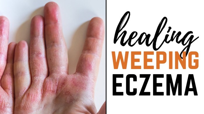
Detailed Illustration of Weeping Eczema Lesions
Weeping eczema lesions are characterized by areas of erythema (redness) and weeping or oozing fluid. The fluid may be clear, yellowish, or contain crusts. The skin often appears inflamed, with a moist and sometimes raw surface. The lesions can vary in size and shape, often appearing in clusters. The affected skin may feel hot to the touch, and itching is a common symptom.
In severe cases, the lesions can be widespread, affecting large areas of the body. The weeping can lead to skin cracking and discomfort.
Visual Representation of Different Stages of Weeping Eczema
Visual representation of weeping eczema stages can illustrate the progressive nature of the condition. Early stages often manifest as small, red papules or plaques, with minimal oozing. As the condition progresses, the lesions enlarge and become more inflamed, exhibiting increased weeping and crusting. The lesions can vary in severity, with some exhibiting only minor weeping, while others have extensive and persistent weeping.
Severe cases can show deep skin fissures and significant tissue damage. Understanding these stages is critical for appropriate diagnosis and tailored treatment.
Prevention Strategies
Weeping eczema, while frustrating, is manageable with proactive strategies. Prevention focuses on identifying and avoiding triggers, establishing a consistent routine, and understanding your body’s unique responses. This allows for better control over flare-ups and minimizes the discomfort associated with weeping eczema.Effective eczema prevention isn’t about a one-size-fits-all approach. It’s about tailoring a personalized strategy to your specific triggers and lifestyle.
By understanding your eczema’s quirks, you can create a plan that works for you, not against you.
Trigger Avoidance Strategies
Identifying and eliminating triggers is paramount in preventing weeping eczema flare-ups. Common triggers include certain fabrics, harsh chemicals, and environmental factors. A detailed log of your eczema episodes can be invaluable in pinpointing patterns and specific irritants.
- Environmental Triggers: Extreme temperatures (both hot and cold), humidity, and even certain weather conditions can aggravate eczema. Understanding your body’s reactions to these elements can help you adapt your environment to minimize flare-ups.
- Allergens: Substances like pollen, dust mites, pet dander, and certain foods can trigger allergic reactions that manifest as eczema. Avoiding contact with these allergens is crucial.
- Irritants: Harsh soaps, detergents, and certain fabrics (like wool) can irritate the skin and exacerbate eczema. Switching to gentler alternatives and choosing soft fabrics can make a significant difference.
- Stress: While not a direct cause, stress can worsen eczema symptoms. Incorporating stress-reducing activities like yoga, meditation, or spending time in nature can be beneficial.
Personalized Eczema Management Plan
A personalized eczema management plan is crucial for long-term control. This plan should be adaptable and flexible to accommodate your daily life and specific needs. It’s not just about avoiding triggers, but about creating a skin-friendly routine.
- Skincare Routine: A gentle, fragrance-free skincare routine, including moisturizing creams and barrier repair products, is essential. Regular application of emollients, particularly after bathing, helps maintain skin hydration and prevents dryness, a major contributor to eczema flare-ups.
- Trigger Log: Maintain a detailed log of potential triggers, including foods, environmental factors, and activities. This helps identify patterns and allows you to make adjustments to your lifestyle accordingly.
- Professional Guidance: Consulting a dermatologist or healthcare professional provides valuable insights into your specific needs. They can offer personalized recommendations, including specific treatments and tailored avoidance strategies.
- Stress Management: Chronic stress can significantly worsen eczema symptoms. Incorporate stress-reducing activities, such as exercise, meditation, or spending time in nature, into your daily routine.
Preventative Strategies for Daily Routines
Implementing preventative strategies into your daily routines can significantly reduce the frequency and severity of weeping eczema flare-ups. These strategies should become an integral part of your daily life, not just a temporary fix.
- Gentle Washing: Use lukewarm water and fragrance-free, hypoallergenic soaps or cleansers. Avoid harsh scrubbing or excessive washing.
- Moisturizing: Apply a moisturizer, especially after showering or bathing, to maintain skin hydration. This creates a protective barrier that helps prevent moisture loss and irritation.
- Avoiding Irritants: Be mindful of clothing materials, detergents, and other products that may irritate your skin. Opt for natural fabrics and hypoallergenic cleaning products.
- Stress Reduction Techniques: Incorporate relaxation techniques like meditation, deep breathing exercises, or spending time in nature into your daily routine. Stress can significantly worsen eczema symptoms.
Additional Resources

Staying informed and connected with the right resources is crucial for managing weeping eczema effectively. This section provides valuable avenues for further learning and support, empowering you to navigate your eczema journey with confidence. Understanding the available resources can help you access comprehensive information and expert advice, promoting better self-management and overall well-being.
Reputable Websites and Organizations
Numerous websites and organizations offer in-depth information on eczema, including weeping eczema. These platforms often provide evidence-based content, support groups, and educational materials to help individuals understand and manage their condition.
- The National Eczema Association (NEA): A leading organization dedicated to eczema research, education, and support. They offer comprehensive resources, including articles, webinars, and information on various eczema types, including weeping eczema.
- The American Academy of Dermatology (AAD): A reputable source of information on skin conditions, including eczema. Their website provides up-to-date information on diagnosis, treatment, and management strategies for various forms of eczema.
- Mayo Clinic: Known for its extensive medical information, the Mayo Clinic website offers reliable information on eczema, including causes, symptoms, and treatment options. They also provide guidance on managing chronic skin conditions.
- Cleveland Clinic: Another trusted source for health information, the Cleveland Clinic website provides detailed insights into eczema, including different types, potential triggers, and treatment approaches. They offer specific information on managing skin conditions effectively.
Healthcare Professionals
Consulting with healthcare professionals specializing in eczema management is essential for tailored care and effective treatment plans. Finding a dermatologist or allergist familiar with eczema can significantly improve your experience and outcomes.
- Dermatologists: These physicians are specifically trained in diagnosing and treating skin conditions like eczema. They can provide accurate diagnoses, recommend appropriate treatments, and monitor your condition over time.
- Allergists: Allergists can identify and manage potential triggers for eczema, helping you avoid specific environmental factors that exacerbate your condition. They can also provide guidance on desensitization therapies if needed.
- Primary Care Physicians: Your primary care physician can offer initial guidance, make referrals to specialists, and manage your overall health, including your eczema. They are a valuable first point of contact for many individuals.
Books, Articles, and Other Resources
Numerous books, articles, and other resources offer practical advice and insights into managing weeping eczema. Seeking out these resources can enhance your understanding of the condition and its management.
- The Eczema Solution by Dr. [Author Name]: This book might offer insights into dietary considerations, lifestyle modifications, and treatment approaches. Verify the author’s credentials to ensure reliability.
- Eczema and Skin Conditions by [Author Name]: Articles and resources available online can offer a wide range of information and perspectives on various skin conditions, including eczema.
- Support groups and online forums: Connecting with others who experience weeping eczema can provide valuable emotional support, shared experiences, and practical tips for managing the condition.
Table of Reliable Eczema Information Sources
This table provides quick access to reputable sources for further research and information.
| Source | Website |
|---|---|
| National Eczema Association | [Link to NEA website] |
| American Academy of Dermatology | [Link to AAD website] |
| Mayo Clinic | [Link to Mayo Clinic website] |
| Cleveland Clinic | [Link to Cleveland Clinic website] |
Last Word
In conclusion, weeping eczema, while often frustrating, is manageable with the right knowledge and approach. By understanding its causes, symptoms, and treatment options, individuals can take proactive steps toward controlling their symptoms and improving their overall well-being. This overview provides a roadmap for navigating this skin condition, empowering readers to take charge of their health and lead fulfilling lives.