Tag: rehabilitation

  • ACL Tears After Surgery A Comprehensive Guide

    ACL Tears After Surgery A Comprehensive Guide

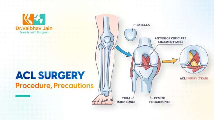
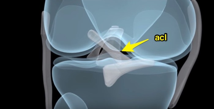
    ACL tears after surgery present a unique challenge, requiring careful consideration of the recovery process. This guide delves into the complexities of ACL reconstruction, from surgical techniques to potential complications and the crucial role of post-surgical rehabilitation. Understanding the nuances of this journey is key to successful outcomes.

    Anterior cruciate ligament (ACL) tears are a common injury, particularly in athletes. Surgical repair, often involving ACL reconstruction, aims to restore stability and function. Common procedures include using hamstring or patellar tendon grafts. This detailed look at ACL tears after surgery provides a clear picture of the entire process, from initial surgery to long-term outcomes.

    Introduction to ACL Tears After Surgery

    Acl tears after surgery

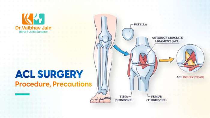
    An anterior cruciate ligament (ACL) tear is a common injury, particularly among athletes. ACL tears often require surgical repair, usually involving reconstruction. This surgical process aims to restore stability to the knee joint, allowing for a return to normal activities. The recovery process after surgery is crucial for successful rehabilitation and long-term joint health.Surgical reconstruction aims to replace the torn ligament with a graft, typically taken from another part of the body, like the hamstring or patellar tendon.

    The choice of graft depends on individual factors and the surgeon’s preference. Post-operative rehabilitation plays a significant role in regaining strength, flexibility, and range of motion.

    Surgical Procedures for ACL Reconstruction

    Various surgical techniques are employed in ACL reconstruction. These procedures involve replacing the torn ACL with a substitute ligament, often from a patient’s own body tissue. The primary goal is to restore the knee’s stability and functionality.

    Common Graft Types

    The choice of graft material significantly impacts the recovery process. Understanding the different graft types is crucial for patients to make informed decisions about their treatment.

    Graft Type Source Advantages Disadvantages
    Hamstring Grafts Hamstring tendons Generally easier to harvest, less invasive Potential for hamstring weakness if not carefully handled
    Patellar Tendon Grafts Patellar tendon Strong graft, often associated with faster recovery Potential for patellar tendonitis or pain in the patella region
    Quadriceps Tendon Grafts Quadriceps tendon Excellent strength, potentially faster recovery More invasive harvesting, potential for quadriceps weakness

    Typical Recovery Timeline

    The recovery timeline following ACL reconstruction surgery is variable, depending on several factors. These include the patient’s age, activity level, and adherence to the rehabilitation protocol. A realistic understanding of the recovery process is essential for patient management.A typical recovery timeline might involve several phases:

    • Initial Phase (Weeks 1-4): Focus on pain management, controlled range of motion exercises, and early mobilization. Patients will likely use crutches or other assistive devices during this phase. Examples include controlled knee flexion and extension exercises, as well as light range of motion exercises. This phase prioritizes minimizing swelling and protecting the healing graft.
    • Intermediate Phase (Weeks 4-12): Gradually increase the intensity and range of motion exercises. Begin strengthening exercises for the surrounding muscles. The goal is to restore the knee’s stability and functionality. Examples include progressively increasing resistance during strengthening exercises and the introduction of sport-specific exercises.
    • Advanced Phase (Weeks 12-24+): Return to normal activities, including sports, is a gradual process. Continued strengthening and proprioception exercises are vital for preventing re-injury. This phase may include more complex agility and balance training. Examples include progressing from simple to more complex movements, such as running, jumping, and agility drills, while gradually increasing intensity and duration.

    Potential Complications: Acl Tears After Surgery

    While ACL reconstruction surgery is a highly effective procedure, potential complications can arise. These complications range from relatively minor issues to more serious problems that may impact long-term recovery and function. Understanding the potential complications and their likelihood is crucial for both patients and surgeons to make informed decisions. Careful planning and meticulous surgical technique can minimize the risk of these complications.Post-operative complications are a multifaceted concern, influenced by individual patient factors, surgical technique, and post-operative care.

    The likelihood of specific complications varies, and factors like age, overall health, and adherence to post-operative instructions can significantly impact the outcome.

    Recovering from an ACL tear after surgery is tough, but fueling your body with the right foods is key to healing. Focus on nutrient-rich options like lean protein and complex carbohydrates to support your body’s repair process. Eating a balanced diet, like those found in foods that give you energy , will give you the energy you need for physical therapy and daily activities.

    This will ultimately help you regain strength and get back to your active lifestyle faster. A good diet is essential for successful ACL recovery.

    Infection

    Infection is a serious complication following any surgical procedure, and ACL reconstruction is no exception. Bacterial infections can lead to significant tissue damage, potentially requiring additional surgeries and prolonged recovery periods.

    • Causes of infection often stem from contamination during surgery, poor wound care, or underlying health conditions that compromise the immune system. Surgical site infections (SSIs) can result from inadequate sterilization procedures or the introduction of bacteria during the procedure.
    • The likelihood of infection is generally low, but factors like the presence of pre-existing infections, or a compromised immune system increase the risk. This is why thorough pre-operative assessments and meticulous surgical technique are essential.

    Stiffness

    Post-operative stiffness is a common concern, potentially limiting the range of motion in the knee joint. This can result from various factors, including scar tissue formation, inflammation, or inadequate rehabilitation.

    • Early and aggressive physiotherapy plays a vital role in minimizing stiffness. Consistent exercises and gentle stretching routines, guided by a physical therapist, help maintain joint flexibility and prevent the development of adhesions and contractures.
    • In some cases, stiffness can persist despite diligent rehabilitation. In such situations, specific treatment modalities like joint injections or manipulation might be considered to improve mobility.

    Delayed Healing

    Delayed healing encompasses a spectrum of issues, from slow wound closure to persistent pain and delayed return to activity. Several factors can contribute to delayed healing.

    • Factors such as poor blood supply to the surgical area, inadequate nutrition, or pre-existing medical conditions can hinder the body’s natural healing processes.
    • The healing process varies significantly from person to person. Some individuals heal faster than others, while others may experience prolonged recovery times.

    Failure of Graft

    Failure of the graft refers to the ACL graft not properly integrating with the surrounding tissues or not functioning as intended, which can lead to knee instability. This is a more serious complication.

    • Factors such as improper graft selection, poor surgical technique, or patient factors (e.g., high activity levels) can potentially contribute to graft failure. Understanding patient activity level and expectations is crucial in determining the right surgical approach.
    • The likelihood of graft failure is lower when the surgical procedure is performed correctly, and the patient actively participates in the rehabilitation process.

    Other Complications, Acl tears after surgery

    Other potential complications include: nerve damage, blood clots, and loosening of the hardware used in the procedure.

    Complication Signs and Symptoms
    Nerve Damage Numbness, tingling, or pain along the nerve pathway.
    Blood Clots Swelling, pain, warmth, or redness in the affected limb.
    Hardware Loosening Pain, clicking, or catching sensations in the knee.

    Post-Surgical Rehabilitation

    Recovering from ACL reconstruction surgery requires a structured and diligent approach to rehabilitation. This phase isn’t just about regaining range of motion; it’s about building strength, restoring stability, and ultimately, returning to your pre-injury activity level. The process is progressive, requiring careful monitoring and adherence to a personalized plan. Success depends heavily on patient commitment and the guidance of a qualified physical therapist.The post-surgical rehabilitation program is designed to address the specific needs of each patient.

    It carefully progresses through distinct phases, gradually increasing the intensity and complexity of exercises. Understanding the different phases and their associated exercises is crucial for successful recovery. This knowledge empowers patients to actively participate in their rehabilitation journey and accelerate their return to normal activities.

    Phases of Post-Surgical Rehabilitation

    The rehabilitation process following ACL reconstruction is typically divided into several distinct phases. Each phase builds upon the previous one, gradually increasing the demands placed on the knee and surrounding tissues. A crucial aspect of these phases is the gradual introduction of controlled stress to facilitate healing and prevent re-injury.

    Phase 1: Early Recovery (First 4-6 Weeks)

    This initial phase focuses on protecting the healing graft and minimizing swelling. The primary goals are pain management, regaining a full range of motion, and beginning the process of regaining strength and stability. Activities are carefully monitored to ensure the healing tissues are not stressed beyond their capacity.

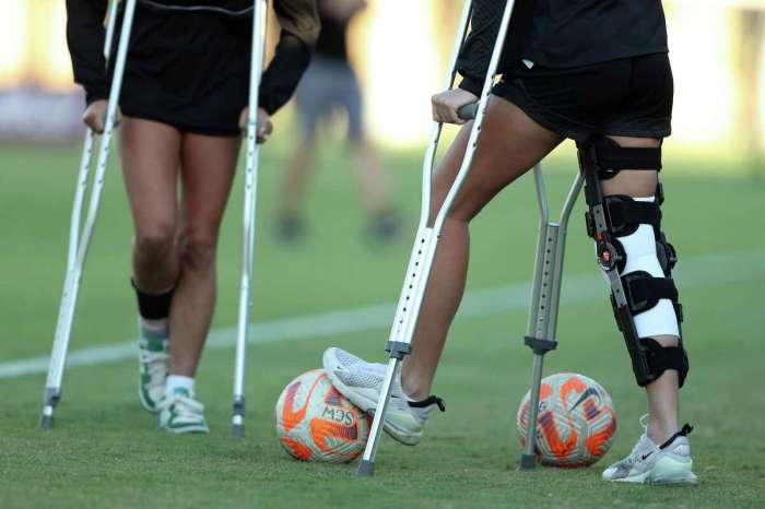
    • Protecting the healing graft: Activities are designed to prevent excessive stress on the repaired ligament. This often involves using crutches, knee braces, and limited weight-bearing. Emphasis is placed on avoiding activities that could jeopardize the healing process.
    • Range of motion exercises: Gentle exercises are performed to maintain the range of motion of the knee joint. These exercises include passive range of motion (assisted by a therapist or another person) and active assisted range of motion (with some assistance from the patient). Simple exercises like knee flexion and extension are typically performed.
    • Gentle strengthening exercises: Initial strengthening exercises are focused on the quadriceps muscle, targeting the front of the thigh. These exercises may include isometric quad sets (squeezing the quadriceps muscle without moving the knee) and gentle hamstring exercises.

    Phase 2: Strengthening and Proprioception (6-12 Weeks)

    This phase gradually increases the intensity of exercises, focusing on improving muscle strength, balance, and proprioception (the body’s awareness of its position in space). The focus shifts towards controlled movements that gradually challenge the healing tissue while also promoting stability and coordination.

    • Progressive strengthening exercises: Exercises are progressively loaded to enhance quadriceps and hamstring strength. These include exercises like straight leg raises, and more advanced hamstring exercises, ensuring the exercises don’t compromise the healing process.
    • Proprioceptive exercises: These exercises help improve the knee’s ability to sense its position and movement. Examples include balance exercises on unstable surfaces, such as wobble boards or foam pads, and exercises that challenge the knee’s coordination.
    • Cardiovascular exercises: Low-impact cardio, such as stationary cycling and swimming, are introduced to gradually improve cardiovascular fitness.

    Phase 3: Functional Training (12-24 Weeks)

    This phase prepares the patient for a return to sport or other activities. The focus is on functional movements that mimic activities of daily living and sports-specific movements. The aim is to fully integrate the knee into normal movement patterns.

    • Functional exercises: Exercises are designed to mimic activities like stair climbing, squatting, and running. These exercises are progressively introduced to ensure the knee can handle the stresses associated with these movements.
    • Sport-specific training: If applicable, specific exercises for the patient’s sport are introduced. This may involve agility drills, plyometrics (exercises that involve jumping and explosive movements), and other sport-specific activities, all under careful supervision.
    • Return to sport progression: A gradual return to sports activities is carefully monitored. This often involves a progression of drills and activities that gradually increase the demands on the knee joint.

    Progression of Exercises Across Rehabilitation Phases

    Phase Exercises
    Phase 1 Isometric quad sets, passive range of motion, gentle hamstring exercises
    Phase 2 Straight leg raises, hamstring curls, balance exercises, low-impact cardio
    Phase 3 Functional exercises, sport-specific training, plyometrics, return to sport progression

    Importance of Adherence to the Rehabilitation Program

    Adherence to the rehabilitation program is paramount for successful recovery and a full return to activity. A commitment to the prescribed exercises and activities is crucial for ensuring the healing process progresses as expected. A dedicated effort is necessary to maximize the benefits of the rehabilitation plan.

    Factors Affecting Recovery

    Acl tears after surgery

    The road to recovery after ACL surgery is a journey unique to each individual. While the surgical procedure itself is a significant step, the factors influencing the healing process are just as crucial. Understanding these variables allows for a more personalized approach to rehabilitation, maximizing the chances of a successful outcome.Factors like age, activity level, and pre-existing conditions play pivotal roles in determining the length and nature of the recovery period.

    Recovering from an ACL tear surgery can be a long haul, and while physical therapy is key, some unexpected symptoms can pop up. One common complaint is tinnitus, or ringing in the ears. Understanding the potential causes and treatments for this symptom is crucial for a smooth recovery. For a deeper dive into the causes, symptoms, and available treatments for tinnitus, check out this helpful resource on ringing in ears symptoms causes and treatment.

    Ultimately, addressing these unexpected symptoms alongside the standard physical therapy will help you get back on your feet faster after your ACL surgery.

    Furthermore, adherence to the rehabilitation plan and overall lifestyle choices greatly impact the final results.

    Age and Recovery

    Age significantly influences the body’s natural healing capacity. Younger individuals often experience faster tissue regeneration and quicker recovery times compared to older adults. This is due to the inherent differences in cellular turnover and tissue repair mechanisms. While age isn’t a barrier to successful recovery, it often necessitates a more cautious and potentially longer rehabilitation protocol. For instance, an older athlete might require a more gradual progression of exercises to avoid undue stress on the healing tissues.

    The emphasis shifts towards maintaining joint function and mobility while minimizing the risk of complications.

    Activity Level and Recovery

    An individual’s pre-injury activity level plays a critical role in the recovery process. Professional athletes, for example, will need a more intensive rehabilitation program to regain the high level of strength and endurance required for their sport. Their rehabilitation will likely involve more specialized exercises and a more rigorous schedule compared to someone with a less demanding lifestyle.

    The focus will be on not only regaining the lost function but also surpassing it to reach their previous performance level. Conversely, individuals with lower pre-injury activity levels will have a more manageable recovery trajectory, with a focus on building a solid foundation of strength and mobility before progressing to more complex movements.

    Pre-Existing Conditions and Recovery

    Pre-existing conditions can also influence the recovery process. Conditions like diabetes, obesity, or cardiovascular issues can affect the body’s overall healing response. These conditions may necessitate modifications to the rehabilitation program to address any potential complications or limitations. For instance, someone with diabetes might require more careful monitoring of blood sugar levels during the rehabilitation phase to prevent complications.

    The rehabilitation plan will be customized to accommodate the individual’s specific health needs and limitations.

    Optimizing Recovery for Specific Characteristics

    Individuals with specific characteristics can benefit from tailored rehabilitation strategies. For instance, athletes can incorporate sport-specific exercises into their rehabilitation plan to ensure they can return to their pre-injury performance levels. This personalized approach allows for a more efficient recovery and a quicker return to the activity they love. Similarly, individuals with pre-existing conditions can work with their physical therapists to adjust the program based on their individual needs and limitations.

    Different Rehabilitation Approaches

    Various rehabilitation approaches can be employed to address individual needs. A structured program combining physical therapy, strength training, and potentially bracing or supportive devices, might be beneficial. The rehabilitation plan should consider factors like the individual’s pain tolerance, activity level, and overall health status. For example, a progressive resistance program designed by a physical therapist can help build strength and improve joint stability, gradually increasing the intensity and complexity of exercises as the individual progresses.

    Furthermore, incorporating lifestyle modifications, such as nutrition and sleep, can further support the healing process and optimize the recovery journey.

    Long-Term Outcomes

    ACL reconstruction surgery aims to restore knee stability and function. However, the journey to long-term success isn’t always straightforward. Understanding the potential outcomes, both positive and challenging, is crucial for patients and their healthcare providers. Factors influencing the long-term results range from the surgical technique itself to the patient’s adherence to rehabilitation protocols.Successful ACL reconstruction, while common, isn’t a guaranteed outcome.

    Recovering from an ACL tear after surgery can be a long journey, and sometimes you might notice a lump under the skin. This is a common concern, and understanding the potential causes and when to seek medical attention is crucial. Knowing that a lump could be a normal part of the healing process or a sign of a complication is important.

    Checking out resources like lump under skin causes and when to worry can help you differentiate between the two. Ultimately, consistent communication with your doctor about any changes is key to a successful ACL recovery.

    Post-operative complications and individual factors can influence the long-term functionality of the reconstructed ligament. This section explores the key elements that contribute to successful long-term results and addresses potential challenges.

    Success Rates of ACL Reconstruction

    Success rates for ACL reconstruction vary depending on several factors, including the surgeon’s experience, the patient’s age, activity level, and adherence to rehabilitation. Studies have shown that the procedure can achieve high success rates, typically exceeding 80% in many cases. However, factors such as graft choice, surgical technique, and patient compliance play a significant role. These results are often measured by return to pre-injury activity levels and the absence of significant pain or instability.

    Factors Contributing to Successful Long-Term Outcomes

    Numerous factors influence the long-term success of ACL reconstruction. Patient compliance with rehabilitation protocols is paramount. A comprehensive rehabilitation program, including exercises to strengthen supporting muscles, improve range of motion, and regain balance, is crucial. Furthermore, a patient’s overall health, age, and activity level significantly impact recovery. Younger patients generally have better long-term outcomes compared to older patients.

    The choice of graft type also plays a role; different grafts (autografts, allografts) might exhibit varying degrees of integration and durability.

    Potential for Re-injury

    Re-injury after ACL reconstruction is a possibility, although not a certainty. Factors that increase the risk of re-injury include inadequate rehabilitation, a return to high-impact activities too soon, and a lack of proper conditioning. The severity and type of pre-injury activity and the individual’s inherent predisposition to injury also influence the likelihood of re-injury. For instance, athletes participating in contact sports often face a higher risk of re-injury than those involved in non-contact activities.

    Comparison of Surgical Techniques

    Different surgical techniques for ACL reconstruction exist, each with its own advantages and disadvantages. Arthroscopic techniques are commonly used and involve minimally invasive procedures. These procedures often lead to quicker recovery times and less scarring compared to open procedures. However, the long-term outcomes of various techniques can differ based on the specific details of the surgery and the patient’s individual response.

    Studies examining long-term outcomes of different techniques often consider factors such as graft survival, stability, and the ability to return to previous activity levels.

    Long-Term Functional Outcomes

    Long-term functional outcomes following ACL reconstruction vary. Some patients experience a complete return to pre-injury activity levels, while others may experience limitations in specific activities. The success of the surgery depends on several factors, including the severity of the initial injury, the patient’s compliance with rehabilitation, and the surgical technique employed. A patient’s age, activity level, and overall health are also critical factors influencing long-term functional outcomes.

    Patient Experiences and Perspectives

    The journey of recovering from an ACL tear and reconstruction is profoundly personal. Beyond the physical rehabilitation, patients face a complex emotional and psychological landscape. Understanding these experiences is crucial for providing comprehensive care and support. This section delves into the diverse perspectives and challenges encountered by individuals navigating this process.

    Patient Experiences: A Summary

    Patient experiences following ACL reconstruction are varied, influenced by factors such as pre-injury activity levels, personality, support systems, and the surgical technique. There are common threads, however, of both triumph and struggle. The following table summarizes some key experiences.

    Aspect Common Experience
    Physical Pain and Discomfort Patients commonly report varying degrees of pain and discomfort during the rehabilitation process, especially during the initial stages. The intensity and duration of this pain can differ significantly based on individual factors.
    Psychological Impact Anxiety, frustration, and depression are not uncommon, especially during periods of plateau in progress. Fear of re-injury or concerns about long-term functionality can contribute to emotional distress.
    Social Adjustment Changes in daily routines and social activities can lead to feelings of isolation or frustration. Adjustments to limitations and the recovery process can impact relationships with friends and family.
    Emotional Well-being Maintaining a positive attitude and managing emotional challenges are crucial aspects of the recovery process. Patients may experience emotional ups and downs, but support systems can significantly influence their journey.
    Motivation and Perseverance Maintaining motivation throughout the recovery period is a significant challenge. Progress can be slow, and setbacks are common. Patients need to understand the importance of consistent effort and perseverance.

    Successful Recovery Stories

    Numerous patients have achieved successful recoveries following ACL reconstruction. These stories often highlight the importance of a dedicated rehabilitation program, strong support networks, and a positive attitude. One example is a high school athlete who, through diligent physical therapy and a supportive family, regained full range of motion and athletic abilities. They were able to return to their sport at a high level and continue playing for many years.

    Less Successful Recovery Stories

    Unfortunately, some patients experience less successful recoveries. Factors like inadequate adherence to rehabilitation protocols, pre-existing conditions, or psychological challenges can hinder progress. One example involves a patient who struggled with motivation and adherence to the prescribed rehabilitation exercises. This led to delayed recovery, potential complications, and a prolonged recovery time. This emphasizes the importance of patient engagement and consistent follow-up.

    Psychological Impact of Injury and Surgery

    ACL injuries and subsequent surgeries can significantly impact a patient’s psychological well-being. The fear of re-injury, concerns about future athletic participation, and the physical and emotional demands of rehabilitation can lead to anxiety, depression, and feelings of isolation. Addressing these psychological aspects is crucial for optimal recovery. For example, cognitive behavioral therapy (CBT) can help patients manage their thoughts and feelings related to the injury.

    Emotional Journey During Recovery

    The emotional journey of a patient recovering from an ACL injury and surgery is multifaceted and often unpredictable. Patients may experience a range of emotions, from initial shock and disappointment to frustration and anger during setbacks, and finally, joy and satisfaction as they progress toward recovery. Acknowledging and addressing these emotions is essential for a smoother recovery process.

    It’s important for patients to allow themselves to feel the full spectrum of emotions and seek support when needed.

    Surgical Techniques & Procedures

    Choosing the right surgical technique for ACL reconstruction is crucial for a successful recovery. Different methods cater to individual patient needs and preferences, while also considering the surgeon’s expertise and the specific characteristics of the injury. Understanding these options empowers patients to make informed decisions with their healthcare providers.

    Different Surgical Techniques

    Various surgical techniques exist for ACL reconstruction, each with its own set of advantages and disadvantages. These techniques primarily differ in the approach to graft harvesting and placement. Open procedures, while once common, are now less frequently used due to the greater precision and reduced invasiveness offered by arthroscopic techniques. Arthroscopic surgery uses small incisions and specialized instruments, allowing for a less disruptive procedure.

    Arthroscopic ACL Reconstruction

    Arthroscopic ACL reconstruction is the most common surgical technique. It involves minimally invasive incisions, typically 2-3 small portals. Surgeons use specialized instruments to visualize and repair the damaged ligament. This approach generally results in less pain, quicker recovery, and smaller scars. It is also less disruptive to surrounding tissues.

    Graft Types and Their Advantages/Disadvantages

    The choice of graft material significantly influences the recovery process and long-term outcomes. The most common grafts are derived from the patient’s own body tissues, including hamstring tendons and patellar tendons. Other options include allograft tissues (from a donor).

    Comparison of Graft Types

    Graft Type Source Advantages Disadvantages
    Hamstring Tendon Patient’s hamstring muscle Generally quicker recovery, less risk of patellar tendonitis, lower risk of anterior knee pain. Potential for hamstring weakness, slightly higher risk of graft rupture in the first year.
    Patellar Tendon Patient’s patellar tendon Stronger graft, potentially better long-term stability, less risk of hamstring weakness. Potential for anterior knee pain, potential for patellar tendonitis, slightly longer recovery time.
    Allograft Donor tissue May be useful in cases where autograft is not ideal. Risk of rejection, potential for disease transmission if not properly screened and treated, more expensive.

    Impact on Recovery

    The specific surgical technique and the chosen graft type directly impact the recovery period. For example, arthroscopic surgery with a hamstring tendon graft might lead to a faster return to activities compared to an open procedure with a patellar tendon graft. Individual patient factors like age, activity level, and overall health play a crucial role in determining the best approach.

    This individualized approach ensures the optimal balance between speed of recovery and long-term stability.

    Factors Influencing Surgical Technique Selection

    Several factors influence the surgeon’s decision regarding the surgical technique and graft choice. These factors include the severity of the tear, the patient’s age, activity level, and overall health. The surgeon’s experience and preferences also play a significant role. Open procedures are less common, but still an option for complex injuries. The primary goal is to select the procedure that provides the best chance for a successful and complete recovery.

    Monitoring and Evaluation

    Post-operative monitoring and evaluation are crucial for ensuring successful ACL reconstruction and minimizing complications. These procedures help track the healing process, identify potential issues early, and adjust the rehabilitation plan as needed. Regular assessments allow healthcare providers to gauge the patient’s progress, ensuring they’re on track to achieve optimal outcomes.

    Methods for Monitoring Healing

    Regular check-ups and physical assessments are fundamental in monitoring the healing process after ACL reconstruction. These assessments help evaluate the extent of tissue integration, joint stability, and overall function. A combination of clinical examinations, imaging studies, and functional tests provide a comprehensive picture of the patient’s progress. These methods enable healthcare professionals to adapt the rehabilitation program based on the patient’s individual needs and response to treatment.

    Importance of Regular Check-ups

    Regular check-ups are essential to assess the healing process and identify potential problems early. They provide a platform to evaluate the patient’s progress and ensure adherence to the prescribed rehabilitation plan. Prompt detection of complications, such as infection or persistent swelling, can lead to timely interventions, minimizing the risk of long-term complications. Early intervention is critical to maximizing the chances of a successful recovery.

    Diagnostic Tools for Monitoring Progress

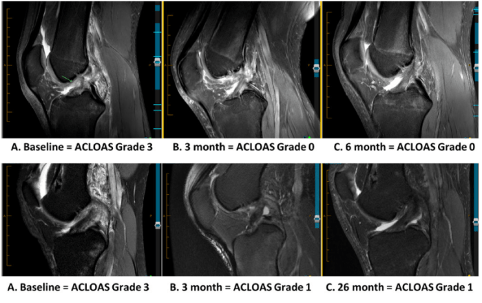
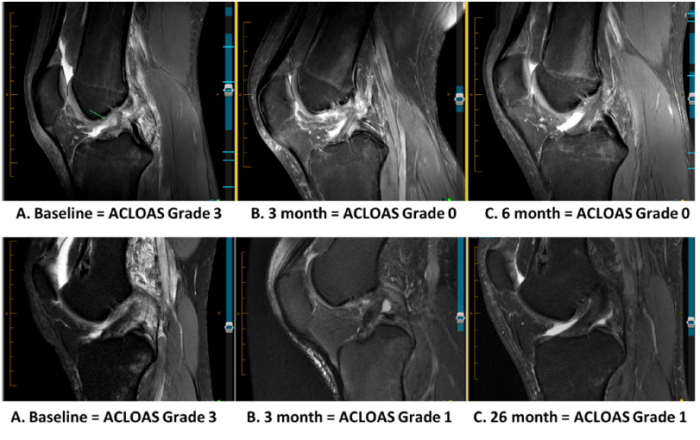
    Various diagnostic tools are employed to monitor the healing process. These tools include physical examinations, which assess range of motion, strength, and stability of the knee joint. Radiographic imaging, such as X-rays and MRI scans, provides detailed images of the bone and soft tissues, allowing visualization of the graft integration and potential complications. Functional tests, like the Lachman test and anterior drawer test, are used to evaluate the stability of the ACL reconstruction.

    These tests are vital in assessing the effectiveness of the surgery and the patient’s ability to perform daily activities.

    Post-Operative Imaging Techniques

    A crucial aspect of monitoring the healing process is employing appropriate imaging techniques. These techniques provide valuable insights into the status of the reconstructed ACL, the surrounding tissues, and the overall healing process. Early detection of complications or delayed healing is possible through these methods. Accurate imaging guides decisions about rehabilitation progression and surgical interventions if needed.

    Imaging Technique Description Use in ACL Monitoring
    X-ray Two-dimensional radiographic image of bones. Evaluates bone alignment, assesses for fracture, or signs of loosening.
    MRI (Magnetic Resonance Imaging) Three-dimensional detailed image of soft tissues. Visualizes the ACL graft, surrounding ligaments, cartilage, and tendons. Identifies any signs of inflammation, swelling, or graft tears.
    CT (Computed Tomography) Three-dimensional image of the bones and soft tissues. Provides detailed anatomical information about bone structures and can evaluate the graft integration with the bone.
    US (Ultrasound) Real-time image of soft tissues using sound waves. Assess for soft tissue swelling, inflammation, and evaluate the healing process in the early stages post-surgery.

    Addressing Specific Concerns

    Navigating the journey after ACL reconstruction surgery can be filled with anxieties and uncertainties. This section focuses on common concerns and provides practical strategies to address them, helping you feel more empowered and prepared for the road ahead. From pain management to returning to your favorite activities, we’ll explore actionable solutions.

    Pain Management Strategies

    Managing post-operative pain is crucial for a smooth recovery. A multi-faceted approach, incorporating medication, physical therapy techniques, and lifestyle adjustments, is essential.

    • Medication adherence is vital. Follow your physician’s instructions precisely regarding dosage and timing of pain relievers. Over-the-counter options like ibuprofen can help manage mild to moderate discomfort. Prescription medication, such as opioids, may be necessary for more severe pain, but use these medications only as directed by your physician and be aware of potential side effects.

      Discuss alternative pain management options with your healthcare team if needed.

    • Ice packs and compression are beneficial for reducing swelling and pain. Applying ice for 15-20 minutes at a time, several times a day, can help minimize discomfort. Proper compression helps to control swelling and support the healing tissues.
    • Physical therapy techniques like gentle range-of-motion exercises and manual therapy can aid in pain management. These methods, guided by a qualified physical therapist, promote healing and reduce stiffness.

    Return to Sports

    Returning to sports after ACL reconstruction is a significant goal for many patients. A gradual and well-structured approach, emphasizing functional strength and stability, is key to successful return.

    • A phased return-to-sport protocol, designed by your physical therapist, is critical. This program gradually increases the intensity and complexity of activities, ensuring that the knee is adequately prepared for the demands of the sport. Each phase should be completed before progressing to the next, to avoid potential setbacks or reinjury.
    • Strength training is essential to improve knee stability and muscle strength around the joint. Exercises should be tailored to your specific needs and progress gradually. Focus on building strength in the quadriceps, hamstrings, and surrounding muscles to support the knee joint. Progressive resistance exercises are essential to ensure that you do not overload the tissues.
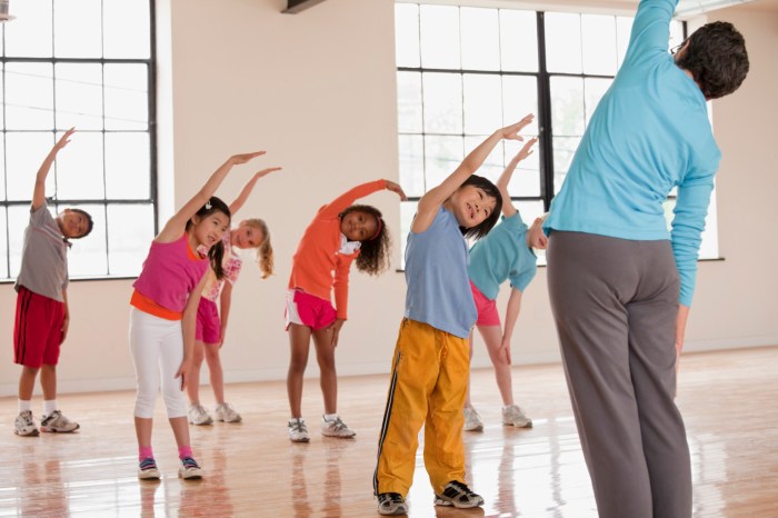
    • Proper warm-up and cool-down routines are essential before and after physical activity. These routines prepare the muscles and tissues for activity and help prevent injury.

    Potential Limitations

    ACL reconstruction surgery does not guarantee a complete return to your pre-injury activity level. Patients need to acknowledge potential limitations and adjust their expectations accordingly.

    • Some activities might require modifications or adjustments. For instance, high-impact activities might need to be avoided or modified to reduce stress on the reconstructed ligament.
    • Gradual return to activities is essential. Jumping, pivoting, and other demanding movements should be reintroduced gradually to minimize the risk of re-injury. Listen to your body, and adjust your activities accordingly.
    • Long-term monitoring and follow-up care are crucial. Regular check-ups with your orthopedic surgeon are essential to ensure that the healing process is progressing as expected and to address any potential complications.

    Common Patient Concerns & Responses

    Common Question Appropriate Response
    How long will it take to fully recover? Recovery time varies depending on the individual, but a typical timeline is several months. Factors like age, activity level, and adherence to the rehabilitation program influence recovery.
    Will I be able to return to my previous activity level? While a full return is often possible, it’s essential to understand that there might be limitations. A phased return-to-sport program, designed in consultation with your healthcare team, is crucial.
    How can I manage the pain effectively? Pain management strategies often involve a combination of medication, ice packs, compression, physical therapy, and lifestyle adjustments. Always follow your physician’s instructions.

    Addressing Rehabilitation Challenges

    Specific challenges in rehabilitation can be addressed through individualized strategies.

    • Motivation and adherence to the rehabilitation program are crucial. Setting realistic goals, celebrating milestones, and maintaining a positive attitude can enhance motivation. Consider seeking support from family and friends or support groups.
    • Managing pain and swelling is critical during rehabilitation. Proper techniques, like icing, compression, and elevation, can help manage discomfort and facilitate the healing process. Discuss pain management options with your healthcare team.
    • Overcoming psychological barriers like fear of re-injury or anxiety about returning to sports can be addressed with a supportive environment and open communication with your healthcare team.

    End of Discussion

    In conclusion, navigating the path of ACL tears after surgery demands a comprehensive approach, encompassing surgical techniques, potential complications, and personalized rehabilitation plans. Factors like age, activity level, and pre-existing conditions significantly impact recovery. A strong support system, adherence to the rehabilitation program, and open communication with healthcare professionals are vital for achieving optimal long-term outcomes. This guide aims to equip you with the knowledge and insights necessary for a successful recovery.

  • Physical Therapy After Proximal Humeral Fracture A Guide

    Physical Therapy After Proximal Humeral Fracture A Guide

    Physical therapy after a proximal humeral fracture is crucial for regaining strength, mobility, and function in the shoulder. This comprehensive guide delves into the evaluation, rehabilitation phases, and considerations for various patient populations, ensuring a smooth recovery journey. We’ll cover everything from initial assessments to advanced exercises, addressing common complications and outlining successful return-to-activity strategies.

    The process involves a multi-stage approach tailored to individual needs. Early rehabilitation focuses on regaining range of motion and pain management, while intermediate phases progressively increase strength and stability. Ultimately, the goal is a safe and effective return to daily activities, with a focus on injury prevention.

    Introduction to Proximal Humeral Fracture

    A proximal humeral fracture is a break in the upper part of the humerus bone, the long bone extending from the shoulder to the elbow. This type of fracture can occur at various points along the proximal humerus, leading to significant pain and functional limitations. Understanding the causes, types, and symptoms is crucial for effective diagnosis and treatment.Proximal humeral fractures are often the result of significant trauma, such as a fall or a high-impact collision.

    Recovering from a proximal humeral fracture means a dedicated physical therapy regimen. It’s about more than just strengthening muscles; it’s about regaining full range of motion. While working on exercises, it’s fascinating to consider unusual ways to burn calories, like dancing or even playing a sport like badminton. This can help maintain your overall fitness during the physical therapy process, and ultimately speed up the recovery process from the proximal humeral fracture.

    Unusual ways to burn calories can definitely add a fun element to your healing journey! I’m finding that a variety of activities, not just traditional exercises, can be helpful.

    However, they can also occur in individuals with underlying osteoporosis or other conditions that weaken the bone structure. Factors such as age, activity level, and the presence of pre-existing medical conditions influence the likelihood of such a fracture.

    Types of Proximal Humeral Fractures

    The proximal humerus has a complex anatomical structure, and fractures can involve various parts of the bone. Anatomical classifications help surgeons and physical therapists to better understand the injury and develop an appropriate treatment plan. These classifications typically categorize fractures based on the specific anatomical location of the break and the associated bone fragments.

    Common Causes of Proximal Humeral Fractures

    Falls, especially from a height, are the most common cause of proximal humeral fractures. High-impact trauma, such as a motor vehicle accident, can also result in these fractures. In some cases, fractures may be a consequence of a direct blow to the shoulder. Additionally, underlying osteoporosis or other bone-weakening conditions can make individuals more susceptible to this type of fracture, even with relatively minor trauma.

    Symptoms of Proximal Humeral Fractures

    The symptoms of a proximal humeral fracture often include significant pain and tenderness in the shoulder region, particularly around the greater tuberosity and the surgical neck. The patient may experience pain when attempting to move the shoulder, or even with simple movements like raising their arm. There may also be visible swelling and bruising around the shoulder joint.

    In some cases, the shoulder may appear deformed or out of alignment.

    Comparison of Different Fracture Types

    Type Cause Symptoms Common Treatment
    Surgical Neck Fracture Falls, direct trauma, or sports injuries. Pain, swelling, bruising around the shoulder, limited range of motion, and potential deformity. Nonsurgical treatment with immobilization, physical therapy, and pain management are common for less severe cases. Surgical intervention, such as pinning or plating, may be necessary for displaced fractures.
    Greater Tuberosity Fracture Direct trauma, falls, or sports injuries. Pain, swelling, tenderness at the top of the shoulder, and potential difficulty in lifting the arm. Nonsurgical management, such as immobilization and pain relief, may be sufficient for some patients. Surgical repair may be necessary if the fracture is unstable or displaced.
    Lesser Tuberosity Fracture Direct trauma, falls, or sports injuries. Pain, swelling, tenderness at the front of the shoulder. The pain may radiate to the elbow or arm. Nonsurgical management is often attempted, but surgery may be necessary for displaced fractures to restore proper shoulder function.
    Humeral Shaft Fracture High-energy trauma, such as motor vehicle accidents or falls from significant heights. Severe pain, swelling, bruising, and significant loss of shoulder function. Surgical intervention, including plating or intramedullary nailing, is typically necessary for stability and proper healing.

    Physical Therapy Evaluation Following Fracture

    Navigating the recovery journey after a proximal humeral fracture requires a meticulous approach, especially in the initial physical therapy evaluation. This phase is crucial for establishing a baseline understanding of the patient’s condition and tailoring a treatment plan that promotes optimal healing and restoration of function. A thorough evaluation helps predict the patient’s progress and potential complications.

    Initial Physical Therapy Evaluation Process

    The initial evaluation involves a systematic assessment of the patient’s physical abilities, pain levels, and overall well-being. This process is designed to gather comprehensive data to guide the physical therapist in developing an individualized treatment plan.

    Assessment of Range of Motion

    Assessing range of motion (ROM) is paramount in evaluating the affected shoulder joint. This involves measuring the extent of movement in all directions, including flexion, extension, abduction, adduction, internal and external rotation. This evaluation helps determine the limitations imposed by the fracture and any potential adhesions or soft tissue restrictions.

    • Active range of motion (AROM): The patient performs the movements independently, indicating their current level of muscle control and joint mobility.
    • Passive range of motion (PROM): The therapist guides the patient’s limb through the available range of motion, revealing limitations that may be due to pain, muscle tightness, or joint stiffness.
    • Resisted range of motion (PROM): Resistance is applied during movement to assess the strength of the surrounding muscles and their ability to control the joint.

    Assessment of Strength

    Evaluating muscle strength is essential to determine the functional capacity of the shoulder musculature. This assessment is critical for determining the patient’s ability to perform activities of daily living and return to their previous level of activity.

    • Manual Muscle Testing (MMT): A standardized method for grading muscle strength by assessing the patient’s ability to resist the therapist’s force against specific movements. Each muscle group is assessed individually.
    • Isometric Exercises: The patient contracts the muscles against resistance without any visible movement, offering a measure of the muscle’s static strength.

    Assessment of Pain Levels

    Pain management is a crucial component of the rehabilitation process. Understanding the patient’s pain levels and their response to different modalities is vital for creating an effective pain management strategy.

    • Visual Analog Scale (VAS): A simple scale that allows the patient to rate their pain level on a scale of 0 to 10, with 0 representing no pain and 10 representing the worst pain imaginable.
    • Numeric Rating Scale (NRS): A numerical scale that provides a similar assessment of pain intensity as the VAS.
    • Patient-reported outcome measures (PROMs): Questionnaires or interviews to gauge the impact of pain on the patient’s daily life, functional abilities, and overall well-being.

    Specific Tests/Measures

    Several specific tests are used to assess the condition of the shoulder, including stability and neurovascular function.

    • Apley scratch test: Assesses glenohumeral (shoulder) range of motion, particularly for internal and external rotation.
    • Hawkins-Kennedy test: Evaluates for impingement syndrome.
    • Empty Can test: Assesses the supraspinatus muscle strength.

    Pre- and Post-Treatment Evaluation Results

    Evaluation Parameter Pre-Treatment Example Post-Treatment Example (after 2 weeks)
    Active ROM (flexion) 60 degrees 90 degrees
    Strength (supraspinatus) 3/5 4/5
    Pain (VAS) 7 3

    Key Considerations in the Evaluation

    Factor Description
    Patient’s Age Younger patients may have a faster recovery rate compared to older patients.
    Activity Level Patients with higher activity levels may require a more intense rehabilitation program.
    Fracture Severity More severe fractures may require longer rehabilitation periods and more extensive interventions.

    Phase 1: Early Rehabilitation

    Physical therapy after a proximal humeral fracture

    The initial phase of physical therapy following a proximal humeral fracture focuses on protecting the healing bone and regaining essential functions, such as pain management, gentle range of motion, and early mobilization. This crucial phase lays the foundation for a successful recovery, preventing complications like stiffness and muscle atrophy. The primary goal is to reduce pain, improve joint mobility, and promote healing while minimizing the risk of further injury.

    Goals of Early Physical Therapy

    The goals of early physical therapy after a proximal humeral fracture are multifaceted. These include pain management, preservation of joint mobility, and the prevention of muscle atrophy. A comprehensive approach to these goals is essential for a positive recovery trajectory.

    Typical Exercises and Activities

    During the first few weeks following surgery or reduction, exercises focus on gentle range of motion and pain-free movements. These exercises aim to prevent stiffness and maintain muscle function. Active and passive range of motion exercises are essential components.

    • Passive range of motion (PROM): A therapist gently moves the affected arm through its full range of motion. This is particularly helpful in the early stages when active movement is painful or limited. This approach helps to maintain joint flexibility and prevent adhesions.
    • Active assisted range of motion (AAROM): The patient actively participates in moving the affected arm, with assistance from the therapist or a device. This type of exercise increases the patient’s involvement and builds strength, while still ensuring pain-free movement.
    • Gentle active range of motion (AROM): As pain allows, the patient performs movements independently. This gradually increases the patient’s control and participation in their recovery process.
    • Isometric exercises: These exercises involve contracting the muscles without moving the joint. They help maintain muscle strength and tone while avoiding excessive stress on the healing fracture site. Examples include squeezing a hand-held device or contracting shoulder muscles against resistance. These exercises are important for preventing muscle atrophy and maintaining strength.

    Pain Management Strategies

    Pain management is paramount during the early rehabilitation phase. This often involves a combination of strategies, including medication, ice, heat, and techniques to help the patient cope with the discomfort.

    • Medication: Pain relievers, as prescribed by the physician, can significantly reduce discomfort. This allows for more effective and comfortable therapy sessions.
    • Ice: Applying ice packs to the affected area can help reduce swelling and inflammation. This can be used in conjunction with other pain management techniques.
    • Heat: Applying heat can help relax muscles and improve blood flow. This can be a beneficial adjunct to other pain management strategies, especially as the patient progresses in therapy.
    • Relaxation techniques: Techniques like deep breathing exercises and mindfulness can help patients manage pain and anxiety associated with the recovery process.

    Progression of Exercises

    The progression of exercises is carefully monitored and adjusted based on the individual’s progress, pain levels, and healing. It is important to gradually increase the intensity and complexity of exercises to maintain and improve range of motion and strength.

    Week Primary Focus Typical Exercises
    1-2 Pain management, PROM, AAROM Gentle range of motion exercises, isometric exercises, passive stretching, light hand exercises
    3-4 Increased AROM, light strengthening Increasing active range of motion, light resistance exercises, gradually increasing intensity of isometric exercises
    5-6 Improving strength and endurance More challenging active exercises, increasing resistance, light functional tasks (e.g., controlled reaching)
    7-8 Functional activities, gradual return to ADLs Functional exercises, gradually increasing activity level as tolerated

    Phase 2: Intermediate Rehabilitation

    Physical therapy after a proximal humeral fracture

    Welcome back to our journey through proximal humeral fracture recovery! Phase 2 of your physical therapy marks a significant step toward regaining full shoulder function. This phase focuses on progressively challenging your shoulder, building strength and stability, and preparing for more complex movements. It’s crucial to remember that each individual’s recovery timeline is unique, and your therapist will tailor exercises and activities to your specific needs and progress.

    Goals of Intermediate Rehabilitation

    The primary goals in this phase are to enhance shoulder strength, increase range of motion, improve joint stability, and begin incorporating functional activities. These activities help patients transition from passive exercises to active, controlled movements, allowing for a more gradual and controlled return to daily activities.

    Progressive Strength Training

    Increasing strength gradually is essential to avoid re-injury. Begin with lighter weights or resistance bands and gradually increase the load as tolerated. Focus on controlled movements and avoid jerky motions. Listen to your body and don’t push through pain. Your therapist will guide you on appropriate weight progressions.

    Shoulder Stability and Mobility Exercises

    Improving shoulder stability and mobility is crucial for preventing future problems and enhancing overall function. Here are some examples:

    • External Rotation Strengthening: Using resistance bands or light weights, perform external rotation exercises with the arm at various angles. This strengthens the muscles that stabilize the shoulder joint and help with tasks like reaching behind you.
    • Internal Rotation Strengthening: Similar to external rotation, use resistance to build strength in the internal rotator muscles. This helps with tasks like reaching across your body.
    • Shoulder Blade Exercises (Scapular Stabilization): Exercises focusing on strengthening the muscles around the shoulder blade are crucial for proper shoulder mechanics. These exercises often involve scapular retractions, protractions, and upward/downward rotations. This helps ensure the shoulder blade moves correctly during various movements, reducing stress on the joint.
    • Range of Motion Exercises: Gentle but consistent range of motion exercises are vital to maintain and increase flexibility. This can include passive and active-assisted exercises that gradually restore the full range of motion.

    Addressing Lingering Pain or Discomfort

    Persistent pain or discomfort should be promptly addressed with your physical therapist. They will help you identify the source of the pain and adjust your exercises or activities accordingly. Rest and ice can be used to manage discomfort between therapy sessions. Remember, it’s essential to prioritize pain management to avoid setbacks in your recovery.

    Functional Activities

    As your strength and stability improve, functional activities will be introduced. These activities mimic everyday movements and help you prepare for returning to your normal activities. Examples include:

    • Reaching and Grasping: Practice reaching for objects at different heights and grasping items with varying sizes and shapes.
    • Dressing and Grooming: Gradually reintroduce tasks like putting on clothes and performing personal care routines. This helps with daily living activities.
    • Light Household Chores: Depending on your progress, you may begin incorporating light household tasks like loading/unloading dishes, or folding clothes, gradually increasing the intensity and duration as your condition allows. Consult with your physical therapist to ensure the activities are safe and appropriate.

    Phase 3: Return to Function and Activities

    This final phase of physical therapy focuses on restoring your pre-injury function and preparing you for a safe and successful return to daily activities. It’s a crucial period for fine-tuning movement patterns, building confidence, and preventing future problems. This stage emphasizes gradual progression, tailored to your individual needs and recovery trajectory.

    Goals of Physical Therapy in the Final Phase

    The primary goals in this phase are to achieve a full range of motion, restore strength and endurance comparable to pre-injury levels, and regain optimal functional use of the affected arm. This includes tasks like lifting, carrying, and reaching. Success also hinges on establishing proper body mechanics to prevent future injuries.

    Transitioning Back to Pre-Injury Activities

    A gradual and progressive return to your pre-injury activities is paramount. Avoid rushing the process. Start with light activities and gradually increase the intensity and duration as your strength and mobility improve. This phased approach minimizes the risk of re-injury and allows for proper adaptation. Examples include light household chores, such as washing dishes or dusting, initially performed with reduced weight or effort.

    Addressing Functional Limitations

    Identifying and addressing any functional limitations is crucial. This may involve modifications to daily tasks, adaptive equipment, or further therapy sessions to target specific areas of weakness or pain. For example, if you have difficulty lifting objects, using a grabber tool can make the task easier and safer. It’s essential to seek guidance from your physical therapist to tailor strategies to your individual limitations.

    Strategies for Preventing Future Injuries

    Preventing future injuries is a vital aspect of this phase. Proper body mechanics and posture are key. This includes maintaining good posture during daily tasks and using supportive equipment if needed. Strengthening the surrounding shoulder muscles beyond the immediate fracture site is also essential for a strong and stable shoulder joint. A personalized exercise program designed by your therapist will be vital.

    Progressing Activities to Simulate Daily Tasks

    To prepare you for your daily routine, activities are progressively modified to simulate real-life scenarios. For example, practicing carrying groceries, lifting a child, or reaching for objects at varying heights can be incorporated into your therapy sessions. These activities should be performed under the supervision of your physical therapist to ensure proper form and technique. Gradual increases in the weight or resistance of the activities will simulate the demands of daily tasks.

    A detailed program will guide your progression from simple tasks to more complex activities.

    Considerations for Specific Patient Populations

    Rehabilitating a proximal humeral fracture requires tailoring the approach to individual patient needs. Different factors, such as age, activity level, and pre-existing conditions, significantly influence the recovery process. Understanding these variations is crucial for developing effective and personalized treatment plans. This section will explore considerations for specific patient populations, focusing on the nuances of rehabilitation for various groups.Effective rehabilitation programs recognize the importance of adapting strategies to individual patient needs.

    By acknowledging the unique characteristics of different patient groups, therapists can create more personalized and effective treatment plans. This approach not only enhances the patient’s chances of recovery but also promotes a positive and supportive therapeutic environment.

    Elderly Patient Considerations

    Elderly patients often present with comorbidities, reduced bone density, and decreased muscle mass, which can affect their healing and recovery. A slower progression through rehabilitation phases is often necessary, focusing on maintaining functional independence. Careful consideration of balance and fall prevention is paramount. Exercises should emphasize strength training to maintain bone density and muscle mass, as well as activities that promote balance and coordination.

    This patient population often requires more frequent monitoring and support during the recovery period.

    Athlete Patient Considerations

    Athletes require a more intensive and specialized approach to rehabilitation, aiming for a swift return to pre-injury activity levels. The focus is on regaining strength, power, and agility, along with a rigorous regimen of functional exercises that mimic their specific sport. This necessitates a thorough understanding of the athlete’s training regimen and injury history. Close monitoring is crucial to prevent re-injury and to ensure a safe return to sport.

    A structured program of progressive overload is essential for athletes, gradually increasing the intensity and complexity of exercises as they recover.

    Patient Compliance and Motivation

    Patient compliance and motivation play a pivotal role in the success of any rehabilitation program. Patient education and clear communication are essential for fostering a positive attitude and understanding of the recovery process. Establishing realistic goals and providing regular feedback and encouragement are crucial to maintaining motivation. Therapists should tailor their approach to address individual patient needs and concerns, actively fostering a collaborative partnership.

    Recovering from a proximal humeral fracture involves a lot of physical therapy, focusing on regaining strength and range of motion. A crucial part of the initial recovery process often involves the use of Kirschner wires, which are small, thin wires inserted to stabilize the bone. Learning more about these wires can be helpful for understanding the recovery process what is a Kirschner wire.

    Physical therapy after the wires are removed continues to be key in restoring full function and avoiding long-term issues.

    Potential Complications and Management

    Potential complications during rehabilitation can range from delayed healing to stiffness and pain. Regular assessment and prompt intervention are crucial for managing these complications. Identifying and addressing potential issues early can significantly improve the patient’s overall outcome. Early intervention for complications like shoulder impingement, adhesive capsulitis, or persistent pain can prevent long-term problems.

    Patient Education and Self-Management Strategies

    Patient education empowers individuals to actively participate in their recovery. Providing clear instructions on exercises, home programs, and activity modifications is essential. Detailed handouts, videos, and online resources can further aid in self-management strategies. Empowering patients with the knowledge and tools to manage their rehabilitation independently improves long-term outcomes.

    Assistive Devices and Incorporation

    Assistive devices, such as slings, braces, and walkers, can aid in managing pain, supporting the injured limb, and promoting functional mobility. Appropriate assistive devices are selected based on the patient’s specific needs and functional limitations. Incorporating these devices into the rehabilitation program ensures a safe and effective approach to recovery. Gradual and supervised progression in the use and removal of assistive devices is crucial to prevent complications and restore independence.

    Recovering from a proximal humeral fracture requires diligent physical therapy, focusing on regaining strength and range of motion. While focusing on exercises, it’s also important to consider things like oral hygiene. For example, if you’re wondering about gargling with peroxide, it’s always best to consult a medical professional. can you gargle with peroxide This will help you understand the safest and most effective way to care for your mouth while you’re undergoing physical therapy for your arm.

    The key is to keep up with your therapy to get back to your usual activities as quickly as possible.

    Common Complications and Management: Physical Therapy After A Proximal Humeral Fracture

    Navigating the road to recovery after a proximal humeral fracture often involves unexpected hurdles. While physical therapy plays a crucial role in restoring function, potential complications can arise. Understanding these complications and their management strategies is essential for successful rehabilitation and preventing long-term problems. This section delves into common challenges and how to address them effectively.

    Potential Complications

    Post-fracture complications can significantly impact recovery. These range from seemingly minor issues like stiffness and pain to more complex problems such as instability. Addressing these complications proactively is vital for a smooth recovery trajectory.

    Stiffness Management

    Joint stiffness is a frequent complication following proximal humeral fractures. It arises from a combination of disuse, inflammation, and scar tissue formation. Early mobilization, gentle range-of-motion exercises, and manual therapy techniques are crucial in mitigating stiffness. A well-structured exercise program, tailored to the patient’s specific needs, plays a key role in maintaining joint flexibility.

    Pain Management Strategies

    Pain is a common and often debilitating consequence of proximal humeral fractures. The severity of pain can vary considerably, and management strategies need to be individualized. This often involves a multi-faceted approach, including medication, physical therapy modalities (like heat or ice), and pain-reducing exercises. Patient education on pain management techniques is essential for empowering them to actively participate in their recovery.

    Instability Considerations, Physical therapy after a proximal humeral fracture

    Instability, in the context of a proximal humeral fracture, refers to a compromised joint stability. This can be a result of damage to the surrounding ligaments and muscles. Restorative exercises, bracing, and, in some cases, surgical intervention, may be necessary to restore joint stability. Close monitoring and a gradual progression of exercises are essential to prevent further injury.

    Interdisciplinary Care

    Successful management of complications requires a collaborative approach. Interdisciplinary care, involving physical therapists, physicians, nurses, and other healthcare professionals, is crucial. The diverse expertise of these professionals ensures that the patient receives comprehensive care tailored to their specific needs. This ensures a holistic and effective approach to treatment.

    Table of Common Complications and Treatment Strategies

    Common Complications Treatment Strategies
    Stiffness Early mobilization, range-of-motion exercises, manual therapy, patient education
    Pain Medication, physical therapy modalities (heat/ice), pain-reducing exercises, patient education
    Instability Restorative exercises, bracing, possible surgical intervention, close monitoring
    Delayed Union/Nonunion Continued physical therapy, potential surgical intervention, nutritional support

    Ongoing Follow-up and Addressing Concerns

    Post-discharge follow-up care is essential for monitoring progress and addressing any concerns. Regular check-ins with the physical therapist and physician are crucial. Open communication channels between the patient and healthcare team are vital for prompt intervention and problem-solving. Addressing concerns promptly prevents potential complications and ensures optimal recovery.

    Tools and Techniques

    Physical therapy for proximal humeral fractures employs a variety of tools and techniques to promote healing, restore function, and prevent complications. This section details the specific approaches used, including modalities, manual therapy, and assistive devices, to guide the patient through the rehabilitation process effectively.Restoring mobility and strength after a proximal humeral fracture requires a multifaceted approach. The choice of tools and techniques is tailored to the individual patient’s needs and progress, ensuring optimal outcomes.

    Specific Tools and Techniques

    The therapeutic approach involves a combination of active and passive techniques. Passive techniques, like mobilization, help restore range of motion, while active exercises enhance strength and function. Early mobilization is crucial to prevent stiffness and maintain joint integrity.

    Modalities

    Modalities like ultrasound, ice, and heat play a significant role in managing pain, inflammation, and promoting tissue healing. These tools are used in conjunction with other therapeutic interventions to provide comprehensive care.

    • Ultrasound: Ultrasound therapy uses high-frequency sound waves to promote tissue healing. It can help reduce pain and inflammation by increasing blood flow to the injured area. Therapeutic ultrasound can be particularly useful in the early stages of rehabilitation to aid in the healing process. It’s important to follow the specific parameters recommended by the physical therapist, as over-application can be detrimental.

    • Ice: Cryotherapy, or the application of ice, is often used to manage acute pain and inflammation in the immediate aftermath of the fracture. Applying ice packs for 15-20 minutes at a time, several times a day, can help control swelling and reduce discomfort.
    • Heat: Heat therapy can be beneficial in later stages of rehabilitation. It can improve blood flow to the area, easing muscle stiffness and promoting relaxation. Heat is usually applied for 15-20 minutes, and the specific type of heat application (e.g., hot packs, paraffin wax baths) will be guided by the physical therapist.

    Manual Therapy Techniques

    Manual therapy techniques, such as soft tissue mobilization and joint mobilization, are essential for restoring normal range of motion and function. These techniques address specific restrictions or adhesions that may have developed as a result of the fracture. A physical therapist’s skilled hands can gently manipulate the injured tissues, restoring flexibility and promoting healing.

    Assistive Devices and Adaptive Techniques

    Assistive devices and adaptive techniques are crucial for supporting the patient during the rehabilitation process. These tools are often necessary to aid in performing daily tasks and activities while the patient recovers strength and function. Examples include slings, splints, and adaptive utensils. Proper instruction on how to use these devices is essential to prevent further injury and promote optimal function.

    • Slings: Slings are used to support the arm and shoulder, reducing stress on the healing humerus. Proper sling use allows the patient to perform activities of daily living while minimizing strain on the fracture site. The physical therapist will demonstrate the correct way to wear the sling and provide guidance on safe activities.
    • Splints: Splints provide additional support and protection to the injured area, particularly during the early stages of rehabilitation. They can limit excessive movement and allow for gradual restoration of range of motion. Splints are often used in conjunction with other modalities to ensure a complete and holistic approach to rehabilitation.
    • Adaptive Utensils: These devices can assist with eating and other tasks, minimizing the strain on the affected arm. They are especially useful in the intermediate phases of recovery when the patient is working on regaining strength and function. Adaptive utensils are designed to support the patient in performing daily activities safely and efficiently.

    Modality Comparison

    Modality Effectiveness Potential Side Effects
    Ultrasound Promotes tissue healing, reduces pain and inflammation Skin irritation, discomfort if parameters are incorrect
    Ice Reduces pain, inflammation, and swelling Skin numbness, cold intolerance
    Heat Improves blood flow, reduces muscle stiffness Skin burns, discomfort if applied incorrectly

    End of Discussion

    In conclusion, navigating physical therapy after a proximal humeral fracture requires a multifaceted approach. Understanding the various phases, patient considerations, and potential complications empowers both patients and therapists to work collaboratively towards a successful recovery. A strong partnership, proactive management, and a personalized plan are key elements in achieving optimal outcomes. Remember, consistent effort and adherence to the prescribed therapy are essential for a full recovery.

  • Physical Therapy After Fracture Hardware Removal Your Recovery Guide

    Physical Therapy After Fracture Hardware Removal Your Recovery Guide

    Physical therapy after fracture hardware removal is crucial for a successful recovery. This comprehensive guide provides a detailed overview of the process, from initial assessments to returning to daily activities. We’ll explore the different types of fractures requiring hardware removal, common exercises, pain management strategies, and the importance of gradual mobility restoration. We’ll also discuss potential complications and how to prevent them.

    Understanding the specifics of physical therapy after fracture hardware removal can empower you to make informed decisions about your recovery journey. This guide will equip you with the knowledge and tools to navigate each stage of rehabilitation effectively and confidently.

    Introduction to Physical Therapy Post-Hardware Removal

    After the removal of fracture hardware, a carefully structured physical therapy program is crucial for restoring function and preventing long-term complications. This process focuses on regaining strength, mobility, and flexibility, ensuring a smooth transition back to normal activities. The specific program depends on the type and location of the fracture and the patient’s individual needs.

    Typical Physical Therapy Process

    Post-hardware removal physical therapy typically begins with gentle range-of-motion exercises to improve joint flexibility and reduce stiffness. These exercises are progressively intensified as the patient’s strength and tolerance increase. Therapeutic exercises, including strengthening and endurance training, are also incorporated to rebuild muscle mass and improve overall functional capacity. Manual therapy techniques may also be used to address any lingering pain or restrictions in movement.

    Common Goals of Physical Therapy

    The primary goals of physical therapy after hardware removal are to regain full range of motion, restore strength and endurance, and reduce pain. This includes regaining the ability to perform daily activities without limitations, such as walking, climbing stairs, or lifting objects. Functional training plays a critical role in preparing the patient for returning to their pre-injury activities and preventing future injury.

    Importance of Proper Rehabilitation

    Proper rehabilitation after fracture hardware removal is essential for a successful recovery. It minimizes the risk of complications like muscle atrophy, joint stiffness, and chronic pain. Adequate rehabilitation helps to ensure that the patient regains their pre-injury level of function and prevents potential future issues. This is especially important for fractures in weight-bearing areas.

    Examples of Fractures Requiring Hardware Removal

    Hardware removal is necessary in various fracture scenarios. Common examples include fractures of the femur, clavicle, humerus, and tibia. The specific type of hardware used varies depending on the fracture type and location. For instance, plates and screws are often used for larger bone fractures, while pins and wires might be employed for smaller or more complex fractures.

    In all cases, the goal is to remove the hardware once it is no longer necessary for the healing process.

    Rehabilitation Protocols for Different Fracture Types

    Fracture Type Rehabilitation Focus Expected Timeline
    Femur Fracture Initially focusing on regaining range of motion in the hip and knee joints, followed by strengthening exercises for the surrounding muscles. Weight-bearing exercises are gradually introduced as tolerated, with a strong emphasis on restoring gait and balance. Typically 6-12 months, depending on the severity of the fracture and the patient’s response to treatment.
    Clavicle Fracture Emphasis on restoring shoulder range of motion and strengthening the surrounding muscles, particularly those involved in shoulder elevation and stabilization. Gentle exercises are started early, and progressive resistance training is implemented as tolerated. Usually 6-8 weeks, with complete recovery taking around 3 months.
    Humerus Fracture Focuses on restoring shoulder range of motion, elbow flexibility, and upper extremity strength. This includes exercises to improve shoulder function, elbow extension, and grip strength. Generally 3-6 months, depending on the location and severity of the fracture.
    Tibia Fracture Rehabilitation initially focuses on restoring ankle and knee range of motion, and strengthening lower leg muscles. Gradual introduction of weight-bearing exercises and functional training is critical to restore normal gait. Typically 6-12 months, depending on the severity and location of the fracture.

    Pre-Therapy Assessment and Evaluation

    Following the removal of fracture hardware, a comprehensive pre-therapy assessment is crucial for establishing a personalized treatment plan. This initial evaluation serves as a baseline, identifying the patient’s current functional status and limitations. It allows the physical therapist to tailor exercises and interventions to address specific needs, optimizing recovery and preventing complications.

    Initial Evaluation Process

    The evaluation process begins with a thorough history taking, encompassing details about the injury, surgical procedure, pain levels, and any pre-existing medical conditions. This information forms the foundation for understanding the patient’s unique situation and expected recovery trajectory. The therapist also assesses the patient’s overall functional ability and daily activities, considering how the injury and hardware removal affect their independence.

    Assessments to Determine Baseline Function and Limitations

    Several assessments are employed to determine the patient’s baseline function and limitations. These assessments provide quantifiable data, allowing the therapist to track progress throughout the rehabilitation process. The evaluations typically encompass a range of measurements and observations, enabling a holistic understanding of the patient’s physical status.

    Key Factors Considered

    Pain levels, range of motion (ROM), strength, and balance are paramount factors in the assessment. Pain is evaluated using standardized scales like the Visual Analog Scale (VAS) or Numerical Rating Scale (NRS). ROM is measured using goniometry, assessing the extent of movement at various joints. Strength is assessed using manual muscle testing, evaluating the patient’s ability to exert force against resistance.

    Balance is evaluated through specific tests, such as the Berg Balance Scale, which identifies potential risk factors for falls.

    Specific Tests and Measurements

    Assessment Description Purpose
    ROM Assessment Using a goniometer, the therapist measures the active and passive range of motion at affected joints. This involves carefully positioning the joint and measuring the angle of movement. Examples include elbow flexion/extension, shoulder abduction/adduction, and hip flexion/extension. To determine the extent of joint mobility and identify any limitations or restrictions in movement.
    Strength Testing Manual muscle testing assesses the patient’s ability to contract muscles against resistance. Specific muscle groups are tested, and the therapist grades the strength using a standardized scale, ranging from 0 (no muscle contraction) to 5 (normal strength). Examples include testing shoulder abduction, quadriceps strength, and wrist extensor strength. To evaluate the patient’s muscular strength and identify areas requiring strengthening exercises.
    Pain Assessment Pain is assessed using a visual analog scale (VAS) or numerical rating scale (NRS). The patient rates their pain level on a scale, typically from 0 to 10, with 0 representing no pain and 10 representing the worst possible pain. To quantify pain levels and track changes in pain throughout the therapy process.
    Functional Capacity Assessment Activities of daily living (ADLs) are assessed to determine how the patient performs basic tasks such as dressing, bathing, eating, and transferring. This assessment provides insights into the impact of the injury and hardware removal on daily activities. To identify functional limitations and tailor therapy to improve independence in ADLs.
    Balance Assessment Tests such as the Berg Balance Scale or Timed Up and Go (TUG) are used to evaluate the patient’s balance and risk of falls. To identify balance deficits and develop strategies to improve stability and reduce fall risk.

    Post-Surgical Physical Therapy Exercises

    Recovering from a fracture and hardware removal requires a structured physical therapy program. This program is crucial for restoring strength, mobility, and function in the affected area. The exercises detailed below are tailored to gradually increase the load on the healing tissues while minimizing the risk of complications. A diligent approach and adherence to the prescribed program are vital for optimal recovery.

    Common Exercises in Post-Fracture Physical Therapy

    This section details common exercises used in physical therapy after fracture hardware removal. These exercises are designed to improve range of motion, strength, and overall function.

    • Range of Motion Exercises: These exercises focus on restoring the full range of motion (ROM) in the affected joint. They include passive movements (therapist guided) and active assisted movements (patient and therapist collaborate). Active range of motion exercises are essential for promoting muscle activation and joint mobility. Examples include gentle flexion, extension, abduction, and adduction exercises for the affected limb.

      These movements are essential to prevent stiffness and promote healthy joint function.

    • Isometric Exercises: These exercises involve contracting muscles without changing the length of the muscle. They are crucial in the early stages of recovery because they help build muscle strength without putting excessive stress on the healing bone and soft tissues. Examples include squeezing a hand-held ball, or pushing against a wall or resistance band. Isometric exercises improve muscle tone and help prevent muscle atrophy, a significant concern during immobility.

    • Progressive Resistive Exercises: As the healing progresses, these exercises gradually increase the resistance applied to the muscles. These exercises use resistance bands, weights, or other tools to strengthen the muscles surrounding the affected area. Progressive resistance exercises are crucial for restoring functional strength and improving the ability to perform daily activities.
    • Balance and Proprioception Exercises: Balance and proprioception exercises focus on regaining the body’s awareness of its position in space and maintaining equilibrium. These exercises are vital for preventing falls and improving overall stability. Examples include standing on one leg, heel-toe walking, or performing exercises on an unstable surface like a balance board. Improved balance is essential for safe and efficient movement.

    • Functional Exercises: These exercises focus on restoring the ability to perform daily activities. These exercises involve activities such as walking, climbing stairs, and getting in and out of chairs. These exercises help patients transition from the therapy setting to their everyday lives with confidence and ease.

    Sample Exercise Program for Tibial Fracture Post-Hardware Removal

    A sample exercise program for a patient recovering from a tibial fracture with hardware removal is presented below. This program is a guideline and should be adjusted based on the patient’s individual needs and progress. It’s essential to consult with a physical therapist for personalized guidance.
    Recovering from fracture hardware removal often involves physical therapy to regain strength and range of motion. While focusing on those exercises, it’s also important to consider your overall well-being, such as managing any chest congestion. For example, exploring natural remedies like those found in home remedies for chest congestion can be a great way to address any discomfort.

    This approach, combined with diligent physical therapy, can accelerate your recovery journey.

     
    Exercise Program: Tibial Fracture Post-Hardware Removal
    Day 1: Light range of motion exercises, isometric exercises
    Day 2:  Increase range of motion exercises, add ankle pumps and toe exercises
    Day 3:  Continue with previous exercises, introduce light weight-bearing exercises with assistive devices
    Day 4:  Continue with previous exercises, increase weight-bearing time and intensity
    Day 5:  Continue with previous exercises, introduce balance exercises (e.g., standing on one leg)
    Day 6:  Continue with previous exercises, increase functional exercises (e.g., walking)
    Day 7:  Continue with previous exercises, increase duration of functional exercises and introduce more complex functional activities.
    Day 8-14:  Continue with all exercises, gradually increase intensity and duration based on tolerance and progress.
    Day 15-28: Focus on functional activities, gradually introduce activities of daily living (ADLs)
    Day 29-42:  Increase exercise intensity and duration, focus on advanced balance and coordination exercises.
    Day 43+: Continue with all exercises as tolerated, progress to sport-specific activities if applicable.


    Addressing Pain and Swelling

    Post-surgery, managing pain and swelling is crucial for a smooth recovery. Effective pain and swelling management techniques can significantly improve comfort, facilitate healing, and promote optimal range of motion. Understanding these strategies empowers you to actively participate in your recovery journey.

    Pain and swelling are common after fracture hardware removal surgery. These reactions are the body’s natural response to the trauma of the procedure and the healing process. The key is to implement strategies that effectively reduce and manage these symptoms.

    Pain Management Strategies

    Managing pain effectively allows you to engage in physical therapy exercises and activities more comfortably, promoting faster healing and recovery. The strategies include utilizing pain medication as prescribed, along with complementary therapies to alleviate discomfort.

    • Medication Adherence: Strictly follow the prescribed dosage and schedule of pain medication to control pain effectively. Consult your doctor if you experience any adverse effects or if the medication is not adequately managing your pain.
    • Non-Pharmacological Techniques: Explore non-pharmacological approaches such as relaxation techniques, guided imagery, or gentle massage to complement medication and reduce discomfort.

    Swelling Reduction Techniques

    Reducing swelling is vital to prevent complications and allow for optimal function. Proper techniques, including elevation and compression, are crucial for minimizing swelling and promoting healing.

    • Elevation: Elevating the affected limb above the heart reduces the gravitational pull on blood and fluid, minimizing swelling. Maintain this position as much as possible, especially during rest periods. Ensure the elevated position is comfortable and allows for good circulation.
    • Compression: Compression wraps or bandages support the tissues and help reduce fluid buildup. Use elastic bandages or compression sleeves as prescribed by your physical therapist. Proper application ensures effective compression without compromising circulation.

    Modality Application, Physical therapy after fracture hardware removal

    Modalities like ice and heat play significant roles in managing pain and inflammation. Understanding how to effectively apply these modalities is key for optimal healing.

    • Ice Application: Applying ice packs to the affected area helps reduce inflammation and pain. Wrap the ice pack in a thin towel to prevent direct skin contact and avoid frostbite. Apply ice for 15-20 minutes at a time, several times a day, as prescribed by your physical therapist. Avoid prolonged exposure.
    • Heat Application: Heat therapy can improve blood flow to the area, easing muscle soreness and stiffness. Apply heat using a heating pad or warm compress for 15-20 minutes at a time. Ensure the heat is not too intense and that you use a towel to protect your skin from direct contact.

    Positioning for Swelling Reduction

    Proper positioning is essential for reducing swelling and promoting healing. Specific positions can assist in optimal fluid drainage.

    Recovering from fracture hardware removal often involves diligent physical therapy. Building strength and regaining range of motion is crucial, but sometimes other health concerns can impact recovery. For example, understanding the broader implications of conditions like outside the exam room metastatic breast cancer can provide a deeper perspective on the challenges faced by patients navigating the complexities of their treatment and recovery journeys.

    Ultimately, a personalized physical therapy plan is key to successful rehabilitation after hardware removal.

    • Elevation: Maintaining the elevated position of the affected limb as described previously aids in reducing swelling.
    • Leg Elevation: Elevating the leg while lying down is another crucial strategy to promote venous return. Maintain this position as needed to manage swelling effectively.

    Restoring Mobility and Function

    Physical therapy after fracture hardware removal

    Recovering lost mobility and function after hardware removal is a crucial part of the physical therapy process. This phase focuses on regaining strength, flexibility, and the ability to perform daily activities without limitations. It’s important to understand that the rate of recovery varies greatly depending on individual factors, including the type of fracture, the extent of the injury, and the patient’s overall health.

    Patience and a consistent approach are key to success.

    The goal is not just to get back to the pre-injury level of function, but also to build resilience and prevent future problems. This involves careful progression of exercises, appropriate use of assistive devices, and a strong emphasis on patient education.

    Recovering from a fracture and having the hardware removed? Physical therapy is crucial for regaining strength and mobility. It’s all about carefully rebuilding muscle and joint function. While on this journey, you might also be researching treatments for other health concerns, such as those related to hormone balance. For instance, some men explore options like estrogen blockers for men, like these , but physical therapy after hardware removal remains a key focus.

    The goal is to get back to your active lifestyle as quickly and safely as possible.

    Regaining Lost Mobility

    Regaining lost mobility involves a combination of targeted exercises, stretching, and gradual increases in activity levels. Early exercises focus on restoring range of motion in the affected joint. This is achieved by gentle movements and stretches, performed within the pain-free range. As the patient progresses, the exercises become more complex, emphasizing strength and endurance.

    Assistive Devices and Adaptive Techniques

    Assistive devices can play a vital role in restoring mobility and function. They provide support and stability, allowing patients to perform activities they might otherwise find challenging. Examples include crutches, walkers, and forearm crutches.

    • Crutches provide support for the upper body, reducing the load on the injured limb. They are particularly helpful for patients with fractures in the lower extremities, allowing them to bear less weight initially and gradually increase as their condition improves.
    • Walkers offer greater stability than crutches, making them suitable for patients who have difficulty with balance or require more support. They are generally used for patients with fractures in both lower extremities, or those who need more support for balance issues.
    • Forearm crutches provide more support and balance compared to standard crutches, particularly for those who require more stability or have pain or discomfort in their wrists or hands. This is often a good choice for patients with upper body injuries.

    Proper technique for using assistive devices is crucial for safety and effectiveness. Patients should be carefully instructed on how to use each device correctly to avoid injury and maximize support. Adaptive techniques, such as using raised toilet seats or grab bars, can also make daily tasks easier and safer.

    Gradual Increase in Activity Levels

    A gradual increase in activity levels is essential for restoring function. Starting with low-impact activities and gradually increasing the intensity and duration is crucial to avoid setbacks. This approach minimizes the risk of reinjury and allows the body to adapt progressively. This can include simple tasks like walking short distances, then gradually increasing the distance and duration.

    It’s crucial to monitor the patient’s response and adjust the program accordingly.

    Patient Education and Compliance

    Patient education plays a significant role in the success of the therapy program. Understanding the importance of exercises, the proper use of assistive devices, and the importance of gradual increases in activity levels empowers the patient to actively participate in their recovery. Clear communication and a supportive environment foster compliance with the therapy program.

    Comparison of Assistive Devices

    Device Pros Cons
    Crutches Lightweight, easy to transport, good for short distances Less stable than walkers, can cause wrist or shoulder pain if not used properly
    Walkers More stable than crutches, provide support for both legs Less portable than crutches, can be cumbersome for some patients
    Forearm crutches More stability than standard crutches, better for patients with wrist or hand issues Can be more challenging to learn, might require more upper body strength

    Crutches vs. walkers – The choice between crutches and walkers depends on the patient’s individual needs and abilities. Factors such as the type of fracture, pain level, and balance issues influence the optimal device selection.

    Return to Activity and Sports

    Returning to normal activities and, ultimately, sports after fracture hardware removal is a gradual process. It’s crucial to prioritize healing and restoring full strength and range of motion before pushing the limits. A tailored approach, guided by your physical therapist, is essential to prevent setbacks and ensure a safe and successful return.

    Criteria for Returning to Normal Activities

    Determining readiness for returning to normal activities involves a multifaceted evaluation. Factors considered include the type of fracture, the location of the hardware, the extent of healing, the patient’s pain tolerance, and the patient’s ability to perform specific functional tasks. Your physical therapist will assess your progress in each of these areas. For instance, if you had a wrist fracture, the therapist will closely monitor your grip strength and wrist flexibility before allowing you to resume activities like using a computer or writing.

    Gradual Increase in Activity Levels

    A gradual increase in activity levels is paramount for a safe return to normal activities. Starting with low-impact activities and progressively increasing intensity and duration is crucial. This approach minimizes the risk of re-injury and allows the body to adapt to the increased demands. For example, if you previously participated in running, the physical therapist might recommend starting with walking for short periods, gradually increasing the duration and distance.

    Factors Influencing Return to Sports

    Several factors influence the timeframe for returning to sports or strenuous activities. The complexity of the fracture, the type of hardware used, and the patient’s overall physical condition play a significant role. For instance, a patient with a complex tibial plateau fracture will require a longer recovery period than someone with a simple distal radius fracture. Individual responses to therapy and the adherence to the prescribed exercise program also influence the return to sports timeline.

    Furthermore, the intensity and demands of the specific sport will also affect the timeframe for a successful return.

    Preparing for Return to Sports

    Specific activities and exercises can help prepare for a return to sports. These exercises should be tailored to the specific demands of the sport. For instance, a basketball player might incorporate exercises that strengthen the muscles used for jumping, shooting, and pivoting. Examples include plyometrics, agility drills, and sport-specific drills, like shooting practice with light weights. These activities help to progressively load the tissues and improve coordination.

    Regular strength training is also crucial to building the necessary muscle strength and endurance for a successful return to sport. Incorporating these activities will help prevent potential injuries and ensure a safe return to your sport.

    Common Complications and Prevention

    Recovering from fracture hardware removal surgery is a journey of restoration, but it’s crucial to understand potential setbacks to proactively address them. This section delves into common complications that can arise during the recovery process, Artikels preventive strategies, and explains how physical therapy plays a vital role in managing these issues. Understanding these potential challenges empowers patients and therapists to work collaboratively toward a smooth and successful recovery.

    Post-operative complications, while not inevitable, are possible after any surgical procedure. By understanding the potential risks and implementing preventive measures, patients can significantly reduce the likelihood of encountering these complications. Physical therapy, through its tailored exercises and interventions, plays a key role in both preventing and managing these complications, enabling a more efficient and effective recovery process. Furthermore, consistent monitoring and follow-up appointments with healthcare professionals are essential in mitigating any potential problems and ensuring optimal outcomes.

    Potential Complications

    The removal of fracture hardware can sometimes lead to complications like infection, pain that persists or worsens, delayed healing, stiffness, or altered range of motion. The severity of these complications can vary greatly, and the recovery time can be prolonged in certain cases. The exact nature and severity of these complications can depend on individual factors, including the patient’s overall health, the extent of the initial fracture, and the specific type of hardware used.

    Infection Prevention

    Maintaining meticulous hygiene is paramount to preventing infection. This includes diligently following post-operative wound care instructions, ensuring proper hand hygiene, and promptly reporting any signs of infection, such as redness, swelling, warmth, or pus drainage, to the healthcare team. The use of prescribed antibiotics, if necessary, is critical in preventing and managing infections. Adherence to the prescribed medication regimen is crucial for effective treatment.

    Early detection and prompt treatment are key to preventing complications and ensuring a faster recovery.

    Pain Management Strategies

    Persistent or worsening pain after hardware removal can be a significant concern. Managing pain effectively involves a multifaceted approach that combines medication, physical therapy interventions, and lifestyle adjustments. This may include over-the-counter pain relievers, prescription medications, and alternative pain relief methods such as heat therapy or cold packs. Physical therapy techniques like gentle range-of-motion exercises and targeted muscle releases can help alleviate pain and promote healing.

    Crucially, regular communication with the healthcare team is vital to adjust the pain management strategy as needed and to promptly address any concerns about pain progression.

    Maintaining Mobility and Range of Motion

    Delayed healing or reduced range of motion can occur due to stiffness or lack of movement. Early mobilization, initiated by the physical therapist, is crucial to maintain and improve range of motion. The prescribed exercises, including passive and active range-of-motion exercises, are tailored to the patient’s specific needs and the type of fracture hardware removed. Consistency in performing these exercises is key to preventing stiffness and improving mobility.

    Patients should actively participate in their rehabilitation program and report any unusual limitations or pain experienced during exercises.

    Ongoing Monitoring and Follow-up

    Regular follow-up appointments are essential for monitoring the recovery process and identifying any potential complications early. This includes evaluating the healing progress, assessing pain levels, and monitoring for any signs of infection or other complications. The frequency of these appointments may vary depending on the individual’s progress and the healthcare provider’s recommendations. This ongoing monitoring allows for early intervention if any problems arise, ensuring the best possible outcomes.

    Follow-up appointments are crucial for proactive management of any potential issues.

    Ultimate Conclusion: Physical Therapy After Fracture Hardware Removal

    Physical therapy after fracture hardware removal

    In conclusion, physical therapy after fracture hardware removal is a personalized journey demanding patience and adherence to the prescribed program. Proper assessment, targeted exercises, pain management techniques, and gradual restoration of mobility are key elements for successful recovery. By understanding the specific needs of your fracture type, you can work closely with your physical therapist to maximize your recovery and return to a fulfilling life.

    Remember, consistent effort and open communication with your healthcare team are vital for achieving optimal results.

  • Reverse Walking in Physical Therapy A Deep Dive

    Reverse Walking in Physical Therapy A Deep Dive

    Reverse walking in physical therapy is a powerful technique gaining traction for improving balance, gait, and strength. This approach challenges the typical forward motion, offering unique benefits for various patient populations. We’ll explore the biomechanics, benefits, and safety considerations, along with exercise progressions and modifications.

    This comprehensive guide dives into the specifics of reverse walking, outlining its application in various physical therapy settings. We’ll discuss how this exercise impacts muscle activation, balance, and overall functional improvement, alongside the important factors of safety and assessment.

    Introduction to Reverse Walking in Physical Therapy

    Reverse walking in physical therapy

    Reverse walking, a seemingly simple activity, plays a crucial role in physical therapy. It’s not just about walking backward; it’s a carefully structured exercise that targets specific muscle groups, improves balance, and aids in the rehabilitation process for a variety of conditions. This exercise is often used in conjunction with other therapies to help patients regain lost function and mobility.Reverse walking in physical therapy is a controlled exercise, meticulously designed to challenge and strengthen specific muscle groups, often in a way that forward walking does not.

    By reversing the movement pattern, therapists can focus on different muscle activation patterns and address specific functional deficits. This controlled environment allows for personalized adjustments and close monitoring, making it a valuable tool in rehabilitation programs.

    Definition of Reverse Walking

    Reverse walking is the act of walking backward. It’s a deliberate movement pattern, not simply a reversal of forward walking. The specific technique and control involved make it a distinct exercise with different therapeutic applications.

    Purpose of Reverse Walking in Physical Therapy

    Reverse walking exercises in physical therapy are used to enhance balance, coordination, and strength. By requiring the body to engage different muscle groups and adapt to a novel movement pattern, it helps improve proprioception (the body’s awareness of its position in space) and reaction time. The controlled environment allows therapists to assess and modify the exercise to suit individual needs and goals.

    Reverse walking in physical therapy can be a surprisingly effective technique for improving balance and gait. It’s a great way to build strength and coordination, but sometimes it’s tough to explain to someone why it’s necessary. Just like when explaining skin conditions like vitiligo to others, explaining vitiligo to others requires patience and clear communication. Ultimately, though, understanding the process behind reverse walking, like understanding the specifics of any physical therapy, helps patients focus on the positive outcomes.

    It’s all about a tailored approach to healing.

    Common Conditions Where Reverse Walking is Used

    Reverse walking is often incorporated into rehabilitation programs for various conditions affecting balance, coordination, and lower body strength. These conditions include stroke, traumatic brain injury, multiple sclerosis, and neurological disorders that affect motor function. It’s also utilized for improving gait patterns after surgery or injury.

    Stages of Reverse Walking Exercises

    Stage Description Key Movements Therapeutic Benefits
    Stage 1: Basic Reverse Steps Initial phase focusing on establishing the proper backward gait pattern. Maintaining a controlled and slow pace, ensuring proper posture and alignment. Small, deliberate steps with minimal arm swing. Improves proprioception and balance. Builds foundational strength and stability in the lower extremities.
    Stage 2: Controlled Reverse Walking Progressing to longer strides and increasing the speed while maintaining stability. Increasing step length and pace, while maintaining upright posture and balance. Gradual increase in arm swing (controlled). Develops more advanced balance and coordination skills. Enhances endurance and strengthens postural muscles.
    Stage 3: Advanced Reverse Walking Incorporating turns and changes in direction to enhance agility. Including turns, pivots, and changes in direction within the backward walking pattern. Maintaining speed and balance. Improves agility, responsiveness, and dynamic balance. Develops neuromuscular control and reaction time.
    Stage 4: Functional Reverse Walking Integrating reverse walking into everyday tasks or activities. Reverse walking in real-world scenarios such as navigating obstacles, stair climbing, or carrying objects. Improves functional mobility, confidence, and independence. Translates therapy gains into everyday activities.

    Biomechanics of Reverse Walking

    Reverse walking, a seemingly simple act, presents unique biomechanical challenges compared to forward walking. Understanding these differences is crucial for physical therapists working with patients recovering from injuries or neurological conditions. The altered loading patterns and muscle activation strategies demand a nuanced approach to rehabilitation.Reverse walking requires a significant reorganization of the body’s movement patterns. The interplay between muscle activation, joint angles, and ground reaction forces differs substantially from the established biomechanics of forward walking.

    This necessitates a deeper understanding of the specific adaptations needed for effective reverse walking rehabilitation.

    Comparison of Forward and Reverse Walking

    Forward and reverse walking, while both utilizing gait cycles, exhibit fundamental differences in their biomechanical characteristics. The direction of progression significantly alters the distribution of forces and the recruitment of muscles. In forward walking, the body propels itself forward, relying on a specific sequence of muscle contractions. Reverse walking, conversely, necessitates a reversal of these forces and movements, demanding a distinct set of muscular actions to move the body backward.

    Muscle Activation During Reverse Walking

    The muscle activation patterns in reverse walking are distinct from those in forward walking. Muscles primarily responsible for forward propulsion, such as the gluteus maximus and hamstrings, experience altered roles and activation timings in reverse walking. Instead of pushing the body forward, these muscles now work to counteract the tendency to fall backward. Furthermore, muscles responsible for stabilizing the body, such as the core muscles and hip abductors, become even more crucial in reverse walking to maintain balance and control.

    Muscle Activation Patterns

    Muscle Group Forward Walking Activation Reverse Walking Activation
    Gluteus Maximus Primary extensor of the hip, propelling the body forward Significant activation to stabilize and counteract backward movement; may also be less prominent compared to forward walking
    Hamstrings Assist in hip extension and knee flexion during the swing phase Assist in maintaining balance and controlling the backward movement of the leg, may experience higher activation in the early swing phase to compensate for the opposite movement.
    Quadriceps Femoris Primary extensor of the knee, crucial for maintaining forward momentum. Less prominent compared to forward walking; primarily involved in controlling knee extension to maintain balance.
    Gastrocnemius Assists in plantarflexion, maintaining stability and propelling the body forward Important for maintaining balance during the backward movement; plantarflexion occurs during the stance phase but is modified to stabilize against the backward movement.
    Tibialis Anterior Assists in dorsiflexion, contributing to a smooth stride and forward progression. Crucial for controlling ankle movement to maintain stability during the backward movement.
    Core Muscles Crucial for trunk stability and maintaining posture during the entire gait cycle Even more crucial for maintaining balance and posture; increased activation during the backward movement to counteract the tendency to lean forward.

    Benefits and Applications

    Reverse walking, a seemingly simple exercise, holds significant potential for rehabilitation and improving overall physical function. It offers a unique challenge to the body, promoting balance, strength, and coordination in a controlled environment. This approach can be especially beneficial for patients recovering from injuries or conditions that affect their gait.The exercise’s effectiveness lies in its ability to engage multiple muscle groups, promoting functional strength and balance, crucial for everyday activities.

    Furthermore, the controlled nature of reverse walking allows therapists to adapt the exercise to suit the specific needs and abilities of individual patients.

    Benefits of Reverse Walking Exercises

    Reverse walking provides a multifaceted approach to rehabilitation. It actively challenges the body’s balance mechanisms and strengthens crucial muscle groups, thereby improving stability and gait. The emphasis on controlled movement reduces the risk of falls and injuries, allowing patients to progress safely and effectively. Patients often experience a noticeable improvement in their confidence and ability to perform everyday tasks.

    Role in Improving Balance and Gait

    Reverse walking directly impacts balance and gait by requiring the body to compensate for the atypical movement pattern. This process enhances proprioception, the body’s awareness of its position in space. By practicing reverse walking, patients refine their strategies for maintaining balance, which can translate to improvements in their overall gait pattern during forward walking. Consistent practice strengthens the neural pathways responsible for coordinated movement.

    Role in Strengthening Specific Muscle Groups

    Reverse walking targets a wide array of muscle groups, contributing to overall functional strength. The exercise particularly strengthens muscles in the legs, core, and back, improving stability and reducing the risk of falls. This increased strength also enhances the ability to perform daily activities such as climbing stairs or rising from a chair. By working against gravity in an atypical manner, reverse walking strengthens postural muscles and deep core stabilisers.

    Patient Populations Who Could Benefit

    Reverse walking can be beneficial for a variety of patient populations, including individuals recovering from stroke, neurological conditions, or orthopedic injuries. The controlled environment of a therapy session allows for gradual progression and tailoring of the exercise to meet the individual’s specific needs. It can also be a valuable tool for older adults to maintain or improve their balance and mobility.

    Reverse walking in physical therapy can be a surprisingly effective exercise, but it’s crucial to manage any pain that comes with it. Sometimes, you might need ibuprofen to ease the discomfort, but it’s essential to understand how much is too much. Knowing the safe dosage is key to avoiding potential side effects, and you can find helpful information on that front at how much ibuprofen is too much.

    Remember to listen to your body and adjust your reverse walking routine accordingly, prioritizing pain management and recovery for optimal results.

    Moreover, patients with cerebral palsy, multiple sclerosis, or Parkinson’s disease could benefit from reverse walking, given careful consideration and adaptation to their individual conditions.

    Adapting Reverse Walking for Different Levels of Ability

    The exercise’s versatility allows for adaptations to accommodate different levels of ability. Patients with limited mobility can begin with shorter distances or reduced speed, while those with greater capacity can gradually increase the intensity and duration of their training. The environment, support, and guidance provided by a physical therapist are crucial for a safe and effective progression. Furthermore, patients can use assistive devices, such as walkers or canes, to maintain stability and safety during reverse walking.

    Potential Adaptations for Different Patient Needs

    • Reduced Speed and Distance: Starting with short distances and slow speeds allows patients to gradually acclimate to the exercise without overwhelming them. This is particularly important for patients with limited mobility or balance issues.
    • Assisted Walking: Using handrails, walkers, or other assistive devices can help maintain balance and safety, especially for patients with significant balance deficits. The level of assistance should be adjusted as the patient progresses.
    • Varying Terrain: Practicing reverse walking on different surfaces, such as flat ground, inclines, or declines, can progressively challenge balance and coordination. The level of difficulty should be adjusted based on the patient’s capabilities.
    • Resistance Training: Incorporating resistance bands or weights can enhance the strengthening effect of reverse walking, increasing the challenge and promoting functional strength for specific muscle groups.
    • Focus on Balance: During reverse walking, focusing on maintaining balance and control is essential. Visual cues, such as stationary points, can be used to help patients maintain their balance, as well as improve proprioception.

    Exercise Progressions and Modifications

    Reverse walking, while beneficial, requires a structured approach to ensure safety and effectiveness. Progression should be tailored to each patient’s individual needs and abilities, building upon previous successes and accommodating limitations. This careful gradation minimizes risk of injury and maximizes therapeutic gains.

    Exercise Progression Stages

    Gradual progression is crucial in reverse walking exercises. Starting with simple movements and progressively increasing the complexity ensures patients build strength and confidence before tackling more challenging tasks. This approach allows for optimal adaptation and prevents frustration or discouragement.

    Stage Description Equipment Therapeutic Goals
    Stage 1: Initiation Short, controlled steps backward, maintaining a stable stance. Focus on maintaining balance and proper posture. None initially, but potentially a wall or railing for support. Develop balance and proprioception in the reverse direction. Establish a safe starting point for the exercise.
    Stage 2: Controlled Movement Increasing the distance of reverse walking steps, maintaining a steady pace. Maintaining a neutral spine and avoiding excessive hip flexion or extension is important. Potentially light ankle weights. Enhance motor control and coordination in reverse walking. Improve strength and endurance in posterior chain muscles.
    Stage 3: Increased Pace Increasing the speed of reverse walking while maintaining proper form. Gradual increase in speed is essential to prevent falls or injuries. None, or resistance bands for increased challenge. Improve cardiovascular fitness and endurance. Develop dynamic balance and coordination.
    Stage 4: Advanced Challenges Adding variations such as reverse walking with arm movements, or stepping over obstacles. Resistance bands, balance pads, cones for obstacles. Increase functional capacity. Develop more complex motor patterns for functional activities.

    Modifications for Specific Impairments

    Modifications are essential for accommodating patients with diverse impairments, ensuring they can safely participate in reverse walking exercises. Adapting the exercise ensures effectiveness while mitigating risk.

    Impairment Modification Explanation
    Balance Issues Perform reverse walking near a wall or support surface for added stability. Begin with shorter distances and gradually increase the steps. Providing a stable environment reduces the risk of falls and allows the patient to focus on proper form.
    Knee Pain Use a cane or walker for support during reverse walking. Consider using a knee brace for added stability. Minimizing stress on the knee joint through support structures.
    Hip Weakness Use resistance bands around the knees or ankles to increase the resistance and strengthen the hip muscles. Perform reverse walking with a slight bend in the knees to reduce the strain on the hips. Utilizing resistance increases the challenge while focusing on hip strengthening and proper form.
    Foot Drop Use a foot drop brace to maintain proper foot position. Ensure the patient has appropriate footwear for support and stability. Maintaining a functional foot position is critical to maintaining balance and avoiding falls.
    Reduced Range of Motion Use assistive devices to support the affected limb. Modify the exercise to avoid positions that exacerbate pain or stiffness. Avoiding exacerbating the impairment is paramount for effective therapy.

    Safety Considerations and Precautions

    Reverse walking, while beneficial for rehabilitation, necessitates careful consideration of safety protocols. Implementing these precautions minimizes the risk of injury and maximizes the effectiveness of the exercise program. A thorough understanding of potential risks and proactive strategies for prevention are crucial for both therapists and patients.Proper supervision and meticulous patient assessment are paramount to successful reverse walking. An individualized approach, tailored to the patient’s specific needs and limitations, is essential for optimizing safety and progress.

    Importance of Proper Supervision

    Thorough supervision is crucial to ensure patient safety and prevent potential injuries during reverse walking exercises. Qualified physical therapists or trained professionals should closely monitor patients, adjusting the exercise program as needed. Constant observation allows for immediate intervention in case of balance issues, pain, or discomfort. This ensures that the patient remains within their capabilities and avoids overexertion.

    Patient Assessment and Evaluation

    Pre-exercise assessments are vital for identifying potential risks and tailoring the reverse walking program. A comprehensive evaluation should include a thorough medical history, gait analysis, range of motion assessment, strength testing, and balance evaluations. These assessments help determine the patient’s baseline capabilities, identify any pre-existing conditions, and establish appropriate exercise progressions. Identifying and addressing potential issues like joint instability, muscle weakness, or balance problems proactively reduces the risk of injuries.

    Potential Risks and Complications

    Reverse walking, while generally safe, carries potential risks. These include falls, muscle strains, joint pain, and exacerbation of pre-existing conditions. Patients with conditions like osteoporosis, arthritis, or recent injuries are at a higher risk. A careful evaluation and gradual progression are crucial to mitigate these risks. For example, a patient with knee osteoarthritis might experience increased pain if the reverse walking exercises are too vigorous.

    Preventing Common Injuries

    Preventing injuries during reverse walking exercises involves several strategies. First, ensuring appropriate footwear is essential for proper support and balance. Secondly, ensuring the exercise environment is safe and free of obstacles is vital. Thirdly, a gradual increase in intensity and duration is recommended. This gradual progression allows the body to adapt to the new movement pattern and reduces the risk of overuse injuries.

    Mitigation Strategies, Reverse walking in physical therapy

    Several strategies can mitigate risks associated with reverse walking. Using assistive devices, such as canes or walkers, can provide additional support and stability. Modifying the exercise environment, like reducing the surface area, can decrease the risk of falls. Implementing proper warm-up and cool-down routines is essential to prepare the muscles and joints for the exercise and prevent muscle soreness.

    Communication is key, encouraging patients to report any pain or discomfort promptly allows for immediate intervention.

    Reverse walking in physical therapy can be a surprisingly effective exercise, especially for those with specific mobility challenges. While focusing on proper form is key, sometimes underlying health conditions can impact progress. For example, if you’re experiencing hypothyroidism, exploring options like using armour thyroid for hypothyroidism might be beneficial alongside your physical therapy routine. Ultimately, reverse walking, when approached thoughtfully, can lead to improved balance and overall physical well-being.

    Assessment and Evaluation

    Reverse walking, while beneficial, requires careful assessment to ensure patient safety and optimal progress. A thorough evaluation helps determine the appropriate exercise intensity, modifications, and overall program design for each individual. This process allows for personalized treatment plans, maximizing outcomes and minimizing risks.Understanding a patient’s readiness for reverse walking is crucial for successful rehabilitation. This involves evaluating various factors, from their physical capabilities to their cognitive understanding of the exercise.

    A structured approach, combining objective measurements and subjective feedback, ensures a safe and effective rehabilitation journey.

    Patient Readiness Assessment

    A comprehensive assessment prior to introducing reverse walking exercises is paramount. This assessment should not be a cursory check but a detailed evaluation of the patient’s current physical and cognitive state. This will determine their suitability for the exercise, identifying any potential limitations or risks.

    • Medical History Review: A detailed review of the patient’s medical history is essential. This includes any pre-existing conditions, recent surgeries, medications, and any known allergies. This ensures that reverse walking is appropriate and does not exacerbate underlying health issues. For example, a patient with a history of heart conditions might require closer monitoring during the exercise.
    • Range of Motion Assessment: Assessing the patient’s range of motion (ROM) in the hips, knees, ankles, and spine is crucial. Limited ROM in any joint can significantly impact the ability to perform reverse walking. A thorough examination of the patient’s flexibility in these areas is important for successful exercise execution.
    • Balance and Gait Analysis: Evaluating the patient’s balance and gait is fundamental. This involves observing their posture, stance, and walking pattern in a variety of settings, including flat surfaces and potentially uneven terrain. Identifying existing gait abnormalities, such as a tendency to lean, can be addressed through exercise progressions.
    • Strength Assessment: Evaluating the patient’s lower extremity strength is vital. This includes assessing the strength of muscles involved in propulsion and balance during walking. A strength deficit in the lower limbs may necessitate modifications or a slower progression.
    • Cognitive Functioning Evaluation: Assessing the patient’s cognitive ability to understand and follow instructions is critical. Clear communication and proper instruction are essential for patient compliance and safety during exercise.

    Patient Assessment Checklist

    A standardized checklist aids in ensuring a thorough assessment. This provides structure and helps track key data points for each patient.

    Assessment Item Criteria Evaluation
    Medical History Any pre-existing conditions, recent surgeries, medications, allergies Reviewed and documented
    Range of Motion (ROM) Hip, knee, ankle, spine Measured and recorded
    Balance Posture, stance, walking pattern on various surfaces Observed and documented
    Strength Lower extremity muscles (e.g., quadriceps, hamstrings, glutes) Evaluated using standardized tests (e.g., manual muscle testing)
    Cognitive Function Understanding instructions, following commands Observed and documented

    Progress Evaluation Metrics

    Tracking progress is crucial for adapting the exercise program and motivating the patient. Monitoring key metrics provides evidence of improvement.

    • Gait Speed and Distance: Measuring the patient’s walking speed and distance covered in a set time frame allows for objective tracking of improvement in mobility. For example, a patient who initially walks 20 meters in 30 seconds might improve to 30 meters in 30 seconds over time.
    • Balance Metrics: Using tools like the Berg Balance Scale or Timed Up and Go test, allows for objective quantification of balance improvement. Changes in these scores reflect progress in regaining balance control.
    • Gait Analysis: Analyzing gait patterns before and after the intervention can provide detailed insights into improvements in stride length, cadence, and posture. Video recordings, supplemented with gait analysis software, are useful.
    • Patient Reported Outcomes: Collecting patient feedback on their perceived ability to perform reverse walking, pain levels, and any other relevant factors, offers a crucial subjective perspective. This subjective information is vital in evaluating overall improvement.

    Monitoring Exercise Effectiveness

    Monitoring the effectiveness of reverse walking exercises requires a structured approach. Regular evaluation is key to ensuring the exercise program remains appropriate for the patient’s evolving needs.

    • Regular Follow-up Appointments: Scheduling regular follow-up appointments allows for consistent monitoring of progress, adjustments to the exercise program, and identification of any emerging issues.
    • Objective Data Collection: Collecting objective data (e.g., gait speed, balance scores) at each follow-up appointment provides quantifiable evidence of the exercise’s impact.
    • Subjective Feedback: Encouraging patients to provide feedback on their experience with reverse walking exercises helps gauge their comfort level and identify any areas where modifications might be needed.
    • Documentation and Analysis: Thoroughly documenting all assessment findings and progress notes facilitates analysis and adaptation of the rehabilitation plan as needed.

    Equipment and Resources: Reverse Walking In Physical Therapy

    Reverse walking, while a beneficial exercise, can be tailored and supported by various equipment and resources. Understanding the tools available can enhance the safety and effectiveness of the therapy process. Choosing the right equipment is crucial for optimizing patient outcomes.This section details essential equipment for reverse walking exercises, along with valuable supplementary resources. Knowledge of assistive devices can greatly improve the patient experience and therapeutic approach.

    Equipment for Reverse Walking Exercises

    Appropriate equipment can significantly impact the effectiveness and safety of reverse walking exercises. This includes ensuring proper support, stability, and guidance during the exercise.

    • Walking Aids: Canes, walkers, and crutches are crucial for patients with reduced mobility or balance issues. These tools provide additional support, allowing for more controlled and confident reverse walking. The type of assistive device selected will depend on the individual’s specific needs and limitations. For example, a quad cane might be more appropriate for someone with hip or knee pain, while a walker could be better suited for someone with significant lower extremity weakness.

    • Resistance Bands: These bands can be used to increase the difficulty of the reverse walking exercise, particularly for patients who are already able to perform reverse walking without assistance. Resistance bands can be strategically positioned to target specific muscle groups, enhancing the effectiveness of the exercise. For example, a resistance band placed around the ankles can increase the demand on the leg muscles, promoting strength and endurance.

    • Balance Boards: These boards can be used to challenge the patient’s balance and coordination, making reverse walking more challenging. This type of exercise is beneficial for patients with a history of falls or balance issues. For example, a balance board can be incorporated into reverse walking exercises to challenge the stability of the movement, gradually increasing the difficulty over time.

    • Adjustable Height Platforms: These platforms allow for varying degrees of elevation, useful for reverse walking exercises focused on strengthening or stretching specific muscle groups. For instance, by adjusting the height, the exercise can be modified to address different needs or progress to more advanced levels.

    Additional Resources for Learning More

    Several resources are available to expand knowledge about reverse walking in physical therapy. Staying updated with the latest research and techniques is crucial for providing optimal care.

    • Professional Journals: Publications like the Journal of Physical Therapy Science and the Archives of Physical Medicine and Rehabilitation regularly publish research articles on various aspects of physical therapy, including reverse walking. These journals provide valuable insights into the latest research findings and best practices.
    • Online Courses and Workshops: Numerous online platforms offer courses and workshops on physical therapy techniques, including reverse walking. These resources provide practical training and knowledge exchange opportunities.
    • Conferences and Seminars: Attending conferences and seminars dedicated to physical therapy provides an opportunity to learn from leading experts in the field. These events often feature presentations and workshops on topics such as reverse walking, enhancing understanding and skills.
    • Expert Consultations: Consulting with experienced physical therapists specializing in gait rehabilitation can provide personalized guidance and insights into reverse walking techniques for specific patient cases. This individualized approach is particularly important for complex or challenging cases.

    Examples of Assistive Devices

    Assistive devices play a vital role in supporting patients during reverse walking exercises.

    • Walkers: Walkers provide substantial support and stability, enabling individuals with reduced mobility or balance issues to perform reverse walking with greater confidence. Different walker types exist, each with varying levels of support. The choice of walker will depend on the patient’s specific needs.
    • Canes: Canes offer a lighter form of support than walkers, assisting with balance and reducing strain on specific joints during reverse walking. Single-point and quad canes provide different levels of support, aiding the patient in maintaining their balance while performing the exercise.
    • Crutches: Crutches are primarily used for patients with upper body or shoulder impairments, offering support and assisting in weight-bearing during reverse walking exercises. They can be used to reduce stress on the affected limb, facilitating the exercise safely and effectively.

    Last Recap

    Reverse walking in physical therapy

    Reverse walking in physical therapy presents a valuable tool for rehabilitation, offering a nuanced approach to improving gait and balance. By understanding the biomechanics, benefits, and safety precautions, therapists and patients can effectively utilize this technique to achieve optimal outcomes. We’ve covered the key aspects, from initial assessments to advanced progressions, enabling a more comprehensive understanding of this method.

  • How ACL Surgery Is Performed A Comprehensive Guide

    How ACL Surgery Is Performed A Comprehensive Guide

    How ACL surgery is performed is a complex procedure, but understanding the process can ease anxieties. This in-depth look explores the various stages, from initial diagnosis to post-operative rehabilitation, offering a clear and concise overview of the entire journey. We’ll delve into pre-operative assessments, surgical techniques, and the critical role of post-operative care in achieving optimal recovery. Get ready to gain valuable insights into this essential medical procedure.

    The anterior cruciate ligament (ACL) plays a crucial role in knee stability. Tears to this vital ligament often require surgical intervention. This guide details the steps involved in ACL reconstruction, from diagnosing the injury to the long-term rehabilitation process. Different surgical approaches, graft options, and post-operative care protocols are also discussed, providing a holistic understanding of the procedure.

    Introduction to ACL Surgery

    The anterior cruciate ligament (ACL) is a crucial part of the knee joint, acting as a stabilizer, preventing excessive forward movement of the tibia (shinbone) relative to the femur (thighbone). It’s a key component in maintaining knee joint stability during various activities, especially those involving twisting, pivoting, and cutting. Understanding its function is essential to grasping the impact of an injury.Common causes of ACL tears include sudden stops and changes in direction, direct blows to the knee, and landing awkwardly from a jump.

    These forceful movements can overstretch or tear the ACL, often resulting in significant pain and instability. The severity of the injury can vary greatly, from a minor sprain to a complete tear. Knowing the potential causes helps in prevention strategies and early intervention.Prompt diagnosis and treatment are vital for optimal recovery and minimizing long-term complications. Delayed intervention can lead to chronic knee instability, osteoarthritis, and a diminished quality of life.

    Early diagnosis allows for timely intervention, potentially avoiding the need for surgical repair in less severe cases. This underscores the importance of seeking medical attention promptly when experiencing knee pain or instability.The overarching goals of ACL surgery are to restore knee stability, alleviate pain, and allow patients to return to their pre-injury activity levels. This often involves a surgical procedure to reconstruct the ACL using grafts from other parts of the body or from donor tissue.

    The recovery process typically includes physical therapy to regain strength and range of motion.

    History of ACL Surgical Techniques

    Early attempts at ACL repair involved simple suturing techniques. These methods, however, often proved insufficient to restore proper knee function. Over time, advancements in surgical techniques and materials led to the development of more robust and effective repair methods. Early 20th-century approaches focused on securing the ligament ends with sutures, but these often failed to provide adequate stability.

    As our understanding of the ligament and the knee joint improved, the use of autografts (grafts from the patient’s own body) gained traction. These methods, while still requiring significant rehabilitation, provided more predictable results. More recent techniques have incorporated advancements in graft harvesting and fixation methods, further refining the surgical process and improving long-term outcomes. One significant advancement involved the use of allograft tissue, which provides an alternative source of graft material.

    These techniques are now considered standard practice, demonstrating the continuous evolution of ACL surgical procedures.

    Common Causes of ACL Tears

    Sudden stops and changes in direction are frequent causes of ACL tears. For example, a basketball player abruptly stopping to avoid a collision often puts immense stress on the knee, potentially leading to a tear. Direct blows to the knee can also cause damage. In a football game, a forceful impact to the side of the knee can directly injure the ACL.

    Landing awkwardly from a jump is another common cause, such as in volleyball or basketball. The forces generated during landing, especially when the foot is not properly positioned, can result in a tear.

    Importance of Timely Diagnosis and Treatment

    Early diagnosis and prompt treatment are critical in minimizing long-term problems. For instance, a patient who delays seeking medical attention might develop chronic knee instability, leading to a higher risk of subsequent injuries. Delayed treatment can also increase the likelihood of developing osteoarthritis in the affected knee. This underscores the significance of early intervention and appropriate medical care.

    Overall Goals of ACL Surgery

    Restoring knee stability is a primary goal. This is achieved through surgical reconstruction of the ACL, using a variety of grafts and techniques. Pain relief is another important objective. The surgical procedure aims to reduce pain and discomfort associated with the injury. Ultimately, enabling patients to resume their pre-injury activity levels is the desired outcome.

    This involves a rigorous rehabilitation program, aimed at strengthening the knee muscles and improving range of motion.

    Pre-operative Assessment and Planning

    Before embarking on ACL reconstruction surgery, a thorough pre-operative assessment is crucial. This meticulous process ensures the patient’s well-being, the surgeon’s understanding of the injury, and the optimal selection of the surgical technique. This phase goes beyond simply confirming the tear; it involves a comprehensive evaluation to personalize the surgical approach for the best possible outcome.Accurate diagnosis and meticulous planning are fundamental to successful ACL surgery.

    This phase focuses on identifying the precise nature of the injury, evaluating the patient’s overall health, and tailoring the surgical approach to individual needs. The pre-operative process aims to minimize risks and maximize the chance of a positive recovery.

    ACL surgery involves meticulously repairing or replacing the anterior cruciate ligament, often using grafts from the patient’s own body or synthetic materials. Understanding the nuances of pain, like differentiating kidney pain from back pain, can be crucial in diagnosing and treating sports injuries. For example, if you’re experiencing pain in your lower back or side, knowing the distinction between kidney pain vs back pain could significantly impact the path to recovery.

    The surgical procedure itself requires careful attention to detail and precise placement of the new ligament to ensure long-term stability and function.

    Diagnostic Procedures for ACL Tears

    Various diagnostic procedures are employed to confirm an ACL tear. These procedures include physical examinations, imaging studies, and potentially arthroscopy. Physical examinations involve evaluating joint stability, range of motion, and palpating for tenderness or swelling.Imaging studies, such as MRI (Magnetic Resonance Imaging), are crucial for visualizing the ligaments and surrounding tissues. MRI scans provide detailed images of the knee joint, allowing for the precise identification of the ACL tear, its extent, and associated injuries.

    X-rays may also be taken to rule out any fractures or other bony abnormalities. Arthroscopy, a minimally invasive procedure, involves inserting a small camera into the knee joint to directly visualize the ACL and other structures. This direct visualization can confirm the diagnosis and potentially identify associated damage. Combining these techniques ensures a definitive diagnosis and informs the surgical plan.

    Patient Evaluation Steps

    Comprehensive patient evaluation involves a series of steps to assess their suitability for surgery. A detailed medical history, including previous injuries and medical conditions, is essential. This information helps identify potential risks and complications. Physical examinations assess the knee’s range of motion, strength, and stability. Neurological assessments are performed to ensure proper nerve function and sensation in the affected limb.This process also involves a detailed discussion of the patient’s expectations and any concerns.

    Open communication with the patient is crucial for building trust and addressing their questions. This includes explaining the surgical procedure, potential risks, and the recovery process. This holistic approach ensures informed consent and shared decision-making.

    Surgical Technique Selection Factors

    Several factors influence the choice of surgical technique. These include the severity of the tear, the patient’s age, activity level, and any pre-existing conditions. The surgeon also considers the desired outcome, aiming for optimal knee stability and function. Graded severity of ACL tears and associated damage are assessed.Different surgical approaches cater to various patient profiles. The choice between a traditional graft or a newer, less invasive approach, considers factors like the patient’s activity level and desired recovery time.

    Each technique has advantages and disadvantages, and the optimal approach is tailored to the individual.

    Importance of Patient Preparation and Counseling

    Thorough patient preparation and counseling are vital for a successful surgical outcome. This involves educating the patient about the surgical procedure, potential risks and complications, and the post-operative recovery process. This preparation also includes discussing realistic expectations for recovery and rehabilitation.Counseling helps manage anxiety and concerns, fostering a positive attitude toward the recovery journey. This proactive approach empowers the patient and contributes to better adherence to post-operative instructions.

    Essential Pre-operative Information

    Category Information
    Medical History Previous injuries, allergies, medications, medical conditions, and current health status.
    Imaging Results MRI, X-ray findings, and any other relevant imaging studies.
    Physical Assessments Range of motion, strength, stability, and neurological function of the affected knee.
    Activity Level Patient’s sport or activity level, desired return to activity timeline, and lifestyle.
    Patient Expectations Understanding the patient’s expectations, concerns, and goals for surgery.

    Surgical Techniques

    ACL reconstruction surgery involves carefully restoring the torn anterior cruciate ligament (ACL) using various techniques and materials. The choice of approach and graft type depends on several factors, including the patient’s age, activity level, and the extent of the injury. Surgical precision and meticulous attention to detail are crucial for optimal outcomes and minimizing the risk of complications.

    Surgical Approaches

    Different surgical approaches are used for ACL reconstruction, each with its own advantages and disadvantages. Minimally invasive techniques are increasingly popular, offering smaller incisions and potentially faster recovery times. These approaches often use specialized instruments and techniques to access the knee joint. Traditional open approaches, while providing greater visualization, may result in larger scars and a slightly longer recovery period.

    Graft Options

    Choosing the right graft is essential for ACL reconstruction. Autografts, derived from the patient’s own tissue, and allografts, sourced from a donor, are the two main types. The selection of a graft type often depends on factors like the patient’s overall health, the specific characteristics of the injury, and the surgeon’s preference.

    Autograft Harvesting

    Autografts are frequently harvested from the patient’s own body. Common sources include the hamstring tendons and the patellar tendon. Hamstring tendon harvesting involves taking a portion of the semitendinosus and gracilis tendons. Patellar tendon autografts are derived from the central portion of the patellar tendon, along with a segment of the bone from the tibia and patella.

    Proper surgical technique during harvesting is critical to minimize potential complications like nerve damage, muscle weakness, and stiffness.

    • Hamstring Tendon Autograft: This technique involves carefully isolating and preparing the semitendinosus and gracilis tendons, preserving their blood supply to promote healing. The tendons are then prepared for implantation, often by carefully shaping and sizing them.
    • Patellar Tendon Autograft: In this approach, a section of the patellar tendon, along with a portion of the bone from the patella and tibia, is carefully harvested. This technique necessitates precise surgical incisions to minimize potential complications and ensure a strong graft. Post-operative care and rehabilitation are crucial to minimize potential complications.

    Allograft Preparation and Implantation

    Allografts, derived from cadaveric tissue, provide a readily available alternative to autografts. They undergo rigorous testing and processing to ensure safety and compatibility. Allograft preparation involves sterilization and processing to remove any potential contaminants or disease agents. Implants are meticulously sized and prepared to fit the patient’s anatomy, ensuring a secure and stable reconstruction.

    Graft Fixation Techniques

    Various techniques are employed to secure the graft within the knee joint. These methods include using screws, interference devices, or a combination of both. The choice of fixation technique depends on several factors, including the graft type and the surgeon’s preference. Careful placement and tensioning of the graft are essential for optimal function and stability.

    Comparison of Graft Types

    Graft Type Advantages Disadvantages
    Hamstring Autograft Generally considered to have a low risk of complications, potential for a faster recovery, lower risk of anterior knee pain. May lead to hamstring weakness or discomfort.
    Patellar Tendon Autograft Strong graft, often a reliable option for patients requiring high-demand activities. Higher risk of anterior knee pain, potential for patellar tracking issues.
    Allograft Readily available, minimizes the risk of complications. Potential for rejection, possibility of disease transmission (extremely rare with modern sterilization methods).

    Minimally Invasive Techniques, How acl surgery is performed

    Minimally invasive ACL reconstruction techniques involve smaller incisions and specialized instruments. These methods often utilize arthroscopic procedures, allowing for precise visualization and manipulation of the knee joint. The benefits include less postoperative pain, faster recovery, and smaller scars. However, these techniques demand highly skilled surgeons with expertise in arthroscopic surgery.

    Post-operative Care and Rehabilitation

    The journey to recovery after ACL surgery is a marathon, not a sprint. Post-operative care and rehabilitation are crucial for a successful return to activity. This phase focuses on restoring strength, range of motion, and function while minimizing the risk of complications. Patience and adherence to the prescribed plan are paramount.Immediate post-operative care lays the foundation for a smooth recovery.

    Close monitoring and adherence to the prescribed protocol are essential for optimal healing and minimizing potential complications. This crucial period sets the stage for the long-term rehabilitation process.

    Immediate Post-Operative Care

    The first few days following ACL surgery are critical. Pain management, swelling control, and meticulous wound care are paramount. Ice packs, compression bandages, and elevation are vital to minimize swelling and promote healing. Medications prescribed by the surgeon are critical for managing pain and inflammation. Early mobilization, with guidance from physical therapists, is essential to prevent stiffness and promote circulation.

    A dedicated support system is invaluable, helping with daily tasks and providing emotional support.

    Importance of Physical Therapy

    Physical therapy is an integral part of the ACL rehabilitation process. It provides tailored exercises and guidance to restore strength, mobility, and function. A qualified physical therapist designs a personalized plan that addresses individual needs and goals. This plan helps to regain full range of motion, restore muscle strength, and improve proprioception (the body’s awareness of its position in space).

    Physical therapy is essential for preventing long-term complications and ensuring a safe and effective recovery.

    Phases of Rehabilitation

    Rehabilitation typically involves distinct phases, each with specific goals. These phases are designed to progressively challenge the healing tissues and restore function. Understanding each phase and its objectives is essential for patient compliance and success.

    Phase 1: Early Recovery (0-4 weeks)

    This phase prioritizes pain management, swelling reduction, and regaining a full range of motion. Exercises focus on gentle range-of-motion exercises, isometric contractions (muscle contractions without movement), and light cardiovascular activities. Examples include gentle knee flexion and extension exercises, ankle pumps, and stationary bike riding. A gradual increase in activity is crucial, avoiding overexertion.

    Phase 2: Strengthening and Proprioception (4-8 weeks)

    This phase emphasizes the strengthening of the muscles surrounding the knee joint and improving proprioception. Exercises progress to include light resistance training, balance exercises, and functional activities. Examples include hamstring curls, quadriceps sets, and exercises that challenge balance on unstable surfaces. Progression should be gradual and pain-free.

    Phase 3: Functional Training (8-12 weeks)

    This phase focuses on preparing the patient for return to sport-specific activities. Exercises incorporate agility drills, plyometrics (jumping and hopping exercises), and sport-specific movements. Examples include lateral shuffles, single-leg hops, and drills mimicking the movements of the patient’s sport. This phase ensures the patient is prepared to safely return to their chosen activities.

    ACL surgery involves carefully repairing or replacing the torn ligament. It’s a complex procedure, but thankfully, advancements in techniques have made it less invasive. While I’m no doctor, I’m fascinated by the different approaches to reconstructing this crucial knee ligament. Learning about the various types of toenail fungus types of toenail fungus is fascinating too, but getting back to the surgery, it’s all about precise placement and the use of grafts or other materials to restore full function.

    Ultimately, the goal is always to get patients back on their feet, so to speak.

    Phase 4: Return to Sport (12+ weeks)

    The final phase focuses on returning to the patient’s pre-injury level of activity. This includes advanced agility training, plyometrics, and sport-specific drills. Exercises are tailored to the specific demands of the patient’s sport. It is crucial to monitor for any signs of pain or instability.

    Potential Complications and Management

    Potential complications include persistent pain, swelling, stiffness, and instability. These issues are addressed through a combination of medication, physical therapy, and in some cases, further surgical intervention. Early detection and appropriate management are critical in minimizing the impact of these complications. A dedicated team of healthcare professionals ensures the patient receives comprehensive care.

    Key Milestones in ACL Rehabilitation

    Phase Timeline (Weeks) Key Exercises
    Early Recovery 0-4 Gentle range-of-motion exercises, isometric contractions, light cardio
    Strengthening & Proprioception 4-8 Resistance training, balance exercises, functional activities
    Functional Training 8-12 Agility drills, plyometrics, sport-specific movements
    Return to Sport 12+ Advanced agility, plyometrics, sport-specific drills

    Creating a Structured Exercise Plan

    A structured exercise plan for ACL rehabilitation is crucial. This plan should be personalized, taking into account the patient’s specific needs and goals. It should include a progressive increase in intensity and difficulty, ensuring that exercises are performed correctly and pain-free. Regular progress assessments are vital to ensure that the plan remains effective and safe.

    Considerations for Different Patient Profiles: How Acl Surgery Is Performed

    ACL reconstruction surgery is a complex procedure, and its success hinges on carefully considering individual patient needs. Factors like age, activity level, and pre-existing conditions significantly influence surgical technique, rehabilitation protocols, and long-term outcomes. Tailoring the approach to each patient profile ensures optimal results and minimizes potential complications.

    ACL Reconstruction in Children

    Children’s growing skeletons present unique challenges in ACL reconstruction. The goal is to rebuild the ligament while preserving the growth plates, which are crucial for proper bone development. Techniques often involve smaller incisions and less invasive approaches to minimize the risk of impacting the growth plates. The use of autografts (tissue from the patient) may be preferred over allografts (tissue from a donor) in some cases due to the potential for graft rejection with allografts.

    The long-term success of reconstruction in children is also influenced by their growth and activity levels, demanding meticulous monitoring and rehabilitation plans tailored to their age.

    Surgical Considerations for Athletes

    Athletes, particularly those in high-impact sports, require specific surgical and rehabilitation strategies to ensure they can return to their pre-injury performance levels. Surgical techniques must consider the demanding nature of their sport and the need for rapid healing and strengthening. This often involves meticulous graft placement, precise fixation techniques, and rigorous post-operative rehabilitation programs that are tailored to the specific demands of the sport.

    Examples include customized strength training programs, agility drills, and sport-specific exercises.

    Special Needs of Patients with Pre-existing Conditions

    Patients with pre-existing conditions, such as diabetes, obesity, or cardiovascular issues, require careful assessment and modification of the surgical plan. These conditions can impact the healing process and increase the risk of complications. The surgical team must consider these factors when determining the optimal approach, anesthesia protocols, and post-operative care. For example, patients with diabetes may need closer monitoring of blood sugar levels during and after surgery.

    Importance of Patient Education and Compliance

    Patient education and compliance are critical to the success of ACL reconstruction. Understanding the procedure, rehabilitation protocols, and potential risks empowers patients to actively participate in their recovery. Clear communication about the expected recovery timeline, home exercises, and potential setbacks is crucial. Compliance with post-operative instructions, including medication adherence and adherence to rehabilitation schedules, directly affects the likelihood of a successful outcome.

    Furthermore, understanding the potential need for long-term follow-up is essential for patients to proactively manage their recovery.

    Importance of Long-Term Follow-Up and Monitoring

    Long-term follow-up is essential to assess the long-term success of ACL reconstruction. Regular check-ups allow the monitoring of the graft’s integration, stability, and the patient’s overall joint health. These checkups also allow for the early detection of any complications, such as osteoarthritis or recurrent instability, enabling timely intervention and management.

    Key Differences in ACL Surgery for Different Age Groups

    Age Group Surgical Technique Considerations Rehabilitation Protocol Considerations Long-Term Follow-up Needs
    Children (pre-puberty) Minimally invasive techniques, growth plate preservation, smaller incisions Gradual progression, focus on functional recovery, close monitoring of growth plates Long-term monitoring for growth and potential instability
    Adolescents (puberty) Balancing growth plate preservation with ligament reconstruction, potential for growth spurts Gradual progression with focus on strength and flexibility, consideration of future sport participation Monitoring for long-term stability, potential for osteoarthritis
    Adults Various graft options, precise surgical technique, focus on stability and function Gradual progression, focus on functional recovery, sport-specific training if applicable Monitoring for long-term stability, potential for osteoarthritis

    Surgical Tools and Instruments

    How acl surgery is performed

    The meticulous selection and precise use of surgical tools are critical to the success of ACL reconstruction. Careful consideration of each instrument’s functionality, combined with the surgeon’s experience and the patient’s specific needs, minimizes complications and maximizes positive outcomes. This section delves into the essential tools and instruments used in ACL reconstruction, focusing on their roles in graft preparation, fixation, and arthroscopic visualization.Surgical instruments, ranging from specialized arthroscopic shavers to precise bone-drilling devices, are carefully chosen to match the surgeon’s technique and the patient’s unique anatomy.

    Factors such as the type of graft used, the patient’s bone structure, and the surgeon’s preference all contribute to the selection process. Proper instrumentation minimizes tissue trauma, promotes optimal healing, and contributes to a faster recovery for the patient.

    ACL surgery involves meticulously repairing or reconstructing the anterior cruciate ligament. Doctors use tiny instruments and precise techniques to reattach the torn ligament, often using grafts from the patient’s own body or donor tissue. While recovering from this procedure, it’s important to focus on a healthy diet, and understanding the different treatments for iron deficiency anemia options can also be beneficial for overall healing.

    This is because proper nutrition is crucial for tissue repair and a strong immune response, making it essential for a speedy recovery from ACL surgery.

    Graft Preparation Instruments

    A critical step in ACL reconstruction involves preparing the graft tissue for implantation. The precise handling of the graft is essential to ensure its optimal strength and biocompatibility. This involves specialized instruments designed to maintain the integrity of the graft while shaping it to fit the recipient site.

    • Arthroscopic Graft Shavers: These tools are used to carefully trim and shape the graft, ensuring a precise fit within the bone tunnels. Their sharp edges and controlled cutting action help to maintain the integrity of the graft and prevent damage to surrounding tissues. They come in various sizes and shapes, allowing for tailored adjustments based on the graft type and the patient’s anatomy.

      For example, a hamstring graft might require a different shaver profile than a patellar tendon graft.

    • Bone-Drilling Instruments: Used to create the tunnels in the femur and tibia, these tools are crucial for securing the graft. High-speed drills with precise control are essential to avoid damaging the surrounding structures and ensuring the tunnels are of the correct diameter and depth. Variations in drill bits accommodate different graft types and bone densities.
    • Osteotomes and Elevators: These instruments are used to carefully elevate and prepare the bone for tunnel creation, ensuring a smooth and precise surgical approach. They are crucial for shaping and preparing the bone surface to receive the graft. Careful use of these instruments minimizes the risk of damage to the surrounding tissues and promotes a stable healing environment.

    Fixation Instruments

    The fixation of the graft is equally important to ensure long-term stability and functionality. Specialized instruments are used to secure the graft within the bone tunnels, providing the necessary tensile strength for the knee joint.

    • Arthroscopic Anchors and Screws: These instruments are used to secure the graft to the bone tunnels. Different types of anchors and screws are available to match various graft types and bone conditions. They provide a strong and stable fixation point, preventing the graft from loosening over time. For instance, bioabsorbable screws might be suitable for certain patients, offering an alternative to metal screws.

    • Bioabsorbable Fixation Devices: These devices offer a temporary fixation point during the initial healing phase, with the advantage of gradually dissolving over time. This minimizes the risk of metal allergies or infections, potentially improving long-term outcomes.

    Arthroscopic Cameras and Accessories

    The arthroscopic camera plays a vital role in providing real-time visualization during the surgical procedure. High-quality cameras with advanced features are essential to ensure optimal visualization and precision.

    • Arthroscopic Cameras: Different cameras offer various features, including different magnification levels, image clarity, and the ability to capture high-resolution images and videos. These cameras aid in the visualization of the surgical site and allow the surgeon to accurately assess the condition of the ACL and surrounding tissues. The choice between different cameras often depends on the surgeon’s preference and the complexity of the procedure.

    • Arthroscopic Light Sources: The quality of the light source significantly affects the visibility of the surgical site. Strong, focused light helps the surgeon see the critical structures and perform the procedure with greater precision.

    Factors Influencing Tool Selection

    The selection of surgical tools is influenced by various factors, including the type of graft, the patient’s specific anatomy, the surgeon’s technique, and the level of experience.

    • Graft Type: The type of graft (hamstring, patellar tendon, or allograft) directly influences the choice of instruments for preparation and fixation. Different grafts require different techniques and tools for optimal results.
    • Patient Anatomy: Variations in bone structure, joint alignment, and soft tissue conditions influence the selection of specific instruments for accurate and efficient tunnel creation and graft fixation.
    • Surgeon’s Experience and Preference: The surgeon’s level of expertise and personal preference play a role in selecting the specific instruments and techniques used during the procedure.

    Surgical Tool Illustrations

    Unfortunately, I cannot create images or illustrations. However, reliable online resources and medical textbooks often contain visual representations of the instruments described above. Searching for “ACL reconstruction instruments” on reputable medical websites can provide detailed images.

    Potential Complications and Prevention

    How acl surgery is performed

    ACL surgery, while a crucial procedure for restoring knee function, carries potential risks. Understanding these complications and the strategies to prevent them is essential for both patients and surgeons. Proper pre-operative assessment, meticulous surgical technique, and a comprehensive post-operative rehabilitation program all contribute to minimizing these risks. This section will delve into potential complications, their causes, and effective prevention and management strategies.

    Potential Complications

    ACL reconstruction, like any surgical procedure, carries the risk of complications. These can range from minor issues like infection to more significant problems like graft failure or persistent pain. Recognizing these potential problems is crucial for developing proactive strategies to mitigate them. Thorough post-operative monitoring and a patient-centered approach are paramount in addressing these complications effectively.

    Infection

    Infections, although less frequent, are a potential concern after any surgical procedure. The risk of infection is directly correlated with the extent of the surgical procedure and the patient’s overall health. Maintaining a sterile surgical field, administering prophylactic antibiotics, and monitoring the patient closely for signs of infection are critical in minimizing this risk. Strict adherence to post-operative wound care instructions plays a significant role in preventing infection.

    Graft Failure

    Graft failure is a significant concern that can impact the long-term success of ACL reconstruction. The graft, a tendon or ligament substitute, can weaken or tear over time. Factors contributing to graft failure include inadequate graft healing, improper surgical technique, and patient-specific factors, such as poor compliance with rehabilitation protocols. The choice of graft type, surgical technique, and patient adherence to post-operative instructions influence the risk of graft failure.

    Persistent Pain

    Persistent pain after ACL surgery is a common complaint, often stemming from factors such as inadequate pain management, improper healing, or complications related to the surgical procedure. Careful pain management strategies, including medication and physical therapy, are crucial in managing pain post-operatively. Prompt and accurate diagnosis of the source of persistent pain is essential for effective management.

    Nerve Damage

    Nerve damage is a rare but possible complication of ACL surgery. It can result from surgical instruments or techniques, potentially affecting sensation and function in the affected area. Minimizing the risk involves careful surgical technique, precise instrument placement, and ongoing assessment of nerve function. Proper patient positioning and the use of advanced surgical instruments can help mitigate nerve damage.

    Arthrofibrosis

    Arthrofibrosis, the formation of scar tissue within the joint, can restrict knee motion and function. It can result from various factors, including inflammation, inadequate mobilization, and certain surgical techniques. Early and aggressive physiotherapy, focusing on range of motion exercises, can help prevent arthrofibrosis. This meticulous attention to rehabilitation protocols is crucial in preventing this complication.

    Table of Potential Complications

    Potential Complications Causes Prevention Strategies
    Infection Compromised surgical technique, poor wound care, weakened immune system Strict surgical asepsis, prophylactic antibiotics, meticulous wound care, vigilant monitoring
    Graft Failure Inadequate graft healing, improper surgical technique, patient non-compliance with rehabilitation Appropriate graft selection, meticulous surgical technique, thorough post-operative rehabilitation
    Persistent Pain Inadequate pain management, improper healing, surgical complications Effective pain management protocols, diligent monitoring for complications, appropriate rehabilitation
    Nerve Damage Surgical instruments or techniques, improper patient positioning Careful surgical technique, precise instrument placement, patient positioning, nerve monitoring
    Arthrofibrosis Inflammation, inadequate mobilization, certain surgical techniques Early and aggressive physiotherapy, range of motion exercises, close monitoring

    Ongoing Patient Monitoring

    Ongoing patient monitoring is critical for identifying and addressing complications promptly. Regular follow-up appointments, physical examinations, and functional assessments help detect any signs of complications early. This proactive approach ensures that any problems are addressed before they escalate. Patient education about recognizing potential problems and promptly reporting symptoms is crucial for effective management.

    Conclusive Thoughts

    In conclusion, ACL surgery is a significant undertaking, demanding meticulous planning and execution. From the initial assessment and surgical techniques to the crucial post-operative rehabilitation, every step is critical in ensuring a successful outcome. This guide highlights the multifaceted nature of ACL reconstruction, emphasizing the importance of individual patient needs and the tailored approach necessary for optimal recovery.

    Understanding the procedure from start to finish can empower patients and their families to make informed decisions about their healthcare.