Tag: orthopedics

  • Hip Labrum Surgery Is It the Right Treatment?

    Hip Labrum Surgery Is It the Right Treatment?

    Hip labrum surgery is it the right treatment? This exploration delves into the complexities of hip labrum tears, examining the various treatment options, from conservative methods to surgical interventions. We’ll explore the causes, symptoms, and diagnostic procedures, providing a comprehensive overview of the factors that influence treatment decisions.

    Understanding the nuances of hip labrum tears is crucial for informed decision-making. This guide will help you navigate the process, from initial diagnosis to recovery, equipping you with the knowledge to discuss treatment options with your healthcare provider.

    Introduction to Hip Labrum Surgery

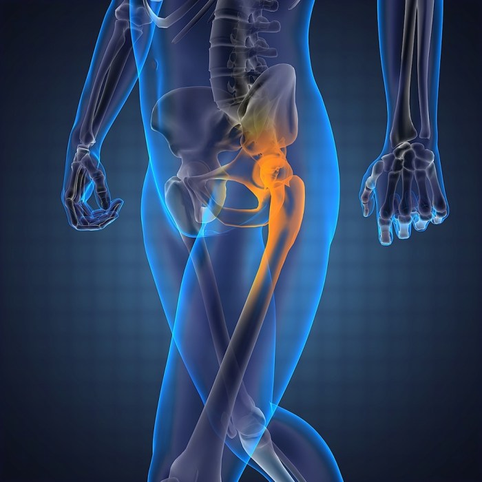
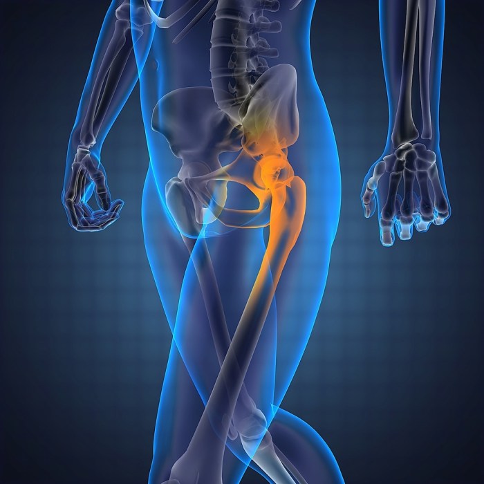
    The hip labrum is a ring of cartilage that lines the socket of the hip joint. It acts as a gasket, deepening the socket and improving the joint’s stability. A tear in this cartilage, known as a hip labrum tear, can result in pain, instability, and decreased range of motion. Various factors can contribute to such tears, including repetitive stress, trauma, or underlying conditions.

    Understanding the causes, function, and symptoms of hip labrum tears is crucial for proper diagnosis and treatment.Hip labrum tears can significantly impact the mechanics of the hip joint. The labrum’s primary role is to increase the contact area between the ball and socket, thus improving joint stability and reducing friction. Damage to the labrum can lead to abnormal movement patterns, increased wear and tear on the joint, and a predisposition to further injuries.

    This disruption of normal joint mechanics is a key factor in the development of pain and disability.

    Hip Labrum Tear Causes

    Several factors can contribute to hip labrum tears. Repetitive stress, such as from sports activities or certain occupations, can gradually wear down the labrum. Trauma, like a fall or direct blow to the hip, can cause an acute tear. Underlying conditions like hip dysplasia, which affects the shape of the hip socket, can predispose individuals to labrum tears.

    Furthermore, some individuals may have a genetic predisposition to weaker or more susceptible labral tissue.

    Hip Labrum Tear Symptoms

    Common symptoms of hip labrum tears include pain, often felt in the groin, hip, or buttock area. The pain can vary in intensity and may be aggravated by specific movements, such as walking, running, or climbing stairs. Limited range of motion, clicking or popping sounds in the hip, and a feeling of instability or catching in the hip joint are also potential indicators.

    Pain may also radiate down the leg.

    Types of Hip Labrum Tears

    Different types of hip labrum tears can affect the hip joint differently. The location of the tear plays a role in the resulting symptoms and treatment options. Here’s a comparison of the common types:

    Type Location Potential Symptoms Treatment Considerations
    Anterior Front of the hip socket Pain in the groin, difficulty with hip flexion (bending the hip), and potentially instability during walking. May require surgical repair or conservative management depending on the severity.
    Posterior Back of the hip socket Pain in the buttock or back of the hip, difficulty with hip extension (straightening the leg), and potential instability during certain movements. Often requires surgical repair, especially if the tear is significant.
    Superior Top of the hip socket Pain in the groin and upper hip region, potential for catching or clicking sensations, and reduced range of motion. Treatment may range from physical therapy to surgery, depending on the severity of the tear.
    Inferior Bottom of the hip socket Pain in the groin, potentially radiating down the inner thigh, difficulty with hip abduction (moving leg away from the body). Surgical intervention may be necessary for significant tears.

    Diagnostic Procedures and Evaluation

    Pinpointing the cause of hip pain, especially when a labral tear is suspected, requires a systematic approach. Accurate diagnosis is crucial for determining the appropriate treatment strategy. This often involves a combination of physical examinations, imaging studies, and potentially arthroscopy. Understanding the information each procedure provides is essential for making well-informed decisions.

    Physical Examinations

    Thorough physical examinations are the initial step in evaluating hip pain. Clinicians assess range of motion, palpating for tenderness, clicking, or grinding sensations. Specific tests, such as the impingement test and flexion abduction external rotation test, are used to evaluate for labral tears and other possible causes of pain. These tests help determine the location and nature of the pain, providing valuable clues about the potential involvement of the hip labrum.

    Pain reproduction during specific movements, coupled with observed limitations in range of motion, can strongly suggest labral pathology.

    Imaging Studies

    Imaging plays a vital role in confirming suspected labral tears and evaluating the extent of the damage. Different imaging modalities offer varying levels of detail and information.

    X-rays

    X-rays are a fundamental initial step in evaluating the hip joint. They primarily assess for bony abnormalities, such as fractures, arthritis, or other bony injuries. X-rays provide a general overview of the hip structure, helping to rule out significant bony pathology. While not specifically designed to visualize soft tissues like the labrum, they can help identify potential contributing factors to the pain.

    MRIs

    Magnetic resonance imaging (MRI) is a powerful tool for visualizing soft tissues, including the labrum. MRI scans offer detailed images of the hip joint, showcasing the cartilage, ligaments, tendons, and the labrum itself. They can clearly identify labral tears, demonstrating their size, location, and degree of detachment from the underlying bone. An MRI can also reveal other soft tissue injuries that may coexist with a labral tear.

    Arthroscopy

    Arthroscopy is an arthroscopic procedure that involves inserting a small camera into the hip joint through a small incision. This allows direct visualization of the joint structures, providing a high-resolution view of the labrum and other surrounding tissues. Arthroscopy is particularly useful for confirming the diagnosis and evaluating the extent of the labral tear in detail. It enables the surgeon to identify the precise location, size, and nature of the tear.

    This direct visualization allows for precise assessment and can also guide surgical interventions, ensuring a tailored approach to the individual patient’s needs.

    Importance of Proper Diagnosis

    A precise diagnosis is crucial for developing the most effective treatment plan. An accurate assessment helps determine the severity of the tear and whether conservative or surgical intervention is necessary. Incorrect diagnoses can lead to inappropriate treatment choices, potentially prolonging the recovery period or causing further complications. Therefore, careful consideration of all diagnostic procedures is paramount in ensuring optimal patient outcomes.

    Summary Table of Typical Findings

    Imaging Modality Typical Findings in Labral Tears
    X-ray May reveal signs of osteoarthritis, hip dysplasia, or other bony abnormalities. No direct visualization of the labrum.
    MRI Demonstrates labral tears as focal defects or detachment of the labrum from the underlying bone. Provides details on the size, location, and extent of the tear. May also show associated cartilage damage or other soft tissue injuries.

    Non-Surgical Treatment Options

    Hip labrum tears, while sometimes requiring surgical intervention, often respond well to non-surgical treatments. Conservative approaches can be highly effective in managing pain and restoring function, avoiding the risks and recovery time associated with surgery. These methods aim to reduce inflammation, promote healing, and improve mobility through targeted exercises and lifestyle modifications.Conservative treatment strategies focus on alleviating symptoms and promoting healing without the need for invasive procedures.

    The effectiveness of each method can vary depending on the severity of the tear, the individual’s response to treatment, and the presence of any other contributing factors. A thorough evaluation by a healthcare professional is crucial to determine the most suitable course of action.

    Physical Therapy, Hip labrum surgery is it the right treatment

    Physical therapy plays a pivotal role in the non-surgical management of hip labrum tears. It’s a cornerstone of conservative treatment, focusing on restoring strength, flexibility, and range of motion in the affected hip joint. Exercises are tailored to address specific needs, and the progression of exercises is crucial for optimal results. The key is to gradually increase the intensity and complexity of the exercises as the patient’s condition improves.

    Rest

    Rest is an essential component of managing hip labrum tears, especially in the acute phase. It allows the injured tissues to heal and reduces further stress on the affected area. While complete bed rest might not always be necessary, avoiding activities that aggravate pain is crucial. This could involve modifying work tasks, avoiding high-impact sports, or adjusting daily routines to minimize hip strain.

    Proper rest is vital for allowing the body to initiate the healing process.

    Anti-inflammatory Medications

    Nonsteroidal anti-inflammatory drugs (NSAIDs) such as ibuprofen or naproxen can help reduce inflammation and pain associated with hip labrum tears. These medications can be particularly helpful in managing the initial inflammatory response, which often contributes to pain and discomfort. However, it’s essential to use these medications as directed by a healthcare professional, as they may have potential side effects, including gastrointestinal issues.

    Considering hip labrum surgery? It’s a big decision, and figuring out if it’s the right treatment for you is key. While hip pain can be debilitating, understanding the common locations for skin conditions like eczema might offer some insight into the overall picture of your health. For example, knowing the most common location for eczema can help you identify potential triggers and improve your treatment plan.

    Ultimately, a consultation with your doctor is crucial in deciding if hip labrum surgery is the right choice for your specific needs. most common location for eczema

    Always consult with a doctor before taking any new medication, especially if you have pre-existing conditions.

    Typical Physical Therapy Exercises and Stretches

    Exercise/Stretch Description
    Hip Flexor Stretch Standing or seated, gently pull the knee towards the chest, holding for 20-30 seconds.
    Hip Abduction Exercises Side-lying with the affected leg raised, keeping the knee straight, to strengthen hip abductors.
    Hip Extension Exercises Prone position, raising the leg straight up to strengthen hip extensors.
    Glute Bridges Lying on the back, lifting the hips off the ground, contracting gluteal muscles.
    Clamshells Side-lying with knees bent, raising the top knee, targeting hip abductors.
    Prone Hip Rotations Lying on stomach, rotating the leg outward and inward, improving hip external and internal rotation.

    The specific exercises and stretches prescribed by a physical therapist will depend on the individual’s specific needs and the stage of healing.

    Surgical Procedures for Hip Labrum Tears

    Hip labral tears, often caused by repetitive stress, trauma, or a combination of factors, can lead to significant pain and dysfunction. Surgical intervention is sometimes necessary to restore hip joint stability and alleviate symptoms. This section will delve into the various surgical techniques employed for hip labral repair or reconstruction.Surgical procedures for hip labrum tears aim to address the underlying cause of the problem and restore the structural integrity of the labrum.

    Different techniques exist, each with its own set of advantages and potential risks. Understanding these techniques allows patients and medical professionals to make informed decisions about the best course of action for their specific needs.

    Arthroscopic Labral Repair

    Arthroscopic labral repair is a minimally invasive surgical procedure that utilizes small incisions and specialized instruments to access and repair the damaged labrum. This approach is generally preferred for its smaller incision size, reduced recovery time, and decreased post-operative pain compared to open surgical techniques.The procedure typically involves the following steps:

    • The surgeon makes small incisions in the hip joint, inserting a specialized camera (arthroscope) and small surgical instruments.
    • The arthroscope allows visualization of the damaged labrum, enabling the surgeon to identify the extent of the tear.
    • Using specialized instruments, the surgeon carefully cleans the area around the tear, removing any loose tissue or debris.
    • Depending on the type of tear, the surgeon may use sutures, anchors, or other techniques to repair the labrum.
    • The joint is flushed with saline solution to remove any residual debris.
    • The incisions are closed, and a bandage is applied to the surgical area.

    Open Labral Repair

    Open labral repair is a more extensive surgical procedure, often reserved for cases where arthroscopic techniques are insufficient or not suitable. The surgeon makes a larger incision to directly access the hip joint, enabling a more comprehensive view and greater control over the repair.

    • A larger incision is made in the hip joint, providing direct access to the damaged labrum.
    • The surgeon carefully dissects the surrounding tissues to expose the labrum tear.
    • The damaged labrum is cleaned and repaired using sutures, anchors, or other appropriate techniques.
    • The surrounding tissues are carefully repositioned to restore normal joint anatomy.
    • The incision is closed, and the surgical area is bandaged.

    Comparison of Surgical Approaches

    Arthroscopic surgery offers a less invasive approach, resulting in a quicker recovery, less pain, and smaller scars. However, it may not be suitable for all types of labral tears, particularly those involving significant bone damage or complex tear patterns. Open surgery, while more invasive, allows for greater visualization and control, which can be beneficial for complex repairs.

    Surgical Instruments Used in Hip Labral Repair

    The choice of surgical instruments depends on the specific procedure and the surgeon’s preference. Here’s a table outlining some common instruments used in hip labral repair procedures:

    Instrument Description
    Arthroscope A small camera and light source used to visualize the hip joint interior.
    Shaver A tool for removing damaged or loose tissue from the joint.
    Scissors Used for precise tissue dissection.
    Sutures Used to repair the torn labrum.
    Anchors Used to secure sutures to the bone.
    Graspers Used to hold and manipulate tissues.

    Recovery and Rehabilitation After Hip Labrum Surgery: Hip Labrum Surgery Is It The Right Treatment

    Recovering from hip labrum surgery is a journey, not a sprint. A diligent and structured rehabilitation program is crucial for a successful outcome. This involves a combination of pain management, targeted physical therapy, and gradual return to activities. Understanding the process and adhering to the prescribed protocol will significantly impact the recovery time and overall success of the surgery.The recovery period after hip labrum surgery is designed to restore hip function and strength while minimizing the risk of re-injury.

    This phase involves a gradual increase in activity, starting with gentle movements and progressing to more challenging exercises. Patience and consistency are key components to a successful recovery.

    Considering hip labrum surgery? It’s a complex decision, and the right treatment depends heavily on the individual’s specific case. Factors like the severity of the tear and the patient’s overall health play a crucial role. Understanding the potential costs, including those related to tariffs and OTC drug pricing fluctuations, can be critical when making your choice. For example, tariffs and OTC drugs can influence the price of necessary medications and supplies, which can impact the overall financial picture.

    Ultimately, talking to a healthcare professional is key to determining if hip labrum surgery is the best path for you.

    Pain Management

    Effective pain management is essential for allowing patients to participate fully in their rehabilitation program. Strategies typically involve a combination of medication, physical therapy modalities, and lifestyle adjustments. Over-the-counter pain relievers may be sufficient for mild discomfort, while stronger medications might be necessary for more severe pain. Physical therapy modalities, such as ice packs, heat therapy, and electrical stimulation, can also help manage pain and inflammation.

    Regular communication with the surgeon and physical therapist is crucial to ensure the pain management plan is optimized throughout the recovery process.

    Physical Therapy Phases

    A structured physical therapy program is tailored to each individual’s needs and recovery progress. It’s designed to gradually restore hip strength, flexibility, and range of motion. A typical progression of exercises and activities can be broken down into distinct phases.

    Phase Focus Duration (Approximate) Key Activities
    Phase 1 (Initial Post-Op) Protecting the surgical site, managing pain, and regaining basic mobility. 1-4 weeks Gentle range of motion exercises, isometric exercises (contracting muscles without movement), and pain-free walking with assistive devices as needed.
    Phase 2 (Early Strengthening) Increasing hip strength and improving flexibility. 4-8 weeks Progressive resistance exercises (using light weights or resistance bands), balance exercises, and controlled walking. Activities such as stationary cycling and swimming can be incorporated.
    Phase 3 (Advanced Strengthening) Improving functional strength and preparing for return to activities. 8-12 weeks More challenging exercises, including plyometrics (jumping and hopping), sport-specific drills (if applicable), and functional exercises mimicking daily activities. Gradual return to impact activities, such as jogging or stairs, is also introduced.
    Phase 4 (Return to Activity) Full functional recovery and return to desired activity level. 12+ weeks Maintenance exercises to maintain gains, and progression to pre-injury activity levels.

    This table provides a general guideline. The duration of each phase may vary depending on the individual’s progress and the complexity of the surgery. Always follow the recommendations of your physical therapist and surgeon.

    Return to Activity Timelines

    The timeline for returning to various activities depends on the extent of the injury, the surgical procedure, and the individual’s progress through the rehabilitation program. Light activities like walking can often begin within a few days of surgery, while more strenuous activities, such as running or contact sports, typically require several months of recovery. It’s crucial to avoid overexertion and allow the body adequate time to heal to prevent re-injury.

    Factors Influencing Recovery Time

    Several factors can influence the recovery time after hip labrum surgery. These include the severity of the tear, the individual’s overall health, adherence to the rehabilitation program, and the surgeon’s expertise. Age, pre-existing conditions, and the patient’s motivation also play a significant role. For instance, a more extensive tear or a patient with underlying medical conditions may take longer to recover.

    Furthermore, a strong commitment to the rehabilitation protocol is crucial for a faster and more complete recovery.

    Potential Complications

    Potential complications after hip labrum surgery are generally rare but include infection, blood clots, persistent pain, stiffness, or incomplete recovery. Addressing any concerns promptly with the surgeon is essential for effective management. Regular follow-up appointments and adherence to the recovery plan are critical in mitigating these risks. Open communication with the surgical team allows for proactive intervention if any complications arise.

    Factors Affecting Treatment Decisions

    Deciding whether hip labrum surgery is the right course of action isn’t a simple yes or no. It’s a nuanced evaluation considering various factors unique to each patient. A surgeon carefully weighs the potential benefits and risks against the patient’s individual circumstances to determine the most appropriate treatment path.Surgeons aren’t simply looking at the tear itself. They consider the patient’s overall health, activity level, and how the labrum tear impacts their daily life.

    This holistic approach ensures the best possible outcome, balancing the need for intervention with the desire for a minimally invasive and effective solution.

    Severity of the Tear

    The extent of the hip labrum tear significantly influences treatment decisions. A small, stable tear might not require surgery, whereas a large, unstable tear causing significant pain and dysfunction may necessitate surgical intervention. Surgical repair aims to restore the labrum’s integrity and function, while non-surgical approaches focus on managing symptoms and promoting healing. The surgeon carefully assesses the tear’s size, location, and the presence of any associated damage to other structures in the hip joint.

    Patient’s Activity Level

    A patient’s activity level plays a crucial role in determining the best course of action. An individual leading a sedentary lifestyle might respond well to non-surgical treatments like physical therapy and activity modification. Conversely, an athlete requiring high-impact movements or frequent weight-bearing activities might benefit more from surgical repair to restore function and prevent further damage. A professional athlete with a large tear affecting their performance would likely be a strong candidate for surgery.

    Overall Patient Health

    Pre-existing medical conditions, such as arthritis or other joint problems, can influence the suitability of hip labrum surgery. The presence of other health concerns may necessitate a more conservative approach to avoid additional risks. For instance, a patient with significant cardiovascular issues might not be an ideal candidate for surgery, even with a severe labrum tear. A careful assessment of the patient’s medical history is essential to determine the safest and most effective treatment plan.

    Patient’s Goals and Expectations

    A key factor is the patient’s expectations and desired outcomes. Open communication between the patient and the surgeon is crucial to ensure mutual understanding and agreement on the treatment plan. A patient who desires complete restoration of function and the ability to return to their pre-injury activity level may be a better candidate for surgery. Conversely, a patient prioritizing symptom relief and improved quality of life might find non-surgical management more suitable.

    Comparison of Surgical and Non-Surgical Options

    Feature Non-Surgical Options Surgical Options
    Potential Benefits Reduced risk of surgery-related complications, quicker recovery, and potentially more cost-effective. Restoration of labrum integrity and function, potentially addressing underlying causes of pain and dysfunction, and returning to previous activity levels.
    Potential Risks Limited effectiveness in severe cases, potential for persistent pain, and possible need for future interventions. Potential complications like infection, nerve damage, or blood clots, longer recovery period, and higher costs.
    Recovery Time Typically shorter than surgical recovery. Typically longer recovery period, requiring a period of rehabilitation and physical therapy.
    Success Rate Variable depending on the severity of the tear and the patient’s compliance with the treatment plan. Generally higher success rates in addressing severe tears and restoring full function, especially for athletes.

    “The ultimate decision rests with the patient, in consultation with their surgeon, weighing the potential benefits and risks of each approach.”

    Potential Complications and Risks

    Hip labrum surgery, while often effective, carries potential risks. Understanding these complications is crucial for informed decision-making, alongside a thorough discussion with your surgeon. Proper preparation and proactive risk mitigation strategies can significantly reduce the likelihood of encountering these issues.

    Potential Complications

    Post-operative complications can arise from various factors, including the surgical procedure itself, individual patient characteristics, and post-operative care. Careful monitoring and management are essential to address these issues promptly and effectively.

    Infection

    Surgical procedures, by their nature, introduce a risk of infection. This risk is not unique to hip labrum surgery but is a concern across various surgical interventions. The prevalence of infection following hip labrum surgery is relatively low, typically reported in the range of 0.5% to 1.5%. Severity can range from localized skin infections to more systemic infections requiring intravenous antibiotics.

    Maintaining meticulous surgical technique, proper wound care, and vigilant monitoring for signs of infection are crucial strategies to minimize this risk.

    Blood Clots (Deep Vein Thrombosis – DVT)

    Blood clots, particularly deep vein thrombosis (DVT), are a possible complication following any surgical procedure that involves prolonged immobilization. DVTs can develop in the deep veins of the leg and, in rare cases, can travel to the lungs, causing a pulmonary embolism (PE). While the exact prevalence varies, studies suggest that DVTs occur in a percentage of patients undergoing hip surgery.

    Strategies to reduce the risk of DVT include early mobilization, compression stockings, and medication like anticoagulants.

    Persistent Pain

    Although hip labrum surgery aims to alleviate pain, some patients may experience persistent or even increased pain after the procedure. This can stem from various factors, including incomplete healing, scar tissue formation, or other underlying conditions. While not all cases result in persistent pain, the percentage of patients experiencing some level of persistent pain can range from a small percentage to a higher number depending on the individual factors.

    Rehabilitation programs tailored to individual needs, and proactive management of pain are essential strategies.

    Other Potential Complications

    Other potential complications include, but are not limited to, nerve damage, numbness, or weakness in the affected leg, and, in rare instances, a reaction to anesthesia. These complications are less frequent but can still occur. Factors such as the surgeon’s experience and surgical technique can play a role in the likelihood of these complications.

    Signs and Symptoms of Complications

    Potential Complication Signs and Symptoms
    Infection Redness, swelling, warmth, pain, pus or drainage from the incision site, fever, chills.
    DVT Swelling, pain, redness, warmth in the affected leg, especially in the calf, difficulty moving the leg.
    Persistent Pain Continued or increased pain in the hip area, even after rehabilitation.
    Nerve Damage Numbness, tingling, weakness, or loss of sensation in the affected leg or foot.

    Alternatives and Other Considerations

    Hip labrum surgery is a significant procedure, and it’s crucial to explore all available options before making a decision. While often the most effective treatment for severe hip labrum tears, it’s not always the right choice for every patient. Factors like the severity of the tear, the patient’s overall health, and lifestyle play a critical role in determining the best course of action.

    This section will delve into alternative treatments and essential considerations beyond surgery.Alternative approaches to treating hip labrum tears exist, offering potential benefits for specific cases. These range from conservative management to minimally invasive procedures, each with its own set of advantages and disadvantages. Understanding these alternatives allows patients to make informed decisions in conjunction with their healthcare providers.

    Alternative Treatment Options

    Conservative treatment often forms the initial approach for hip labrum tears. This typically involves a combination of physical therapy, activity modification, and pain management strategies. Physical therapy focuses on strengthening the muscles surrounding the hip joint, improving flexibility, and restoring proper biomechanics. Activity modification aims to reduce the impact on the injured area, while pain management can include over-the-counter pain relievers or, in more severe cases, prescription medications.

    In some instances, corticosteroid injections may be used to reduce inflammation. These conservative measures can be effective in managing mild to moderate hip labrum tears, allowing for a non-surgical solution.

    Patient-Specific Factors

    Patient-specific factors significantly influence the choice of treatment. Age is a crucial consideration. Younger patients, particularly athletes, may benefit from surgical intervention to maintain optimal joint function and prevent long-term complications. Older patients might find non-surgical options more suitable, given their potential for a slower recovery period. Overall health conditions, such as arthritis or other joint issues, also impact treatment choices.

    Deciding if hip labrum surgery is the right choice can be tough. It’s important to consider all your options, and understanding your health insurance is key. For example, knowing the difference between an HSA and an FSA can significantly impact your out-of-pocket costs, especially when considering a procedure like this. hsa vs fsa whats the difference Ultimately, talking to your doctor about your specific situation and carefully weighing the pros and cons of surgery is crucial for making an informed decision.

    Lifestyle factors, like the patient’s occupation and level of physical activity, play a role in determining the suitability of various treatments. For example, a professional athlete with a significant hip labrum tear will likely require surgical intervention to maintain their performance level, while a sedentary individual might be able to manage the condition with non-surgical therapies.

    Comparison of Treatment Approaches

    A comprehensive comparison of hip labrum surgery with other treatments is crucial for informed decision-making. Surgery is often reserved for severe cases, including those with significant instability or persistent pain not responding to conservative measures. In contrast, conservative management provides a less invasive approach, suitable for individuals with milder symptoms. Minimally invasive procedures, such as arthroscopy, represent a middle ground, offering a less extensive surgical intervention than traditional open surgery.

    The success rate of each approach depends on factors such as the severity of the tear, the individual’s response to treatment, and the expertise of the medical team.

    Non-Surgical Treatment Options

    Non-surgical approaches are valuable options for managing hip labrum tears, especially in the initial stages or for individuals not suitable for surgery. These methods often focus on reducing pain, improving function, and preventing further damage.

    Treatment Option Description Potential Benefits Potential Limitations
    Physical Therapy Exercises to strengthen hip muscles, improve flexibility, and restore proper mechanics. Improved joint stability, reduced pain, and enhanced function. Requires commitment and adherence to a prescribed regimen.
    Activity Modification Adjusting activities to reduce stress on the hip joint. Reduces pain and inflammation. May require significant lifestyle adjustments.
    Pain Management Over-the-counter or prescription medications, or corticosteroid injections. Reduces pain and inflammation. May have side effects, and injections may not address the root cause.
    Injections (e.g., Corticosteroids) Administering corticosteroids directly into the hip joint. Temporary pain relief and reduced inflammation. Potential for side effects, and not a long-term solution.

    Patient Experiences and Success Stories (Illustrative)
    Hip labrum surgery is it the right treatment

    Hip labrum surgery, while often a necessary procedure, can evoke a range of emotions and expectations in patients. Understanding real-life experiences can provide valuable insights into the recovery process and the potential outcomes. This section highlights illustrative cases to illustrate successful outcomes and emphasize the importance of realistic patient expectations.

    Illustrative Patient Experiences

    Many patients report significant improvements in their quality of life following hip labrum surgery. The recovery journey, however, varies greatly depending on individual factors such as age, pre-existing conditions, and the extent of the tear.

    Successful Outcomes and Positive Aspects

    A common theme in positive patient experiences is a noticeable reduction in pain and improved mobility. This improvement often translates to increased participation in daily activities and hobbies. For instance, a 35-year-old professional athlete who underwent hip labrum repair reported a return to competitive play within six months, albeit with a modified training regimen. This illustrates how surgery can enable a return to a fulfilling lifestyle, albeit with careful consideration of physical limitations during recovery.

    Another patient, a 60-year-old retiree, regained the ability to walk comfortably and participate in social activities like gardening. These experiences demonstrate the positive impact of surgery on overall well-being.

    Lifestyle Changes and Benefits

    Hip labrum surgery can lead to significant lifestyle changes, and these changes are often perceived positively by patients. Many patients report being able to engage in activities they previously found painful or impossible, like dancing, hiking, or simply enjoying a longer walk with their loved ones. The ability to resume everyday tasks without pain significantly enhances their quality of life.

    Importance of Patient Expectations and Realistic Outcomes

    It’s crucial for patients to understand that complete recovery and a full return to previous activity levels may not be immediate. While surgery can be a significant step toward improvement, realistic expectations about recovery time and potential limitations are essential for a positive experience. A thorough discussion with the surgeon about potential limitations and necessary physical therapy is critical to manage expectations and avoid disappointment.

    The surgeon’s role in counseling patients about the potential range of outcomes is paramount. This includes discussing possible limitations, long-term care needs, and the need for ongoing physical therapy. Realistic expectations prevent disappointment and promote a more positive and sustainable recovery.

    Ultimate Conclusion
    Hip labrum surgery is it the right treatment

    Ultimately, the decision of whether or not hip labrum surgery is the right treatment is a personal one. Weighing the potential benefits and risks, alongside your individual circumstances, is key. This detailed analysis should empower you to make an informed choice, working closely with your doctor to determine the best course of action for your specific needs.
  • Cobbs Angle and Scoliosis A Deep Dive

    Cobbs Angle and Scoliosis A Deep Dive

    Cobbs angle and scoliosis – Cobb’s angle and scoliosis are crucial concepts in understanding spinal deformities. This comprehensive exploration delves into the intricacies of Cobb’s angle, from its definition and significance in diagnosing scoliosis to its role in treatment planning and monitoring progression. We’ll explore different types of scoliosis, measurement methods, and the clinical implications of Cobb’s angle in managing this condition. This discussion also encompasses the importance of accurate measurements and the various imaging techniques employed.

    Understanding the relationship between Cobb’s angle and the severity of scoliosis is key to effective treatment. Different types of scoliosis, like idiopathic and neuromuscular, present unique challenges, and the severity of the curve, as quantified by Cobb’s angle, plays a significant role in determining the appropriate treatment strategy. This guide provides a practical overview, including tables illustrating the correlation between Cobb’s angle and treatment options, and factors influencing the progression of scoliosis.

    Introduction to Cobb’s Angle and Scoliosis

    Scoliosis is a sideways curvature of the spine. Understanding the degree of this curvature is crucial for diagnosis and treatment planning. Cobb’s Angle is a key measurement in this process, providing a standardized way to quantify the severity of scoliosis. This helps healthcare professionals determine the appropriate course of action for each patient.Cobb’s Angle is a clinically important tool used to measure the degree of spinal curvature in scoliosis.

    It’s a precise numerical value, essential in evaluating the severity of the condition, guiding treatment decisions, and tracking the progression of the curvature over time. The angle is calculated from the upper and lower end vertebrae, providing a reliable metric for the severity of the scoliosis.

    Definition of Cobb’s Angle

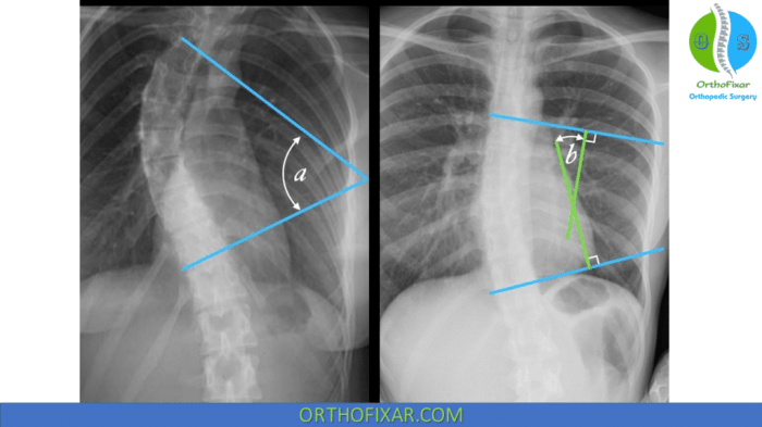
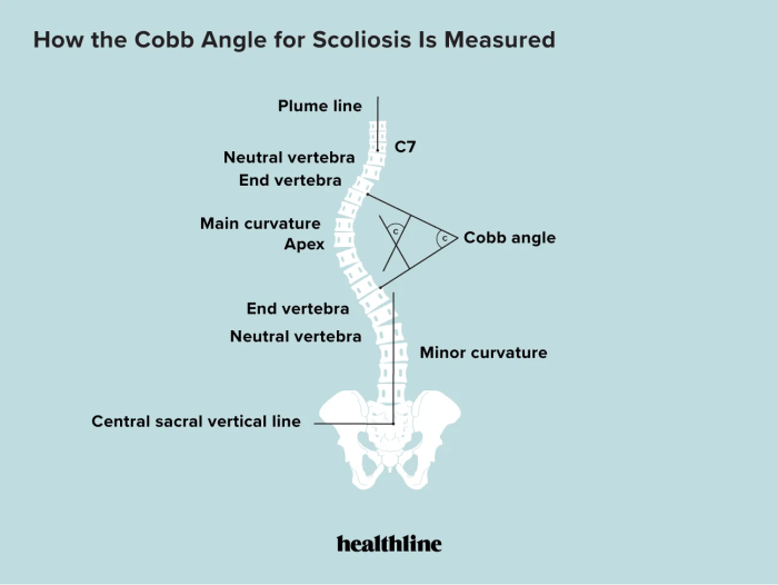
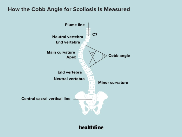
    Cobb’s Angle is a measurement of the degree of spinal curvature in scoliosis. It’s calculated by drawing lines along the top and bottom vertebrae that are most involved in the curve, then measuring the angle between these lines. This standardized measurement allows healthcare providers to objectively quantify the severity of the scoliosis and track its progression. A higher Cobb’s Angle typically indicates a more severe curve.

    Significance of Cobb’s Angle in Scoliosis Diagnosis

    Cobb’s Angle is vital in scoliosis diagnosis because it provides a standardized and objective way to measure the degree of spinal curvature. This measurement allows healthcare professionals to assess the severity of the condition, compare it to established guidelines, and plan appropriate treatment strategies. The Cobb’s Angle is a crucial factor in determining the necessity and type of intervention required.

    Relationship Between Cobb’s Angle and Severity of Scoliosis

    The relationship between Cobb’s Angle and the severity of scoliosis is directly proportional. A larger Cobb’s Angle indicates a more severe spinal curve. This correlation is a fundamental aspect of scoliosis assessment, guiding the selection of appropriate treatment strategies. Different treatment options are tailored to the severity of the curvature, as reflected in the Cobb’s Angle. For example, a Cobb’s Angle of 20 degrees might require observation, while a Cobb’s Angle of 40 degrees might necessitate bracing or surgery.

    Types of Scoliosis

    Scoliosis can be categorized into different types, each with its own characteristics and potential causes. Understanding these variations is important for appropriate diagnosis and treatment planning. This categorization helps doctors identify the specific factors that contribute to the curvature, leading to personalized care strategies.

    Table Comparing Types of Scoliosis

    Type Cause Characteristics
    Idiopathic Scoliosis Unknown cause; often genetic predisposition and environmental factors are suspected. Most common type, typically diagnosed in childhood or adolescence. The curvature is primarily in the thoracic spine, but it can occur in other regions.
    Neuromuscular Scoliosis Associated with neurological conditions like cerebral palsy, muscular dystrophy, or spina bifida. Often presents with additional physical limitations and may be accompanied by other medical issues. The curve can be variable in location and severity.
    Congenital Scoliosis Structural abnormalities of the spine present at birth. Characterized by specific structural defects in the spine, such as hemivertebrae or ribs. The curvature is typically present from birth or early childhood.

    Measuring Cobb’s Angle

    Understanding Cobb’s angle measurement is crucial for evaluating scoliosis severity and guiding treatment plans. Accurate determination of this angle is essential for proper diagnosis and prognosis. Precise measurement ensures that the treatment strategy is tailored to the specific curvature of the spine.Accurate measurement of Cobb’s angle relies on standardized techniques. These techniques ensure consistent and reliable results across different healthcare settings.

    This allows for effective comparison of results over time and facilitates the evaluation of treatment efficacy.

    Radiographic Measurement Procedures

    Radiographic imaging, typically a posterior-anterior (PA) X-ray of the spine, is the primary method for measuring Cobb’s angle. The procedure involves identifying the apex vertebrae of the curve and then defining the superior and inferior endplates of the vertebrae. The accuracy of the angle measurement directly impacts the accuracy of the scoliosis diagnosis.

    The steps involved in the radiographic measurement of Cobb’s angle are as follows:

    1. Identify the apex vertebrae: The apex vertebra is the most severely angled vertebra within the curve.
    2. Locate the superior and inferior endplates: The superior and inferior endplates are the top and bottom surfaces of the vertebrae that form the boundaries of the curve.
    3. Draw lines tangent to the superior and inferior endplates: These lines should be perpendicular to the endplates, creating a clear visualization of the curve’s extent.
    4. Determine the intersection of the lines: The intersection point of these two lines defines the angle, representing the Cobb’s angle.
    5. Measure the angle: The angle formed between these two lines is the Cobb’s angle. This measurement is crucial for assessing the severity of scoliosis.

    Visual Representation of the Measurement Process

    Imagine a diagram of a spine on a radiograph. The spine is depicted as a series of vertebrae. Two distinct lines, perpendicular to the superior and inferior endplates of two vertebrae at the apex of the curvature, are drawn. The angle formed by the intersection of these two lines represents Cobb’s angle. A clearly labeled diagram illustrating this would show the vertebrae, the superior and inferior endplates, the tangent lines, and the measured Cobb’s angle.

    The diagram should be well-labeled with arrows indicating the direction of the tangent lines and highlighting the key components involved in the measurement process. The angle should be clearly marked with the corresponding numerical value.

    Equipment for Cobb’s Angle Measurement

    Accurate Cobb’s angle measurement relies on appropriate equipment. The following table Artikels the necessary equipment:

    Equipment Description
    X-ray machine Produces the radiographic images of the spine.
    X-ray film or digital imaging system Captures the images of the spine for analysis.
    Radiation shielding Protects personnel from radiation exposure during the X-ray procedure.
    Measuring device (e.g., protractor or specialized software) Used to precisely measure the Cobb’s angle from the radiograph.
    Appropriate imaging technique Ensure the radiograph is taken in a standard way (e.g., posterior-anterior view)

    Clinical Significance of Cobb’s Angle

    Cobbs angle and scoliosis

    Understanding Cobb’s angle is crucial for evaluating the severity of scoliosis and determining the appropriate course of treatment. This angle, measured on X-rays, quantifies the degree of curvature in the spine, providing a standardized metric for assessing the structural abnormality. The relationship between Cobb’s angle and the need for intervention is a complex one, influenced by several factors beyond the numerical value itself.The clinical significance extends beyond simply measuring the curve.

    A higher Cobb’s angle generally correlates with a greater degree of spinal deformity, potentially leading to more significant functional limitations and complications. However, other factors, like the patient’s age, skeletal maturity, and the location and type of curve, also play pivotal roles in determining the necessary intervention.

    Relationship Between Cobb’s Angle and Treatment

    The decision to recommend treatment for scoliosis is multifaceted. While a higher Cobb’s angle often indicates a greater need for intervention, the specific treatment plan is not solely dictated by the numerical value. Age and skeletal maturity are key considerations. For example, a younger patient with a smaller Cobb’s angle might still require intervention if the curve is rapidly progressing.

    Understanding Cobb’s angle and scoliosis often involves delving into the patient’s overall well-being, including sleep quality. Poor sleep can sometimes contribute to musculoskeletal issues, and the Pittsburgh Sleep Quality Index (PSQI) can be a valuable tool in assessing sleep patterns. Ultimately, a thorough evaluation of Cobb’s angle and scoliosis requires a holistic approach that considers various factors, including sleep quality.

    Factors Influencing Treatment Decisions

    Several factors beyond the Cobb’s angle contribute to the choice of treatment. These include the patient’s age, skeletal maturity, the location and type of the spinal curve (thoracic, lumbar, or thoracolumbar), and the presence of any associated neurological symptoms. For instance, a rapidly progressing curve in a younger patient might warrant intervention even with a relatively small Cobb’s angle.

    Comparison of Treatment Options

    Treatment options for scoliosis vary based on the severity of the curvature. For mild curves, observation and regular follow-up may suffice. As the Cobb’s angle increases, bracing becomes a potential option to halt or slow the progression of the curve. Surgical intervention is typically reserved for curves that are large, rapidly progressing, or causing significant pain or functional impairment.

    Bracing is usually employed in adolescent patients whose spines are still growing, whereas surgery may be necessary for those who have reached skeletal maturity.

    Correlation Between Cobb’s Angle and Treatment

    Cobb’s Angle Treatment
    0-20 degrees Observation, regular follow-up
    21-40 degrees Bracing, regular follow-up
    41-50 degrees Bracing, close monitoring, potential surgery
    51-60 degrees Bracing, surgical intervention likely
    >60 degrees Surgical intervention typically necessary

    This table provides a general guideline, and the actual treatment decision should always be made in consultation with a qualified medical professional. Individualized factors, as mentioned previously, will be taken into account when determining the best course of action for each patient. For example, a 15-year-old with a 45-degree Cobb’s angle might be recommended bracing, while an adult with the same angle might not require any intervention.

    Progression of Scoliosis and Cobb’s Angle

    Cobbs angle and scoliosis

    Understanding how scoliosis progresses is crucial for effective management and treatment. This involves recognizing the factors that contribute to the curve worsening and how Cobb’s Angle measurements help track the disease’s advancement. Monitoring Cobb’s Angle over time is a critical tool for assessing the effectiveness of interventions.The progression of scoliosis, a sideways curvature of the spine, isn’t always consistent and can be influenced by several factors.

    The rate of progression can vary significantly from person to person. Early detection and appropriate intervention are essential to minimize the potential for long-term complications.

    Factors Contributing to Scoliosis Progression

    Several factors can influence the progression of scoliosis. Genetic predisposition plays a role, and certain conditions can increase the risk of curve worsening. Age and growth spurts are also key considerations. During periods of rapid growth, the spine is more susceptible to developing or worsening curves. Furthermore, muscle imbalances and postural habits can contribute to the progression of the deformity.

    Role of Cobb’s Angle in Monitoring Progression

    Cobb’s Angle, a measurement of the spinal curvature, is fundamental in monitoring scoliosis progression. It’s a standardized method for quantifying the degree of the spinal deformity. Regular assessments of Cobb’s Angle allow clinicians to track the change in the spinal curve over time. This allows for a more accurate prediction of future progression and enables the evaluation of treatment effectiveness.

    Using Cobb’s Angle to Track Treatment Effectiveness

    Monitoring Cobb’s Angle is essential for evaluating the efficacy of scoliosis treatment. By comparing Cobb’s Angle measurements before, during, and after treatment, clinicians can assess the response to therapy. A decrease in Cobb’s Angle indicates a positive response to treatment, while a continued or increasing angle suggests that alternative strategies might be necessary. This helps in making informed decisions regarding the most appropriate course of action.

    Example of Cobb’s Angle Changes Over Time

    The following table illustrates the potential changes in Cobb’s Angle over time, with and without treatment:

    Time Period Cobb’s Angle (without treatment) Cobb’s Angle (with treatment)
    Initial Assessment 25 degrees 25 degrees
    6 Months Later 30 degrees 27 degrees
    1 Year Later 35 degrees 28 degrees
    2 Years Later 40 degrees 29 degrees

    Note: These are examples, and actual changes will vary depending on the individual patient’s condition, treatment plan, and response to intervention. This table highlights the potential for a significant increase in Cobb’s Angle without treatment, whereas treatment can help maintain or slow down the progression of the curve.

    Imaging Techniques for Cobb’s Angle Measurement

    Understanding scoliosis and its progression requires accurate assessment of the spinal curvature. Precise measurement of the Cobb’s angle, a crucial indicator of scoliosis severity, relies heavily on appropriate imaging techniques. Different modalities offer varying levels of detail and utility, each with its own set of advantages and disadvantages.Accurate imaging is vital for assessing the extent of spinal deformity and guiding treatment decisions.

    Different imaging modalities provide varying degrees of information about the spine, making it essential to understand the strengths and weaknesses of each method to ensure proper diagnosis and monitoring.

    Radiography (X-rays)

    Radiographs are the most common initial imaging technique for evaluating scoliosis. They are relatively inexpensive, readily available, and provide a good overview of the spine’s overall alignment.

    • Advantages: Low cost, widespread availability, relatively quick acquisition, and good visualization of bony structures. They allow for a general assessment of the spinal alignment and identify any potential fractures or other bony abnormalities.
    • Disadvantages: X-rays expose patients to ionizing radiation, although the dose is generally considered low. They do not provide detailed soft tissue information. Interpreting the angle can be subjective, depending on the radiologist’s experience and technique.

    Example: A patient presents with back pain and a suspected scoliosis. An initial scoliosis X-ray is obtained. The radiologist can assess the degree of curvature and measure the Cobb’s angle from the X-ray image. This information helps to determine the severity of the condition and guide subsequent treatment decisions.

    Interpreting a Scoliosis X-ray: To measure Cobb’s angle, the radiologist identifies the most superior and inferior endplates of the vertebrae involved in the curve. These are the points where the curve begins and ends. Lines are drawn along the superior and inferior endplates of these vertebrae. The angle formed by these two lines is the Cobb’s angle. Specialized software can often aid in this measurement, increasing accuracy.

    It’s crucial that the X-ray is taken with the patient standing upright and maintaining a neutral posture.

    Cobbs angle and scoliosis are fascinating, but complex. Understanding how the spine curves can be tricky, and sometimes, you might be wondering about other aspects of your health. For example, if you’re looking at birth control, you might be curious about how long it takes to work, which you can find out more about here. Ultimately, though, it’s important to remember that proper scoliosis care involves a lot more than just a single factor, and seeking medical advice is key.

    Computed Tomography (CT) Scans

    CT scans offer detailed cross-sectional images of the spine. They provide a more comprehensive view of the bony structures and soft tissues than plain radiographs.

    • Advantages: High-resolution images of bone and soft tissue, enabling precise measurements of vertebral morphology and allowing for assessment of any associated bony abnormalities. They provide detailed information about the spinal canal, which can be helpful in identifying stenosis.
    • Disadvantages: Higher radiation exposure than X-rays, can be more expensive, and may not always be the most appropriate initial imaging method.

    Example: A patient with a complex scoliosis might require a CT scan to assess the degree of vertebral rotation or the presence of bony anomalies. The detailed cross-sectional images can assist in determining the extent of the deformity and the involvement of soft tissues.

    Magnetic Resonance Imaging (MRI) Scans

    MRI scans provide detailed images of soft tissues, including muscles, ligaments, and nerves. This can be useful in assessing the overall health of the spine and the presence of any associated soft tissue abnormalities.

    Understanding Cobb’s angle and scoliosis is crucial for spinal health, and a healthy lifestyle plays a vital role. A balanced diet, like the one outlined in the longevity diet plan overview , can significantly impact overall well-being, potentially influencing the progression of scoliosis. This, in turn, can help maintain a healthy Cobb’s angle, ultimately contributing to a better quality of life.

    • Advantages: Excellent soft tissue visualization, enabling assessment of the spinal cord and nerve roots, crucial in cases with neurological symptoms. No ionizing radiation exposure.
    • Disadvantages: MRI scans can be more expensive than X-rays or CT scans. They can be time-consuming, and the patient may experience discomfort due to the enclosed environment. Metal implants can affect the quality of the images.

    Example: A patient with scoliosis and associated nerve root compression might benefit from an MRI to assess the extent of the compression and guide treatment decisions. The high-resolution images can delineate the location and extent of any nerve root impingement.

    Variations in Cobb’s Angle Measurement

    Understanding Cobb’s angle is crucial for evaluating scoliosis, but accurate measurement is paramount. Variations in the technique used to determine this angle can significantly impact the diagnosis and subsequent treatment plan. Inaccurate measurements can lead to misdiagnosis, inappropriate interventions, or, in severe cases, potentially delayed or inadequate treatment. Therefore, it’s vital to understand the factors that contribute to variability and the strategies for ensuring consistent and reliable measurements.

    Potential Sources of Error

    Numerous factors can influence the precision of Cobb’s angle measurement. These factors range from the inherent limitations of radiographic imaging to the variability in the technique employed by different healthcare professionals. Inconsistent application of the measurement technique across different healthcare providers or at various points in time can result in inaccurate or misleading interpretations of scoliosis progression. These errors must be meticulously addressed to maintain the integrity of the measurement and ensure reliable clinical assessment.

    Importance of Consistent Measurement Techniques

    Consistent measurement techniques are essential for accurate and reliable Cobb’s angle assessments. Standardized procedures, clearly defined criteria, and rigorous training for healthcare professionals are vital to minimize discrepancies in measurements. This consistency allows for more accurate tracking of scoliosis progression over time, facilitating better treatment planning and outcomes. Using a consistent methodology across different facilities and healthcare providers minimizes the potential for errors.

    Factors Affecting Accuracy

    Several factors can affect the accuracy of Cobb’s angle measurement. These include the quality of the radiographic image, the alignment of the spine on the X-ray, the experience and skill of the radiologist or technician performing the measurement, and the choice of specific measurement technique. The quality of the radiograph, including adequate exposure and positioning, is crucial. Poor quality images may lead to inaccurate interpretations and measurements.

    Impact of Different Radiographic Views, Cobbs angle and scoliosis

    The radiographic view used significantly impacts the Cobb’s angle measurement. Different projections can yield different measurements due to the varying anatomical orientations of the spine. For example, an anteroposterior (AP) view might not accurately represent the curvature in a lateral view. To obtain a comprehensive assessment, it’s essential to consider the specific limitations of each view and utilize multiple views as needed to obtain the most accurate and comprehensive evaluation of scoliosis.

    Table of Sources of Error in Cobb’s Angle Measurements

    Source of Error Description
    Inadequate Radiographic Quality Blurred images, poor positioning, insufficient exposure, or improper alignment can lead to inaccuracies.
    Inconsistent Measurement Technique Variations in the method of identifying the end vertebrae or in the application of the lines to the radiograph can produce differing results.
    Observer Variability Differences in the experience and training of the individuals performing the measurements can lead to discrepancies in the identified angles.
    Patient Positioning Improper patient positioning during the X-ray can affect the spinal alignment, leading to inaccurate measurements.
    Image Artifacts Foreign objects or artifacts on the X-ray can interfere with the measurement process, causing errors.

    Final Thoughts: Cobbs Angle And Scoliosis

    In conclusion, Cobb’s angle serves as a critical diagnostic and monitoring tool for scoliosis. Understanding its measurement, clinical significance, and potential sources of error is essential for healthcare professionals and individuals seeking knowledge about this spinal condition. We’ve explored the various aspects of Cobb’s angle, from its measurement methods and the different types of scoliosis to its implications in treatment and monitoring progression.

    The diverse imaging techniques, the potential for error, and the varying needs of different types of scoliosis highlight the complexity of this condition and the need for careful evaluation.

  • PRP Injections A Deep Dive

    PRP Injections A Deep Dive

    Platelet rich plasma prp injections – Platelet-rich plasma (PRP) injections offer a fascinating approach to tissue repair and regeneration. The process involves isolating platelets from blood, harnessing their potent healing properties. Understanding the different preparation methods, mechanisms of action, and applications is key to appreciating the potential of PRP. From joint pain relief to hair growth stimulation, PRP injections are proving to be a versatile tool in various medical fields.

    Let’s explore the details, advantages, and considerations surrounding this innovative treatment.

    This comprehensive guide will delve into the intricacies of PRP injections, covering everything from the fundamental science behind its workings to the clinical applications and safety precautions. We’ll explore the various preparation methods, comparing their efficacy and potential side effects, to provide a holistic understanding of this evolving medical technology. The information presented here is for educational purposes only and should not be considered medical advice.

    Always consult with a qualified healthcare professional before considering any treatment.

    Introduction to Platelet-Rich Plasma (PRP) Injections

    Platelet-rich plasma (PRP) injections are a minimally invasive treatment that harnesses the body’s natural healing abilities. This innovative therapy leverages the concentrated power of platelets, a crucial component of blood, to stimulate tissue repair and regeneration in various areas of the body. The process involves isolating platelets from a patient’s blood and concentrating them into a plasma solution. This concentrated solution is then injected into the target area, where the platelets release growth factors that promote healing and tissue regeneration.PRP injections are increasingly popular in various medical fields due to their potential to accelerate healing and reduce recovery time.

    The process focuses on the remarkable capacity of platelets to initiate and facilitate tissue repair and regeneration. The specific mechanisms behind this effect are complex, but it is generally understood that the growth factors released by platelets trigger a cascade of cellular events, leading to enhanced tissue repair.

    Platelet Function in Tissue Repair

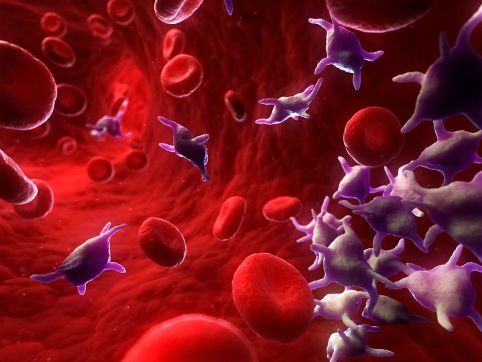
    Platelets, also known as thrombocytes, are small, irregular-shaped blood cells that play a critical role in blood clotting. Beyond their crucial function in hemostasis, platelets are now recognized for their multifaceted role in tissue repair and regeneration. They release a variety of growth factors, including platelet-derived growth factor (PDGF), transforming growth factor-beta (TGF-β), vascular endothelial growth factor (VEGF), and others.

    These growth factors stimulate the proliferation and differentiation of various cell types, promoting angiogenesis (formation of new blood vessels), collagen synthesis, and tissue remodeling. This cascade of events ultimately contributes to the healing and regeneration of damaged tissues.

    PRP Preparation Methods

    The process of isolating platelets from blood and concentrating them into a plasma solution involves several methods. Each method varies in terms of complexity, cost, and the resulting concentration of platelets in the PRP.

    • Centrifugation: This is the most common method, relying on centrifugation to separate the platelets from other blood components. The specific protocols can vary, affecting the final platelet concentration and the required time for preparation. The success of this method depends heavily on the centrifugation technique and the precision in controlling the centrifugation parameters, which can vary depending on the centrifuge’s specifications.

      PRP injections, or platelet-rich plasma, are a fascinating treatment for various health concerns. While I’m currently researching the effectiveness of PRP for hair growth, I’ve been thinking about how wearable tech like the Oura Ring and Apple Watch might complement these therapies. Comparing the features of these devices, like their sleep tracking capabilities, could offer insights into how patients respond to PRP treatments.

      For a deeper dive into the pros and cons of each wearable, check out this comparison of the Oura Ring vs Apple Watch oura ring vs apple watch. Ultimately, the goal is to leverage data to tailor PRP treatment plans for optimal results.

    Comparison of PRP Preparation Methods

    Method Platelet Concentration Preparation Time Cost Advantages Disadvantages
    Centrifugation (e.g., single-step) High Short Moderate Faster, potentially higher yield Risk of contamination, potential for inconsistent results if not properly controlled
    Centrifugation (e.g., multi-step) Moderate to High Longer Low More cost-effective, less risk of complications Slower process, may not achieve desired concentration if not optimized

    Medical Applications of PRP Injections

    PRP injections are utilized in various medical fields to address a wide range of conditions, with promising results in several areas. These include:

    • Orthopedic Conditions: PRP is used to treat osteoarthritis, tendonitis, and ligament injuries, promoting cartilage repair and tissue regeneration. Successful applications often involve precise targeting of the injured area to ensure optimal delivery of the concentrated platelets.
    • Dental Procedures: PRP is increasingly used in dental procedures to enhance bone regeneration, promote wound healing, and improve periodontal health. This application often relies on careful planning and precise injection techniques to maximize the benefits of PRP.
    • Hair Loss Treatment: PRP therapy is gaining popularity as a treatment for androgenetic alopecia (male pattern baldness), stimulating hair follicle regeneration and improving hair growth. The effectiveness of this method varies significantly depending on the individual’s specific condition.

    Mechanisms of Action

    PRP injections harness the body’s natural healing processes to stimulate tissue repair. This potent therapy leverages the concentrated platelets within the plasma, rich in growth factors and cytokines, to trigger a cascade of events that ultimately promote regeneration and healing. The specific mechanisms involved are complex and multifaceted, but understanding the underlying biological pathways is crucial for appreciating the therapeutic potential of PRP.Platelets, crucial components of the blood, are not merely passive participants in the healing process.

    Their activation within the target tissue triggers a cascade of biological responses, laying the groundwork for enhanced tissue repair. These responses are intricately linked to the release of growth factors and cytokines, proteins that act as messengers, orchestrating the intricate dance of cellular activity and tissue regeneration.

    Growth Factor and Cytokine Release

    Platelets, upon activation, release a diverse array of growth factors and cytokines. These bioactive molecules are essential for initiating and directing the healing process. Examples include transforming growth factor-β (TGF-β), vascular endothelial growth factor (VEGF), platelet-derived growth factor (PDGF), and insulin-like growth factor (IGF). These factors exert their influence on various cell types, orchestrating their behavior and promoting tissue regeneration.

    The precise concentrations and types of growth factors released by platelets influence the specific healing response.

    Stimulation of Cell Proliferation and Tissue Regeneration

    The release of growth factors and cytokines acts as a potent stimulus for cell proliferation and tissue regeneration. These factors interact with target cells, such as fibroblasts, chondrocytes, and osteoblasts, triggering their proliferation and differentiation. This increase in cell numbers and their specialization contribute to the restoration of damaged tissue structure and function. For example, in cartilage repair, chondrocytes stimulated by PRP proliferate and synthesize extracellular matrix components, contributing to the rebuilding of the cartilage tissue.

    Possible Pathways Involved in Tissue Healing

    Several pathways are involved in the complex process of tissue healing after PRP injection. These pathways include angiogenesis (new blood vessel formation), extracellular matrix remodeling, and the modulation of inflammatory responses. PRP’s effect on angiogenesis helps restore blood supply to the injured site, providing the necessary nutrients and oxygen for tissue repair. Extracellular matrix remodeling involves the breakdown and synthesis of the structural framework of the tissue, allowing for the rebuilding of the tissue’s architecture.

    PRP injections are a popular treatment for various conditions, and while they’re often used for hair growth, they can also help with other issues. Sometimes, dizziness can be a symptom of nasal polyps, which can affect breathing and lead to a variety of symptoms, including dizziness. If you’re experiencing this combination of symptoms, it’s important to understand the potential causes, such as nasal polyps symptoms dizzy , and discuss them with a healthcare professional.

    PRP injections can potentially help with the underlying issues, but they shouldn’t be your first line of defense without a proper diagnosis.

    PRP’s modulation of inflammatory responses helps to balance the healing process, preventing excessive inflammation and promoting resolution.

    Flowchart of PRP’s Healing Process

    Flowchart of PRP Healing Process

    Applications and Procedures

    PRP injections are rapidly gaining popularity as a non-surgical treatment option for a range of musculoskeletal and aesthetic concerns. This versatility stems from the unique regenerative properties of platelets, which are crucial for tissue repair and healing. The precise application and evaluation methods play a significant role in determining the effectiveness of PRP therapy.Understanding the specific procedures and evaluating the outcomes are vital for achieving optimal results.

    Different areas of the body require tailored approaches, and the evaluation process must be standardized to accurately assess treatment success.

    Medical Applications of PRP Injections

    PRP injections are utilized in various medical fields, extending beyond traditional orthopaedic applications. Platelet-rich plasma has shown promise in promoting tissue regeneration and healing in diverse areas, including sports medicine, dentistry, and dermatology. The versatility of PRP stems from its ability to stimulate the body’s natural healing mechanisms.

    Procedures for Administering PRP Injections

    The procedure for PRP injection varies based on the targeted area and the condition being treated. A standard procedure typically involves drawing a blood sample from the patient. The blood is then processed to isolate the platelets, concentrating them into a platelet-rich plasma solution. The concentrated PRP is then carefully injected into the affected area using a precise technique.

    The specific procedure for each application is tailored to ensure accurate placement and minimal discomfort.

    Evaluating PRP Efficacy

    Several methods are used to evaluate the effectiveness of PRP therapy. These include subjective assessments, such as pain reduction or improved range of motion, and objective measurements, such as imaging studies or laboratory tests. Patient feedback and clinical observations are also important factors in determining the success of PRP treatment.

    Common Medical Conditions Treated with PRP

    Condition Body Area Expected Outcomes
    Osteoarthritis Joints (e.g., knees, hips) Reduced pain, improved joint mobility, and potentially slowed disease progression. Individual results vary depending on the severity of the condition and the patient’s overall health.
    Tendinopathy (e.g., tennis elbow, Achilles tendinitis) Tendons Reduced pain, inflammation, and improved tendon function. Successful outcomes often correlate with early intervention and adherence to rehabilitation protocols.
    Hair Loss Scalp Increased hair growth, improved hair density, and slowed hair loss. The efficacy can vary based on the underlying cause of hair loss and individual patient response.
    Dental Procedures Gums, Teeth Accelerated wound healing, reduced inflammation, and improved bone regeneration in dental procedures.
    Soft Tissue Injuries (e.g., muscle strains, sprains) Muscles, ligaments Reduced pain, inflammation, and improved tissue repair and healing.

    Safety and Risks

    Platelet rich plasma prp injections

    PRP injections, while generally safe, carry potential risks. Understanding these risks and the importance of proper procedures is crucial for patient safety and successful outcomes. Thorough preparation, sterile techniques, and informed consent are paramount to minimizing complications. The specific risks can vary depending on the application.

    Potential Side Effects and Complications

    PRP injections, like any medical procedure, can result in adverse reactions. Common side effects are typically mild and temporary, while more serious complications are rare. The potential complications are often related to the injection site, the underlying condition being treated, and the individual patient’s response to the procedure.

    Importance of Proper Preparation and Sterile Techniques

    Maintaining strict sterile techniques during PRP preparation and injection is critical. Contamination can lead to infections, allergic reactions, and other complications. Proper preparation of the PRP sample, using aseptic techniques throughout the process, minimizes the risk of introducing pathogens. This includes using sterile equipment, appropriate personal protective equipment (PPE), and meticulous handling of the sample.

    Potential Risks Specific to Different Applications

    The risk profile of PRP injections can vary depending on the target area and the specific condition being treated. For example, injections into the joints might cause temporary pain or swelling, while injections into the hair follicles might lead to mild irritation. A thorough evaluation of the patient’s medical history, current medications, and the specific application is essential to assess the potential risks.

    Clinicians should carefully consider the potential benefits versus risks before proceeding with a PRP treatment plan.

    Necessity of Informed Consent

    Prior to administering PRP injections, it is imperative to obtain informed consent from the patient. This process involves explaining the procedure, potential benefits, and possible risks in clear and understandable language. The patient must comprehend the procedure, its potential outcomes, and the associated risks before consenting to the treatment. This ensures the patient’s autonomy and their ability to make an informed decision about their healthcare.

    Table of Potential Side Effects

    Side Effect Frequency Severity Treatment
    Pain at injection site Common Mild to Moderate Ice packs, over-the-counter pain relievers
    Swelling at injection site Common Mild to Moderate Elevation, cold compresses, compression bandages
    Bruising at injection site Common Mild Observation; typically resolves spontaneously
    Infection at injection site Rare Moderate to Severe Antibiotics, possibly surgical intervention
    Allergic reaction Rare Moderate to Severe Antihistamines, epinephrine (if severe)
    Nerve damage (rare) Very Rare Severe Rehabilitation, pain management
    Hematoma formation Rare Mild to Moderate Compression, observation; resolves over time

    Clinical Evidence and Research: Platelet Rich Plasma Prp Injections

    Platelet-rich plasma (PRP) injections have shown promise in various clinical settings, but robust, high-quality research is crucial to solidify their effectiveness and safety profile. The current body of evidence, while encouraging, requires careful consideration of study methodologies and outcomes to understand the full picture. This section delves into the clinical studies investigating PRP’s effectiveness and highlights key findings, limitations, and future research directions.A substantial amount of research explores the use of PRP in musculoskeletal conditions, aiming to understand its potential in promoting healing and reducing pain.

    The variety of conditions studied—from osteoarthritis to ligament injuries—reflects the broad scope of potential applications. Analyzing these studies is essential for determining the appropriate use of PRP in different scenarios.

    Summary of Current Clinical Studies

    The existing clinical studies on PRP are diverse in their methodologies, patient populations, and outcomes. Some studies focus on specific conditions like osteoarthritis, while others examine a wider range of injuries. Crucially, the quality of the research varies significantly, impacting the reliability of the conclusions drawn. This variation emphasizes the need for more rigorous, well-designed trials to validate the observed benefits.

    Examples of Published Research

    Numerous studies have investigated the efficacy of PRP in various conditions. For instance, a meta-analysis published in the

    • American Journal of Sports Medicine* summarized findings from multiple trials, concluding that PRP showed potential in treating certain types of tendinopathies. The meta-analysis highlighted the importance of standardized protocols and proper PRP preparation techniques. Another study, published in the
    • Journal of Orthopaedic Research*, focused on the effects of PRP on cartilage regeneration in patients with knee osteoarthritis. The study demonstrated a positive trend in cartilage repair, although further large-scale studies are needed to confirm these initial results.

    Limitations of Current Research, Platelet rich plasma prp injections

    Several limitations characterize current PRP research. First, the sample sizes in many studies are relatively small, making it difficult to draw definitive conclusions about the overall effectiveness. Second, variations in PRP preparation methods and injection techniques can influence treatment outcomes. The heterogeneity of study designs and the lack of standardized protocols pose challenges in comparing results across different investigations.

    Lastly, long-term follow-up data are often lacking, making it difficult to assess the sustained benefits of PRP treatment.

    Structured Reporting of Research Findings

    A well-structured reporting of research findings includes several key elements:

    • Sample Size: A larger sample size generally leads to more reliable results, allowing for a better understanding of the treatment’s effect on a wider population. Small sample sizes can obscure true effects or yield misleading conclusions.
    • Study Design: Randomized controlled trials (RCTs) are considered the gold standard for evaluating the effectiveness of interventions. RCTs help minimize bias and ensure a more accurate comparison between treatment groups.
    • Outcomes: Clear, measurable outcomes are essential for evaluating the impact of PRP injections. Examples include pain reduction, functional improvement, and radiological changes (e.g., cartilage repair). Specific outcome measures should be clearly defined and consistently applied throughout the study.

    Areas for Future Investigation

    Future research should focus on addressing the limitations of current studies. This includes larger, well-designed RCTs, standardized PRP preparation protocols, and long-term follow-up studies. Further research should also explore the optimal timing and dosage of PRP injections, as well as the mechanisms through which PRP exerts its effects. Additionally, investigations into the identification of patient sub-groups most likely to respond favorably to PRP treatment are essential.

    Understanding patient factors that correlate with treatment success would allow for more personalized approaches.

    Patient Considerations

    PRP injections offer a promising approach to various medical conditions, but understanding the process and expectations is crucial for a positive outcome. This section delves into the key considerations for patients considering PRP therapy, from preparation to recovery and the importance of selecting qualified professionals. Informed decisions are paramount to achieving the best possible results.

    Essential Information for Patients

    Patients need a comprehensive understanding of PRP therapy’s potential benefits and limitations. This includes awareness of the specific condition being treated, potential risks, and realistic expectations about the procedure’s efficacy. Thorough communication between the patient and medical professional is essential to ensure shared understanding and manage expectations effectively.

    PRP injections are becoming a popular treatment option for various musculoskeletal issues. They’re often used to stimulate tissue repair, and can potentially help with bone density issues. For example, in cases of osteoporosis-related fractures, treatments like those detailed in osteoporosis fractures treatment and prevention may be combined with PRP to encourage bone regeneration and healing.

    Ultimately, PRP injections could play a role in strengthening bones and preventing future fractures.

    Preparation and Expectations Before the Procedure

    Careful preparation significantly impacts the PRP injection’s success. Patients should follow the medical professional’s instructions meticulously. This includes abstaining from certain medications, informing the medical team about current health conditions, and managing any pre-existing medical issues that could potentially influence the procedure. A detailed discussion of any concerns or questions is encouraged before the procedure.

    Post-Injection Care and Recovery Time

    Post-injection care is vital for optimal healing and minimizing potential complications. Patients should adhere to specific instructions provided by the medical professional. This may include recommendations for rest, activity limitations, ice application, and pain management strategies. Recovery time varies depending on the treated area and individual factors, but patients should be prepared for some level of discomfort and potential swelling.

    It’s crucial to follow the recommended recovery timeline diligently.

    Choosing Qualified Medical Professionals

    Selecting a qualified medical professional is paramount. Look for practitioners with experience in administering PRP injections, a strong understanding of the specific condition being treated, and a proven track record of successful outcomes. Verify the practitioner’s credentials, licenses, and any relevant certifications. Seeking recommendations from trusted sources, such as other patients or healthcare providers, can also be beneficial.

    A thorough consultation is crucial to assess the suitability of the therapy for the individual patient.

    Questions to Ask Before Undergoing PRP Therapy

    A proactive approach to questions before undergoing PRP therapy is essential for informed decision-making. A comprehensive understanding of the procedure is critical. Here’s a list of essential questions to consider:

    • What is the specific condition being treated, and what are the potential benefits of PRP therapy for this condition?
    • What are the potential risks and complications associated with PRP injections?
    • What is the success rate of PRP therapy for this specific condition based on your experience and clinical evidence?
    • What is the expected recovery time, and what are the post-injection care instructions?
    • What are the alternative treatment options, and how does PRP compare to these options?
    • What are the costs associated with the procedure, including potential follow-up visits?
    • What are the qualifications and experience of the medical professional administering the PRP injections?
    • What are the protocols for managing any potential adverse effects?

    Future Directions

    PRP therapy is rapidly evolving, driven by ongoing research and technological advancements. This dynamic field promises exciting possibilities for enhancing treatment outcomes and expanding applications beyond current practices. The future of PRP likely involves a more personalized and integrated approach to patient care, leveraging technology to optimize treatment efficacy and safety.

    Emerging Trends and Technologies

    Advancements in PRP preparation techniques are constantly refining the process. Improved methods for isolating platelets and separating them from other blood components lead to higher concentrations of growth factors. This translates to potentially more effective treatments with fewer side effects. For instance, novel centrifugation protocols and filtration techniques are yielding more pure PRP preparations, enabling greater control over the concentration of growth factors and minimizing the presence of potentially harmful substances.

    Potential Advancements in PRP Preparation and Application Techniques

    Innovations in PRP application techniques are equally important. Minimally invasive approaches are being explored, offering greater precision and reduced patient discomfort. The development of targeted drug delivery systems, combined with PRP, is also gaining traction. Imagine a scenario where growth factors are precisely delivered to damaged tissues using nanocarriers, maximizing their therapeutic impact and minimizing side effects.

    This could lead to more precise and targeted healing in various applications, such as sports injuries or dermatological procedures.

    Role of Personalized Medicine in PRP Treatment Protocols

    Personalized medicine is poised to revolutionize PRP therapy. Analyzing individual patient factors, such as genetic predispositions, specific tissue damage, and the patient’s response to other therapies, can optimize PRP treatment protocols. This involves using genetic testing and other biological markers to tailor the PRP preparation and application strategy for optimal results. For example, a patient with a specific genetic predisposition to slower wound healing could receive a personalized PRP protocol designed to accelerate the healing process.

    Integration of PRP Therapy with Other Medical Treatments

    The integration of PRP with other medical treatments is another promising area. Combining PRP with stem cell therapy or other regenerative medicine approaches holds great potential. For example, PRP injections might be used in conjunction with physical therapy to accelerate recovery from musculoskeletal injuries. This synergistic approach could lead to improved outcomes and a more comprehensive treatment plan for patients.

    Closing Notes

    Platelet rich plasma prp injections

    In conclusion, platelet-rich plasma injections offer a promising avenue for tissue regeneration and pain management. While promising, it’s crucial to acknowledge the evolving nature of research and the need for ongoing studies to fully understand its efficacy and potential limitations. The diverse applications, ranging from joint repair to hair restoration, highlight the versatility of this treatment. Ultimately, responsible and informed decision-making, coupled with the guidance of qualified healthcare professionals, are essential when considering PRP injections.

    Further research and clinical trials will be critical in expanding our knowledge of PRP’s potential and ensuring its safe and effective use.