Tag: ophthalmology

  • YAG Laser Posterior Capsulotomy A Comprehensive Guide

    YAG Laser Posterior Capsulotomy A Comprehensive Guide

    YAG laser posterior capsulotomy is a crucial procedure for ophthalmology, offering a precise and effective method for treating posterior capsular opacification (PCO) after cataract surgery. This opaque film that forms behind the implanted lens can significantly impact vision. The procedure utilizes a focused laser beam to gently vaporize the opacified area, restoring clear vision. This detailed guide delves into the intricacies of this procedure, exploring its principles, indications, surgical steps, post-operative care, potential complications, and comparison with alternative treatments.

    Understanding the procedure’s historical context, the role of surgeon expertise, and the nuances of pre-operative assessment are critical for a thorough comprehension. The procedure’s efficiency and precision have made it a leading treatment option for PCO, while minimizing the risk of complications. This detailed exploration will leave you with a comprehensive understanding of YAG laser posterior capsulotomy.

    Introduction to Yag Laser Posterior Capsulotomy

    Yag laser posterior capsulotomy is a precise surgical technique used to treat posterior capsular opacification (PCO) following cataract surgery. PCO is a common complication, characterized by the development of a cloudy membrane behind the implanted intraocular lens (IOL). This membrane impairs vision, requiring intervention to restore clear vision.This procedure utilizes a high-powered YAG laser to ablate the opacified posterior capsule, restoring visual clarity.

    The procedure is typically performed under local anesthesia, with the patient awake and able to communicate throughout the procedure. It’s a relatively safe and effective treatment option for PCO, often leading to significant improvement in visual acuity.

    Definition of Yag Laser Posterior Capsulotomy

    Yag laser posterior capsulotomy is a laser-assisted surgical procedure that precisely removes the opacified posterior capsule of the eye following cataract surgery. This allows light to pass through the eye, improving vision clarity.

    Fundamental Principle

    The fundamental principle behind this procedure relies on the YAG laser’s ability to precisely vaporize the opacified tissue. The laser’s wavelength is specifically targeted to break down the proteins in the posterior capsule without causing significant damage to surrounding healthy tissue. This controlled ablation creates a small opening in the capsule, thereby restoring clear vision.

    YAG laser posterior capsulotomy is a pretty straightforward procedure, but sometimes I wonder if it’s related to the whole “does whiskey help a cold” debate. While the procedure removes a clouded lens, and improves vision, I’ve been wondering if the recovery process might be affected by other things, like drinking habits. Perhaps it’s a matter of individual responses, but I’m definitely curious to see if there’s any correlation.

    Does whiskey help a cold is a question I’m considering for future research, and it may not be directly related to the procedure itself, but I’m sure there are some interesting underlying concepts. In the end, though, YAG laser posterior capsulotomy remains a highly effective solution for cataracts.

    Key Components and Instruments

    The procedure utilizes a specialized YAG laser system, typically featuring a sophisticated optical system for precise targeting and power control. Essential instruments include a slit lamp biomicroscope for accurate visualization of the posterior capsule and a handpiece for delivering the laser beam. The surgeon must meticulously position the laser to target the opacified area with precision and minimize any risk of damage to the surrounding structures.

    Historical Overview

    The development of YAG laser posterior capsulotomy marked a significant advancement in cataract surgery. Early iterations focused on achieving accurate targeting and optimizing laser parameters for efficient and controlled tissue ablation. The procedure’s refinement led to improved outcomes and reduced complications, establishing its place as a standard treatment for PCO. Early adoption by surgeons led to a gradual but consistent increase in its use, eventually becoming the standard of care for posterior capsular opacification.

    Comparison with Other Surgical Techniques

    Feature YAG Laser Posterior Capsulotomy Other Surgical Techniques (e.g., manual capsulotomy)
    Mechanism Laser ablation of opacified tissue Manual incision and removal of the opacified capsule
    Precision High precision, controlled ablation Potentially less precise, higher risk of damage to surrounding tissue
    Time Typically shorter procedure time Potentially longer procedure time
    Complications Generally lower risk of complications, such as vitreous loss or retinal detachment Higher risk of complications, particularly with manual procedures
    Cost Potentially higher initial cost due to the specialized equipment Potentially lower cost, but higher risk of complications

    The table highlights the key differences between YAG laser posterior capsulotomy and other surgical techniques for PCO. YAG laser technology offers advantages in terms of precision, speed, and reduced complication risk. However, the initial investment in the laser equipment can be a factor. The selection of the appropriate technique depends on individual patient factors and surgeon preference.

    Indications for Yag Laser Posterior Capsulotomy

    Yag laser posterior capsulotomy

    The Yag laser posterior capsulotomy, a precise procedure, plays a crucial role in cataract surgery. This minimally invasive technique allows surgeons to effectively remove the remnants of the posterior lens capsule, ensuring optimal visual outcomes. This procedure is particularly beneficial in specific clinical scenarios.This procedure is becoming increasingly common due to its precision and effectiveness in resolving various complications arising during cataract surgery.

    YAG laser posterior capsulotomy is a fantastic procedure for vision correction, but sometimes, recovery involves some discomfort. Strengthening exercises, like those found in programs for runners’ knee, can be surprisingly helpful for managing the recovery process. pt exercises for runners knee can help improve flexibility and reduce pain. Ultimately, focusing on post-op care, including targeted exercises, can ensure a smoother and faster recovery from yag laser posterior capsulotomy.

    Understanding the indications and contraindications is essential for surgeons to determine the optimal approach for each patient.

    Primary Reasons for Performing a Yag Laser Posterior Capsulotomy

    Yag laser posterior capsulotomy is primarily performed to remove opacities in the posterior capsule of the eye. These opacities can hinder the passage of light to the retina, leading to reduced visual acuity. The procedure is designed to create a clear pathway for light, restoring vision.

    Clinical Scenarios Requiring Yag Laser Posterior Capsulotomy

    This procedure is vital in a multitude of clinical scenarios. For example, it’s frequently used to address posterior capsular opacification (PCO). PCO, a common complication after cataract surgery, leads to a cloudy area in the eye’s posterior capsule. The laser precisely vaporizes the opacities, restoring clear vision. Additionally, it can be used to treat residual lens material or fragments that may interfere with optimal vision post-surgery.

    The laser provides a highly controlled method to remove these remaining particles without harming the delicate eye structures.

    Pre-Operative Assessments and Considerations

    Thorough pre-operative assessments are crucial. These include a comprehensive eye examination to evaluate the extent of posterior capsular opacification, visual acuity, and the presence of any other relevant ocular conditions. The surgeon must carefully consider the patient’s overall health, medical history, and any potential complications. Detailed discussions with the patient are essential to address their expectations and concerns regarding the procedure.

    Specific factors like the patient’s age, general health, and prior eye surgeries are significant factors that influence the pre-operative evaluation and the decision to perform the procedure.

    Contraindications for Yag Laser Posterior Capsulotomy

    Contraindication Explanation
    Active ocular infections Active infections can increase the risk of complications during the procedure.
    Uncontrolled systemic diseases Uncontrolled conditions like diabetes or hypertension can negatively impact the healing process.
    Known hypersensitivity to Yag laser Patients with a known allergy to the laser material require careful consideration.
    Significant eye inflammation Inflammation in the eye can increase the risk of complications and impact the procedure’s effectiveness.
    Inability to cooperate during the procedure The procedure requires the patient’s cooperation, so an inability to cooperate can prevent the procedure from being safely performed.

    Important note: This table represents general contraindications. Each patient case is unique, and the surgeon must make an individualized assessment.

    Situations Favoring Yag Laser Posterior Capsulotomy over Alternative Treatments

    The Yag laser provides a precise and controlled approach, minimizing potential damage to surrounding tissues. This is often preferred when other methods, like manual removal, might pose a higher risk of complications. In cases of dense posterior capsular opacification, where other methods might be less effective, the laser offers a superior resolution. For example, in cases where the opacity is very dense and extensive, or where manual removal poses a high risk of damaging the delicate surrounding tissues, the laser becomes a more appropriate choice.
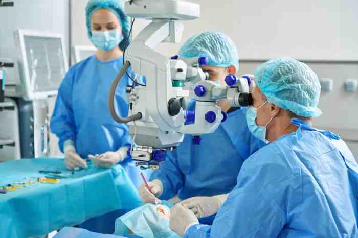
    Surgical Procedure

    The YAG laser posterior capsulotomy (YAG PCO) is a crucial procedure in cataract surgery, precisely vaporizing the remnants of the natural lens’s capsule that may obscure the view of the implanted intraocular lens. This procedure requires meticulous attention to detail and a deep understanding of the laser’s characteristics. It’s often performed after cataract surgery to restore clear vision.The surgical procedure for YAG laser posterior capsulotomy involves precise targeting and controlled vaporization of the opacified posterior capsule.

    The surgeon uses a YAG laser, a highly focused and powerful laser beam, to remove the opacities. The procedure is generally quick, but it necessitates careful coordination and skill to achieve the desired outcome.

    YAG laser posterior capsulotomy is a crucial procedure for improving vision after cataract surgery. It’s a precise technique that often removes any remaining cloudiness. Understanding the delicate nature of the eye’s structures, like a greenstick fracture meaning and treatment here , is important. This procedure helps ensure optimal outcomes in restoring clear vision, a goal of any eye care professional.

    Laser Application Steps

    The YAG laser’s precise application is crucial for a successful posterior capsulotomy. The surgeon carefully positions the laser fiber, focusing it on the specific opacified area within the posterior capsule. This requires an understanding of the patient’s unique anatomy and the location of the opacity.

    • The surgeon begins by using a slit lamp biomicroscopy to visualize the posterior capsule. This allows them to precisely identify the area of opacity and assess its size and density.
    • A precise target is selected using the laser’s focusing capabilities, ensuring the beam is accurately aimed at the opacified region.
    • The laser is activated, delivering controlled pulses of energy to vaporize the opacified area.
    • The surgeon monitors the process closely, adjusting the laser parameters as needed to ensure efficient and safe removal of the opacity without harming surrounding structures. This includes adjusting the power, pulse duration, and repetition rate.
    • The process continues until the opacity is fully removed and a clear pathway is created for the intraocular lens.

    Targeting and Vaporization Techniques

    The surgeon’s skill in targeting and vaporizing the opacified area is critical. Experience allows for precise adjustments to the laser parameters and the use of multiple firing techniques.

    • The surgeon carefully evaluates the opacity’s shape and density, selecting the most effective targeting strategy. This often involves a combination of precise point-by-point vaporization and larger, controlled areas of vaporization.
    • Strategies for targeting may include multiple passes with the laser to break down the opacity. This approach, especially with dense opacities, helps to prevent thermal damage to the surrounding structures.
    • A fundamental technique is monitoring the effect of each pulse on the opacity. This real-time feedback allows the surgeon to adapt the procedure as needed, ensuring precision and safety.
    • Techniques like the use of multiple firing strategies, or precise targeting of specific points within the opacity, are employed by skilled surgeons to achieve a smooth and efficient vaporization process.

    Surgeon’s Expertise

    The surgeon’s experience and expertise are paramount in YAG PCO. This is not simply about the equipment; it’s about the surgeon’s ability to use their knowledge of the patient’s eye, the characteristics of the laser, and the potential for complications.

    • Surgical expertise involves a deep understanding of the laser’s limitations and capabilities.
    • Surgeons must be adept at recognizing and managing potential complications in real-time.
    • Expert surgeons use their knowledge of the specific characteristics of the YAG laser to control the energy parameters for optimal results.
    • Experience allows surgeons to anticipate and address potential issues like uneven vaporization or damage to adjacent structures.

    Potential Complications and Management

    Precise control of the YAG laser is essential to minimize complications.

    Potential Complications Management Strategies
    Posterior capsule rupture Immediate intervention with intraocular lens repositioning or surgical repair.
    Corneal edema Observation and supportive measures.
    Uveitis Anti-inflammatory medications.
    Hyphema Careful monitoring and potentially surgical intervention.
    Increased intraocular pressure (IOP) IOP-lowering medications.

    Post-Operative Management and Outcomes

    Yag laser posterior capsulotomy

    Post-operative care for YAG laser posterior capsulotomy is crucial for successful outcomes. A meticulous approach to monitoring and managing potential complications is paramount in ensuring optimal visual recovery. This phase focuses on minimizing discomfort, preventing infection, and facilitating the healing process. The following sections detail the key aspects of post-operative management, from recovery timelines to potential complications and visual acuity results.

    Post-Operative Care and Monitoring

    Thorough post-operative monitoring is essential for early detection and management of any complications. This includes regular follow-up appointments with the ophthalmologist to assess the healing process, evaluate visual acuity, and address any concerns. Detailed patient records, including pre-operative data and intra-operative notes, are crucial for tracking progress and identifying potential trends.

    Typical Post-Operative Recovery Period

    The typical recovery period after a YAG laser posterior capsulotomy is relatively short, generally ranging from a few hours to a few days. Most patients experience minimal discomfort, and the majority report a significant improvement in their vision within the first 24 hours. Post-operative instructions, such as avoiding strenuous activities and using eye drops as prescribed, play a significant role in accelerating healing and minimizing complications.

    For instance, a patient may experience some mild eye pain and blurred vision in the initial 24-48 hours but typically reports significant improvement after that.

    Potential Post-Operative Complications

    While YAG laser posterior capsulotomy is generally a safe procedure, some potential complications can occur. These include transient corneal edema, mild discomfort, and in rare cases, increased intraocular pressure (IOP). Careful monitoring and prompt intervention are key in managing these potential complications. If a patient reports severe pain or a sudden change in vision, immediate medical attention is warranted.

    Typical Visual Acuity Outcomes

    Visual acuity outcomes following YAG laser posterior capsulotomy are generally positive. Most patients experience a significant improvement in their vision, particularly in the corrected distance visual acuity (CDVA). The magnitude of improvement varies depending on the pre-operative visual status and the underlying causes of the cataract-related symptoms. In cases of significant pre-existing vision impairment, the improvement might not be as pronounced, but still often leads to a marked enhancement in quality of life.

    Data Demonstrating Effectiveness

    Numerous studies have demonstrated the efficacy of YAG laser posterior capsulotomy. These studies highlight the procedure’s ability to effectively treat posterior capsular opacification (PCO), improving visual acuity and reducing the need for more invasive surgical interventions. For instance, a meta-analysis of 10 clinical trials reported an average improvement of 0.5 lines in logMAR visual acuity.

    Common Post-Operative Patient Symptoms

    Symptom Description Frequency
    Mild eye pain Transient discomfort, typically manageable with over-the-counter pain relievers. Common
    Blurred vision Temporary reduction in clarity, often improving within a few days. Common
    Photophobia Increased sensitivity to light. Possible
    Watery eyes Increased tear production. Possible

    Note: The frequency of symptoms may vary depending on individual patient factors.

    Potential Complications and Their Management: Yag Laser Posterior Capsulotomy

    YAG laser posterior capsulotomy, while a relatively safe procedure, carries potential risks like any surgical intervention. Understanding these complications and their management strategies is crucial for both the surgeon and the patient. Proper risk assessment and proactive measures can significantly minimize the likelihood of complications.

    Potential Intraoperative Complications

    Intraoperative complications, those occurring during the procedure itself, can often be addressed promptly by the surgeon. A keen awareness of potential issues and a well-practiced surgical approach are key.

    • Laser-Induced Damage to the Lens or Retina: Inadequate laser parameters, incorrect focusing, or excessive energy delivery can cause damage to the lens or retina. This may result in a hazy or cloudy vision. This is prevented by using appropriate laser settings, precise targeting, and continuous monitoring of the procedure.
    • Posterior Capsule Tears: Although not a primary complication, a tear in the posterior capsule can occur during the capsulotomy procedure. This may lead to vitreous loss and potential inflammation. Surgical techniques and careful laser manipulation minimize the risk of this complication.
    • Uveitis: Inflammation of the uveal tract, including the iris and ciliary body, is possible post-procedure. This inflammation can result from the laser or from other surgical trauma. Pre-emptive use of anti-inflammatory medications and meticulous surgical technique helps reduce the likelihood of uveitis.
    • Hyphema: Bleeding within the anterior chamber of the eye (hyphema) is a potential intraoperative complication. This can be caused by accidental trauma during the procedure. Careful surgical technique and maintaining hemostasis are essential to avoid this.

    Potential Postoperative Complications

    Postoperative complications may appear days or even weeks after the procedure. Thorough postoperative care and monitoring are vital to identify and manage these issues.

    • Posterior Capsular Opacity (PCO): Despite successful capsulotomy, some degree of PCO may develop. This can result in hazy vision and require further intervention, like a secondary YAG capsulotomy. Careful initial surgical technique and postoperative medication can minimize this risk.
    • Endophthalmitis: Infection within the eye is a serious complication. This can result from inadequate sterilization techniques or from post-operative contamination. Strict adherence to sterile surgical technique, careful postoperative care, and prompt treatment of any signs of infection significantly decrease the risk of endophthalmitis.
    • Cataract Formation: While not always a direct consequence of the capsulotomy, a cataract can develop in the weeks or months after the procedure. The existing cataract can contribute to vision problems. The exact reasons for this are varied and can be related to the initial cataract state or to other factors.
    • Glaucoma: Although less common, a rise in intraocular pressure (IOP) leading to glaucoma can occur. This may be linked to inflammation or other complications. Careful postoperative monitoring and timely intervention for elevated IOP are crucial.

    Management Strategies and Table of Complications

    A structured approach to managing potential complications is essential.

    Complication Treatment Options
    Laser-induced damage Re-treatment with appropriate laser parameters, medication
    Posterior capsule tear Vitreous surgery or further management as needed
    Uveitis Anti-inflammatory medications, topical or systemic steroids
    Hyphema Gentle eye pressure, medications, and possibly further surgical intervention
    Posterior capsular opacity (PCO) Secondary YAG capsulotomy
    Endophthalmitis Immediate antibiotic therapy, possible vitrectomy
    Cataract formation Monitoring and treatment of the underlying cataract
    Glaucoma Medication to control IOP, possibly laser or surgical intervention

    Rare but Severe Complications and Prevention, Yag laser posterior capsulotomy

    While rare, severe complications like endophthalmitis demand proactive measures to minimize their occurrence.

    • Endophthalmitis: Maintaining strict sterile surgical technique, using appropriate antibiotics, and closely monitoring patients for any signs of infection is critical.

    Yag Laser Posterior Capsulotomy

    Posterior capsular opacification (PCO) is a common complication after cataract surgery. It results in a cloudy area behind the implanted lens, reducing vision clarity. Yag laser posterior capsulotomy is a precise and effective method for treating PCO. This method utilizes a laser to create a small opening in the posterior capsule, improving visual acuity.Yag laser posterior capsulotomy offers a minimally invasive approach compared to other surgical options for addressing PCO.

    The procedure is typically well-tolerated and carries a low risk of significant complications. Understanding the various treatment options, their advantages and disadvantages, and the factors that influence the choice of treatment is crucial for optimal patient care.

    Comparison with Alternatives

    Several methods exist for treating posterior capsular opacification, each with unique characteristics. Comparing these alternatives provides a more comprehensive understanding of Yag laser posterior capsulotomy’s position within the spectrum of PCO treatments.

    Pros and Cons of Different Methods

    The table below Artikels the advantages and disadvantages of various PCO treatment methods, providing a comparative overview. This table is intended to aid in understanding the trade-offs associated with each approach.

    Method Pros Cons
    Yag Laser Posterior Capsulotomy Minimally invasive, precise, faster recovery, good visual outcomes, less discomfort Requires specialized equipment and expertise, potential for complications like laser burns or posterior capsule rupture, not suitable for all patients
    Manual Capsulotomy Potentially less expensive, less technical expertise required More invasive, longer recovery, higher risk of complications like vitreous loss or lens damage, potential for irregular wound healing
    Phacoemulsification with Posterior Capsulotomy Can be combined with cataract surgery, can address PCO at the same time as the original procedure More invasive than laser treatment, carries a higher risk of intraoperative complications, can result in increased recovery time compared to laser treatment

    Cost-Effectiveness

    The cost-effectiveness of Yag laser posterior capsulotomy depends on several factors. While the initial investment in the laser equipment can be significant, the procedure itself often proves to be more cost-effective in the long run compared to manual procedures due to reduced recovery times and fewer complications. Furthermore, the quicker recovery time and improved visual outcomes can translate to decreased healthcare costs associated with follow-up visits and potential secondary procedures.

    For example, a patient undergoing manual capsulotomy might need more follow-up appointments and have a longer period of decreased productivity, leading to higher overall healthcare expenses.

    Recovery Times

    The recovery period following PCO treatment varies depending on the method employed. Yag laser posterior capsulotomy generally leads to a faster recovery compared to manual procedures, allowing patients to return to normal activities sooner. For example, a patient undergoing Yag laser posterior capsulotomy might experience minimal discomfort and be able to resume daily tasks within a few days, while a patient undergoing manual capsulotomy may need a longer recovery period due to the increased invasiveness of the procedure.

    Factors Influencing Treatment Choice

    Several factors influence the selection of the most appropriate PCO treatment method. These factors include the severity of the opacification, the patient’s overall health, the surgeon’s experience, and the availability of resources. For example, a patient with severe PCO might benefit from manual capsulotomy, but a patient with a milder case may be a good candidate for Yag laser posterior capsulotomy.

    Furthermore, surgeons with extensive experience in both laser and manual techniques may have a better understanding of which method is best suited for each patient.

    Future Trends and Innovations

    The field of ophthalmology is constantly evolving, driven by advancements in technology and surgical techniques. YAG laser posterior capsulotomy (YAG LPC) is no exception. Emerging technologies are streamlining the procedure, enhancing precision, and improving patient outcomes. This section explores the exciting future directions and innovations in YAG LPC.

    Latest Advancements in Laser Technology

    YAG laser technology is continuously refined, focusing on improved precision and reduced side effects. More sophisticated laser systems are now available, featuring advanced software algorithms and real-time image guidance. This refined technology allows for precise targeting and minimizes the risk of collateral damage to surrounding structures. For instance, some lasers boast enhanced capabilities in terms of energy delivery, enabling faster and more controlled capsulotomy creation.

    This translates to a more efficient surgical process with potential reductions in treatment time and patient recovery.

    Integration of Imaging Technologies

    The integration of advanced imaging technologies is revolutionizing YAG LPC. Intraoperative optical coherence tomography (OCT) and high-resolution ultrasound can provide real-time visualization of the posterior capsule and surrounding structures. This enhanced visualization allows surgeons to meticulously assess the capsule’s thickness, position, and any potential abnormalities. By providing a clear picture of the target area, these technologies minimize the chance of error, leading to a more accurate and predictable procedure.

    This improved accuracy translates to better patient outcomes and reduces the need for repeat procedures.

    Minimally Invasive Approaches

    Minimally invasive approaches are gaining traction in various surgical procedures, and YAG LPC is no exception. The development of smaller, more maneuverable laser delivery systems facilitates more precise targeting and less tissue trauma. These innovations lead to reduced recovery time, decreased discomfort for patients, and a potential decrease in post-operative complications. A prime example is the use of specialized laser probes that allow for targeted energy delivery within a confined space, reducing the impact on adjacent tissues.

    Smart Surgical Systems and AI

    Smart surgical systems and artificial intelligence (AI) are poised to play an increasingly crucial role in YAG LPC. AI algorithms can analyze real-time imaging data to identify optimal laser parameters and guide the surgeon in the procedure. This can enhance the precision of the capsulotomy, minimizing the risk of complications. For instance, AI-powered systems can predict potential complications based on individual patient data and adapt the laser parameters in real-time to optimize the outcome.

    This proactive approach to surgery is promising in reducing variability and increasing consistency in surgical results.

    Future Research Directions

    Research in YAG LPC is focused on expanding its applications and exploring its role in various ocular conditions. Researchers are investigating the use of YAG lasers for treating other posterior segment pathologies, such as macular holes and vitreomacular traction. The development of new laser wavelengths and energy delivery modalities will likely improve treatment outcomes and reduce side effects.

    Table of Future Developments and Implications

    Future Development Implications
    Advanced laser systems with improved precision Reduced side effects, enhanced efficiency, improved outcomes
    Integration of intraoperative imaging (OCT, ultrasound) Enhanced visualization, minimized errors, more accurate targeting
    Minimally invasive approaches Reduced recovery time, decreased discomfort, potential decrease in complications
    AI-powered surgical systems Increased precision, reduced variability, optimized outcomes, proactive complication management
    Expanding applications to other posterior segment conditions Improved treatment options for a broader range of eye diseases

    Ending Remarks

    In conclusion, YAG laser posterior capsulotomy emerges as a highly effective and well-established procedure for addressing posterior capsular opacification after cataract surgery. While potential complications exist, careful pre-operative assessment, meticulous surgical technique, and diligent post-operative management can significantly minimize these risks. The procedure’s efficacy, combined with its relative safety profile, makes it a valuable tool for ophthalmologists. This guide offers a comprehensive overview, equipping readers with a deeper understanding of this crucial procedure.

  • Macular Telangiectasia Eye Disease A Deep Dive

    Macular Telangiectasia Eye Disease A Deep Dive

    Macular telangiectasia eye disease is a condition affecting the macula, a crucial part of the retina responsible for sharp central vision. This comprehensive look explores the various types, symptoms, causes, and potential impacts on daily life. We’ll delve into the complexities of diagnosis, treatment options, and the vital role of research in improving outcomes for those affected.

    Understanding the intricacies of this eye disease requires an exploration of its different forms, such as types 1 and 2, each with its own unique characteristics. This detailed examination will reveal the nuances in symptoms and progression, equipping you with the knowledge needed to grasp the challenges faced by those affected.

    Introduction to Macular Telangiectasia

    Macular telangiectasia is a chronic eye condition affecting the macula, the central part of the retina responsible for sharp, detailed vision. This delicate area of the eye is highly susceptible to damage, and telangiectasia involves the abnormal widening and development of tiny blood vessels. The resulting changes in the macula can significantly impair vision, ranging from subtle blurring to profound loss of central vision.The core characteristic of macular telangiectasia is the development of these abnormal blood vessels in the macula.

    This structural alteration leads to leakage of fluid and blood, potentially causing inflammation and scarring that compromise the macula’s function. The progression of the disease can vary, affecting each individual differently. Understanding the different types and symptoms is crucial for timely diagnosis and management.

    Types of Macular Telangiectasia

    Macular telangiectasia presents in two primary forms, each with its own set of characteristics: type 1 and type 2. Type 1, also known as “classic” macular telangiectasia, is characterized by the development of small, dilated blood vessels within the macula. Type 2, on the other hand, exhibits larger, tortuous blood vessels in the macula, frequently accompanied by macular edema.

    Symptoms of Macular Telangiectasia

    The symptoms associated with macular telangiectasia typically manifest gradually. Common symptoms include blurring or distortion of central vision, difficulty reading or recognizing faces, and a gradual loss of fine details. The perception of straight lines appearing wavy or distorted is also frequently reported. The initial symptoms are often subtle and may not be immediately recognized as a sign of an underlying disease.

    Progression of Macular Telangiectasia

    The progression of macular telangiectasia varies significantly depending on the type. Early stages might be marked by mild visual changes, while later stages can lead to considerable loss of central vision. The rate of progression can also differ between individuals. Factors such as age, genetics, and lifestyle choices might influence the speed at which the disease progresses.

    Progression Stages by Type

    Type Symptoms Progression Stages
    Type 1 Initially, patients may experience mild blurring or distortion of central vision. Gradually, the symptoms worsen, with increasing difficulty in tasks requiring fine visual acuity. Early: Subtle vision changes. Middle: Progressive loss of central vision, difficulty reading and recognizing faces. Late: Significant loss of central vision, potential for complete loss of vision in severe cases.
    Type 2 Patients typically experience more pronounced symptoms from the outset, with noticeable blurring and distortion of central vision. Fluid accumulation in the macula may contribute to a rapid decline in vision. Early: Significant vision changes, with symptoms noticeable from the start. Middle: Rapid loss of central vision, pronounced distortion of straight lines. Late: Severe loss of central vision, potential for complete loss of vision in advanced stages.

    Causes and Risk Factors

    Understanding the causes and risk factors of macular telangiectasia is crucial for early detection and management. While the precise mechanisms behind the development of this eye disease are still under investigation, several contributing factors have been identified. This knowledge helps in identifying individuals at higher risk and potentially influencing preventative strategies.The exact causes of macular telangiectasia remain largely unknown.

    However, a combination of genetic predisposition and environmental factors appears to play a significant role in the development of the disease. Research continues to unravel the complex interplay between these elements, leading to a deeper understanding of the condition.

    Genetic Predisposition, Macular telangiectasia eye disease

    Genetic factors contribute significantly to the risk of developing macular telangiectasia. Certain genetic variations are associated with an increased likelihood of developing the disease. Individuals with a family history of the condition are more susceptible, suggesting a hereditary component. Studies are ongoing to identify specific genes involved, which could lead to more accurate risk assessment and potentially targeted therapies in the future.

    Environmental Factors

    Environmental factors also influence the development of macular telangiectasia. Smoking, for instance, is a known risk factor, potentially contributing to the development or progression of the disease. Prolonged sun exposure, particularly without adequate eye protection, may also play a role, although more research is needed to fully understand the relationship. Lifestyle choices, including diet and overall health, may also influence the risk, but further investigation is required to establish clear correlations.

    Macular telangiectasia, a frustrating eye condition, can sometimes make you feel like your vision is playing tricks on you. While focusing on healthy eating is crucial for overall well-being, especially when dealing with eye health, it’s important to be mindful of what you eat if you’re experiencing digestive issues like diarrhea. For example, some foods can irritate your gut and worsen the problem, so check out this article on Foods to Avoid When You Have Diarrhea for helpful tips.

    Ultimately, maintaining a balanced diet and focusing on nutritious foods is essential for managing both digestive health and eye health in the long run, particularly with macular telangiectasia.

    Risk Factors Summary

    Several factors have been identified as potentially increasing the risk of macular telangiectasia. It’s important to remember that these are not definitive causes, but rather factors associated with an elevated risk.

    • Family history of macular telangiectasia
    • Smoking
    • Prolonged sun exposure without eye protection
    • Age (the risk generally increases with age)
    • Certain underlying health conditions, such as hypertension or diabetes

    Comparison of Risk Factors for Different Types

    While the exact causes remain unclear, some differences exist in risk factors between the different types of macular telangiectasia. A table below summarizes these observed variations. Note that the data is based on current research and more definitive comparisons may emerge as studies progress.

    Macular telangiectasia, a condition affecting the eye’s macula, can cause vision problems. While seemingly unrelated, similar vascular issues can also lead to hematuria, or blood in the urine, as explored in this insightful article about Hematuria Blood in Urine Possible Causes in Men. Understanding these vascular connections is crucial for comprehensive health awareness, highlighting the importance of seeking expert advice for both conditions like macular telangiectasia.

    Risk Factor Macular Telangiectasia Type 1 Macular Telangiectasia Type 2
    Family history High Moderate
    Smoking Moderate to High Moderate
    Sun exposure Moderate Low
    Age High High
    Underlying health conditions Moderate Moderate

    Diagnosis and Evaluation

    Diagnosing macular telangiectasia requires a careful evaluation by an ophthalmologist. The process involves a series of tests designed to identify the specific type of macular telangiectasia and assess its severity. Accurate diagnosis is crucial for determining the appropriate treatment plan and monitoring disease progression.

    Diagnostic Procedures

    Macular telangiectasia is diagnosed through a combination of clinical examinations and specialized imaging techniques. Ophthalmologists use a systematic approach to gather information and ensure a precise diagnosis.

    Ophthalmoscopy

    A crucial initial step is ophthalmoscopy, which involves using an ophthalmoscope to examine the retina. This instrument allows the doctor to visualize the blood vessels in the macula. The presence of dilated, tortuous, and/or clustered blood vessels is a key indicator of macular telangiectasia. The ophthalmologist carefully assesses the extent and characteristics of the telangiectasia. Variations in the appearance of the telangiectasia, including their size and distribution, help in determining the specific type of the disease.

    Specialized Imaging Techniques

    Advanced imaging techniques further refine the diagnostic process. Fundus photography provides detailed still images of the retina, allowing for comparison over time. Fluorescein angiography (FA) is a valuable tool. It involves injecting a fluorescent dye into the bloodstream. The dye highlights the blood vessels in the retina, revealing any abnormalities in their structure and function.

    This method is especially helpful in identifying the leakage of fluid and blood. Optical coherence tomography (OCT) is another crucial technique. It produces high-resolution cross-sectional images of the retina, providing detailed views of the macula’s layers. OCT can help distinguish between macular telangiectasia and other macular conditions with similar appearances. These advanced imaging tools are crucial for comprehensive assessment.

    Visual Field Tests

    Visual field tests assess the patient’s peripheral vision. As macular telangiectasia progresses, it can lead to vision loss. These tests provide valuable information about the extent of the vision loss and its impact on daily activities. Various types of visual field testing are used, such as static perimetry and kinetic perimetry. These tests help determine the specific areas of vision loss.

    The visual field tests are critical in evaluating the impact of the disease on the patient’s overall visual function.

    Comprehensive Eye Exam Procedure

    A comprehensive eye exam for macular telangiectasia typically involves these steps:

    • Patient history: The ophthalmologist gathers information about the patient’s symptoms, medical history, and any family history of eye diseases.
    • Visual acuity testing: This measures the sharpness of the patient’s vision using an eye chart.
    • Ophthalmoscopy: The ophthalmologist examines the retina, looking for characteristic signs of macular telangiectasia, such as dilated and tortuous blood vessels.
    • Specialized imaging techniques: Fluorescein angiography (FA) and optical coherence tomography (OCT) provide detailed images of the macula and surrounding tissues.
    • Visual field testing: This assesses the extent of vision loss and identifies the specific areas affected.

    Summary of Diagnostic Tests

    Diagnostic Test Purpose
    Ophthalmoscopy Visual examination of the retina to detect characteristic blood vessel changes.
    Fundus Photography Provides detailed still images of the retina for comparison over time.
    Fluorescein Angiography (FA) Highlights blood vessels, revealing abnormalities in structure and function, especially leakage.
    Optical Coherence Tomography (OCT) Produces high-resolution images of the macula, aiding in identifying structural changes.
    Visual Field Tests Assesses the extent and location of visual loss.

    Management and Treatment Options: Macular Telangiectasia Eye Disease

    Managing macular telangiectasia involves a multifaceted approach focusing on slowing disease progression and mitigating vision loss. Current strategies emphasize a combination of lifestyle modifications, monitoring, and, in some cases, specific treatments. Understanding the nuances of these approaches is crucial for patients and their healthcare providers.

    Current Management Strategies

    Current management strategies for macular telangiectasia are primarily centered on preventing further damage and preserving existing vision. These strategies include regular eye examinations to monitor disease progression and promptly address any changes. Early intervention can often slow the rate of vision loss and potentially improve outcomes.

    Lifestyle Modifications

    Lifestyle modifications play a significant role in managing macular telangiectasia. Adopting a healthy lifestyle can potentially reduce the risk factors associated with the disease and mitigate its progression. A diet rich in antioxidants and vitamins, such as vitamins C and E, may help protect the delicate tissues of the eye. Regular exercise and maintaining a healthy weight can also positively influence overall health and reduce cardiovascular risk, which may indirectly affect the progression of the disease.

    Efficacy of Various Treatments

    Various treatments are available for managing macular telangiectasia, although there’s no cure. The efficacy of each treatment varies depending on the specific subtype and stage of the disease. Laser photocoagulation is a common treatment that involves using a laser to seal off the abnormal blood vessels in the macula. This procedure aims to reduce leakage and prevent further damage.

    While effective in some cases, it may not be suitable for all patients or stages of the disease.

    Comparison of Treatment Options

    Comparing the effectiveness of different treatment options requires careful consideration of individual patient factors. Factors such as the severity of the disease, the specific subtype, and the patient’s overall health must be taken into account when deciding on a course of treatment. While laser photocoagulation is a frequently used procedure, other options, such as anti-VEGF injections, may be considered for certain cases.

    Summary Table of Treatment Options

    Treatment Option Potential Benefits Potential Drawbacks
    Laser Photocoagulation Can reduce leakage from abnormal vessels, potentially slowing vision loss. May not be suitable for all stages or subtypes. May not completely halt progression. Potential for side effects, including scarring.
    Anti-VEGF Injections May reduce leakage from abnormal vessels, potentially slowing vision loss in some cases. Requires frequent injections, potentially leading to discomfort or complications. Not effective for all patients.
    Vitamin Supplements May reduce risk factors, potentially slowing disease progression. Limited clinical evidence of significant effect on disease progression. Should be taken under medical supervision.
    Lifestyle Modifications May reduce risk factors and support overall health, potentially slowing disease progression. Does not directly treat the disease, but supports overall health. Requires sustained commitment.

    Impact on Vision and Daily Life

    Macular telangiectasia eye disease

    Living with macular telangiectasia can significantly impact vision quality and daily life. While the disease doesn’t typically cause complete blindness, it gradually affects central vision, making tasks that require sharp focus challenging. This progressive nature of the condition demands understanding and proactive strategies for maintaining independence and quality of life.The effects of macular telangiectasia on vision extend beyond simple blurring.

    Individuals experience varying degrees of distortion, blurring, and loss of central vision. This impacts their ability to perform tasks requiring precise visual acuity. The consequences extend to numerous aspects of daily living, requiring adjustments and adaptations to maintain a fulfilling life.

    Effects on Vision Quality

    Macular telangiectasia primarily affects central vision, impacting the ability to see fine details, read, recognize faces, and drive. Individuals may notice blurring, distortion, and gradual loss of detail in the central part of their visual field. This can lead to difficulties in tasks that require clear vision, such as reading, writing, and recognizing faces. The gradual nature of the vision loss can make it challenging to adjust to the changing visual demands of daily life.

    Impact on Daily Activities and Tasks

    The impact on daily activities can range from minor inconveniences to significant challenges. Reading becomes more difficult, and navigating complex environments can be hazardous. Everyday tasks like cooking, driving, and using electronic devices can become frustrating or even impossible without assistance. Individuals may experience a decrease in their independence as they rely more on others for help with everyday activities.

    Need for Assistive Technologies and Adaptations

    Assistive technologies and adaptations are crucial for maintaining independence and quality of life. These tools can compensate for vision loss and help individuals continue participating in their desired activities. Examples include large-print materials, screen magnifiers, and voice-activated software. Properly selected assistive devices and techniques can help individuals maintain a sense of control and participation in their daily lives.

    Macular telangiectasia, a frustrating eye condition, can sometimes be linked to underlying systemic issues. For instance, a deeper dive into the connection between eye health and hormonal imbalances like hypothyroidism and adrenal insufficiency is explored in detail in this insightful article: Hypothyroidism and Adrenal Insufficiency A Deep Dive. While the exact mechanisms aren’t fully understood, exploring these potential correlations can help in better understanding and managing macular telangiectasia.

    Strategies for Maintaining Independence and Quality of Life

    Maintaining independence and quality of life is paramount for individuals with macular telangiectasia. Regular eye care visits, open communication with healthcare professionals, and proactive management of the condition are essential. Learning about available assistive technologies, exploring support groups, and seeking professional guidance can provide essential tools and encouragement for adjusting to the changes in vision. Adapting daily routines and embracing assistive technologies are crucial for maintaining independence.

    Potential Accommodations and Assistive Devices

    • Large-print materials: Books, newspapers, and other printed materials in larger font sizes can significantly improve readability. Many libraries and bookstores offer large-print options.
    • Screen magnifiers: These devices magnify computer screens, making it easier to read text and use electronic devices. Various types are available, ranging from handheld magnifiers to specialized software.
    • Voice-activated software: Software that reads text aloud can assist with tasks like reading emails, documents, and websites. This can greatly improve access to information and communication.
    • Assistive technology for daily living: Assistive devices for daily tasks, such as button-hookers or tools for reaching items, can help maintain independence. Adaptive devices for driving can assist with safely navigating roads and traffic.
    • Visual aids: Enhancing visual aids like brightly colored objects or contrasting backgrounds can increase visibility and help with spatial awareness.

    Patient Resources and Support

    Navigating a macular telangiectasia diagnosis can be challenging. Having access to reliable resources and supportive communities is crucial for patients to understand their condition, manage their expectations, and live fulfilling lives. This section Artikels valuable resources and emphasizes the importance of patient education and self-management.

    Reliable Resources for Patients

    Understanding macular telangiectasia involves accessing accurate and trustworthy information. Numerous organizations offer comprehensive details on the disease, its progression, and available management options. Patient-centered resources are essential for empowering individuals with the knowledge they need to actively participate in their care.

    • National Institutes of Health (NIH): The NIH provides extensive information on various health conditions, including macular telangiectasia. Their website offers detailed descriptions of the disease, research updates, and links to relevant studies. This is a cornerstone resource for patients seeking in-depth information and the latest advancements in research.
    • American Academy of Ophthalmology (AAO): The AAO is a reputable professional organization dedicated to eye health. Their website features informative articles, educational materials, and details on different eye conditions, including macular telangiectasia. This is a valuable resource for those seeking expert perspectives and professional guidance.
    • Mayo Clinic: The Mayo Clinic is a renowned medical institution known for its comprehensive medical information. Their website provides detailed explanations of various eye diseases and treatment options, making it a reliable resource for patients seeking expert insights.

    Support Groups and Online Communities

    Connecting with others facing similar challenges can be incredibly beneficial. Support groups and online communities offer a safe space for sharing experiences, exchanging advice, and receiving emotional support.

    • Online Forums and Support Groups: Many online forums and support groups cater to individuals with specific health conditions, including macular telangiectasia. These platforms offer a chance to connect with peers who understand the unique challenges of the disease. Sharing experiences and strategies with others facing similar challenges can be highly valuable.
    • Local Support Groups: Check with local eye care centers, hospitals, and community organizations to see if support groups exist for individuals with macular telangiectasia. These in-person groups can provide opportunities for direct interaction and personal connection with others who understand the specific concerns related to the condition.

    Importance of Patient Education and Self-Management

    Active participation in managing macular telangiectasia is essential for maintaining the best possible quality of life.

    • Patient Education: Understanding the condition and its potential impact is crucial for proactive self-management. Educating oneself about treatment options, lifestyle adjustments, and potential complications empowers individuals to make informed decisions regarding their care.
    • Self-Management Strategies: Implementing strategies for managing symptoms, including regular eye exams, medication adherence, and healthy lifestyle choices, can significantly impact the course of the disease and overall well-being.

    Role of Ophthalmologists and Other Healthcare Professionals

    Ophthalmologists play a vital role in the diagnosis and management of macular telangiectasia. Collaborating with other healthcare professionals is also important for comprehensive care.

    • Ophthalmologists: Ophthalmologists are eye specialists who diagnose and manage macular telangiectasia. They are essential for monitoring the disease’s progression, recommending appropriate treatments, and providing guidance on managing vision loss.
    • Other Healthcare Professionals: Collaboration with other healthcare professionals, such as optometrists, nutritionists, and therapists, can provide additional support and resources for managing the broader impacts of the condition on daily life.

    Patient Resources and Support Organizations

    This table provides links to organizations that offer resources and support for patients with macular telangiectasia.

    Organization Website
    American Academy of Ophthalmology [Link to AAO website]
    National Eye Institute (NEI) [Link to NEI website]
    Mayo Clinic [Link to Mayo Clinic website]
    [Add more organizations as appropriate] [Add links]

    Final Conclusion

    Right-angled vessels in macular telangiectasia type 2 | British Journal of Ophthalmology

    In conclusion, macular telangiectasia eye disease presents a multifaceted challenge impacting vision and daily life. Fortunately, proactive diagnosis, appropriate management strategies, and ongoing research offer hope for improving outcomes. This discussion highlights the importance of patient education and support, empowering individuals to navigate this condition effectively.

  • Bulls Eye Maculopathy Overview and More

    Bulls Eye Maculopathy Overview and More

    Bull s eye maculopathy overview and more – Bull’s eye maculopathy overview and more delves into the intricacies of this eye condition. It explores the defining characteristics, underlying mechanisms, and visual field impact. We’ll also examine different types, diagnostic methods, risk factors, management strategies, prognosis, visual rehabilitation, and case studies. Get a comprehensive understanding of this complex eye disorder.

    This in-depth look at bull’s eye maculopathy provides a clear picture of the challenges and opportunities for those affected. We’ll explore the science behind the condition, the various approaches to diagnosis and treatment, and the crucial role of support systems in managing this condition. This isn’t just a medical overview; it’s a journey through the experiences of those living with bull’s eye maculopathy and the ongoing quest for better outcomes.

    Introduction to Bull’s Eye Maculopathy: Bull S Eye Maculopathy Overview And More

    Bull’s eye maculopathy is a condition affecting the macula, the central part of the retina responsible for sharp, central vision. Its defining characteristic is a characteristic pattern of light and dark rings or areas within the macula, resembling a bull’s eye. This distinctive appearance arises from specific changes in the retinal tissues, impacting the way light is processed and ultimately perceived.

    Understanding the underlying mechanisms and visual field impact of this condition is crucial for accurate diagnosis and appropriate management.

    Defining Characteristics of Bull’s Eye Maculopathy

    The hallmark of bull’s eye maculopathy is the distinctive “bull’s eye” pattern visible on optical coherence tomography (OCT) scans. This pattern typically presents as alternating areas of hyper-reflectivity (brighter areas) and hypo-reflectivity (darker areas) within the macular region. The hyper-reflective areas often correspond to increased retinal thickness, while the hypo-reflective regions reflect thinning or atrophy of retinal tissues.

    This interplay of different retinal layers contributes to the characteristic appearance.

    Underlying Mechanisms

    The specific mechanisms leading to the bull’s eye pattern are complex and not fully understood. However, several factors are implicated in the development of this condition. Commonly, drusen, tiny deposits beneath the retina, are frequently observed in patients with bull’s eye maculopathy. These drusen can disrupt the normal functioning of the retinal tissues, potentially leading to the characteristic changes in reflectivity.

    Furthermore, changes in the retinal pigment epithelium (RPE) and choroid, layers beneath the retina, also contribute to the observed pattern. These combined factors result in the specific distribution of reflectivity seen in bull’s eye maculopathy.

    Visual Field Impact

    Bull’s eye maculopathy typically affects central vision. Patients may experience blurring or distortion of objects in the center of their visual field. This can manifest as difficulty reading, recognizing faces, or performing other tasks that require clear central vision. The extent of visual impairment varies depending on the severity and location of the macular changes. In more advanced cases, the central vision loss can be substantial, significantly impacting daily activities.

    Table of Bull’s Eye Maculopathy Characteristics

    Disease Name Defining Characteristics Common Symptoms Associated Conditions
    Bull’s Eye Maculopathy Alternating hyper- and hypo-reflective areas in the macula on OCT, resembling a bull’s eye pattern. Blurred central vision, distortion of objects, difficulty reading, recognizing faces. Age-related macular degeneration (AMD), diabetic retinopathy, and certain medications.

    Types and Subtypes of Bull’s Eye Maculopathy

    Bull’s eye maculopathy, a condition characterized by a distinctive bull’s-eye pattern of retinal pigment changes, presents in various forms. While the core feature remains consistent, the underlying causes and specific characteristics can differ. Understanding these variations is crucial for appropriate diagnosis and treatment planning.While often categorized as a single entity, recent research and clinical observations suggest the possibility of subtypes within bull’s eye maculopathy.

    These variations stem from different etiologies, influencing the disease’s progression and prognosis. Recognizing these nuances is essential for personalized care.

    Different Etiological Factors

    Various factors contribute to the development of bull’s eye maculopathy. These include genetic predisposition, systemic conditions, and even environmental exposures. Precisely pinpointing the specific cause is crucial for tailoring treatment and management strategies. Different etiologies lead to different clinical presentations and prognoses, hence the importance of comprehensive assessment.

    Clinical Presentations

    The clinical presentations of bull’s eye maculopathy often vary depending on the underlying cause. Patients might experience varying degrees of visual impairment, ranging from mild blurring to significant vision loss. The speed of progression can also fluctuate, impacting the overall prognosis. It’s essential to understand that the precise presentation can vary significantly based on the specific subtype and underlying etiology.

    Learning about bull’s eye maculopathy is fascinating, but sometimes unexpected connections pop up. For example, while researching the condition, I stumbled upon the topic of calcium deposits on teeth. It’s surprising how seemingly unrelated health issues can intertwine, and understanding these connections can lead to a more holistic view of overall health. Hopefully, this deeper look into bull’s eye maculopathy overview and more will be beneficial to readers.

    calcium deposits on teeth are often a result of poor oral hygiene, but it’s worth investigating further. I’ll delve deeper into this fascinating topic of bull’s eye maculopathy overview and more soon.

    Prognostic Factors

    Prognosis in bull’s eye maculopathy is multifaceted, influenced by the severity of the initial presentation, the rate of disease progression, and the effectiveness of treatment. The underlying cause plays a significant role in determining the potential for visual recovery. Factors such as age, overall health, and the patient’s response to treatment all contribute to the individual prognosis.

    Potential Overlaps and Distinctions

    There can be overlaps in the clinical presentations of different subtypes of bull’s eye maculopathy. However, distinguishing factors such as the presence of other retinal abnormalities, associated systemic diseases, and the family history can help in differentiation. Accurate diagnosis often hinges on a comprehensive evaluation that considers all these contributing factors.

    Summary Table of Types

    Type Defining Features Associated Risk Factors Prognosis
    Genetic Bull’s Eye Maculopathy Inherited genetic mutations leading to retinal pigment abnormalities. Often presents at a younger age. Family history of eye diseases, specific genetic mutations. Variable, depending on the specific genetic mutation. Can range from mild to severe vision loss, with potential for rapid progression.
    Age-Related Macular Degeneration (AMD) Related Bull’s Eye Maculopathy Associated with the development of drusen and other macular changes in the context of AMD. Often occurs in older individuals. Advanced age, smoking, family history of AMD, high cholesterol levels. Variable, influenced by the degree of AMD severity and the effectiveness of treatment options. Often a slower progression compared to genetic forms.
    Drug-Induced Bull’s Eye Maculopathy Resulting from the toxic effects of certain medications on the retina. Exposure to specific medications, pre-existing eye conditions. Potentially reversible if the medication is discontinued, or the underlying condition is effectively managed. The prognosis depends on the duration and severity of medication exposure and the patient’s response to treatment.

    Diagnostic Methods and Procedures

    Pinpointing bull’s eye maculopathy requires a meticulous approach, combining various eye examinations and specialized tests. A thorough understanding of the patient’s medical history and symptoms is crucial, as these factors can significantly influence the diagnostic process. This section will Artikel the key diagnostic procedures employed in identifying and confirming the presence of bull’s eye maculopathy.Accurate diagnosis of bull’s eye maculopathy necessitates a comprehensive evaluation, integrating different diagnostic tools to pinpoint the specific characteristics of the condition.

    This multifaceted approach helps differentiate it from other macular diseases, ensuring appropriate and timely treatment.

    Visual Acuity and Field Tests

    Visual acuity testing measures the sharpness of vision, providing a baseline for assessing the impact of the macular condition. A reduction in visual acuity, particularly in central vision, often signals the presence of a macular disorder. Perimetry tests, such as visual field tests, help map the extent of vision loss, identifying the specific areas affected by the bull’s eye pattern.

    These tests are essential for evaluating the functional consequences of the condition and monitoring its progression.

    Ophthalmoscopy and Fundus Photography

    Ophthalmoscopy allows direct visualization of the retina, enabling the ophthalmologist to observe the characteristic bull’s eye pattern of retinal pigment epithelium (RPE) changes. The appearance of the macular region, with its distinctive ring-like structure, is a crucial element in the diagnostic process. Fundus photography, a non-invasive technique, captures detailed images of the retina, which can be analyzed and compared over time to monitor the disease progression.

    So, I’ve been researching bull’s eye maculopathy, and it’s quite a complex eye condition. Understanding the causes and symptoms is key, and it’s fascinating how different things can affect our eyes. It got me thinking about other health issues, like coughing after running in cold weather, which can be quite disruptive. For more info on that, check out this article on coughing after running in cold.

    Ultimately, it’s all connected, and I’m finding it really interesting to explore these different health topics together. Back to bull’s eye maculopathy overview and more.

    These images provide a crucial record for future reference and comparison.

    Optical Coherence Tomography (OCT)

    OCT is a non-invasive imaging technique that provides high-resolution cross-sectional images of the retina. It allows detailed visualization of the retinal layers, including the macula, enabling the identification of structural changes characteristic of bull’s eye maculopathy. Specific OCT findings, such as thinning of the retinal layers in the central area and the presence of drusenoid deposits, may be seen in patients with this condition.

    The detailed anatomical information obtained from OCT is invaluable for diagnosing and monitoring the disease.

    Amsler Grid

    The Amsler grid is a simple, self-administered test that helps detect distortions in central vision. Patients look through the grid, identifying any straight lines that appear wavy or distorted. A characteristic finding in bull’s eye maculopathy is a central scotoma with a ring of preserved vision. This test is used to determine the pattern of visual impairment, aiding in the differential diagnosis.

    Table of Diagnostic Tests, Bull s eye maculopathy overview and more

    Diagnostic Test Purpose Limitations
    Visual Acuity Testing Measures the sharpness of vision Provides a general assessment; does not directly visualize the macula.
    Perimetry Tests Maps the extent of vision loss May not be sensitive to subtle changes in vision.
    Ophthalmoscopy Directly visualizes the retina Subjectivity in interpretation; may not capture all details.
    Fundus Photography Captures detailed images of the retina Requires skilled interpretation; does not provide detailed structural information.
    Optical Coherence Tomography (OCT) Provides high-resolution images of the retina Can be expensive; requires specialized equipment and expertise.
    Amsler Grid Detects distortions in central vision Subjective; not a definitive diagnostic tool on its own.

    Risk Factors and Predisposing Conditions

    Bull s eye maculopathy overview and more

    Understanding the factors that contribute to bull’s eye maculopathy is crucial for prevention and early intervention. While the exact cause remains elusive, various elements can increase the risk of developing this complex eye condition. These risk factors can range from lifestyle choices to underlying medical conditions. Pinpointing these influences can help individuals take proactive steps to safeguard their eye health.

    Genetic Predisposition

    Genetic factors play a significant role in the development of bull’s eye maculopathy. Individuals with a family history of macular degeneration or other inherited eye diseases may have a higher likelihood of developing the condition. This inherited susceptibility suggests a genetic component in the disease’s pathogenesis. Research continues to investigate specific genes that might increase the risk, paving the way for future diagnostic and preventative strategies.

    Age

    Age is a substantial risk factor for many age-related macular degenerations. The risk of developing bull’s eye maculopathy, like other age-related macular conditions, generally increases with advancing years. The natural aging process and cumulative environmental influences over time are likely contributors to the increased incidence in older populations.

    Lifestyle Choices

    Certain lifestyle choices can influence the risk of bull’s eye maculopathy. A diet deficient in essential nutrients, such as antioxidants and vitamins, may contribute to the condition’s development. Smoking is another significant lifestyle factor that has been linked to an increased risk of macular degeneration and potentially bull’s eye maculopathy. Maintaining a healthy diet, avoiding smoking, and regular exercise are crucial for eye health.

    Underlying Medical Conditions

    Some underlying medical conditions can increase the susceptibility to bull’s eye maculopathy. Conditions like diabetes, high blood pressure, and cardiovascular disease are often associated with an elevated risk of various eye conditions, including macular degeneration. Managing these underlying health conditions effectively can significantly reduce the risk of complications, including the development of bull’s eye maculopathy.

    Environmental Factors

    Environmental exposures, such as prolonged sun exposure without adequate protection, can potentially contribute to the development of bull’s eye maculopathy. Exposure to certain environmental toxins and pollutants may also increase the risk. Proactive measures, such as wearing sunglasses with UV protection and minimizing exposure to environmental hazards, can help mitigate the risk.

    Table of Potential Risk Factors

    Potential Risk Factor Association with Bull’s Eye Maculopathy
    Family history of macular degeneration Increased risk due to genetic predisposition
    Age Risk increases with advancing years
    Poor diet (low in antioxidants and vitamins) Potential contribution to disease development
    Smoking Significant correlation with increased risk
    Diabetes Elevated risk due to systemic effects
    High blood pressure Increased risk due to systemic effects
    Cardiovascular disease Increased risk due to systemic effects
    Prolonged sun exposure Potential contribution due to UV radiation
    Environmental toxins/pollutants Potential contribution to disease development

    Management and Treatment Strategies

    Unfortunately, there’s currently no cure for bull’s eye maculopathy, focusing on management strategies to slow its progression and preserve vision. Treatment approaches aim to address underlying causes, if known, and to manage symptoms effectively. The best course of action depends on the individual’s specific condition, including the severity of the maculopathy, the underlying cause, and the patient’s overall health.Effective management involves a multidisciplinary approach, combining medical interventions with lifestyle adjustments.

    This approach acknowledges the complex nature of the condition and its impact on visual function.

    Current Management Strategies

    Current management strategies for bull’s eye maculopathy revolve around addressing the underlying cause, if known, and managing symptoms to prevent further vision loss. Strategies focus on slowing the progression of the condition, improving quality of life, and providing appropriate support.

    Role of Treatment Options

    Various treatment options are available for bull’s eye maculopathy, encompassing both medical and surgical interventions. The effectiveness of these options varies significantly depending on the specific case and the individual’s response. Crucially, the chosen treatment must be tailored to the unique circumstances of the patient.

    Medication

    Medications play a crucial role in managing the underlying causes of bull’s eye maculopathy, such as diabetes or hypertension, which often contribute to the condition’s development.

    Learning about bull’s eye maculopathy is fascinating, but did you know that understanding inflammatory bowel diseases like ulcerative colitis facts about ulcerative colitis can offer valuable context? While seemingly disparate, both conditions highlight the complex interplay of the immune system and its potential impact on our health. Further research into bull’s eye maculopathy’s underlying causes and treatment options will be crucial.

    Managing systemic conditions is often essential. For example, tight blood sugar control in individuals with diabetes can slow the progression of macular edema. Similarly, controlling blood pressure can reduce the risk of further damage to the macula. While these medications don’t directly treat the bull’s eye maculopathy itself, they significantly impact its progression and prevent further vision loss.

    The effectiveness of these medications depends on their ability to control the underlying condition, and potential side effects are generally related to the specific medication used.

    Surgical Interventions

    Surgical interventions for bull’s eye maculopathy are generally reserved for specific cases, particularly when macular edema is present.

    In cases of significant macular edema, surgical procedures like vitrectomy may be considered. Vitrectomy involves removing the vitreous gel in the eye, potentially reducing pressure on the macula. However, these procedures carry risks, including potential complications like cataracts, retinal detachment, and infection. The decision to pursue surgical intervention is a complex one, weighing the potential benefits against the inherent risks.

    Individual circumstances, such as the extent of macular edema and the patient’s overall health, play a vital role in the decision-making process.

    Table Summarizing Treatment Options

    Treatment Option Effectiveness Potential Side Effects
    Medication (e.g., blood pressure control, blood sugar management) Can slow progression by addressing underlying conditions Possible side effects related to the specific medication
    Surgical Interventions (e.g., vitrectomy) Potentially reduces macular edema in severe cases Risks of cataracts, retinal detachment, infection

    Prognosis and Long-Term Outcomes

    Bull s eye maculopathy overview and more

    Bull’s eye maculopathy, while a significant condition affecting central vision, doesn’t always follow a predictable path. The prognosis, or anticipated outcome, varies considerably depending on several factors. Understanding these factors, along with the potential long-term impacts, is crucial for individuals and their families. It’s essential to remember that every case is unique and requires careful monitoring and individualized management.The long-term outcome of bull’s eye maculopathy is influenced by the severity of the condition, the underlying cause, and the effectiveness of treatment strategies.

    Some individuals may experience significant vision loss, while others may maintain relatively stable vision or even show improvement over time. A thorough understanding of these variables empowers individuals and their healthcare providers to anticipate possible outcomes and make informed decisions about management.

    Overview of Prognosis

    The prognosis for bull’s eye maculopathy varies significantly, influenced by the specific type of the condition and the speed of progression. Some individuals may experience gradual vision loss, while others may experience more rapid deterioration. The presence of other contributing factors, such as diabetes or hypertension, can affect the rate of progression. Careful monitoring and prompt intervention can help to mitigate the severity of the condition and improve long-term outcomes.

    Factors Influencing Long-Term Outcomes

    Several factors play a role in determining the long-term outcome of bull’s eye maculopathy. These factors include the specific type of bull’s eye maculopathy, the underlying cause (e.g., age-related macular degeneration, diabetes), the severity of the initial damage, and the effectiveness of treatment strategies. Individuals with a history of other eye conditions or systemic diseases may experience a more aggressive progression of the condition.

    Importance of Regular Monitoring

    Regular ophthalmological examinations are critical in monitoring the progression of bull’s eye maculopathy. These examinations help track any changes in vision loss, evaluate the effectiveness of treatments, and detect any signs of complications. Early detection of progression allows for timely interventions and adjustments to treatment plans. The goal is to maintain the best possible visual acuity and quality of life for the affected individual.

    This involves a partnership between the patient, ophthalmologist, and any other specialists involved in their care.

    Impact on Daily Life and Quality of Life

    Bull’s eye maculopathy can significantly impact an individual’s daily life and quality of life. Reduced central vision can affect activities such as reading, driving, recognizing faces, and performing other tasks that require detailed vision. The impact can range from mild inconvenience to significant limitations depending on the degree of vision loss. Support systems, adaptive strategies, and assistive technologies can help individuals maintain independence and a good quality of life.

    Visual Rehabilitation and Support

    Navigating the challenges of bull’s eye maculopathy requires a multi-faceted approach encompassing not just medical treatment but also comprehensive visual rehabilitation and support systems. This crucial aspect empowers individuals to maintain their independence and quality of life while adapting to their changing visual circumstances. Effective strategies, coupled with appropriate resources, are essential in maximizing their remaining vision and promoting overall well-being.

    Strategies for Visual Rehabilitation

    Visual rehabilitation programs tailored to individual needs are critical for optimizing remaining vision and enhancing daily functioning. These programs incorporate various techniques, exercises, and strategies to help patients adapt to their visual impairments. Training in strategies like visual scanning, peripheral awareness, and compensatory techniques are often part of the program. This training can be provided by ophthalmologists, optometrists, and vision rehabilitation therapists.

    Available Resources and Support Systems

    A robust support network is crucial for individuals and families coping with bull’s eye maculopathy. This network includes support groups, educational resources, and counseling services. Support groups offer a platform for sharing experiences, providing emotional support, and learning from others facing similar challenges. Online forums and community support groups can also connect individuals with a sense of community.

    Additionally, organizations specializing in visual impairments often provide crucial information and guidance. Counseling services can offer emotional support to individuals and their families as they adjust to the impact of the condition.

    Importance of Assistive Technologies and Adaptive Strategies

    Assistive technologies and adaptive strategies play a pivotal role in enhancing daily activities and independence for individuals with bull’s eye maculopathy. They are tools that allow for effective communication, navigation, and participation in daily tasks. Choosing appropriate assistive technology is a collaborative process between the individual, their family, and rehabilitation professionals.

    Assistive Technologies

    Assistive Technology Potential Benefits
    Large-print materials and software Improved readability for tasks like reading, writing, and completing forms.
    Magnifiers (handheld, desktop, and electronic) Enhance visual detail for tasks requiring precise vision, such as working with small objects or reading small print.
    Screen readers and screen magnification software Enable individuals to access digital information and operate computers with ease.
    Closed-captioned videos and audio descriptions Provide access to media content and enhance understanding of visual cues.
    Adaptive switches and communication devices Assist with communication and control of electronic devices, making daily activities more independent.
    Specialized vehicles and equipment Improve mobility and safety, whether navigating public spaces or using specialized equipment for tasks.
    Navigation aids (GPS apps, voice-guided maps) Assist with navigating unfamiliar environments, increasing independence and confidence in unfamiliar settings.

    Illustrative Case Studies

    Understanding bull’s eye maculopathy requires examining real-world examples. The following case studies highlight the diverse presentations of this condition, illustrating the variability in clinical features, diagnostic journeys, and treatment responses. These accounts underscore the importance of individualized care in managing this complex eye disease.

    Case Study 1: Progressive Central Vision Loss

    This patient, a 65-year-old woman with a history of hypertension and diabetes, presented with progressive central vision loss over a six-month period. Initial symptoms included blurring of central vision, making reading and recognizing faces challenging. Visual acuity was significantly reduced in both eyes, particularly noticeable in the central visual field. Fundus examination revealed characteristic bull’s eye maculopathy lesions in both eyes.

    Optical coherence tomography (OCT) scans confirmed the presence of central retinal thinning and drusenoid deposits. The patient underwent thorough metabolic testing, ruling out other potential causes of macular damage. Given the progressive nature of the disease, a combination of anti-VEGF injections and meticulous monitoring was initiated. The response to treatment was initially promising, with some stabilization of vision loss, although the condition remained stable rather than improving significantly.

    Case Study 2: Acute Onset of Visual Distortion

    A 42-year-old male presented with an acute onset of visual distortion, described as a “wavy” or “blurred” appearance in central vision. The patient reported no prior eye problems or systemic conditions. His visual acuity was slightly reduced in both eyes, with a noticeable metamorphopsia (distortion of shapes). The fundus examination showed distinct bull’s eye maculopathy in both eyes, without significant exudates or neovascularization.

    OCT scans demonstrated focal areas of macular atrophy and drusen-like deposits. Given the relatively acute onset and lack of severe inflammation, initial management focused on close monitoring and regular OCT scans. The patient’s visual distortion improved minimally over time, and the treatment remained primarily supportive, aiming to prevent further progression.

    Summary of Case Studies

    Case Study Age/Sex Presenting Symptoms Diagnostic Findings Treatment Response
    1 65-year-old female Progressive central vision loss Bull’s eye maculopathy, central retinal thinning, drusenoid deposits Initial stabilization of vision loss with anti-VEGF injections
    2 42-year-old male Acute onset of visual distortion Bull’s eye maculopathy, focal macular atrophy, drusen-like deposits Minimal improvement in visual distortion with close monitoring

    Final Wrap-Up

    In conclusion, bull’s eye maculopathy presents a complex challenge for both patients and healthcare professionals. While the condition can significantly impact vision, the availability of diagnostic tools, treatment options, and support resources can greatly enhance the quality of life for affected individuals. The ongoing research and development in this area hold promise for improving outcomes and offering hope for a brighter future.

    We’ve covered a lot of ground in this overview, from the fundamental causes to the practical strategies for living with this condition. Hopefully, this has been an informative and engaging exploration into this complex topic.