Tag: muscle weakness

  • How Myasthenia Gravis Is Diagnosed A Comprehensive Guide

    How Myasthenia Gravis Is Diagnosed A Comprehensive Guide

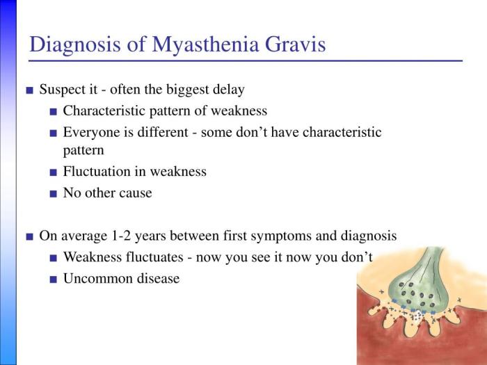
    How myasthenia gravis is diagnosed is a crucial process, often involving a multifaceted approach. This journey begins with recognizing the subtle and sometimes overlooked initial symptoms, progressing through neurological examinations, and culminating in a series of diagnostic tests. Understanding the various methods used in the diagnostic process can help patients and their families navigate this complex health challenge with greater awareness and confidence.

    This comprehensive guide explores the different steps involved in diagnosing myasthenia gravis, from initial symptom assessment to advanced laboratory tests and imaging techniques. We’ll delve into the nuances of muscle weakness, neurological evaluations, and the crucial role of diagnostic tests such as the Edrophonium test and repetitive nerve stimulation. A detailed understanding of these processes can help those affected better navigate the diagnostic pathway.

    Initial Symptoms and Presenting Complaints

    How myasthenia gravis is diagnosed

    Myasthenia gravis (MG) is a chronic autoimmune neuromuscular disease characterized by fluctuating muscle weakness. Understanding the initial symptoms and presenting complaints is crucial for early diagnosis and effective management. Recognizing the diverse ways MG manifests is essential to avoid delayed diagnosis, which can lead to a poorer prognosis. The symptoms vary widely, depending on the specific muscles affected.

    Common Initial Symptoms

    Initial symptoms of myasthenia gravis often begin subtly, with gradual worsening of muscle weakness over time. The hallmark feature is fluctuating muscle weakness, meaning the strength of the affected muscles can change throughout the day, and sometimes even within hours. This fluctuation can make it challenging for patients to accurately describe their symptoms. Common initial complaints often involve muscles used for repetitive or sustained activity.

    Presenting Complaints by Muscle Group

    The presenting complaints in myasthenia gravis can vary greatly depending on the muscle groups affected. Patients might experience difficulty with various tasks due to the varying weakness.

    • Ocular Symptoms: Double vision (diplopia) is a frequent and often the first symptom. It typically affects one or both eyes, often worse at the end of the day or after prolonged use. Ptosis (drooping eyelids) is another common ocular symptom, often occurring in conjunction with diplopia.
    • Facial Symptoms: Difficulty smiling, chewing, or making facial expressions is common. Weakness of facial muscles can lead to a characteristic “mask-like” appearance. Difficulty with swallowing (dysphagia) can also present as an early symptom.
    • Bulbar Symptoms: Difficulty speaking, swallowing, or chewing (bulbar weakness) can indicate involvement of the cranial nerves controlling these functions. These symptoms often manifest as slurred speech, difficulty with mastication, and progressive dysphagia.
    • Limb Symptoms: Weakness in the arms and legs, often affecting both sides equally, can occur. This weakness can present as difficulty with tasks like lifting objects, climbing stairs, or walking.

    Typical Onset and Progression of Different MG Subtypes

    The onset and progression of myasthenia gravis can vary significantly, even within the same patient. This variability necessitates a comprehensive understanding of the disease.

    Subtype Typical Onset Progression Notes
    Ocular MG Often insidious, starting with ptosis or diplopia Slowly progressive, may not affect other muscle groups Frequently the first subtype to appear
    Generalized MG Can start in any muscle group Rapid progression affecting multiple muscle groups Symptoms often more severe and widespread
    Bulbar MG Often starts with dysphagia, dysarthria, or both Can lead to significant swallowing and speaking difficulties May progress to respiratory compromise

    Triggers and Exacerbating Factors

    Several factors can exacerbate symptoms of myasthenia gravis. These factors are important to consider when assessing a patient’s condition.

    • Fatigue: Physical or mental exertion can significantly worsen muscle weakness. This is often a major factor for patients, leading to reduced functional capacity.
    • Stress: Emotional stress and anxiety can trigger or worsen symptoms. This can be an important consideration for managing symptoms.
    • Infection: Infections, especially viral infections, can temporarily worsen the symptoms of myasthenia gravis.
    • Certain Medications: Some medications, including antibiotics, can temporarily exacerbate symptoms.

    Indicators of Myasthenia Gravis in General Weakness

    General weakness can be a symptom of many conditions, but several indicators can suggest myasthenia gravis.

    • Fluctuating Weakness: Weakness that comes and goes, improving with rest and worsening with activity, is a strong indicator. This is a key differentiating feature from other causes of weakness.
    • Presence of Ocular Symptoms: The presence of ptosis or diplopia, even in the absence of other symptoms, should raise suspicion for myasthenia gravis.
    • Family History: A family history of autoimmune disorders may increase the likelihood of myasthenia gravis.
    • Exacerbation with Repetitive Activity: If the weakness worsens with repeated muscle use, it might suggest a neuromuscular junction problem like myasthenia gravis.

    Neurological Examination Procedures

    Unraveling the complexities of Myasthenia Gravis often hinges on meticulous neurological examinations. These procedures provide crucial insights into the patient’s muscle strength, function, and the specific involvement of the neuromuscular junction. By identifying characteristic patterns of weakness and evaluating cranial nerve function, clinicians can narrow down the diagnosis and tailor treatment strategies.

    Muscle Strength and Function Assessment

    Neurological examinations for myasthenia gravis (MG) focus on assessing muscle strength and fatiguability. Clinicians utilize various techniques to evaluate the extent and pattern of weakness. These techniques are designed to identify the characteristic fatigability patterns, a key diagnostic feature of MG. Precise documentation of these findings is vital for monitoring disease progression and treatment efficacy.

    Neuromuscular Junction Evaluation

    Specific tests are employed to evaluate the neuromuscular junction (NMJ), the site where nerves communicate with muscles. These tests aim to assess the efficiency of nerve impulse transmission. The rationale behind each procedure is to pinpoint any disruptions in the NMJ, which may be indicative of MG.

    Key Neurological Findings in Myasthenia Gravis

    Neurological Finding Description Clinical Significance Example
    Muscle Weakness Varying degrees of muscle weakness, particularly in muscles controlling eye movements, facial expression, swallowing, and limb movements. Weakness often worsens with repetitive use or prolonged activity. A hallmark symptom of MG, indicating impaired neuromuscular transmission. Difficulty raising eyelids, drooping eyelids (ptosis), double vision (diplopia), facial weakness, difficulty chewing, or swallowing.
    Cranial Nerve Dysfunction Impairment in cranial nerves, most commonly affecting those controlling eye movement (III, IV, VI), facial expression (VII), and swallowing (IX, X). Suggests the presence of neuromuscular dysfunction affecting specific nerve pathways. Ocular palsies, dysarthria, dysphagia, and facial weakness.
    Fatiguability Progressive weakening of muscles with sustained or repetitive use. A crucial diagnostic sign in MG, reflecting the impaired ability of the NMJ to maintain the necessary neurotransmitter release. Weakness worsens after sustained activity or repetition, like sustained eye movements.
    Response to Ice Packs Temporary improvement in muscle strength after applying ice packs to affected muscles. Can indicate the presence of a myasthenic crisis. Temporary improvement in eye movement after ice application.

    Detecting Characteristic Patterns of Muscle Weakness

    Identifying the characteristic patterns of muscle weakness in MG involves careful observation and testing. Clinicians evaluate the extent and location of weakness, noting whether it affects specific muscle groups or cranial nerves. They observe how weakness changes with rest and activity, specifically if the weakness increases with repeated use or activity. Furthermore, the presence of fatiguability during specific tasks like sustained eye movements is crucial for diagnosis.

    Importance of Cranial Nerve Function Evaluation

    Evaluating cranial nerve function is essential in a patient suspected of having myasthenia gravis. Cranial nerves innervate vital muscles involved in vision, facial expression, chewing, swallowing, and speech. Any impairment in these functions can significantly impact a patient’s quality of life. The presence of specific cranial nerve palsies, particularly those affecting ocular muscles, strongly suggests the possibility of MG.

    Thorough testing and documentation of cranial nerve function provide vital clues for diagnosis and management.

    Diagnostic Tests

    Navigating the diagnostic journey for Myasthenia Gravis (MG) often involves a combination of clinical assessments and specific tests. A thorough understanding of these tests is crucial for accurate diagnosis and effective management of the condition. The goal is to pinpoint the underlying neuromuscular dysfunction that characterizes MG, enabling the right treatment plan to be implemented.

    Diagnosing myasthenia gravis often involves a combination of physical exams, neurological assessments, and blood tests. Doctors look for specific symptoms, like muscle weakness that worships throughout the day, and often, try to rule out other conditions. Switching to healthier alternatives to full-fat dairy products, like healthy alternatives to full fat dairy products , can be a beneficial part of a comprehensive treatment plan, though it’s crucial to remember that this is a complex disease and dietary changes alone won’t diagnose or cure it.

    Ultimately, a team of specialists and thorough testing are key to accurate diagnosis.

    Edrophonium (Tensilon) Test, How myasthenia gravis is diagnosed

    This test is a crucial diagnostic tool in evaluating suspected myasthenia gravis. It involves administering edrophonium, a cholinesterase inhibitor, intravenously. The rationale behind this test is that cholinesterase inhibitors increase the amount of acetylcholine available at the neuromuscular junction. This, in turn, can temporarily improve muscle strength in individuals with MG.

    The mechanism of action is straightforward: edrophonium inhibits acetylcholinesterase, the enzyme that breaks down acetylcholine. This leads to an increased concentration of acetylcholine in the synaptic cleft, stimulating muscle contraction more effectively.

    During the test, muscle strength is meticulously observed before and after the injection. A positive response, characterized by a marked improvement in muscle strength within minutes of the injection, strongly suggests MG. Conversely, a lack of improvement or a minimal response typically indicates that MG is not the primary cause of the symptoms. Important considerations include potential side effects such as bradycardia, nausea, or muscle fasciculations.

    The test should only be performed under strict medical supervision.

    Diagnosing myasthenia gravis often involves a combination of physical exams, neurological tests, and sometimes even blood tests. It’s a complex process, much like figuring out how to manage a stuffy nose at night, which can often be a real struggle. Fortunately, there are some simple strategies to try, like using a saline nasal spray, which can help clear up congestion and make breathing easier.

    This process of elimination and targeted approaches, similar to figuring out the right combination of strategies for a stuffy nose, is important in diagnosing myasthenia gravis accurately. Ultimately, a doctor will need to consider various factors to make a precise diagnosis. how to reduce a stuffy nose at night can help find relief.

    Repetitive Nerve Stimulation (RNS) Testing

    RNS testing assesses the neuromuscular junction’s response to repeated nerve stimulation. The rationale is that in MG, repeated nerve stimulation leads to a quicker decline in muscle strength compared to healthy individuals. This decline is a reflection of the fatigued neuromuscular junction’s reduced ability to produce acetylcholine effectively.

    The procedure involves electrically stimulating a nerve repeatedly, measuring the strength of the resulting muscle contractions. The test identifies the pattern of muscle weakness as a result of repeated stimulation. A progressive decrease in the amplitude of muscle action potentials is a significant finding suggestive of MG.

    Electromyography (EMG)

    EMG is a diagnostic procedure that evaluates the electrical activity of muscles. It plays a crucial role in assessing neuromuscular transmission, the process by which nerve impulses trigger muscle contractions. The rationale for using EMG is that it can reveal subtle abnormalities in the electrical signals produced by muscles and nerves, helping to distinguish between different neuromuscular disorders.

    Diagnosing myasthenia gravis often involves a combination of physical exams, neurological tests, and sometimes even blood tests. It’s a tricky diagnosis, and sometimes, symptoms can overlap with other conditions like, for example, what is tmj disorder , making accurate identification crucial. Ultimately, a thorough evaluation by a neurologist is key to pinpointing the cause and receiving the right treatment for myasthenia gravis.

    During EMG, electrodes are inserted into specific muscles to record their electrical activity. Normal muscle activity is characterized by specific patterns of electrical signals. EMG can reveal characteristic abnormalities in MG, such as reduced amplitude of muscle action potentials and prolonged relaxation times. It’s important to note that EMG findings alone are not always definitive for MG and may require correlation with other clinical and test results.

    Comparison of Diagnostic Tests

    Test Strengths Limitations Typical Findings in MG
    Edrophonium (Tensilon) Test Quick, relatively simple, and can provide immediate diagnostic clues. Can cause adverse reactions, may not be conclusive in all cases, and is not suitable for everyone. Marked improvement in muscle strength after edrophonium administration.
    Repetitive Nerve Stimulation (RNS) Objective measure of neuromuscular junction fatigue, highly sensitive to early signs of MG. Requires specialized equipment and interpretation by trained personnel. Progressive decrease in muscle action potential amplitude with repeated stimulation.
    Electromyography (EMG) Provides detailed information about muscle and nerve function, helpful in ruling out other conditions. Can be invasive, requires specialized interpretation, and may not always directly pinpoint MG. Reduced amplitude of muscle action potentials and prolonged relaxation times.

    Laboratory Tests: How Myasthenia Gravis Is Diagnosed

    Beyond the crucial neurological examination, laboratory tests play a vital role in confirming and characterizing myasthenia gravis (MG). These tests help identify specific immune system responses and rule out other conditions with similar symptoms. They provide critical supporting evidence to aid in the diagnosis and management of MG.Laboratory tests are invaluable tools in the diagnostic process for myasthenia gravis, supplementing clinical findings and neurological examination results.

    These tests help differentiate MG from other neuromuscular disorders and provide insights into the underlying immune mechanisms.

    Serum Acetylcholine Receptor (AChR) Antibody Testing

    A crucial laboratory test in MG diagnosis is the serum AChR antibody test. AChR antibodies are autoantibodies that target the acetylcholine receptors (AChRs) at the neuromuscular junction. The presence of these antibodies strongly suggests MG.The procedure involves drawing a blood sample and analyzing it for the presence and level of AChR antibodies. Various methods exist for detecting these antibodies, including enzyme-linked immunosorbent assays (ELISAs).

    The interpretation of the results is critical. A positive result, indicating the presence of AChR antibodies, significantly strengthens the suspicion of MG, especially when coupled with clinical symptoms and a neurological examination. The level of antibodies can also provide information about the severity of the disease.

    Muscle-Specific Kinase (MuSK) Antibody Testing

    MuSK antibodies are another type of autoantibody implicated in MG. These antibodies target the muscle-specific kinase (MuSK) protein, also crucial at the neuromuscular junction. MuSK antibodies are present in a subset of MG patients, often those who do not have detectable AChR antibodies. This finding highlights the complexity of MG and the diverse mechanisms driving the disease.The procedure for MuSK antibody testing is similar to AChR antibody testing, involving blood draws and specialized laboratory analysis.

    Interpretation of MuSK antibody test results is essential. A positive result, indicating the presence of MuSK antibodies, suggests a form of MG, particularly when clinical symptoms and neurological examination support the diagnosis.

    Other Relevant Laboratory Tests

    Thyroid function tests are routinely included in the laboratory workup for MG, even though they are not directly related to the disease. This is because thyroid disorders can sometimes mimic MG symptoms. Other laboratory tests, such as complete blood counts (CBCs), electrolytes, and creatine kinase (CK) levels, might be ordered to rule out other conditions or assess overall health.

    These tests are vital for a comprehensive evaluation.

    Summary of Laboratory Test Results in Myasthenia Gravis

    Test Typical Finding in MG Deviation from Normal Clinical Significance
    Serum AChR Antibodies Positive Elevated levels Strong indication of MG; level may correlate with disease severity
    Serum MuSK Antibodies Positive (in some cases) Elevated levels Suggests a subtype of MG; often seen in patients negative for AChR antibodies
    Thyroid Function Tests Normal Abnormal thyroid hormone levels (hypothyroidism or hyperthyroidism) May mimic MG symptoms; necessitates further investigation
    Complete Blood Count (CBC) Normal Anemia, lymphopenia, or other abnormalities Could suggest other conditions or complications
    Electrolytes Normal Imbalances May indicate underlying issues or complications
    Creatine Kinase (CK) Normal Elevated levels Suggests muscle damage; may indicate a complication or other underlying conditions

    Imaging Techniques

    How myasthenia gravis is diagnosed

    Imaging techniques play a limited role in the diagnosis of myasthenia gravis (MG). While they can be helpful in excluding other conditions with similar symptoms, they don’t typically provide definitive evidence of MG. Their utility is most often in ruling out structural abnormalities in the neuromuscular junction or other neurological conditions that might mimic MG.Imaging studies are typically not the first-line diagnostic tools for MG.

    A comprehensive neurological examination, along with a detailed medical history and laboratory tests, are usually more critical in reaching a diagnosis. Imaging is often employed only when other tests are inconclusive or when there’s a strong suspicion of an underlying structural problem that might be contributing to the symptoms.

    Role of Imaging in MG Diagnosis

    Imaging plays a supportive role in the diagnostic process. It can help rule out alternative diagnoses or reveal structural abnormalities that might be affecting neuromuscular transmission, potentially mimicking the symptoms of MG. However, imaging alone is insufficient to establish a definitive diagnosis of MG.

    Specific Imaging Modalities

    Several imaging techniques may be employed in the evaluation of patients suspected of having MG. The choice depends on the specific clinical questions and the suspected location of any potential abnormality.

    Neuroimaging

    Neuroimaging techniques, including magnetic resonance imaging (MRI) and computed tomography (CT), are commonly used to evaluate the brain, spinal cord, and other neural structures. These studies can reveal the presence of masses, lesions, or other abnormalities that might be affecting the nervous system. While these techniques are valuable for ruling out other neurological conditions, they rarely show specific findings directly related to MG.

    Muscle Imaging

    Muscle imaging, using techniques like magnetic resonance imaging (MRI), can be utilized in certain situations. This is typically done when there’s a strong suspicion of muscle involvement that isn’t easily explained by other tests. Muscle MRI can potentially show evidence of muscle atrophy or other structural changes. However, these findings are often not pathognomonic for MG.

    Table of Imaging Modalities in MG

    Imaging Modality Usefulness in Identifying Associated Abnormalities Relevant Image Findings (Possible but not Specific) Limitations
    MRI of Brain and Spinal Cord Useful for ruling out other neurological conditions (e.g., tumors, multiple sclerosis). May show lesions or masses in the brain or spinal cord. Does not directly visualize neuromuscular junction.
    MRI of Muscles May reveal muscle atrophy or structural changes in cases of suspected myopathic involvement. Potential for showing atrophy or inflammation of specific muscle groups. Less sensitive for detecting the subtle neuromuscular changes of MG.
    CT Scan Less frequently used than MRI for evaluating the nervous system. May be helpful for detecting certain types of masses or bony abnormalities. May show bony abnormalities or lesions. Lower resolution compared to MRI for soft tissue structures.

    Differential Diagnosis

    Myasthenia gravis (MG) can sometimes masquerade as other neurological disorders, making accurate diagnosis crucial. A thorough differential diagnosis is essential to distinguish MG from conditions with overlapping symptoms, ensuring the correct treatment plan is implemented. This process involves ruling out various possibilities that might cause similar symptoms, such as muscle weakness or fatigue. Failure to identify and exclude these conditions can lead to inappropriate therapies and potentially delay the appropriate treatment for MG.Careful consideration of the patient’s medical history, physical examination findings, and results from various diagnostic tests are vital to differentiate MG from other neuromuscular conditions.

    Precise identification of the underlying cause of the symptoms is essential for appropriate management and to avoid potential misdiagnosis.

    Conditions Mimicking Myasthenia Gravis

    Many neurological and other medical conditions can present with symptoms similar to those of myasthenia gravis, including muscle weakness and fatigue. A comprehensive differential diagnosis considers these possibilities to ensure an accurate assessment of the patient’s condition.

    • Other Neuromuscular Disorders: Conditions such as Lambert-Eaton myasthenic syndrome (LEMS), botulism, and other neuromuscular junction disorders share overlapping symptoms with MG. The distinct characteristics of each condition, such as the presence or absence of specific antibodies or responses to different medications, are crucial for differentiation. For instance, LEMS often presents with muscle weakness that improves with repeated use, while MG typically worsens with repeated use.

    • Autoimmune Diseases: Systemic lupus erythematosus (SLE), rheumatoid arthritis, and other autoimmune disorders can sometimes cause muscle weakness as a secondary effect. The presence of characteristic symptoms and associated laboratory findings helps in distinguishing these conditions from MG.
    • Infectious Diseases: Infections like Lyme disease, poliomyelitis, and others can manifest with muscle weakness. Careful evaluation of the patient’s medical history and laboratory tests, such as serological testing, aid in distinguishing these conditions from MG.
    • Neurodegenerative Diseases: Amyotrophic lateral sclerosis (ALS) and other neurodegenerative diseases can cause progressive muscle weakness. The progressive nature and specific neurological findings associated with these diseases help differentiate them from the fluctuating weakness often seen in MG.
    • Endocrine Disorders: Hypothyroidism, hyperthyroidism, and other endocrine disorders can cause muscle weakness. Appropriate laboratory tests for thyroid function and other relevant endocrine parameters can aid in distinguishing these conditions.
    • Medication Side Effects: Certain medications can cause muscle weakness as a side effect. Carefully reviewing the patient’s medication list and considering the timing of symptom onset in relation to medication use is essential.
    • Structural Neuromuscular Diseases: Conditions like muscular dystrophy, myopathies, and peripheral neuropathies can mimic MG. Detailed neurological examination and specific muscle biopsy results help differentiate these from MG.

    Distinguishing Features

    Accurate diagnosis relies on a thorough understanding of the key differentiating characteristics between MG and other neuromuscular conditions. Precise identification of the distinctive features of each condition helps clinicians arrive at the correct diagnosis.

    Characteristic Myasthenia Gravis Lambert-Eaton Myasthenic Syndrome (LEMS) Amyotrophic Lateral Sclerosis (ALS)
    Muscle Weakness Fluctuating weakness, typically worse with repeated use; improved with rest. Often affecting ocular, bulbar, and limb muscles. Weakness improves with repeated use; characteristically affects proximal muscles first. Progressive, symmetrical muscle weakness affecting both upper and lower extremities.
    Response to Edrophonium (Tensilon) Test Improved muscle strength following edrophonium administration. Muscle strength worsens following edrophonium administration. No significant change in muscle strength with edrophonium.
    EMG Findings May show decremental response to repetitive nerve stimulation. May show incremental response to repetitive nerve stimulation. May show denervation and fibrillation potentials.
    Autoantibodies Presence of acetylcholine receptor antibodies (AChR-Abs) or muscle-specific kinase antibodies (MuSK-Abs). Presence of presynaptic calcium channel antibodies. Absence of specific autoantibodies.

    Closing Summary

    In conclusion, diagnosing myasthenia gravis requires a careful evaluation of symptoms, neurological findings, and a battery of diagnostic tests. This multifaceted approach ensures a more accurate and timely diagnosis, paving the way for appropriate treatment and management strategies. The key is early detection and intervention, allowing individuals to live more fulfilling lives despite the challenges presented by this condition.

  • What is a Myotome A Deep Dive

    What is a Myotome A Deep Dive

    What is a myotome? It’s a crucial concept in understanding how our muscles are controlled by the nervous system. Imagine a specific group of muscles, all working together, coordinated by a particular segment of the spinal cord. That’s essentially a myotome. This isn’t just an academic curiosity; myotome assessment plays a vital role in diagnosing neurological conditions.

    From pinpointing the source of muscle weakness to understanding the intricate pathways of the nervous system, exploring myotomes reveals a fascinating interplay between structure and function.

    This comprehensive guide delves into the world of myotomes, examining their definition, anatomy, function, clinical relevance, and assessment methods. We’ll uncover how these intricate muscle groups are organized, the role of nerve roots, and how evaluating myotomes helps diagnose and understand various neurological issues. Get ready to explore the fascinating science behind your body’s movement!

    Definition and Scope

    A myotome is a group of muscles innervated by a specific spinal nerve root. Understanding myotomes is crucial for clinicians, as they provide a pathway to assess the integrity of the motor pathways of the nervous system. It’s a fundamental concept in neurology, helping pinpoint the location of potential nerve damage.Myotomes and dermatomes, while both associated with spinal nerves, differ in their function.

    Dermatomes map sensory innervation, while myotomes map motor innervation. Damage to a specific nerve root can manifest as weakness or paralysis in the associated myotome, allowing clinicians to pinpoint the affected area. This localized weakness helps determine the level of spinal cord or nerve root compromise.

    Myotome Definition and Relationship to Dermatomes

    A myotome is a group of muscles that are controlled by a single spinal nerve root. This contrasts with dermatomes, which map the sensory areas served by the same nerve root. The relationship between the two is based on the common spinal nerve root origin. Clinical correlation between sensory and motor deficits, observed in dermatomes and myotomes respectively, allows clinicians to pinpoint the affected spinal segment and the extent of the damage.

    A myotome is basically a group of muscles that a single spinal nerve root controls. Understanding these muscle groups is crucial for diagnosing and treating pain, especially when considering active and passive pain management strategies. For example, if you’re experiencing pain in a specific area, evaluating the corresponding myotome can help pinpoint the source of the issue. This kind of assessment can help you determine the best course of action, such as exploring different approaches to pain relief, including active and passive pain management techniques.

    Ultimately, knowing your myotomes can give you a better understanding of your body and how to manage any discomfort effectively.

    This integrative approach is vital in diagnosing and treating various neurological conditions.

    Clinical Significance of Myotomes

    Myotomes are essential in clinical settings for diagnosing nerve root or spinal cord lesions. Weakness or paralysis in a specific myotome can indicate a problem with the corresponding spinal nerve root, often associated with conditions like herniated discs, spinal stenosis, or multiple sclerosis. The pattern of muscle weakness can help determine the precise location and extent of the neurological damage.

    Description of Myotomes

    Myotomes can be described in various ways. An anatomical description focuses on the specific muscles within the group. A functional description emphasizes the movements the myotome enables. Different methods of testing can also provide a detailed description, focusing on the specific movements and muscles involved in the examination. These methods are integral to the clinical evaluation.

    Table of Myotomes

    Myotome Name Associated Muscles Nerve Root Clinical Significance
    Cervical Myotome C5 Deltoid, biceps brachii, brachialis C5 spinal nerve root Weakness in shoulder abduction and elbow flexion can suggest C5 nerve root compression.
    Cervical Myotome C6 Biceps brachii, wrist extensors C6 spinal nerve root Weakness in wrist extension and elbow flexion indicates possible C6 nerve root impingement.
    Lumbar Myotome L2 Hip flexors L2 spinal nerve root Paralysis in hip flexion may point to L2 nerve root compression.
    Lumbar Myotome L4 Knee extensors L4 spinal nerve root Weakness in knee extension often suggests L4 nerve root damage, potentially due to disc herniation.
    Sacral Myotome S1 Plantar flexors, ankle evertors S1 spinal nerve root Weakness in plantar flexion and ankle eversion can indicate S1 nerve root pathology.

    Anatomy and Structure: What Is A Myotome

    What is a myotome

    Myotomes are fascinating segments of the body, directly linked to the intricate network of nerves and muscles. Understanding their anatomical arrangement is crucial for diagnosing and treating neuromuscular disorders. This section delves into the detailed structure of myotomes, illustrating their organization and the specific components that contribute to their function.The arrangement of myotomes reflects the segmental organization of the nervous system.

    They are essentially functional units, each controlled by a specific group of nerve roots. The specific muscles controlled by each myotome are essential in coordinating movement, posture, and other functions. This knowledge is fundamental in assessing neurological integrity.

    Anatomical Arrangement of Myotomes, What is a myotome

    The spinal cord segments and corresponding nerve roots organize myotomes in a precise pattern. Imagine a vertical arrangement of segments, each responsible for controlling muscles in a specific region of the body. This organization allows for precise control and coordination of movement, enabling us to perform a vast array of actions. This arrangement also allows for efficient communication between the brain and different parts of the body.

    Components of a Myotome

    A myotome comprises the nerve root(s) that innervate it and the specific muscle groups it controls. These components are interconnected, forming a functional unit. The nerve roots carry signals from the spinal cord to the muscles, enabling contraction and movement. The muscle groups are the effectors of this signal, performing the intended actions. Together, these elements create a complete functional unit.

    Nerve Root Innervation

    The nerve roots are the essential communication channels connecting the spinal cord to the muscles. Each myotome receives its innervation from specific nerve roots, which originate from the spinal cord. These nerve roots relay signals from the central nervous system to the muscle fibers, triggering contractions. The precise location of these roots is essential in determining the specific myotome.

    Spinal Cord Segments Involved

    The spinal cord is divided into segments, each corresponding to a specific region of the body. These segments are crucial in the formation of myotomes, as they are the source of the nerve roots. Each segment sends signals to the corresponding muscle groups, controlling their actions. The precise location and function of each spinal cord segment are essential in understanding myotome function.

    This information is used to diagnose and treat various neuromuscular conditions.

    Myotome Table

    This table illustrates the relationship between myotome, nerve root(s), and the muscle groups controlled. It highlights the crucial role of nerve roots in activating specific muscle groups, which together create the function of a myotome. This information is critical in assessing neurological function and diagnosing neuromuscular disorders.

    Myotome Nerve Root(s) Innervating It Muscle Groups Controlled
    Cervical Myotomes (C1-C8) C1-C8 Neck, shoulder, arm, hand muscles
    Thoracic Myotomes (T1-T12) T1-T12 Intercostal muscles, back muscles
    Lumbar Myotomes (L1-L5) L1-L5 Hip, thigh, knee muscles
    Sacral Myotomes (S1-S5) S1-S5 Leg, foot muscles

    Neural Pathways Involved in Myotome Function

    The neural pathways involved in myotome function are complex, but they can be described as follows. Sensory input from the muscle and surrounding tissues is conveyed to the spinal cord through sensory nerves. Motor commands are then sent from the spinal cord via motor nerves to the muscle fibers. This two-way communication allows for precise control and coordination of movement.

    The intricate nature of these pathways underscores the importance of a well-functioning nervous system.

    Function and Clinical Relevance

    Myotomes, as we’ve explored, represent a specific group of muscles innervated by a single spinal nerve root. Understanding their function is crucial in evaluating neurological health. This section delves into the practical applications of myotome assessment in clinical settings.Myotome assessment provides valuable insights into the integrity of the peripheral nervous system. Clinicians utilize this information to pinpoint potential nerve root impingements, spinal cord lesions, or peripheral neuropathies.

    The accuracy of this assessment is pivotal in formulating a diagnosis and developing a targeted treatment plan.

    Myotome Function

    Myotomes are responsible for specific movements or actions. Each myotome corresponds to a particular set of muscles, and the coordinated activation of these muscles allows for a range of body movements. For example, the C5 myotome controls shoulder abduction, while the L4 myotome facilitates dorsiflexion of the foot. Understanding the specific movements controlled by each myotome helps clinicians assess the extent of potential neurological damage.

    Myotome Assessment in Clinical Practice

    Clinicians evaluate myotomes through a series of standardized muscle strength tests. These tests involve asking the patient to perform specific movements, and the examiner assesses the strength and range of motion of the corresponding muscles. The examiner grades the strength using a standardized scale, often a 0-5 scale, where 0 indicates no muscle contraction and 5 represents normal strength.

    The precise movements and muscle groups tested depend on the suspected area of neurological impairment. Thorough and careful examination is crucial to ensure accurate assessment.

    Clinical Uses of Myotome Testing

    Myotome testing serves as a vital diagnostic tool in identifying and evaluating neurological conditions. It helps pinpoint the level of spinal cord or nerve root involvement, aiding in differential diagnosis. This testing is particularly useful in cases of suspected nerve root compression, such as from herniated discs, or in conditions like spinal stenosis. Furthermore, myotome assessment can help monitor the progression or response to treatment in various neurological disorders.

    A myotome is a group of muscles that are innervated by a single spinal nerve root. Maintaining good kidney health is crucial for overall well-being, and a balanced diet plays a key role. Eating foods rich in antioxidants and low in sodium, such as those highlighted in this resource on foods for kidney health , can contribute significantly to kidney function.

    Understanding myotomes is important for diagnosing and treating neuromuscular conditions.

    Conditions Where Myotome Assessment is Important

    Myotome assessment is critical in several neurological conditions. The information gathered aids in pinpointing the location and extent of the damage to the nervous system. By evaluating muscle strength, clinicians can identify the specific nerve roots or spinal cord segments that are affected. This allows for a more accurate diagnosis and facilitates appropriate treatment.

    Examples of Conditions and Expected Myotome Weakness

    Condition Expected Myotome Weakness Diagnostic Significance
    Cervical Radiculopathy (C6-C7) Weakness in elbow flexion and wrist extension Indicates nerve root compression at the C6-C7 level, potentially from a herniated disc.
    Lumbar Radiculopathy (L5) Weakness in foot dorsiflexion and great toe extension Suggests nerve root compression at the L5 level, possibly due to spinal stenosis or a herniated disc.
    Spinal Cord Injury (Thoracic Level) Weakness in shoulder abduction, elbow flexion, and wrist extension. Indicates a spinal cord lesion, and the level of the lesion can be determined by the specific myotomes affected.
    Peripheral Neuropathy Gradual, symmetric weakness in multiple myotomes Suggests a diffuse or systemic process affecting the peripheral nerves.

    Assessment and Testing

    Assessing myotome function is crucial for evaluating neuromuscular health and identifying potential nerve or muscle pathologies. Proper myotome testing involves a systematic approach to evaluate the strength and power of specific muscle groups innervated by particular spinal nerve roots. This allows clinicians to pinpoint the location of potential damage or dysfunction.

    Methods for Assessing Myotome Function

    Various methods are employed to assess myotome function, each with its strengths and limitations. These methods range from simple manual muscle testing to more sophisticated electromyographic (EMG) studies. Manual muscle testing is a widely used, cost-effective method that relies on the clinician’s observation of the patient’s ability to perform specific movements against resistance. EMG studies provide a more detailed evaluation of muscle electrical activity, allowing for a deeper understanding of muscle function and identifying any neuromuscular disorders.

    Steps Involved in a Myotome Examination

    A comprehensive myotome examination involves several key steps. First, a thorough patient history and neurological examination are essential to understand the patient’s presenting complaint and potential contributing factors. Second, the examination should focus on identifying the specific muscle groups associated with the relevant myotomes. Third, standardized manual muscle testing procedures should be meticulously followed to ensure accuracy and reliability.

    Lastly, interpretation of the findings should be performed with careful consideration of the patient’s overall clinical presentation.

    Importance of Proper Technique in Myotome Testing

    Proper technique is paramount in myotome testing to ensure accurate and reliable results. Inconsistent or improper techniques can lead to inaccurate assessment and potentially misdiagnosis. Clinicians should adhere to standardized procedures and ensure that the patient is properly positioned and relaxed during the test. Accurate measurement and consistent evaluation criteria are crucial for reliable results. Standardized protocols should be used to maintain consistency and reproducibility.

    Step-by-Step Procedure for Testing Specific Myotomes

    1. Biceps Myotome (C5-C6): Assess the patient’s ability to flex the elbow against resistance. The examiner should position the patient supine or sitting with the elbow flexed at 90 degrees. The examiner applies resistance to the patient’s forearm while the patient attempts to maintain elbow flexion. Note the degree of resistance the patient can overcome. The examiner should observe for any weakness, atrophy, or tremors during the test.

    2. Triceps Myotome (C6-C7): Assess the patient’s ability to extend the elbow against resistance. The examiner should position the patient supine or sitting with the elbow flexed at 90 degrees. The examiner applies resistance to the patient’s forearm while the patient attempts to extend the elbow. Observe the degree of resistance the patient can overcome. Look for any weakness, atrophy, or tremors during the test.

      So, what exactly is a myotome? Basically, it’s a group of muscles that are innervated by a single spinal nerve root. Understanding these muscle groups is crucial for diagnosing nerve damage, and for people exploring natural health options like AHCC supplements, for example, AHCC benefits and safety is an important consideration. Knowing how AHCC might interact with your myotomes is key to potential therapeutic benefits.

      Ultimately, a good understanding of myotomes is essential for anyone working with the body’s complex systems.

      Note that a weak triceps response can suggest damage to the radial nerve.

    3. Wrist Extensors (C6-C7): Assess the patient’s ability to extend the wrist against resistance. The patient should be positioned supine or sitting with the forearm supported and the wrist in a neutral position. The examiner applies resistance to the dorsal aspect of the hand as the patient attempts to extend the wrist. Assess the strength and note any weakness. This test evaluates the integrity of the radial nerve.

    4. Iliopsoas Myotome (L2-L3): Assess hip flexion. The patient should be supine with the knee flexed and the hip at a 90-degree angle. The examiner should apply resistance to the patient’s thigh while they attempt to maintain hip flexion. Record the patient’s ability to maintain the flexion against resistance. Assess for any weakness, atrophy, or other abnormalities.

      This is crucial for assessing the integrity of the lumbar plexus and the associated nerves.

    Clinical Cases and Examples

    Myotome testing plays a crucial role in diagnosing and localizing neurological impairments. By assessing the strength and function of specific muscle groups, clinicians can pinpoint the level of the nervous system affected, whether it’s a peripheral nerve, spinal cord, or brain. Understanding the clinical presentation and interpretation of myotome weakness is vital for developing an accurate diagnosis and appropriate treatment plan.Interpreting myotome testing results requires careful consideration of the patient’s overall neurological presentation.

    The presence of other symptoms, such as sensory changes, reflexes, and cognitive impairments, provides valuable context and helps differentiate between various neurological conditions. These combined findings contribute to a comprehensive picture of the patient’s neurological status.

    Myotome Weakness in Cervical Radiculopathy

    Cervical radiculopathy, often caused by nerve root compression in the neck, frequently leads to specific myotome weakness. The affected myotomes are typically those innervated by the affected nerve roots. For example, C5 and C6 nerve root involvement might result in weakness of the biceps and deltoid muscles, affecting shoulder abduction and flexion.

    Myotome Weakness in Spinal Cord Injury

    Spinal cord injuries can result in varying degrees of myotome weakness, depending on the level and extent of the damage. A complete spinal cord lesion at the C5 level, for example, would likely produce significant weakness in the muscles innervated by the C5 nerve root and below, leading to weakness in shoulder abduction, elbow flexion, wrist extension, and hand movements.

    Myotome Weakness in Peripheral Neuropathy

    Peripheral neuropathies encompass a broad range of conditions affecting peripheral nerves. These conditions can lead to varying degrees of myotome weakness depending on the affected nerves. For instance, a diabetic neuropathy can lead to distal muscle weakness affecting the muscles of the feet and legs, impacting ambulation and balance.

    Interpreting Myotome Testing Results

    Interpreting myotome testing results in conjunction with other neurological findings is crucial for accurate diagnosis. For example, if a patient presents with weakness in the C7 myotome (triceps) but normal reflexes, the cause might be localized to the peripheral nerve rather than the spinal cord. Consideration of the patient’s medical history, symptoms, and other neurological examination findings further enhances diagnostic accuracy.

    Case Studies of Myotome Involvement

    Case Scenario Myotome Affected Symptoms Diagnosis
    Patient with neck pain, radiating down the arm, and weakness in biceps and wrist extensors. C5, C6, C7 Pain, numbness, tingling in the arm and hand, weakness in elbow flexion and wrist extension. Cervical radiculopathy
    Patient with a history of a fall, presenting with decreased sensation in the lower extremities and weakness in hip flexion and knee extension. L2, L3, L4 Numbness and tingling in the lower extremities, weakness in hip flexion, knee extension, and dorsiflexion. Spinal cord injury
    Patient with a history of diabetes, experiencing gradual weakness in the foot and ankle. L4, L5, S1 Gradual weakness and numbness in the feet and ankles, difficulty with ambulation. Diabetic neuropathy

    Variations and Considerations

    Myotomes, while providing a valuable framework for assessing neuromuscular function, aren’t a perfect fit for every individual. Variations in their organization and the influence of various factors can significantly affect how a myotome is assessed and interpreted. Understanding these variations and considerations is crucial for accurate diagnoses and effective treatment plans.The consistency in myotome organization, though a useful guide, is not absolute.

    Individual variations in muscle fiber type, size, and innervation patterns can affect the specific muscles that make up a myotome. This variability in anatomy necessitates a careful and nuanced approach to myotome assessment, always considering the individual’s unique characteristics.

    Variations in Myotome Organization

    Individual differences in muscle anatomy and innervation can lead to variations in myotome boundaries. These variations can sometimes result in a muscle being innervated by a nerve root slightly different from the expected pattern. This can cause a myotome to be slightly larger or smaller in some individuals than in others. Factors like genetic predisposition and previous injuries can also play a role in these anatomical variations.

    Factors Influencing Myotome Assessment

    Several factors can impact the accuracy of myotome testing. Patient cooperation and motivation are critical, as muscle strength is affected by psychological factors like fear and anxiety. Pain, fatigue, and other medical conditions can also influence the ability to perform muscle tests, potentially leading to inaccurate or misleading results. Furthermore, the examiner’s skill and experience in performing the tests directly impacts the reliability of the assessment.

    Careful consideration of these influencing factors is paramount to obtaining meaningful and reliable results.

    Limitations of Myotome Testing

    Myotome testing, despite its usefulness, has limitations. It provides a general indication of nerve root function but doesn’t offer a precise measurement of the extent of the nerve damage. The test’s resolution is limited, and subtle deficits may be missed. Additionally, myotome testing relies on the patient’s ability to cooperate, which can be affected by factors like pain or cognitive impairment.

    It’s important to recognize that myotome testing is a component of a broader neurological assessment, not a stand-alone diagnostic tool.

    Considerations for Assessing Myotomes in Different Age Groups

    Age significantly impacts muscle strength and the ability to perform motor tasks. Children and older adults may have different baseline strength levels, requiring adjusted assessment methods and interpretations. The developmental stages of children, for example, need to be taken into account. In children, the myotome assessment might focus on milestones and developmental norms, rather than precise strength measurements.

    Table: Age Group Considerations for Myotome Assessment

    Age Group Potential Variations Assessment Considerations
    Infants (0-1 year) Immature neuromuscular system, limited voluntary movement Focus on developmental milestones, use of reflexes, parental input
    Children (2-12 years) Varying developmental stages, rapid growth spurts Use age-appropriate tasks, consider developmental norms, assess cooperation
    Adults (18-65 years) Stable muscle mass, generally consistent motor abilities Standard myotome testing methods are generally applicable
    Older Adults (65+) Decreased muscle mass, decreased strength, potential comorbidities Adjust testing to accommodate limitations, assess for confounding factors, use caution

    Imaging and Techniques

    What is a myotome

    Visualizing myotomes, the groups of muscles innervated by a single spinal nerve root, presents unique challenges. While direct visualization of muscle function isn’t possible with standard imaging, these techniques can provide valuable information about the structures involved, aiding in the diagnosis and understanding of neuromuscular conditions. Understanding the limitations of these techniques is equally crucial for accurate interpretation.Imaging modalities offer insights into the anatomy of myotomes, but don’t directly measure their functional capacity.

    This is critical to remember when interpreting results, as imaging findings must be correlated with other assessments like electromyography (EMG) and nerve conduction studies (NCS) to gain a comprehensive picture. Indirect assessments of myotome involvement are possible through visualization of neural pathways and structures related to muscle function.

    MRI and CT Imaging

    Magnetic resonance imaging (MRI) and computed tomography (CT) scans are valuable tools for visualizing the anatomy of the spine, spinal cord, and surrounding tissues. These techniques can depict the spinal nerves, which are crucial for understanding myotome function. MRI excels at providing detailed soft tissue images, highlighting potential spinal cord compression or nerve root impingement, which can affect the associated myotomes.

    CT, on the other hand, provides excellent bone detail, aiding in identifying bony structures that might contribute to nerve root compression.

    Limitations of Imaging Techniques

    Imaging techniques primarily visualize thestructures* involved in myotome function, not the function itself. For example, an MRI might reveal a herniated disc compressing a nerve root, but it doesn’t directly demonstrate the resultant weakness in the muscles of the corresponding myotome. Other factors, like the severity of the compression or the individual’s response to the condition, can also affect the myotome’s function and are not directly measurable by these techniques.

    Visualizing Neural Pathways

    Visualization of neural pathways involves advanced techniques. Advanced MRI techniques, such as diffusion tensor imaging (DTI), can map the pathways of nerve fibers, providing insights into the integrity of neural connections. This is crucial in cases of suspected nerve damage or disease, aiding in identifying the affected myotomes.

    Electrodiagnostic Studies (EMG and NCS)

    Electromyography (EMG) and nerve conduction studies (NCS) are essential for evaluating the electrical activity of muscles and nerves. EMG measures the electrical activity of muscle fibers, while NCS assesses the speed and amplitude of nerve impulses. These studies can directly assess the functional integrity of the myotomes, detecting issues like denervation (loss of nerve supply) or axonal damage.

    For instance, if a specific myotome shows reduced or absent electrical activity, it suggests a potential problem with the nerve root or the muscles it supplies.

    Comparison of Imaging Modalities

    Imaging Modality Strengths Limitations Application in Myotome Assessment
    MRI Excellent soft tissue detail, visualization of spinal cord and nerve roots Limited visualization of bone structures, can be expensive Identifying spinal cord compression, nerve root impingement
    CT Excellent bone detail, rapid acquisition Limited soft tissue contrast, higher radiation exposure Identifying bony structures contributing to nerve root compression
    DTI Visualization of neural pathways Requires specialized software and interpretation, more expensive than standard MRI Assessing nerve fiber integrity, identifying areas of potential damage
    EMG/NCS Direct assessment of muscle and nerve function Invasive procedure, requires specialized expertise for interpretation Evaluating the functional integrity of the myotomes, detecting denervation or axonal damage

    Ending Remarks

    In conclusion, understanding myotomes provides a critical lens through which we can appreciate the intricate relationship between the nervous system and our muscles. From a clinical standpoint, myotome assessment is invaluable for pinpointing the source of neurological issues. This guide has explored the key aspects of myotomes, offering a clear understanding of their definition, anatomy, function, and clinical applications.

    We’ve seen how these muscle groups, coordinated by specific nerve roots, are crucial for our everyday movements and how their assessment is a valuable tool in diagnosing various conditions. Myotomes are more than just anatomical structures; they’re essential for our health and well-being.