What to expect in an MRI sets the stage for a detailed look at the entire experience, from preparation to post-scan recovery. We’ll explore everything from the essential pre-scan instructions to the sensations you might feel during the procedure itself. This comprehensive guide will walk you through the steps, highlighting potential anxieties, and offering insights into the various types of MRIs and their uses.
Preparing for an MRI often involves specific instructions. This guide will cover common pre-scan requirements, including fasting, removing metal objects, and managing medications. We’ll also discuss modifications for specific medical conditions, ensuring you understand the unique aspects of your situation.
Pre-MRI Preparation
Preparing for a Magnetic Resonance Imaging (MRI) scan involves specific steps to ensure accurate results and patient safety. Adherence to these pre-MRI instructions is crucial for the quality of the images and the overall effectiveness of the diagnostic process. Ignoring these guidelines can lead to complications and unnecessary delays.Following the pre-MRI instructions is essential to minimize potential risks and ensure the quality of the diagnostic images.
This includes a thorough understanding of the specific procedures required for your particular scan type.
Importance of Adhering to Pre-MRI Instructions
Adherence to pre-MRI instructions is vital for obtaining clear and accurate diagnostic images. These instructions are designed to optimize the scan process and minimize potential complications. A clear image is critical for the radiologist to interpret and provide an accurate diagnosis. Failure to comply can lead to repeat scans, increased costs, and a potentially delayed diagnosis.
Common Pre-MRI Instructions for Patients
A range of preparations is necessary for a successful MRI scan. These preparations aim to eliminate any factors that could interfere with the magnetic field or compromise image quality.
- Fasting: For some MRI examinations, particularly those involving the abdomen or pelvis, fasting is necessary. This is to reduce the presence of gas in the digestive tract, which can interfere with image clarity. The duration of fasting varies depending on the specific type of scan and the patient’s individual circumstances. Specific guidelines for fasting should be provided by the facility conducting the MRI.
- Removing Metal Objects: MRI machines use powerful magnets. Metal objects can be drawn into the magnetic field, potentially causing injury to the patient or damage to the equipment. Patients should remove all metal objects, including jewelry, watches, piercings, and hair clips. This includes metal in clothing, if any concerns arise, medical professionals should be consulted.
- Medications: Certain medications may interfere with the MRI scan or affect the results. Patients should inform their healthcare provider about all medications they are taking, including prescription drugs, over-the-counter medications, and supplements. The healthcare provider will advise on whether to continue or temporarily discontinue certain medications before the scan.
Potential Consequences of Not Following Pre-MRI Instructions
Failure to adhere to pre-MRI instructions can result in a variety of negative consequences. These include:
- Blurred or Inaccurate Images: If gas or metal interferes with the magnetic field, the resulting images may be blurry or unclear, making accurate diagnosis difficult.
- Repeat Scans: Unclear images necessitate repeat scans, which are time-consuming, costly, and inconvenient for the patient.
- Delayed Diagnosis: If the initial scan is inconclusive due to improper preparation, a diagnosis may be delayed, potentially impacting treatment decisions.
- Equipment Damage: Metal objects drawn into the magnetic field can damage the MRI equipment.
- Patient Injury: In extreme cases, improperly prepared patients with metallic implants or objects could experience injury due to the strong magnetic field.
Modifications to Pre-MRI Instructions
In some situations, modifications to standard pre-MRI instructions may be necessary. This is particularly true for patients with specific medical conditions or implanted devices.
- Patients with Pacemakers or Other Implantable Devices: Patients with pacemakers or other implanted metallic devices require careful consideration. The presence of these devices might necessitate adjustments to the scan protocol or even preclude an MRI in some cases. Always consult with a healthcare professional to determine the best course of action.
- Patients with Specific Medical Conditions: Medical conditions such as pregnancy, claustrophobia, or other medical conditions might require specific modifications to the standard pre-MRI preparation guidelines. Consult with the healthcare provider for the necessary modifications.
Pre-MRI Instructions Table
| Category | Details | Reason | Example |
|---|---|---|---|
| Fasting | Abstain from food and drink for a specified time period before the scan. | Reduces gas in the digestive tract, improving image quality. | Fasting for 4-6 hours before an abdominal MRI. |
| Removing Metal | Remove all metal objects from the body. | Metal objects can interfere with the magnetic field. | Removing jewelry, watches, and hairpins. |
| Medications | Inform the facility about all medications. | Some medications may interfere with the scan or the results. | Reporting use of iron supplements, blood thinners, or other medications. |
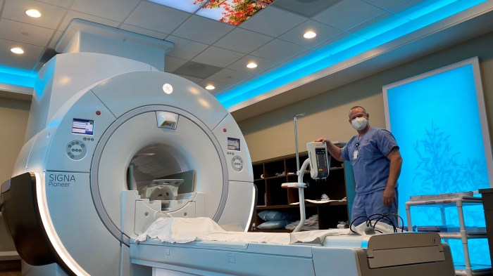
The MRI Procedure Itself
Getting ready for your MRI scan can be a bit nerve-wracking, but understanding the procedure can help alleviate some of those anxieties. This section will walk you through the typical steps, sensations, and considerations involved in an MRI scan, so you can approach the experience with more confidence.The MRI machine itself is a sophisticated tool that uses powerful magnetic fields and radio waves to create detailed images of the inside of your body.
This allows doctors to diagnose a wide range of conditions and monitor treatment effectiveness.
Getting an MRI can feel a bit daunting, but it’s usually straightforward. You’ll likely lie on a table that slides into a large tube, and you might hear some thumping noises. It’s important to remain still during the procedure, and you’ll need to remove any metal objects. Understanding what to expect in a medical procedure like an MRI is key, and sometimes comparing it to other similar procedures can help.
For example, if you’re curious about the role of someone who specializes in skin care, learning about what an esthetician does might help you understand the different types of medical and beauty professionals out there. what is an esthetician Ultimately, the key to a good MRI experience is relaxation and cooperation with the medical staff.
So, remember to take a deep breath and be prepared!
Typical Steps in an MRI Scan
The MRI scan process is generally standardized, though minor variations may occur depending on the specific part of the body being imaged and the individual patient. A typical scan involves several key steps:
- Patient Positioning: You’ll be positioned on a table that slides into the MRI machine’s bore. Proper positioning is critical for obtaining clear images and minimizing movement artifacts. This positioning is carefully tailored to the area of interest.
- Coil Placement: Depending on the body part being scanned, specialized coils may be placed on or around the area. These coils help to focus the radio waves and enhance image quality.
- Image Acquisition: Once positioned and ready, the MRI machine will begin generating the magnetic fields and radio waves. The machine will produce a series of pulses and measurements, which are then processed by a computer to create the images.
- Image Review: The radiologist or technician will then review the images and determine if additional scans are needed. This process is essential to ensuring accuracy and complete diagnosis.
Sensations During an MRI Scan
You might experience a few sensations during the scan. The most common is a thumping or clicking sound, which is produced by the machine as it generates the magnetic fields and radio waves. Some patients also feel a slight pressure or warmth, particularly around the coil. The duration of the scan can vary depending on the body part being imaged.
Description of the MRI Machine and Its Components
The MRI machine consists of a large, cylindrical magnet known as the bore. Inside the bore, there’s a strong magnetic field, which interacts with the hydrogen atoms in your body. The radio waves are used to excite these atoms, and the signals they emit are captured by the machine’s detectors.
- Magnet: The powerful magnet generates the strong magnetic field essential for the procedure. The strength of the magnetic field is measured in Tesla (T). Higher Tesla strength typically results in higher image resolution but may also increase patient discomfort.
- Gradient Coils: These coils are positioned around the magnet. They modify the magnetic field strength in specific locations, allowing for precise imaging of different parts of the body.
- Radiofrequency (RF) Coils: These coils are positioned around the body part being examined. They emit and receive the radio waves, providing detailed information about the internal structures.
- Control Console: The technician controls the MRI machine from a separate control console. This console allows them to adjust parameters such as scan time, field strength, and image resolution.
Potential Concerns and Anxieties
Some patients experience anxiety about enclosed spaces (claustrophobia) or the loud noises produced by the machine. For those with claustrophobia, open MRI machines may be a better option. Communication with the technician or radiologist can ease these concerns. If claustrophobia is a significant concern, consider discussing options with your healthcare provider in advance.
Flow Chart of the MRI Scan Procedure
(A flow chart would be a visual representation here, but text-based description follows.)The procedure typically starts with the patient being positioned on the MRI table. Next, the technician or radiologist places the necessary coils. The machine then generates the magnetic fields and radio waves, capturing the signals emitted by the body’s hydrogen atoms. Once the scan is complete, the technician or radiologist reviews the images.
If additional scans are needed, they’ll be performed.
Comparison of MRI Machine Types
| Machine Type | Features | Advantages | Disadvantages |
|---|---|---|---|
| Open MRI | A less enclosed design | Can reduce claustrophobia concerns | May have lower image resolution compared to closed MRIs, potentially requiring longer scan times |
| Closed MRI | A traditional, enclosed design | Typically provides higher image resolution and faster scan times | Can induce claustrophobia in some patients |
Post-MRI Experience

The MRI procedure itself is just one part of the diagnostic journey. Understanding what happens afterward, from receiving results to potential recovery, is crucial for managing expectations and ensuring a smooth transition. This section will delve into the post-MRI experience, covering everything from post-scan procedures to potential anxieties.
Post-MRI Procedures
Following the MRI scan, you’ll typically be directed to a waiting area where you can rest and recover. A member of the imaging center staff will then escort you to a location where you’ll be given any necessary instructions and be informed of the next steps in the process. This might include a review of your vital signs, such as your heart rate and blood pressure.
Getting an MRI can be a bit unnerving, but it’s generally straightforward. You’ll lie on a table that slides into a large tube-like machine. The machine uses strong magnetic fields and radio waves to create detailed images of your body. Understanding the complex interactions of genes, DNA, and chromosomes, like what are genes dna and chromosomes , is fascinating, but not directly relevant to the MRI procedure itself.
Ultimately, you can expect to be in the machine for a while, and potentially hear some loud thumping noises, but the whole process is usually quick and painless.
Sometimes, additional imaging may be required to provide a more comprehensive view.
Getting ready for an MRI? Expect some preliminary questions about your medical history, and possibly some light sedation. You’ll likely be asked to remove any metal objects, like jewelry or even certain clothing. Understanding potential issues like rectal ulcers can be important in preparing for the procedure. For a comprehensive overview of rectal ulcer symptoms, causes, and treatment options, check out this helpful resource on rectal ulcer overview and more.
Ultimately, your experience will depend on your specific situation, but remember to communicate any concerns to your doctor beforehand.
Receiving and Interpreting MRI Results
The speed at which you receive your MRI results depends on several factors. These factors include the complexity of the scan, the availability of radiologists, and the workload of the facility. Typically, you can expect to receive your results within a few days to a couple of weeks. Your physician or a designated member of the healthcare team will review the images and provide you with a detailed interpretation of the findings.
The report will usually include information about any abnormalities or normal findings.
Potential Recovery Times
Recovery times following an MRI scan are generally minimal. Most patients can resume their normal activities immediately. However, the specific recovery time may vary depending on the type of MRI scan. For example, a simple brain MRI may not require any significant recovery time, whereas a more complex scan of the abdomen might necessitate a slightly longer period of observation.
Potential Side Effects or Complications
MRI scans are generally considered safe procedures. However, some rare side effects or complications can occur. These include, but are not limited to, allergic reactions to contrast agents (in some cases), mild discomfort at the injection site, or in rare instances, nerve damage. Always report any unusual symptoms to your physician.
Managing Anxiety or Discomfort
It’s completely normal to feel some anxiety or discomfort after an MRI scan. Deep breathing exercises, relaxation techniques, and communication with your healthcare team can help manage these feelings. If anxiety is persistent, consider discussing it with your doctor.
Common Questions Patients Might Have After an MRI
- What if I experience discomfort after the scan? Mild discomfort is common, especially at the injection site for contrast-enhanced scans. Severe or persistent discomfort requires immediate medical attention.
- How long will it take to get my results? This varies based on factors such as scan complexity and radiologist availability. Your healthcare provider will provide a timeline for results.
- What should I do if I have concerns about the results? Discuss any concerns you have with your physician. They will address your questions and provide clarity regarding the results.
- Can I eat or drink before the MRI? This will depend on the specific scan and your doctor’s instructions. Be sure to follow the instructions provided to you before the procedure.
- Will I need to take any medication after the scan? This is usually not necessary unless your doctor has instructed you to do so.
Types of MRIs and Their Applications: What To Expect In An Mri
MRI technology offers a wide range of applications beyond just general body scans. Different types of MRI scans are tailored to specific body parts and diagnostic needs, allowing for a more precise and focused examination. Understanding the various types of MRIs and their respective applications is crucial for both patients and healthcare professionals.Different types of MRI scans use varying parameters and sequences to target specific tissues or structures.
This tailored approach ensures that the most relevant information is captured, enabling more accurate diagnoses and treatment plans.
Brain MRI
Brain MRIs are crucial for identifying various neurological conditions. They can reveal abnormalities in brain structure, such as tumors, cysts, or strokes. The detailed images allow physicians to assess the extent of damage and guide treatment decisions. These scans are also helpful in evaluating conditions like multiple sclerosis, where inflammation and damage to the myelin sheath are key factors.
The high-resolution images enable precise localization of lesions and aid in monitoring disease progression.
Spine MRI
Spine MRIs provide detailed images of the spinal cord, discs, and surrounding structures. These scans are essential for diagnosing conditions like herniated discs, spinal stenosis, and tumors affecting the spine. The detailed images help identify the exact location and extent of the problem, enabling more accurate surgical planning and treatment strategies. Degenerative conditions of the spine can also be evaluated with these scans.
Early detection of abnormalities can significantly impact treatment outcomes.
Knee MRI
Knee MRIs are frequently used to diagnose injuries and diseases affecting the knee joint. They are valuable for evaluating meniscus tears, ligament sprains (ACL, MCL, PCL), cartilage damage, and bone fractures. The precise images allow for a detailed assessment of the affected structures and help in determining the severity of the injury. This enables appropriate treatment decisions, from physical therapy to surgical interventions.
Furthermore, they help in monitoring the healing process after surgery or injury.
Comparison of MRI Scan Types
| Scan Type | Body Part | Purpose |
|---|---|---|
| Brain MRI | Brain | Identifying neurological conditions, assessing brain structure abnormalities (tumors, cysts, strokes), evaluating conditions like multiple sclerosis. |
| Spine MRI | Spine, spinal cord, and surrounding structures | Diagnosing herniated discs, spinal stenosis, tumors affecting the spine, evaluating degenerative conditions, and guiding surgical planning. |
| Knee MRI | Knee joint | Diagnosing meniscus tears, ligament sprains (ACL, MCL, PCL), cartilage damage, bone fractures, and monitoring healing after injury or surgery. |
Understanding the Images
MRI scans produce detailed images of the body’s internal structures, offering invaluable insights into potential health issues. These images, unlike X-rays, utilize strong magnetic fields and radio waves to create detailed cross-sectional views of the body. This allows doctors to visualize tissues and organs with exceptional clarity.MRI images are not simply a snapshot of the body; they are meticulously constructed representations based on the responses of hydrogen atoms within the body to magnetic fields.
Understanding how these images are generated and interpreted is crucial for appreciating their value in medical diagnostics.
MRI Image Generation
The process of creating MRI images involves exciting hydrogen atoms within the body using radio waves, which are then measured by the scanner. The strength and duration of these signals depend on the surrounding tissues. This information is then used to construct detailed images. The unique characteristics of different tissues allow for their differentiation in the final image.
Types of MRI Images
Different orientations of the MRI scanner provide various perspectives of the body. These perspectives are referred to as image planes.
- Axial (or transverse) images show horizontal slices of the body, providing a top-down view. These images are particularly useful for assessing structures like the brain, spinal cord, and abdomen.
- Sagittal images display vertical slices running from the front to the back of the body. These views are important for visualizing the spinal cord, ligaments, and other long structures.
- Coronal images show vertical slices running from the side to the side of the body. These are valuable for examining the structures of the head, torso, and extremities.
Image Characteristics
The quality of MRI images depends on several factors, with the most crucial being:
- Contrast: Contrast refers to the differences in signal intensity between various tissues. High contrast allows for clear differentiation between tissues with varying densities. For instance, fat and water often appear differently on MRI scans, leading to good contrast between them.
- Signal Intensity: Signal intensity on an MRI image is a measure of the strength of the radio waves emitted by the hydrogen atoms in the tissue. Different tissues exhibit different signal intensities, reflecting their composition and physical properties. For example, gray matter in the brain appears with a different signal intensity than white matter.
Radiologist Interpretation, What to expect in an mri
Radiologists are medical professionals specializing in interpreting medical images, including MRI scans. They use their expertise to analyze the images, identify anomalies, and provide reports to physicians. Their expertise in anatomy and pathology is crucial for accurate interpretation.
Normal and Abnormal Findings
- Normal: Normal brain MRI scans typically show clear, well-defined structures with expected signal intensities for different tissues. For example, the ventricles of the brain should be the correct size and shape, and the gray matter should appear with its characteristic signal intensity.
- Abnormal: Abnormal findings might include lesions, tumors, or edema, each exhibiting unique signal intensities and patterns. For example, a tumor in the brain might appear as a mass with abnormal signal intensity, different from the surrounding healthy tissue.
Interpreting Signal Intensities
Different signal intensities on MRI scans provide crucial information about the tissues being examined. A high signal intensity often suggests the presence of water or fat, whereas a low signal intensity may indicate bone or dense tissue. This knowledge is used to identify areas of concern or abnormality.
Closure

Navigating an MRI can seem daunting, but understanding the process from start to finish can ease any anxieties. This guide provides a comprehensive overview, equipping you with the knowledge to feel confident and prepared for your MRI experience. From pre-scan preparations to post-procedure follow-up, we’ve covered it all. Remember, if you have any questions or concerns, please consult with your healthcare provider.