Tuberculous meningitis overview and more delves into the complexities of this often-overlooked neurological disease. From its insidious origins to the intricate diagnostic processes, we’ll explore the multifaceted nature of this infection, highlighting its unique challenges and the critical need for early intervention.
This comprehensive overview covers everything from the defining symptoms and diagnostic methods to the intricate pathophysiology, treatment approaches, and ultimately, the potential long-term outcomes. We’ll examine the crucial role of a multidisciplinary approach in patient care and offer a glimpse into the challenges of managing such a complex case.
Introduction to Tuberculous Meningitis
Tuberculous meningitis (TBM) is a serious and potentially life-threatening infection of the membranes surrounding the brain and spinal cord. It’s a form of meningitis, caused by the bacteriumMycobacterium tuberculosis*, the same organism responsible for tuberculosis. Understanding its causes, symptoms, and diagnosis is crucial for timely intervention and improved patient outcomes.Tuberculous meningitis arises when theMycobacterium tuberculosis* bacteria, typically present in the lungs, spreads to the central nervous system (CNS).
The exact pathogenesis, or how the disease develops, is complex, involving the body’s immune response to the infection. In essence, the immune system’s attempts to fight off the bacteria can cause inflammation and damage to the meninges, leading to a range of neurological symptoms.
Definition of Tuberculous Meningitis
Tuberculous meningitis (TBM) is an inflammation of the meninges (the protective membranes surrounding the brain and spinal cord) caused by the bacteriumMycobacterium tuberculosis*. This inflammatory response leads to a range of neurological symptoms and complications.
Etiology and Pathogenesis
Tuberculosis, caused byMycobacterium tuberculosis*, is the underlying cause of TBM. The bacteria often initially infect the lungs, but they can spread to other parts of the body, including the central nervous system. This dissemination happens through the bloodstream, leading to the inflammation of the meninges. The immune system’s response to the infection plays a crucial role in the pathogenesis.
While the immune system attempts to control the infection, it can also contribute to the damage of the meninges, resulting in the characteristic symptoms of TBM.
Typical Presentation of Symptoms
Patients with TBM typically present with a gradual onset of symptoms. Early symptoms may mimic other illnesses, making diagnosis challenging. Common symptoms include:
- Headache: Often persistent and worsening over time, sometimes described as a throbbing or band-like sensation around the head.
- Fever: Usually high, and often accompanied by chills and sweating.
- Nausea and vomiting: Can be severe and occur frequently.
- Stiff neck (meningismus): A common symptom indicative of meningeal irritation.
- Neurological dysfunction: Varying symptoms, including confusion, lethargy, seizures, focal neurological deficits (e.g., weakness on one side of the body), and cranial nerve palsies (e.g., impaired vision or hearing).
These symptoms often develop subtly, with a progression that can be insidious.
Common Diagnostic Methods
Diagnosis of TBM relies on a combination of clinical evaluation, laboratory tests, and imaging studies. Key methods include:
- Lumbar puncture (spinal tap): A crucial diagnostic procedure that involves obtaining cerebrospinal fluid (CSF) for analysis. Examination of the CSF for presence of bacteria, inflammatory cells, and other markers can aid in diagnosis.
- Chest X-ray: Used to look for evidence of active pulmonary tuberculosis, a common source of the infection.
- Blood tests: May reveal evidence of an infection, but aren’t specific to TBM.
- Polymerase Chain Reaction (PCR) testing: A molecular test that can detect the presence of
-Mycobacterium tuberculosis* DNA in CSF, providing a rapid and highly sensitive method for diagnosis.
Comparison of Tuberculous Meningitis Symptoms with Other Meningitis Types, Tuberculous meningitis overview and more
| Symptom | Tuberculous Meningitis | Viral Meningitis | Bacterial Meningitis |
|---|---|---|---|
| Headache | Often persistent, worsening | May occur, often less severe | Sudden, severe |
| Fever | Often high, gradual onset | May occur, often mild | Often high, rapid onset |
| Neck Stiffness | Present | May be present | Present |
| Neurological Dysfunction | Common, progressive | Less common, generally mild | Common, potentially severe and rapidly evolving |
| CSF Findings | Lymphocytic pleocytosis, elevated protein, low glucose | Lymphocytic pleocytosis, normal or slightly elevated protein, normal glucose | Neutrophilic pleocytosis, elevated protein, low glucose |
Note that the CSF findings (cerebrospinal fluid) are particularly important in differentiating between different types of meningitis. Variations in cell counts, protein levels, and glucose levels provide clues about the underlying cause of the meningitis.
Pathophysiology
Tuberculous meningitis (TBM) arises from the complex interplay between Mycobacterium tuberculosis (Mtb) infection and the host’s immune response within the central nervous system (CNS). The infection’s progression isn’t a straightforward process but involves a cascade of events, ultimately leading to inflammation and neurological damage. Understanding the pathophysiology is crucial for developing effective diagnostic and therapeutic strategies.The immune system’s initial response to Mtb in the CNS is characterized by the activation of macrophages and the release of inflammatory cytokines.
These initial inflammatory responses, while crucial in containing the infection, can also contribute to the development of the disease. The subsequent immune response within the CNS leads to the formation of granulomas, which can cause localized damage. The presence of Mtb within the CNS triggers a series of events that ultimately lead to the clinical manifestations of TBM.
Immune Response to Mycobacterium tuberculosis in the CNS
The immune response to Mtb in the CNS is multifaceted and involves various cell types and mediators. Macrophages, a crucial part of the innate immune system, engulf Mtb and initiate an inflammatory response. This response, while intended to eliminate the pathogen, can lead to significant inflammation and tissue damage within the CNS. The subsequent activation of T cells and the release of cytokines further contribute to the inflammatory cascade.
Inflammatory Processes in the Development of TBM
The inflammatory processes in TBM are complex and involve multiple signaling pathways. The release of cytokines like TNF-α, IL-1β, and IFN-γ, crucial components of the immune response, promotes inflammation. These cytokines can cause increased vascular permeability, leading to edema and further tissue damage within the CNS. The presence of inflammatory cells, such as neutrophils and lymphocytes, contributes to the inflammatory infiltrate within the CNS, further exacerbating the damage.
Mechanisms of Neurological Damage
Neurological damage in TBM stems from several factors. The inflammatory process itself causes direct damage to neuronal cells and their supporting structures. The accumulation of inflammatory cells and the resulting edema lead to compression of brain structures, causing neurological dysfunction. The presence of granulomas and the formation of fibrous tissue can obstruct cerebrospinal fluid flow and contribute to hydrocephalus.
In severe cases, this damage can lead to permanent neurological sequelae.
Role of Granuloma Formation
Granuloma formation is a key characteristic of TBM. These structures, formed by immune cells and containing Mtb, attempt to contain the infection. However, the presence of granulomas within the CNS can cause mechanical damage, disrupting brain tissue and leading to neurological deficits. The size and location of granulomas significantly influence the severity of the neurological manifestations. Granulomas can also cause compression of vital structures within the brain, potentially resulting in focal neurological deficits.
Stages of Tuberculous Meningitis Progression
| Stage | Characteristics | Clinical Manifestations |
|---|---|---|
| Early Stage | Initial infection and mild inflammation; often asymptomatic or with nonspecific symptoms | Headache, fever, malaise, anorexia, lethargy |
| Intermediate Stage | Progression of inflammation; increased intracranial pressure | Severe headache, stiff neck (meningismus), photophobia, nausea, vomiting, seizures, cranial nerve palsies |
| Late Stage | Extensive inflammation and neurological damage; potential for complications | Focal neurological deficits (e.g., hemiparesis, aphasia), hydrocephalus, coma, death |
The progression of TBM is highly variable, influenced by the host’s immune response, the virulence of the infecting Mtb strain, and the timely initiation of treatment. Monitoring and tracking the disease’s progression are crucial to ensure appropriate therapeutic intervention and minimize long-term neurological sequelae.
Clinical Manifestations
Tuberculous meningitis (TBM) presents a complex tapestry of neurological and systemic symptoms, often varying significantly between individuals and even within the same patient over time. Recognizing these manifestations, both subtle and dramatic, is crucial for timely diagnosis and appropriate management. The spectrum of presentations underscores the importance of a high index of suspicion, particularly in individuals with risk factors for TB exposure.
Early diagnosis and intervention are vital to mitigate long-term neurological sequelae.The clinical picture of TBM is often characterized by insidious onset, with initial symptoms frequently subtle and easily overlooked. This insidious nature often leads to delayed diagnosis. Recognizing the potential for these subtle manifestations and the possibility of TBM in vulnerable populations is critical. Prompt evaluation is crucial to avoid potential neurological complications.
Neurological Symptoms
A wide array of neurological symptoms can emerge in TBM. These range from mild headache and fever to more severe manifestations like seizures, cranial nerve palsies, and altered mental status. The progression of neurological dysfunction is often gradual, but can also exhibit sudden exacerbations.
- Headache: A persistent headache, often described as throbbing or severe, is a frequent initial symptom. It may be accompanied by fever and stiffness of the neck. Its intensity may fluctuate, and its localization may be diffuse or focal, mimicking other neurological conditions.
- Fever: High fever is a common presenting symptom. Its presence, coupled with other symptoms, may heighten suspicion for TBM.
- Neck Stiffness (Nuchal Rigidity): This symptom often accompanies meningitis, reflecting inflammation of the meninges. It is usually a late symptom in the progression of TBM.
- Cranial Nerve Palsies: Inflammation around the cranial nerves can lead to a range of impairments. These can include blurred vision, difficulty with eye movements, facial weakness, and difficulty swallowing. The specific cranial nerve affected can offer clues about the location of the inflammatory process.
- Seizures: These can be focal or generalized, reflecting the underlying inflammatory process and potential involvement of brain tissue.
- Altered Mental Status: This can manifest as confusion, drowsiness, lethargy, or even coma. The degree of mental status change can fluctuate, sometimes correlating with the severity of the infection.
- Focal neurological deficits: These can include weakness, numbness, or sensory loss in specific body parts, indicating localized brain involvement.
Associated Systemic Symptoms
Beyond neurological manifestations, TBM can also present with systemic symptoms, which can often mimic other infectious illnesses. These may be present in varying degrees of severity.
- Weight loss: This can be significant, reflecting the body’s response to the chronic inflammatory process.
- Fatigue: Persistent fatigue and weakness are common, contributing to the overall decline in function.
- Anorexia: Loss of appetite is often present, compounding the patient’s overall decline.
- Night sweats: These can be a prominent symptom, adding to the diagnostic considerations.
- Cough: In some cases, a cough may be present, possibly reflecting pulmonary involvement in the disease.
- General malaise: This is a non-specific symptom encompassing a feeling of discomfort and unease, often accompanying various illnesses.
Variability in Clinical Presentation
The clinical presentation of TBM can vary considerably, depending on factors such as age, immune status, and the extent of the disease. This variability can complicate early diagnosis.
- Age: Infants and young children may present with nonspecific symptoms such as irritability, feeding difficulties, and lethargy, making diagnosis even more challenging.
- Immunocompromised individuals: Patients with weakened immune systems, such as those with HIV/AIDS, may have atypical or muted presentations, making it harder to differentiate TBM from other infections.
- Co-morbidities: Pre-existing conditions can influence the presentation and severity of TBM symptoms.
Importance of Early Diagnosis
Early diagnosis of TBM is paramount. Prompt treatment significantly reduces the risk of long-term neurological complications. Delay in diagnosis can lead to irreversible brain damage and even death.
- Reduced neurological sequelae: Early treatment can help minimize permanent neurological damage.
- Improved patient outcomes: Treatment initiated early can significantly enhance patient survival and functional recovery.
Differentiating from Other Neurological Conditions
Differentiating TBM from other neurological conditions requires a comprehensive evaluation, including detailed history, physical examination, and appropriate laboratory investigations.
- Other types of meningitis: Bacterial meningitis, viral meningitis, and fungal meningitis may present with overlapping symptoms, necessitating careful laboratory investigation.
- Brain tumors: Some brain tumors can mimic the symptoms of meningitis, but usually present with more persistent and slowly evolving symptoms.
- Neurodegenerative diseases: Certain neurodegenerative conditions may exhibit symptoms that overlap with those of TBM. However, the progression and presentation are usually different.
Diagnosis
Diagnosing tuberculous meningitis (TBM) can be challenging, often requiring a multifaceted approach. The presentation can mimic other types of meningitis, making a definitive diagnosis crucial for timely and appropriate treatment. Accurate diagnosis hinges on a thorough history, physical examination, and a battery of laboratory investigations, particularly cerebrospinal fluid (CSF) analysis.A crucial aspect of the diagnostic process is recognizing the subtle but important clues in the patient’s history and physical findings.
Early recognition and prompt investigation are essential to reduce morbidity and mortality associated with this condition.
Diagnostic Criteria
Tuberculous meningitis diagnosis relies on a combination of clinical and laboratory findings. No single test definitively confirms TBM. The diagnosis often involves ruling out other potential causes of meningitis while accumulating suggestive evidence. The presence of granulomas and acid-fast bacilli (AFB) in the cerebrospinal fluid (CSF) is highly suggestive, though not always present.
Importance of History and Physical Examination
A detailed history, focusing on recent or prolonged exposure to individuals with tuberculosis (TB), is crucial. This includes evaluating any potential contact with individuals exhibiting symptoms of pulmonary TB, or even asymptomatic individuals. Physical examination should meticulously assess neurological status, looking for signs of meningeal irritation, such as nuchal rigidity, or focal neurological deficits. These subtle clinical findings, combined with the patient’s history, can significantly aid in the diagnostic process.
Laboratory Investigations
Several laboratory investigations are employed to aid in the diagnosis of TBM. A comprehensive blood work-up, including a complete blood count (CBC), erythrocyte sedimentation rate (ESR), and C-reactive protein (CRP), can offer insights into the patient’s overall health and inflammatory state. Importantly, the cerebrospinal fluid (CSF) analysis is paramount.
CSF Analysis
Cerebrospinal fluid (CSF) analysis is a cornerstone of TBM diagnosis. It’s crucial to analyze the CSF for various parameters. This includes an examination for cells, protein, glucose, and other biomarkers. The typical CSF findings in TBM often include an elevated white blood cell count (WBC), with a predominance of lymphocytes, an elevated protein level, and a decreased glucose level.
These findings, when considered alongside the clinical picture, provide valuable clues.
CSF Findings Comparison Table
| Feature | Tuberculous Meningitis | Other Meningitis Types ||—|—|—|| Cell Count | Lymphocytic pleocytosis (predominantly lymphocytes) | Variable, can include neutrophils or lymphocytes || Protein | Elevated | Variable, can be elevated || Glucose | Decreased | Normal or slightly decreased (in some cases) || Gram Stain | Negative | Variable, may be positive || Culture | Positive for
Mycobacterium tuberculosis* | Variable |
| Acid-Fast Bacilli (AFB) | Positive | Negative |
Radiological Findings
Neuroimaging plays a crucial role in evaluating the extent of the disease. Computed tomography (CT) scans and magnetic resonance imaging (MRI) scans are valuable tools in assessing the brain and meninges. CT scans may reveal hydrocephalus, which is often observed in severe cases. MRI scans can depict the presence of inflammatory lesions, which are characteristic of TBM.
In addition, the location of the lesions within the brain can offer clues to the underlying pathology.
Treatment and Management
Tuberculous meningitis, a severe form of infection, necessitates a multifaceted approach to treatment and management. Early diagnosis and aggressive intervention are crucial to minimizing neurological damage and improving patient outcomes. This includes a combination of anti-tubercular drugs, supportive care, and careful monitoring. Successful management relies heavily on adherence to the prescribed treatment regimen.Standard treatment for tuberculous meningitis typically involves a combination of multiple drugs targeting the causative bacteria, Mycobacterium tuberculosis.
This multi-drug therapy (MDT) approach is vital in combating the development of drug resistance. Supportive care plays a significant role in addressing the symptoms and complications that often accompany this disease.
Standard Treatment Regimens
The standard treatment for tuberculous meningitis encompasses a multi-drug regimen, typically lasting for 12 to 24 months. This extended duration reflects the need for thorough eradication of the bacteria from the central nervous system. The specific medications and dosages are carefully tailored to individual patient needs and response. A crucial aspect of treatment is close monitoring of the patient’s condition throughout the duration of therapy.
Importance of Multi-Drug Therapy
Multi-drug therapy (MDT) is crucial in the treatment of tuberculous meningitis. It significantly reduces the risk of developing drug resistance in Mycobacterium tuberculosis, a critical factor in the long-term effectiveness of the treatment. This approach utilizes a combination of medications with different mechanisms of action, which hinders the emergence of resistant strains and enhances the overall success rate of the treatment.
Failure to adhere to MDT can lead to treatment failure and potentially life-threatening complications.
Role of Supportive Care
Supportive care is integral to the management of tuberculous meningitis. It addresses the various symptoms and complications that may arise, including fever, headache, seizures, and altered mental status. Close monitoring of vital signs, fluid balance, and nutritional status is essential. Appropriate management of seizures, if they occur, is crucial to prevent further neurological damage. Adequate hydration and nutrition are paramount to supporting the body’s response to treatment.
Tuberculous meningitis, a serious infection, requires careful medical attention. Understanding the causes and symptoms is crucial for early diagnosis and treatment. While researching this, I stumbled upon an interesting question: can blood donation lower cholesterol levels? The answer, as explored in this resource, can blood donation lower cholesterol levels , is complex and multifaceted. Ultimately, the best approach to managing tuberculous meningitis, and overall health, is a holistic one involving medical guidance and proactive lifestyle choices.
Potential Complications and Management
Tuberculous meningitis can lead to a range of complications, including hydrocephalus, cranial nerve palsies, and cerebral infarction. Hydrocephalus, characterized by the accumulation of cerebrospinal fluid in the brain, may necessitate surgical intervention to relieve pressure. Cranial nerve palsies can result in impaired motor or sensory function, requiring close monitoring and supportive care. Cerebral infarction, a blockage of blood flow to the brain, can lead to permanent neurological deficits.
Treatment focuses on addressing the underlying cause, managing symptoms, and preventing further complications. Early detection and prompt intervention are vital in mitigating the severity of these complications.
Specific Medications in the Treatment Regimen
| Medication | Mechanism of Action | Potential Side Effects |
|---|---|---|
| Isoniazid (INH) | Inhibits the synthesis of mycolic acids, essential components of the mycobacterial cell wall. | Peripheral neuropathy, liver damage |
| Rifampicin (RIF) | Inhibits RNA synthesis in mycobacteria. | Hepatotoxicity, discoloration of body fluids |
| Pyrazinamide (PZA) | Inhibits bacterial growth at an acidic pH. | Hepatotoxicity, hyperuricemia |
| Ethambutol (EMB) | Disrupts the synthesis of the mycobacterial cell wall. | Optic neuritis |
| Streptomycin (STR) | Inhibits protein synthesis in mycobacteria. | Ototoxicity, nephrotoxicity |
Note: This table provides a general overview. Specific dosages and durations of treatment should be determined by a qualified healthcare professional based on individual patient factors and response to therapy.
Prognosis and Outcomes
Tuberculous meningitis (TBM) is a serious and potentially devastating infection of the brain and spinal cord. The prognosis for TBM varies significantly depending on several factors, including the speed of diagnosis and treatment initiation, the severity of the initial infection, and the individual’s overall health. Understanding these factors is crucial for developing effective management strategies and improving patient outcomes.
Factors Influencing Prognosis
The prognosis of TBM is significantly influenced by several key factors. These include the time taken to diagnose and initiate appropriate treatment, the extent of brain involvement, the presence of complications like hydrocephalus, and the patient’s pre-existing health conditions. Early detection and prompt initiation of therapy are paramount to minimize long-term neurological damage. Patients with more extensive involvement of the meninges or brain parenchyma tend to have a poorer prognosis.
Furthermore, the presence of hydrocephalus, a buildup of cerebrospinal fluid, often complicates the clinical course and necessitates surgical intervention to reduce pressure on the brain. Pre-existing medical conditions, such as HIV infection or malnutrition, can exacerbate the severity of TBM and negatively impact the prognosis.
Potential Long-Term Neurological Sequelae
TBM can lead to a range of long-term neurological sequelae, affecting various cognitive, motor, and sensory functions. These sequelae can include cognitive impairment, such as memory loss, difficulty concentrating, or learning disabilities. Motor deficits, like paralysis or weakness in limbs, are also common. Sensory disturbances, including vision or hearing problems, can also occur. The severity of these sequelae is highly dependent on the extent of brain damage caused by the infection and the effectiveness of treatment.
For instance, a patient with minimal involvement and prompt treatment might experience only mild cognitive difficulties, whereas someone with widespread infection and delayed treatment could suffer significant, long-lasting neurological impairments.
Importance of Early Intervention and Appropriate Treatment
Early diagnosis and treatment are crucial for minimizing the risk of long-term neurological damage. Prompt initiation of anti-tuberculosis therapy, often administered intravenously, is essential to control the infection and prevent further damage to the brain. Early recognition of symptoms, such as headache, fever, stiff neck, and altered mental status, is vital to initiate timely intervention. Proper adherence to the prescribed treatment regimen is equally important to ensure the effectiveness of therapy and prevent relapse.
A well-structured treatment plan, carefully monitored by medical professionals, is crucial for optimizing patient outcomes.
Monitoring Procedures for Assessing Treatment Effectiveness and Prognosis
Monitoring the effectiveness of treatment and prognosis involves a combination of clinical assessments and laboratory tests. Neurological examinations, including assessments of motor function, sensory perception, and cognitive abilities, are routinely performed to evaluate the progression or improvement in the patient’s condition. Serial cerebrospinal fluid (CSF) analyses are essential to monitor the decline in the number of inflammatory cells and the reduction in the presence of bacteria.
Tuberculous meningitis, a serious infection, often starts subtly like a typical cold. But unlike a common cold, it requires immediate medical attention. Learning the subtle signs your cold is getting better, like a reduced cough or a clearer nose, can be a helpful comparison, but it’s crucial to remember that tuberculous meningitis needs a doctor’s diagnosis. Understanding the overview and more about this condition is vital for early detection and treatment.
For a detailed guide on recognizing cold improvement, check out this helpful resource: signs your cold is getting better. Ultimately, any unusual symptoms should prompt a visit to your doctor.
Regular monitoring of vital signs, such as temperature, blood pressure, and heart rate, is also crucial for identifying any complications or deterioration. Radiological imaging, such as CT scans or MRI, may be used to evaluate brain structure and identify any signs of complications.
Correlation Between Treatment Duration and Recovery Outcomes
The duration of treatment for TBM significantly correlates with the likelihood of full recovery. Patients who adhere to the complete treatment regimen, typically lasting for several months, have a higher chance of achieving a full recovery. A shorter duration of treatment might be associated with a higher risk of relapse or incomplete recovery. Early intervention, prompt initiation of therapy, and consistent monitoring are critical to maximizing the chances of a successful outcome.
| Treatment Duration (Months) | Recovery Outcome |
|---|---|
| 6-9 | Good recovery, minimal sequelae |
| 9-12 | Moderate recovery, some sequelae |
| >12 | Poor recovery, significant sequelae |
The table above provides a general guideline. Individual responses to treatment vary, and the actual outcomes may differ based on the specific patient’s condition and adherence to treatment.
Prevention and Public Health
Tuberculous meningitis, a devastating complication of tuberculosis, underscores the critical need for proactive prevention and robust public health strategies. Effective interventions at individual and societal levels are essential to minimize the risk of transmission and the devastating consequences of this disease. This section will Artikel key preventive measures, emphasizing the importance of early detection and treatment, and highlighting public health strategies for controlling outbreaks.Effective prevention hinges on understanding the transmission pathways and implementing appropriate measures.
The transmission ofMycobacterium tuberculosis* occurs primarily through respiratory droplets. This means individuals who are infected and coughing or sneezing can spread the bacteria to those nearby. Preventing transmission involves mitigating exposure to these droplets.
Measures to Prevent Transmission
Preventing the transmission ofMycobacterium tuberculosis* relies on multiple strategies. These strategies aim to reduce exposure to the bacteria and to ensure prompt treatment of active cases. Individuals with active tuberculosis must take precautions to prevent the spread of the disease. This often involves the use of respiratory protection, such as masks, and isolation to minimize contact with others.
Role of Vaccination
Vaccination plays a crucial role in preventing tuberculosis, particularly in high-risk populations. The BCG vaccine, while not completely protective against all forms of tuberculosis, has proven effective in reducing the incidence of severe forms of the disease, including meningitis. This vaccine is frequently used in many parts of the world, especially in children.
Importance of Early Detection and Treatment
Early detection and treatment of tuberculosis are critical in preventing the development of tuberculous meningitis. Early diagnosis allows for prompt initiation of treatment, reducing the risk of transmission and the development of severe complications. Regular screening and monitoring of high-risk populations are essential components of this strategy.
Public Health Strategies for Controlling Tuberculosis Outbreaks
Public health strategies for controlling tuberculosis outbreaks encompass a wide range of measures. These strategies are multifaceted and involve community engagement, contact tracing, and the provision of appropriate healthcare services. Comprehensive tuberculosis control programs are essential for effective disease management and prevention of outbreaks. Such programs often include:
- Contact Tracing: Identifying and monitoring individuals who have had close contact with individuals with active tuberculosis is crucial for preventing further transmission. This involves interviewing individuals who were exposed to determine their risk and providing them with necessary preventative measures or testing.
- Screening and Testing: Regular screening and testing of high-risk populations, such as healthcare workers and individuals with HIV, are important for early detection of tuberculosis and preventing the development of severe complications like meningitis.
- Treatment Adherence Support: Ensuring patients with tuberculosis complete their treatment regimen is essential to prevent the emergence of drug-resistant strains and the spread of the disease. This includes addressing social and economic barriers that may hinder treatment adherence.
- Improved Infrastructure and Resources: Strengthening healthcare infrastructure and ensuring adequate resources for tuberculosis diagnosis and treatment are vital for effective disease control. This includes ensuring access to diagnostic tools and trained healthcare personnel.
Preventive Measures for Individuals at Risk
Individuals at increased risk of developing tuberculosis, such as those with weakened immune systems or those with close contact to individuals with active tuberculosis, should take proactive measures to minimize their risk of infection. These measures include:
- Hygiene Practices: Maintaining good hygiene practices, such as frequent handwashing, covering coughs and sneezes, and avoiding close contact with individuals with active tuberculosis, is crucial for preventing the spread of the disease.
- Healthy Lifestyle Choices: Maintaining a healthy lifestyle, including adequate nutrition and regular exercise, can strengthen the immune system and make individuals less susceptible to tuberculosis infection.
- Regular Check-ups: Regular check-ups with healthcare providers, particularly for those with underlying health conditions or known exposures, can facilitate early detection of any potential tuberculosis infection.
- Vaccination (if eligible): Individuals eligible for vaccination, particularly those in high-risk populations, should consider vaccination with BCG.
Illustrative Case Studies

Tuberculous meningitis (TBM) presents a complex diagnostic and therapeutic challenge, often requiring a multidisciplinary approach. Early recognition and prompt treatment are crucial to minimize neurological sequelae. The following case studies illustrate the diverse presentations, diagnostic dilemmas, and management strategies involved in TBM.
Tuberculous meningitis, a serious infection of the brain’s membranes, requires careful medical attention. Understanding the various symptoms is crucial, but sometimes similar symptoms can arise from other conditions. For instance, back pain and nerve-related issues, like those stemming from herniated discs, what are herniated disc symptoms , can mimic certain aspects of meningitis. This is why a proper diagnosis is essential to ensure the right treatment.
Further research into the different causes and management strategies for tuberculous meningitis is important to promote awareness and effective care.
Case Study 1: A Young Adult with Gradual Neurological Deterioration
This case involves a 25-year-old male presenting with a progressive headache, fever, and gradually worsening confusion over a 3-week period. He denied any recent travel or contact with individuals with known infections. Physical examination revealed meningeal signs, including stiff neck and Brudzinski’s sign. A lumbar puncture yielded cerebrospinal fluid (CSF) analysis showing elevated protein, lymphocytic pleocytosis, and a positive acid-fast bacilli smear.
PCR for Mycobacterium tuberculosis was also positive. The patient was diagnosed with tuberculous meningitis and started on a four-drug regimen (isoniazid, rifampicin, pyrazinamide, and ethambutol). He was also administered corticosteroids to reduce inflammation.Challenges encountered included the insidious onset of symptoms, which delayed the diagnosis. The absence of travel history or obvious risk factors further complicated the initial assessment.
The patient’s progressive neurological deterioration required intensive monitoring and supportive care, including respiratory support.A multidisciplinary approach, involving neurologists, infectious disease specialists, and pulmonologists, was crucial for optimal patient management. The team coordinated treatment, monitored for side effects, and addressed the patient’s psychosocial needs.The patient responded well to treatment, showing gradual improvement in neurological function and resolution of symptoms.
Follow-up care and monitoring were essential to ensure long-term recovery.
Case Study 2: A Child with Acute Meningitis
A 6-year-old child presented with high fever, severe headache, and vomiting. The child exhibited stiff neck, altered consciousness, and a positive Kernig’s sign. The CSF analysis revealed elevated protein, lymphocytic pleocytosis, and a positive culture for Mycobacterium tuberculosis. Diagnosis was further confirmed with PCR. The patient received the same four-drug regimen as in Case Study 1, supplemented with close monitoring for complications.
The child’s condition was initially critical, requiring intensive care and close observation for signs of increased intracranial pressure.The challenges in this case included the difficulty in distinguishing TBM from other forms of acute meningitis, especially in a pediatric population. Early diagnosis was crucial to prevent severe neurological damage. The multidisciplinary approach ensured close collaboration between pediatric specialists, neurologists, and infectious disease specialists.The child responded to treatment with a positive trend in the CSF analysis.
However, significant neurological sequelae, including cognitive impairment, could not be entirely ruled out, highlighting the importance of long-term rehabilitation.
Tuberculous meningitis requires a comprehensive approach encompassing early diagnosis, aggressive treatment, and a strong multidisciplinary team. Factors like the patient’s age, underlying health conditions, and adherence to treatment play crucial roles in the outcome. Proper follow-up care is essential to assess and address potential complications.
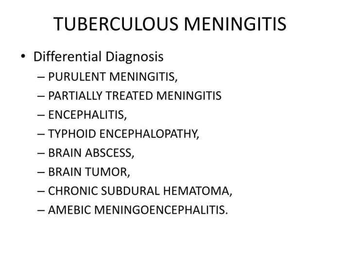
Differential Diagnosis: Tuberculous Meningitis Overview And More

Tuberculous meningitis (TBM) can mimic other neurological conditions, making accurate diagnosis crucial. The overlapping symptoms and subtle differences in presentation can lead to diagnostic challenges. Early and precise identification of TBM is vital for timely treatment and favorable outcomes. This necessitates a thorough understanding of the differential diagnoses and the specific diagnostic tests used to distinguish TBM from other causes of meningitis.Differentiating TBM from other forms of meningitis and neurological illnesses is essential to provide appropriate and effective treatment.
Failure to accurately identify TBM can lead to delays in initiating crucial anti-tuberculosis therapy, potentially resulting in poor neurological outcomes. A systematic approach, considering the clinical presentation, epidemiological factors, and specific diagnostic tests, is paramount in arriving at a definitive diagnosis.
Comparison with Other Meningitis Causes
Identifying the specific cause of meningitis is a critical aspect of patient management. Different types of meningitis have varying clinical presentations, diagnostic findings, and treatment strategies. Accurate differentiation is crucial for initiating the appropriate management plan.
- Viral Meningitis: Viral meningitis is frequently characterized by a more benign course compared to TBM. Symptoms are often less severe and typically resolve within a few weeks. Laboratory findings, such as cerebrospinal fluid (CSF) analysis, often show a lymphocytic pleocytosis, with a higher proportion of lymphocytes compared to TBM. The presence of characteristic viral pathogens in the CSF can further aid in diagnosis.
Viral meningitis is typically self-limiting, and treatment focuses on symptom management.
- Bacterial Meningitis: Bacterial meningitis, often caused by pathogens like
-Streptococcus pneumoniae* or
-Neisseria meningitidis*, presents with a more rapid and severe onset of symptoms compared to TBM. CSF analysis typically reveals a neutrophilic pleocytosis, high protein levels, and low glucose levels. Rapid initiation of broad-spectrum antibiotics is crucial for bacterial meningitis. The clinical presentation, particularly the rapid progression and severe symptoms, helps distinguish it from TBM. - Fungal Meningitis: Fungal meningitis, often caused by
-Cryptococcus neoformans*, may present with a subacute or chronic course. CSF analysis may reveal lymphocytic pleocytosis, but the presence of specific fungal elements can aid in diagnosis. Treatment strategies differ significantly from TBM and involve antifungal agents. - Other Neurological Conditions: Conditions such as brain abscesses, stroke, and encephalitis can present with symptoms overlapping with TBM, especially concerning neurological deficits. Imaging studies, like CT scans and MRIs, can aid in distinguishing these conditions from TBM. The presence of focal neurological deficits or specific imaging findings is key to differentiating them from the more diffuse involvement seen in TBM.
Diagnostic Tests for Differential Diagnosis
A comprehensive diagnostic approach is crucial in distinguishing TBM from other neurological conditions. This involves a combination of clinical evaluation, laboratory tests, and imaging studies.
- Cerebrospinal Fluid (CSF) Analysis: CSF analysis is a fundamental diagnostic tool. The presence of lymphocytes, low glucose levels, and elevated protein levels, along with the presence of acid-fast bacilli (AFB) in the CSF, is indicative of TBM. Comparing these findings with other causes of meningitis helps establish a differential diagnosis. Furthermore, the cellular composition and other markers in the CSF help differentiate between the various causes of meningitis.
- Imaging Studies: Imaging studies, such as CT scans and MRIs, are essential to rule out other neurological conditions. These scans can reveal abnormalities indicative of brain abscesses, stroke, or other focal lesions. CT scans can help identify signs of intracranial pressure elevation, which is common in TBM, and MRIs can further delineate the extent of the pathology and identify specific regions of involvement.
- Sputum and Tissue Culture: Culture of sputum or tissue samples for
-Mycobacterium tuberculosis* can confirm the diagnosis of TBM. This is important for accurate identification of the causative agent, which is critical for effective treatment.
Overlap of Symptoms and Challenges
The overlapping symptoms between TBM and other neurological conditions can make differentiation challenging. For example, both TBM and viral meningitis can manifest with fever, headache, and altered mental status. A meticulous clinical history, a thorough neurological examination, and the combination of laboratory findings are crucial for distinguishing TBM from other causes.
Table of Key Differences
| Characteristic | Tuberculous Meningitis | Viral Meningitis | Bacterial Meningitis |
|---|---|---|---|
| CSF Findings | Lymphocytic pleocytosis, low glucose, elevated protein, AFB | Lymphocytic pleocytosis, normal glucose, normal or slightly elevated protein | Neutrophilic pleocytosis, low glucose, elevated protein |
| Course | Insidious, subacute | Acute, self-limiting | Rapid, severe |
| Symptoms | Headache, fever, altered mental status, neurological deficits | Headache, fever, altered mental status, malaise | Severe headache, high fever, stiff neck, altered mental status |
Conclusion
In conclusion, tuberculous meningitis, though a formidable adversary, is not insurmountable. By understanding its multifaceted nature, from its insidious onset to the potential for long-term sequelae, we can better equip ourselves to combat this disease. Early diagnosis and appropriate treatment remain paramount, underscoring the vital role of public health initiatives and research in prevention and improved outcomes.