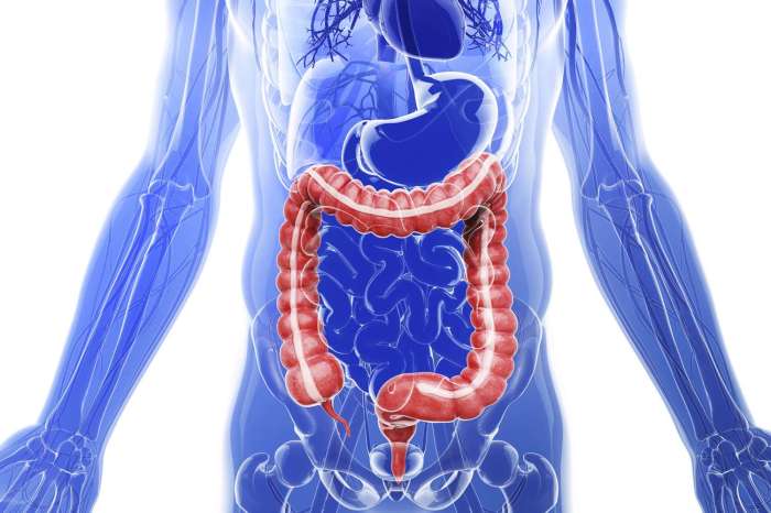
Tag: human body

  • What Happens to My Body Right After I Die?

    What Happens to My Body Right After I Die?

    What happens to my body right after I die? This exploration delves into the immediate physiological shifts, cellular processes, and external manifestations that mark the transition from life to death. We’ll examine the cascade of events, from the cessation of brain function to the visible signs of rigor mortis, livor mortis, and algor mortis. The journey will be both scientific and thought-provoking, offering insights into the fascinating yet often overlooked processes of the human body’s final moments.

    The immediate cessation of brain function triggers a domino effect throughout the body. Respiration stops, blood circulation ceases, and various organs experience profound changes. These changes, though swift, are complex, and we’ll unpack the biochemical processes that take place within the first few minutes after death. The subsequent cellular breakdown, influenced by enzymes and environmental factors, further shapes the body’s transformation.

    We’ll also examine the visible signs of death, which offer clues about the time of death.

    Immediate Physiological Changes

    What happens to my body right after i die

    The moment life ceases, a cascade of physiological changes begins, unfolding in a predictable sequence. These changes, though often unsettling, are a natural consequence of the body’s intricate systems shutting down. Understanding these immediate transformations offers a glimpse into the final moments of a biological existence.

    Cessation of Brain Function

    The brain, the command center of the body, is the first to exhibit significant changes. The cessation of electrical activity, or brain death, is the hallmark of irreversible cessation of all brain functions. This immediate loss of electrical impulses halts the brain’s ability to regulate bodily processes, including breathing and circulation. The lack of conscious thought and perception is a direct result of the cessation of neural activity.

    Sequence of Events After Respiration Cessation

    Following the cessation of respiration, the body rapidly depletes its oxygen supply. This leads to a cascade of events, affecting every organ system. The lack of oxygen deprives the cells of the energy they need to function, leading to their gradual deterioration. The heart, deprived of its own oxygen supply, loses its ability to pump blood, initiating the chain reaction of organ failure.

    Cellular metabolism slows, and eventually ceases.

    Effects of Blood Circulation Loss

    The cessation of blood circulation, triggered by cardiac arrest, has profound and immediate effects on various organs. Oxygen deprivation quickly leads to cellular damage and death in tissues and organs throughout the body. The lack of blood flow prevents the delivery of nutrients and oxygen to vital organs like the brain, heart, and kidneys, leading to their dysfunction.

    The brain is particularly vulnerable, as it is highly sensitive to oxygen deprivation. Damage occurs within minutes, progressing to irreversible harm.

    Biochemical Changes in the First Few Minutes

    Within the first few minutes after death, biochemical changes become increasingly evident. The body’s metabolic processes begin to shut down, and cellular respiration ceases. This leads to the accumulation of waste products, such as lactic acid, within the cells. Autolysis, the self-digestion of cells, commences, further contributing to the decomposition process. The breakdown of cellular components results in the release of various enzymes and chemicals, impacting the surrounding tissues.

    Physiological Changes in Different Organs and Systems

    Organ/System Change Timeframe Impact
    Brain Cessation of electrical activity Immediate Loss of consciousness
    Heart Cessation of pumping Immediate Loss of blood circulation
    Lungs Cessation of respiration Immediate Lack of oxygen
    Kidneys Loss of filtration function Minutes Accumulation of waste products
    Liver Loss of metabolic functions Minutes to hours Biochemical imbalances
    Gastrointestinal Tract Loss of motility Minutes to hours Digestion stops
    Skin Changes in appearance (e.g., pallor) Minutes to hours Visual indication of circulatory cessation

    Visual Representation of Circulatory System Changes

    Imagine a diagram of the human circulatory system. The heart, normally depicted as a powerful pump, would be depicted as a still organ, no longer contracting. The arteries and veins, once filled with flowing blood, would now appear empty, with the blood pooling in the lower extremities. This stagnation of blood flow is a direct consequence of the heart’s cessation of function.

    The once vibrant network of capillaries, responsible for nutrient and oxygen exchange, would appear as a network of static channels. This visual representation highlights the profound impact of cardiac arrest on the entire circulatory system.

    Ever wondered what happens to your body right after you kick the bucket? Well, your cells start shutting down, and your skin, which needs protection from the sun, especially if you have tattoos, might need some serious TLC. To keep your ink looking vibrant, finding the best sunscreen for tattoos is crucial. What is the best sunscreen for tattoos will help you in that regard.

    But back to the body’s post-mortem processes, it’s a fascinating (and slightly morbid) journey.

    Cellular Processes After Death

    What happens to my body right after i die

    The cessation of life marks a profound shift in the body’s internal environment, triggering a cascade of cellular changes. These processes, driven by the absence of metabolic functions and the activation of degradative enzymes, are essential for the eventual decomposition of the body. Understanding these cellular processes offers insights into the natural biological cycle and the complex mechanisms governing life’s transition.Cellular processes following death involve the breakdown of cellular structures and the release of cellular contents.

    So, what exactly happens to your body right after you kick the bucket? Well, your muscles relax, and your breathing stops. Interestingly, if you’re experiencing a tension headache, trying some simple techniques like applying a warm compress or practicing deep breathing exercises might help ease the discomfort. Finding a method that works for you, like the ones explained in this helpful guide on how to relieve a tension headache , can make a big difference.

    After that, your body starts to cool down, and eventually, decomposition sets in. Fascinating, isn’t it? Just don’t let a headache get in the way of your next philosophical pondering.

    The rate and extent of these processes vary considerably depending on factors such as environmental conditions, the individual’s health prior to death, and the specific tissues involved.

    Cellular Structure and Function Degradation

    The cessation of cellular respiration and other metabolic processes immediately following death leads to a significant disruption of cellular structure and function. Mitochondria, the powerhouses of the cell, cease their energy production, resulting in the depletion of ATP, the cellular energy currency. This depletion disrupts the integrity of cell membranes, leading to leakage of intracellular components. The loss of osmotic balance further contributes to cellular swelling and eventual rupture.

    Cellular Component Degradation and Release

    Following the initial disruption, cellular components begin to degrade. Proteins, the workhorses of the cell, are broken down into smaller peptides and amino acids by proteolytic enzymes. Nucleic acids, the genetic material, undergo hydrolysis, releasing their constituent nucleotides. Lipids, crucial for membrane structure, are broken down into fatty acids and glycerol. The release of these cellular contents into the surrounding environment contributes to the overall decomposition process.

    Enzymes in Decomposition, What happens to my body right after i die

    Enzymes play a critical role in the degradation of cellular components after death. These proteins act as biological catalysts, speeding up the breakdown of complex molecules into simpler ones. Examples include proteases, lipases, and nucleases. These enzymes are naturally present within cells or are released from lysosomes, cellular organelles responsible for waste disposal. The activation of these enzymes, often triggered by the increase in pH or the presence of other chemical signals, initiates the decomposition cascade.

    Rate of Cellular Breakdown in Different Tissues and Organs

    The rate of cellular breakdown varies considerably across different tissues and organs. For example, tissues with high protein content, such as muscle, will show faster degradation than tissues with lower protein content, such as bone. The presence of oxygen and moisture also influences the speed of decomposition. For example, a body in a dry environment will decompose more slowly than a body in a moist environment.

    Cellular Processes After Death by Organ System

    Organ System Cellular Processes Timeframe Impact
    Nervous System Neuronal disintegration, synaptic breakdown Hours/Days Cell death, tissue breakdown, loss of neurological function
    Muscular System Muscle protein breakdown, rigor mortis Hours/Days Muscle rigidity, decomposition
    Cardiovascular System Blood coagulation, tissue ischemia Minutes/Hours Loss of blood circulation, oxygen deprivation to tissues
    Respiratory System Lung tissue breakdown, loss of respiratory function Hours/Days Accumulation of fluids, cessation of gas exchange
    Digestive System Enzyme activation, tissue breakdown Days/Weeks Decomposition of ingested material, further tissue degradation
    Integumentary System Skin protein degradation, dehydration Days/Weeks Skin discoloration, dehydration, and breakdown

    Mechanisms Triggering Cellular Damage After Death

    The cessation of life triggers a multitude of cellular processes that result in damage and degradation. Depletion of ATP, the primary energy source, disrupts cellular functions. The accumulation of cellular waste products and the activation of enzymes contribute to the breakdown of cellular structures. Furthermore, the loss of oxygen and nutrient supply further exacerbates cellular damage. These combined effects lead to the eventual decomposition of the body.

    External Manifestations of Death

    The transition from life to death is marked by a series of observable changes in the body. These changes, collectively known as post-mortem changes, provide valuable clues for forensic scientists and medical examiners in determining the time of death and understanding the circumstances surrounding the demise. Understanding these external signs is crucial for accurately assessing the post-mortem interval (PMI).These changes are not simply a matter of aesthetics; they represent a complex interplay of biological processes that cease once life ceases.

    Careful observation and documentation of these changes can provide critical information for investigations.

    Visible Signs of Death

    The external signs of death are a combination of chemical and physical processes that begin immediately following cessation of the circulatory system. These processes include rigor mortis, livor mortis, and algor mortis. Each of these processes unfolds in a predictable sequence and is influenced by various factors.

    Rigor Mortis

    Rigor mortis, meaning “stiffness of death,” is the post-mortem stiffening of muscles. This phenomenon is due to the depletion of ATP (adenosine triphosphate), the energy currency of cells. Without ATP, the myosin heads in muscle fibers cannot detach from the actin filaments, leading to a sustained contraction.The onset of rigor mortis typically begins several hours after death and peaks within 6 to 12 hours.

    Following the peak, the muscles gradually relax over the next few days as the proteins begin to degrade. Factors such as ambient temperature, muscle mass, and the cause of death can influence the speed and intensity of rigor mortis. For example, in a cold environment, the process might be slower, while individuals with larger muscle mass will exhibit rigor mortis more intensely.

    So, what actually happens to my body right after I die? Well, the cooling process begins, a fascinating biological phenomenon. Interestingly, some people experience a distinct “hot sleeper” or “cold sleeper” phenomenon, which can be related to underlying health conditions, but ultimately doesn’t alter the eventual cooling of the body. Understanding how the body temperature changes post-mortem can provide valuable insights into the broader process of decay.

    For more on the nuances of body temperature regulation during sleep, check out this informative article on hot sleeper vs cold sleeper. It’s all part of the complex dance of life and death.

    Livor Mortis

    Livor mortis, also known as post-mortem lividity, is the purplish discoloration of the skin due to the settling of blood in the lower parts of the body. Gravity pulls the blood, which is no longer being circulated, to the dependent areas, resulting in a bluish-purple discoloration.Livor mortis typically begins within a few hours of death and becomes fixed within 8 to 12 hours.

    The distribution of the discoloration provides clues about the body’s position after death. If the body is moved after livor mortis has begun to develop, the discoloration may not be evenly distributed.

    Algor Mortis

    Algor mortis is the cooling of the body after death. The body loses heat to the surrounding environment at a predictable rate. The rate of cooling depends on factors such as the environmental temperature and the clothing or covering of the body.Algor mortis is a more gradual process, typically occurring over several hours or even days, depending on the surrounding conditions.

    The body temperature decreases at a predictable rate and can be used to estimate the time of death. For instance, in a cold environment, the body will cool significantly slower than in a hot environment.

    Factors Influencing the Rate of Post-Mortem Changes

    The rate of post-mortem changes is influenced by a number of factors, including:

    • Ambient temperature:
    • A warmer environment accelerates the rate of decomposition, while a cooler environment slows it down.

    • Body mass:
    • Larger individuals tend to retain heat for longer periods, thus slowing the cooling process.

    • Clothing:
    • Clothing can insulate the body, influencing both cooling and the development of rigor mortis.

    • Cause of death:
    • Certain conditions, such as exertion or trauma, can accelerate or decelerate the onset of these changes.

    Estimating Time of Death

    A combination of these external signs can assist in estimating the time since death. For instance, if rigor mortis is fully developed, and livor mortis is fixed, then the body has been deceased for a period of time. The forensic scientist considers all the factors, and together with the observed signs, they provide an estimate of the time of death.

    Table of Post-Mortem Changes

    Post-Mortem Change Description Timeframe Factors influencing rate
    Rigor Mortis Stiffening of muscles Hours to days Temperature, muscle mass, cause of death
    Livor Mortis Discoloration of skin Hours Blood settling, body position, and movement after death
    Algor Mortis Cooling of body Hours/Days Environmental temperature, body mass, clothing

    Flow Chart of External Post-Mortem Changes

    (A complex flow chart is not possible to represent in text format. A visual diagram would be necessary to represent the order and interconnectedness of these post-mortem changes effectively.)

    Decomposition Processes

    The final stage of the human journey after death is decomposition, a complex and fascinating process driven by the tireless activity of microorganisms and influenced by the environment. Understanding this process offers a glimpse into the intricate interplay between life, death, and the natural world. It also plays a vital role in forensic science, enabling estimations of time since death and the identification of potential contributing factors.The decomposition of a body is a gradual and multifaceted process, driven by a symphony of biological and chemical reactions.

    Microorganisms, including bacteria and fungi, play a crucial role in breaking down the complex organic molecules of the body. Environmental factors like temperature, humidity, and access to oxygen significantly affect the speed and nature of this process. This intricate dance between biology and environment shapes the visible stages of decomposition, allowing forensic experts to gather vital clues.

    Microorganisms in Decomposition

    The decomposition process is heavily reliant on the presence of microorganisms. These tiny organisms, including bacteria and fungi, thrive on the organic matter of the body. Their enzymes break down proteins, carbohydrates, and lipids, ultimately reducing the body to simpler substances. Different types of microorganisms thrive in various conditions, dictating the rate and manner of decomposition. This microbial activity is the driving force behind the various stages of decomposition.

    Stages of Decomposition

    Decomposition progresses through distinct stages, each marked by specific characteristics. The stages are influenced by factors such as the body’s location, the surrounding environment, and the presence of specific microorganisms.

    • Fresh Stage: This initial stage immediately follows death. No significant changes are visible to the naked eye. Cellular processes within the body are still active, though gradually ceasing.
    • Bloated Stage: Gas production by bacteria leads to swelling, primarily in the abdominal region. The skin may appear taut and the body may emit foul odors. This stage typically lasts for several days. The body’s internal environment becomes increasingly anaerobic, creating an environment conducive to the growth of different microorganisms.
    • Decay Stage: The decomposition process intensifies, with tissues breaking down and liquefying. The body’s structure begins to disintegrate, and the skin may start to slough off. This stage can last for several days to weeks. The release of gases and fluids from the body becomes more pronounced. The foul odor intensifies, and the body becomes increasingly vulnerable to insect colonization.

    Factors Influencing Decomposition Rate

    Numerous factors influence the rate of decomposition. These factors are critical in forensic investigations.

    • Temperature: Higher temperatures generally accelerate decomposition, while lower temperatures slow it down. A warmer climate will lead to faster decomposition compared to a cold one. For example, a body left in a desert will decompose faster than a body buried in a frozen tundra.
    • Humidity: High humidity can accelerate decomposition, providing a favorable environment for microorganisms to thrive. Conversely, extremely dry conditions can slow down the process. The moisture content in the air can significantly affect the microbial activity, which, in turn, dictates the pace of decomposition.
    • Environmental Conditions: Factors like access to oxygen, burial depth, and the presence of scavengers all impact the decomposition rate. A body left in an open field will decompose faster than a body buried deep underground. The presence of scavengers can accelerate the decomposition process.

    Role of Insects in Decomposition

    Insects play a significant role in the decomposition process, particularly in the later stages. They are attracted to the decaying body, and their activity contributes to the breakdown of tissues. Different insects colonize the body at different stages, and this pattern can provide valuable information for forensic scientists. The sequence of insect colonization can aid in estimating the time of death.

    Stages of Decomposition Table

    Stage Characteristics Timeframe Indicators
    Fresh No significant changes Immediate No obvious changes
    Bloated Swelling, gas formation Days Abdominal distension, foul odor
    Decay Decomposition, liquefaction Days/Weeks Tissue disintegration, skin slippage, odor intensification
    Post-Decay Skeletonization, dry remains Weeks/Months Skeleton remains, dry skin

    Complete Breakdown of the Body

    The complete breakdown of the body involves the progressive degradation of tissues and the conversion of complex organic molecules into simpler substances. This process is driven by microbial action and environmental factors. The body eventually reduces to its basic components, which are then released into the environment. This breakdown is essential for the natural recycling of nutrients.

    Conclusive Thoughts: What Happens To My Body Right After I Die

    In conclusion, the transition from life to death is a fascinating and complex process, marked by a cascade of physiological and cellular changes. Understanding these events offers a unique perspective on the human body’s remarkable capabilities and the inevitable journey towards decomposition. From the immediate cessation of brain function to the gradual breakdown of tissues, the body’s response to death is a symphony of biological events.

    We’ve explored the scientific details of this journey, but the larger philosophical implications are also significant. This journey through death is a reminder of the cyclical nature of life, and the profound impact our bodies have on the world around us.

  • Trachea Function and Conditions and Diagram A Deep Dive

    Trachea Function and Conditions and Diagram A Deep Dive

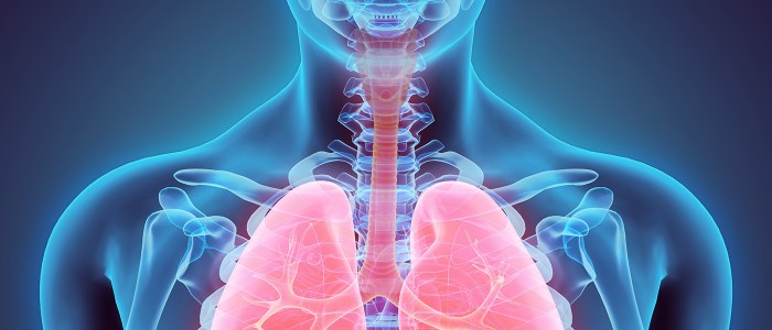
    Trachea function and conditions and diagram provides a comprehensive overview of the windpipe, its crucial role in respiration, and the various conditions that can affect it. We’ll explore its intricate structure, examining how air travels through it, its impact on the overall respiratory system, and the diseases that can impact this vital part of our anatomy. This guide also includes a detailed diagram to visualize the trachea’s position and components within the respiratory system.

    This exploration delves into the mechanics of air flow, highlighting the trachea’s role in warming, humidifying, and filtering the air we breathe. We’ll also analyze the impact of different conditions and diseases on trachea function, discussing the symptoms, causes, and treatment options. A detailed look at the relationship between the trachea and surrounding structures, like the esophagus and blood vessels, will further illuminate its complexity.

    Introduction to the Trachea

    The trachea, commonly known as the windpipe, is a vital component of the respiratory system, situated in the anterior neck and mediastinum of the chest. It acts as a crucial conduit for air to travel between the larynx (voice box) and the lungs. Understanding its structure and function is essential for comprehending the mechanics of breathing and diagnosing respiratory conditions.The trachea’s role in the respiratory system is primarily to facilitate the unimpeded flow of air.

    It’s a flexible tube, allowing for some movement during breathing cycles. This flexibility, combined with its robust structure, ensures that air can pass smoothly, even during shifts in body position. Its position, anterior to the esophagus, also allows for simultaneous passage of food and air.

    Tracheal Structure and Components

    The trachea’s structure is well-suited to its function as an air passage. It’s composed of a series of C-shaped cartilaginous rings that provide structural support and prevent the trachea from collapsing. These rings are incomplete posteriorly, allowing for flexibility and accommodating the esophagus’s expansion during swallowing. The tracheal wall also contains smooth muscle tissue, which further regulates the diameter of the trachea, influencing airflow.

    Understanding the trachea’s function, like its role in breathing and common conditions affecting it, is crucial, as seen in diagrams. However, similar to how the frontal bone’s anatomy, function, and treatment options are vital to understand, as detailed in this article on frontal bone anatomy function and treatment , a healthy airway is fundamental. Knowing about the intricate details of the trachea and its potential issues is key to maintaining overall respiratory health.

    The inner lining, or mucosa, is covered with a ciliated epithelium. These cilia constantly beat, moving mucus and trapped particles upward toward the pharynx, preventing them from entering the lungs.

    Trachea’s Role in Air Passage

    The trachea acts as a critical conduit for air, carrying oxygen-rich air from the larynx to the lungs. The branching of the trachea into the right and left bronchi marks the transition into the lower respiratory tract. This branching ensures that air is directed to each lung. The rhythmic expansion and contraction of the chest, driven by the diaphragm and intercostal muscles, creates pressure differentials that facilitate the movement of air in and out of the lungs.

    The cartilage rings, as previously mentioned, are essential in maintaining the patency of the airway, preventing collapse during inhalation. This structural integrity is crucial for proper ventilation.

    Tracheal Connections and Respiratory Organs

    The trachea connects the larynx superiorly and bifurcates into the left and right main bronchi inferiorly. The main bronchi further branch into smaller bronchioles, eventually reaching the alveoli, the tiny air sacs in the lungs where gas exchange occurs. This branching network ensures efficient delivery of air to all parts of the lungs, maximizing oxygen uptake. The smooth muscle in the tracheal walls and bronchioles allows for adjustments in airflow based on the body’s needs.

    Table: Trachea Anatomy and Function

    Anatomical Structure Function Location Image Description
    Cartilage Rings Provide structural support, prevent collapse, and allow for flexibility. Surrounding the trachea, forming a C-shape. A diagram showing the trachea with clearly visible C-shaped cartilaginous rings. The rings should be depicted as a series of interconnected C’s that encircle the tube.
    Mucosa Line the trachea’s inner surface; cilia move mucus and trapped particles upward. Lines the interior of the trachea. A close-up view of the trachea’s inner lining, highlighting the ciliated epithelial cells and the mucus layer.
    Smooth Muscle Regulates airflow by adjusting the trachea’s diameter. Within the tracheal wall. A diagram illustrating the smooth muscle fibers within the tracheal wall, showing how they surround the cartilage rings.
    Larynx Superior connection to the trachea, conduit for air. Above the trachea in the neck. A diagram showing the trachea with a distinct connection to the larynx. The larynx should be clearly visible and labeled.

    Trachea Function in Respiration

    The trachea, often called the windpipe, plays a crucial role in the respiratory system, enabling the passage of air to and from the lungs. Its structure and function are intricately linked to the efficient exchange of gases vital for life. Understanding the trachea’s function during inhalation and exhalation, as well as its role in conditioning inspired air, is essential to comprehending the overall respiratory process.The trachea acts as a conduit, facilitating the movement of air between the external environment and the lungs.

    This passageway is critical for the continuous supply of oxygen needed for cellular respiration and the removal of carbon dioxide, a waste product. The unique structure of the trachea allows for efficient airflow, warming, humidifying, and filtering the inhaled air before it reaches the delicate lung tissues.

    Mechanics of Airflow

    Air moves through the trachea due to pressure differences between the atmosphere and the lungs. During inhalation, the diaphragm contracts, increasing the volume of the thoracic cavity. This expansion creates a lower pressure within the lungs than in the atmosphere, causing air to flow into the trachea and subsequently into the lungs. Conversely, during exhalation, the diaphragm relaxes, decreasing the volume of the thoracic cavity, and increasing the pressure within the lungs.

    This higher pressure forces air out of the lungs, through the trachea, and into the atmosphere.

    Trachea’s Role in Air Conditioning

    The trachea is lined with specialized cells that play a vital role in preparing the inhaled air for the delicate structures of the lungs. These cells warm the air to body temperature, ensuring the lungs are not subjected to extreme temperature fluctuations. They also humidify the air, adding moisture to prevent the delicate lung tissues from drying out.

    Furthermore, the trachea’s lining contains cilia and mucus, which filter out foreign particles, such as dust and pathogens, preventing them from reaching the sensitive lung tissues.

    Tracheal Structure for Efficient Airflow

    The trachea’s rigid structure, composed of C-shaped cartilaginous rings, provides crucial support and prevents the trachea from collapsing during inhalation and exhalation. This structure allows for flexibility, enabling the trachea to move and adapt to changes in posture and body movement without impeding airflow. The C-shape also allows for flexibility in the esophagus, permitting food passage without obstructing the airway.

    This structure, combined with the smooth muscle lining, ensures efficient and unobstructed air passage.

    Trachea’s Role in Gas Exchange

    The trachea’s role in gas exchange is primarily as a conduit. It does not directly participate in the actual exchange of oxygen and carbon dioxide. The gas exchange occurs in the alveoli, the tiny air sacs within the lungs. The trachea simply provides the pathway for the air to reach these vital gas-exchange sites. The efficient movement of air through the trachea is critical for maintaining the continuous supply of oxygen to the body’s tissues and the removal of carbon dioxide.

    Comparison of Trachea, Bronchi, and Alveoli

    Feature Trachea Bronchi Alveoli
    Location Superior part of the respiratory system Branching from the trachea Terminal air sacs in the lungs
    Structure C-shaped cartilaginous rings Cartilage decreases in size; smooth muscle increases Thin-walled sacs with extensive capillary network
    Function Conduits air to and from lungs; warms, humidifies, filters air Conduits air to the alveoli; regulates airflow Gas exchange; oxygen diffuses into blood; carbon dioxide diffuses out
    Importance in Respiration Essential for initial air passage; crucial for preventing damage Vital for directing air to specific areas of the lungs Site of the critical gas exchange process

    The table above highlights the unique roles of the trachea, bronchi, and alveoli in the respiratory system, emphasizing their distinct structural and functional characteristics. Each component plays a vital part in the overall respiratory process, from initial air intake to the final gas exchange.

    Conditions Affecting Trachea Function

    Trachea function and conditions and diagram

    The trachea, a vital component of the respiratory system, is susceptible to various conditions that can impair its function and, consequently, the overall respiratory process. Understanding these conditions is crucial for recognizing symptoms, seeking timely medical intervention, and ultimately improving patient outcomes. These conditions can range from relatively benign irritations to life-threatening obstructions.The trachea’s role in facilitating airflow is paramount.

    Any disruption to this function can lead to significant respiratory distress. Conditions affecting the trachea can be broadly categorized as obstructive, inflammatory, or neoplastic. Obstructive conditions hinder the passage of air, inflammatory conditions cause swelling and irritation, and neoplastic conditions involve abnormal tissue growth. Recognizing the underlying causes and symptoms is critical for appropriate diagnosis and treatment.

    Obstructive Conditions

    Obstructive conditions impede the flow of air through the trachea. These obstructions can stem from various sources, including foreign bodies, tumors, or narrowing of the airway. Symptoms typically manifest as difficulty breathing, especially during exertion. These conditions can significantly impact the efficiency of the respiratory system, leading to reduced oxygen intake and carbon dioxide elimination. The severity of these conditions can range from mild discomfort to life-threatening situations, demanding immediate medical attention.

    Inflammatory Conditions

    Inflammatory conditions, such as tracheitis, involve swelling and irritation of the tracheal lining. These conditions are frequently triggered by infections, irritants, or allergic reactions. Symptoms often include a persistent cough, chest discomfort, and sometimes fever. The inflammatory response can affect the entire respiratory system, potentially leading to complications such as pneumonia or bronchitis. The severity of inflammatory conditions depends on the underlying cause and the individual’s immune response.

    Neoplastic Conditions

    Neoplastic conditions, including tumors, can develop within or around the trachea. These growths can exert pressure on the airway, leading to narrowing and obstruction. Symptoms may vary depending on the size and location of the tumor. In some cases, the symptoms are subtle, but in others, they can be severe and life-threatening. The impact of neoplastic conditions on the respiratory system can be profound, potentially leading to respiratory failure if left untreated.

    Common Conditions Affecting Trachea Function

    Condition Cause Symptoms Treatment
    Tracheitis Viral or bacterial infections, irritants, allergies Cough, chest discomfort, fever, difficulty breathing Antibiotics (if bacterial), pain relievers, rest, hydration
    Tracheal stenosis Traumatic injury, inflammatory diseases, congenital abnormalities Shortness of breath, wheezing, chronic cough, chest pain Bronchoscopy, dilation, surgery (in severe cases)
    Tracheal tumor Abnormal cell growth (benign or malignant) Cough, hoarseness, dyspnea, hemoptysis, chest pain, dysphagia Surgery, radiation therapy, chemotherapy (depending on tumor type and stage)
    Foreign body aspiration Inhalation of foreign objects (e.g., food, toys) Sudden onset of respiratory distress, coughing, choking, wheezing Immediate medical intervention (e.g., bronchoscopy for removal)

    Trachea-Related Diseases

    The trachea, a vital component of the respiratory system, is susceptible to various diseases that can significantly impact breathing and overall health. Understanding these conditions is crucial for timely diagnosis and effective treatment. These diseases can range from relatively benign irritations to life-threatening obstructions. This section will delve into specific tracheal diseases, exploring their causes, mechanisms, symptoms, and diagnostic approaches.

    Specific Tracheal Diseases

    Various conditions can affect the trachea, impacting its structure and function. These include infections, inflammatory responses, and structural abnormalities. Understanding these diseases helps in identifying the underlying cause and implementing appropriate treatment strategies.

    Inflammatory Tracheal Conditions

    Inflammation of the trachea, often triggered by infections or irritants, can lead to various symptoms. Examples include tracheitis and tracheobronchitis. Tracheitis is characterized by inflammation of the tracheal lining, often associated with acute infections, whereas tracheobronchitis extends the inflammation to involve the bronchi as well.

    • Tracheitis: This condition, typically caused by viral or bacterial infections, presents with symptoms like a cough, hoarseness, and chest discomfort. The inflammation can narrow the airway, leading to breathing difficulties. Pathophysiologically, the infection triggers an inflammatory response in the tracheal mucosa, resulting in edema and cellular infiltration. The clinical presentation includes the aforementioned symptoms, and diagnosis often relies on physical examination, including listening to lung sounds, and possibly imaging studies.

      Treatment involves supportive care, such as rest, fluids, and over-the-counter pain relievers. Antibiotics might be necessary if a bacterial infection is suspected.

    • Tracheobronchitis: This more extensive inflammation encompasses both the trachea and bronchi, often following an upper respiratory infection. It typically presents with a persistent cough, which may produce mucus, and shortness of breath. The pathophysiology involves the spread of infection from the upper respiratory tract to the lower airways. Diagnosis is often made based on clinical presentation and physical examination.

      Treatment, similar to tracheitis, focuses on supportive care and potentially antibiotics if bacterial involvement is suspected. Monitoring for complications is important in both conditions.

    Tracheal Tumors

    Neoplasms, both benign and malignant, can develop within the trachea. These growths can obstruct the airway, leading to breathing difficulties.

    • Benign Tumors: These growths, such as papillomas or fibromas, typically cause a gradual narrowing of the airway. They are often slow-growing and may present with symptoms like persistent cough, wheezing, and a sensation of tightness in the chest. Diagnosis involves imaging studies like CT scans or bronchoscopy. Treatment options often involve surgical removal or other minimally invasive procedures.

    • Malignant Tumors: Malignant tumors, such as squamous cell carcinoma or adenocarcinomas, can grow rapidly and aggressively. They may cause severe airway obstruction, leading to life-threatening respiratory distress. Clinical presentation includes persistent cough, hemoptysis (coughing up blood), and dyspnea. Diagnostic procedures include bronchoscopy with biopsy, CT scans, and PET scans. Treatment strategies often involve surgery, radiation therapy, and chemotherapy, depending on the tumor’s stage and location.

    Diagnostic Procedures

    Diagnosing tracheal diseases involves a combination of methods, including physical examination, imaging studies, and endoscopic procedures.

    • Bronchoscopy: A flexible or rigid tube with a camera is inserted through the nose or mouth into the airways. This allows direct visualization of the trachea and surrounding structures, enabling the identification of abnormalities, like tumors or foreign bodies.
    • Imaging Studies: X-rays, CT scans, and MRI scans provide detailed images of the trachea and surrounding tissues, allowing for the detection of structural abnormalities, masses, or infections. The choice of imaging modality depends on the suspected condition and clinical presentation.

    Comparison of Tracheal Diseases

    | Disease | Description | Symptoms | Treatment Options ||—|—|—|—|| Tracheitis | Inflammation of the tracheal lining | Cough, hoarseness, chest discomfort | Rest, fluids, pain relievers; antibiotics if bacterial || Tracheobronchitis | Inflammation of the trachea and bronchi | Persistent cough, mucus production, shortness of breath | Rest, fluids, pain relievers; antibiotics if bacterial || Benign Tracheal Tumors | Non-cancerous growths in the trachea | Persistent cough, wheezing, chest tightness | Surgical removal, other minimally invasive procedures || Malignant Tracheal Tumors | Cancerous growths in the trachea | Persistent cough, hemoptysis, dyspnea | Surgery, radiation therapy, chemotherapy |

    Diagram of the Trachea: Trachea Function And Conditions And Diagram

    The trachea, often called the windpipe, is a crucial component of the respiratory system. Understanding its structure and position within the body is essential for comprehending how air travels to and from the lungs. This section delves into a visual representation of the trachea, highlighting its key anatomical features and variations.The trachea’s complex structure allows for efficient airflow while also protecting against foreign particles.

    This intricate design is vital for proper respiration and overall health. A clear understanding of its anatomy and position within the respiratory system is key to appreciating the delicate balance of the body’s breathing mechanisms.

    Trachea Structure Diagram

    This diagram will illustrate the trachea’s position within the respiratory system, along with its key anatomical features. It’s crucial to visualize the trachea’s relationship to other vital organs, such as the esophagus and the lungs. This aids in understanding how respiratory ailments can affect other bodily functions.

    Anterior View of the Trachea

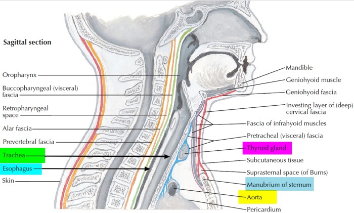
    The trachea is a flexible tube situated in the anterior part of the neck and the mediastinum of the thorax. It’s positioned anterior to the esophagus, with its cartilaginous rings providing structural support. The diagram below depicts this anterior view, showing the trachea’s characteristic C-shaped cartilaginous rings and its connection to the larynx and bronchi. This visualization helps identify its placement and relationship with other surrounding structures.

    [Imagine a diagram here. It should show the trachea positioned in the front of the neck and upper chest, connecting the larynx to the bronchi. The C-shaped cartilaginous rings should be clearly visible. Label the larynx, trachea, bronchi, and esophagus.]

    Cross-Sectional View of the Trachea, Trachea function and conditions and diagram

    This view provides a detailed look at the trachea’s internal structure. The diagram below illustrates the trachea’s cross-section, highlighting the mucosa lining, submucosa, hyaline cartilage rings, and adventitia. This detailed representation aids in understanding the trachea’s functional layers and structural integrity.

    [Imagine a diagram here. The diagram should show a cross-section of the trachea, clearly labeling the mucosa, submucosa, hyaline cartilage rings, and adventitia. The diagram should also show the lumen of the trachea.]

    Key Anatomical Features Table

    The table below summarizes the trachea’s key anatomical features, their locations, functions, and descriptions. This structured approach aids in understanding the relationship between structure and function in this vital respiratory organ.

    Structure Location Function Description
    C-shaped Cartilage Rings Surrounding the trachea Provide structural support and prevent collapse These rings maintain the trachea’s open shape, ensuring unobstructed airflow.
    Mucosa Lines the trachea Protects against foreign particles and pathogens The mucosa contains cilia that sweep mucus and trapped particles upward, aiding in the body’s defense mechanism.
    Submucosa Beneath the mucosa Connective tissue layer This layer contains glands that secrete mucus, aiding in moistening the air and trapping foreign particles.
    Adventitia Outermost layer Anchors the trachea to surrounding structures Connective tissue that holds the trachea in place.

    Trachea and Surrounding Structures

    The trachea, or windpipe, is a crucial part of the respiratory system, but it doesn’t operate in isolation. It’s surrounded by vital structures that influence its function and are themselves affected by respiratory movements. Understanding these relationships is key to comprehending how the respiratory system works as a whole.The trachea is nestled within the anterior mediastinum, a region of the chest cavity.

    This anatomical positioning places it close to other essential organs and tissues, creating a complex interplay.

    Learning about the trachea’s function and potential conditions, like tracheitis or tumors, is fascinating, and diagrams really help visualize it all. However, I’ve recently been researching how magnesium might help with period cramps, which is something I’m personally interested in, and the connection to overall bodily function is interesting. Magnesium for period cramps seems to be a promising area of study.

    Ultimately, understanding the trachea’s role in breathing and how it can be affected by various issues is still my primary focus.

    Surrounding Structures and Their Roles

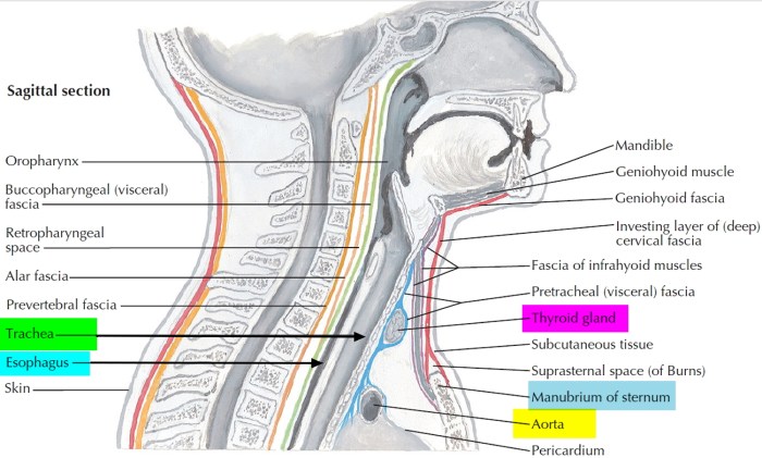
    The trachea is intimately associated with several structures. Its immediate neighbors include the esophagus, major blood vessels (like the aorta and its branches), and the nerves of the mediastinum. These structures interact with the trachea in various ways.

    • Esophagus: The esophagus, a muscular tube that carries food from the mouth to the stomach, lies posterior to the trachea. This proximity necessitates a close relationship and coordinated movement. The esophagus’s rhythmic contractions during swallowing must not impede the trachea’s function during respiration. The cartilaginous rings of the trachea provide structural support, preventing the trachea from collapsing during inhalation.

      The flexible nature of the esophagus allows for the passage of food without obstructing the airway.

    • Blood Vessels: The aorta and its branches, along with other major blood vessels, encircle and run alongside the trachea. These vessels deliver oxygenated blood to the body. The close proximity ensures efficient blood supply to the surrounding tissues, including the trachea itself. The pulsations of the blood vessels are negligible, but the overall blood flow is vital for maintaining the health of the trachea and surrounding tissues.

    • Nerves: Nerves of the mediastinum run near the trachea. These nerves are crucial for regulating various functions, including the trachea’s smooth muscle contractions and reflexes involved in coughing. The presence of these nerves is critical for maintaining the integrity and function of the respiratory system. Damage to these nerves can disrupt normal breathing patterns. These nerves facilitate coordination with other respiratory muscles and regulate the airway’s response to stimuli.

    Interaction During Respiratory Movements

    During inhalation, the expansion of the chest cavity creates a negative pressure within the lungs, drawing air into the respiratory system. Simultaneously, the diaphragm and intercostal muscles contract, increasing the volume of the thoracic cavity. The trachea, supported by its cartilaginous rings, remains open and allows for the unobstructed flow of air.During exhalation, the chest cavity contracts, decreasing the volume of the lungs.

    The elastic recoil of the lungs and the relaxation of the respiratory muscles facilitate the expulsion of air. The surrounding structures, like the esophagus, accommodate these respiratory movements without hindering the airflow through the trachea. The coordination between the trachea and its neighboring structures is critical for efficient respiration.

    Diagram of Trachea and Surrounding Structures

    [Imagine a diagram here showing the trachea in the center, with the esophagus positioned posteriorly, major blood vessels (aorta, etc.) encircling it, and nerves positioned around the trachea. The diagram should illustrate the relative positions of these structures and the spaces between them. Label the trachea, esophagus, blood vessels, and nerves clearly.]The diagram should visually represent the anatomical relationship between the trachea and its neighboring structures, highlighting the close proximity and potential for interaction.

    This visualization emphasizes the importance of coordinated movement between the trachea and its surrounding structures during respiration.

    Understanding the trachea’s role in breathing is crucial, and diagrams help visualize its function and potential conditions. However, it’s also important to be aware of other health concerns, like sexually transmitted infections (STIs). For a comprehensive look at various STI symptoms, check out this helpful resource: std symptoms gallery std pictures. Ultimately, a healthy respiratory system, as represented by the trachea, is essential, and recognizing potential issues is key.

    This knowledge can lead to early detection and appropriate treatment.

    Clinical Significance of Trachea Function

    Trachea function and conditions and diagram

    The trachea, often overlooked, plays a crucial role in maintaining overall health. Understanding its function and the potential for dysfunction is vital for diagnosing and treating various respiratory and systemic issues. The implications of tracheal problems extend beyond the respiratory system, affecting other organs and potentially leading to serious complications.Thorough comprehension of tracheal function and conditions empowers healthcare professionals to intervene effectively, improving patient outcomes and reducing the risk of severe consequences.

    Early detection and appropriate management are paramount in minimizing the impact of tracheal disorders on the body.

    Importance of Diagnosing and Treating Trachea-Related Problems

    Prompt diagnosis and treatment of tracheal issues are critical for preventing severe complications and maintaining respiratory health. Early intervention can mitigate the progression of diseases and improve patient quality of life. Failure to address tracheal problems can lead to a cascade of adverse effects, impacting various organ systems.

    Examples of How Tracheal Dysfunction Can Affect Other Bodily Systems

    Tracheal dysfunction can have a significant impact on other systems due to its role in respiration. For instance, impaired airflow due to tracheal narrowing or obstruction can lead to hypoxia (low oxygen levels) in the blood, affecting oxygen delivery to all tissues and organs. This can cause damage to vital organs like the heart and brain. Further, chronic respiratory distress associated with tracheal conditions can strain the cardiovascular system, potentially leading to heart failure.

    Moreover, impaired ventilation can impact acid-base balance, potentially causing metabolic acidosis or alkalosis.

    Potential Complications of Untreated Trachea Conditions

    Untreated tracheal conditions can lead to a multitude of serious complications, including severe respiratory distress, life-threatening infections, and even death. Failure to address conditions like tracheal stenosis (narrowing) or tumors can lead to complete airway obstruction, requiring urgent intervention. Furthermore, chronic inflammation and infection can lead to permanent lung damage, reducing lung capacity and increasing susceptibility to further infections.

    Clinical Significance Table

    Condition Impact on Other Systems Diagnostic Methods Potential Complications
    Tracheal Stenosis Impaired airflow, hypoxia, strain on cardiovascular system, potential for metabolic imbalances. Bronchoscopy, CT scans, X-rays, and possibly a flexible fiberoptic bronchoscopy. Respiratory failure, heart failure, infections, and permanent lung damage. Potentially requiring surgical intervention, such as a tracheostomy or dilation.
    Tracheal Tumors Partial or complete airway obstruction, compromised oxygenation, and potential for spread to other parts of the body. Bronchoscopy, CT scans, biopsy. May involve imaging techniques such as PET scans. Respiratory distress, lung damage, infections, spread of the tumor to other parts of the body, and even death in severe cases. Surgical removal may be necessary.
    Tracheomalacia Irregular airflow, potentially causing recurrent respiratory infections, and potentially impacting the cardiovascular system with long-term effects. Bronchoscopy, X-rays during respiration, and CT scans. Possibly lung function tests. Respiratory infections, increased susceptibility to lung complications, and difficulty breathing, particularly during exertion. May need supportive measures such as positive airway pressure.
    Tracheitis Inflammation, reduced airflow, potentially leading to pneumonia, and potentially causing an infection to spread to other areas. Physical examination, X-rays, blood tests (CBC and CRP), possibly bronchoscopy. Pneumonia, severe infections, and potential complications of severe respiratory distress, particularly in individuals with compromised immune systems.

    Ultimate Conclusion

    In conclusion, understanding the trachea’s function and potential conditions is crucial for comprehending the intricate workings of the respiratory system. This comprehensive guide has highlighted the trachea’s vital role in respiration, its intricate structure, the impact of various conditions, and the importance of early diagnosis and treatment. By grasping the information presented here, you can gain a deeper appreciation for the remarkable mechanics of the human body.

  • Tour the Digestive System A Journey Inside

    Tour the Digestive System A Journey Inside

    Tour the digestive system, a fascinating journey through the complex machinery of your body! This exploration dives into the intricate process of breaking down food, highlighting the vital roles of each organ from mouth to anus. We’ll uncover the amazing chemical and mechanical processes that transform a meal into usable energy, and explore the fascinating interplay between the digestive system and other bodily functions.

    Prepare to be amazed by the wonders within!

    This comprehensive tour will guide you through the stages of digestion, from ingestion to elimination. We’ll examine the structure and function of key organs like the stomach, small intestine, and liver, and discover how they work together in a symphony of biological processes. We’ll also explore common digestive disorders and ways to maintain a healthy digestive system through lifestyle choices.

    Introduction to the Digestive System: Tour The Digestive System

    The human digestive system is a complex network of organs working together to break down food into nutrients that the body can absorb and use for energy, growth, and repair. This intricate process, starting from the moment we ingest food, involves a coordinated effort from various organs, each playing a specific role in the overall digestive journey. Understanding this process allows us to appreciate the remarkable efficiency of our bodies and the importance of a healthy diet.The digestive system’s primary function is to transform ingested food into absorbable molecules.

    This transformation involves mechanical and chemical processes that break down large food particles into smaller components that can be transported across the intestinal lining and into the bloodstream. The resulting nutrients then fuel various bodily functions, supporting everything from basic metabolism to complex bodily processes.

    Key Organs and Their Functions

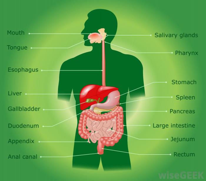
    The digestive system is a continuous tube, starting from the mouth and ending at the anus, with various specialized organs along the way. Each organ plays a crucial role in the digestive process.

    • Mouth: The mouth is the entry point for food. Mechanical digestion begins here through chewing (mastication), which breaks down food into smaller pieces. Saliva, secreted by salivary glands, contains enzymes that initiate the chemical breakdown of carbohydrates. This initial breakdown is crucial for efficient digestion later in the process.
    • Esophagus: The esophagus is a muscular tube connecting the mouth to the stomach. Peristalsis, wave-like muscle contractions, propels food down the esophagus, ensuring its uninterrupted journey to the stomach.
    • Stomach: The stomach is a muscular sac where food is further broken down. The stomach’s muscular walls churn the food, mixing it with gastric juices containing enzymes (like pepsin) that begin protein digestion. The acidic environment of the stomach also helps to kill harmful bacteria.
    • Small Intestine: The small intestine is the primary site of nutrient absorption. Its long, folded structure maximizes the surface area available for absorption. Enzymes secreted by the pancreas and bile from the liver further break down carbohydrates, proteins, and fats into their smallest components. Nutrients are then absorbed into the bloodstream through the intestinal lining.
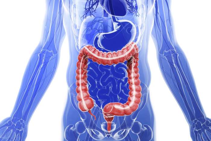
    • Large Intestine: The large intestine absorbs water and electrolytes from the remaining indigestible food matter. Bacteria in the large intestine ferment any remaining undigested material, producing some vitamins. The waste products are then compacted and stored until elimination.
    • Rectum: The rectum is a short, muscular tube that stores feces until elimination.
    • Anus: The anus is the final part of the digestive tract. It controls the release of feces from the body.

    Stages of Digestion

    Digestion involves several key stages.

    1. Ingestion: Ingestion is the intake of food into the body. It’s the initial step in the digestive process, starting the journey of food through the system.
    2. Digestion: Digestion involves the mechanical and chemical breakdown of food. This process transforms large food molecules into smaller, absorbable units. Mechanical digestion involves physical breakdown (chewing, churning), while chemical digestion uses enzymes to break down food into simpler molecules.
    3. Absorption: Absorption is the process by which the body takes up nutrients from digested food. The small intestine is the primary site of absorption, with its vast surface area facilitating the uptake of nutrients into the bloodstream.
    4. Elimination: Elimination is the removal of undigested food and waste products from the body. Feces, the solid waste product of digestion, is expelled through the anus.

    Diagram of the Digestive System

    Organ Description
    Mouth Food enters the system
    Esophagus Tube connecting mouth to stomach
    Stomach Churns and mixes food with enzymes
    Small Intestine Main site of nutrient absorption
    Large Intestine Absorbs water and electrolytes
    Rectum Stores waste before elimination
    Anus Waste is expelled from the body

    Organs of the Digestive System

    The digestive system is a complex network of organs working together to break down food into absorbable nutrients. This process, essential for energy and growth, involves mechanical and chemical breakdown. Understanding the structure and function of each organ is crucial to comprehending the intricate dance of digestion.

    Mouth

    The mouth, the initial entry point for food, plays a vital role in both mechanical and chemical digestion. Teeth mechanically break down food into smaller pieces, while saliva, containing enzymes like amylase, begins the chemical breakdown of carbohydrates. The tongue mixes the food with saliva, forming a bolus that is ready for swallowing.

    Esophagus

    The esophagus, a muscular tube connecting the mouth to the stomach, propels the bolus through peristalsis, a series of coordinated muscle contractions. This involuntary process ensures that food moves unidirectionally, preventing backflow. The esophagus’s smooth muscle lining facilitates this controlled transport.

    Stomach

    The stomach, a muscular sac, continues the mechanical breakdown of food. Its churning action mixes food with gastric juices, which contain hydrochloric acid and enzymes like pepsin. Hydrochloric acid creates an acidic environment essential for pepsin’s activity in protein digestion. This acidic environment also helps kill harmful bacteria that may be present in the food.

    Small Intestine, Tour the digestive system

    The small intestine, a long, coiled tube, is the primary site of nutrient absorption. Its large surface area, due to villi and microvilli, maximizes contact with digested food. Enzymes from the pancreas and the small intestine itself further break down carbohydrates, proteins, and fats. The absorbed nutrients are then transported into the bloodstream for use by the body.

    Large Intestine

    The large intestine, a shorter but wider tube than the small intestine, absorbs water and electrolytes from the remaining undigested material. Bacteria in the large intestine ferment some undigested material, producing vitamins like vitamin K. The remaining waste, now solidified into feces, is stored in the rectum before elimination.

    Liver

    The liver, a vital accessory organ, plays a crucial role in many bodily functions, including digestion. It produces bile, a fluid crucial for fat emulsification, breaking down large fat globules into smaller droplets, which increases the surface area for enzyme action. The liver also processes absorbed nutrients, detoxifies harmful substances, and synthesizes essential proteins.

    Pancreas

    The pancreas, another accessory organ, produces a variety of enzymes crucial for digestion. These enzymes, released into the small intestine, break down carbohydrates, proteins, and fats. The pancreas also regulates blood sugar levels by producing insulin and glucagon.

    Gallbladder

    The gallbladder, a small, pear-shaped sac, stores and concentrates bile produced by the liver. When food containing fat enters the small intestine, the gallbladder releases bile to aid in fat digestion. This ensures that the body has a readily available supply of bile when needed.

    Ever wanted to tour the digestive system? It’s a fascinating journey, isn’t it? Learning how our bodies process food is truly incredible. And for those looking to maintain a healthy weight, a successful weight-loss story like zepbound keeps weight off 3 years might provide some inspiration. Understanding the intricate processes within our bodies, like the digestive system, can be a powerful tool in our wellness journeys.

    This knowledge can be really valuable when trying to make healthy lifestyle choices.

    Enzymes in Digestion

    Enzymes are biological catalysts that speed up chemical reactions in the body, including those involved in digestion. Different enzymes target different types of nutrients. Amylase breaks down carbohydrates, pepsin breaks down proteins, and lipase breaks down fats. The specific environment (pH) and the precise substrate dictate which enzyme is most effective.

    Accessory Organ Comparison

    The liver, pancreas, and gallbladder, though distinct organs, work in concert to support digestion. The liver produces bile, the pancreas secretes digestive enzymes, and the gallbladder stores and releases bile. These accessory organs, acting in synergy, are essential for complete and efficient nutrient breakdown and absorption.

    Ever wanted to tour the digestive system? It’s fascinating how our bodies process food, isn’t it? Knowing how your body breaks down nutrients is key to making smart food choices, like selecting snacks for weight loss that support your goals. Learning about the digestive process can help you understand the impact of different foods, ultimately leading to a more informed approach to overall health.

    A better understanding of the digestive system can empower you to make more thoughtful choices, and ultimately improve your well-being.

    Organ Summary

    Organ Location Function Key Features
    Mouth Oral cavity Mechanical and chemical digestion begins Teeth, tongue, saliva
    Esophagus Connects mouth to stomach Transports food to stomach Peristalsis
    Stomach Upper abdomen Mechanical and chemical digestion of proteins Gastric juices, churning
    Small Intestine Lower abdomen Nutrient absorption Villi, microvilli, enzymes
    Large Intestine Lower abdomen Water absorption, waste elimination Bacteria fermentation
    Liver Upper right abdomen Bile production, nutrient processing Detoxification
    Pancreas Behind stomach Enzyme production, blood sugar regulation Insulin, glucagon
    Gallbladder Below liver Bile storage and release Concentrates bile

    Digestive Processes

    The journey of food through our bodies is a complex interplay of mechanical and chemical processes. From the initial chewing in the mouth to the final absorption in the small intestine, each step is carefully orchestrated to extract nutrients and eliminate waste. This intricate process is crucial for maintaining our overall health and well-being.The digestive system’s efficiency depends on the precise coordination of various mechanisms.

    These include physical breakdown of food, enzymatic reactions that break down complex molecules, and the intricate absorption of nutrients into the bloodstream. Understanding these processes allows us to appreciate the remarkable complexity of our own bodies.

    Ever wanted to tour the digestive system? It’s a fascinating journey, but did you know that sometimes, even our bodies’ internal pathways can be confused about the difference between a simple cold sore and herpes? Learning about the intricacies of the digestive system can help you better understand how your body works, including the importance of proper hygiene to avoid potential infections, like those that cause cold sores or herpes.

    For a deeper dive into the world of oral health and differentiating between these conditions, check out this helpful resource on cold sore vs herpes. Understanding these nuances is key to maintaining overall wellness, and the digestive system plays a significant role in this.

    Mechanical Digestion

    Mechanical digestion involves physically breaking down food into smaller pieces. This process increases the surface area exposed to digestive enzymes, facilitating the chemical breakdown that follows. The act of chewing, for instance, mechanically reduces large pieces of food into smaller particles. This process begins in the mouth and continues throughout the digestive tract.

    Chemical Digestion

    Chemical digestion involves the use of enzymes to break down complex food molecules into simpler absorbable forms. Enzymes are biological catalysts that speed up chemical reactions. Different enzymes target different types of molecules, such as carbohydrates, proteins, and fats. This process is crucial for extracting usable nutrients from the ingested food.

    Peristalsis and Segmentation

    Peristalsis is a series of rhythmic muscular contractions that propel food through the digestive tract. These wave-like movements occur in the esophagus, stomach, and intestines, ensuring that food moves unidirectionally from the mouth to the anus. Segmentation, on the other hand, involves the contraction and relaxation of smooth muscles in the intestines. This process mixes the food with digestive enzymes and further enhances the exposure of food particles to enzymes.

    These coordinated movements facilitate efficient digestion and nutrient absorption.

    Nutrient Absorption in the Small Intestine

    The small intestine is the primary site of nutrient absorption. Its highly folded inner lining, called the villi and microvilli, dramatically increases the surface area available for absorption. Nutrients, such as glucose, amino acids, and fatty acids, are absorbed across the intestinal lining and transported into the bloodstream for distribution throughout the body. This process is vital for providing the body with the building blocks needed for energy, growth, and repair.

    Digestive System Interactions

    The digestive system doesn’t operate in isolation. It interacts with other systems in the body, such as the circulatory system and the endocrine system. The circulatory system transports absorbed nutrients to various tissues and organs. The endocrine system regulates digestive processes through the release of hormones, such as gastrin and secretin. These hormonal signals control the release of digestive enzymes and the movement of food through the digestive tract.

    This interconnectedness highlights the intricate and sophisticated nature of human physiology.

    Comparison of Mechanical and Chemical Digestion

    Characteristic Mechanical Digestion Chemical Digestion
    Mechanism Physical breakdown of food into smaller pieces Breakdown of food molecules into simpler forms using enzymes
    Location Mouth, stomach, intestines Mouth, stomach, intestines
    Examples Chewing, churning in the stomach, segmentation Enzymatic breakdown of carbohydrates (amylase), proteins (proteases), and fats (lipases)
    Effect on food Increases surface area for chemical digestion Breaks down complex molecules into absorbable forms

    Health and Disorders of the Digestive System

    Tour the digestive system

    Our digestive system, a complex network of organs, is vital for our overall health. However, various disorders can disrupt its normal functioning, leading to discomfort and even serious health complications. Understanding these disorders, their causes, and available treatments is crucial for maintaining a healthy digestive system.

    Common Digestive Disorders

    Numerous conditions can affect the digestive system, ranging from mild inconveniences to more severe illnesses. Recognizing the signs and symptoms of these disorders is the first step towards seeking appropriate medical care.

    Heartburn

    Heartburn, a common digestive ailment, is characterized by a burning sensation in the chest, often radiating to the throat. This discomfort typically arises from stomach acid refluxing into the esophagus. Factors such as eating large meals, lying down soon after eating, and certain foods (e.g., fatty foods, citrus fruits, chocolate) can trigger heartburn. Lifestyle modifications, such as avoiding trigger foods, maintaining a healthy weight, and elevating the head of the bed, often provide relief.

    Over-the-counter medications can also effectively manage symptoms.

    Peptic Ulcers

    Peptic ulcers are sores that develop in the lining of the stomach or the upper portion of the small intestine. These ulcers are often caused by a bacterial infection (Helicobacter pylori) or the long-term use of nonsteroidal anti-inflammatory drugs (NSAIDs). Symptoms may include abdominal pain, nausea, and vomiting. Treatment typically involves antibiotics to eliminate the bacteria, medications to reduce stomach acid production, and lifestyle changes to minimize triggers.

    Irritable Bowel Syndrome (IBS)

    Irritable bowel syndrome (IBS) is a chronic condition affecting the large intestine. The exact cause of IBS remains unknown, but factors such as stress, diet, and hormonal changes may play a role. Symptoms of IBS can vary but often include abdominal pain, bloating, gas, diarrhea, and/or constipation. Management of IBS typically involves dietary modifications, stress reduction techniques, and medications to alleviate specific symptoms.

    Other Digestive Disorders

    • Gastroesophageal Reflux Disease (GERD): A more severe form of heartburn, GERD involves frequent and persistent acid reflux, potentially causing esophageal damage. Treatment options include medication, lifestyle changes, and in some cases, surgery.
    • Crohn’s Disease and Ulcerative Colitis: These are inflammatory bowel diseases (IBDs) that cause chronic inflammation in the digestive tract. Symptoms can range from mild to severe and include abdominal pain, diarrhea, weight loss, and fatigue. Treatment focuses on reducing inflammation and managing symptoms.
    • Celiac Disease: An autoimmune disorder triggered by gluten, a protein found in wheat, barley, and rye. Symptoms can vary but often include abdominal pain, diarrhea, and nutrient deficiencies. Treatment involves strict adherence to a gluten-free diet.

    Preventing Digestive Problems

    A healthy diet and lifestyle play a crucial role in preventing digestive issues. Regular exercise, maintaining a healthy weight, and managing stress are all important preventative measures. Choosing nutrient-rich foods, avoiding processed foods and excessive caffeine, and drinking plenty of water can also contribute to digestive health.

    Healthy Dietary Habits

    Adopting healthy dietary habits is key to supporting a healthy digestive system.

    • Fiber-rich foods: Include fruits, vegetables, and whole grains in your diet. Fiber promotes regular bowel movements and aids digestion.
    • Probiotics: These beneficial bacteria can support gut health. They are found in fermented foods such as yogurt, kefir, and sauerkraut.
    • Hydration: Drinking plenty of water helps maintain proper bowel function and prevents constipation.
    • Portion control: Avoiding large meals can help prevent digestive discomfort.

    Table of Common Digestive Disorders and Symptoms

    Disorder Symptoms
    Heartburn Burning sensation in the chest, possibly radiating to the throat; indigestion
    Peptic Ulcers Abdominal pain, nausea, vomiting, bloating, indigestion
    Irritable Bowel Syndrome (IBS) Abdominal pain, bloating, gas, diarrhea, constipation
    Gastroesophageal Reflux Disease (GERD) Frequent or persistent heartburn, acid reflux, potential esophageal damage
    Crohn’s Disease and Ulcerative Colitis Abdominal pain, diarrhea, weight loss, fatigue, fever, bloody stools
    Celiac Disease Abdominal pain, diarrhea, bloating, fatigue, nutrient deficiencies

    Illustrations and Visualizations

    Tour the digestive system

    Delving deeper into the fascinating world of the digestive system, we now move towards visualizing its intricate mechanisms. Understanding the structure and function of key organs through detailed imagery is crucial for grasping the complex processes involved in nutrient breakdown and absorption. These visualizations will serve as valuable tools for a better comprehension of the digestive system’s inner workings.

    Stomach Muscular Structure

    The stomach’s muscular structure is essential for its churning action, crucial for breaking down food. Imagine a thick-walled sac, lined with three layers of smooth muscle tissue: the longitudinal, circular, and oblique layers. These layers work in coordinated contractions, creating a churning motion that mixes food with gastric juices. The oblique layer, in particular, plays a vital role in the forceful mixing action.

    This coordinated contraction efficiently breaks down food particles into smaller components.

    • Longitudinal layer: These muscle fibers run along the length of the stomach, contributing to the overall churning and mixing of food contents.
    • Circular layer: These muscle fibers encircle the stomach, further aiding in the mixing and grinding of food.
    • Oblique layer: These muscle fibers run diagonally across the stomach, adding another dimension to the churning and compressing action, intensifying the mixing process.

    Villi in the Small Intestine

    The small intestine, the primary site of nutrient absorption, is lined with tiny finger-like projections called villi. These structures significantly increase the surface area for absorption. Think of them as miniature trees lining the intestinal wall, greatly expanding the space for nutrients to be absorbed into the bloodstream. Each villus is further covered in even smaller projections called microvilli, creating a brush border effect, which further multiplies the absorption area.

    • Structure: Villi are small, finger-like protrusions that project into the lumen of the small intestine.
    • Function: They dramatically increase the surface area for nutrient absorption, facilitating the efficient uptake of digested food molecules.
    • Microvilli: These microscopic hair-like structures on the surface of each villus further increase the surface area, maximizing the efficiency of nutrient absorption.

    Liver’s Role in Bile Production

    The liver plays a crucial role in producing bile, a substance essential for the digestion and absorption of fats. Imagine the liver as a chemical factory producing this important digestive fluid. Bile is stored in the gallbladder and released into the small intestine when needed. The liver continuously produces bile, which aids in the emulsification of fats, breaking them down into smaller droplets, making them easier for enzymes to digest.

    • Bile Production: The liver continuously manufactures bile, a greenish-yellow fluid.
    • Emulsification of Fats: Bile salts within bile break down large fat globules into smaller droplets, making them more accessible to digestive enzymes.
    • Absorption: This process enhances the absorption of fatty acids and fat-soluble vitamins.

    Nutrient Absorption in the Small Intestine

    The small intestine is the primary site for nutrient absorption, a complex process involving the movement of digested nutrients from the lumen of the intestine into the bloodstream. Nutrients are transported across the intestinal lining through a combination of passive and active transport mechanisms. Think of this as a sophisticated conveyor belt system where nutrients are carefully selected and transported into the circulatory system for distribution throughout the body.

    • Passive Transport: Some nutrients, like water and certain small molecules, can passively diffuse across the intestinal lining.
    • Active Transport: Other nutrients, like glucose and amino acids, require energy to be transported against their concentration gradient, emphasizing the small intestine’s active role in nutrient uptake.

    Large Intestine’s Function in Water Absorption

    The large intestine’s primary function is water absorption. Imagine the large intestine as a water reclamation center, reabsorbing water from the indigestible food residue. This process is critical for maintaining fluid balance in the body. Undigested materials are compacted and prepared for elimination.

    • Water Reabsorption: The large intestine absorbs water from the remaining undigested food matter.
    • Waste Compaction: The remaining waste material is compacted into solid feces.
    • Elimination: The feces are expelled from the body.

    Interactive Elements

    Embarking on a virtual journey through the fascinating world of the digestive system requires engaging tools that go beyond static information. Interactive elements breathe life into the learning experience, allowing for active participation and deeper comprehension. Interactive elements transform passive reading into an active learning process, making the subject matter more memorable and relatable.

    Interactive Quiz

    A well-designed quiz is an effective way to test knowledge retention and pinpoint areas needing further clarification. This section presents a sample quiz designed to assess comprehension of the digestive system.

    Question Options Correct Answer
    Which organ is responsible for the majority of nutrient absorption? a) Stomach
    b) Small Intestine
    c) Large Intestine
    d) Esophagus
    b) Small Intestine
    What is the primary function of the liver? a) Mechanical digestion
    b) Chemical digestion
    c) Nutrient absorption
    d) Bile production
    d) Bile production
    What is the role of the pancreas in digestion? a) Stores bile
    b) Produces enzymes for digestion
    c) Absorbs water
    d) Transports food
    b) Produces enzymes for digestion

    Digestive Process Flowchart

    Visualizing the intricate process of digestion can be challenging. A flowchart provides a clear and concise representation of the steps involved, enabling a better understanding of the sequential nature of digestion. This is essential for comprehending how different parts of the system work together.

    Food Ingestion --> Mechanical Digestion (Mouth) --> Chemical Digestion (Stomach) --> Movement through Small Intestine --> Nutrient Absorption --> Movement through Large Intestine --> Waste Elimination
    

    Animated Journey Through the Digestive Tract

    An animated depiction of food’s journey through the digestive tract offers a dynamic and engaging way to illustrate the process. This visualization brings the abstract concepts to life. Imagine seeing the food being broken down and absorbed as it travels through the various organs.

    Stage Description
    Mouth Food is chewed and mixed with saliva, initiating the mechanical and chemical breakdown.
    Esophagus Food is transported from the mouth to the stomach via peristaltic waves.
    Stomach Food is churned and mixed with stomach acid and enzymes.
    Small Intestine Nutrients are absorbed into the bloodstream.
    Large Intestine Water is absorbed, and waste is prepared for elimination.

    Interactive Organ Matching Game

    A matching game allows users to actively engage with the information and solidify their understanding of the relationship between digestive organs and their functions. This approach is particularly effective for learners who benefit from hands-on activities.

    Organ Function
    Stomach Mechanical and chemical breakdown of food
    Small Intestine Nutrient absorption
    Liver Produces bile for fat digestion
    Pancreas Produces enzymes for digestion

    Virtual Tour Design

    Creating a virtual tour of the digestive system necessitates the integration of interactive elements to enhance engagement. This involves incorporating interactive 3D models of organs, allowing users to explore the anatomy in detail. Interactive elements like clickable labels and pop-up information can also provide context.

    Element Description
    Interactive 3D Models Allow users to rotate, zoom, and explore the structure of organs.
    Clickable Labels Provide detailed information about each organ when clicked.
    Pop-up Information Offer detailed descriptions of functions and processes.
    Animation of Processes Show the journey of food through the system and the actions of various organs.

    Closing Notes

    In conclusion, our tour of the digestive system has unveiled a truly remarkable biological marvel. From the initial breakdown of food in the mouth to the final absorption of nutrients in the intestines, the digestive system’s intricate workings are essential for life. We’ve explored the organs, processes, and potential health concerns associated with this vital system. Understanding the digestive system is crucial for overall well-being, emphasizing the importance of a healthy diet and lifestyle choices for optimal function.

    Hopefully, this tour has sparked your curiosity and left you with a greater appreciation for the complexity and beauty of the human body.

  • How Long Live Without Food Survival Limits

    How Long Live Without Food Survival Limits

    How long live without food? This question delves into the fascinating, and often grim, realities of human survival in the face of starvation. Our bodies are remarkably resilient, but how long can they withstand the absence of essential nutrients? This exploration examines the complex interplay of physiology, factors influencing survival, consequences of prolonged fasting, and even cultural and historical perspectives on this fundamental human challenge.

    We’ll explore the body’s metabolic responses, examining the energy sources it taps into during fasting, and how the body’s composition, age, and pre-existing health conditions can significantly impact survival duration. We’ll also discuss the profound consequences of prolonged starvation on the body, from organ damage to neurological changes. Beyond the biological, we’ll look at historical examples, cultural beliefs, and even medical interventions for severe malnutrition.

    Human Survival Without Food

    Our bodies are remarkably resilient, capable of enduring periods without food. Understanding how our metabolism adapts to fasting is crucial for comprehending the physiological challenges and eventual consequences of prolonged food deprivation. This exploration delves into the intricate processes our bodies employ to survive without sustenance.The human body, when deprived of food, must rely on its own internal stores for energy.

    This intricate process, involving a cascade of hormonal and metabolic adjustments, ensures survival for a limited time. The primary goal is to maintain essential functions while breaking down stored energy sources.

    Metabolic Processes During Fasting

    The body’s initial response to fasting involves mobilizing stored glycogen, the primary carbohydrate reserve, in the liver and muscles. This process, known as glycogenolysis, provides a readily available source of glucose for energy. However, glycogen stores are limited.

    Glycogen stores are sufficient for only a short period, typically around 12-24 hours without food intake.

    Subsequently, the body shifts to utilizing fat reserves. Lipolysis, the breakdown of fats into fatty acids, occurs in adipose tissue. These fatty acids are transported to the liver, where they are converted into ketones.

    Ketones are alternative fuel sources that the brain can utilize when glucose is scarce.

    Wondering how long you can survive without food? While the answer depends on various factors, it’s fascinating to consider the incredible nutritional value of plants like moringa, which could potentially play a role in extending survival time. Exploring the uses and benefits of moringa the uses and benefits of moringa reveals its impressive array of vitamins and minerals, hinting at its potential as a supplementary food source.

    Ultimately, however, relying solely on any single food for extended survival isn’t practical. Staying healthy and prepared for emergencies is a different matter entirely.

    This metabolic shift, from glucose to ketones, is a crucial adaptation to prolonged fasting, allowing the brain to continue functioning without relying solely on glucose.

    Sequence of Energy Depletion

    The body’s utilization of energy sources proceeds in a specific sequence:

    • Initially, the body prioritizes glucose from glycogen stores in the liver and muscles. This phase is relatively short, lasting for approximately 12-24 hours.
    • Once glycogen stores are depleted, the body transitions to using fat reserves as its primary energy source. Lipolysis becomes prominent, and ketones are produced as an alternative fuel for the brain and other tissues. This stage is characterized by a gradual decline in blood glucose levels.
    • As fat reserves are depleted, the body starts breaking down proteins for energy. This process, known as gluconeogenesis, generates glucose from amino acids. This is a less efficient energy source and is used as a last resort.

    Physiological Changes Over Time

    Prolonged fasting leads to a series of physiological changes. These changes are crucial for survival but can also be detrimental if the fast is prolonged. Reduced metabolic rate and hormonal adjustments are key adaptations to conserve energy.

    Stages of Starvation

    The following table Artikels the different stages of starvation, their typical timeframes, and the associated symptoms.

    Wondering how long you can survive without food? It’s a fascinating, albeit morbid, question. While the exact timeframe varies greatly based on individual factors, it’s clear that prolonged periods without sustenance are dangerous. This brings me to a related, but perhaps less immediately life-threatening, concern: fear of crowds, also known as enochlophobia fear of crowds enochlophobia.

    This fear, while often quite manageable, can still impact daily life. Ultimately, though, focusing on how long the human body can endure without food is a far more immediate and urgent concern.

    Stage Timeframe (approx.) Symptoms Body’s Response
    Early Starvation First few days Mild hunger, fatigue, reduced physical activity, decreased body temperature. Body starts using glycogen stores.
    Moderate Starvation Several days to a few weeks Significant weight loss, decreased muscle mass, increased heart rate, and decreased blood pressure. Body shifts to fat reserves, producing ketones.
    Severe Starvation Weeks to months Extreme weight loss, severe muscle wasting, organ damage, electrolyte imbalances, impaired cognitive function, and eventual organ failure. Body starts breaking down proteins for energy.

    Factors Affecting Survival Time

    Human survival without food depends on a complex interplay of various factors. While a precise timeline is impossible to predict for every individual, understanding these influences can provide a better grasp of the physiological challenges faced during prolonged fasting. This understanding can also be crucial in situations where access to food is limited or challenging.Survival time without food isn’t a simple equation; it’s a dynamic process influenced by numerous factors, ranging from individual physical attributes to environmental conditions.

    The body’s response to starvation is a remarkable feat of adaptation, but the capacity to withstand it is highly variable.

    Body Weight and Composition

    Individual body weight and composition play a crucial role in determining how long a person can survive without food. Heavier individuals typically have larger energy reserves, which translates to a longer potential survival time compared to lighter individuals. Body fat stores provide a significant source of energy during fasting, and the proportion of fat to lean muscle mass influences the body’s ability to conserve essential nutrients.

    For example, a person with a higher percentage of body fat may endure starvation for a longer duration compared to someone with a predominantly lean body composition.

    Age and Sex

    Age and sex also significantly affect the body’s ability to endure starvation. Younger individuals generally have a greater capacity to withstand starvation than older individuals due to their more efficient metabolic processes and higher levels of organ function. Similarly, the physiological differences between males and females also impact their ability to endure starvation. For example, women often have a higher percentage of body fat than men, potentially providing them with a slight advantage in terms of energy reserves.

    However, this advantage is not always substantial, and individual variations are significant.

    Pre-Existing Health Conditions

    Pre-existing health conditions significantly impact the duration of survival without food. Individuals with conditions like diabetes, kidney disease, or cardiovascular issues often have compromised metabolic processes, making them more vulnerable to the adverse effects of starvation. These conditions can exacerbate the effects of malnutrition, potentially leading to rapid organ damage or even death. For instance, a person with uncontrolled diabetes might experience a more rapid decline in health and a shorter survival time compared to someone without such conditions.

    Physical Activity Level During Fasting

    Physical activity level during fasting dramatically impacts survival time. Increased physical activity requires more energy, leading to a faster depletion of energy stores. Maintaining a high level of physical activity during a period of fasting would accelerate the body’s reliance on internal resources, potentially shortening the period of survival without food. For instance, an athlete undertaking strenuous exercise during a period of fasting would likely experience a more rapid decline in health and energy levels.

    Factors Affecting Survival Time: A Summary

    Factor Potential Impact Severity Level Examples
    Body Weight/Composition Higher body fat stores generally lead to longer survival time. Moderate Obese individuals may survive longer than lean individuals.
    Age Younger individuals generally have a greater capacity to withstand starvation. High Infants and children are more vulnerable to starvation.
    Sex Women may have a slightly longer survival time due to higher body fat percentage. Low Individual variation is significant.
    Pre-existing Health Conditions Compromised metabolic processes and increased vulnerability to starvation. High Diabetes, kidney disease, cardiovascular issues.
    Physical Activity Increased activity accelerates energy depletion. High Strenuous exercise during fasting.

    Consequences of Prolonged Fasting

    How long live without food

    Prolonged fasting, while a subject of interest in some circles, carries significant and potentially severe consequences for the human body. Understanding these effects is crucial for anyone considering or engaging in such practices, as they can range from immediate discomfort to long-term, irreversible damage. It’s vital to remember that the human body is incredibly complex, and drastic measures like prolonged fasting can disrupt its intricate systems.The body’s response to prolonged calorie restriction is multifaceted and often unpredictable.

    Initially, the body may enter a state of adaptation, utilizing stored energy reserves. However, as the period of fasting extends, the body begins to break down its own tissues for fuel, which can lead to a cascade of negative consequences. This is a far cry from the controlled, medically supervised fasts practiced in some cultural contexts or medical settings.

    Immediate Effects of Severe Calorie Restriction, How long live without food

    The immediate effects of severe calorie restriction are primarily related to the body’s efforts to maintain homeostasis. Symptoms can include fatigue, weakness, dizziness, and headaches. The body is actively trying to conserve energy and may experience a reduction in basal metabolic rate, further exacerbating feelings of fatigue and weakness. There may also be a decrease in cognitive function, including difficulty concentrating and remembering.

    Long-Term Effects on Organs and Systems

    Prolonged starvation can cause significant damage to various organs and systems. The body, deprived of essential nutrients, begins to utilize its own proteins and fats for energy, leading to muscle loss, organ atrophy, and potentially, irreversible damage. The gastrointestinal tract, heart, and kidneys are particularly vulnerable to the effects of starvation. The breakdown of fat stores can also lead to the release of harmful substances that can further stress these organs.

    Impact on the Nervous System

    The nervous system is particularly sensitive to the effects of prolonged starvation. The brain, needing a constant supply of glucose for energy, becomes compromised when fasting for extended periods. This can lead to a range of neurological changes, including impaired cognitive function, depression, anxiety, and even seizures. The body may also exhibit symptoms of confusion, irritability, and mood swings.

    Severe cases can result in irreversible neurological damage.

    Detailed Neurological Changes

    Prolonged starvation can result in a cascade of neurological changes, impacting various aspects of brain function. Glucose deprivation leads to impaired neuronal communication, affecting memory, concentration, and decision-making. The brain’s structural integrity can also be compromised, leading to shrinkage and potential loss of function. The body’s own response to stress hormones, such as cortisol, can also affect the brain, contributing to further neurological complications.

    While the human body can surprisingly withstand a few days without food, the long-term effects of such deprivation are severe. Recent research into the impact of the pandemic on teen mental health, like the pandemic teen mental health study , highlights the profound toll that prolonged stress can take. Ultimately, however, the human body’s ability to survive without food is a complex and unfortunate reality, especially when considering the factors that can influence the duration.

    Table: Impact of Prolonged Fasting on Organs and Systems

    Organ/System Symptoms Severity Level Description
    Muscles Wasting, weakness, fatigue High Muscle tissue is broken down for energy.
    Heart Arrhythmias, reduced output High The heart may struggle to maintain adequate blood flow.
    Kidneys Reduced function, electrolyte imbalance High The kidneys struggle to filter waste products.
    Liver Fatty infiltration, impaired function Moderate to High Fat accumulation can disrupt liver function.
    Gastrointestinal Tract Loss of appetite, nausea, vomiting Moderate The digestive system struggles to process food.
    Brain Confusion, irritability, impaired cognitive function High The brain experiences glucose deprivation.

    Cultural and Historical Perspectives

    Humanity’s struggle for survival has been profoundly shaped by its relationship with food. Throughout history, periods of famine have tested the resilience of societies, forcing them to adapt and innovate in extraordinary ways. Examining historical responses to food scarcity reveals a rich tapestry of cultural beliefs, practices, and survival strategies. Understanding these perspectives provides invaluable context for comprehending the human capacity for endurance and the multifaceted nature of our relationship with nourishment.Historical accounts reveal a remarkable diversity in how different cultures have confronted periods of widespread hunger.

    From resourcefulness in foraging to elaborate communal support systems, societies have developed unique strategies to mitigate the devastating impact of famine. These varied responses demonstrate the adaptive nature of human behavior and the importance of social cohesion in times of crisis.

    Historical Famine Events and Cultural Responses

    Understanding the impact of famine requires examining specific historical events and the cultural responses to them. These historical accounts provide valuable insights into the human capacity for endurance, adaptation, and social support systems.

    Examining historical famines reveals the varied and often ingenious ways cultures have sought to ensure the survival of their people. From developing sophisticated agricultural techniques to implementing complex social safety nets, these responses demonstrate the remarkable ability of humans to adapt and innovate in the face of adversity.

    Famine Event Duration (approximate) Cultural Responses Key Strategies
    The Irish Potato Famine (1845-1852) 7 years Mass emigration, widespread poverty, and social unrest People left Ireland in search of better opportunities in North America and elsewhere. There was also a shift to other food sources, and some community support systems were established.
    The Bengal Famine (1943) 1 year Significant loss of life, societal collapse, and political upheaval The British colonial government’s response was criticized for being inadequate and failing to prevent widespread death. There were also instances of individual resilience and community support.
    The Great Chinese Famine (1959-1961) 3 years Devastating loss of life, forced collectivization, and social unrest The policies of the Chinese government during this period led to the deaths of millions. There were also instances of individuals taking desperate measures to survive.
    The Sahel Drought (1970s-1980s) Decades Displacement, mass migration, and conflict Families migrated in search of water and food. There was an increase in dependence on international aid, and also a development of strategies to conserve water.

    Cultural Beliefs and Practices Related to Fasting

    Fasting, a practice with roots in various cultures, often carries significant religious or spiritual connotations. Its importance transcends simple dietary restrictions, deeply influencing societal norms and beliefs.

    Fasting is a practice deeply embedded in various religious and cultural traditions. These practices, while seemingly different, often share a common thread: the idea of spiritual purification or renewal. Understanding the cultural contexts of fasting provides valuable insight into the significance of food in human life.

    • Islam: Muslims observe Ramadan, a month-long period of fasting from dawn to dusk, emphasizing spiritual reflection and connection with the divine. The practice involves abstaining from food, drink, and other physical needs, focusing on spiritual renewal.
    • Hinduism: Fasting in Hinduism varies based on specific festivals and traditions. It is often associated with rituals, penance, and seeking divine blessings.
    • Buddhism: Fasting in Buddhism often relates to practices like meditation and self-discipline. It’s seen as a means of overcoming cravings and attachments, facilitating a deeper understanding of oneself.

    Comparison of Survival Techniques in Different Cultures

    Examining survival techniques across different cultures provides insight into human adaptability and ingenuity in the face of scarcity. From traditional hunting and gathering methods to elaborate agricultural practices, diverse strategies have evolved to ensure food security.

    Different cultures have developed unique and effective survival techniques in response to food scarcity. These techniques highlight the adaptive nature of human behavior and the importance of cultural knowledge and tradition.

    • Traditional Hunting and Gathering Societies: These societies often possess detailed knowledge of local flora and fauna, enabling them to find sustenance in challenging environments.
    • Agricultural Societies: Developing advanced farming techniques and crop diversification has allowed certain societies to become more resilient to fluctuations in food availability.
    • Developed Societies: Modern food systems and distribution networks have helped to mitigate the impact of famine in many parts of the world, though vulnerabilities still exist.

    Medical Interventions and Considerations

    Severe starvation, whether caused by voluntary fasting, illness, or other factors, poses significant medical challenges. Prompt and appropriate medical intervention is crucial to mitigate the devastating consequences and restore health. The body undergoes profound changes during prolonged periods without food, impacting various organ systems. Early recognition and treatment are vital to prevent irreversible damage and improve outcomes.

    Medical Treatments for Starvation

    Medical interventions for severe starvation are multifaceted, focusing on restoring nutritional balance and addressing the complications arising from malnutrition. These interventions are tailored to the individual’s specific needs and the severity of their condition. Early diagnosis and initiation of treatment are paramount to successful recovery.

    • Nutritional Support: Initial treatment typically involves providing essential nutrients intravenously (IV) to rapidly replenish depleted stores. This often includes a carefully formulated mix of carbohydrates, proteins, fats, vitamins, and minerals, delivered directly into the bloodstream. This approach is crucial for restoring the body’s metabolic functions and preventing further deterioration.
    • Electrolyte Balance: Prolonged fasting frequently leads to electrolyte imbalances, especially concerning potassium, sodium, and magnesium. Monitoring and correcting these imbalances is critical to prevent cardiac arrhythmias and other life-threatening complications. Electrolyte replacement therapy is often administered intravenously or orally, depending on the severity of the imbalance.
    • Treating Underlying Conditions: In many cases, severe malnutrition is a symptom of an underlying medical condition. Identifying and treating this underlying condition is essential for long-term recovery. For example, infections, digestive disorders, or mental health issues can contribute to or exacerbate malnutrition. Addressing these issues is a crucial part of the recovery process.
    • Monitoring Vital Signs: Continuous monitoring of vital signs, including heart rate, blood pressure, body temperature, and respiration, is essential to detect any complications or deterioration. This allows for prompt intervention and adjustments to the treatment plan.

    Importance of Early Intervention

    Early intervention in cases of severe malnutrition is critical. The longer the period of starvation, the greater the risk of irreversible organ damage, and the more challenging the recovery process. The body’s adaptive mechanisms can become compromised, leading to complications that can be difficult, and sometimes impossible, to reverse. In such cases, immediate medical attention can significantly improve the chances of a full recovery.

    For instance, a person experiencing severe malnutrition due to an eating disorder might experience organ failure if treatment is delayed.

    Specific Considerations for Managing Severe Starvation Cases

    Managing severe starvation requires careful consideration of the patient’s overall health, the duration and cause of the malnutrition, and potential complications. Factors like pre-existing medical conditions, concurrent illnesses, and psychological well-being play a role in the individualized treatment approach. This is especially true in cases of anorexia nervosa or other eating disorders.

    Medical Treatments Table

    Treatment Effectiveness Potential Side Effects Considerations
    Intravenous Nutritional Support Generally very effective in rapidly restoring nutritional balance. Potential for fluid overload, allergic reactions to specific components, and infection risk if not administered properly. Requires careful monitoring of fluid intake and electrolyte levels.
    Electrolyte Replacement Therapy Effective in correcting electrolyte imbalances. Potential for electrolyte overload, especially if not carefully monitored. Requires frequent blood tests to monitor electrolyte levels.
    Treating Underlying Conditions Improves long-term prognosis by addressing the root cause. Treatment of the underlying condition may have its own set of side effects. Requires collaboration between different medical specialties.
    Management of Complications Essential for preventing and addressing issues like infections or organ failure. Treatment for complications may have their own side effects. Requires close monitoring and potentially aggressive intervention.

    Illustrative Examples of Survival Times: How Long Live Without Food

    Prolonged survival without food, while rare and often extreme, offers valuable insights into human resilience and the body’s remarkable adaptations. Understanding the factors that contribute to these survival feats is crucial for appreciating the intricate interplay between physiological responses and environmental conditions. These cases, though harrowing, provide a window into the limits of human endurance.Examining real-life examples allows us to compare and contrast the varying survival times and identify common threads.

    The specific circumstances surrounding each individual, including their pre-existing health, access to resources, and the environment, heavily influence the outcome. Furthermore, understanding the physiological adaptations and consequences of prolonged fasting becomes clearer through the analysis of documented cases.

    Real-Life Cases of Prolonged Fasting

    Real-life cases of prolonged fasting, while often tragic, can illuminate the remarkable resilience of the human body. These instances are not mere anecdotal accounts; they often involve medical observation and documentation.

    Factors Contributing to Survival

    Several factors significantly influenced the survival duration of individuals experiencing prolonged fasting. These factors are complex and often intertwined. Physical condition, access to water and minimal sustenance, environmental factors, and mental fortitude all play a critical role.

    Comparison and Contrast of Examples

    Comparing and contrasting different survival cases highlights the intricate relationship between factors like pre-existing health, access to water, and the environment. The variation in survival time is largely due to the interplay of these factors. Some individuals, with robust health and access to limited sustenance, may fare better than others facing severe dehydration and extreme conditions.

    Reasons for Variation in Survival Time

    The variation in survival time stems from a complex interplay of factors. Pre-existing health conditions, access to resources like water and minimal nourishment, the harshness of the environment, and the individual’s psychological resilience all contribute significantly to the outcome. Individual metabolic rates and the body’s ability to adapt to the extreme conditions are crucial elements.

    Table of Case Studies

    Case Study Survival Duration (approximate) Factors Contributing to Survival Notes
    The “Lost Boy” of the Amazon (Example) ~2 months Access to limited food sources, adequate water supply, and remarkable mental fortitude. This is a hypothetical example to illustrate a possible case study.
    The Castaways of the Island (Example) ~3 months Water collection and limited hunting and fishing, shared resources and social support. This is a hypothetical example to illustrate a possible case study.
    The “Starving Artist” (Example) ~2 weeks Poor nutrition and lack of adequate water intake, severe psychological distress. This is a hypothetical example to illustrate a possible case study.

    Ethical Considerations

    The 35 Best Haircut Ideas for Long Hair

    Prolonged fasting, whether voluntary or involuntary, raises complex ethical dilemmas concerning individual autonomy, societal responsibility, and the inherent value of human life. These considerations extend beyond mere physical survival, encompassing psychological well-being and the potential for long-term health consequences. The moral implications of choosing to fast for extended periods, or facing forced starvation, necessitate careful examination.The spectrum of ethical concerns surrounding prolonged fasting is broad and multifaceted, demanding a nuanced understanding of the motivations, circumstances, and potential outcomes for individuals and society.

    This section will explore the ethical implications of such practices, contrasting voluntary and involuntary starvation, and examining the social and psychological repercussions of severe food deprivation.

    Voluntary vs. Involuntary Starvation

    Voluntary starvation, often motivated by spiritual or health-related beliefs, raises ethical questions about individual autonomy and the potential for self-harm. Involuntary starvation, on the other hand, is typically a result of coercion, neglect, or systemic issues, and raises serious ethical concerns about human rights violations and societal failures. Distinguishing between these two situations is crucial for developing appropriate ethical frameworks.

    Ethical Considerations for Individuals Undergoing Prolonged Fasting

    The ethical landscape for individuals choosing prolonged fasting involves a careful assessment of their motivations, knowledge of potential risks, and the availability of support systems. Individuals must fully understand the potential physical and mental health consequences before undertaking such a practice. Access to medical supervision and ongoing monitoring are paramount to ensure safety and well-being.

    Social and Psychological Implications of Severe Food Deprivation

    Prolonged fasting can lead to significant social and psychological consequences. Individuals experiencing severe food deprivation may experience social isolation, emotional distress, and cognitive impairment. The loss of social connections and the impact on mental health are serious concerns that require careful consideration. The societal impact of widespread food insecurity is also substantial, leading to increased rates of crime, social unrest, and overall decreased well-being.

    Ethical Dilemmas of Prolonged Fasting

    Ethical Dilemma Perspectives Involved Possible Solutions Illustrative Example
    Balancing individual autonomy with potential harm Individual’s right to self-determination vs. risk of severe health consequences Providing comprehensive information about potential risks and benefits, encouraging consultation with medical professionals, and establishing safeguards against self-harm An individual fasting for religious reasons but neglecting medical advice, leading to severe complications.
    Responsibility of caregivers and institutions in cases of involuntary starvation Obligation to provide essential resources vs. individual autonomy in cases of severe illness or disability. Establishing clear protocols for identifying and responding to cases of involuntary starvation, increasing public awareness, strengthening social safety nets, and providing resources for vulnerable populations. A child experiencing malnutrition due to neglect by their parents.
    Potential for exploitation and abuse in cases of voluntary or involuntary fasting Vulnerable individuals exploited for financial gain, personal beliefs, or societal pressure. Protecting vulnerable individuals through robust legal frameworks and social support systems, increasing transparency in religious or spiritual practices related to fasting. Individuals coerced into prolonged fasting for religious or cultural reasons, potentially leading to health issues and harm.
    Impact of prolonged fasting on mental and physical health Balancing individual well-being against potential consequences of prolonged deprivation. Promoting access to mental health support for individuals experiencing prolonged fasting, providing nutritional counseling, and implementing robust monitoring systems. A person experiencing severe depression who fasts for extended periods, leading to further deterioration of mental health.

    Final Review

    In conclusion, our ability to survive without food is a complex interplay of physiological processes, external factors, and even historical and cultural contexts. While the human body is remarkably adaptable, prolonged fasting has profound and potentially devastating consequences. Understanding the interplay of these factors provides valuable insights into human resilience and vulnerability, while also emphasizing the importance of medical interventions and ethical considerations surrounding food deprivation.