Tag: digestive system

  • Gallbladder Anatomy, Function, and Treatment

    Gallbladder Anatomy, Function, and Treatment

    The gallbladder anatomy function and treatment is a crucial topic for understanding digestive health. This exploration delves into the gallbladder’s role in the human body, from its intricate structure to the various disorders that can affect it. We’ll cover everything from its location and function in the digestive process to the different treatment options available for gallbladder problems.

    Understanding the gallbladder’s anatomy is key to grasping its role in bile storage and release. Its shape, size, and position within the abdomen are critical factors in how it interacts with other organs. The gallbladder concentrates bile, a crucial component of fat digestion, and its proper functioning is essential for overall health. This discussion will also highlight common gallbladder disorders, their causes, and the available treatments.

    Gallbladder Anatomy

    The gallbladder anatomy function and treatment

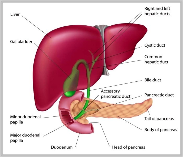
    The gallbladder, a small, pear-shaped organ, plays a crucial role in the digestive process. Understanding its location, structure, and relationship to other organs is vital for comprehending its function and potential issues. This section delves into the detailed anatomy of the gallbladder.The gallbladder is situated beneath the liver, nestled in a shallow depression on its inferior surface. Its position, relative to other abdominal organs, allows it to efficiently collect and concentrate bile produced by the liver.

    Gallbladder Location

    The gallbladder rests in a fossa, a shallow depression, on the inferior surface of the liver. Its head points toward the right side of the body, while the tail curves slightly downward and toward the midline. The gallbladder is positioned in close proximity to the duodenum, a part of the small intestine. This anatomical arrangement allows bile to travel directly from the gallbladder to the duodenum when needed.

    Gallbladder Structure

    The gallbladder is a hollow, muscular organ, roughly 7-10 cm long and 3-4 cm wide. Its shape resembles a pear, with a wider, rounded fundus (the bottom portion), a narrow neck, and a body connecting the two. The fundus typically extends slightly beyond the liver’s edge. Its walls are relatively thin, composed of smooth muscle fibers that contract to release bile.

    A mucous membrane lines the inner surface, allowing for the storage and movement of bile.

    Gallbladder Wall Layers, The gallbladder anatomy function and treatment

    The gallbladder wall consists of three distinct layers:

    • The outer serosa, a thin layer of connective tissue, is the outermost layer. It provides protection and helps to hold the gallbladder in place. It’s a vital component of the peritoneum, the membrane that lines the abdominal cavity.
    • The middle muscularis layer is comprised of smooth muscle fibers. These fibers contract to propel bile into the cystic duct. The strength of these contractions is essential for efficient bile release.
    • The inner mucosa is a thin layer of epithelium, highly folded to increase surface area. This structure is important for the absorption of water from the bile and maintaining the consistency of the bile while it is stored in the gallbladder.

    Dimensions and Shape Comparison

    The following table compares the dimensions and shape of the gallbladder with other related organs.

    Organ Dimensions (approximate) Shape
    Gallbladder 7-10 cm long, 3-4 cm wide Pear-shaped
    Liver 12-15 cm long, 7-8 cm wide Irregularly lobed
    Common Bile Duct 5-10 mm in diameter Tube-like

    Visual Representation

    Imagine the liver as a large, reddish-brown, irregular-shaped organ dominating the upper right quadrant of the abdomen. Beneath it, tucked into a shallow depression, is the gallbladder. Its pear shape is clearly visible, with the wider fundus extending slightly beyond the liver’s edge. The neck of the gallbladder connects to the cystic duct, which then merges with the common bile duct.

    This duct leads to the duodenum, where bile aids in the digestion of fats. The gallbladder’s position allows for efficient storage and release of bile into the small intestine.

    Gallbladder Function

    The gallbladder, a small pear-shaped organ nestled beneath the liver, plays a crucial role in the digestive process. It acts as a temporary storage reservoir for bile, a fluid produced by the liver essential for breaking down fats in the small intestine. Understanding its function is vital for comprehending how our bodies efficiently process nutrients.The gallbladder’s primary function is not to produce bile, but to concentrate and store it.

    This allows for a controlled release of bile into the small intestine precisely when needed, maximizing its effectiveness in fat digestion. This process is tightly regulated, ensuring that the right amount of bile is available at the right time.

    Bile Storage and Concentration

    The liver constantly produces bile, which drains into the common bile duct. This duct leads to the gallbladder, where bile is temporarily stored. Inside the gallbladder, specialized cells absorb water and electrolytes from the bile, effectively concentrating it. This concentration process significantly increases the bile’s potency in breaking down fats, ensuring optimal efficiency in the digestive process.

    The concentrated bile is stored until stimulated by the presence of fats in the duodenum. This process allows for the controlled release of concentrated bile, optimizing its effect.

    Bile Release into the Small Intestine

    The gallbladder’s release of bile is triggered by the presence of fats in the duodenum, the first part of the small intestine. This triggers the release of cholecystokinin (CCK), a hormone. CCK signals the gallbladder to contract, pushing the concentrated bile into the common bile duct. The sphincter of Oddi, a muscular valve at the junction of the common bile duct and the duodenum, relaxes, allowing the bile to flow into the small intestine.

    Understanding the gallbladder’s role in digestion is crucial – it stores and releases bile, essential for breaking down fats. Different treatments for gallbladder issues range from lifestyle changes to surgery. However, sometimes, medication choices like comparing the effectiveness of Tegretol and Lamictal in managing certain conditions are important. For a deeper dive into the nuances of tegretol vs lamictal how do they differ , explore the detailed analysis on their respective benefits and potential side effects.

    Ultimately, proper gallbladder function is vital for overall health and well-being.

    This precise mechanism ensures that bile is released only when needed, optimizing fat digestion.

    Importance of Bile in Fat Digestion and Absorption

    Bile plays a vital role in the digestion and absorption of fats. It emulsifies fats, breaking them down into smaller droplets, increasing the surface area exposed to digestive enzymes. This process makes it much easier for the enzymes to break down the fats into fatty acids and glycerol, which can then be absorbed into the bloodstream. Without bile, fat digestion would be significantly impaired, leading to malabsorption of essential nutrients.

    This is crucial for the body to utilize fats for energy, hormone production, and cell function.

    Bile Secretion, Storage, and Release Flowchart

    The following flowchart illustrates the process of bile secretion, storage, and release:

    Step Description
    1 Liver produces bile.
    2 Bile flows into the common bile duct.
    3 Bile enters the gallbladder for storage.
    4 Fats enter the duodenum.
    5 Cholecystokinin (CCK) is released.
    6 Gallbladder contracts, releasing bile.
    7 Sphincter of Oddi relaxes.
    8 Bile enters the small intestine.
    9 Bile emulsifies fats, aiding digestion and absorption.

    Gallbladder Disorders

    The gallbladder, a small, pear-shaped organ, plays a crucial role in fat digestion. However, various disorders can affect its function and health. Understanding these disorders is vital for recognizing symptoms and seeking appropriate medical care. Gallbladder issues, ranging from relatively benign gallstones to more severe cholecystitis, can significantly impact a person’s quality of life.Gallbladder disorders encompass a spectrum of conditions, from the relatively common presence of gallstones to the more serious inflammation of the gallbladder known as cholecystitis.

    These conditions can lead to discomfort, pain, and, in some cases, require surgical intervention. Identifying the specific type of disorder and its underlying cause is essential for effective treatment.

    Gallstones

    Gallstones are hardened deposits that form within the gallbladder. These stones can vary in size and composition, impacting their potential for causing symptoms. A variety of factors contribute to their formation.Common causes of gallstones include imbalances in the bile’s chemical composition, such as an excess of cholesterol or bilirubin. Certain lifestyle factors, such as obesity, rapid weight loss, and a diet high in fat, can also increase the risk.

    Understanding gallbladder function, from its anatomy to potential treatments, is crucial for overall health. A healthy gallbladder helps process fats in our diet, but sometimes issues arise. Interestingly, a balanced diet, like the one often recommended by a high protein vanilla matcha latte dietitian, like this one , can play a role in supporting gallbladder health. Lifestyle choices, including dietary modifications, are often part of the treatment plan for gallbladder issues, highlighting the connection between nutrition and digestive health.

    Genetic predisposition and certain medical conditions can also play a role in gallstone formation. Furthermore, pregnancy and prolonged fasting can also increase the risk. For example, individuals with a family history of gallstones or those who have undergone significant weight loss procedures may be at higher risk.

    Cholecystitis

    Cholecystitis is an inflammation of the gallbladder, often associated with the presence of gallstones. The inflammation can vary in severity, ranging from mild discomfort to severe pain and potentially life-threatening complications.The primary cause of cholecystitis is gallstones that obstruct the cystic duct, the tube connecting the gallbladder to the common bile duct. This blockage can lead to inflammation and infection within the gallbladder.

    Other causes of cholecystitis, although less common, can include infections, tumors, or trauma to the gallbladder.

    Symptoms and Diagnosis

    The symptoms of gallstones and cholecystitis can overlap, making diagnosis challenging. However, some key differences exist.Gallstones may cause episodic pain, often located in the upper right abdomen or radiating to the back or right shoulder. This pain is typically severe and can be accompanied by nausea and vomiting. Cholecystitis pain is typically more persistent and intense, often accompanied by fever and chills.Diagnostic methods for both conditions include physical examinations, blood tests to assess inflammation markers, and imaging techniques.

    Ultrasound is a common initial diagnostic tool. It provides images of the gallbladder and surrounding structures, allowing for the detection of gallstones and assessing the degree of inflammation. CT scans and MRCP (Magnetic Resonance Cholangiopancreatography) may be used in certain cases to obtain a more detailed view of the biliary system, especially when ultrasound results are inconclusive.

    Gallstone Types

    Gallstone Type Composition Associated Risks
    Cholesterol Stones Predominantly cholesterol Obesity, rapid weight loss, high-fat diet
    Pigment Stones Predominantly bilirubin Liver conditions, hemolytic anemia
    Mixed Stones Combination of cholesterol and bilirubin Combination of risk factors for cholesterol and pigment stones

    Diagnostic Tests

    Several diagnostic tests are used to identify gallbladder disorders. Blood tests can help evaluate inflammation levels. Imaging techniques, such as ultrasound, CT scans, and MRCP, provide detailed visual representations of the gallbladder and biliary system, allowing for the identification of gallstones, blockages, and inflammation. A physician will determine the most appropriate tests based on the patient’s symptoms and medical history.

    Gallbladder Treatment Options

    Treating gallbladder disorders depends on the severity of the condition and the patient’s overall health. Effective management aims to alleviate symptoms, prevent complications, and improve quality of life. The available options range from lifestyle modifications to surgical interventions, each with its own set of benefits and risks.

    Surgical Treatment Options

    Surgical intervention, particularly laparoscopic cholecystectomy, is often the preferred approach for symptomatic gallbladder disease. This minimally invasive procedure offers several advantages over traditional open surgery, including reduced pain, shorter hospital stays, and faster recovery times.

    Laparoscopic Cholecystectomy

    Laparoscopic cholecystectomy involves making small incisions in the abdomen to insert specialized instruments and a tiny camera (laparoscope). The surgeon uses these tools to visualize the gallbladder and carefully remove it. This procedure is performed under general anesthesia.

    “The use of specialized instruments and a laparoscope allows for precise removal of the gallbladder with minimal trauma to surrounding tissues.”

    The procedure generally takes about 45-60 minutes.

    Recovery Process Following Gallbladder Surgery

    The recovery period after laparoscopic cholecystectomy is typically brief. Patients usually experience mild to moderate pain, which can be managed with prescribed medications. A gradual return to normal activities is encouraged, with physical limitations initially. Full recovery usually takes several weeks.

    Risks and Complications

    While laparoscopic cholecystectomy is generally safe, potential risks and complications include bleeding, infection, injury to nearby organs (such as the bile duct or liver), and the formation of scar tissue. Post-operative pain, bile duct injury, and wound infections are possible, although less common. The risk of complications is generally low, especially in experienced hands.

    Understanding gallbladder anatomy, function, and treatment options is crucial for overall health. For example, gallstones can cause significant discomfort, and sometimes surgery is necessary. Interestingly, the diagnostic process for similar skin conditions, like plaque psoriasis, often involves a combination of physical examination and skin biopsy, as detailed in this helpful resource on how plaque psoriasis is diagnosed.

    Ultimately, the right approach to gallbladder issues depends on the individual case and the severity of the problem.

    Non-Surgical Treatment Options

    Non-surgical approaches are considered for individuals with less severe gallbladder conditions or those who are not suitable candidates for surgery. These methods primarily focus on managing symptoms and improving overall health.

    Dietary Modifications

    Dietary changes can play a crucial role in managing gallbladder symptoms. A low-fat diet is often recommended to reduce the strain on the gallbladder. Eating smaller, more frequent meals and avoiding fatty foods, especially fried or greasy meals, can significantly help alleviate discomfort. It’s essential to consult a healthcare professional for personalized dietary recommendations.

    Medical Management

    Medication, such as pain relievers and medications to aid digestion, may be prescribed to manage symptoms. These medications help reduce inflammation and improve digestion, but they do not address the underlying cause of the gallbladder disorder.

    Comparison of Surgical and Non-Surgical Treatment Options

    Feature Surgical (Laparoscopic Cholecystectomy) Non-Surgical
    Procedure Type Invasive, involves surgical removal of the gallbladder Non-invasive, focuses on symptom management
    Recovery Time Several weeks Can vary depending on the severity of symptoms
    Effectiveness Permanent resolution of the condition Temporary relief of symptoms
    Suitable for Individuals with symptomatic gallbladder disease Individuals with less severe conditions or those unsuitable for surgery
    Risks Bleeding, infection, injury to surrounding organs Minimal, but can include side effects from medications

    Prevention and Management

    Protecting your gallbladder involves a proactive approach, focusing on lifestyle choices and preventative measures. Understanding the factors that influence gallbladder health allows for informed decisions to reduce the risk of developing issues and manage any existing conditions effectively. A balanced approach combining dietary adjustments, regular checkups, and long-term management strategies can significantly impact overall well-being.

    Lifestyle Factors Affecting Gallbladder Health

    Maintaining a healthy weight and engaging in regular physical activity are crucial for gallbladder health. Obesity is a significant risk factor for gallstones, and regular exercise can help manage weight and reduce the likelihood of developing this condition. A sedentary lifestyle, conversely, increases the risk. Smoking also plays a role; research indicates a potential correlation between smoking and gallstone formation.

    Genetic predisposition is another factor; a family history of gallstones can increase individual risk.

    Dietary Recommendations for Gallstone Prevention

    Dietary modifications play a key role in reducing the risk of gallstones. A diet low in saturated and trans fats, along with a balanced intake of fiber-rich foods, can be beneficial. Reducing refined carbohydrates and sugary foods can also contribute to overall health and lower the likelihood of developing gallstones. Individuals should prioritize whole, unprocessed foods. Small, frequent meals can also aid in preventing gallstone formation.

    Importance of Regular Checkups and Screenings

    Regular checkups are vital for early detection of potential gallbladder issues. A healthcare professional can identify risk factors and recommend appropriate screenings based on individual circumstances. These screenings can include blood tests, imaging scans, or other diagnostic procedures to monitor gallbladder health. Early detection allows for timely intervention and treatment if necessary, potentially preventing complications.

    Long-Term Management Strategies for Gallbladder Disease

    Long-term management of gallbladder disease focuses on mitigating symptoms and preventing recurrence. This may involve adhering to specific dietary guidelines, maintaining a healthy weight, and managing underlying conditions. Regular follow-up appointments with a healthcare professional are essential to monitor progress and adjust treatment plans as needed. For individuals who have undergone gallbladder removal, lifestyle modifications are crucial to prevent complications from developing.

    Preventive Measures for Good Gallbladder Health

    Maintaining good gallbladder health involves a combination of proactive measures.

    • Maintain a healthy weight: Obesity is a significant risk factor for gallstones. Regular exercise and a balanced diet are crucial for weight management.
    • Adopt a balanced diet: A diet low in saturated and trans fats, with adequate fiber intake, can reduce the risk of gallstones. Reducing refined carbohydrates and sugary foods is also recommended.
    • Engage in regular physical activity: Regular exercise can help manage weight and improve overall health, lowering the risk of gallbladder issues.
    • Maintain a healthy blood lipid profile: High cholesterol and other blood lipid abnormalities can contribute to gallstone formation. Consult with a healthcare professional for advice.
    • Avoid rapid weight loss: Significant weight loss too quickly can increase the risk of gallstone formation.
    • Regular medical checkups: Routine checkups are essential for early detection of potential gallbladder issues and risk factor assessment.

    Final Thoughts: The Gallbladder Anatomy Function And Treatment

    The gallbladder anatomy function and treatment

    In conclusion, the gallbladder’s complex anatomy and function are essential for optimal digestion. This detailed look at its structure, role in the digestive process, and potential disorders provides a comprehensive understanding. The various treatment options, ranging from lifestyle changes to surgical interventions, offer hope for those affected by gallbladder issues. Remember that early diagnosis and proper management are crucial for maintaining digestive health.

  • Tour the Digestive System A Journey Inside

    Tour the Digestive System A Journey Inside

    Tour the digestive system, a fascinating journey through the complex machinery of your body! This exploration dives into the intricate process of breaking down food, highlighting the vital roles of each organ from mouth to anus. We’ll uncover the amazing chemical and mechanical processes that transform a meal into usable energy, and explore the fascinating interplay between the digestive system and other bodily functions.

    Prepare to be amazed by the wonders within!

    This comprehensive tour will guide you through the stages of digestion, from ingestion to elimination. We’ll examine the structure and function of key organs like the stomach, small intestine, and liver, and discover how they work together in a symphony of biological processes. We’ll also explore common digestive disorders and ways to maintain a healthy digestive system through lifestyle choices.

    Introduction to the Digestive System: Tour The Digestive System

    The human digestive system is a complex network of organs working together to break down food into nutrients that the body can absorb and use for energy, growth, and repair. This intricate process, starting from the moment we ingest food, involves a coordinated effort from various organs, each playing a specific role in the overall digestive journey. Understanding this process allows us to appreciate the remarkable efficiency of our bodies and the importance of a healthy diet.The digestive system’s primary function is to transform ingested food into absorbable molecules.

    This transformation involves mechanical and chemical processes that break down large food particles into smaller components that can be transported across the intestinal lining and into the bloodstream. The resulting nutrients then fuel various bodily functions, supporting everything from basic metabolism to complex bodily processes.

    Key Organs and Their Functions

    The digestive system is a continuous tube, starting from the mouth and ending at the anus, with various specialized organs along the way. Each organ plays a crucial role in the digestive process.

    • Mouth: The mouth is the entry point for food. Mechanical digestion begins here through chewing (mastication), which breaks down food into smaller pieces. Saliva, secreted by salivary glands, contains enzymes that initiate the chemical breakdown of carbohydrates. This initial breakdown is crucial for efficient digestion later in the process.
    • Esophagus: The esophagus is a muscular tube connecting the mouth to the stomach. Peristalsis, wave-like muscle contractions, propels food down the esophagus, ensuring its uninterrupted journey to the stomach.
    • Stomach: The stomach is a muscular sac where food is further broken down. The stomach’s muscular walls churn the food, mixing it with gastric juices containing enzymes (like pepsin) that begin protein digestion. The acidic environment of the stomach also helps to kill harmful bacteria.
    • Small Intestine: The small intestine is the primary site of nutrient absorption. Its long, folded structure maximizes the surface area available for absorption. Enzymes secreted by the pancreas and bile from the liver further break down carbohydrates, proteins, and fats into their smallest components. Nutrients are then absorbed into the bloodstream through the intestinal lining.
    • Large Intestine: The large intestine absorbs water and electrolytes from the remaining indigestible food matter. Bacteria in the large intestine ferment any remaining undigested material, producing some vitamins. The waste products are then compacted and stored until elimination.
    • Rectum: The rectum is a short, muscular tube that stores feces until elimination.
    • Anus: The anus is the final part of the digestive tract. It controls the release of feces from the body.

    Stages of Digestion

    Digestion involves several key stages.

    1. Ingestion: Ingestion is the intake of food into the body. It’s the initial step in the digestive process, starting the journey of food through the system.
    2. Digestion: Digestion involves the mechanical and chemical breakdown of food. This process transforms large food molecules into smaller, absorbable units. Mechanical digestion involves physical breakdown (chewing, churning), while chemical digestion uses enzymes to break down food into simpler molecules.
    3. Absorption: Absorption is the process by which the body takes up nutrients from digested food. The small intestine is the primary site of absorption, with its vast surface area facilitating the uptake of nutrients into the bloodstream.
    4. Elimination: Elimination is the removal of undigested food and waste products from the body. Feces, the solid waste product of digestion, is expelled through the anus.

    Diagram of the Digestive System

    Organ Description
    Mouth Food enters the system
    Esophagus Tube connecting mouth to stomach
    Stomach Churns and mixes food with enzymes
    Small Intestine Main site of nutrient absorption
    Large Intestine Absorbs water and electrolytes
    Rectum Stores waste before elimination
    Anus Waste is expelled from the body

    Organs of the Digestive System

    The digestive system is a complex network of organs working together to break down food into absorbable nutrients. This process, essential for energy and growth, involves mechanical and chemical breakdown. Understanding the structure and function of each organ is crucial to comprehending the intricate dance of digestion.

    Mouth

    The mouth, the initial entry point for food, plays a vital role in both mechanical and chemical digestion. Teeth mechanically break down food into smaller pieces, while saliva, containing enzymes like amylase, begins the chemical breakdown of carbohydrates. The tongue mixes the food with saliva, forming a bolus that is ready for swallowing.

    Esophagus

    The esophagus, a muscular tube connecting the mouth to the stomach, propels the bolus through peristalsis, a series of coordinated muscle contractions. This involuntary process ensures that food moves unidirectionally, preventing backflow. The esophagus’s smooth muscle lining facilitates this controlled transport.

    Stomach

    The stomach, a muscular sac, continues the mechanical breakdown of food. Its churning action mixes food with gastric juices, which contain hydrochloric acid and enzymes like pepsin. Hydrochloric acid creates an acidic environment essential for pepsin’s activity in protein digestion. This acidic environment also helps kill harmful bacteria that may be present in the food.

    Small Intestine, Tour the digestive system

    The small intestine, a long, coiled tube, is the primary site of nutrient absorption. Its large surface area, due to villi and microvilli, maximizes contact with digested food. Enzymes from the pancreas and the small intestine itself further break down carbohydrates, proteins, and fats. The absorbed nutrients are then transported into the bloodstream for use by the body.

    Large Intestine

    The large intestine, a shorter but wider tube than the small intestine, absorbs water and electrolytes from the remaining undigested material. Bacteria in the large intestine ferment some undigested material, producing vitamins like vitamin K. The remaining waste, now solidified into feces, is stored in the rectum before elimination.

    Liver

    The liver, a vital accessory organ, plays a crucial role in many bodily functions, including digestion. It produces bile, a fluid crucial for fat emulsification, breaking down large fat globules into smaller droplets, which increases the surface area for enzyme action. The liver also processes absorbed nutrients, detoxifies harmful substances, and synthesizes essential proteins.

    Pancreas

    The pancreas, another accessory organ, produces a variety of enzymes crucial for digestion. These enzymes, released into the small intestine, break down carbohydrates, proteins, and fats. The pancreas also regulates blood sugar levels by producing insulin and glucagon.

    Gallbladder

    The gallbladder, a small, pear-shaped sac, stores and concentrates bile produced by the liver. When food containing fat enters the small intestine, the gallbladder releases bile to aid in fat digestion. This ensures that the body has a readily available supply of bile when needed.

    Ever wanted to tour the digestive system? It’s a fascinating journey, isn’t it? Learning how our bodies process food is truly incredible. And for those looking to maintain a healthy weight, a successful weight-loss story like zepbound keeps weight off 3 years might provide some inspiration. Understanding the intricate processes within our bodies, like the digestive system, can be a powerful tool in our wellness journeys.

    This knowledge can be really valuable when trying to make healthy lifestyle choices.

    Enzymes in Digestion

    Enzymes are biological catalysts that speed up chemical reactions in the body, including those involved in digestion. Different enzymes target different types of nutrients. Amylase breaks down carbohydrates, pepsin breaks down proteins, and lipase breaks down fats. The specific environment (pH) and the precise substrate dictate which enzyme is most effective.

    Accessory Organ Comparison

    The liver, pancreas, and gallbladder, though distinct organs, work in concert to support digestion. The liver produces bile, the pancreas secretes digestive enzymes, and the gallbladder stores and releases bile. These accessory organs, acting in synergy, are essential for complete and efficient nutrient breakdown and absorption.

    Ever wanted to tour the digestive system? It’s fascinating how our bodies process food, isn’t it? Knowing how your body breaks down nutrients is key to making smart food choices, like selecting snacks for weight loss that support your goals. Learning about the digestive process can help you understand the impact of different foods, ultimately leading to a more informed approach to overall health.

    A better understanding of the digestive system can empower you to make more thoughtful choices, and ultimately improve your well-being.

    Organ Summary

    Organ Location Function Key Features
    Mouth Oral cavity Mechanical and chemical digestion begins Teeth, tongue, saliva
    Esophagus Connects mouth to stomach Transports food to stomach Peristalsis
    Stomach Upper abdomen Mechanical and chemical digestion of proteins Gastric juices, churning
    Small Intestine Lower abdomen Nutrient absorption Villi, microvilli, enzymes
    Large Intestine Lower abdomen Water absorption, waste elimination Bacteria fermentation
    Liver Upper right abdomen Bile production, nutrient processing Detoxification
    Pancreas Behind stomach Enzyme production, blood sugar regulation Insulin, glucagon
    Gallbladder Below liver Bile storage and release Concentrates bile

    Digestive Processes

    The journey of food through our bodies is a complex interplay of mechanical and chemical processes. From the initial chewing in the mouth to the final absorption in the small intestine, each step is carefully orchestrated to extract nutrients and eliminate waste. This intricate process is crucial for maintaining our overall health and well-being.The digestive system’s efficiency depends on the precise coordination of various mechanisms.

    These include physical breakdown of food, enzymatic reactions that break down complex molecules, and the intricate absorption of nutrients into the bloodstream. Understanding these processes allows us to appreciate the remarkable complexity of our own bodies.

    Ever wanted to tour the digestive system? It’s a fascinating journey, but did you know that sometimes, even our bodies’ internal pathways can be confused about the difference between a simple cold sore and herpes? Learning about the intricacies of the digestive system can help you better understand how your body works, including the importance of proper hygiene to avoid potential infections, like those that cause cold sores or herpes.

    For a deeper dive into the world of oral health and differentiating between these conditions, check out this helpful resource on cold sore vs herpes. Understanding these nuances is key to maintaining overall wellness, and the digestive system plays a significant role in this.

    Mechanical Digestion

    Mechanical digestion involves physically breaking down food into smaller pieces. This process increases the surface area exposed to digestive enzymes, facilitating the chemical breakdown that follows. The act of chewing, for instance, mechanically reduces large pieces of food into smaller particles. This process begins in the mouth and continues throughout the digestive tract.

    Chemical Digestion

    Chemical digestion involves the use of enzymes to break down complex food molecules into simpler absorbable forms. Enzymes are biological catalysts that speed up chemical reactions. Different enzymes target different types of molecules, such as carbohydrates, proteins, and fats. This process is crucial for extracting usable nutrients from the ingested food.

    Peristalsis and Segmentation

    Peristalsis is a series of rhythmic muscular contractions that propel food through the digestive tract. These wave-like movements occur in the esophagus, stomach, and intestines, ensuring that food moves unidirectionally from the mouth to the anus. Segmentation, on the other hand, involves the contraction and relaxation of smooth muscles in the intestines. This process mixes the food with digestive enzymes and further enhances the exposure of food particles to enzymes.

    These coordinated movements facilitate efficient digestion and nutrient absorption.

    Nutrient Absorption in the Small Intestine

    The small intestine is the primary site of nutrient absorption. Its highly folded inner lining, called the villi and microvilli, dramatically increases the surface area available for absorption. Nutrients, such as glucose, amino acids, and fatty acids, are absorbed across the intestinal lining and transported into the bloodstream for distribution throughout the body. This process is vital for providing the body with the building blocks needed for energy, growth, and repair.

    Digestive System Interactions

    The digestive system doesn’t operate in isolation. It interacts with other systems in the body, such as the circulatory system and the endocrine system. The circulatory system transports absorbed nutrients to various tissues and organs. The endocrine system regulates digestive processes through the release of hormones, such as gastrin and secretin. These hormonal signals control the release of digestive enzymes and the movement of food through the digestive tract.

    This interconnectedness highlights the intricate and sophisticated nature of human physiology.

    Comparison of Mechanical and Chemical Digestion

    Characteristic Mechanical Digestion Chemical Digestion
    Mechanism Physical breakdown of food into smaller pieces Breakdown of food molecules into simpler forms using enzymes
    Location Mouth, stomach, intestines Mouth, stomach, intestines
    Examples Chewing, churning in the stomach, segmentation Enzymatic breakdown of carbohydrates (amylase), proteins (proteases), and fats (lipases)
    Effect on food Increases surface area for chemical digestion Breaks down complex molecules into absorbable forms

    Health and Disorders of the Digestive System

    Tour the digestive system

    Our digestive system, a complex network of organs, is vital for our overall health. However, various disorders can disrupt its normal functioning, leading to discomfort and even serious health complications. Understanding these disorders, their causes, and available treatments is crucial for maintaining a healthy digestive system.

    Common Digestive Disorders

    Numerous conditions can affect the digestive system, ranging from mild inconveniences to more severe illnesses. Recognizing the signs and symptoms of these disorders is the first step towards seeking appropriate medical care.

    Heartburn

    Heartburn, a common digestive ailment, is characterized by a burning sensation in the chest, often radiating to the throat. This discomfort typically arises from stomach acid refluxing into the esophagus. Factors such as eating large meals, lying down soon after eating, and certain foods (e.g., fatty foods, citrus fruits, chocolate) can trigger heartburn. Lifestyle modifications, such as avoiding trigger foods, maintaining a healthy weight, and elevating the head of the bed, often provide relief.

    Over-the-counter medications can also effectively manage symptoms.

    Peptic Ulcers

    Peptic ulcers are sores that develop in the lining of the stomach or the upper portion of the small intestine. These ulcers are often caused by a bacterial infection (Helicobacter pylori) or the long-term use of nonsteroidal anti-inflammatory drugs (NSAIDs). Symptoms may include abdominal pain, nausea, and vomiting. Treatment typically involves antibiotics to eliminate the bacteria, medications to reduce stomach acid production, and lifestyle changes to minimize triggers.

    Irritable Bowel Syndrome (IBS)

    Irritable bowel syndrome (IBS) is a chronic condition affecting the large intestine. The exact cause of IBS remains unknown, but factors such as stress, diet, and hormonal changes may play a role. Symptoms of IBS can vary but often include abdominal pain, bloating, gas, diarrhea, and/or constipation. Management of IBS typically involves dietary modifications, stress reduction techniques, and medications to alleviate specific symptoms.

    Other Digestive Disorders

    • Gastroesophageal Reflux Disease (GERD): A more severe form of heartburn, GERD involves frequent and persistent acid reflux, potentially causing esophageal damage. Treatment options include medication, lifestyle changes, and in some cases, surgery.
    • Crohn’s Disease and Ulcerative Colitis: These are inflammatory bowel diseases (IBDs) that cause chronic inflammation in the digestive tract. Symptoms can range from mild to severe and include abdominal pain, diarrhea, weight loss, and fatigue. Treatment focuses on reducing inflammation and managing symptoms.
    • Celiac Disease: An autoimmune disorder triggered by gluten, a protein found in wheat, barley, and rye. Symptoms can vary but often include abdominal pain, diarrhea, and nutrient deficiencies. Treatment involves strict adherence to a gluten-free diet.

    Preventing Digestive Problems

    A healthy diet and lifestyle play a crucial role in preventing digestive issues. Regular exercise, maintaining a healthy weight, and managing stress are all important preventative measures. Choosing nutrient-rich foods, avoiding processed foods and excessive caffeine, and drinking plenty of water can also contribute to digestive health.

    Healthy Dietary Habits

    Adopting healthy dietary habits is key to supporting a healthy digestive system.

    • Fiber-rich foods: Include fruits, vegetables, and whole grains in your diet. Fiber promotes regular bowel movements and aids digestion.
    • Probiotics: These beneficial bacteria can support gut health. They are found in fermented foods such as yogurt, kefir, and sauerkraut.
    • Hydration: Drinking plenty of water helps maintain proper bowel function and prevents constipation.
    • Portion control: Avoiding large meals can help prevent digestive discomfort.

    Table of Common Digestive Disorders and Symptoms

    Disorder Symptoms
    Heartburn Burning sensation in the chest, possibly radiating to the throat; indigestion
    Peptic Ulcers Abdominal pain, nausea, vomiting, bloating, indigestion
    Irritable Bowel Syndrome (IBS) Abdominal pain, bloating, gas, diarrhea, constipation
    Gastroesophageal Reflux Disease (GERD) Frequent or persistent heartburn, acid reflux, potential esophageal damage
    Crohn’s Disease and Ulcerative Colitis Abdominal pain, diarrhea, weight loss, fatigue, fever, bloody stools
    Celiac Disease Abdominal pain, diarrhea, bloating, fatigue, nutrient deficiencies

    Illustrations and Visualizations

    Tour the digestive system

    Delving deeper into the fascinating world of the digestive system, we now move towards visualizing its intricate mechanisms. Understanding the structure and function of key organs through detailed imagery is crucial for grasping the complex processes involved in nutrient breakdown and absorption. These visualizations will serve as valuable tools for a better comprehension of the digestive system’s inner workings.

    Stomach Muscular Structure

    The stomach’s muscular structure is essential for its churning action, crucial for breaking down food. Imagine a thick-walled sac, lined with three layers of smooth muscle tissue: the longitudinal, circular, and oblique layers. These layers work in coordinated contractions, creating a churning motion that mixes food with gastric juices. The oblique layer, in particular, plays a vital role in the forceful mixing action.

    This coordinated contraction efficiently breaks down food particles into smaller components.

    • Longitudinal layer: These muscle fibers run along the length of the stomach, contributing to the overall churning and mixing of food contents.
    • Circular layer: These muscle fibers encircle the stomach, further aiding in the mixing and grinding of food.
    • Oblique layer: These muscle fibers run diagonally across the stomach, adding another dimension to the churning and compressing action, intensifying the mixing process.

    Villi in the Small Intestine

    The small intestine, the primary site of nutrient absorption, is lined with tiny finger-like projections called villi. These structures significantly increase the surface area for absorption. Think of them as miniature trees lining the intestinal wall, greatly expanding the space for nutrients to be absorbed into the bloodstream. Each villus is further covered in even smaller projections called microvilli, creating a brush border effect, which further multiplies the absorption area.

    • Structure: Villi are small, finger-like protrusions that project into the lumen of the small intestine.
    • Function: They dramatically increase the surface area for nutrient absorption, facilitating the efficient uptake of digested food molecules.
    • Microvilli: These microscopic hair-like structures on the surface of each villus further increase the surface area, maximizing the efficiency of nutrient absorption.

    Liver’s Role in Bile Production

    The liver plays a crucial role in producing bile, a substance essential for the digestion and absorption of fats. Imagine the liver as a chemical factory producing this important digestive fluid. Bile is stored in the gallbladder and released into the small intestine when needed. The liver continuously produces bile, which aids in the emulsification of fats, breaking them down into smaller droplets, making them easier for enzymes to digest.

    • Bile Production: The liver continuously manufactures bile, a greenish-yellow fluid.
    • Emulsification of Fats: Bile salts within bile break down large fat globules into smaller droplets, making them more accessible to digestive enzymes.
    • Absorption: This process enhances the absorption of fatty acids and fat-soluble vitamins.

    Nutrient Absorption in the Small Intestine

    The small intestine is the primary site for nutrient absorption, a complex process involving the movement of digested nutrients from the lumen of the intestine into the bloodstream. Nutrients are transported across the intestinal lining through a combination of passive and active transport mechanisms. Think of this as a sophisticated conveyor belt system where nutrients are carefully selected and transported into the circulatory system for distribution throughout the body.

    • Passive Transport: Some nutrients, like water and certain small molecules, can passively diffuse across the intestinal lining.
    • Active Transport: Other nutrients, like glucose and amino acids, require energy to be transported against their concentration gradient, emphasizing the small intestine’s active role in nutrient uptake.

    Large Intestine’s Function in Water Absorption

    The large intestine’s primary function is water absorption. Imagine the large intestine as a water reclamation center, reabsorbing water from the indigestible food residue. This process is critical for maintaining fluid balance in the body. Undigested materials are compacted and prepared for elimination.

    • Water Reabsorption: The large intestine absorbs water from the remaining undigested food matter.
    • Waste Compaction: The remaining waste material is compacted into solid feces.
    • Elimination: The feces are expelled from the body.

    Interactive Elements

    Embarking on a virtual journey through the fascinating world of the digestive system requires engaging tools that go beyond static information. Interactive elements breathe life into the learning experience, allowing for active participation and deeper comprehension. Interactive elements transform passive reading into an active learning process, making the subject matter more memorable and relatable.

    Interactive Quiz

    A well-designed quiz is an effective way to test knowledge retention and pinpoint areas needing further clarification. This section presents a sample quiz designed to assess comprehension of the digestive system.

    Question Options Correct Answer
    Which organ is responsible for the majority of nutrient absorption? a) Stomach
    b) Small Intestine
    c) Large Intestine
    d) Esophagus
    b) Small Intestine
    What is the primary function of the liver? a) Mechanical digestion
    b) Chemical digestion
    c) Nutrient absorption
    d) Bile production
    d) Bile production
    What is the role of the pancreas in digestion? a) Stores bile
    b) Produces enzymes for digestion
    c) Absorbs water
    d) Transports food
    b) Produces enzymes for digestion

    Digestive Process Flowchart

    Visualizing the intricate process of digestion can be challenging. A flowchart provides a clear and concise representation of the steps involved, enabling a better understanding of the sequential nature of digestion. This is essential for comprehending how different parts of the system work together.

    Food Ingestion --> Mechanical Digestion (Mouth) --> Chemical Digestion (Stomach) --> Movement through Small Intestine --> Nutrient Absorption --> Movement through Large Intestine --> Waste Elimination
    

    Animated Journey Through the Digestive Tract

    An animated depiction of food’s journey through the digestive tract offers a dynamic and engaging way to illustrate the process. This visualization brings the abstract concepts to life. Imagine seeing the food being broken down and absorbed as it travels through the various organs.

    Stage Description
    Mouth Food is chewed and mixed with saliva, initiating the mechanical and chemical breakdown.
    Esophagus Food is transported from the mouth to the stomach via peristaltic waves.
    Stomach Food is churned and mixed with stomach acid and enzymes.
    Small Intestine Nutrients are absorbed into the bloodstream.
    Large Intestine Water is absorbed, and waste is prepared for elimination.

    Interactive Organ Matching Game

    A matching game allows users to actively engage with the information and solidify their understanding of the relationship between digestive organs and their functions. This approach is particularly effective for learners who benefit from hands-on activities.

    Organ Function
    Stomach Mechanical and chemical breakdown of food
    Small Intestine Nutrient absorption
    Liver Produces bile for fat digestion
    Pancreas Produces enzymes for digestion

    Virtual Tour Design

    Creating a virtual tour of the digestive system necessitates the integration of interactive elements to enhance engagement. This involves incorporating interactive 3D models of organs, allowing users to explore the anatomy in detail. Interactive elements like clickable labels and pop-up information can also provide context.

    Element Description
    Interactive 3D Models Allow users to rotate, zoom, and explore the structure of organs.
    Clickable Labels Provide detailed information about each organ when clicked.
    Pop-up Information Offer detailed descriptions of functions and processes.
    Animation of Processes Show the journey of food through the system and the actions of various organs.

    Closing Notes

    In conclusion, our tour of the digestive system has unveiled a truly remarkable biological marvel. From the initial breakdown of food in the mouth to the final absorption of nutrients in the intestines, the digestive system’s intricate workings are essential for life. We’ve explored the organs, processes, and potential health concerns associated with this vital system. Understanding the digestive system is crucial for overall well-being, emphasizing the importance of a healthy diet and lifestyle choices for optimal function.

    Hopefully, this tour has sparked your curiosity and left you with a greater appreciation for the complexity and beauty of the human body.

  • Spicy Food & Diarrhea The Science Behind the Spikes

    Spicy Food & Diarrhea The Science Behind the Spikes

    Why does spicy food cause diarrhea? This burning sensation often leads to an unwelcome consequence – the runs. We’ll delve into the science behind this common digestive reaction, exploring the chemical compounds in spicy foods, how they irritate the digestive tract, and the individual variations in how our bodies respond.

    From the fiery heat of jalapeños to the intense burn of habaneros, different chili peppers pack varying levels of capsaicinoids, the compounds responsible for that fiery sensation. Understanding the physiological effects of these compounds is key to understanding why some people experience diarrhea after consuming spicy foods, while others don’t.

    Understanding the Components of Spicy Food: Why Does Spicy Food Cause Diarrhea

    Spicy foods, often enjoyed for their flavor and perceived heat, owe their properties to a variety of chemical compounds. These compounds interact with our sensory receptors and digestive systems in unique ways, leading to a range of sensations, including the burning sensation associated with spiciness and, in some cases, digestive issues. A deeper understanding of these compounds provides insight into the diverse effects of spicy foods on our bodies.

    Common Chemical Compounds in Spicy Foods

    The primary chemical compounds responsible for the “heat” in spicy foods are capsaicinoids. These are a group of pungent compounds found primarily in chili peppers. They stimulate sensory receptors in the mouth and digestive tract, triggering a burning sensation. Beyond capsaicinoids, other compounds contribute to the overall flavor profile of spicy foods. These include various alkaloids, flavonoids, and volatile compounds that contribute to the complex aromas and tastes often associated with spicy cuisines.

    Capsaicinoids and Their Effects

    Capsaicinoids are the main players in the fiery experience. They are primarily found in the placenta, membranes, and inner tissues of chili peppers. Capsaicin, the most prevalent capsaicinoid, is responsible for the burning sensation. Its chemical structure involves a conjugated phenol group, which is crucial for its interaction with TRPV1 receptors in our nerve endings. This interaction triggers the release of pain signals, perceived as heat or burning.

    Different Chili Peppers and Their Effects

    The level of spiciness varies greatly among different chili pepper types. This difference is directly related to the concentration and type of capsaicinoids present. For instance, jalapeños contain lower concentrations of capsaicinoids compared to habaneros, leading to a milder burning sensation. The spiciness of habaneros, on the other hand, is much more intense. This difference in spiciness significantly impacts the physiological effects on the digestive system.

    Capsaicinoid Levels in Chili Peppers

    The concentration of capsaicinoids, especially capsaicin and dihydrocapsaicin, varies widely among chili pepper types. This difference is reflected in the Scoville Heat Units (SHU) scale, a measure of pungency. Higher SHU values indicate a greater concentration of capsaicinoids and, consequently, a more intense burning sensation.

    Comparison of Chili Pepper Types

    Chili Pepper Type Heat Level (Scoville Heat Units) Main Capsaicinoids Description of Chemical Structure
    Jalapeño 2,500-8,000 SHU Capsaicin Capsaicin is a lipophilic molecule, possessing an amide group and a conjugated diene system. This structure is crucial for its interaction with TRPV1 receptors. A specific isomeric form, trans-N-vanillyl-6-nonenamide, plays a key role in the perception of heat.
    Habanero 100,000-500,000 SHU Capsaicin, Dihydrocapsaicin Dihydrocapsaicin has a similar structure to capsaicin, but with a reduced degree of unsaturation. This slight difference in chemical structure contributes to the varying effects on the TRPV1 receptors, and thus to the perception of heat.
    Scotch Bonnet 100,000-350,000 SHU Capsaicin, Dihydrocapsaicin Similar chemical structure and effects to Habanero.
    Carolina Reaper 1,500,000-2,200,000 SHU Capsaicin, Dihydrocapsaicin, Nordihydrocapsaicin Further variations in capsaicinoid structure, leading to the extremely high heat level.

    Mechanisms of Diarrhea Triggered by Spicy Food

    Why does spicy food cause diarrhea

    Spicy foods, often enjoyed for their flavor and heat, can sometimes lead to uncomfortable digestive issues like diarrhea. Understanding the physiological mechanisms behind this response is key to appreciating the complexities of our digestive systems and the impact of certain food components. The sensation of heat from spicy foods is primarily due to the presence of capsaicinoids, a group of compounds that trigger a cascade of events within the digestive tract.The digestive system’s response to spicy food is a complex interplay of chemical and neural signals.

    Capsaicin, the primary culprit, interacts with specialized receptors in the digestive lining, initiating a cascade of events that ultimately result in the increased production of digestive fluids and, in some cases, diarrhea. The precise mechanisms and intensity of the response vary significantly between individuals based on factors like tolerance levels, the type of spice consumed, and overall health conditions.

    Physiological Responses to Capsaicin

    Capsaicin, a pungent compound found in chili peppers, acts as a chemical irritant. Its primary mode of action involves binding to the transient receptor potential vanilloid 1 (TRPV1) receptor, a specialized protein embedded in the cell membranes of the digestive tract. This binding triggers a cascade of cellular responses, leading to the activation of other receptors and neurotransmitters within the digestive lining.

    Irritating the Digestive Lining

    The binding of capsaicin to TRPV1 receptors initiates a cascade of signaling events. The immediate effect is a local irritation of the digestive lining, causing inflammation and increased permeability of the intestinal walls. This process can lead to the release of inflammatory mediators, further exacerbating the irritation. The extent of this irritation and the resulting discomfort varies significantly among individuals.

    Some individuals may experience only mild discomfort, while others may have more severe symptoms.

    Role of Receptors and Neurotransmitters

    TRPV1 receptors are not the only players in the spicy food-diarrhea equation. Other receptors and neurotransmitters in the digestive system, such as substance P and calcitonin gene-related peptide (CGRP), are also activated by the presence of capsaicin. These neurotransmitters mediate the transmission of signals between nerve cells, ultimately contributing to the inflammatory response and the increased production of digestive fluids.

    Increased Digestive Fluid Production

    The irritation and activation of these signaling pathways lead to an increased production of digestive fluids, including mucus and water. This increased fluid volume in the intestines, combined with the altered intestinal motility, is a key contributor to the onset of diarrhea. The release of these fluids can occur rapidly, sometimes resulting in diarrhea within a short period after consuming spicy food.

    This response is the body’s attempt to flush out the irritant, but it can also cause discomfort.

    Flowchart of Events from Spicy Food Consumption to Diarrhea Onset

    Spicy food consumption -->
      |
      V
    Binding of capsaicin to TRPV1 receptors -->
      |
      V
    Activation of other receptors and neurotransmitters (e.g., substance P, CGRP) -->
      |
      V
    Increased permeability of intestinal walls -->
      |
      V
    Release of inflammatory mediators -->
      |
      V
    Increased production of digestive fluids (mucus, water) -->
      |
      V
    Increased intestinal motility -->
      |
      V
    Diarrhea onset
     

    Individual Variations in Response to Spicy Food

    The human experience with spicy food is remarkably diverse.

    Spicy food often leads to diarrhea due to the capsaicin it contains, which irritates the intestines. This irritation can cause increased bowel movements. Interestingly, high fiber low carb foods high fiber low carb foods can also have a similar effect, but they typically cause a more gradual and less intense form of diarrhea. This is because the increased fiber can stimulate the digestive tract more, leading to increased water absorption in the intestines.

    So, while both types of food can affect digestion, the triggers are different, leading to various levels of discomfort. Ultimately, the culprit behind the spicy food diarrhea is the capsaicin’s effect on the intestines.

    While some individuals can consume fiery dishes without experiencing any discomfort, others find even a small amount of spice to be intensely irritating. This variation in sensitivity arises from a complex interplay of factors, including genetics, pre-existing conditions, age, and gender. Understanding these individual differences is crucial for predicting and managing potential adverse reactions.

    Genetic Factors Influencing Spicy Food Tolerance

    The ability to tolerate capsaicin, the compound responsible for the “heat” in chili peppers, is partly determined by our genes. Specific variations in genes involved in the metabolism and perception of capsaicin can significantly influence an individual’s tolerance level. For instance, variations in the TRPV1 receptor gene, which is responsible for detecting capsaicin, can affect how strongly the receptor responds to the compound.

    Individuals with certain genetic profiles may have a higher threshold for capsaicin, making them more resilient to spicy foods.

    Pre-existing Conditions and Spicy Food Sensitivity, Why does spicy food cause diarrhea

    Individuals with certain pre-existing gastrointestinal conditions, such as irritable bowel syndrome (IBS) or inflammatory bowel disease (IBD), often exhibit heightened sensitivity to spicy foods. The gut inflammation and altered motility associated with these conditions can exacerbate diarrhea and discomfort when triggered by spicy foods. Furthermore, the presence of ulcers or other digestive tract irritations can make individuals more vulnerable to the irritating effects of capsaicin.

    For example, an individual with IBS might experience more severe abdominal cramps and diarrhea after consuming spicy food compared to someone without the condition.

    Age and Gender Considerations

    Age and gender also play a role in how individuals react to spicy food. Studies have shown that younger individuals may be more sensitive to the heat of spicy foods than older individuals, potentially due to variations in gut microbiome composition and the maturation of digestive processes. Similarly, the prevalence and severity of spicy food-induced diarrhea might differ between men and women, although the precise mechanisms are still being investigated.

    It’s important to note that these are generalizations, and individual experiences can vary significantly.

    Influence of Dietary Habits

    Consistent dietary habits also influence the body’s response to spicy food. Individuals who regularly consume spicy foods may develop a degree of tolerance over time. This tolerance, however, is not universal and depends on factors like the type of spicy food, frequency of consumption, and individual metabolic processes. For instance, a person who eats spicy food frequently may not experience the same level of diarrhea as someone who only occasionally consumes it.

    Factors Influencing Individual Reactions to Spicy Food

    Factor Explanation Impact on Diarrhea Risk
    Genetics Variations in genes related to capsaicin metabolism and TRPV1 receptor sensitivity can influence the perception of spiciness and the likelihood of experiencing diarrhea. Genetic variations can make some individuals more susceptible to spicy food-induced diarrhea, while others can tolerate it better.
    Pre-existing conditions (e.g., IBS, IBD) Conditions like irritable bowel syndrome (IBS) and inflammatory bowel disease (IBD) can increase gut sensitivity and inflammation, making individuals more prone to diarrhea triggered by spicy foods. Pre-existing conditions can exacerbate the symptoms of spicy food-induced diarrhea, leading to more severe and prolonged episodes.
    Age Younger individuals may be more susceptible to spicy food-induced diarrhea compared to older individuals, potentially due to variations in gut microbiome and digestive system maturity. Age-related variations in gut microbiome and digestive processes can influence the response to spicy foods.
    Gender Studies suggest potential differences in the prevalence and severity of spicy food-induced diarrhea between men and women, but the underlying mechanisms are still under investigation. The impact of gender on spicy food tolerance and diarrhea risk is not fully understood.
    Dietary Habits Regular consumption of spicy foods can lead to a degree of tolerance, but individual responses vary. Regular consumption of spicy food may build tolerance, but it does not guarantee a lack of reaction in all individuals.

    Dietary Considerations and Prevention Strategies

    Spicy food’s fiery kick can be a delightful experience for many, but for others, it can trigger unwelcome digestive distress. Understanding how to navigate spicy food consumption, and what to eat when diarrhea strikes, is key to enjoying these flavorful dishes without unpleasant consequences. This section delves into practical dietary considerations and prevention strategies to minimize the risk of spicy food-induced diarrhea.

    Foods That Exacerbate Diarrhea

    Certain foods can aggravate existing digestive issues or trigger diarrhea, especially when combined with spicy foods. These include high-fat foods, processed foods, and foods high in sugar. These foods can put extra stress on the digestive system, making it more susceptible to irritation from spices.

    • High-fat foods: Fatty foods, like fried foods and rich sauces, can slow down digestion and increase the likelihood of diarrhea, particularly when combined with spicy ingredients. The slower digestion allows the spices more time to irritate the intestines.
    • Processed foods: Processed foods often contain artificial ingredients and additives that can further irritate the digestive system, especially when combined with the heat of spices.
    • Foods high in sugar: Sugary foods can cause an imbalance in the gut bacteria, making the intestines more sensitive to spices. This imbalance can lead to more frequent bowel movements.
    • Dairy products: For individuals sensitive to dairy, consuming dairy products alongside spicy food can exacerbate digestive issues. The lactose in dairy can be difficult to digest, leading to discomfort and diarrhea.

    Managing Spicy Food-Induced Diarrhea

    When diarrhea strikes after a spicy meal, it’s crucial to address the symptoms effectively. This involves replenishing lost fluids and electrolytes, and providing the gut with the rest it needs.

    • Rest and hydration: The body needs rest and fluids to recover from diarrhea. Drinking plenty of clear fluids like water, broth, and clear juice will help to replace lost fluids and prevent dehydration.
    • BRAT diet: A BRAT diet (Bananas, Rice, Applesauce, and Toast) can help soothe the digestive tract. These foods are easy to digest and can help to firm up the stool.
    • Avoid trigger foods: Identifying and avoiding foods that exacerbate the diarrhea is crucial for recovery. This includes spicy foods, high-fat foods, and foods high in sugar.

    Reducing the Risk of Diarrhea

    Taking preventive measures can significantly reduce the likelihood of spicy food-induced diarrhea. These measures involve careful consideration of the body’s response to spices and portion sizes.

    Spicy food often leads to diarrhea because those fiery ingredients can irritate the lining of your intestines. It’s a bit like getting a microdermabrasion treatment for your gut, though instead of exfoliating skin, it’s irritating the digestive system. If you’re considering a microdermabrasion treatment, be sure to check out what to expect during a microdermabrasion treatment to understand the process.

    The result of this irritation is often a need to visit the bathroom, which is just your body’s way of trying to get rid of the offending spices. So, be mindful of those spicy dishes, especially if your system is sensitive!

    • Start slowly: When introducing spicy foods, begin with small portions and gradually increase the spice level over time to allow your digestive system to adjust.
    • Listen to your body: Pay attention to how your body reacts to different levels of spiciness. If you experience discomfort, reduce the amount of spice or avoid it altogether.
    • Hydrate properly: Drinking plenty of water before, during, and after consuming spicy food can help to prevent dehydration.

    Foods to Avoid When Experiencing Diarrhea

    Certain foods can worsen diarrhea symptoms. Avoiding these foods can accelerate recovery.

    • High-fiber foods: Foods high in fiber, like beans and leafy greens, can stimulate bowel movements and worsen diarrhea.
    • Fatty foods: Fatty foods can be difficult to digest and may worsen diarrhea symptoms.
    • Dairy products: If dairy products are a trigger, avoid them to minimize digestive distress.
    • Caffeinated beverages: Caffeinated drinks can irritate the digestive system and worsen diarrhea.

    Foods for Recovery

    Certain foods can help the body recover from diarrhea. These foods provide essential nutrients and help to restore balance in the gut.

    • Bananas: Bananas are rich in potassium and can help to replenish electrolytes lost through diarrhea.
    • Plain yogurt: Plain yogurt (without added sugar) can help restore beneficial gut bacteria.
    • Broth: Broth provides essential electrolytes and fluids, aiding in hydration.
    • Rice: Plain rice is easily digestible and can help to firm up the stool.

    Recommendations for Managing Spicy Food Consumption

    Managing spicy food consumption involves a mindful approach, considering individual tolerance and potential digestive consequences.

    Spicy food often leads to diarrhea due to the capsaicin it contains, which irritates the intestines. This irritation can cause increased bowel movements, sometimes resulting in loose stools. While the amount of exercise you get, like how many miles should I walk a day how many miles should I walk a day , can influence your overall health, it’s not a direct factor in the diarrhea caused by spicy foods.

    Ultimately, the gut reaction to spicy food is a common digestive response for many people.

    • Gradual introduction: Introduce spicy foods gradually, starting with mild levels and increasing intensity slowly.
    • Portion control: Controlling the portion size of spicy foods can help to manage the digestive impact.
    • Consider individual tolerance: Pay close attention to how your body responds to spicy food and adjust your consumption accordingly.
    • Hydration: Drinking plenty of water before, during, and after consuming spicy food is essential to prevent dehydration.

    Illustrative Examples and Case Studies

    Why does spicy food cause diarrhea

    Spicy food, a culinary delight for many, can sometimes lead to an unwelcome digestive response. Understanding how individual reactions manifest and the potential mechanisms involved is crucial for managing such experiences. This section delves into illustrative case studies and relevant research to shed light on the relationship between spicy food and diarrhea.

    Hypothetical Case Study: A Spicy Sichuan Dinner

    A 30-year-old woman, Sarah, enjoys a flavorful Sichuan dinner featuring numerous chili peppers and Sichuan peppercorns. While the meal is enjoyable, within a few hours, she experiences mild cramping in her abdomen followed by loose stools. The frequency of bowel movements increases, and she notices the stools have a watery consistency. The symptoms persist for about 12 hours, gradually subsiding as the day progresses.

    Sarah reports no other significant symptoms, such as fever or vomiting. She attributes the episode to the spicy nature of the meal.

    Symptoms and Progression of the Case

    Sarah’s experience demonstrates a common pattern. The symptoms, including abdominal cramping and watery diarrhea, typically arise within a few hours of consuming spicy food. The duration of the episode can vary, often lasting from a few hours to a day or two. Crucially, the symptoms are generally mild and self-limiting, resolving without specific treatment. This is typical for a case of spicy food-induced diarrhea.

    Relevant Research and Studies on Spicy Food and Diarrhea

    Numerous studies have investigated the link between capsaicin, the active compound in chili peppers, and gastrointestinal responses. Research suggests that capsaicin can stimulate the release of certain hormones and neurotransmitters in the gut, potentially leading to increased intestinal motility and fluid secretion. This effect can result in diarrhea in susceptible individuals. Some studies highlight variations in individual responses, emphasizing the role of factors such as gut microbiome composition and individual sensitivity to capsaicin.

    It’s crucial to note that more research is ongoing to fully elucidate the complex interplay between spicy food, gut responses, and individual susceptibility.

    Visual Representation of the Digestive Tract

    Imagine a simplified representation of the digestive tract. The small intestine, a key area for nutrient absorption, is depicted as a series of folds. The consumption of spicy food, particularly capsaicin, is shown as triggering increased activity in the intestinal lining. This enhanced activity can lead to heightened contractions and increased fluid secretion into the intestinal lumen, resulting in loose stools.

    Visual Representation of the Digestive System’s Response to Capsaicin

    A diagram illustrating the digestive system’s response to capsaicin would show capsaicin molecules interacting with specific receptors in the intestinal lining. This interaction triggers a cascade of events, ultimately leading to heightened intestinal motility and fluid secretion. The diagram would clearly differentiate the various stages involved in the process, from the initial interaction with capsaicin to the eventual expulsion of loose stools.

    This visual aids in understanding the mechanistic link between capsaicin and diarrhea.

    Final Summary

    In conclusion, spicy food-induced diarrhea isn’t a simple matter of tolerance; it’s a complex interplay of chemical compounds, physiological responses, and individual factors. Understanding the mechanisms behind this digestive reaction, along with individual sensitivities and dietary considerations, empowers us to navigate spicy food consumption more effectively and manage any potential discomfort. From managing symptoms to preventing future episodes, we’ll equip you with the knowledge to enjoy spicy cuisine with confidence.

  • What is a Sphincter? Bodys Gatekeepers

    What is a Sphincter? Bodys Gatekeepers

    What is a sphincter? These remarkable little muscles act as gatekeepers throughout the human body, controlling the flow of substances. From regulating digestion to managing urination, sphincters play a crucial role in maintaining bodily functions. This exploration delves into the fascinating world of sphincters, examining their diverse types, mechanisms, associated disorders, and specific examples in detail. We’ll also compare their structure and function across various species, providing a comprehensive overview of these essential body components.

    Sphincters are circular muscles that constrict or relax to control the passage of materials. They’re found in various locations throughout the body, including the digestive, urinary, and respiratory tracts. Understanding their intricate workings is key to grasping how our bodies function seamlessly.

    Defining Sphincters

    What is a sphincter

    Sphincters are crucial components of the human body, acting as valves to regulate the passage of substances through various openings. They play a vital role in maintaining homeostasis and preventing unwanted leakage or flow. Understanding their diverse functions and locations is essential for comprehending overall bodily processes.A sphincter is a ring-like muscle that contracts and relaxes to control the opening and closing of a body passage.

    Their ability to regulate flow is paramount in preventing the backflow or leakage of substances, whether that’s food, waste, or other fluids. These muscles are strategically positioned to perform this vital function.

    Types of Sphincters

    Sphincters are classified into different categories based on their location and function. Recognizing these distinctions is crucial to understanding their specific roles.

    • Gastrointestinal Sphincters: These sphincters are situated within the digestive system, regulating the passage of food and other substances along the alimentary canal. Their precise positioning and controlled contractions are vital for efficient digestion and absorption.
    • Urinary Sphincters: Located in the urinary tract, these sphincters manage the flow of urine from the bladder to the outside of the body. Their ability to contract and relax is essential for voluntary and involuntary control of urination.
    • Anal Sphincters: These sphincters, comprising both internal and external components, regulate the passage of feces from the rectum to the anus. The interplay of these muscles is key to maintaining bowel control.
    • Vascular Sphincters: These are found in the circulatory system. They control blood flow within capillaries, adjusting the amount of blood reaching specific tissues. This is vital for temperature regulation and other physiological responses.

    Anatomical Locations of Sphincters

    Understanding the precise location of sphincters is vital for grasping their specific functions. The strategic placement of these muscles is crucial to their roles in regulating the flow of substances through various openings.

    • Gastrointestinal: The esophagus, stomach, small intestine, and large intestine all have sphincters. The esophageal sphincter regulates the passage of food from the esophagus into the stomach, while the pyloric sphincter controls the flow of food from the stomach to the small intestine. The ileocecal sphincter connects the small and large intestines. Numerous others are present throughout the gastrointestinal tract.

    • Urinary: The internal urethral sphincter is located at the junction of the bladder and the urethra. The external urethral sphincter surrounds the urethra as it passes through the pelvic floor. These sphincters work together to control the release of urine.
    • Anal: The internal anal sphincter is located within the rectum, while the external anal sphincter is located in the pelvic floor muscles. Both are crucial for maintaining continence.
    • Vascular: Vascular sphincters are situated within the walls of arterioles, the smallest arteries, and capillaries.

    Comparative Analysis of Sphincter Types

    This table summarizes the key characteristics of different sphincter types.

    Sphincter Type Location Function Structure
    Gastrointestinal Esophagus, stomach, intestines Regulate food and waste movement Circular muscles
    Urinary Bladder and urethra Control urine flow Circular muscles
    Anal Rectum and anus Control fecal expulsion Circular and longitudinal muscles
    Vascular Arterioles and capillaries Control blood flow Smooth muscle

    Sphincter Mechanisms

    Sphincters, those crucial ring-like muscles, are far more than simple gatekeepers. Their intricate mechanisms ensure controlled flow of substances, from food to waste, through various bodily pathways. Understanding these mechanisms is key to appreciating the sophistication and precision of the human body. The following exploration delves into the muscular, neurophysiological, and hormonal interplay that dictates sphincter function.Sphincter action is a finely tuned dance between muscular contraction and relaxation, orchestrated by complex neurophysiological pathways and subtly influenced by hormones.

    These mechanisms ensure precise control over the passage of materials through the body, safeguarding against leaks and ensuring efficient digestion and waste elimination.

    Muscular Mechanisms in Sphincter Function

    The precise functioning of sphincters hinges on the interplay of various muscle types. These muscles are not monolithic entities but rather intricate networks of contractile fibers. Smooth muscle, with its slow, rhythmic contractions, plays a critical role in maintaining basal tone and responding to stimuli. Skeletal muscle, controlled consciously, allows for voluntary control in some sphincters.

    • Smooth Muscle: This involuntary muscle type, found in the walls of internal organs, exhibits sustained contractions for maintaining a baseline pressure gradient, known as basal tone. This constant tension prevents unwanted passage and is crucial for maintaining the integrity of the canal.
    • Skeletal Muscle: Skeletal muscle, innervated by the somatic nervous system, allows for conscious control over sphincter function. This is particularly important for voluntary actions like urination and defecation.

    Neurophysiological Control of Sphincters

    Neurological control is fundamental to sphincter regulation. A complex interplay of nerves, signals, and pathways governs the intricate dance of contraction and relaxation. The autonomic nervous system, comprised of the sympathetic and parasympathetic branches, plays a pivotal role.

    • Autonomic Nervous System: The autonomic nervous system, divided into sympathetic and parasympathetic branches, regulates the involuntary contractions and relaxations. The sympathetic system generally inhibits the sphincter, while the parasympathetic system promotes relaxation, allowing the passage of substances.
    • Sensory Input: Sensory receptors in the surrounding tissues send signals to the brain, providing feedback on the status of the sphincter. This sensory input is crucial for adjusting the degree of contraction or relaxation, ensuring precise control.

    Role of Hormones in Sphincter Regulation

    Hormones, chemical messengers of the body, can also modulate sphincter activity. Their influence, although often subtle, can be significant in certain circumstances.

    • Hormonal Influences: Hormones, such as those involved in the digestive process, can subtly influence sphincter activity. For example, certain hormones can affect the motility of the gut, thereby impacting the tone and responsiveness of the sphincters along the digestive tract.

    Steps in Sphincter Contraction and Relaxation

    The process of sphincter contraction and relaxation involves a coordinated sequence of events.

    1. Stimulus: A variety of stimuli, ranging from nervous signals to hormonal cues, initiate the process.
    2. Signal Transmission: The signals are transmitted through nerves, triggering the necessary muscular responses.
    3. Muscle Contraction/Relaxation: Smooth muscle contracts to close the sphincter or relaxes to open it, while skeletal muscle responds according to voluntary commands. The exact mechanism depends on the type of muscle involved.
    4. Feedback: The body constantly monitors the state of the sphincter, ensuring proper function.

    Physiological Changes During Sphincter Opening and Closing

    Opening and closing a sphincter are accompanied by noticeable physiological changes. The pressure gradient across the sphincter is the key factor.

    Action Pressure Gradient Physiological Changes
    Sphincter Opening Decreased pressure Relaxation of the sphincter muscles, allowing passage of materials
    Sphincter Closing Increased pressure Contraction of the sphincter muscles, obstructing passage

    Sphincter Disorders

    Sphincters, those crucial gatekeepers of our bodies, can malfunction, leading to a range of uncomfortable and sometimes debilitating conditions. Understanding these disorders is essential for recognizing the symptoms, seeking appropriate treatment, and ultimately improving quality of life for those affected. From the subtle discomfort of occasional leakage to the more severe complications of chronic dysfunction, sphincter disorders impact various aspects of daily life.

    So, what exactly is a sphincter? Basically, it’s a ring of muscle that controls the opening and closing of a passageway in the body. Think about how important these are in everyday functions, like digestion and controlling the flow of waste. And while we’re on the topic of things that control flow, have you ever wondered about the impact of certain foods on hormones?

    For example, Foods with Estrogen Eat or Avoid? can be a real factor in overall well-being. Knowing what we eat can have a surprising impact on these crucial functions, including the ones controlled by sphincters.

    Common Sphincter Malfunctions

    Sphincter malfunctions encompass a spectrum of problems, varying in severity and origin. These issues can affect both the voluntary and involuntary control of sphincter muscles, impacting bowel and bladder function. The diverse range of causes underscores the complexity of these disorders.

    Examples of Sphincter Dysfunction

    Examples of sphincter dysfunction include incontinence, both urinary and fecal, and anal fissures. Urinary incontinence can manifest as a frequent, involuntary leakage of urine, impacting daily routines and social interactions. Fecal incontinence involves involuntary loss of stool, causing significant discomfort and potentially leading to embarrassment. Anal fissures, small tears in the anal lining, can result in painful bowel movements, and are often associated with straining during defecation.

    Sphincters are those amazing little muscles that control things like bowel movements and bladder release. Understanding how they work is crucial, and it’s often linked to the need for proper health insurance. For example, if you’re considering a new job or have a pre-existing condition, knowing What is Minimum Essential Coverage and Why Does It Matter? becomes incredibly important.

    This knowledge can help you ensure you have the right coverage to protect your health, which ultimately influences the health of those important sphincter muscles, too.

    These are just a few examples, highlighting the diverse ways sphincter dysfunction can impact daily life.

    Ever wondered what a sphincter is? Basically, it’s a ring of muscle that controls the opening and closing of certain body passages. Thinking about that, I was curious about how air purifiers affect dust, especially considering their role in keeping things like the airways clear. This article on Do Air Purifiers Help With Dust? A Deep Dive dives into that question, and ultimately, it’s fascinating to see how similar mechanisms work in both the human body and our efforts to control the air around us.

    So, next time you think about a sphincter, remember its job of controlling openings – it’s a pretty crucial function!

    Symptoms of Sphincter-Related Problems

    Symptoms associated with sphincter-related problems can range from mild to severe. Common symptoms include urinary incontinence (frequent, involuntary urination), fecal incontinence (involuntary loss of stool), painful bowel movements (especially with anal fissures), and a sensation of incomplete emptying of the bladder or bowel. These symptoms can significantly impact quality of life, causing anxiety, social isolation, and emotional distress.

    Causes of Sphincter Disorders

    The causes of sphincter disorders are varied and often complex. These can include neurological conditions like multiple sclerosis or spinal cord injuries, which can disrupt the nerve signals controlling sphincter muscles. Chronic conditions like diabetes can also impact sphincter function. Damage to the sphincter muscles from childbirth, trauma, or surgery can also be a factor. Furthermore, certain medications and lifestyle choices, such as prolonged straining during bowel movements or inadequate fluid intake, can exacerbate existing conditions.

    Understanding these diverse factors is crucial for effective diagnosis and treatment.

    Treatment Options for Sphincter Dysfunction

    Treatment options for sphincter dysfunction are tailored to the specific cause and severity of the condition. Conservative approaches, such as dietary modifications, pelvic floor exercises, and biofeedback therapy, can often improve symptoms. Medications, including those that address underlying conditions like diabetes, may also play a role in treatment. In more severe cases, surgical interventions, such as sphincteroplasty (repair or reconstruction of the sphincter), may be necessary.

    The decision of the most appropriate treatment plan is made collaboratively between the patient and healthcare provider.

    Table of Sphincter Disorders and Treatments, What is a sphincter

    Disorder Symptoms Causes Treatment
    Urinary Incontinence Frequent, involuntary urination Neurological conditions, childbirth trauma, medications, aging Pelvic floor exercises, biofeedback, medications, surgery (in severe cases)
    Fecal Incontinence Involuntary loss of stool Neurological conditions, chronic constipation, childbirth trauma, surgery Dietary modifications, pelvic floor exercises, medications, surgery (in severe cases)
    Anal Fissures Painful bowel movements, bleeding Straining during bowel movements, chronic constipation, inflammatory bowel disease Dietary changes, stool softeners, topical creams, surgery (in severe cases)

    Sphincter Examples in Detail

    Sphincters, those ring-like muscles, play a crucial role in regulating the passage of substances through various parts of the body. Understanding their function is key to comprehending how our digestive and urinary systems operate. From controlling food movement to preventing accidental leakage, sphincters are essential components of human physiology.

    Anal Sphincter in Digestion

    The anal sphincter, composed of two major muscles, the internal and external anal sphincters, is a crucial component in the digestive process. The internal sphincter, involuntary, maintains continence during rest. The external sphincter, under voluntary control, allows for conscious control of bowel movements. These muscles work in concert to prevent leakage and regulate the expulsion of feces.

    This coordinated action ensures that bowel movements occur when desired, avoiding accidental soiling.

    Urinary Sphincter in Urination

    The urinary sphincter complex comprises two parts: the internal urethral sphincter and the external urethral sphincter. The internal sphincter, an involuntary muscle, prevents urine leakage while the bladder is filling. The external urethral sphincter, a voluntary muscle, provides conscious control over urination. This intricate interplay of voluntary and involuntary muscles allows for precise control of urination, enabling individuals to urinate when and where it is appropriate.

    Esophageal Sphincter in Swallowing

    The esophageal sphincter, specifically the lower esophageal sphincter (LES), is a critical component in the swallowing process. It acts as a valve, relaxing to allow food to pass into the stomach and contracting to prevent reflux of stomach contents into the esophagus. The rhythmic contractions and relaxation of the LES ensure that food moves unidirectionally, preventing regurgitation. A malfunctioning LES can lead to acid reflux, a common digestive issue.

    Pyloric Sphincter in Digestion

    The pyloric sphincter is a ring of muscle at the junction of the stomach and the duodenum (the first part of the small intestine). Its role is to regulate the passage of partially digested food (chyme) from the stomach into the small intestine. The pyloric sphincter’s controlled opening and closing ensures that chyme is released gradually into the small intestine, allowing for optimal digestion and nutrient absorption.

    Bladder Sphincter in Urinary Retention

    The bladder sphincter, a complex of muscles including the internal and external urethral sphincters, plays a pivotal role in urinary retention. The internal sphincter, involuntary, prevents leakage during bladder filling. The external sphincter, voluntary, allows for conscious control of urination. The coordinated function of these muscles ensures that urine is retained in the bladder until the individual is ready to urinate.

    Structure of the Cardiac Sphincter

    The cardiac sphincter, also known as the lower esophageal sphincter (LES), is not a distinct, separate structure, but rather a specialized region of the esophageal musculature. It is formed by the circular muscles of the esophagus at the junction with the stomach. The LES’s function is to prevent stomach acid from refluxing into the esophagus. Its structure consists of a thickened ring of smooth muscle fibers.

    Illustrations of Sphincters

    Anal Sphincter: A cross-section of the anal canal would show the internal and external anal sphincter muscles encircling the anal canal. The internal sphincter, located deeper, is involuntary. The external sphincter, closer to the surface, is under voluntary control.
    Urinary Sphincter: A diagram of the bladder and the surrounding structures would depict the internal urethral sphincter as a ring of muscle at the bladder neck, and the external urethral sphincter located more distally.

    Comparative Analysis of Sphincters

    What is a sphincter

    Sphincters, those crucial ring-like muscles, play a vital role in controlling the passage of substances throughout the body. Their diverse structures and functions are essential for maintaining homeostasis. This section delves into a comparative analysis, highlighting the similarities and differences in structure, function, mechanisms, evolutionary adaptations, and the roles they play across various species.Different sphincters, despite their shared function, exhibit variations in their structural makeup and functional mechanisms, reflecting the diverse demands of different bodily systems.

    Understanding these variations provides a deeper appreciation for the complexity and elegance of biological design.

    Structure and Function of Different Sphincters

    Various sphincters exhibit distinct structural features tailored to their specific roles. The internal anal sphincter, for instance, is a smooth muscle, whereas the external anal sphincter is composed of skeletal muscle. These differences in muscle type correlate directly with the function and control of each sphincter. The smooth muscle of the internal sphincter provides involuntary control, while the skeletal muscle of the external sphincter allows for voluntary control.

    This dual control system ensures proper bowel function. Similarly, the pyloric sphincter in the stomach has a unique structure that facilitates the regulated release of chyme into the small intestine.

    Mechanisms of Action of Various Sphincters

    The mechanisms by which sphincters regulate passage vary significantly. The pyloric sphincter, for example, uses a complex interplay of smooth muscle contractions and relaxation to regulate the emptying of the stomach. These contractions are coordinated by intricate neural signals and hormones. In contrast, the urinary sphincters, composed of both smooth and skeletal muscles, utilize both involuntary and voluntary control mechanisms.

    The smooth muscle provides continuous tone, while the skeletal muscle allows for conscious control over urination.

    Evolutionary Adaptations of Sphincters

    The evolution of sphincters reflects the adaptation of organisms to different environmental pressures and dietary habits. In herbivores, for example, the sphincters regulating the digestive tract are often more complex and elaborate compared to carnivores, due to the greater complexity of digesting plant matter. This evolutionary pressure resulted in specialized adaptations to accommodate different diets and lifestyles.

    Role of Sphincters in Different Species

    Sphincters are critical for the survival of numerous species. In marine animals, sphincters regulate the expulsion of waste products. In birds, they control the passage of food through the digestive system. The specific roles of sphincters can vary greatly depending on the species and its unique ecological niche.

    Comparative Analysis Table

    Sphincter Structure Function Mechanism
    Pyloric Sphincter Smooth muscle Regulates emptying of stomach contents into small intestine Contractions and relaxation coordinated by neural and hormonal signals
    Internal Anal Sphincter Smooth muscle Provides involuntary control over bowel movements Continuous tonic contraction regulated by autonomic nervous system
    External Anal Sphincter Skeletal muscle Provides voluntary control over bowel movements Conscious control via somatic nervous system
    Urethral Sphincters Smooth and skeletal muscle Regulates the flow of urine Involuntary tone from smooth muscle; voluntary control from skeletal muscle

    Ultimate Conclusion: What Is A Sphincter

    In conclusion, sphincters are vital for maintaining homeostasis within the body. Their precise control over the passage of substances is critical for various bodily processes. We’ve explored their mechanisms, disorders, and diverse roles in the body. The next time you experience a natural bodily function, remember the remarkable sphincters working diligently behind the scenes to ensure smooth operation.

    Understanding their importance can lead to better appreciation of the complexity and efficiency of the human body.