Tag: dermatology

  • Mohs Surgery on Nose A Comprehensive Guide

    Mohs Surgery on Nose A Comprehensive Guide

    Mohs surgery on nose is a specialized procedure used to treat skin cancers on the delicate nasal area. This in-depth guide explores every facet of the process, from the initial evaluation to long-term management. We’ll delve into the reasons for choosing Mohs surgery, the surgical technique, and the crucial post-operative care required for optimal outcomes. Understanding the intricate connection between facial aesthetics and function is key to comprehending the nuanced approach to this procedure.

    The unique challenges presented by the nose’s prominence on the face, and its vital role in both appearance and breathing, necessitate meticulous attention to detail throughout the entire Mohs surgery process. This article provides a comprehensive overview of the pre-operative considerations, surgical procedure, post-operative care, aesthetic outcomes, and long-term management strategies.

    Introduction to Mohs Surgery on the Nose

    Mohs surgery is a specialized surgical technique used to treat skin cancers. It involves carefully removing layers of skin, examining them under a microscope, and then repeating the process until all cancerous cells are removed. This precise approach minimizes the risk of leaving behind any cancerous cells, reducing the chance of recurrence. The technique is particularly valuable for delicate areas like the nose, where preserving as much healthy tissue as possible is crucial.The nose plays a vital role in facial aesthetics and function.

    It is a prominent feature, and any abnormality can significantly impact a person’s appearance and self-esteem. Preserving the natural shape and structure of the nose is paramount in Mohs surgery, as this delicate procedure aims to remove cancerous tissue while minimizing scarring and preserving its overall functionality.

    Reasons for Performing Mohs Surgery on the Nose

    Skin cancers, including basal cell carcinoma and squamous cell carcinoma, frequently develop on the nose due to its exposure to the sun. These cancers, if left untreated, can spread and potentially cause serious complications. Early detection and prompt treatment are essential. Mohs surgery is frequently employed on the nose to remove these cancerous lesions because it provides a high cure rate with minimal damage to surrounding healthy tissue.

    It is particularly useful for skin cancers in hard-to-reach areas of the nose, as well as those with indistinct borders, which are more challenging to treat effectively using other methods.

    Significance of the Nose in Facial Aesthetics and Function

    The nose’s unique position on the face makes it a central element in facial harmony. Its shape and size contribute significantly to a person’s overall appearance. Functional aspects of the nose include breathing, smell, and sound resonance. Preserving the nose’s natural contours and functionality is critical during any surgical procedure, especially in Mohs surgery where the goal is to achieve a complete cure while minimizing visible scarring and preserving its aesthetic appeal.

    Comparison of Mohs Surgery with Other Procedures

    Procedure Name Description Benefits Risks
    Mohs Surgery on the Nose A specialized surgical technique that removes cancerous skin lesions layer by layer, with microscopic examination of each layer. This process is repeated until all cancerous cells are removed, minimizing the risk of recurrence and preserving healthy tissue. High cure rate, minimal scarring, preservation of healthy tissue, precise removal of cancerous cells. Potential for some discomfort or pain during and after the procedure, temporary swelling or bruising, risk of infection, and possible cosmetic outcome variations, though these are minimized by surgical expertise.

    Pre-Operative Considerations

    Before embarking on Mohs surgery for a suspected skin cancer on the nose, a thorough evaluation of the patient is paramount. This meticulous process ensures the safety and efficacy of the procedure, maximizing the chances of complete removal and minimizing the risk of recurrence. A comprehensive understanding of the patient’s medical history, current condition, and the specific characteristics of the lesion is crucial.The pre-operative evaluation aims to precisely define the extent and nature of the skin cancer, enabling the surgeon to tailor the surgical approach for optimal results.

    This meticulous approach is particularly vital for lesions located on a delicate area like the nose, where cosmetic outcomes are paramount.

    Patient Evaluation Process

    The patient evaluation process involves a multifaceted approach encompassing a detailed patient history, a comprehensive physical examination, and potentially various diagnostic tests. This multi-pronged approach allows the surgeon to gather a complete picture of the patient’s situation, aiding in the most appropriate treatment plan. It is not enough to just see the lesion; understanding the entire clinical context is essential.

    Role of Diagnostic Tests

    Diagnostic tests play a critical role in confirming the presence and extent of skin cancer. These tests aid in determining the precise margins of the lesion, which is vital for complete removal and prevention of recurrence. Histology, for instance, is frequently used to confirm the diagnosis and determine the type of skin cancer, guiding the surgical approach. Dermoscopy is a non-invasive technique that aids in assessing the characteristics of the lesion, providing crucial visual information for diagnosis.

    Importance of Patient History and Physical Examination

    The patient’s history, including any prior skin cancers, sun exposure habits, and family history of skin cancer, provides valuable context. A thorough physical examination allows the surgeon to visually assess the lesion’s size, shape, color, and location. For instance, a history of significant sun exposure might suggest a higher likelihood of squamous cell carcinoma. The physical examination provides the surgeon with a crucial first impression.

    These two aspects are the bedrock of the initial assessment.

    Common Skin Conditions Necessitating Mohs Surgery, Mohs surgery on nose

    Several skin conditions can necessitate Mohs surgery on the nose, most commonly various forms of skin cancer. Basal cell carcinoma, squamous cell carcinoma, and melanoma are the primary concerns. Each presents unique characteristics and requires a tailored surgical approach. In addition, precancerous lesions like actinic keratoses can sometimes warrant Mohs surgery to prevent malignant transformation.

    Table Comparing Diagnostic Methods

    Diagnostic Method Description Strengths Limitations
    Dermoscopy Non-invasive technique using a dermatoscope to examine the lesion. Provides detailed visualization, aids in early detection, and can be performed in the office setting. Requires specialized training and may not always provide definitive diagnosis.
    Biopsy Removal of a small tissue sample for microscopic examination. Provides a definitive diagnosis, enabling precise identification of the cancer type. Can be invasive, may not always capture the entire lesion’s extent.
    Histopathology Microscopic examination of tissue samples. Provides definitive diagnosis, enabling characterization of cancer type and grade. Requires laboratory processing, may involve a delay in obtaining results.

    Surgical Procedure

    The Mohs surgical procedure, a meticulous technique, is specifically tailored for skin cancers, particularly those located on delicate areas like the nose. This precision-driven approach minimizes damage to healthy tissue, preserving the natural structure and aesthetics of the nose. The goal is to remove all cancerous cells while preserving as much normal tissue as possible. This approach is crucial for patients, especially those with skin cancers on the face, as it ensures the best possible outcome in terms of both functional and cosmetic results.

    Excision Technique

    The Mohs technique for excision of skin cancers on the nose involves a precise and staged removal of tissue. The surgeon carefully removes a thin layer of skin containing the suspected cancerous cells, followed by microscopic examination. Based on the microscopic findings, the surgeon determines whether additional tissue removal is necessary. This iterative process continues until no cancerous cells are detected in the margins of the excised tissue.

    This step-by-step approach ensures complete removal of the cancer while preserving healthy tissue.

    Instruments and Tools

    A variety of specialized instruments and tools are employed during Mohs surgery. These include scalpels, micro-knives, and specialized instruments for precise tissue dissection. Furthermore, precise instruments are employed for meticulous tissue handling. The selection and use of these instruments depend on the specific location and characteristics of the skin cancer. This meticulousness is vital for the overall success of the procedure.

    Mohs surgery on the nose can be a delicate procedure, often requiring precise removal of skin cancer. While the focus is on healing and restoring the nose’s appearance, it’s also important to consider the potential impact on mental well-being. For example, some patients might find that certain antidepressants, like comparing Celexa vs Lexapro in terms of uses, efficacy, and safety, can help manage anxieties related to the surgery and recovery process.

    celexa vs lexapro uses efficacy safety Ultimately, the goal is to get the nose back to its best possible shape, free from cancerous cells and supported by a holistic approach to mental health.

    Meticulous Tissue Handling

    Maintaining meticulous tissue handling is paramount during Mohs surgery on the nose. Careful handling of the tissue samples is crucial for accurate microscopic analysis. The surgeon must take great care to avoid contamination or damage to the specimen. This includes using sterile instruments, maintaining a clean surgical field, and preserving the tissue’s integrity. The precise handling of the tissue is directly related to the accuracy of the microscopic evaluation and, ultimately, the success of the procedure.

    Mohs Procedure on the Nose – Step-by-Step

    Step Description Instruments Used Microscopic Analysis
    1 Initial excision of the suspected cancerous lesion. A small, carefully shaped margin of healthy skin is removed. Scalpels, micro-knives, tissue forceps The excised tissue is immediately prepared for microscopic analysis. Pathologists analyze the tissue under a microscope for the presence of cancer cells.
    2 Microscopic analysis of the tissue. Pathologists identify the margins of the lesion. Microscope, slides, staining solutions Results are analyzed for cancer cell presence in the margin.
    3 If cancer cells are present in the margin, additional tissue is removed in a precisely defined shape. Scalpels, micro-knives, tissue forceps The tissue is again prepared for microscopic analysis, and the process repeats.
    4 This process repeats until no cancerous cells are found in the margin. The surgeon ensures that all cancerous cells are removed while minimizing the amount of healthy tissue removed. Scalpels, micro-knives, tissue forceps Final microscopic examination verifies the absence of cancerous cells in the margin.

    Post-Operative Care

    Navigating the post-operative period after Mohs surgery on the nose requires careful attention to detail. This crucial phase focuses on promoting healing, managing potential complications, and ensuring the best possible outcome. Proper wound care and adherence to your dermatologist’s instructions are paramount for a smooth recovery.Post-operative care is designed to minimize complications, promote optimal healing, and reduce the risk of infection or scarring.

    It’s essential to follow your dermatologist’s specific instructions closely, as individual needs may vary.

    Wound Care Instructions

    Proper wound care is essential for preventing infection and promoting healing. This involves meticulously cleaning the surgical site, applying dressings, and managing any discomfort. Your dermatologist will provide detailed instructions on how to clean and care for the wound. Failure to adhere to these instructions can delay healing and increase the risk of complications.

    • Regularly clean the wound with a saline solution or prescribed antiseptic solution, as directed by your dermatologist. Avoid using harsh soaps or alcohol-based cleansers, which can irritate the wound.
    • Apply prescribed dressings and change them according to the instructions provided by your dermatologist. The frequency of dressing changes will depend on the type of dressing used and the healing process.
    • Report any signs of increased pain, swelling, redness, or drainage from the surgical site immediately to your dermatologist. These could indicate an infection or other complications.

    Follow-up Appointments

    Regular follow-up appointments are crucial for monitoring the healing process and addressing any concerns. Your dermatologist will assess the wound, evaluate its healing, and provide any necessary adjustments to your care plan. Missing appointments can jeopardize the healing process and increase the risk of complications.

    Dealing with a skin concern on your nose? Mohs surgery is a precise technique used to treat skin cancers on the nose. It’s really important to support your body’s healing process. For example, some people find that supplementing with specific magnesium forms, like magnesium l threonate vs magnesium glycinate , can help. Regardless of the choices you make, remember that a healthy lifestyle and post-procedure care are key to a successful outcome for Mohs surgery on the nose.

    • Schedule and attend all follow-up appointments as directed by your dermatologist. These appointments are essential for monitoring healing and identifying any potential problems early.
    • Thoroughly document any changes in the wound, such as increased pain, swelling, or drainage. Reporting these changes during your appointments will help your dermatologist promptly address any concerns.
    • Communicate openly with your dermatologist about any questions or concerns you have regarding your recovery. Addressing these concerns promptly can help avoid potential complications.

    Potential Complications

    While Mohs surgery is generally safe, potential complications can arise. These complications, although infrequent, can be managed effectively with prompt medical intervention.

    • Infection: Signs of infection include increased pain, swelling, redness, warmth, and purulent drainage from the surgical site. Prompt treatment with antibiotics is often necessary.
    • Bleeding: While uncommon, excessive bleeding can occur. This usually resolves with pressure or additional interventions.
    • Scarring: Scarring is a possibility, but the extent and visibility can vary depending on the size and location of the surgical site. Minimizing scarring is a priority of Mohs surgery and post-operative care.
    • Delayed healing: In some cases, the healing process may take longer than expected. This is usually related to the depth and size of the wound or other factors.

    Healing Process

    The healing process after Mohs surgery on the nose is gradual and typically takes several weeks. Initial swelling and bruising are common. The wound gradually closes and the skin regenerates. Complete healing can take several weeks to months, depending on the individual and the extent of the procedure.

    • The initial stages involve swelling, bruising, and mild discomfort. These symptoms usually subside within a few days.
    • The wound begins to close and the skin regenerates over the following weeks.
    • The final healing phase involves the formation of a scar, which may vary in appearance and size.

    Potential Post-Operative Complications and Management

    Potential Complications Management
    Infection Prompt antibiotic treatment, close monitoring, and adherence to wound care instructions.
    Bleeding Applying pressure to the wound, possible need for cauterization or stitches.
    Excessive Scarring Addressing the issue during the initial healing phase, possible scar revision procedures if needed.
    Delayed Healing Regular follow-up appointments, and addressing underlying factors affecting healing.
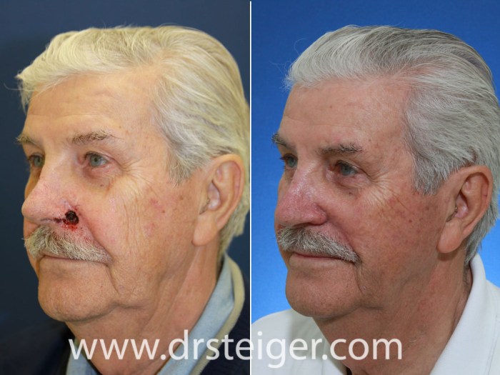
    Aesthetic Outcomes

    Mohs surgery on nose

    Mohs surgery, while crucial for precise cancer removal, demands meticulous attention to preserving the natural aesthetics of the treated area, especially on the delicate nose. Achieving a seamless, natural-looking result is paramount for patient satisfaction and psychological well-being. This section delves into the strategies employed to minimize scarring and maximize functional restoration, showcasing successful outcomes and highlighting the nuances of pre- and post-operative appearance.

    Mohs surgery on the nose can be a delicate procedure, requiring precision and expertise. A highly regarded dermatologist, like Jurairat J Molina MD MBA , is often instrumental in achieving the best possible outcomes. This specialized approach, crucial for preserving healthy tissue around the nose, makes it a preferred option for skin cancers in that area.

    Minimizing Scarring

    Minimizing scarring is a cornerstone of Mohs surgery, particularly on the nose. Techniques employed to achieve this include meticulous surgical planning, precise excision, and careful closure techniques. The surgeon’s experience and skill play a vital role in achieving minimal scarring, as does the selection of the appropriate closure method. The use of advanced surgical techniques, such as undermining and skin grafting, significantly reduces the likelihood of noticeable scarring.

    For example, a skilled surgeon may utilize a Z-plasty or other skin advancement techniques to reposition tissue and minimize the appearance of a linear scar.

    Functional Restoration

    Beyond aesthetic considerations, Mohs surgery on the nose must address potential functional impairments. This involves meticulous reconstruction of the nasal structure, ensuring that the repaired area seamlessly integrates with the surrounding tissue. The surgeon may utilize skin grafts, flaps, or other reconstructive techniques, meticulously matching the color, texture, and thickness of the surrounding nasal skin to ensure a natural appearance.

    The surgeon’s expertise in reconstructive surgery is crucial in achieving a result that not only removes the cancerous tissue but also restores the nose’s natural form and function.

    Successful Outcomes

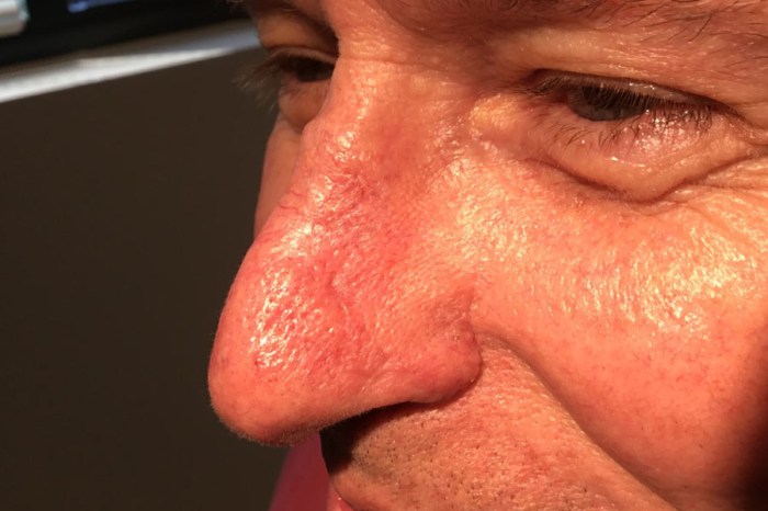
    A successful outcome in Mohs surgery on the nose is often characterized by subtle but significant improvements. For instance, a patient with a noticeable lesion on the tip of their nose might experience a nearly imperceptible difference in their nasal profile after the procedure, demonstrating the precise nature of the intervention. The goal is not merely to remove the cancerous lesion but to restore the nose’s original form and contour, leaving no visible trace of the surgical intervention.

    Post-operative photographs often highlight the restoration of symmetry and proportion, a crucial element in aesthetic outcomes.

    Comparison of Surgical Techniques

    Surgical Technique Description Advantages Disadvantages
    Skin Grafting Transfer of skin from a donor site to the defect. Can cover large defects, provides a durable result. Potential for donor site morbidity, may require multiple procedures.
    Local Flaps Tissue repositioning from adjacent areas. Preserves the natural contours of the nose, less donor site morbidity. Suitable for smaller defects, technical skill required.
    Z-Plasty Reconstructive technique that repositions tissue to reduce scar tension. Minimizes scar visibility, improves scar shape and length. Requires careful planning and precise execution.
    V-Y Advancement Flap Reshaping tissue to reduce tension and create a more natural appearance. Reduces scar tension, good for linear defects. May not be suitable for all defect types, requires precise planning.

    The table above highlights the various surgical techniques employed in Mohs surgery on the nose, demonstrating the wide range of options available to restore both the function and aesthetic appeal of the nasal structure.

    Long-Term Management

    Mohs surgery offers a high success rate in treating skin cancers, particularly on the nose, but long-term vigilance is crucial. This phase extends beyond the initial procedure, focusing on preventing recurrence and managing any potential skin concerns that might arise. Proactive follow-up and a clear understanding of potential issues are essential for long-term well-being and aesthetic satisfaction.

    Importance of Long-Term Follow-Up

    Regular follow-up appointments are vital for monitoring the treated area and detecting any signs of recurrence early. Early detection allows for prompt intervention, minimizing the impact of potential complications. This proactive approach ensures optimal long-term outcomes and preserves the aesthetic integrity of the nose.

    Role of Regular Skin Examinations

    Dermatologists employ visual inspections and, when necessary, biopsies to assess the treated area for any suspicious changes. These examinations are tailored to the individual patient’s needs and risk factors, ensuring that any developing abnormalities are identified quickly. Early intervention is crucial to prevent further growth or spread of cancerous cells.

    Strategies for Managing Potential Skin Concerns

    Skin reactions, such as redness, swelling, or scarring, can occur post-Mohs surgery. Managing these concerns involves meticulous post-operative care and appropriate medical intervention, as directed by the dermatologist. These strategies can help alleviate discomfort and promote healing. For instance, using prescribed topical medications and keeping the area clean and protected are crucial. A dermatologist can provide personalized recommendations based on the individual’s specific situation.

    Questions Patients Should Ask Their Dermatologist

    Patients should actively engage in discussions with their dermatologist regarding long-term care. This involves seeking clarification on specific post-operative instructions, recognizing potential complications, and understanding preventative measures. Key questions should include:

    • What are the signs of recurrence that I should be aware of?
    • How often should I schedule follow-up appointments?
    • What are the potential long-term skin concerns, and how can they be managed?
    • What skin care regimen should I follow to promote healing and prevent further irritation?
    • Are there any specific lifestyle factors that could influence the healing process?

    These questions are crucial for ensuring a smooth and successful recovery. Open communication and collaboration with the dermatologist are essential to achieve the best possible long-term outcome.

    Case Studies: Mohs Surgery On Nose

    Mohs surgery on nose

    Mohs micrographic surgery, a specialized technique for skin cancer removal, offers a high cure rate and minimizes tissue loss, particularly crucial in delicate areas like the nose. Detailed case studies illuminate the nuances of this procedure, demonstrating the importance of personalized treatment plans tailored to each patient’s unique situation. These examples showcase the complexities of skin cancer, the precision of Mohs surgery, and the ultimate goal of restoring both function and aesthetics.Understanding the diverse presentation of skin cancers on the nose and the subsequent surgical approach is critical.

    Each case study below highlights a different aspect of Mohs surgery, including challenges, considerations, and long-term outcomes, emphasizing the importance of meticulous surgical planning and post-operative care.

    Case Study 1: Basal Cell Carcinoma with Recurrent Nodule

    This patient presented with a persistent, recurrent basal cell carcinoma (BCC) nodule on the nasal dorsum. Initial examination revealed a small, firm, pearly papule with telangiectasia. Microscopic examination confirmed the diagnosis of BCC. Image description: Initial condition of BCC nodule on the nasal dorsum, showing a small, firm, pearly papule with telangiectasia.The Mohs procedure involved meticulous removal of the tumor in stages, with microscopic examination of each layer. The surgical approach focused on precise excision of the tumor and its surrounding margins to ensure complete removal. The surgical procedure was performed under local anesthesia.Image description: Mohs surgical procedure in progress, showing the precise excision of the tumor and surrounding margins.Post-operative healing was uneventful. The patient experienced minimal scarring. Long-term follow-up revealed no recurrence of the BCC.Image description: Post-operative result, showing minimal scarring and a near-normal nasal dorsum.The challenge in this case was the recurrent nature of the tumor. The careful attention to precise margin assessment during the Mohs procedure was essential for complete tumor removal.

    Case Study 2: Squamous Cell Carcinoma with Extensive Involvement

    A patient presented with a large, ulcerated squamous cell carcinoma (SCC) on the right nasal ala. The lesion exhibited significant tissue destruction and undermining. The patient’s history included a history of sun exposure. Image description: Initial condition of SCC on the right nasal ala, showing a large, ulcerated lesion with tissue destruction.The Mohs procedure was extensive, involving multiple stages of excision and microscopic analysis. The surgical approach focused on complete tumor removal while preserving as much healthy tissue as possible.Image description: Mohs surgical procedure showing the meticulous removal of the tumor, highlighting the multiple stages and microscopic examination.Post-operative care included meticulous wound care and skin grafting to close the defect. The patient experienced some temporary discomfort. The long-term outcome demonstrated complete eradication of the SCC without recurrence.Image description: Post-operative result, showing the repaired nasal ala and minimal visible scarring.The challenges in this case included the large size and depth of the lesion, as well as the need for reconstructive procedures.

    Case Study Summary

    Case Study Patient Demographics Type of Cancer Treatment Outcome
    1 (Age, Gender, Ethnicity) BCC, Recurrent Complete eradication, no recurrence
    2 (Age, Gender, Ethnicity) SCC, Extensive Complete eradication, no recurrence

    Epilogue

    In conclusion, Mohs surgery on the nose offers a targeted approach to treating skin cancer while minimizing the impact on facial aesthetics and function. The detailed procedures and post-operative care, along with long-term follow-up, are vital for successful outcomes. This comprehensive guide aims to empower patients and their healthcare providers with the knowledge needed to navigate this intricate procedure.

  • Sebaceous Hyperplasia Causes, Symptoms, and Treatments

    Sebaceous Hyperplasia Causes, Symptoms, and Treatments

    Sebaceous hyperplasia causes symptoms and treatments are explored in detail. This comprehensive guide dives into the causes, symptoms, and various treatment options for sebaceous hyperplasia, a common skin condition. We’ll examine the underlying reasons for its development, the visible signs it presents, and the different approaches to managing it. Understanding this condition empowers individuals to make informed decisions about their health and well-being.

    Sebaceous hyperplasia, often appearing as small, yellowish bumps on the face, neck, or upper chest, is frequently misunderstood. This condition, often benign and not requiring immediate medical attention, can be easily confused with other skin issues. However, a thorough understanding of its characteristics, causes, and potential treatments is crucial for appropriate management.

    Introduction to Sebaceous Hyperplasia

    Sebaceous hyperplasia is a common, benign skin condition characterized by the overgrowth of sebaceous glands. These glands, responsible for producing sebum (oil), become enlarged and form small, often flesh-colored or yellowish bumps on the skin. Understanding this condition is crucial for differentiating it from other skin issues with similar appearances and for managing patient concerns.

    Definition of Sebaceous Hyperplasia

    Sebaceous hyperplasia is a benign (non-cancerous) proliferation of sebaceous glands, typically occurring in middle-aged and older adults. It’s a common finding, often appearing as small, raised bumps on the skin.

    Locations of Sebaceous Hyperplasia

    Sebaceous hyperplasia commonly appears on areas of the body with a high concentration of sebaceous glands. These locations include the face (especially the nose, cheeks, and forehead), chest, back, and shoulders. The distribution often mirrors areas with higher sebum production.

    Appearance of Sebaceous Hyperplasia

    Sebaceous hyperplasia typically presents as small, smooth, flesh-colored or yellowish papules (small, solid bumps). They are usually less than 5 millimeters in diameter and may appear slightly raised above the skin surface. The bumps often have a pearly or waxy texture. Their appearance can vary from subtle to more noticeable, depending on the individual and the affected area.

    Comparison to Other Skin Conditions

    Condition Typical Appearance Location Key Differences
    Sebaceous Hyperplasia Small, smooth, flesh-colored or yellowish papules, less than 5 mm in diameter Face, chest, back, shoulders Benign, often multiple, typically asymptomatic.
    Basal Cell Carcinoma Small, pearly, translucent, or reddish papules or nodules; may have a slightly crusted or ulcerated surface Face, ears, neck, scalp Can be cancerous; often has a non-healing or bleeding characteristic; may have a rolled or pearly border
    Moles (Melanocytic Nevi) Various colors, shapes, and sizes; typically dark brown or black Anywhere on the skin May have irregular borders; can be raised or flat; a change in size, shape, or color can be a warning sign.
    Acne Red, inflamed papules, pustules, or nodules; often accompanied by blackheads or whiteheads Face, back, chest Often accompanied by inflammation and pain; commonly seen in adolescents and young adults

    Risk Factors for Sebaceous Hyperplasia

    Risk Factor Description Potential Impact Supporting Evidence
    Age Increased incidence with advancing age Higher probability of developing sebaceous hyperplasia as one ages. Numerous studies show a correlation between age and prevalence.
    Genetics Family history of sebaceous hyperplasia Genetic predisposition may increase the risk of developing the condition. Family history studies suggest a potential genetic component.
    Hormonal changes Hormonal fluctuations (e.g., during puberty or pregnancy) Possible influence on sebaceous gland activity. Limited direct evidence, but hormonal fluctuations are known to affect skin conditions.
    Sun exposure Prolonged or intense exposure to sunlight No direct evidence linking sun exposure to the development of sebaceous hyperplasia. Other skin conditions can be affected by sun exposure. Data is inconclusive. The condition itself is not directly sun-related.

    Causes of Sebaceous Hyperplasia: Sebaceous Hyperplasia Causes Symptoms And Treatments

    Sebaceous hyperplasia causes symptoms and treatments

    Sebaceous hyperplasia, benign skin growths, often appear as small, yellowish bumps, particularly on the face, chest, and back. While their exact cause remains somewhat elusive, researchers are uncovering crucial insights into the factors that contribute to their development. Understanding these factors is vital for both diagnosis and management strategies.The underlying causes of sebaceous hyperplasia are complex and not fully understood.

    Current research suggests a combination of genetic predispositions and environmental influences. This interplay is likely the driving force behind the formation of these common skin lesions.

    Genetic Predisposition

    Genetic factors play a significant role in the development of sebaceous hyperplasia. Family history often reveals a pattern of these lesions. Inherited variations in genes related to skin cell growth and function might increase an individual’s susceptibility to developing these bumps. Studies on large families with a high prevalence of sebaceous hyperplasia have identified potential candidate genes, although definitive links are still being investigated.

    The exact mechanisms through which these genetic variations influence sebaceous hyperplasia remain a subject of ongoing research.

    Environmental Factors

    Environmental factors may also contribute to the development or exacerbation of sebaceous hyperplasia. Exposure to certain irritants or chronic inflammation could potentially trigger the growth of these benign lesions. Hormonal fluctuations, especially in individuals with hormonal imbalances, could also influence the proliferation of sebaceous glands. For instance, individuals experiencing significant hormonal shifts during puberty or pregnancy may see an increase in sebaceous hyperplasia lesions.

    Comparison of Theories

    Different theories exist regarding the precise cause of sebaceous hyperplasia. One prominent theory emphasizes the role of genetic predisposition, suggesting that individuals with a specific genetic makeup are more prone to developing these lesions, irrespective of environmental factors. Another theory highlights the importance of environmental factors, suggesting that prolonged exposure to specific irritants or chronic inflammation could trigger or exacerbate sebaceous hyperplasia in genetically susceptible individuals.

    These theories aren’t mutually exclusive, and a more accurate explanation likely involves an interplay between genetic predisposition and environmental triggers.

    Triggers for Exacerbation

    While the exact causes of sebaceous hyperplasia are not fully understood, several factors could potentially exacerbate the growth or appearance of existing lesions. For example, irritation from frequent rubbing or pressure on the affected area might lead to inflammation and a visible increase in lesion size. Likewise, individuals with sensitive skin might experience flare-ups when exposed to harsh skincare products.

    The effects of prolonged sun exposure on sebaceous hyperplasia lesions are also a potential factor, although further research is needed to determine the extent of this effect.

    Hypotheses on Triggers

    Hypothesis Description Supporting Evidence Limitations
    Genetic Predisposition Individuals inherit a predisposition to develop sebaceous hyperplasia due to specific gene variations. Family history studies show clustering of cases in families. Candidate genes have been identified, although definitive links remain to be established. Doesn’t explain all cases; environmental factors likely play a role. The specific genes and their interactions are still under investigation.
    Chronic Inflammation Chronic skin irritation or inflammation may trigger or exacerbate the development of sebaceous hyperplasia. Some studies suggest a correlation between chronic inflammation and sebaceous hyperplasia. Irritants like certain cosmetics or friction could contribute. Difficult to isolate inflammation as the sole cause; genetic predisposition likely plays a significant role. Studies on the specific inflammatory pathways are limited.
    Hormonal Fluctuations Changes in hormone levels, especially in puberty or pregnancy, could influence sebaceous gland activity and hyperplasia. Observations of increased lesions during hormonal shifts in some individuals suggest a potential link. Correlation does not equal causation; other factors may contribute. More rigorous studies are needed to assess the precise hormonal impact.
    Environmental Irritants Prolonged exposure to specific irritants or chemicals could contribute to the development or exacerbation of sebaceous hyperplasia. Certain cosmetics or products may trigger inflammation. Friction or pressure on the affected area could also exacerbate the condition. Identifying specific triggers and quantifying their impact on sebaceous hyperplasia remains challenging. Individual responses to irritants vary significantly.

    Symptoms of Sebaceous Hyperplasia

    Sebaceous hyperplasia, a common benign skin condition, often presents with subtle changes that can be easily overlooked. Understanding the typical symptoms is crucial for distinguishing it from other skin issues and for appropriate management. While generally harmless, it’s important to recognize any unusual developments.

    Common Symptoms

    Sebaceous hyperplasia is typically characterized by small, raised, and yellowish or flesh-colored bumps. These lesions are usually smooth and round, and they vary in size, often ranging from a few millimeters to a few millimeters in diameter. They are frequently found on the face, particularly on the cheeks, nose, and forehead, but can also appear on the chest, back, and shoulders.

    These lesions are typically painless and do not cause any discomfort. Their appearance can be subtle, with the bumps sometimes being so small they may be barely noticeable.

    Distinguishing Features

    Several key features help differentiate sebaceous hyperplasia from other skin conditions. Unlike skin cancers, sebaceous hyperplasia lesions are usually well-defined and do not have irregular borders. They tend to appear in clusters, rather than scattered individually. They are often associated with areas of increased sebum production, a factor that can be noted in some cases. It is important to note that sebaceous hyperplasia typically does not cause itching, bleeding, or pain.

    Potential Complications

    While sebaceous hyperplasia is generally benign, potential complications are rare. The following table Artikels some possibilities and their associated symptoms.

    Complication Description Symptoms Severity
    Inflammation Localized redness, swelling, and tenderness around the lesion. Pain, warmth, redness, potential swelling, discomfort. Mild to moderate. Often resolves with time.
    Secondary Infection Bacterial or fungal infection of the lesion. Pus-filled sores, increased redness, pain, fever. Moderate to severe. Requires medical intervention.
    Cosmetic Concerns Patient dissatisfaction with the appearance of lesions. Psychological distress, desire for treatment, concerns about social interaction. Mild to moderate. Management varies based on patient preference.
    Accidental Trauma Injury or damage to the lesion from external factors. Bleeding, broken skin, pain. Mild to severe depending on the extent of injury.

    Variations in Symptoms

    The appearance and extent of symptoms can vary depending on the location and extent of the sebaceous hyperplasia. Lesions on sun-exposed areas may be slightly darker in color. Larger clusters of lesions may cause a noticeable thickening of the skin in the affected area. In cases where the lesions are numerous and widespread, the appearance may be more noticeable.

    Individual variations in skin tone and texture can also affect the overall visual impression of the lesions.

    Sebaceous hyperplasia, a common skin condition, often presents with small, yellowish bumps, typically on the face and chest. While usually harmless, understanding its causes, symptoms, and treatments is important. Interestingly, some research suggests a potential link between certain medications, like NSAIDs (non-steroidal anti-inflammatory drugs), and inflammatory bowel disease (IBD), which can sometimes complicate the picture. For a deeper dive into the relationship between NSAIDs and IBD, check out this informative article: nsaids non steroidal anti inflammatories and ibd.

    Fortunately, various treatment options exist for sebaceous hyperplasia, ranging from topical creams to surgical removal, depending on the severity and location of the lesions.

    Treatments for Sebaceous Hyperplasia

    Sebaceous hyperplasia, while typically harmless, can be cosmetically bothersome for some individuals. Fortunately, various treatment options are available to address the lesions and improve appearance. The best approach depends on factors such as the size, number, and location of the lesions, as well as individual preferences and potential risks.Effective treatment strategies aim to reduce the size, flatten the surface, or remove the sebaceous hyperplasia lesions, restoring a more even skin tone.

    The decision of whether or not to pursue treatment is a personal one and should be made in consultation with a dermatologist.

    Treatment Options

    Several treatment options exist for sebaceous hyperplasia, each with varying degrees of effectiveness and potential side effects. The choice of treatment often depends on the severity and location of the lesions, as well as the patient’s individual preferences.

    Surgical Removal

    Surgical removal is a definitive treatment option for sebaceous hyperplasia. This procedure involves excising the lesions under local anesthesia. Surgical removal is generally effective in eliminating the lesions and can provide a very satisfactory aesthetic outcome. However, it carries a risk of scarring, which can be more noticeable on certain areas of the body. The extent of scarring depends on the size of the lesion, the surgical technique, and the individual’s skin characteristics.

    This treatment is often considered for larger or more numerous lesions, particularly those on areas of the body where scarring might be less noticeable, such as the back or chest.

    Cryotherapy

    Cryotherapy involves freezing the sebaceous hyperplasia lesion using liquid nitrogen. This method is generally effective in reducing the size and appearance of the lesions. It is often a less invasive approach compared to surgical removal, with a lower risk of scarring. However, it may not be as effective for larger lesions and may take multiple treatments for optimal results.

    Side effects include temporary skin discoloration and discomfort, and, in rare cases, blistering. This treatment is well-suited for smaller lesions on less sensitive skin areas.

    Laser Therapy

    Laser therapy uses focused light beams to target and destroy the sebaceous hyperplasia lesions. Different types of lasers can be used, each with varying effectiveness and potential side effects. Laser therapy can be highly effective in treating sebaceous hyperplasia, leading to a significant reduction in lesion size and improved skin texture. Potential side effects include redness, swelling, and temporary skin discoloration.

    The choice of laser therapy will depend on the type of laser and the individual patient’s skin characteristics. It is often a viable option for treating multiple lesions, especially those on visible areas, and can offer a more precise approach compared to cryotherapy.

    Chemical Peels

    Chemical peels can be used to treat sebaceous hyperplasia, although their effectiveness may vary depending on the concentration and type of chemical used. The treatment involves applying a chemical solution to the skin to remove the top layers, potentially causing the sebaceous hyperplasia lesions to become less noticeable. Chemical peels can be effective in treating sebaceous hyperplasia, particularly when used in conjunction with other treatments.

    Side effects can include temporary redness, swelling, and skin sensitivity. This treatment is often considered for less severe cases or in combination with other methods.

    Comparison Table

    Treatment Description Effectiveness Potential Side Effects
    Surgical Removal Excision of lesions under local anesthesia Generally very effective Scarring possible, depending on location and technique
    Cryotherapy Freezing lesions with liquid nitrogen Effective for smaller lesions, multiple treatments may be needed Temporary discoloration, discomfort, possible blistering
    Laser Therapy Using focused light beams to target lesions Highly effective, often precise Redness, swelling, temporary discoloration
    Chemical Peels Applying chemical solution to remove skin layers Variable effectiveness, often used in combination Temporary redness, swelling, skin sensitivity

    Spontaneous Resolution

    In some cases, sebaceous hyperplasia lesions may spontaneously resolve without any treatment. However, this is not a reliable outcome and the time it takes for resolution is unpredictable. The likelihood of spontaneous resolution varies depending on factors such as the size, location, and individual characteristics of the lesions.

    Diagnosis of Sebaceous Hyperplasia

    Sebaceous hyperplasia causes symptoms and treatments

    Sebaceous hyperplasia, while often benign and easily recognizable, requires a proper diagnosis to rule out similar skin conditions. Accurate identification is crucial for appropriate management and to ensure patients receive the right treatment. This process typically involves a combination of visual assessment and, sometimes, additional diagnostic tools.Proper diagnosis of sebaceous hyperplasia is vital to differentiate it from other skin lesions that may mimic its appearance.

    This distinction helps guide treatment and avoid unnecessary interventions. The process often begins with a detailed physical examination, focusing on the characteristics of the lesions.

    Sebaceous hyperplasia, those little bumps on your skin, are usually harmless and often caused by aging or hormones. While some remedies suggest turmeric for potential benefits, it’s important to understand how much turmeric is too much, as with any supplement. This page will give you more details on the safe dosage of turmeric. Ultimately, proper medical advice from a dermatologist remains the best approach for addressing sebaceous hyperplasia causes, symptoms, and effective treatments.

    Physical Examination, Sebaceous hyperplasia causes symptoms and treatments

    A thorough physical examination plays a key role in diagnosing sebaceous hyperplasia. Clinicians visually inspect the skin lesions, paying close attention to their size, shape, color, and distribution. The presence of multiple, well-defined, yellowish or flesh-colored papules or plaques, often clustered together, is a strong indicator. The location of the lesions, typically on the face, neck, chest, or back, can also provide valuable clues.

    Careful observation of the surrounding skin and the absence of any alarming symptoms like inflammation or ulceration further support the diagnosis.

    Differential Diagnosis

    Differentiating sebaceous hyperplasia from other skin conditions is crucial. Similar-appearing lesions, such as basal cell carcinoma, seborrheic keratosis, or other benign growths, can require further investigation. The clinician must carefully assess the characteristics of the lesion to rule out these potential alternatives. Features like irregular borders, ulceration, rapid growth, or bleeding strongly suggest the need for additional tests.

    Diagnostic Criteria

    Careful evaluation of diagnostic criteria is critical to confirm a diagnosis of sebaceous hyperplasia. This systematic approach helps clinicians determine if the lesion meets the criteria for sebaceous hyperplasia.

    Criterion Description Diagnostic Method Significance
    Appearance Well-defined, yellowish or flesh-colored papules or plaques, often clustered together. Visual inspection during physical examination. Suggests the presence of sebaceous hyperplasia.
    Location Typically found on the face, neck, chest, or back. Physical examination. Provides additional context and supports the diagnosis.
    Size Usually small, ranging from a few millimeters to a few centimeters in diameter. Physical examination. Helps differentiate from larger, potentially concerning lesions.
    Consistency Soft and slightly raised; often with a smooth surface. Palpation during physical examination. Assists in distinguishing from other lesions with different consistencies.

    Imaging Techniques

    While not always necessary, imaging techniques can aid in the diagnosis, particularly when differential diagnoses are suspected. Dermoscopy, a non-invasive technique that uses a specialized microscope to examine the skin’s surface, can provide detailed images of the lesion’s structure and characteristics. This can be useful in differentiating sebaceous hyperplasia from other skin conditions with similar appearances. In cases of doubt, a biopsy may be performed to obtain a tissue sample for microscopic examination.

    This is the definitive diagnostic method, confirming the presence and nature of the lesion.

    Sebaceous hyperplasia, those little bumps on your skin, often have simple causes like genetics or aging. Understanding the different types of carbs, like good vs bad carbs , might not directly impact the condition, but a balanced diet can certainly contribute to overall health and potentially influence skin health. Treatments range from topical creams to laser procedures, depending on the severity and location of the hyperplasia.

    Prevention of Sebaceous Hyperplasia

    Sebaceous hyperplasia, while typically harmless, can be bothersome due to its appearance. Unfortunately, there are no definitive preventive measures currently available to stop the development of these benign skin growths. Understanding the factors that might increase the risk, however, can help in managing potential triggers.Currently, the primary focus in managing sebaceous hyperplasia is on symptom management and treatment when necessary, rather than on prevention.

    This is because the exact causes of the condition are not fully understood. Research continues, but for now, there’s no proven method to prevent their formation.

    Lifestyle Factors Influencing Development

    Several lifestyle factors could potentially influence the likelihood of developing sebaceous hyperplasia. These factors, however, are not definitive causes, and further research is needed to establish clear links. It’s important to note that this is not an exhaustive list and more research is necessary to determine the complete picture.

    • Genetics: A family history of sebaceous hyperplasia might increase a person’s predisposition to developing the condition. However, this doesn’t guarantee development, and many individuals with a family history do not experience it.
    • Age: Sebaceous hyperplasia typically appears in middle-aged and older adults. This suggests an association with aging, but it doesn’t mean younger individuals are immune. The exact mechanisms behind this correlation are still under investigation.
    • Hormonal Changes: While not fully understood, hormonal fluctuations might play a role. However, there’s no conclusive evidence to support this theory, and further studies are needed to establish a definite link.

    Managing Potential Risk Factors

    While definitive prevention is not yet possible, managing potential risk factors can help in reducing the likelihood of developing sebaceous hyperplasia. These approaches focus on general health and well-being rather than on targeting the specific causes.

    • Healthy Diet: A balanced diet rich in fruits, vegetables, and whole grains may contribute to overall health and well-being. This may indirectly influence the body’s response to factors that might contribute to sebaceous hyperplasia.
    • Regular Exercise: Physical activity can contribute to a healthier lifestyle, which might help in managing potential risk factors. The exact impact on sebaceous hyperplasia development is not yet fully understood.
    • Stress Management: Managing stress effectively through techniques like yoga, meditation, or spending time in nature can have positive effects on overall health. There is some evidence linking stress to skin conditions, but further research is needed to explore its relationship with sebaceous hyperplasia.

    Potential Effectiveness of Preventive Measures

    The effectiveness of these preventive measures is difficult to assess due to the lack of definitive research on the causes of sebaceous hyperplasia. While adopting a healthy lifestyle can improve overall health, there’s no guarantee it will prevent the development of sebaceous hyperplasia. The current understanding is that these strategies contribute to general well-being, which might indirectly influence the development of various skin conditions.

    However, further research is crucial to determine a definitive link between lifestyle choices and the prevention of sebaceous hyperplasia.

    Illustrations and Visual Aids

    Visual aids are crucial for understanding and recognizing sebaceous hyperplasia. They allow for a clear comparison with other skin conditions and facilitate the identification of various stages and locations of the lesions. This section provides detailed visual representations of sebaceous hyperplasia in different contexts.Visual representations are a valuable tool for learning about the nuances of sebaceous hyperplasia. By visualizing the different stages of development, locations, microscopic structures, and comparisons with other skin conditions, one can gain a deeper understanding of this benign skin lesion.

    Detailed Visual Representation of Sebaceous Hyperplasia in Various Stages

    Sebaceous hyperplasia typically presents as small, yellowish or flesh-colored bumps. Early-stage lesions appear as slightly raised, smooth papules. As the lesions mature, they can become more prominent, slightly larger, and potentially develop a slightly rough or slightly pearly texture. The color might vary slightly, but generally remains in the yellowish or flesh-colored range. Advanced stages may show slightly more noticeable enlargement and a slight increase in the number of lesions.

    These visual representations help differentiate between benign and malignant lesions, particularly important for medical professionals.

    Graphic Illustration of Common Locations

    Sebaceous hyperplasia frequently appears on the face, particularly on the cheeks, nose, forehead, and chin. It can also appear on the back, chest, and shoulders. A visual representation of these common locations would show the distribution patterns on different areas of the body. The illustrations would highlight the clustering tendency of these lesions in certain areas, providing context for potential misdiagnosis.

    For example, a cluster of lesions on the nose might be mistaken for another skin condition.

    Microscopic Structure of Sebaceous Hyperplasia

    A microscopic view of sebaceous hyperplasia reveals an accumulation of sebaceous glands. These glands are often enlarged and appear as clusters of cells, with a slightly irregular arrangement. The cells within the glands might show some degree of atypia but generally maintain a benign appearance. The surrounding skin tissue usually appears normal, lacking significant inflammation or other cellular abnormalities.

    A clear visual comparison with a normal sebaceous gland would be valuable for educational purposes.

    Visual Comparison with Other Skin Conditions

    Differentiating sebaceous hyperplasia from other skin conditions, like basal cell carcinoma or acne, can be crucial. A visual comparison chart, including images of each condition, would highlight the key differences. Sebaceous hyperplasia often presents as a more rounded, slightly raised bump, while basal cell carcinoma might have a slightly ulcerated or pearly appearance. Acne lesions, in contrast, might be inflamed and have a pustular or papular appearance.

    Accurate visual representations help medical professionals make informed diagnoses.

    Recognizing Sebaceous Hyperplasia: A Detailed Illustration

    A detailed illustration should guide the viewer through the process of identifying sebaceous hyperplasia. The illustration could begin with a general overview of the appearance of the lesion, progressing to close-up views showing the characteristic features. Specific details, such as the size, shape, color, and consistency of the lesions, should be highlighted. This visual representation could be combined with a key identifying the key characteristics to aid in diagnosis.

    Concluding Remarks

    In conclusion, sebaceous hyperplasia, while generally harmless, warrants careful consideration. Understanding its causes, symptoms, and treatment options is key to managing this condition effectively. This discussion highlights the importance of accurate diagnosis and the various approaches available to address sebaceous hyperplasia, from watchful waiting to more interventionist treatments. Remember, consulting a dermatologist is crucial for personalized guidance and effective management.

  • What is Acne Surgery? A Deep Dive

    What is Acne Surgery? A Deep Dive

    What is acne surgery? This exploration delves into the world of surgical interventions for acne scars, differentiating it from other acne treatments. We’ll examine the various types of acne requiring surgical intervention, the different surgical procedures used, and the meticulous pre- and post-operative care necessary for successful outcomes. From excision to dermabrasion, we’ll compare techniques, highlighting their respective advantages and disadvantages, and examine the potential complications.

    Understanding the intricacies of acne surgery involves considering patient suitability, potential risks and benefits, and alternative treatments. The cost and insurance coverage aspects will be thoroughly examined, as well as success stories, case studies, and the recovery timelines for different procedures. We’ll conclude with a comprehensive overview, leaving you well-informed about this specialized area of dermatological care.

    Introduction to Acne Surgery

    What is acne surgery

    Acne surgery represents a specialized approach to treating severe acne and its lingering consequences, distinct from topical treatments, oral medications, or laser therapies. It’s a more invasive procedure, often reserved for cases where other methods have proven ineffective or have caused unacceptable side effects. This surgical intervention focuses on the removal of acne lesions, scars, and other problematic skin formations.Acne surgery addresses specific skin issues that other treatments might not fully resolve.

    It’s particularly valuable for managing deep, cystic acne, severe nodular acne, and acne scars that have resisted standard therapies. The surgical approach targets the root cause of the problem, aiming to restore the skin’s normal structure and appearance.

    Types of Acne Requiring Surgery

    Acne presents in various forms, with some cases necessitating surgical intervention. Severe cystic acne, characterized by large, pus-filled lesions, often leads to significant inflammation and scarring. Nodular acne, marked by painful, inflamed bumps beneath the skin’s surface, also may benefit from surgical treatment to minimize long-term damage. Persistent, recalcitrant acne that doesn’t respond to other treatments might warrant surgical exploration.

    Finally, extensive acne scarring, resulting from previous inflammatory lesions, is a common reason for considering surgical interventions.

    Surgical Procedures for Acne Scars

    Surgical techniques for acne scars aim to improve skin texture and appearance. Different approaches address various scar types and severities. These techniques vary significantly in their invasiveness, recovery time, and potential for complications.

    Comparison of Surgical Techniques for Acne Scars

    Technique Description Pros Cons
    Excision This procedure involves surgically removing the scar tissue. The surgeon carefully cuts out the affected area, often followed by skin grafting to restore the area’s appearance and functionality. Effective for deep, raised scars; potentially restores skin texture. More invasive; longer recovery period; potential for scarring; increased risk of infection.
    Dermabrasion Dermabrasion uses a rotating instrument to abrade the skin’s surface, removing the top layer of damaged skin. This procedure aims to smooth out irregularities and promote new skin cell growth. Can improve the appearance of mild to moderate acne scars; relatively less invasive than excision. Higher risk of temporary skin discoloration; potential for prolonged redness; possible complications such as infection or hyperpigmentation.
    Laser resurfacing Laser resurfacing uses precisely controlled beams of light to target and vaporize or ablate skin tissue. This procedure can remove superficial scars and improve skin texture. Minimally invasive; faster recovery time than other surgical options; effective for various scar types; often less painful than dermabrasion. Potential for skin discoloration, blistering, or infection; possible scarring if not performed correctly; may not be suitable for all types of acne scars.
    Chemical peels Chemical peels use strong acids to remove the top layers of skin, promoting new skin cell growth. The depth of the peel is adjusted to target specific skin concerns, including acne scars. Relatively non-invasive; effective for mild to moderate acne scars; often less expensive than other surgical techniques. Potential for skin irritation, redness, or discomfort; may not be suitable for deep or severe acne scars; recovery time can vary.

    Surgical Procedures

    Acne scar revision surgery offers a targeted approach to improving the appearance of acne scars, addressing a wide range of scar types and severities. These procedures aim to reshape the skin, reduce the prominence of scars, and restore a smoother, more even skin texture. The specific surgical technique employed depends on the type and depth of the scars, as well as the patient’s individual needs and expectations.

    Common Acne Scar Revision Procedures

    Different surgical techniques target various types of acne scars. A common procedure involves excision, where the scar tissue is removed and the surrounding skin is reshaped. Other techniques, such as skin grafting, may be employed to replace damaged skin with healthy skin from another area of the body. The choice of procedure depends on the type and extent of the scarring, and a consultation with a qualified dermatologist or plastic surgeon is crucial to determine the most appropriate treatment plan.

    Acne surgery, a procedure to treat severe acne, often involves removing damaged skin. While different from the topical treatments many people use, it’s important to understand the various approaches to managing skin conditions like eczema, as seen in biologics for eczema mechanisms options considerations. Ultimately, acne surgery is a specialized treatment option for persistent and problematic acne.

    Steps Involved in a Common Acne Scar Revision Surgery

    The steps in a common acne scar revision surgery typically include:

    • Pre-operative Evaluation: A thorough examination of the scars, including their type, depth, and distribution, is conducted. This evaluation helps determine the best surgical approach and to assess the patient’s overall health and suitability for the procedure. The dermatologist or plastic surgeon will also discuss the patient’s expectations and desired outcomes.
    • Anesthesia: Local or general anesthesia is administered to ensure patient comfort and relaxation during the surgical procedure.
    • Surgical Incisions: Precise incisions are made around the scar tissue to be corrected. The depth and shape of the incisions will depend on the specific type of scar.
    • Scar Tissue Removal: The scar tissue is carefully excised and removed.
    • Tissue Reshaping: The surrounding skin is reshaped to reduce the prominence of the scar and create a smoother appearance. This may involve techniques like skin grafting or tissue flaps.
    • Closure: The surgical site is carefully closed using sutures or staples.
    • Post-operative Care: The patient is provided with detailed instructions for post-operative care, including wound care, medication management, and activity restrictions.

    Pre-operative and Post-operative Care Procedures

    Strict adherence to pre-operative and post-operative care instructions is crucial for optimal healing and minimizing complications. Pre-operative care often involves blood tests, consultations with an anesthesiologist, and cessation of certain medications. Post-operative care includes meticulous wound care, pain management, and strict adherence to prescribed medication regimens. Failure to follow these guidelines can significantly increase the risk of infection, delayed healing, and other complications.

    Recovery Time and Potential Complications

    Recovery time varies depending on the complexity of the procedure and the individual patient’s healing capacity. Simple procedures might take a few weeks to heal, while more complex procedures could require several months. Potential complications include infection, excessive scarring, bleeding, and nerve damage. Individual experiences can vary greatly.

    Comparison of Recovery Periods for Different Acne Scar Surgeries

    The recovery period for different acne scar surgeries varies significantly. Procedures that involve minimal tissue manipulation and smaller incisions generally have a shorter recovery time than those that involve extensive tissue reshaping or skin grafting. Factors like the patient’s age, overall health, and the individual healing response also influence the recovery duration.

    Acne surgery, often a last resort, involves procedures like laser treatments or chemical peels to target stubborn blemishes. Considering how and when to start birth control can be a factor in some cases, as hormonal changes can sometimes influence acne. The right approach to birth control, as detailed in this helpful guide how and when to start birth control , could potentially help manage acne alongside other treatment options.

    Ultimately, the best course of action for acne surgery depends on the individual’s specific needs and the severity of their condition.

    Potential Complications of Different Procedures

    Procedure Potential Complications Frequency Mitigation
    Excision Infection, excessive scarring, bleeding, nerve damage Low to moderate Careful surgical technique, meticulous post-operative care, antibiotic use
    Skin Grafting Rejection of graft, infection, prolonged healing, hypertrophic scarring Moderate Proper graft selection, meticulous wound care, immunosuppressants (in some cases)
    Laser resurfacing Infection, scarring, uneven skin tone, skin discoloration Low to moderate Proper laser settings, post-operative care, follow-up visits

    Candidates for Acne Surgery: What Is Acne Surgery

    What is acne surgery

    Acne surgery, while effective for certain cases, isn’t a one-size-fits-all solution. Identifying the right candidates is crucial for achieving positive outcomes and avoiding unnecessary procedures. Understanding the characteristics of suitable patients and those who aren’t ideal is paramount for ensuring the best possible results.Careful evaluation of a patient’s acne history, type of lesions, and overall skin health is essential before recommending acne surgery.

    This evaluation will help determine if the benefits outweigh the potential risks and if the patient is likely to achieve a satisfactory outcome.

    Patient Characteristics Suitable for Acne Surgery

    Careful patient selection is critical for successful acne surgery. Suitable candidates typically exhibit specific characteristics related to their acne and skin type. These patients have a well-defined pattern of acne lesions, often with significant scarring or cystic acne that hasn’t responded well to other treatments.

    • History of persistent acne: Patients with a history of moderate to severe acne that has not responded to conventional treatments, such as topical medications, oral antibiotics, or oral contraceptives, are often good candidates for acne surgery.
    • Specific lesion type: Surgery is often targeted at specific lesions like deep, inflamed nodules or cysts. The presence of these lesions, which are often resistant to other treatments, suggests a potential benefit from surgical intervention.
    • Extent of scarring: Patients with significant acne scarring, including hypertrophic or keloid scars, or those with pitted or depressed scars, might be ideal candidates for acne scar revision surgery. The extent of scarring is often a significant factor in determining the appropriateness of surgery.
    • Realistic expectations: Patients who have realistic expectations about the procedure’s limitations and potential outcomes are more likely to have a positive experience. They understand that surgery may not completely eliminate all acne-related problems but can improve their condition.

    Patient Characteristics Unsuitable for Acne Surgery

    Certain patient characteristics might make them unsuitable for acne surgery. These factors often indicate that other treatment options might be more appropriate or that the risks of the procedure outweigh the potential benefits.

    • Active, untreated infections: Patients with active infections in the area of concern are not suitable for acne surgery until the infection is controlled. Surgical intervention on infected tissue can worsen the situation.
    • Poor skin healing: Patients with a history of poor wound healing or those with underlying skin conditions that may interfere with healing are less suitable for surgery. Factors like diabetes or autoimmune diseases can negatively impact wound healing.
    • Unrealistic expectations: Patients who have unrealistic expectations about the procedure’s results, or those who are not compliant with post-operative instructions, are often not ideal candidates. Understanding the limitations of surgery and commitment to post-operative care are crucial.
    • Certain skin types: Patients with very sensitive or thin skin might experience complications from acne surgery. This is particularly true for certain procedures, such as deep excisions.

    Role of Consultation in Assessing Suitability

    A comprehensive consultation plays a vital role in evaluating a patient’s suitability for acne surgery. During this consultation, the dermatologist or surgeon thoroughly assesses the patient’s medical history, current condition, and expectations.

    • Detailed medical history: The consultation includes a thorough review of the patient’s medical history, including any pre-existing skin conditions, allergies, or medications. This helps identify potential complications or contraindications.
    • Examination of the acne lesions: A detailed examination of the acne lesions is performed to assess the type, depth, and extent of the acne and associated scarring. This helps determine the appropriate surgical approach.
    • Discussion of expectations: The surgeon discusses the potential benefits and risks of the procedure, including the limitations of acne surgery. Realistic expectations and informed consent are essential for a successful outcome.

    Checklist for Evaluating Patient Suitability for Acne Scar Revision Surgery

    A structured checklist helps in systematically evaluating a patient’s suitability for acne scar revision surgery.

    Acne surgery, a procedure for severe acne, often involves extracting lesions and repairing damaged skin. While researching the best approach for my own skin, I stumbled upon some interesting information about dietary choices and their potential impact on skin health. For example, if you’re wondering whether tomatoes are a good or bad idea when dealing with gout, checking out this resource on are tomatoes bad for gout might be helpful.

    Ultimately, understanding the root causes of acne and finding the right treatment is key to achieving healthy skin.

    Criteria Evaluation
    Medical history (allergies, medications, pre-existing conditions) Document and assess potential risks
    Type and extent of acne scars Evaluate severity and location
    Patient expectations Assess realistic outcomes and understand the limitations of surgery
    Skin type and healing capacity Assess potential complications
    Compliance with post-operative instructions Assess patient’s willingness to follow post-op care

    Risks and Benefits

    Acne surgery, while often effective, carries inherent risks. Understanding these potential downsides alongside the possible benefits is crucial for informed decision-making. This section will detail the risks and benefits associated with different surgical procedures, providing a balanced perspective for patients considering this option.

    Potential Risks of Acne Surgery

    A thorough evaluation of the potential risks is essential before proceeding with acne surgery. These risks vary depending on the specific procedure and the individual patient’s health status.

    • Infection: Surgical procedures, no matter how precise, create small breaches in the skin, making the area vulnerable to bacterial infections. Prompt and appropriate antibiotic treatment is crucial in mitigating this risk. Proper wound care and post-operative hygiene significantly reduce the risk of infection.
    • Scarring: While modern techniques minimize scarring, some degree of scarring is possible, particularly with more aggressive procedures. The type and severity of scarring can vary depending on the individual’s skin type and the extent of the surgical intervention.
    • Bleeding: Any surgical procedure carries a risk of bleeding. Appropriate surgical techniques, careful hemostasis, and post-operative pressure dressings are employed to control bleeding and promote healing.
    • Numbness: Some surgical procedures may result in temporary or permanent numbness in the treated area. The extent and duration of numbness depend on the specific nerves involved in the procedure.
    • Adverse Reactions to Anesthesia: The use of anesthesia always carries a small risk of adverse reactions. Pre-operative assessments and careful monitoring during the procedure are essential to mitigate these risks. Reactions may range from mild discomfort to serious complications.
    • Delayed Healing: Certain medical conditions or factors like poor nutrition can influence the healing process. Careful management of pre-operative and post-operative care is crucial to minimize delays in healing.

    Potential Benefits of Acne Surgery

    Acne surgery offers a potential pathway to improved appearance and self-esteem. The benefits are often substantial for individuals with severe acne that hasn’t responded to other treatments.

    • Improved Appearance: Surgical techniques can effectively remove acne lesions and scars, leading to a noticeable improvement in skin texture and appearance. This can significantly boost a patient’s self-confidence.
    • Reduced Acne: In cases of severe cystic acne, surgical interventions can effectively target and remove the lesions, thus reducing the frequency and severity of future outbreaks. This can provide long-term relief for those with persistent and debilitating acne.
    • Scar Reduction: Certain surgical techniques can address and improve the appearance of existing acne scars. This helps patients achieve a more even skin tone and a smoother texture.

    Comparison of Surgical Procedures

    Different acne surgical procedures offer varying approaches and results. Factors such as the severity of acne, the patient’s skin type, and the surgeon’s expertise all play a role in determining the most appropriate procedure.

    Procedure Description Potential Benefits Potential Risks
    Dermabrasion Removes the top layer of skin to smooth out irregularities. Effective for superficial scars. Increased risk of scarring, infection, and prolonged healing time.
    Laser resurfacing Uses laser energy to target acne lesions and scars. Can reduce inflammation and improve skin tone. Potential for scarring, hyperpigmentation, and discomfort.
    Surgical excision Removes cysts and nodules under the skin. Effective for deep acne lesions. Higher risk of scarring and bleeding.

    Success Stories and Testimonials

    Patients have reported varying degrees of success with acne surgery. The positive impact on self-image and confidence is often cited as a key benefit. A patient who had extensive cystic acne, for example, saw a significant reduction in acne lesions and scar tissue after undergoing a series of surgical procedures. Another patient experienced a noticeable improvement in skin texture and tone following laser resurfacing, leading to an enhanced sense of well-being.

    Cost and Insurance Coverage

    Acne surgery, while effective, can be a significant financial investment. Understanding the potential costs and how insurance might cover these procedures is crucial for patients considering this option. This section will detail the average costs of various acne surgery procedures, explore insurance coverage, and discuss the overall financial implications of pursuing this type of treatment.The cost of acne surgery varies considerably based on the specific procedure, the extent of the treatment needed, the location of the clinic, and the surgeon’s expertise.

    Factors such as the use of advanced technologies, the complexity of the scar revision, and the number of sessions required all play a role in determining the overall price tag.

    Average Costs of Acne Surgery Procedures

    Acne surgery procedures can involve a range of costs, from relatively low-cost treatments to more extensive and costly options. For example, a simple excision of acne lesions might range from $500 to $1500 per session, whereas a more involved procedure such as laser resurfacing or chemical peels for acne scar revision might cost $1000 to $5000 or more, depending on the extent of the treatment.

    It’s essential to discuss pricing with a qualified dermatologist or plastic surgeon to obtain personalized estimates.

    Insurance Coverage for Acne Surgery

    Insurance coverage for acne surgery is not always straightforward. Insurance companies often consider acne surgery a cosmetic procedure, and therefore, coverage is usually limited or nonexistent. Even if a portion of the procedure is deemed medically necessary, for example, to treat severe acne scars, the extent of coverage can vary greatly depending on the specific policy.

    Examples of Insurance Policies and Acne Scar Revision

    Different insurance policies have varying levels of coverage for acne scar revision. A comprehensive health plan might cover a portion of the costs if the scars are considered a result of a pre-existing medical condition, like severe acne. However, plans with a limited scope may not cover any of the treatment at all. It’s essential to review your policy’s specifics and contact your insurance provider directly to understand your coverage options for acne scar revision.

    For example, a patient with a Blue Cross Blue Shield plan might receive partial coverage for a medically necessary scar revision procedure if the procedure is deemed necessary for the treatment of a pre-existing condition. However, a similar procedure performed under a different insurance plan may not receive any coverage.

    Financial Implications of Acne Surgery

    Patients considering acne surgery should carefully assess the financial implications. The costs associated with the procedure itself, potential follow-up appointments, and any associated medications or therapies can accumulate quickly. It’s crucial to develop a realistic budget and consider financing options if needed. For example, some patients might explore payment plans offered by clinics or consider personal loans to manage the costs.

    Patients should also be aware of the potential for out-of-pocket expenses, even with insurance coverage. A thorough understanding of the costs and potential financial responsibilities is essential before committing to acne surgery.

    Alternatives to Acne Surgery

    Acne surgery, while effective for severe cases, isn’t the only option. Many non-surgical treatments can effectively address acne scars and improve skin texture. These alternatives offer varying degrees of efficacy, recovery time, and long-term maintenance, making careful consideration crucial for individuals seeking acne scar improvement.Choosing the right alternative depends on the severity and type of acne scars, individual skin type, and desired outcome.

    A consultation with a dermatologist is essential to determine the most suitable course of action.

    Non-Surgical Acne Scar Treatments

    Various non-surgical approaches target acne scars, ranging from topical medications to advanced laser therapies. Understanding the strengths and weaknesses of each is critical in selecting the most appropriate treatment.

    Chemical Peels

    Chemical peels use chemical solutions to exfoliate the skin, promoting new skin cell growth and improving the appearance of acne scars. Different peel strengths and formulations cater to various skin types and scar depths. Light peels can address superficial scars, while deeper peels tackle more extensive scarring. A professional should always perform chemical peels.

    Microneedling

    Microneedling involves creating microscopic punctures in the skin using tiny needles. This stimulates collagen production, leading to improved skin texture and reduced scar visibility. Multiple sessions are typically required to achieve optimal results, and the recovery time varies depending on the depth and extent of treatment.

    Laser Treatments

    Laser treatments employ different wavelengths of light to target acne scars, stimulating collagen production and reducing inflammation. The type of laser used depends on the scar type and depth. Fractional lasers, for example, create microscopic channels in the skin, promoting collagen regeneration. Laser treatments often require multiple sessions for optimal outcomes.

    Dermal Fillers

    Dermal fillers are injectables used to fill in depressed scars, restoring volume and smoothing the skin’s surface. The filler material is carefully injected into the scar tissue to improve its appearance. Results can vary depending on the type of filler used and the skill of the injector. Maintenance injections may be needed to sustain results.

    Dermabrasion

    Dermabrasion involves using a rotating device with abrasive material to remove the top layer of skin, smoothing out the skin’s surface. This procedure is more aggressive than other treatments and may result in noticeable recovery time. The severity of the acne scar will dictate the extent of the procedure. Dermabrasion is not always the most suitable option, and alternative treatments may be more appropriate in certain cases.

    Comparison of Surgical and Non-Surgical Treatments

    Treatment Effectiveness Recovery Time Cost Maintenance
    Surgical (e.g., excision) High for severe cases Extended High Minimal
    Non-Surgical (e.g., chemical peels, microneedling) Variable, depends on the treatment and severity Variable Variable May require ongoing maintenance

    Advantages and Disadvantages of Non-Surgical Options

    Non-surgical alternatives offer several advantages, including less invasive procedures, shorter recovery times, and often more affordable costs. However, these methods may not be as effective for severe or deep scars, and some treatments require multiple sessions to achieve satisfactory results.

    Long-Term Results and Maintenance

    The long-term results and maintenance requirements vary considerably among non-surgical treatments. Chemical peels, for instance, can yield noticeable improvements, but repeated treatments may be needed to sustain results. Laser treatments often produce lasting results, but follow-up sessions might be required. The duration of treatment effects will depend on the type of treatment and the individual’s skin characteristics.

    Illustrative Cases

    Acne surgery, while often effective, is not a one-size-fits-all solution. Individual responses vary, and the success of the procedure depends on various factors, including the type and severity of acne, the extent of scarring, and the skill of the surgeon. This section presents three case studies, showcasing diverse outcomes and highlighting crucial considerations for patients considering acne surgery.

    Case Study 1: Successful Acne Surgery for Nodular Acne, What is acne surgery

    This patient presented with severe nodular acne, characterized by deep, inflamed lesions that left significant scarring. The surgical approach involved a combination of mechanical extraction of the inflamed lesions and the application of targeted laser treatments to reduce inflammation and improve skin texture. Post-operatively, the patient experienced mild discomfort and swelling, which subsided within a week. A meticulous skincare regimen, including topical antibiotics and anti-inflammatory agents, was prescribed to manage any residual inflammation.

    Over several months, the patient observed a considerable improvement in their skin texture, with a noticeable reduction in the size and appearance of the acne scars. The final outcome demonstrated a substantial improvement in the patient’s overall skin health and self-esteem.

    Case Study 2: Acne Scar Revision with Fractional Laser Resurfacing

    This case study focuses on a patient with moderate acne scarring. Instead of the traditional surgical approach, the patient opted for fractional laser resurfacing. This procedure involves using a laser to create microscopic wounds in the skin, stimulating collagen production and promoting skin regeneration. The procedure resulted in minimal downtime and relatively quick healing. The patient experienced mild redness and swelling post-operatively, which resolved within a few days.

    The treatment plan involved multiple sessions over several weeks to achieve the desired results. The final outcome exhibited a significant improvement in the appearance of the acne scars, smoothing out the skin texture and reducing the overall appearance of the irregularities.

    Case Study 3: Lack of Improvement with Acne Surgery

    This patient presented with a complex pattern of acne scarring, including hypertrophic and atrophic scars. The patient underwent a surgical procedure aimed at addressing the hypertrophic scars, but the results were less than satisfactory. The procedure, though performed skillfully, did not effectively target the underlying causes of the acne scarring. The patient’s skin type and individual response to the treatment contributed to the lack of improvement.

    After careful evaluation, alternative treatments, including topical retinoids and chemical peels, were explored to address the atrophic scars. The patient continued to benefit from the targeted treatments and experienced a moderate improvement in the appearance of the scars over time.

    Different Types of Acne Scars

    Acne scars manifest in various forms, each presenting unique challenges in treatment. Understanding these differences is crucial for selecting the most appropriate surgical or non-surgical approach.

    • Hypertrophic Scars: These scars are raised and thickened areas of skin that result from excessive collagen production. They often appear reddish or purplish and can be quite prominent.
    • Keloid Scars: These scars extend beyond the boundaries of the original wound and grow progressively larger. They are characterized by a firm, raised, and often discolored appearance.
    • Atrophic Scars: These scars are depressed or sunken areas of skin, often appearing as pitted or indented marks. These scars can be difficult to treat and often require multiple treatments to achieve noticeable improvement.
    • Ice Pick Scars: These deep, narrow, and pitted scars are often difficult to treat, as they penetrate deeply into the skin layers.
    • Boxcar Scars: These are flat, depressed scars with distinct, sharp edges, often appearing in a box-like shape.

    Conclusion

    In conclusion, acne surgery presents a specialized approach to treating acne scars, offering potential benefits but also carrying inherent risks. A detailed understanding of the various surgical techniques, patient selection criteria, potential complications, and alternative treatments is crucial for making informed decisions. This comprehensive guide empowers individuals to navigate the complexities of acne surgery, ultimately leading to informed choices for their specific needs and circumstances.

    The information presented here aims to serve as a valuable resource for anyone seeking to learn more about this specialized area of dermatological care.

  • Steroids Topical Steroid Strengths A Deep Dive

    Steroids Topical Steroid Strengths A Deep Dive

    Steroids topical steroid strengths are crucial for treating various skin conditions. This guide delves into the different potencies, their applications, and potential side effects. Understanding the strengths and appropriate usage is vital for effective treatment and minimizing adverse reactions. We’ll explore the diverse range of skin conditions treatable with topical steroids, examining the factors determining the right strength for each case.

    From mild inflammation to severe skin disorders, the right topical steroid strength can significantly impact treatment outcomes. This comprehensive overview will guide you through the potency levels, application methods, and potential side effects associated with various topical steroid strengths. Learn about the importance of following a healthcare professional’s instructions and understanding the unique considerations for specific skin conditions.

    Introduction to Topical Steroids

    Topical steroids, also known as topical corticosteroids, are a crucial part of treating various skin conditions. They are potent anti-inflammatory medications applied directly to the affected skin area. Their effectiveness stems from their ability to reduce inflammation and itching, often providing significant relief from discomfort and improving the appearance of the skin. Understanding how these medications work is essential for both patients and healthcare providers.Topical corticosteroids exert their effects by decreasing the production of inflammatory mediators within the skin.

    This anti-inflammatory action results in a reduction of redness, swelling, and itching, which are common symptoms of many skin conditions. Their mechanisms of action typically involve suppressing the release of substances like histamine and prostaglandins that trigger the inflammatory response. These drugs work by binding to specific receptors in the skin, thus altering cellular processes and reducing inflammation.

    Common Skin Conditions Treated with Topical Steroids

    Topical steroids are frequently prescribed to treat a variety of skin conditions. These medications effectively address inflammatory skin conditions, including eczema, psoriasis, contact dermatitis, and various forms of allergic reactions. They also play a role in managing conditions like seborrheic dermatitis and some types of acne. The selection of the specific topical steroid and its strength will depend on the severity and type of the skin condition.

    Classes of Topical Steroids

    The potency of topical steroids varies significantly. This variation is crucial for treating different skin conditions effectively and minimizing potential side effects. The potency is often categorized into different classes.

    Class Strength Examples Conditions Typically Treated
    Very High 0.05% clobetasol propionate Clobex, Temovate Severe, recalcitrant eczema; psoriasis plaques
    High 0.1% triamcinolone acetonide Kenalog, Aristocort Moderate eczema, psoriasis plaques, contact dermatitis
    Medium 0.05% betamethasone valerate Betnovate, Uticort Mild eczema, seborrheic dermatitis
    Low 0.1% hydrocortisone Hydrocortisone cream, lotion Mild skin irritations, insect bites, rashes

    The table above highlights the different classes of topical steroids, their typical strengths, examples of available medications, and the types of skin conditions they are often used to treat. The choice of steroid depends on the specific condition and its severity. A healthcare provider will determine the most appropriate strength and type of topical steroid based on the individual patient’s needs.

    Topical Steroid Strengths

    Steroids topical steroid strengths

    Choosing the right strength of topical steroid is crucial for effective treatment without causing unwanted side effects. The potency of a topical steroid directly impacts its effectiveness and the potential for adverse reactions. Understanding the different strengths and their appropriate uses is essential for optimal skin health.

    Comparing Topical Steroid Strengths

    Different strengths of topical steroids are available to address various skin conditions with varying degrees of severity. This section presents a table outlining common strengths and their corresponding brand names, offering a practical overview.

    Understanding topical steroid strengths is key for managing skin conditions, but did you know that your health insurance subsidy eligibility might depend on your income? Knowing how to calculate your MAGI, or Modified Adjusted Gross Income, for health insurance subsidy eligibility, is crucial for navigating healthcare costs. This is especially relevant when considering the varying strengths of topical steroids, and how different strengths affect treatment duration and potential side effects.

    For a detailed guide on MAGI calculation, check out this helpful resource: magi calculation for health insurance subsidy eligibility. Ultimately, choosing the right topical steroid strength involves careful consideration of your individual needs and budget, alongside the correct MAGI calculation.

    Strength Common Brand Names Potency
    0.05% Hydrocortisone Low
    0.1% Hydrocortisone Low
    0.5% Triamcinolone acetonide Medium
    1% Betamethasone valerate Medium
    2.5% Fluocinolone acetonide High
    0.05% clobetasol propionate Temovate Very High

    Factors Determining Appropriate Strength

    Several factors influence the selection of the optimal topical steroid strength. The severity of the skin condition, the location of the affected area, and the patient’s overall health are all critical considerations. A dermatologist can assess these factors and tailor the treatment to the individual’s needs. For example, a mild rash might only require a low-potency steroid, whereas a severe inflammatory condition might necessitate a higher potency option.

    Importance of Correct Strength

    Using the correct strength of topical steroid is paramount to achieving optimal results while minimizing the risk of adverse effects. Overuse of high-potency steroids can lead to skin thinning, stretch marks, and potentially even fungal infections. Furthermore, inappropriate use can delay healing and increase the likelihood of rebound inflammation.

    Steroid Potency and Skin Condition Severity

    The potency of the topical steroid should align with the severity of the skin condition. Mild conditions often respond well to low-potency steroids, whereas moderate to severe conditions may require higher potency options. The goal is to use the least potent steroid that effectively controls the inflammation and symptoms.

    Strengths and Potential Side Effects

    The table below contrasts different potency levels of topical steroids with their potential side effects. It’s crucial to remember that these are not exhaustive lists, and individual reactions can vary. Consulting a healthcare professional is always recommended for personalized advice.

    Potency Level Potential Side Effects
    Low Minimal side effects; possible delayed response in severe cases
    Medium Potential for skin thinning and atrophy; use for short periods
    High Increased risk of skin thinning, striae (stretch marks), and fungal infections; use cautiously and for a limited duration
    Very High Significant risk of skin atrophy, striae, and rebound inflammation; use only under strict medical supervision for short periods

    Understanding Potency Levels

    Topical steroids come in varying strengths, and understanding these potency levels is crucial for effective and safe treatment. Choosing the right strength is vital, as using a stronger steroid than necessary can lead to unwanted side effects, while a weaker steroid may not provide sufficient relief. This section delves into the different potency classifications and how they affect clinical practice.

    Potency Classification Explained

    Topical steroids are categorized by their potency, reflecting their ability to reduce inflammation. These categories range from very mild to very strong, allowing physicians to tailor treatment to individual needs. The potency of a topical steroid is determined by its ability to suppress the inflammatory response at the site of application. This is a key factor in choosing the appropriate strength for a particular skin condition.

    Different Potency Levels

    Topical steroids are categorized into various potency classes, each with a unique strength. These potency levels directly influence the effectiveness and potential side effects of the treatment. The choice of potency depends on the severity of the skin condition and the patient’s individual needs. Factors such as the area of application and the duration of treatment are also considered.

    Understanding topical steroid strengths is key, especially if you’re experiencing chest pain when coughing. A common issue is dealing with inflamed areas, and the right strength of topical steroid can make a real difference. If you’re struggling with a persistent cough and chest pain, you might want to explore the potential causes further, such as chest hurts when i cough.

    Different strengths target different levels of inflammation, so finding the right one for your specific needs is important. Ultimately, the best course of action is to consult with a healthcare professional to determine the appropriate topical steroid strength for your situation.

    Examples of Topical Steroids in Each Potency Class

    Different topical steroids exhibit varying potencies. The selection of a specific topical steroid is tailored to the specific skin condition and the severity of the inflammatory response. Understanding the potency of different steroids is essential for appropriate clinical management.

    Typical Potency Scale and Examples

    Potency Level Examples Description
    Very Low Hydrocortisone 1% Suitable for mild skin conditions and widespread applications. Often used for initial treatment or as a maintenance therapy.
    Low Hydrocortisone valerate 0.2% Slightly stronger than very low potency, suitable for mild to moderate skin conditions. May be used for limited areas or more persistent conditions.
    Medium Triamcinolone acetonide 0.1% Effective for moderate skin conditions, particularly when used for shorter periods. May be used in limited areas to prevent the development of skin atrophy.
    High Fluocinolone acetonide 0.025% Reserved for moderate to severe skin conditions that haven’t responded to lower potency steroids. Requires careful monitoring due to potential side effects.
    Very High Clobetasol propionate 0.05% Strongest topical steroids. Used for severe, recalcitrant conditions where other treatments have failed. Must be used judiciously to prevent adverse effects like skin thinning.

    Clinical Implications of Varying Potency Levels

    The choice of topical steroid potency directly influences the clinical outcomes and potential side effects. Using a stronger steroid than necessary may lead to skin thinning, stretch marks, or other adverse reactions. Conversely, using a weaker steroid may not effectively address the underlying inflammation. Therefore, clinicians must carefully evaluate the patient’s condition and select the appropriate potency level for optimal results.

    Application and Usage Guidelines

    Proper application of topical steroids is crucial for maximizing effectiveness and minimizing potential side effects. Incorrect application can lead to inconsistent results and increased risk of complications. Adherence to the prescribed guidelines is paramount for achieving optimal outcomes.Careful attention to application techniques, frequency, and duration is essential for managing skin conditions effectively. Overuse can lead to unwanted side effects, while insufficient application may not provide adequate relief.

    This section will detail the proper methods for applying topical steroids, highlighting the importance of avoiding overuse and carefully monitoring treated areas.

    Proper Application Techniques

    Applying topical steroids correctly is essential for their effectiveness. Using clean hands and applying the medication to the affected area as directed by your healthcare professional is crucial. Using a thin layer of medication is typically sufficient. Rubbing vigorously can be counterproductive, and excessive pressure should be avoided.

    Frequency and Duration of Application

    The frequency and duration of topical steroid application are determined by the specific condition and the prescribed strength of the medication. A healthcare professional will tailor a treatment plan to individual needs. The duration of treatment should be determined by the healthcare provider, as continued use beyond the prescribed period can lead to potential complications.

    Avoiding Overuse and Potential Side Effects

    Overuse of topical steroids can lead to a range of side effects, including skin thinning, redness, irritation, and even the development of fungal infections. These adverse reactions can often be avoided by adhering strictly to the prescribed application guidelines. It’s crucial to follow the dosage and frequency recommended by your healthcare provider.

    Importance of Monitoring Treated Areas

    Careful monitoring of treated areas is essential for detecting any adverse reactions or unexpected side effects. Regular observation of the treated area can help identify issues early on. Changes in skin appearance, such as increased redness, swelling, or unusual discharge, should be promptly reported to your healthcare provider.

    Adherence to Prescribed Guidelines

    Following the guidelines provided by your healthcare professional is essential for the successful and safe management of your skin condition. This includes understanding the prescribed strength, frequency, and duration of application. It’s critical to adhere to the instructions provided by your doctor to prevent unwanted side effects and achieve the best possible results. A healthcare provider can assess your individual needs and tailor a treatment plan that best suits your condition.

    It’s crucial to contact your doctor with any concerns or questions.

    Example Application Schedule (Illustrative – Consult your doctor):

    Day Application Time Frequency Duration (Weeks)
    Day 1-7 Morning and Evening Twice daily 1
    Day 8-14 Morning and Evening Twice daily 2
    Day 15-21 Morning Once daily 3

    This is a sample schedule and should not be used as a substitute for professional medical advice. Your doctor will provide a tailored treatment plan.

    Potential Side Effects and Precautions

    Topical steroids, while effective for many skin conditions, can have side effects. Understanding these potential issues and the precautions to take is crucial for safe and effective use. Proper application and adherence to dosage instructions are vital in minimizing the risk of adverse reactions.Topical steroid use, despite its localized application, can sometimes lead to systemic effects or reactions in susceptible individuals.

    The severity and type of side effects depend heavily on the potency of the steroid and the duration of treatment. Knowing the potential side effects and recognizing the signs of problems is essential for prompt intervention and avoiding complications.

    Potential Side Effects

    Topical steroids, while generally safe when used correctly, can lead to various side effects. These range from mild and temporary to more severe and persistent, and understanding these is crucial for patient management. It’s important to be aware that the risk of side effects increases with higher potency and prolonged use.

    • Skin Irritation and Irritation Worsening: Burning, itching, redness, and dryness are common initial reactions. These can sometimes worsen the original skin condition, particularly if the steroid is not applied correctly or if the skin is already compromised.
    • Thinning of the Skin (Atrophy): Prolonged use of potent topical steroids can lead to thinning of the skin, making it more fragile and prone to bruising. This is particularly true for prolonged or frequent use of higher potency topical steroids. This effect can be minimized by using the lowest effective potency for the shortest duration possible.
    • Striae (Stretch Marks): In some cases, prolonged use of topical steroids, especially in areas prone to stretching, can lead to the formation of stretch marks.
    • Perioral Dermatitis: This skin condition can occur around the mouth, appearing as redness, scaling, and sometimes small bumps. This is more likely with potent topical steroids.
    • Folliculitis: Inflammation of hair follicles can be caused by topical steroids, especially in individuals with pre-existing skin conditions.

    Risk Factors for Adverse Reactions

    Several factors can increase the likelihood of developing adverse reactions to topical steroids. Understanding these risk factors can help healthcare providers tailor treatment plans and monitor patients more closely.

    • Pre-existing Skin Conditions: Individuals with sensitive skin or pre-existing skin conditions, such as eczema or psoriasis, may be more susceptible to side effects.
    • Dosage and Duration of Treatment: Higher potency steroids used for longer durations increase the risk of side effects.
    • Areas of Application: Applying topical steroids to large areas or delicate areas, such as the face or genitals, may increase the risk of certain side effects.
    • Individual Patient Factors: Genetic predispositions or other underlying health conditions may influence an individual’s response to topical steroids.

    Importance of Discontinuing Use

    If any adverse reactions occur during topical steroid treatment, it’s essential to discontinue use immediately and seek medical advice. This is crucial to prevent worsening of the skin condition and potential long-term complications.

    Frequency of Side Effects by Potency Level

    The frequency of side effects varies depending on the potency of the topical steroid. Lower potency steroids generally cause fewer side effects compared to higher potency ones.

    Potency Level Likelihood of Side Effects Examples
    Low Generally Low Hydrocortisone 1%
    Medium Moderate Triamcinolone acetonide 0.1%
    High Higher Fluocinolone acetonide 0.025%

    Precautions

    Adhering to prescribed guidelines and taking precautions can significantly reduce the risk of side effects.

    • Proper Application Technique: Applying the medication precisely as prescribed and avoiding overuse is critical.
    • Duration of Treatment: Limiting the duration of treatment to the minimum effective period is essential to minimize the risk of side effects.
    • Monitoring for Side Effects: Regularly monitoring the treated area for any signs of irritation or unusual reactions is crucial.
    • Seeking Medical Advice: Consulting a healthcare professional if any side effects arise is essential.

    Interactions with Other Medications

    Topical steroids, while generally safe, can interact with other medications you might be taking. Understanding these interactions is crucial for preventing unexpected outcomes and ensuring the optimal effectiveness of your treatment. This section will detail potential interactions, emphasizing the importance of open communication with your healthcare provider.

    Importance of Informing Healthcare Professionals

    Thorough communication about all medications, including over-the-counter drugs, supplements, and herbal remedies, is essential when using topical steroids. Your doctor or pharmacist needs this complete picture to assess potential interactions and adjust your treatment plan if necessary. Failing to disclose all medications could lead to unforeseen reactions or reduced effectiveness of the topical steroid. This comprehensive approach ensures personalized care and avoids potential complications.

    Topical steroid strengths can be surprisingly helpful for various skin conditions, but it’s important to understand their potential side effects. One crucial aspect of overall health, often overlooked, is preventing osteoporosis, which can be greatly influenced by lifestyle choices. Learning how to prevent osteoporosis, like incorporating weight-bearing exercises and a balanced diet rich in calcium and vitamin D, can significantly improve your bone health.

    Ultimately, choosing the right topical steroid strength requires careful consideration, weighing the benefits against potential risks, just as a proactive approach to bone health, like exploring how to prevent osteoporosis , is essential for a healthier life.

    Interactions with Other Topical Treatments

    Topical steroids can interact with other topical medications applied to the same area. For example, applying a topical steroid directly over a topical antibiotic cream could potentially alter the absorption rate of either medication. This can affect the effectiveness of both treatments. Careful consideration of the timing and sequence of topical applications is crucial. It is important to consult your healthcare provider about any potential interactions between topical treatments.

    How Medications Might Alter Topical Steroid Effectiveness

    Certain medications can influence the effectiveness of topical steroids. For instance, some medications may increase or decrease the absorption of the topical steroid, affecting its potency. Similarly, some medications may interfere with the steroid’s ability to reach its target cells, reducing its effectiveness. Therefore, it’s vital to discuss all medications you’re taking with your doctor. This information will help them tailor the topical steroid regimen for optimal results.

    Table of Potential Interactions, Steroids topical steroid strengths

    Topical Steroid Interacting Medication Potential Interaction
    Hydrocortisone Topical antifungal agents Possible alteration in the effectiveness of either medication.
    Betamethasone Topical retinoids May increase the risk of skin irritation or dryness.
    Triamcinolone Topical antibiotics Possible interaction that could affect the absorption or effectiveness of both medications.
    Fluticasone propionate Oral corticosteroids Potential for increased side effects or reduced effectiveness of either medication.
    Any topical steroid Sunscreen (containing certain ingredients) Possible interaction that may affect the absorption of the steroid or the effectiveness of the sunscreen.

    Note: This table is not exhaustive and should not be used as a definitive guide. Always consult with a healthcare professional for personalized advice.

    Special Considerations for Specific Skin Conditions: Steroids Topical Steroid Strengths

    Topical steroids are a valuable treatment option for a wide range of skin conditions, but the appropriate strength and application method can vary significantly depending on the specific issue. Choosing the right steroid for the job is crucial to maximize efficacy and minimize potential side effects. This section delves into the nuanced considerations for different skin conditions, including variations in treatment for children and the elderly.Understanding the unique characteristics of each skin condition is paramount to selecting the optimal topical steroid.

    Factors like the severity, location, and type of inflammation need careful consideration. Furthermore, potential interactions with other medications and pre-existing medical conditions must be taken into account.

    Varying Strengths Based on Skin Condition

    Different skin conditions exhibit varying degrees of inflammation and require different levels of steroid potency. A mild skin condition might respond well to a weaker steroid, while a more severe condition may necessitate a stronger preparation. The selection process should always be guided by a healthcare professional.

    • Contact Dermatitis: This common condition, often triggered by allergens or irritants, usually responds well to lower-strength topical steroids. Mild to moderate cases often see improvement with hydrocortisone 1% or 2.5%. More severe cases may require stronger steroids, like triamcinolone acetonide 0.1%, under medical supervision.
    • Eczema (Atopic Dermatitis): Eczema can manifest in various forms, ranging from mild dryness to severe inflammation. The selection of topical steroid strength depends on the severity of the eczema flare-up. Mild cases might benefit from low-potency hydrocortisone, while moderate to severe cases might need higher-potency steroids, but always under the guidance of a dermatologist.
    • Psoriasis: Psoriasis is characterized by red, scaly patches. Topical steroids can help manage the inflammation and scaling associated with psoriasis. However, the choice of strength often depends on the severity and location of the affected areas. Mild to moderate psoriasis might respond well to lower-potency steroids, while more extensive or severe cases may require higher-potency options, but should be carefully monitored for potential side effects.

    Topical Steroid Use in Children and the Elderly

    Special considerations are needed when prescribing topical steroids to children and the elderly. Children’s skin is generally thinner and more sensitive, and the elderly may have decreased skin barrier function. Therefore, lower-potency steroids are often preferred for children and individuals with sensitive skin. The elderly may also require a shorter duration of treatment.

    • Children: The skin of children is more delicate and prone to thinning. Consequently, lower-potency topical steroids are typically used to minimize potential side effects like skin atrophy or striae (stretch marks). The treatment duration should be as short as possible, and the application area limited.
    • Elderly: Elderly individuals may have decreased skin barrier function, making them more susceptible to side effects from topical steroids. Low-potency steroids and shorter treatment durations are often preferred to mitigate these risks. Regular monitoring of the treated area is critical to ensure the safety and efficacy of the treatment.

    Specific Precautions for Certain Skin Conditions

    Certain skin conditions may require specific precautions when using topical steroids. For example, conditions with a history of infection or those that are located in areas with potential for increased absorption need extra attention. Strict adherence to the prescribed application guidelines is crucial in these situations.

    • Skin Infections: Topical steroids should not be used as a primary treatment for skin infections. If a skin infection is suspected, the appropriate antibiotic or antifungal medication should be used in conjunction with or instead of topical steroids.
    • Facial Applications: Topical steroids applied to the face may be associated with a higher risk of skin thinning. Therefore, lower-potency steroids are generally preferred for facial applications.
    • Intertriginous Areas: Areas of skin folds (intertriginous areas) can trap moisture, which may increase the risk of skin infections. Higher-potency steroids are generally avoided in these areas.

    Examples of Skin Conditions Treated with Topical Steroids

    Topical steroids are commonly used to treat various skin conditions. These include but are not limited to:

    • Contact dermatitis
    • Eczema
    • Psoriasis
    • Dermatitis
    • Seborrheic dermatitis
    • Allergic reactions

    Table of Topical Steroid Strengths for Different Skin Conditions

    Skin Condition Typical Topical Steroid Strength Considerations
    Mild Contact Dermatitis Hydrocortisone 1% Avoid higher-potency steroids unless advised by a doctor.
    Moderate Eczema Triamcinolone acetonide 0.1% Consider other treatment options if symptoms persist.
    Severe Psoriasis Clobetasol propionate 0.05% Use only under the guidance of a dermatologist.
    Facial Dermatitis Hydrocortisone 1% or lower Use for a shorter duration to minimize skin thinning.

    Patient Education and Counseling

    Steroids topical steroid strengths

    Topical steroids are powerful medications that, when used correctly, can effectively treat various skin conditions. However, improper use can lead to adverse effects. Thorough patient education is crucial to ensure safe and effective treatment. A well-informed patient is more likely to adhere to the prescribed regimen and achieve optimal outcomes.Clear and concise communication about the medication’s benefits, risks, and proper application techniques is paramount.

    This empowers patients to actively participate in their care and fosters a collaborative relationship with their healthcare provider.

    Guidelines for Educating Patients

    Effective patient education should cover all aspects of topical steroid use, from application methods to potential side effects. A comprehensive approach that anticipates potential questions and concerns is key to successful patient management. Patients should be informed about the medication’s purpose, how to use it, and what to expect during treatment.

    • Understanding the Condition: Explain the specific skin condition being treated and how topical steroids work to alleviate symptoms. Highlight the importance of addressing the underlying cause, if possible, along with the topical steroid’s role in symptom management.
    • Proper Application Techniques: Demonstrate the correct method of applying the medication, emphasizing the importance of precise dosage and consistent application. This includes avoiding overuse, which can lead to thinning of the skin. Visual aids, such as diagrams or videos, can be highly effective in reinforcing instructions.
    • Frequency and Duration of Use: Clearly Artikel the recommended frequency and duration of treatment, emphasizing the importance of following the prescribed regimen and not exceeding the recommended duration. Patients should understand that discontinuation should be gradual under the supervision of a healthcare provider to avoid rebound effects.
    • Potential Side Effects: Discuss common and rare potential side effects, such as skin irritation, redness, or thinning. Explain how to recognize these side effects and when to contact the healthcare provider. Emphasize the importance of reporting any unusual symptoms immediately.
    • Avoiding Overuse and Areas to Avoid: Educate patients about the potential for skin thinning and other adverse effects from prolonged or excessive use. Explain which areas of the body should not be treated with topical steroids (e.g., eyes, mucous membranes) and why. Examples include the face, especially around the eyes, and the groin area.

    Importance of Clear Communication

    Providing clear and concise information is essential for patient comprehension and adherence. Patients should feel comfortable asking questions and expressing concerns without fear of judgment. Active listening and empathetic communication are crucial components of effective patient counseling. The goal is to foster a collaborative relationship where patients feel empowered to manage their skin condition effectively.

    • Active Listening and Questioning: Encourage patients to ask questions and actively listen to their concerns. Create a safe space for open dialogue and address any misconceptions or anxieties they may have.
    • Use of Plain Language: Avoid medical jargon and use simple, understandable language to explain complex information. Use analogies or examples to clarify concepts. Consider using visual aids to further enhance understanding.
    • Follow-up Appointments: Schedule regular follow-up appointments to monitor treatment progress and address any emerging concerns. This allows for adjustments to the treatment plan as needed, based on individual responses.

    Role of the Healthcare Provider

    The healthcare provider plays a vital role in guiding patients through the complexities of topical steroid use. They are responsible for providing comprehensive information, addressing concerns, and ensuring the safety and effectiveness of the treatment.

    Responsibility Action
    Patient Education Providing clear and concise instructions on application, frequency, and duration of use.
    Monitoring for Adverse Effects Encouraging patients to report any adverse reactions and adjusting treatment plans accordingly.
    Addressing Concerns Creating a supportive environment for open communication and addressing patient anxieties.
    Ensuring Adherence Providing clear instructions and reminders for medication adherence.

    Helping Patients Understand Risks and Benefits

    Presenting the potential risks and benefits of topical steroid use in a balanced way is crucial for informed decision-making. A frank discussion about potential side effects, along with their likelihood and severity, is essential. This allows patients to weigh the benefits against the risks and make choices that align with their individual needs and preferences.

    “A balanced discussion of risks and benefits empowers patients to make informed decisions and actively participate in their care.”

    End of Discussion

    In conclusion, choosing the correct topical steroid strength is paramount for effective and safe treatment. This detailed exploration of strengths, potencies, applications, and potential side effects equips you with the knowledge to navigate this complex area of dermatological care. Remember, consulting a healthcare professional is essential for personalized guidance and optimal results.

  • Treating Psoriasis on Hands and Feet A Comprehensive Guide

    Treating Psoriasis on Hands and Feet A Comprehensive Guide

    Treating psoriasis on your hands and feet can be a challenging journey, but understanding the condition and exploring various treatment options is crucial for managing symptoms effectively. This guide delves into the intricacies of hand and foot psoriasis, providing a comprehensive overview of its causes, symptoms, and a range of treatment approaches, from topical medications to lifestyle modifications and even home remedies.

    We’ll also explore the vital role of professional medical guidance and the emotional impact of living with this condition.

    From identifying the telltale signs of hand and foot psoriasis to understanding its potential triggers, this guide empowers you with the knowledge to take control of your well-being. We’ll compare various treatment options, offering a clear understanding of their pros and cons, while also emphasizing the significance of self-care and proper hygiene. Discover how lifestyle choices, dietary factors, and even home remedies can complement professional medical care.

    Overview of Psoriasis on Hands and Feet

    Psoriasis is a chronic autoimmune skin condition that causes the rapid growth of skin cells. While it can manifest anywhere on the body, its appearance on the hands and feet often presents unique challenges due to the frequent exposure to friction, injury, and environmental factors. Understanding the specific characteristics of hand and foot psoriasis is crucial for effective management and treatment.Psoriasis on the hands and feet typically involves red, inflamed patches of skin covered with silvery-white scales.

    These patches can appear as thick, rough plaques, sometimes with fissures or cracks, particularly on the palms and soles. The affected areas may be itchy and painful, and in severe cases, can become cracked, bleeding, and susceptible to infection. The inflammation can also lead to joint pain and stiffness, a condition known as psoriatic arthritis.

    Symptoms and Signs of Hand and Foot Psoriasis

    The symptoms of hand and foot psoriasis often resemble other skin conditions. Careful observation of the affected areas, including the presence of scales, inflammation, and any associated pain or discomfort, can help differentiate it from other possibilities. Red, inflamed patches covered in silvery-white scales are characteristic of psoriasis. These patches are often raised and thickened, appearing as plaques, particularly on the palms and soles.

    Fissures, or cracks, can develop in the skin, which can be painful and bleed easily. In severe cases, the skin may become excessively thick and painful, interfering with daily activities.

    Common Triggers and Risk Factors

    Several factors can trigger or exacerbate psoriasis on the hands and feet. These include injuries, infections, stress, and certain medications. Skin trauma, such as cuts, scrapes, or blisters, can initiate or worsen psoriasis plaques. Infections, both bacterial and viral, can also act as triggers. Emotional stress is often cited as a contributing factor, and certain medications, such as lithium and some antimalaria drugs, can potentially exacerbate the condition.

    Dealing with psoriasis on your hands and feet can be tough, but incorporating certain nutrients might help. One such nutrient is pantothenic acid, often referred to as vitamin B5, which plays a crucial role in various bodily functions. Understanding its benefits and nutritional sources, as detailed in this article about pantothenic acid benefits and nutrition , could potentially offer a supportive approach to managing psoriasis symptoms on these areas.

    Ultimately, a holistic approach, including diet and lifestyle changes, may be beneficial for treating psoriasis on your hands and feet.

    Genetic predisposition also plays a significant role. A family history of psoriasis increases the risk of developing the condition in these areas.

    Comparison with Other Skin Conditions

    Characteristic Psoriasis (Hands/Feet) Eczema (Hands/Feet) Dermatitis (Hands/Feet)
    Appearance Red, inflamed patches with silvery-white scales, often raised and thickened plaques; fissures/cracks possible. Red, itchy patches, often with weeping or oozing; can be dry and cracked, but less often with scales. Red, inflamed patches, may be dry, itchy, and cracked; sometimes with blisters or oozing. Can be caused by irritants.
    Symptoms Itching, pain, burning, cracking, bleeding. Intense itching, burning, and discomfort. Itching, burning, pain, and possible blistering.
    Triggers Injuries, infections, stress, medications. Irritants, allergens, stress, changes in weather. Irritants, allergens, soaps, detergents, and other environmental factors.

    This table provides a general comparison. A dermatologist’s assessment is essential for accurate diagnosis and personalized treatment plans.

    Treatment Approaches

    Psoriasis on the hands and feet can be incredibly frustrating, impacting both physical comfort and self-esteem. Effective treatment aims to reduce inflammation, slow skin cell growth, and alleviate symptoms like redness, scaling, and pain. A multifaceted approach, often combining different therapies, is usually the most successful strategy.A tailored treatment plan is crucial, considering individual factors like the severity of the condition, personal preferences, and potential side effects.

    Dealing with psoriasis on your hands and feet can be tough, but finding the right approach is key. While there’s no magic bullet, understanding the dosage of supplements like creatine, for example, how much creatine per day , might play a role in overall health, which in turn could indirectly influence your body’s ability to manage the condition.

    Focusing on a balanced diet, regular exercise, and appropriate topical treatments remains essential for effectively treating psoriasis on your hands and feet.

    Consulting with a dermatologist is essential to develop a personalized strategy.

    Topical Medications

    Topical medications are often the first line of defense for hand and foot psoriasis. These creams, ointments, and lotions are applied directly to the affected skin. They work by reducing inflammation and slowing the rapid skin cell turnover characteristic of psoriasis. Common topical treatments include corticosteroids, vitamin D analogs, and coal tar. Corticosteroids are potent anti-inflammatory agents, but prolonged use can lead to skin thinning and other side effects.

    Vitamin D analogs, such as calcipotriene, are often effective and well-tolerated. Coal tar, a natural substance, can help reduce inflammation and itching but may have a strong odor.

    Oral Medications

    Oral medications are often reserved for more severe cases of hand and foot psoriasis that don’t respond adequately to topical treatments. These medications can systemically target the immune response and reduce inflammation throughout the body. Examples include methotrexate, cyclosporine, and apremilast. Methotrexate is a potent immunosuppressant, potentially impacting other bodily functions. Cyclosporine can suppress the immune system, and apremilast targets specific inflammatory pathways.

    Careful monitoring is necessary with oral medications due to potential side effects.

    Light Therapy (Phototherapy)

    Light therapy, or phototherapy, utilizes specific types of ultraviolet (UV) light to target psoriatic plaques. Narrowband UVB phototherapy is a common form, precisely targeting the affected areas with minimal impact on surrounding healthy skin. This method can be highly effective, but patients may experience temporary skin reactions like redness or tenderness. It is important to follow the dermatologist’s instructions carefully and use proper sun protection.

    Dealing with psoriasis on your hands and feet can be tough, and sometimes simple lifestyle changes can make a big difference. For example, avoiding tight-fitting shoes, like those high heels that can put a lot of stress on your feet, is a great step. High heeled shoes bad for the body in many ways, and that stress can potentially exacerbate existing skin conditions.

    So, consider your footwear choices when managing your psoriasis, especially on your extremities.

    Lifestyle Modifications

    Lifestyle modifications can significantly impact the management of hand and foot psoriasis. These modifications often focus on stress reduction and improving overall health. Stress can exacerbate psoriasis symptoms. Regular exercise, a balanced diet, and sufficient sleep can help manage stress and support overall well-being. Quitting smoking and limiting alcohol consumption are also important lifestyle considerations.

    Dietary Factors

    Dietary factors can play a role in managing psoriasis. While a specific diet hasn’t been definitively proven to cure psoriasis, some individuals find that certain foods might influence their symptoms. Avoiding inflammatory foods like processed foods, refined sugars, and excessive saturated fats might be beneficial. A balanced diet rich in fruits, vegetables, and lean protein may support overall health and potentially contribute to symptom management.

    Treatment Options Comparison

    Treatment Option Pros Cons
    Topical Medications Localized treatment, often first-line, readily available May not be effective for severe cases, potential for skin irritation
    Oral Medications Systemic effect for severe cases, potential for significant improvement Potentially serious side effects, need for regular monitoring
    Light Therapy Effective for many, targeted treatment, less systemic impact Potential for skin reactions, requires specific equipment and procedures
    Lifestyle Modifications Improve overall health, potentially reduce symptoms May not be sufficient for severe cases, requires consistent effort
    Dietary Factors Potential to reduce inflammation, support overall well-being No definitive proof of a cure, requires careful monitoring and adjustment

    Home Remedies and Self-Care

    Managing hand and foot psoriasis can involve a combination of medical treatments and self-care strategies. While these remedies are not cures, they can often help alleviate symptoms and improve comfort. Remember, consulting a dermatologist is crucial for a personalized treatment plan and to rule out any underlying conditions.Effective management of hand and foot psoriasis frequently relies on a holistic approach that considers both topical treatments and lifestyle factors.

    Self-care plays a significant role in mitigating the discomfort and reducing the severity of flares.

    Natural Ingredients

    Natural ingredients, like aloe vera and tea tree oil, have shown potential in soothing irritated skin and reducing inflammation associated with psoriasis. Aloe vera’s cooling and moisturizing properties can help calm inflamed skin, while tea tree oil’s antiseptic qualities may help prevent infection.

    • Aloe vera gel, extracted from the aloe vera plant, is often used topically to soothe and moisturize affected areas. Its cooling effect can provide relief from itching and burning sensations.
    • Tea tree oil, known for its antimicrobial properties, is sometimes applied topically to help reduce inflammation and prevent bacterial infections. However, it’s important to dilute it with a carrier oil, like coconut oil, before application, as undiluted tea tree oil can irritate the skin.

    Proper Hand and Foot Hygiene

    Maintaining meticulous hand and foot hygiene is crucial for managing psoriasis. This involves gentle cleansing, thorough drying, and moisturizing. Avoid harsh soaps and excessive scrubbing, as these can irritate the skin further.

    • Use mild, fragrance-free soaps to wash hands and feet.
    • Pat the skin dry gently to avoid further irritation.
    • Apply a moisturizing cream or lotion immediately after washing to hydrate the skin and prevent dryness.

    Effectiveness Comparison of Home Remedies

    The effectiveness of home remedies for psoriasis can vary greatly depending on individual responses and the severity of the condition. A consistent approach and patience are key.

    Home Remedy Potential Benefits Potential Drawbacks Effectiveness Rating (1-5, 5 being highest)
    Aloe Vera Soothing, moisturizing, cooling May not be effective for all individuals 3
    Tea Tree Oil Antiseptic, potential anti-inflammatory Can be irritating if not diluted properly 2
    Oatmeal Baths Soothing, anti-inflammatory May not be suitable for all skin types 3
    Coconut Oil Moisturizing, potential anti-inflammatory May not be suitable for all skin types 3

    Soothing Foot Soak for Psoriasis

    A soothing foot soak can provide significant relief for individuals experiencing hand or foot psoriasis. This procedure can be tailored to individual needs and preferences.

    A soothing foot soak can help relieve inflammation and itching, promoting a sense of calm and well-being.

    1. Gather Supplies: Warm water, Epsom salts, a few drops of tea tree oil (diluted), a basin, and a clean towel.
    2. Prepare the Soak: Fill the basin with warm water. Add a generous amount of Epsom salts. If using, add a few drops of diluted tea tree oil.
    3. Soak Your Feet: Immerse your feet in the prepared soak for 15-20 minutes. Avoid soaking for extended periods to prevent skin irritation.
    4. Dry Your Feet: Gently pat your feet dry with a clean towel.
    5. Moisturize: Apply a moisturizer to your feet immediately after the soak to retain moisture and prevent dryness.

    Professional Medical Guidance

    Seeking professional medical advice is crucial for effectively managing hand and foot psoriasis. Ignoring the condition can lead to worsening symptoms, potentially causing discomfort, reduced mobility, and emotional distress. A dermatologist, a specialist in skin conditions, possesses the expertise to accurately diagnose psoriasis and develop a tailored treatment plan.Dermatologists are trained to distinguish psoriasis from other skin conditions that may present with similar symptoms.

    Their experience allows them to evaluate the severity of the condition, identify potential triggers, and recommend the most appropriate course of action. They can also assess for any complications associated with psoriasis, such as secondary skin infections or joint inflammation.

    Importance of Dermatologist Consultation

    A dermatologist’s expertise is vital in diagnosing and managing psoriasis on the hands and feet. They can accurately identify the type and severity of psoriasis, differentiate it from other skin conditions, and determine the best course of treatment. This specialized knowledge ensures appropriate management tailored to the individual’s needs.

    Questions to Ask a Dermatologist, Treating psoriasis on your hands and feet

    Thorough questioning of your dermatologist is essential for an effective treatment plan. This includes inquiring about the specific type of psoriasis, potential triggers, the effectiveness of various treatment options, and the long-term implications of the condition.

    • What are the potential triggers for my hand and foot psoriasis? Understanding triggers can help you avoid them, reducing flare-ups.
    • What are the various treatment options available, and which one is most suitable for my condition?
    • What are the potential side effects of each treatment, and how can they be managed?
    • What are the long-term implications of my psoriasis? Are there potential complications?
    • What lifestyle changes might help manage my psoriasis?

    Potential Complications of Untreated Psoriasis

    Untreated psoriasis can lead to a range of complications. These complications can affect the skin, joints, and overall well-being. Examples include skin infections, joint pain (psoriatic arthritis), and psychological distress due to the visible nature of the condition. Early intervention and appropriate management are crucial to minimize the risk of these complications.

    • Secondary skin infections can arise from the cracked, inflamed skin associated with severe psoriasis.
    • Psoriatic arthritis, a chronic inflammatory condition, can develop in some individuals with psoriasis. Early diagnosis and treatment are key to preventing joint damage.
    • The visible nature of psoriasis can lead to emotional distress and body image issues.

    Importance of Regular Follow-up Appointments

    Regular follow-up appointments with your dermatologist are crucial for monitoring the effectiveness of treatment and adjusting the plan as needed. This proactive approach helps to ensure that the condition is managed effectively and any potential complications are identified early. These appointments also allow for open communication between patient and doctor, enabling adjustments to treatment as needed.

    Steps to Seek Professional Medical Advice

    This table Artikels the steps involved in seeking professional medical advice for hand and foot psoriasis.

    Step Action
    1 Schedule an appointment with a dermatologist.
    2 Prepare a list of questions regarding your condition and treatment options.
    3 Provide your medical history and any relevant information to the dermatologist.
    4 Actively participate in the diagnostic process by answering questions and providing details about your symptoms.
    5 Follow the dermatologist’s recommendations for treatment and follow-up appointments.

    Understanding the Impact of Psoriasis: Treating Psoriasis On Your Hands And Feet

    Treating psoriasis on your hands and feet

    Psoriasis on the hands and feet, while often a physical concern, can significantly impact a person’s emotional and psychological well-being. The chronic nature of the condition, coupled with the visible nature of the lesions, can lead to a range of challenges beyond the discomfort and inconvenience. This section explores the multifaceted effects of psoriasis on individuals.Living with psoriasis can be emotionally taxing.

    The constant presence of the condition, the potential for pain and discomfort, and the variability in its severity can create a cycle of stress and anxiety. This can affect self-esteem and body image, leading to feelings of isolation and depression.

    Emotional and Psychological Impact

    Psoriasis can trigger a cascade of emotional responses. The visible nature of the skin lesions can lead to feelings of self-consciousness and embarrassment, potentially affecting social interactions and confidence. The chronic nature of the condition often results in feelings of frustration and helplessness, especially when treatments prove ineffective or have side effects. The impact can extend beyond the individual to affect family members and loved ones as well.

    This emotional toll can be profound and requires understanding and support.

    Social Challenges

    The physical manifestations of psoriasis on hands and feet can present social challenges. Simple tasks, like shaking hands or wearing certain types of clothing, can become difficult or even anxiety-provoking. Fear of judgment or discrimination can lead to social isolation and avoidance of social situations. This can negatively impact relationships and create feelings of inadequacy. Understanding these potential social challenges is crucial in developing appropriate support strategies.

    Stress and Anxiety Management Strategies

    Managing stress and anxiety associated with psoriasis requires a multifaceted approach. Stress reduction techniques, such as mindfulness exercises, yoga, and deep breathing, can be invaluable tools. Regular physical activity, even gentle walks, can help alleviate stress and improve mood. Establishing a healthy sleep routine and prioritizing healthy eating can contribute to overall well-being. Seeking professional help from a therapist or counselor can provide valuable support and coping mechanisms.

    Importance of Support Systems

    Strong support systems are essential for individuals living with psoriasis. Family, friends, and support groups can provide emotional comfort, practical assistance, and a sense of belonging. Connecting with others who understand the challenges of living with psoriasis can foster a sense of community and reduce feelings of isolation. This shared experience can provide valuable insights and strategies for coping with the condition.

    Professional support, such as counseling, can further bolster coping mechanisms and overall well-being.

    Testimonial

    “Living with psoriasis on my hands and feet has been incredibly challenging. The constant discomfort and the stares from strangers have made me feel isolated and self-conscious. However, joining a support group has been invaluable. Sharing experiences with others who understand has helped me feel less alone and more empowered to manage the condition. I’m grateful for the community and the support system I’ve found.”

    Sarah M.

    Illustrative Examples

    Psoriasis on the hands and feet can manifest in various ways, impacting individuals differently. Understanding the visual characteristics of different stages of the condition can be crucial for both self-assessment and communication with healthcare professionals. This section provides detailed descriptions of common presentations, along with a case study illustrating the experience of someone living with hand and foot psoriasis.

    Visual Stages of Hand and Foot Psoriasis

    The progression of psoriasis can vary significantly. Initial signs often include small, red, inflamed patches, which may or may not be accompanied by itching. Over time, these patches can become thicker, scaling, and more inflamed. The affected skin may also become cracked, painful, and bleed. The severity of the condition can differ from person to person.

    Detailed Descriptions of Psoriasis Stages

    • Early Stage: Small, red, inflamed patches appear on the skin. These patches may be slightly raised and have a subtle scaling. Itching is often present, but the overall discomfort may be mild. This early stage often goes unnoticed, or is mistaken for other skin conditions.
    • Moderate Stage: The patches enlarge and become more numerous. The redness intensifies, and the scaling becomes more pronounced. The skin may exhibit cracking and some pain. Symptoms such as itching, burning, and dryness are more prominent.
    • Severe Stage: Extensive areas of skin are affected. Patches are thick, inflamed, and covered with thick, silvery scales. Significant cracking, bleeding, and significant pain are evident. Joint pain and swelling may accompany the skin condition.

    Illustrative Image Descriptions

    Image Description
    Image 1 A small, red, slightly raised patch on the back of the hand. The patch appears inflamed and shows mild scaling. Itching is likely present in this early stage.
    Image 2 A larger, more inflamed patch on the sole of the foot. The redness is more intense, and the scaling is thicker. The skin appears cracked in a few areas. Moderate discomfort is expected.
    Image 3 Multiple, thick, inflamed patches on the palms of the hands. The patches are covered with thick, silvery scales. Significant cracking and bleeding are visible. Severe pain and discomfort are likely.

    Case Study: Sarah’s Experience

    Sarah, a 35-year-old woman, experienced hand and foot psoriasis for over a decade. Initially, the condition manifested as small, red patches on her hands. Over time, the psoriasis spread to her feet, becoming more severe, with extensive cracking and bleeding. This led to significant pain and discomfort, impacting her daily life. After consulting multiple dermatologists, she finally found a treatment regimen combining topical corticosteroids, phototherapy, and lifestyle adjustments.

    The treatment significantly reduced the inflammation and scaling, enabling her to manage the condition effectively and return to a more active lifestyle. Her experience highlights the importance of seeking professional medical guidance and exploring various treatment options for optimal management.

    Last Word

    Treating psoriasis on your hands and feet

    In conclusion, treating psoriasis on your hands and feet is a multifaceted process requiring a holistic approach. This guide has highlighted the importance of understanding the condition, exploring diverse treatment options, and prioritizing self-care. Remember that professional medical guidance is essential for accurate diagnosis and effective management. By equipping yourself with the knowledge and resources presented here, you can navigate this journey with greater confidence and find the path towards better symptom control and improved quality of life.

  • Scalp Psoriasis vs Dandruff A Comprehensive Guide

    Scalp Psoriasis vs Dandruff A Comprehensive Guide

    Scalp psoriasis vs dandruff: Understanding these seemingly similar scalp conditions is crucial for effective treatment. This guide dives deep into the differences, symptoms, causes, diagnosis, and treatment options for both, allowing you to better manage your scalp health.

    From the initial symptoms to potential complications, we’ll explore everything you need to know to differentiate between psoriasis and dandruff, ensuring you get the right diagnosis and treatment.

    Introduction to Scalp Psoriasis and Dandruff

    Scalp conditions like psoriasis and dandruff can significantly impact a person’s self-esteem and quality of life. Understanding the differences between these two common conditions is crucial for proper diagnosis and effective management. This section provides a clear overview of both scalp psoriasis and dandruff, highlighting their distinct characteristics and symptoms.Scalp psoriasis and dandruff, while both affecting the scalp, are fundamentally different in their underlying causes and clinical presentations.

    Recognizing these distinctions is key to receiving appropriate medical advice and treatment.

    Definition of Scalp Psoriasis

    Scalp psoriasis is a chronic autoimmune skin condition characterized by red, inflamed patches of skin covered with silvery scales. The immune system mistakenly attacks healthy skin cells, leading to rapid skin cell production and the formation of these characteristic lesions. This rapid cell turnover is a key factor differentiating psoriasis from other scalp conditions.

    Definition of Dandruff

    Dandruff is a common scalp condition characterized by the excessive shedding of dead skin cells. This shedding is typically more noticeable than the normal process, resulting in flakes of white or yellowish skin. Dandruff is often triggered by factors like dryness, yeast overgrowth, or a weakened scalp barrier.

    Key Differences between Scalp Psoriasis and Dandruff

    The primary difference between scalp psoriasis and dandruff lies in their underlying causes and the resulting symptoms. Psoriasis is an autoimmune disorder, while dandruff is typically a reaction to environmental or scalp factors. The appearance and texture of the lesions also differ significantly. Understanding these differences is crucial for proper diagnosis and treatment.

    Common Symptoms of Scalp Psoriasis

    Scalp psoriasis presents with a variety of symptoms. Commonly, affected areas display red, inflamed skin patches. These patches are often covered with silvery-white scales that can be thick and easily visible. Itching is a frequent symptom, ranging from mild to intense. Dryness and cracking of the skin can also accompany the condition.

    Common Symptoms of Dandruff

    Dandruff is characterized by the excessive shedding of dead skin cells. The resulting flakes are typically white or yellowish and may be noticeable on the shoulders or clothing. Mild to moderate itching is common, but it is usually not as severe as in psoriasis. The scalp itself may feel dry or slightly irritated.

    Figuring out if your itchy scalp is psoriasis or just dandruff can be tricky. While both can cause discomfort, psoriasis often presents with more noticeable, flaky patches. Interestingly, understanding the complexities of conditions like metastatic castration resistant prostate cancer metastatic castration resistant prostate cancer can highlight the importance of recognizing the subtle differences between various skin issues.

    Ultimately, if you’re unsure, consulting a dermatologist is always the best course of action for accurate diagnosis and personalized treatment for scalp psoriasis vs dandruff.

    Comparison Table of Symptoms

    Feature Scalp Psoriasis Dandruff
    Appearance of Lesions Red, inflamed patches covered with silvery-white scales; plaques; sometimes thick White or yellowish flakes; often in small amounts; less visually noticeable as plaques
    Itching Moderate to severe, often persistent itching Mild to moderate itching, intermittent
    Scalp Texture Dry, sometimes cracked skin May feel dry, but not as severe as psoriasis
    Inflammation Significant inflammation, redness Mild or no inflammation
    Underlying Cause Autoimmune disorder Environmental factors, yeast overgrowth, or weakened scalp barrier

    Causes and Risk Factors

    Understanding the causes and risk factors for scalp psoriasis and dandruff is crucial for effective management and prevention. These conditions, while often similar in presentation, have distinct underlying mechanisms. Pinpointing the triggers can significantly impact treatment strategies and lifestyle choices.The precise causes of both conditions remain somewhat elusive, but a combination of genetic predisposition and environmental factors play significant roles.

    Research suggests that immune system dysregulation plays a key part in the development of psoriasis, while environmental factors, like stress and infections, are more closely associated with dandruff.

    Causes of Scalp Psoriasis

    Scalp psoriasis arises from an overactive immune response in the skin cells. This response leads to rapid cell growth, resulting in the characteristic red, scaly patches. While the exact trigger remains unknown, several factors are thought to contribute to this immune system malfunction.

    • Genetics: A family history of psoriasis significantly increases the risk of developing the condition. Inherited genes influence the immune system’s response, making individuals more susceptible to this inflammatory skin disorder.
    • Environmental Factors: Certain environmental factors, such as infections (e.g., strep throat), stress, and injuries (e.g., cuts or scrapes), can trigger or worsen psoriasis in susceptible individuals. These factors may act as a catalyst, activating the underlying genetic predisposition.
    • Medications: Certain medications, including lithium and some antimalarials, have been linked to the onset or exacerbation of psoriasis in some patients. The precise mechanism is not fully understood, but it is believed that these medications can influence the immune system’s response in a way that promotes inflammation.

    Causes of Dandruff

    Dandruff, characterized by flaky skin on the scalp, is a common condition primarily caused by an overgrowth of a fungus called Malassezia globosa. This fungus is naturally present on the scalp, but under certain conditions, it can multiply excessively, leading to inflammation and shedding of skin cells.

    • Malassezia Overgrowth: The primary culprit behind dandruff is an overgrowth of the fungus
      -Malassezia globosa*, which normally resides on the scalp. Factors influencing this overgrowth can include hormonal changes, stress, and skin conditions.
    • Sebum Production: The amount of sebum, the oily substance produced by the scalp, can influence the growth of
      -Malassezia globosa*. Increased sebum production can create an environment conducive to fungal growth.
    • Hygiene Practices: While not a direct cause, certain hygiene practices can exacerbate dandruff. Washing the scalp too infrequently may allow the fungus to flourish, while using harsh shampoos or excessively drying the scalp can also contribute to irritation and scaling.

    Risk Factors for Scalp Psoriasis and Dandruff

    The following table summarizes the potential risk factors for both scalp psoriasis and dandruff. It highlights the similarities and differences in their underlying causes.

    Risk Factor Scalp Psoriasis Dandruff
    Genetics High Moderate
    Stress Moderate High
    Certain Infections Moderate Low
    Medications Moderate Low
    Hormonal Changes Low Moderate
    Sebum Production Low High

    Diagnosis and Treatment

    Scalp psoriasis vs dandruff

    Understanding the differences in diagnosis and treatment between scalp psoriasis and dandruff is crucial for effective management. Both conditions can cause similar symptoms, leading to confusion. Accurate diagnosis is the first step in developing a personalized treatment plan.Precise identification of the underlying cause allows for tailored therapies and minimizes the risk of misdiagnosis and ineffective treatments. This section delves into the diagnostic processes and treatment options for both scalp psoriasis and dandruff, highlighting the key differences and providing practical guidance.

    Diagnostic Procedures for Scalp Psoriasis

    Diagnosing scalp psoriasis typically involves a thorough physical examination of the scalp. A dermatologist will visually inspect the affected area, looking for characteristic signs like red, inflamed patches, silvery scales, and plaques. The location and appearance of the lesions can be helpful clues. Sometimes, a skin biopsy may be necessary to confirm the diagnosis, especially if the condition is unclear or atypical.

    The biopsy helps in ruling out other skin conditions with similar symptoms.

    Diagnostic Procedures for Dandruff

    Diagnosing dandruff involves a physical examination of the scalp, looking for symptoms like flaking, itching, and redness. A dermatologist will assess the severity and pattern of the scaling, which can vary significantly. A visual assessment is usually sufficient for diagnosis. In rare cases, further testing might be required to rule out other conditions, particularly if the symptoms are unusually severe or persistent.

    Treatment Options for Scalp Psoriasis

    Scalp psoriasis treatment aims to reduce inflammation, control scaling, and alleviate symptoms. Topical medications are often the first line of defense. These can include corticosteroids, vitamin D analogs, anthralin, and coal tar. Systemic medications, like methotrexate or cyclosporine, may be necessary in severe cases or when topical treatments are insufficient. Phototherapy, such as UVB light therapy, is another effective treatment option.

    This approach targets the inflamed areas with controlled doses of ultraviolet light.

    Figuring out if your flaky scalp is dandruff or psoriasis can be tricky. While both cause those pesky white flakes, the underlying causes differ significantly. Just like managing blood sugar levels, finding the right approach to scalp health often involves exploring different treatment options. For those dealing with diabetes, understanding sugar replacements for diabetics can be crucial for overall well-being.

    Ultimately, proper diagnosis and tailored treatment plans are key to effectively addressing scalp psoriasis or dandruff.

    Treatment Options for Dandruff

    Dandruff treatment focuses on managing the flaking and itching. Topical antifungals, such as selenium sulfide or ketoconazole shampoos, are frequently prescribed. These shampoos help to control the yeast that can contribute to dandruff. Other options include anti-inflammatory shampoos containing salicylic acid or pyrithione zinc. These help to exfoliate the scalp and reduce inflammation.

    Lifestyle factors, such as stress management and proper hydration, can also play a role in managing dandruff.

    Common Treatment Approaches for Both Conditions

    Treatment Approach Scalp Psoriasis Dandruff
    Topical Medications Corticosteroids, vitamin D analogs, anthralin, coal tar Antifungals (selenium sulfide, ketoconazole), anti-inflammatory agents (salicylic acid, pyrithione zinc)
    Systemic Medications Methotrexate, cyclosporine (severe cases) Generally not necessary unless severe
    Phototherapy UVB light therapy Not typically used

    Managing Scalp Psoriasis Symptoms

    Effective symptom management for scalp psoriasis involves a combination of consistent treatment and lifestyle adjustments. Maintaining a regular treatment schedule, as prescribed by a dermatologist, is crucial. Avoiding triggers, such as stress and certain products, can help reduce flare-ups. Using gentle, fragrance-free products on the scalp is also recommended. Moisturizing the scalp regularly can help manage dryness and scaling.

    Managing Dandruff Symptoms

    Managing dandruff involves consistent use of prescribed shampoos and avoiding triggers. Regular scalp hygiene is essential. Choosing appropriate shampoos and avoiding harsh or irritating products can help reduce the frequency and severity of flaking. Stress management techniques, such as meditation or yoga, might also help in mitigating dandruff symptoms. A balanced diet and adequate hydration can also contribute to overall scalp health.

    Figuring out if you have scalp psoriasis or just dandruff can be tricky. Often, a good posture assessment can be helpful in understanding the underlying causes of scalp irritation. Improving posture, by practicing exercises and mindful body alignment, can actually reduce stress and inflammation throughout your body, potentially easing the symptoms of scalp psoriasis. While dandruff is usually a less severe condition, knowing the difference between the two can help you choose the best treatment approach.

    For more detailed information on how to improve posture, check out this helpful guide: how to improve posture. Ultimately, a correct diagnosis is key to finding the right solution for your specific scalp issue.

    Visual Representation: Scalp Psoriasis Vs Dandruff

    Recognizing the differences between scalp psoriasis and dandruff is crucial for accurate diagnosis and appropriate treatment. Visual cues, though not definitive, can be helpful in distinguishing between the two conditions. Careful observation of the affected area, including color, texture, and size of lesions, can provide valuable information to a healthcare professional.

    Scalp Psoriasis Lesions

    Scalp psoriasis lesions typically present as red, inflamed patches on the scalp. These patches can vary in size, from small, pinpoint areas to larger, plaque-like formations. The skin within these patches often appears thickened and rough, with a silvery-white buildup of scales. This scaling can be easily scraped off, revealing a moist, red surface beneath. The texture of the affected skin can feel like sandpaper or rough, dry leather.

    The color of the plaques can range from a pale, pinkish-red to a more vibrant, reddish hue, depending on the individual and the severity of the condition.

    Dandruff Flakes

    Dandruff is characterized by the presence of small, white or yellowish flakes that appear on the scalp. These flakes are typically dry and powdery, often appearing as fine particles that easily fall from the scalp. The texture is typically dry and easily detachable from the scalp, unlike the more adherent scales of psoriasis. The flakes are usually small and numerous, and may be associated with itching and/or a feeling of dryness.

    Comparison Table

    Feature Scalp Psoriasis Dandruff
    Color Red, pinkish-red, or reddish, often with silvery-white scales White or yellowish, appearing as fine flakes
    Texture Thickened, rough, like sandpaper, often with adherent silvery-white scales Dry, powdery, easily detachable flakes
    Size Variable, from small pinpoint areas to large, plaque-like formations Small, numerous flakes
    Distribution Often appears in distinct patches or plaques, sometimes spreading over large areas Typically distributed diffusely over the scalp, often appearing more noticeable at the hairline or around the ears
    Scaling Silvery-white scales that can be easily scraped off, revealing a moist, red surface beneath Dry, powdery flakes that easily fall from the scalp

    Prevention and Management Strategies

    Scalp psoriasis vs dandruff

    Taking proactive steps to manage scalp psoriasis and dandruff can significantly improve your quality of life. Understanding the triggers and employing effective strategies for both conditions can lead to noticeable improvements in scalp health and overall well-being. Consistent effort in prevention and management plays a crucial role in achieving long-term control and minimizing flare-ups.Effective management of scalp conditions requires a multi-faceted approach encompassing lifestyle adjustments, topical treatments, and consistent scalp care.

    This section delves into preventative measures, highlighting crucial lifestyle changes that can significantly impact the progression of both psoriasis and dandruff. The importance of regular scalp care and a tailored approach to managing these conditions is emphasized.

    Preventative Measures for Scalp Psoriasis

    Scalp psoriasis often responds well to preventative measures aimed at minimizing triggers and promoting overall scalp health. Avoiding known triggers can be crucial in preventing flare-ups.

    • Identify and Avoid Triggers: Certain factors can exacerbate psoriasis. These may include stress, certain medications, infections, and changes in weather. Recognizing and avoiding these triggers is essential for preventing flare-ups. For instance, if a particular food seems to worsen your psoriasis, limiting or eliminating it from your diet could be a helpful step. Similarly, stress management techniques, such as meditation or yoga, may help reduce the impact of stress-related psoriasis outbreaks.

    • Maintain a Healthy Diet: A balanced diet rich in fruits, vegetables, and whole grains can support overall health, which may indirectly contribute to better scalp health. Limiting processed foods, sugary drinks, and excessive alcohol consumption may also be beneficial. A nutritious diet can be particularly helpful in conjunction with other preventative measures.
    • Stress Management Techniques: Chronic stress can trigger or worsen psoriasis. Implementing stress-reducing strategies, such as exercise, meditation, or spending time in nature, can help manage stress and potentially reduce psoriasis flare-ups. Consistent practice of stress-reducing activities can be valuable in preventing outbreaks.

    Preventative Measures for Dandruff

    Dandruff, often caused by a combination of factors, can be managed effectively through proactive measures.

    • Regular Scalp Cleansing: Washing your hair with a gentle, dandruff-specific shampoo regularly is crucial. This helps remove excess oil and flakes. Consistency is key in managing dandruff; it is important to follow a regular shampooing routine, tailored to your specific needs.
    • Avoid Over-Washing: While regular cleansing is important, over-washing can disrupt the scalp’s natural oils, leading to dryness and potentially worsening dandruff. Finding a balance between sufficient cleansing and avoiding over-washing is important.
    • Use a Mild Shampoo: Harsh shampoos can irritate the scalp and worsen dandruff. Opting for a gentle, medicated shampoo, if needed, can be beneficial. Using a mild shampoo is a good preventative step against potential irritation.

    Lifestyle Changes for Managing Scalp Psoriasis, Scalp psoriasis vs dandruff

    Implementing lifestyle changes can complement topical treatments and significantly improve the management of scalp psoriasis.

    • Stress Management: Chronic stress can exacerbate psoriasis. Techniques like yoga, meditation, or spending time in nature can help manage stress levels and potentially reduce flare-ups. Incorporating these stress-relieving activities into a routine can contribute to overall well-being and help manage psoriasis.
    • Adequate Sleep: Getting enough sleep is essential for overall health. A lack of sleep can weaken the immune system, potentially impacting psoriasis management. Prioritizing adequate sleep is crucial for both physical and mental well-being.
    • Balanced Diet: A diet rich in fruits, vegetables, and whole grains can support overall health, which may indirectly benefit psoriasis management. This can include limiting processed foods, sugary drinks, and excessive alcohol consumption.

    Lifestyle Changes for Managing Dandruff

    Implementing lifestyle changes can complement topical treatments and significantly improve the management of dandruff.

    • Balanced Diet: A healthy diet may indirectly influence scalp health. A diet rich in fruits, vegetables, and whole grains may contribute to overall well-being, which could benefit dandruff management. This includes limiting processed foods and sugary drinks.
    • Hydration: Drinking plenty of water can support overall health and potentially contribute to a healthier scalp. Proper hydration can be a significant part of overall well-being.
    • Regular Exercise: Exercise can improve circulation and overall health, which might have a positive impact on dandruff management. Maintaining a healthy lifestyle through exercise is a positive step towards managing dandruff.

    Tips for Maintaining Scalp Health

    Maintaining healthy scalp habits is essential for both preventing and managing scalp conditions.

    • Gentle Scalp Scrubs (as needed): A gentle scrub can help remove dead skin cells and promote blood circulation. This is a useful step in maintaining a healthy scalp, but should be used sparingly to avoid irritation.
    • Use a Scalp Massager: A gentle scalp massage can promote blood circulation and reduce stress. This can be an effective way to help maintain scalp health and potentially manage related conditions.
    • Regular Hair Washing: Regular washing removes excess oil and dirt, which can contribute to dandruff and other scalp issues. A consistent hair-washing routine is beneficial for maintaining a healthy scalp.

    Importance of Regular Scalp Care

    Consistent scalp care is essential for preventing and managing both scalp psoriasis and dandruff. A tailored approach, incorporating proper cleansing, moisturizing, and addressing underlying triggers, can significantly improve scalp health and overall well-being.

    Complications and Associated Conditions

    Untreated scalp conditions like psoriasis and dandruff can lead to a range of complications, impacting both physical and mental well-being. Understanding these potential issues is crucial for proactive management and seeking appropriate medical attention. While both conditions can cause significant discomfort, the specific complications and associated risks differ.A thorough understanding of these complications allows individuals to make informed decisions about their health and seek timely medical intervention when necessary.

    This section will delve into the potential consequences of untreated scalp psoriasis and dandruff, and discuss associated conditions that may co-occur with these skin disorders.

    Potential Complications of Untreated Scalp Psoriasis

    Untreated scalp psoriasis can lead to a number of complications, some of which can be quite severe. These complications stem from the chronic inflammation and skin damage characteristic of the condition. Prolonged, untreated psoriasis can result in thickened, inflamed, and painful lesions, potentially leading to significant discomfort and impacting daily activities.

    • Secondary Infections: Cracked, bleeding skin lesions from psoriasis are susceptible to bacterial or fungal infections. These infections can worsen the inflammation and cause further discomfort. Treatment may require antibiotics or antifungals, alongside ongoing psoriasis management.
    • Joint Pain and Psoriatic Arthritis: A significant percentage of individuals with psoriasis develop psoriatic arthritis, a chronic inflammatory condition affecting the joints. Symptoms range from mild stiffness to severe pain and swelling, impacting mobility and quality of life. Early diagnosis and treatment are essential to mitigate the long-term effects of this condition.
    • Emotional Distress: The visible nature of scalp psoriasis can lead to significant emotional distress and impact self-esteem. Social isolation and anxiety are potential consequences of the condition, requiring psychological support alongside medical interventions.

    Potential Complications of Untreated Dandruff

    While dandruff itself isn’t typically a serious condition, untreated cases can lead to complications and potential secondary issues. These complications are generally less severe than those associated with psoriasis, but they can still significantly impact an individual’s quality of life.

    • Severe Scalp Irritation and Inflammation: Persistent dandruff can lead to chronic scalp inflammation, causing significant itching and discomfort. This can make daily activities difficult and frustrating.
    • Scalp Flaking: In severe cases, excessive flaking can lead to a buildup of dry, flaky skin on the scalp. This can result in an uncomfortable and unsightly appearance. While not life-threatening, it significantly impacts the individual’s appearance and self-confidence.
    • Hair Loss: While rare, some individuals experience hair loss associated with chronic, untreated dandruff. This can be attributed to the inflammation and irritation of the scalp, though this is less common than other conditions.

    Associated Conditions Linked to Scalp Psoriasis

    Several medical conditions are often associated with scalp psoriasis. The exact mechanisms are not fully understood, but the link between these conditions suggests a potential underlying inflammatory or immune system component.

    • Cardiovascular Disease: Studies have indicated a correlation between psoriasis and an increased risk of cardiovascular diseases like heart disease and stroke. This connection is attributed to the chronic inflammatory nature of psoriasis.
    • Metabolic Syndrome: Individuals with psoriasis may be more prone to developing metabolic syndrome, a cluster of conditions including high blood pressure, high blood sugar, and abnormal cholesterol levels. Again, the chronic inflammation linked to psoriasis may play a role.
    • Obesity: Some studies suggest a link between psoriasis and obesity, though the exact relationship is not fully established. The potential role of inflammation and immune responses needs further research.

    Associated Conditions Linked to Dandruff

    While dandruff is not typically associated with the same range of serious conditions as psoriasis, it can still be a significant factor in a person’s overall health.

    • Seborrheic Dermatitis: Dandruff is often a symptom of seborrheic dermatitis, a chronic skin condition that affects various areas of the body, including the scalp. This condition can result in more severe scalp inflammation and discomfort than typical dandruff.

    Potential Complications and Associated Conditions Table

    Condition Scalp Psoriasis Dandruff
    Secondary Infections Yes Potentially, if severe
    Joint Pain (Psoriatic Arthritis) Yes No
    Emotional Distress Yes Potentially, if severe
    Severe Scalp Irritation/Inflammation Yes Yes
    Hair Loss Rare Rare
    Cardiovascular Disease Potentially Increased Risk No
    Metabolic Syndrome Potentially Increased Risk No
    Obesity Potentially Increased Risk No
    Seborrheic Dermatitis No Yes

    Last Point

    In conclusion, while both scalp psoriasis and dandruff can cause discomfort, understanding their distinct characteristics is vital for proper diagnosis and treatment. This guide has provided a comprehensive overview, highlighting the key differences, potential causes, and available management strategies. Remember, consulting a dermatologist is essential for accurate diagnosis and personalized care.

  • Skin Lesions Types, Pictures, Causes & Treatment

    Skin Lesions Types, Pictures, Causes & Treatment

    Types of skin lesion pictures causes and treatment is a comprehensive guide to understanding skin abnormalities. From identifying various lesion types and their characteristics, to exploring potential causes and effective treatments, this article provides a thorough overview. We’ll delve into the specifics of primary and secondary lesions, offering visual aids and descriptions to aid in recognition. This will cover the importance of proper diagnosis and treatment options for different types of skin lesions.

    Learning about skin lesions is crucial for recognizing potential health concerns. Knowing the characteristics of different types of lesions, their potential causes, and appropriate treatment options empowers individuals to take proactive steps toward their well-being.

    Introduction to Skin Lesions

    Types of skin lesion pictures causes and treatment

    Skin lesions are any noticeable changes in the appearance or texture of the skin. These changes can range from minor imperfections to serious medical conditions. Understanding skin lesions is crucial for both individuals and healthcare professionals, as early detection and diagnosis can lead to prompt treatment and improved outcomes. Recognizing the different types of lesions is essential for determining potential causes and appropriate management strategies.Skin lesions are broadly classified into primary and secondary lesions.

    Primary lesions are the initial changes in the skin’s structure, while secondary lesions are the changes that occur as a result of the primary lesion or due to other factors like scratching or infection. Accurate identification of these lesions is fundamental to differentiating between benign and malignant conditions. This differentiation is vital for initiating appropriate treatment plans.

    Primary Skin Lesions

    Primary skin lesions represent the initial or intrinsic alterations in the skin’s structure. They are crucial for initial assessment and diagnosis. Understanding their characteristics helps in differentiating various conditions.

    • Macule: A flat, discolored area on the skin, varying in size. Examples include freckles, birthmarks, and flat moles. Macules are typically less than 1 cm in diameter.
    • Papule: A small, solid elevation on the skin, typically less than 1 cm in diameter. Examples include warts, skin tags, and some types of skin cancer.
    • Plaque: A flat-topped, raised area of skin, larger than a papule. Examples include psoriasis and eczema lesions.
    • Nodule: A solid, elevated lesion that extends deeper into the skin than a papule, typically larger than 1 cm in diameter. Examples include lipomas and some types of skin cancer.
    • Tumor: A large, solid mass that can extend deep into the skin. Examples include melanomas and other cancerous tumors.
    • Wheal: A raised, itchy area of skin, often appearing as a hive or welt. Examples include allergic reactions and insect bites.
    • Vesicle: A small, fluid-filled blister on the skin, typically less than 1 cm in diameter. Examples include herpes simplex and chickenpox.
    • Bulla: A large, fluid-filled blister on the skin, larger than 1 cm in diameter. Examples include severe burns and some allergic reactions.
    • Pustule: A pus-filled blister on the skin. Examples include acne and impetigo.
    • Cyst: A sac-like lesion containing fluid or semi-solid material. Examples include sebaceous cysts and epidermal cysts.

    Secondary Skin Lesions

    Secondary skin lesions are alterations to the primary lesion, or they develop as a result of other factors like scratching, infection, or healing. Analyzing secondary lesions can provide insights into the lesion’s evolution and the underlying condition.

    Type Description Examples
    Primary Initial changes in the skin’s structure. Macule, Papule, Plaque, Nodule, Tumor, Wheal, Vesicle, Bulla, Pustule, Cyst
    Secondary Changes resulting from the primary lesion or other factors. Erosion, Excoriation, Fissure, Ulcer, Scar, Crust, Scale, Atrophy
    • Erosion: A superficial loss of the epidermis, leaving a shallow depression in the skin. Examples include skin irritation from scratching.
    • Excoriation: A scratch mark or abrasion on the skin. Examples include intense itching or scratching.
    • Fissure: A linear crack or split in the skin. Examples include athlete’s foot or cracked lips.
    • Ulcer: A deep loss of skin tissue, extending into the dermis. Examples include pressure sores and some skin infections.
    • Scar: A fibrous tissue that forms after the healing of a wound. Examples include surgical scars or acne scars.
    • Crust: A dried, scab-like accumulation of blood, serum, or pus on the skin. Examples include impetigo and skin infections.
    • Scale: Flaky or dry skin, often associated with inflammatory conditions. Examples include psoriasis and eczema.
    • Atrophy: Thinning of the skin, resulting in a depressed appearance. Examples include prolonged sun exposure and some skin conditions.

    Types of Skin Lesions

    Types of skin lesion pictures causes and treatment

    Skin lesions are changes in the skin’s appearance, texture, or color that can be caused by various factors. Understanding different types of skin lesions is crucial for accurate diagnosis and appropriate treatment. This section delves into the characteristics of various skin lesions, providing a visual guide for identification.

    Classifying Skin Lesions by Morphology, Types of skin lesion pictures causes and treatment

    Skin lesions are categorized based on their visual characteristics, including size, shape, color, and elevation from the surrounding skin. These morphological features are key in differentiating between various conditions and aid in proper diagnosis.

    Lesion Type Description Size Range (mm) Color Elevation
    Macule A flat, discolored area on the skin. It does not protrude above or below the surface. Less than 10 Various (e.g., red, brown, black, white, pink) Flat
    Papule A small, solid, elevated lesion. It is typically less than 1 centimeter in diameter. 1-10 Various (e.g., red, brown, pink, dark-red) Elevated
    Plaque A flat-topped, elevated lesion, often larger than a papule. A plaque can be slightly raised or flat-topped, and is typically more than 1 centimeter in diameter. Greater than 10 Various Elevated, but not as prominent as a nodule
    Nodule A solid, elevated lesion that is deeper and firmer than a papule or plaque. It can extend into the dermis and may be palpable. Greater than 10 Various (e.g., red, brown, skin-colored) Elevated and palpable
    Vesicle A small, fluid-filled blister, less than 0.5 centimeters in diameter. Less than 5 Usually translucent or slightly pink Elevated and filled with fluid
    Bullae A large, fluid-filled blister, greater than 0.5 centimeters in diameter. Similar to vesicles, but significantly larger. Greater than 5 Usually translucent or slightly pink Elevated and filled with fluid
    Pustule A small, pus-filled blister. Less than 10 Yellowish or whitish Elevated and filled with pus
    Ulcer A break in the skin’s surface that extends into the dermis. It may be shallow or deep, and may be filled with exudate. Variable Red, yellow, or brown Depressed below the skin surface

    Lesion Shapes

    The shape of a skin lesion can also provide important clues about its potential cause. Different shapes may indicate different types of underlying conditions.

    Shape Description Example
    Round Circular or nearly circular. A mole that is perfectly round
    Oval Elliptical or egg-shaped. A birthmark that follows an oval pattern
    Irregular Not round or oval; often with uneven borders. A skin lesion with a complex, non-symmetrical shape
    Annular Ring-shaped. A ring-like rash
    Linear Long and narrow. A rash that follows a line on the skin

    Pictures of Skin Lesions

    A crucial aspect of understanding skin lesions is visual recognition. Detailed descriptions of skin lesions, highlighting key characteristics like size, shape, color, and texture, are essential for accurate diagnosis. These descriptions help healthcare professionals differentiate between benign and potentially serious conditions. Visual aids, although not directly presented here, can be invaluable tools in confirming the diagnosis.Visual recognition plays a vital role in identifying skin lesions.

    Detailed descriptions of characteristics, including size, shape, color, and texture, are critical for accurate diagnosis. By understanding these visual cues, we can distinguish between benign and potentially concerning conditions.

    Examples of Skin Lesions

    Accurate diagnosis often hinges on the detailed characteristics of a skin lesion. Careful observation of size, shape, color, and texture is crucial. These features contribute significantly to distinguishing between various types of skin lesions.

    Image Description Lesion Type Key Features
    (Imagine a small, round, red, slightly raised lesion, with a well-defined border.) A small, round, red papule, approximately 3-5 mm in diameter, with a smooth, slightly raised surface. The lesion appears erythematous (red) and has a well-defined border. Erythematous Papule Small size, round shape, red color, slightly raised, well-defined border.
    (Imagine a large, irregular, brown, flat lesion with a slightly scaly texture.) A large, irregular, brown macule, approximately 2-3 cm in diameter, with a slightly scaly texture. The lesion is flat and displays a varied, mottled brown coloration. Macule (possibly a lentigo) Large size, irregular shape, brown color, flat, slightly scaly texture.
    (Imagine a raised, firm, flesh-colored nodule with a slightly rough surface.) A raised, firm, flesh-colored nodule, approximately 1-2 cm in diameter, with a slightly rough, or slightly crusted, surface. The lesion appears firm to the touch and is well-circumscribed. Flesh-colored Nodule Raised, firm texture, flesh-colored, slightly rough or crusted surface, well-circumscribed.
    (Imagine a collection of small, fluid-filled blisters on a reddened base.) Multiple small, fluid-filled blisters (vesicles) approximately 2-5 mm in diameter, clustered together on an erythematous base. The blisters are clear or translucent. Vesicular Eruption Multiple small, fluid-filled blisters, clustered, erythematous base, clear or translucent fluid.

    Detailed Descriptions of Skin Lesions

    The following descriptions exemplify the variety of skin lesions, emphasizing their diverse characteristics:

    • Actinic Keratosis: A rough, scaly patch, often appearing reddish-brown, typically found on sun-exposed areas. The patch may be slightly raised and have a rough, sandpaper-like texture. It can vary in size from a few millimeters to a centimeter or more.
    • Seborrheic Keratosis: A well-defined, raised, often warty lesion that is typically brown, black, or tan. It has a somewhat greasy or stuck-on appearance and may be slightly rough to the touch. It is often found on the face, neck, or chest.
    • Psoriasis: A common skin condition characterized by red, inflamed plaques covered with silvery-white scales. The plaques can vary in size and often have well-defined borders. The scaling is a key characteristic of this condition.
    • Basal Cell Carcinoma: A common type of skin cancer, typically appearing as a pearly, flesh-colored, or waxy papule or nodule. It often has a slightly raised border and may have telangiectasias (small blood vessels) visible on the surface. It can slowly grow over time.

    Causes of Skin Lesions

    Skin lesions, ranging from seemingly harmless spots to severe conditions, can arise from a multitude of factors. Understanding the underlying causes is crucial for proper diagnosis and effective treatment. These causes often intertwine, making it essential to consider the interplay of various factors in a given individual. This section delves into the common causes, mechanisms, and associated risk factors.

    Infectious Causes

    Infections are a significant contributor to skin lesions. Microorganisms like bacteria, viruses, and fungi can invade the skin, triggering an inflammatory response that manifests as various lesions. The specific type of lesion often depends on the infecting agent and the individual’s immune response.

    Understanding different skin lesion pictures, their causes, and treatments is crucial for good health. While factors like genetics and sun exposure play a role, a diet high in sodium can also indirectly affect skin health. For example, excessive consumption of processed foods, often loaded with foods high in sodium , can lead to water retention, potentially exacerbating certain skin conditions.

    Ultimately, a balanced diet, coupled with proper skincare, is key to managing various skin lesion types effectively.

    • Bacterial infections, such as impetigo and folliculitis, are characterized by pustules, vesicles, or crusted lesions. These infections typically arise from skin trauma or compromised skin barriers, allowing bacteria to colonize and multiply. Examples include staphylococcal infections that can lead to boils or carbuncles.
    • Viral infections, including herpes simplex and varicella-zoster (chickenpox), typically present with blisters or grouped vesicles. These viruses can remain dormant in the body, reactivating under certain conditions to cause outbreaks. Herpes simplex virus, for instance, can cause cold sores or genital herpes, and varicella-zoster virus causes shingles.
    • Fungal infections, such as ringworm and tinea pedis (athlete’s foot), manifest as red, scaly, or ring-shaped lesions. These infections often occur in areas with moisture or in individuals with weakened immune systems. Tinea capitis is another example, causing hair follicle infections.

    Allergic Reactions

    Allergic reactions can lead to a variety of skin lesions, ranging from mild hives to severe contact dermatitis. These reactions occur when the immune system overreacts to an allergen, a substance that triggers an immune response. The intensity of the reaction depends on the individual’s sensitivity and the amount of allergen encountered.

    • Contact dermatitis, a common allergic reaction, arises from direct contact with an allergen, such as certain plants (poison ivy), chemicals (nickel), or cosmetics. The resulting skin lesions can be itchy, red, and inflamed, often with blisters or oozing.
    • Urticaria (hives) is characterized by raised, itchy wheals on the skin. These lesions are typically caused by allergic reactions to foods, medications, or insect bites. They can vary in size and duration.

    Autoimmune Diseases

    Autoimmune diseases are conditions where the body’s immune system mistakenly attacks healthy tissues. This can manifest in various skin lesions, reflecting the specific tissues affected. The lesions’ appearance can be diverse and vary depending on the particular autoimmune disease.

    • Lupus, a chronic autoimmune disease, can cause a wide range of skin lesions, including butterfly rash (a red rash across the cheeks and nose), discoid lupus (coin-shaped lesions), and other skin manifestations. Lupus is a systemic disease, impacting various organ systems.
    • Psoriasis is a chronic inflammatory condition causing red, scaly plaques on the skin. The exact cause of psoriasis remains unknown, but it’s believed to involve an interplay of genetic and environmental factors.

    Genetic Factors

    Genetic predisposition plays a role in the development of certain skin lesions. Inherited mutations or variations in genes can increase the risk of developing specific skin conditions. This often leads to conditions present from birth or manifesting early in life.

    • Certain inherited skin disorders, such as epidermolysis bullosa, result in fragile skin that blisters or tears easily due to defects in the skin’s structural proteins.
    • Some inherited conditions, like porphyria, can cause skin lesions that are sensitive to sunlight.

    Trauma

    Trauma to the skin, whether physical injury or repetitive friction, can lead to skin lesions. The type of lesion depends on the nature and extent of the trauma.

    • Burns, from various sources like heat, chemicals, or radiation, result in varying degrees of skin damage, leading to blisters, scars, and other lesions.
    • Pressure sores (decubitus ulcers) develop due to prolonged pressure on the skin, often in individuals confined to bed or with limited mobility. These wounds are prone to infections and can be difficult to heal.

    Treatment of Skin Lesions

    Treating skin lesions requires a multifaceted approach, considering the type, cause, and severity of the lesion. Effective treatment aims to eliminate or significantly reduce the lesion while minimizing potential side effects. The choice of treatment depends heavily on the specific diagnosis and individual patient factors. Proper identification and diagnosis by a dermatologist are crucial for selecting the most appropriate and effective course of action.Effective treatment strategies encompass a wide range of options, ranging from topical medications to surgical procedures.

    The selection process often involves weighing the benefits of each approach against potential risks and side effects. Understanding the rationale behind different treatments is essential for informed decision-making and for achieving the best possible outcomes.

    Common Topical Treatments

    Topical treatments are often the first line of defense for many skin lesions. They are frequently used for superficial lesions and offer a less invasive approach. These treatments typically involve applying medications directly to the affected area. The effectiveness and suitability of topical treatments depend on the specific type of lesion.

    Learning about different skin lesion pictures, their causes, and treatments is super helpful. But sometimes, concerning symptoms pop up elsewhere, like lower back pain. If you’re wondering if your lower back pain could be something more serious, like cancer, checking out this resource on is my lower back pain cancer might be a good idea.

    Ultimately, understanding skin lesions and their treatment options remains a crucial aspect of overall health awareness.

    • Corticosteroids: These potent anti-inflammatory medications are frequently used to reduce inflammation and swelling associated with various skin conditions, including eczema and psoriasis. They are particularly useful for inflammatory lesions. Expected outcomes include reduced inflammation and itching. Potential side effects include skin thinning, striae (stretch marks), and local skin atrophy, especially with prolonged use. The duration and strength of the corticosteroid treatment are carefully determined by the healthcare provider.

      Learning about different skin lesions and their pictures, causes, and treatments is fascinating. It’s crucial to understand these visual clues and the underlying reasons for their development. While exploring the causes of sleepwalking, for instance, why do people sleepwalk , can shed light on the complexities of the human body, ultimately leading to a better understanding of various health conditions.

      Ultimately, understanding skin lesion pictures, causes, and treatments is a vital aspect of self-care and proactive health management.

    • Antibiotics: Antibiotics are used to treat bacterial infections of the skin. They target the bacteria causing the lesion, aiming to clear the infection and prevent further spread. Expected outcomes include resolution of the infection and healing of the affected area. Potential side effects can include allergic reactions, antibiotic resistance, and disruption of the skin’s natural microbiome.

    • Antivirals: Used for viral infections, antivirals aim to suppress or eliminate the virus responsible for the lesion. Examples include treatments for herpes simplex virus (HSV) and warts. Expected outcomes include reduction or elimination of viral lesions. Potential side effects may include localized skin irritation, or rarely, systemic side effects. Treatment duration depends on the type and severity of the viral infection.

    Surgical Treatments

    Surgical interventions are often necessary for more complex or deep-seated lesions. These methods vary widely, and the specific procedure depends on the type of lesion and its location.

    • Excision: This involves surgically removing the lesion, typically with a scalpel. It is suitable for various types of skin lesions, including benign tumors, moles, and cysts. Expected outcomes include complete removal of the lesion. Potential complications include scarring, bleeding, and infection. The extent of scarring depends on the size and location of the lesion, and the surgical technique employed.

    • Cryotherapy: This method uses extreme cold (liquid nitrogen) to freeze and destroy the lesion. It is effective for certain skin lesions, such as warts and some precancerous lesions. Expected outcomes include the destruction of the targeted lesion. Potential complications include scarring, blistering, and pain. The choice of cryotherapy depends on the lesion’s size, depth, and location.

    • Laser therapy: Laser treatment utilizes focused light beams to target and destroy specific skin lesions. It is used for various conditions, including vascular lesions, pigmented lesions, and some skin cancers. Expected outcomes include removal or reduction of the lesion. Potential complications include scarring, hyperpigmentation, and pain. The specific type of laser used depends on the lesion type and the desired outcome.

    Treatment Options Table

    Lesion Type Treatment Options Expected Outcomes Potential Complications
    Actinic Keratosis Cryotherapy, topical chemotherapy, excision Resolution of the lesion, prevention of progression to skin cancer Scarring, blistering, pain, allergic reactions to topical agents
    Basal Cell Carcinoma Surgical excision, Mohs surgery, radiation therapy Complete removal of the cancer, preservation of surrounding tissue Scarring, nerve damage, infection, radiation side effects
    Melanoma Surgical excision, radiation therapy, chemotherapy, immunotherapy Complete removal of the cancer, treatment of metastasis Scarring, nerve damage, systemic side effects, fatigue
    Warts Cryotherapy, topical medications, laser therapy, surgical excision Removal or resolution of the lesion Scarring, pain, blistering, allergic reactions
    Skin Tags Surgical excision, ligation, electrocautery Removal of the skin tag Bleeding, infection, scarring

    Diagnosis and Evaluation

    Accurate diagnosis is crucial for effective skin lesion management. A proper diagnosis helps determine the nature of the lesion, its potential severity, and the appropriate course of treatment. This involves careful evaluation of the lesion’s appearance, location, and other relevant factors. Incorrect diagnoses can lead to delayed or inappropriate treatment, potentially impacting the patient’s health and well-being.Understanding the diagnostic process provides a clear framework for identifying and managing skin lesions.

    This includes employing various methods, ranging from a simple physical examination to sophisticated laboratory tests, all tailored to the specific characteristics of the lesion. This meticulous approach is vital to ensure the most accurate and timely diagnosis.

    Diagnostic Methods

    A comprehensive approach to skin lesion diagnosis typically starts with a thorough physical examination. This initial assessment helps to identify key features such as size, shape, color, border definition, and any associated symptoms. Observing the lesion’s location and any surrounding skin changes is also important. The clinician will carefully document these observations, creating a baseline for further investigation.

    Physical Examination

    A physical examination involves a detailed visual inspection of the skin lesion. This includes noting the lesion’s characteristics such as:

    • Size and Shape: Measuring the lesion’s dimensions and describing its shape (e.g., round, oval, irregular) are crucial for differentiating potential causes.
    • Color and Texture: Noting the lesion’s pigmentation (e.g., red, brown, black) and texture (e.g., smooth, rough, scaly) provides additional clues.
    • Location and Distribution: Observing where the lesion is located on the body and whether it appears in clusters or scattered patterns helps to narrow down possible diagnoses.
    • Associated Symptoms: Identifying any accompanying symptoms like itching, pain, bleeding, or tenderness is important for understanding the lesion’s potential nature and severity.

    Biopsy

    A biopsy is a procedure where a small tissue sample is removed from the lesion for microscopic examination. It is a crucial diagnostic tool for suspected cancerous or atypical lesions. This method allows pathologists to examine the cells under a microscope, aiding in the definitive diagnosis. There are different types of biopsies, including incisional and excisional biopsies, each with its specific application.

    Laboratory Tests

    Laboratory tests can be used to aid in the diagnosis of skin lesions. For example, blood tests may be conducted to check for inflammatory markers or specific antibodies. These tests can provide valuable information about the overall health status of the patient and help differentiate various skin conditions.

    Evaluation Process

    The evaluation process for skin lesions involves a systematic approach. This begins with a detailed history and physical examination. Further testing, such as a biopsy or lab tests, may be necessary to confirm the diagnosis. A final diagnosis is typically made based on the combination of all gathered information.

    Examples of Diagnostic Tests

    • Dermoscopy: This non-invasive technique uses a dermatoscope to examine the skin lesion’s surface in detail. It enhances the visualization of fine structures, assisting in differentiating benign and malignant lesions. Dermoscopy is particularly helpful in identifying subtle features of melanocytic lesions (moles). Dermoscopy is a powerful tool in early melanoma detection.
    • Skin Biopsy: A skin biopsy involves removing a small sample of skin tissue for examination under a microscope. It is crucial for diagnosing conditions such as skin cancer, inflammatory skin diseases, and infections. The type of biopsy performed depends on the size and location of the lesion, with options like punch biopsies, shave biopsies, and excisional biopsies.
    • Immunofluorescence Tests: These tests can help identify specific antibodies or antigens associated with certain skin conditions. This aids in diagnosing autoimmune skin diseases, such as bullous pemphigoid, by detecting the presence of particular antibodies.

    Final Conclusion: Types Of Skin Lesion Pictures Causes And Treatment

    In conclusion, understanding skin lesions involves recognizing diverse types, appreciating their varied causes, and selecting appropriate treatments. This comprehensive guide provides a framework for recognizing and managing these conditions. From initial identification to eventual treatment, the information presented will aid in understanding and addressing these often-complex issues. Remember, proper diagnosis and consultation with a healthcare professional are essential for any concerning skin lesion.

  • Ask an Expert Creative Psoriasis Management

    Ask an Expert Creative Psoriasis Management

    Ask an expert creative ways to manage psoriasis: This comprehensive guide delves into a wide range of strategies, from understanding the nuances of psoriasis to exploring innovative lifestyle modifications and cutting-edge treatments. We’ll explore everything from the basics of psoriasis, its diverse types, and contributing factors, to the latest in topical therapies, phototherapy, and systemic treatments. We’ll also touch upon complementary therapies and the critical importance of seeking expert advice.

    From simple dietary changes and stress reduction techniques to advanced medical interventions, this in-depth exploration empowers you with the knowledge to take an active role in managing your psoriasis. We’ll analyze different approaches, considering their potential benefits and challenges, helping you make informed decisions about your care.

    Understanding Psoriasis

    Ask an expert creative ways to manage psoriasis

    Psoriasis is a chronic autoimmune skin condition that causes skin cells to multiply much faster than normal. This rapid cell turnover results in inflamed, red, and scaly patches on the skin. While it can affect anyone, it’s often first diagnosed in young adults and adolescents. Understanding the different types, causes, and progression of psoriasis is crucial for effective management and symptom control.Psoriasis isn’t contagious, and it’s important to remember that it’s not caused by poor hygiene or lifestyle choices.

    The condition arises from a complex interplay of genetic predisposition, environmental triggers, and the body’s immune response. This multifaceted nature makes managing psoriasis a challenge, but understanding its components is a significant step toward improved well-being.

    Types of Psoriasis

    Psoriasis manifests in various forms, each with its own characteristics. The most common types include plaque psoriasis, guttate psoriasis, inverse psoriasis, pustular psoriasis, and erythrodermic psoriasis. These distinctions are crucial for tailored treatment strategies.

    • Plaque Psoriasis: This is the most common type, characterized by raised, red, scaly patches called plaques. These plaques are often found on the elbows, knees, scalp, and lower back. The scales can be silvery-white or grayish-white, and they can be itchy or painful. Plaque psoriasis is the most frequent type, and it usually presents in stages with increasing severity and size of the affected skin areas.

    • Guttate Psoriasis: This type is characterized by small, red, drop-like spots on the skin, often appearing after an infection like strep throat. While often less severe than plaque psoriasis, it can still cause discomfort and affect a larger area over time. Guttate psoriasis usually occurs in children and young adults, and the affected skin areas are often concentrated on the torso and limbs.

    • Inverse Psoriasis: This type affects skin folds, such as the armpits, groin, and under the breasts. It appears as smooth, red patches that are often inflamed and may appear less scaly than other types. Inverse psoriasis is often triggered by friction and moisture in the skin folds, and its symptoms can be exacerbated by tight clothing.
    • Pustular Psoriasis: This type involves the formation of pus-filled blisters on the skin. These pustules can be localized or widespread, and they can be painful and accompanied by fever, chills, and fatigue. Pustular psoriasis is generally more severe and requires prompt medical attention.
    • Erythrodermic Psoriasis: This is the most severe form, involving widespread redness and scaling of the entire skin surface. It can cause severe discomfort, dehydration, and electrolyte imbalances. Erythrodermic psoriasis needs immediate medical intervention to control the inflammation and prevent complications.

    Causes of Psoriasis

    The exact cause of psoriasis remains unknown, but it’s believed to be a combination of genetic and environmental factors.

    • Genetic Predisposition: A family history of psoriasis significantly increases a person’s risk of developing the condition. Genes play a crucial role in determining an individual’s susceptibility to the immune system dysregulation that underlies psoriasis.
    • Immune System Dysfunction: The immune system mistakenly attacks healthy skin cells in psoriasis, leading to inflammation and rapid cell growth. This malfunctioning immune response is a key factor in the development and progression of the disease.
    • Environmental Factors: Certain environmental triggers, such as stress, infections (like strep throat), injuries (like cuts or scrapes), and exposure to certain medications, can trigger or worsen psoriasis symptoms in susceptible individuals. These triggers vary greatly between people, highlighting the need for personalized management strategies.

    Symptoms of Psoriasis

    Psoriasis symptoms can vary depending on the type and severity of the condition.

    • Common Symptoms: Red, inflamed skin patches, silvery-white scales, itching, burning, and pain are common symptoms across most types. These symptoms can vary in intensity and location, impacting daily activities and quality of life.

    Progression of Psoriasis

    The progression of psoriasis varies greatly from person to person. Some individuals experience infrequent flare-ups, while others have more persistent symptoms. Factors such as genetics, environmental triggers, and individual immune responses play a critical role in determining the course of the disease. In some cases, psoriasis can be mild and manageable, while in others, it can be severe and require intensive treatment.

    Figuring out creative ways to manage psoriasis can be tough, but asking an expert is key. While researching, I came across some crucial information about HIV in Black men, and the increased risk factors, testing, and resources available. Knowing these facts, like those detailed in this helpful resource on hiv in black men increased risk facts testing resources , is vital for overall health.

    This understanding can help you tailor your psoriasis management approach, too, highlighting the importance of proactive health choices and seeking professional guidance.

    Factors Contributing to Psoriasis Flare-ups

    Numerous factors can trigger or worsen psoriasis symptoms. Understanding these factors is essential for effective management.

    • Stress: Emotional stress can significantly exacerbate psoriasis symptoms. The body’s response to stress can trigger immune system reactions, leading to increased inflammation and skin cell turnover.
    • Infections: Certain infections, particularly strep throat, can trigger psoriasis flare-ups. The immune system’s response to infection can sometimes lead to a reaction that manifests as psoriasis.
    • Injuries: Minor skin injuries, such as cuts or scrapes, can sometimes trigger or worsen psoriasis. These injuries can create a localized inflammatory response, making the psoriasis worse in the affected area.
    • Medications: Some medications can trigger psoriasis flare-ups. Individuals with psoriasis should discuss potential side effects with their doctor before starting any new medications.
    • Environmental Factors: Exposure to certain environmental factors, such as cold weather, dry air, and extreme temperatures, can sometimes trigger psoriasis flare-ups.

    Lifestyle Modifications

    Managing psoriasis effectively often involves a multi-faceted approach, encompassing lifestyle adjustments. These changes can significantly impact the severity and frequency of flare-ups, contributing to a better quality of life for those affected. Understanding the interplay between lifestyle choices and psoriasis is crucial for developing a personalized management plan.

    Dietary Recommendations

    A balanced diet plays a vital role in overall health and can influence psoriasis. While there’s no specific diet proven to cure psoriasis, certain dietary choices may help manage symptoms. Focus on a diet rich in fruits, vegetables, and whole grains, while limiting processed foods, sugary drinks, and excessive red meat consumption. Evidence suggests that foods high in antioxidants and anti-inflammatory properties might offer some benefit.

    • Fruits and vegetables are excellent sources of vitamins, minerals, and antioxidants, promoting overall health and potentially mitigating inflammation.
    • Whole grains offer complex carbohydrates and fiber, supporting healthy digestion and potentially reducing inflammation.
    • Limit processed foods, sugary drinks, and excessive red meat consumption. These foods may contribute to inflammation.
    • Consider incorporating foods rich in omega-3 fatty acids, like fatty fish, as they have anti-inflammatory properties.

    Stress Management Techniques

    Stress is a known trigger for psoriasis flare-ups. Developing effective stress management techniques is an essential component of psoriasis management. These techniques help regulate the body’s response to stress, thereby reducing inflammation and minimizing flare-ups. Various relaxation methods can be employed to achieve this.

    • Mindfulness practices, such as meditation and deep breathing exercises, can help reduce stress and promote relaxation. Regular practice can cultivate a sense of calm and control over stress responses.
    • Yoga and Tai Chi combine physical postures, breathing techniques, and meditation, promoting physical well-being and stress reduction.
    • Spending time in nature, engaging in hobbies, and pursuing activities that bring joy and relaxation can help reduce stress levels.
    • Prioritizing sufficient sleep and establishing a regular sleep schedule is crucial. Adequate sleep allows the body to recover and restore itself from stress.

    Regular Exercise and Sleep

    Regular exercise and sufficient sleep are fundamental components of a healthy lifestyle and play a significant role in managing psoriasis. Physical activity and adequate rest help the body function optimally, potentially reducing inflammation and promoting a more balanced immune response. These habits are also important for mental well-being, which is closely linked to the management of psoriasis.

    • Regular physical activity, such as brisk walking, swimming, or cycling, can improve cardiovascular health, boost mood, and potentially reduce inflammation.
    • Prioritize sufficient sleep and maintain a consistent sleep schedule to allow the body to repair and restore itself. Sleep deprivation can exacerbate stress and inflammation.

    Comparison of Relaxation Techniques

    Various relaxation techniques can be utilized to manage psoriasis-related stress. Their effectiveness can vary depending on individual preferences and responses.

    • Meditation involves focusing on the present moment, promoting a sense of calm and reducing racing thoughts. Different types of meditation, such as mindfulness meditation and loving-kindness meditation, have unique approaches.
    • Progressive muscle relaxation involves systematically tensing and relaxing different muscle groups, promoting physical and mental relaxation.
    • Yoga combines physical postures, breathing techniques, and meditation, promoting physical well-being and stress reduction. Different styles of yoga emphasize different aspects of relaxation and stress management.

    Lifestyle Changes for Psoriasis Management

    Lifestyle Change Description Benefits Potential Challenges
    Balanced Diet Focus on fruits, vegetables, whole grains, and lean proteins. Limit processed foods, sugary drinks, and excessive red meat. May reduce inflammation, improve overall health. Requires conscious effort and potentially changes in eating habits.
    Stress Management Incorporate mindfulness practices, yoga, or other relaxation techniques. Reduces stress hormones, potentially improving psoriasis symptoms. Requires consistent effort and potentially time commitment.
    Regular Exercise Engage in moderate-intensity physical activity most days of the week. Improves cardiovascular health, reduces stress, potentially boosts mood. Finding time for exercise and adjusting to a new routine can be challenging.
    Sufficient Sleep Prioritize 7-9 hours of quality sleep per night. Allows the body to repair and restore itself, potentially reducing inflammation. Establishing a consistent sleep schedule and addressing sleep disturbances may be difficult.

    Topical Treatments

    Topical treatments are a cornerstone of psoriasis management, often providing significant relief for many individuals. They directly target the affected skin areas, offering a localized approach to reducing inflammation and scaling. This targeted approach can be particularly helpful for those with mild to moderate psoriasis, and in combination with other therapies for more severe cases.Topical treatments for psoriasis come in various forms, including creams, ointments, and solutions.

    The choice of treatment often depends on the severity and location of the psoriasis, as well as individual factors such as skin type and sensitivity. Understanding how these treatments work, their potential side effects, and their suitability for different skin types is crucial for effective and safe self-management.

    Types of Topical Treatments

    A range of topical treatments are available, each with its own mechanism of action and potential side effects. Understanding these distinctions allows individuals to make informed choices about their psoriasis management.

    • Corticosteroids: These are commonly used for their anti-inflammatory properties. They work by suppressing the immune response that contributes to psoriasis inflammation. Commonly available in various strengths, from mild to potent, they are often effective in reducing inflammation and scaling. However, prolonged use of potent corticosteroids can lead to skin thinning, striae (stretch marks), and skin atrophy. Choosing the correct strength and duration of use is critical for optimal results while minimizing potential side effects.

      The precise mechanism involves reducing the production of inflammatory mediators within the skin.

    • Vitamin D analogs: These medications work by regulating cell growth and differentiation, thereby modulating the inflammatory process. They are particularly helpful in managing psoriasis plaques. Examples include calcitriol and calcipotriene. Potential side effects include skin irritation, redness, and burning. The effectiveness varies, with some individuals responding better than others.

    • Salicylic acid: This keratolytic agent helps exfoliate the skin, reducing scaling and allowing other medications to penetrate better. It can be effective in combination with other treatments. Side effects include skin irritation, dryness, and redness, which are often temporary. It’s important to follow usage instructions closely and to use appropriate moisturizers to prevent further skin dryness.
    • Coal tar: This is a naturally occurring substance with anti-inflammatory properties that has been used for many years to treat psoriasis. It can help reduce inflammation and itching. However, it can stain clothing and skin, causing discoloration, and may cause skin irritation. The mechanism of action involves influencing cell growth and reducing inflammation.
    • Anthralin: A potent topical treatment that works by inhibiting cell growth and inflammation. It can be highly effective in treating severe psoriasis, but it can also cause significant skin irritation. Its use requires careful monitoring and a cautious approach.

    Comparison of Topical Treatments

    Different topical treatments have varying levels of effectiveness and potential side effects. The selection of an appropriate treatment should be individualized and guided by a healthcare professional.

    Treatment Type Active Ingredients Potential Benefits Potential Side Effects
    Corticosteroids Hydrocortisone, Betamethasone Effective anti-inflammatory, reduces redness and scaling Skin thinning, striae, atrophy, skin irritation, burning, infection
    Vitamin D analogs Calcipotriene, Calcitriol Regulates cell growth, effective for psoriasis plaques Skin irritation, redness, burning, itching, hypercalcemia (in rare cases)
    Salicylic acid Salicylic acid Exfoliates skin, improves penetration of other treatments Skin irritation, dryness, redness, potential allergic reactions
    Coal tar Coal tar Anti-inflammatory, reduces itching, scaling Skin discoloration, staining, skin irritation, allergic reactions
    Anthralin Anthralin Potent anti-inflammatory, highly effective in severe cases Significant skin irritation, discoloration, potential for skin reactions

    Phototherapy

    Ask an expert creative ways to manage psoriasis

    Phototherapy, a cornerstone of psoriasis treatment, utilizes light to target and reduce inflammation in the skin. It’s a non-invasive method that can significantly improve the appearance and symptoms of psoriasis, often in conjunction with other treatments. The precise mechanisms by which light interacts with skin cells are still under investigation, but the overall effect is demonstrably positive for many patients.

    Principles of Phototherapy in Psoriasis Treatment

    Phototherapy works by altering the skin’s inflammatory response. Different types of light, with varying wavelengths and intensities, trigger changes in immune cells and inflammatory mediators, ultimately reducing the overactive immune response that fuels psoriasis. This process can lead to decreased skin cell production, reduced redness, and scaling, ultimately improving the appearance and comfort of the skin. A critical factor in successful phototherapy is the precise dosage of light, tailored to individual patient needs and skin type.

    Types of Phototherapy Used to Manage Psoriasis

    Several types of phototherapy are used to manage psoriasis. These differ in the source and spectrum of light used.

    • Narrowband UVB Phototherapy: This method uses a specific wavelength of ultraviolet B (UVB) light, typically 311-312 nm. This narrowband approach targets inflammatory cells more effectively than broader UVB light, leading to reduced side effects and faster treatment response in many cases. The targeted nature of narrowband UVB often makes it a preferred first-line option for many patients.
    • Broadband UVB Phototherapy: This technique utilizes a broader range of UVB wavelengths. While effective, it often results in more significant side effects like skin irritation and a higher risk of long-term skin damage compared to narrowband UVB. However, it can be an option when narrowband UVB is not suitable or accessible.
    • PUVA (psoralen plus UVA): This method combines a photosensitizing agent (psoralen) with ultraviolet A (UVA) light. Psoralen makes the skin more sensitive to UVA light, allowing for a lower UVA dose. PUVA is often used for more severe cases of psoriasis that haven’t responded to other treatments. Its higher potential for side effects and need for careful monitoring make it a treatment option typically reserved for more challenging situations.

    • Excimer Laser Phototherapy: This approach uses a highly focused beam of ultraviolet light to target specific cells and structures in the skin. It’s a more precise method than broad-spectrum treatments, and is often used for localized psoriasis patches or in conjunction with other treatments.

    Potential Risks and Side Effects of Phototherapy

    Phototherapy, while effective, carries potential risks. These risks are primarily related to the use of ultraviolet light and can include sunburn, skin irritation, and increased risk of skin cancer. Skin cancer risk is a significant concern, and careful monitoring and adherence to safety protocols are crucial. Patients should be aware of these potential side effects and discuss them thoroughly with their dermatologist before initiating treatment.

    Proper sun protection measures should also be implemented during and after phototherapy sessions.

    Comparison of Phototherapy Modalities

    The efficacy and safety of different phototherapy modalities vary.

    Phototherapy Type Mechanism of Action Advantages Disadvantages
    Narrowband UVB Targets specific inflammatory cells with a narrow wavelength. Generally less side effects than broadband UVB, faster treatment response, lower risk of long-term skin damage. Requires specialized equipment, may not be suitable for all cases.
    Broadband UVB Uses a broader range of UVB wavelengths. Can be effective, often less expensive than narrowband UVB. Higher risk of side effects (e.g., skin irritation, long-term skin damage), longer treatment duration.
    PUVA Psoralen makes skin more sensitive to UVA light. Effective for severe cases that haven’t responded to other treatments. Higher risk of skin cancer, significant side effects (e.g., skin irritation, photosensitivity), requires careful monitoring.
    Excimer Laser Uses a highly focused beam of ultraviolet light to target specific cells. More precise than broad-spectrum treatments, suitable for localized psoriasis. Can be more expensive, may not be as effective for widespread psoriasis.

    Systemic Treatments

    Systemic treatments for psoriasis represent a crucial aspect of managing the condition, especially for individuals with moderate to severe disease. These medications act on the body’s immune system to reduce inflammation and slow the rapid growth of skin cells, which are the primary drivers of psoriasis plaques. They are often a necessary component of a comprehensive treatment plan, particularly when topical therapies and phototherapy prove insufficient.

    Overview of Systemic Treatments

    Systemic treatments encompass a broad range of medications, including both traditional drugs and newer biologics. These medications work in different ways to target the underlying immune processes driving psoriasis. Traditional medications, such as methotrexate and cyclosporine, have been used for many years, while biologics have emerged as more targeted therapies in recent decades.

    Mechanisms of Action

    Different systemic treatments work through various mechanisms. Methotrexate, for example, is an immunosuppressant that interferes with cell growth and division. Cyclosporine works by suppressing the immune response. Biologics, on the other hand, are designed to target specific proteins or cells in the immune system implicated in psoriasis. For instance, tumor necrosis factor (TNF) inhibitors block the activity of TNF-alpha, a key inflammatory cytokine.

    Potential Side Effects

    Systemic treatments for psoriasis can carry a range of potential side effects. These vary depending on the specific medication and the individual patient. Methotrexate, for example, can cause liver damage, nausea, and mouth sores. Cyclosporine can lead to high blood pressure, kidney problems, and increased risk of infection. Biologics, while generally considered safer than traditional drugs, can still cause injection site reactions, infections, and in some cases, more serious immune-related complications.

    Seeking creative ways to manage psoriasis? One area to explore is the potential of natural remedies like turmeric and black pepper. Studies suggest that combining these spices might offer relief from inflammation, a key component in psoriasis management. For more information on the properties of turmeric and black pepper , check out this insightful article. Ultimately, consulting with a dermatologist is still crucial for personalized recommendations and a comprehensive approach to managing psoriasis.

    It’s important to carefully weigh the benefits and risks with your healthcare provider.

    Suitability for Different Patient Populations and Disease Severities, Ask an expert creative ways to manage psoriasis

    The choice of systemic treatment depends on several factors, including the severity of the disease, the patient’s overall health, and any pre-existing medical conditions. Mild psoriasis may respond well to topical treatments or phototherapy, while moderate to severe cases might necessitate systemic intervention. Patients with comorbidities, such as kidney disease, should be evaluated carefully for systemic treatments that are less likely to exacerbate existing health problems.

    Table of Systemic Treatments

    Treatment Type Target Advantages Disadvantages
    Methotrexate Cell growth and division Effective for many patients, often well-tolerated initially Potential for liver damage, nausea, mouth sores, and other side effects
    Cyclosporine Immune response Rapidly effective in some cases Significant side effects, including kidney problems, high blood pressure, and increased risk of infection
    Biologics (e.g., TNF inhibitors) Specific inflammatory proteins/cells Targeted approach, often highly effective, and fewer side effects than traditional drugs in many cases Injection site reactions, infections, potential for immune-related complications

    Complementary and Alternative Therapies: Ask An Expert Creative Ways To Manage Psoriasis

    Exploring complementary and alternative therapies can be a personal journey for individuals seeking additional avenues to manage psoriasis. While these therapies can sometimes offer comfort and potentially aid in symptom reduction, it’s crucial to approach them with a balanced perspective, understanding their limitations and potential risks. They should never replace conventional medical treatments prescribed by a healthcare professional.These therapies often involve practices rooted in various cultural traditions and belief systems.

    They range from herbal remedies and acupuncture to mindfulness techniques and dietary adjustments. Their effectiveness varies, and scientific evidence supporting their efficacy often falls short of that found for standard medical treatments. It’s important to consult with a healthcare professional before incorporating any complementary therapy into your psoriasis management plan, especially if you’re already taking other medications.

    Herbal Remedies

    Herbal remedies, derived from plants, have been used for centuries to treat various ailments. For psoriasis, some herbal preparations are touted for their potential anti-inflammatory and immune-modulating properties. However, the scientific evidence supporting their effectiveness for psoriasis is limited and often inconclusive. Many herbal remedies haven’t undergone rigorous clinical trials, making it difficult to assess their safety and efficacy.

    The active compounds and dosages in herbal preparations can vary significantly, leading to inconsistent results.

    Acupuncture

    Acupuncture, a traditional Chinese medicine technique, involves inserting thin needles into specific points on the body. Proponents suggest that acupuncture can stimulate the body’s natural healing mechanisms and reduce inflammation. Some individuals report experiencing relief from psoriasis symptoms after acupuncture treatments. However, rigorous scientific studies on the efficacy of acupuncture for psoriasis are still limited, and more research is needed to fully understand its potential benefits and risks.

    Other Complementary Therapies

    A wide range of other complementary therapies are available, including dietary modifications, stress reduction techniques, and mindfulness practices. These approaches can indirectly influence psoriasis management by improving overall well-being and reducing stress. For instance, adopting a healthy diet can improve overall health and may indirectly reduce inflammation. Mindfulness and stress-reduction techniques can also play a crucial role in managing the emotional and psychological aspects of living with psoriasis.

    Potential Benefits and Risks

    While some individuals find relief from psoriasis symptoms through complementary and alternative therapies, their benefits aren’t consistently supported by scientific evidence. Furthermore, the potential risks should be carefully considered. Herbal remedies, for example, may interact with prescription medications, potentially leading to adverse effects. It’s important to be aware of potential side effects and to consult with a healthcare professional before starting any new treatment.

    Scientific Evidence

    The scientific evidence supporting the effectiveness of complementary and alternative therapies for psoriasis is generally limited. More high-quality research is needed to determine the true efficacy and safety of these therapies. While anecdotal reports and individual experiences can be valuable, they should not be the sole basis for making decisions about psoriasis treatment.

    Reputable Sources

    For further research, consult reputable sources like the National Center for Complementary and Integrative Health (NCCIH), the Mayo Clinic, and the Cleveland Clinic. These organizations provide information on various complementary and alternative therapies, including their potential benefits, risks, and supporting evidence.

    I’ve been exploring creative ways to manage my psoriasis, and asking experts is key. Learning about different treatment approaches is crucial, and sometimes, even seemingly unrelated topics can offer surprising insights. For example, understanding inhaler colors, like the ones detailed in this helpful guide on inhaler colors what do they mean , can spark fresh ideas about managing chronic conditions.

    Ultimately, finding innovative solutions to psoriasis management involves a blend of expert advice and a bit of outside-the-box thinking.

    Therapy Type Description Potential Benefits Potential Risks
    Herbal Remedies Using plant-derived substances for treatment. Potential anti-inflammatory effects, some report symptom relief. Potential drug interactions, inconsistent quality and dosage, unknown long-term effects.
    Acupuncture Inserting thin needles into specific points on the body. Some report reduced inflammation and symptom relief. Potential for infection, bruising, or discomfort at the insertion site.
    Other Complementary Therapies Dietary modifications, stress reduction, mindfulness. Potential to improve overall well-being and reduce stress, indirectly affecting psoriasis. Effectiveness not consistently supported by scientific evidence. Potential for interactions with medications.

    Expert Advice for Managing Psoriasis

    Navigating the complexities of psoriasis requires a personalized approach. Understanding the nuances of your condition, alongside expert guidance, is crucial for effective management and overall well-being. A collaborative relationship with a healthcare professional is essential to achieving optimal control.Effective psoriasis management hinges on the personalized guidance of a healthcare expert. This involves more than just applying treatments; it encompasses a deep understanding of your individual needs and response to various therapies.

    Finding the right dermatologist or healthcare provider is the first step toward a tailored approach.

    Finding a Suitable Dermatologist or Healthcare Professional

    Finding a dermatologist experienced in psoriasis management is key to navigating the complexities of the condition. This requires careful consideration and research. Begin by seeking recommendations from trusted sources, including family, friends, or other patients with psoriasis. Online reviews and forums can also provide valuable insights.

    Steps in Finding a Suitable Dermatologist

    • Consult your primary care physician for referrals.
    • Research dermatologists in your area specializing in psoriasis or related skin conditions.
    • Check credentials and experience with psoriasis treatment.
    • Read online reviews and testimonials from other patients.
    • Schedule consultations with potential candidates to discuss your concerns and treatment goals.

    A crucial aspect of finding the right dermatologist involves thorough research. Your primary care physician can offer valuable referrals based on their knowledge of local specialists. Online resources, including patient reviews and professional directories, can also aid in your search.

    Effective Communication Strategies

    Open and honest communication with your healthcare provider is essential for successful psoriasis management. Clearly articulate your symptoms, concerns, and treatment preferences. Be prepared to discuss your medical history, current medications, and any allergies. Actively listen to their recommendations and ask clarifying questions.

    • Clearly explain your symptoms and treatment goals.
    • Provide a detailed medical history, including previous treatments and responses.
    • Ask questions about different treatment options and their potential side effects.
    • Document all treatment plans, including dosages and instructions.
    • Maintain open communication regarding treatment adjustments and side effects.

    Effective communication ensures that your healthcare provider understands your needs and preferences, allowing them to develop a personalized treatment plan. This proactive approach enhances the likelihood of successful management.

    Resources for Qualified Dermatologists

    Several resources can help you locate qualified dermatologists specializing in psoriasis.

    • The American Academy of Dermatology (AAD): Provides a searchable database of dermatologists.
    • The National Psoriasis Foundation (NPF): Offers resources and support for patients with psoriasis, including dermatologist referrals.
    • Online medical directories: Many websites provide comprehensive directories of healthcare professionals, allowing for searches based on specialty and location.

    Utilizing these resources can streamline your search for a qualified dermatologist. These tools provide a structured approach to finding experienced professionals specializing in psoriasis management.

    Sample Conversation: Patient and Dermatologist

    Patient: “I’ve been struggling with psoriasis for several years, and the current topical treatments aren’t providing significant relief. I’m interested in exploring other options, like phototherapy.”

    Dermatologist: “I understand your frustration. Phototherapy can be very effective, but it’s important to discuss the potential side effects and how it might fit into your overall treatment plan. We can also consider whether a combination of topical and phototherapy might be optimal. What are your lifestyle preferences and concerns regarding treatment?”

    Patient: “I’m concerned about the time commitment required for phototherapy and the potential for skin irritation. I’d also like to explore dietary changes to manage inflammation.”

    Dermatologist: “Let’s discuss your lifestyle preferences and concerns further. We can explore the various types of phototherapy, discuss potential side effects, and also look into dietary approaches that might help with inflammation. This will help us tailor a comprehensive treatment plan.”

    This sample interaction highlights the importance of open communication and collaborative decision-making between patients and healthcare providers. By actively participating in the discussion, patients can ensure their needs are addressed and a tailored treatment plan is developed.

    Illustrative Cases and Examples

    Understanding psoriasis requires more than just theoretical knowledge; it’s essential to see how the condition manifests in real-life situations. This section presents a case study to illustrate the range of symptoms, treatment approaches, and potential outcomes for individuals living with psoriasis. This detailed example will help solidify your understanding of the complexities of managing this chronic skin condition.Case studies provide valuable insights into the diverse ways psoriasis can affect individuals.

    Each person’s experience is unique, shaped by factors such as the severity of the disease, genetic predisposition, and individual responses to treatment. This specific case study highlights a common scenario and offers a glimpse into the journey of managing psoriasis effectively.

    Case Study: Sarah’s Journey with Psoriasis

    Sarah, a 32-year-old woman, presented with a history of plaque psoriasis, primarily affecting her elbows, knees, and scalp. She reported experiencing significant discomfort and embarrassment due to the visible lesions. Her medical history included a diagnosis of mild anxiety, which she managed through regular exercise and mindfulness practices. Prior to seeking professional help, Sarah had tried various over-the-counter remedies with limited success.

    Symptoms and Medical History

    Sarah’s psoriasis was characterized by well-demarcated, erythematous plaques, covered with silvery-white scales, on her elbows, knees, and scalp. She experienced itching, burning, and occasional pain in affected areas. Her anxiety levels were moderately elevated, and she reported sleep disturbances due to the discomfort and the visual impact of the psoriasis.

    Treatment Strategies

    Sarah’s treatment plan involved a multi-faceted approach, beginning with topical corticosteroids applied directly to the affected skin areas. Moisturizers were also incorporated to alleviate dryness and prevent cracking. Due to the moderate severity and location of her psoriasis, a dermatologist also recommended phototherapy sessions, specifically narrowband UVB therapy. These sessions were gradually increased in frequency and duration under the guidance of a medical professional.

    To address her anxiety, Sarah’s dermatologist recommended a consultation with a therapist specializing in managing anxiety related to chronic conditions.

    Visual Representation of Before and After Treatment

    Before treatment, Sarah’s elbows displayed noticeable, thick, silvery-white plaques, while her knees were also affected by inflamed, red lesions. The scalp exhibited noticeable scaling and inflammation. After several months of consistent treatment, including topical medications and phototherapy, the plaques on Sarah’s elbows and knees showed significant improvement, with reduced redness and scaling. The scalp also showed noticeable improvement, with reduced inflammation and scaling.

    The visual difference between the “before” and “after” states underscores the effectiveness of the combined treatment strategies.

    Caption: A visual representation of Sarah’s psoriasis before and after treatment, showcasing the significant reduction in the size, redness, and scaling of the plaques on her elbows, knees, and scalp. This image serves as a powerful example of how consistent treatment can positively impact the appearance and symptoms of psoriasis.

    Final Thoughts

    In conclusion, effectively managing psoriasis involves a multifaceted approach. By understanding the various treatment options available, from lifestyle adjustments to specialized therapies, individuals can work collaboratively with healthcare professionals to find the most suitable path toward controlling their condition. This guide provides a framework for informed decision-making and highlights the importance of personalized care in managing psoriasis effectively. Remember, consistent communication with your dermatologist is key.