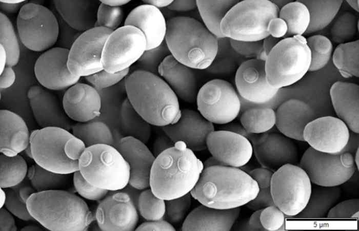
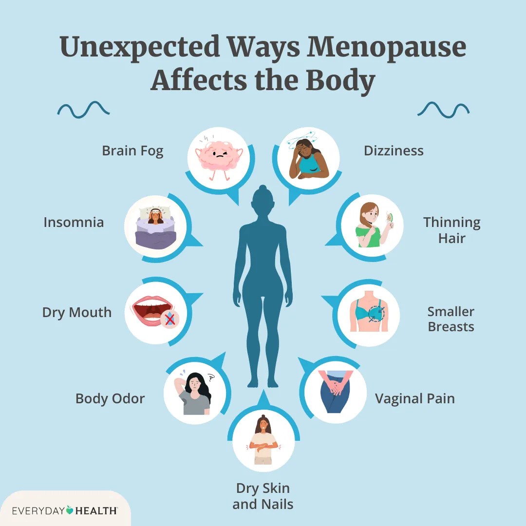
Category: Women’s Health

  • PCOS Diagnosis Delays Adult Years

    PCOS Diagnosis Delays Adult Years

    How could I not know that I had PCOS until adulthood? This journey explores the often-delayed diagnosis of Polycystic Ovary Syndrome (PCOS) in women. We’ll delve into why diagnosis might be delayed, the impact of this delay on health outcomes, and strategies for improving early detection.

    Many women experience a range of symptoms, some subtle, that can be easily dismissed as other issues. This can lead to years of misdiagnosis or a lack of understanding about the underlying condition. This article aims to shed light on the common symptoms, potential reasons for delayed diagnosis, and the importance of proactive health management.

    Understanding the Delays in PCOS Diagnosis in Adults

    Discovering polycystic ovary syndrome (PCOS) in adulthood can often be a frustrating experience, marked by a sense of bewilderment and a feeling of having been misdiagnosed or simply misunderstood. This is often due to the complex and sometimes subtle nature of PCOS symptoms, and the historical limitations in diagnosis and treatment approaches. The good news is that with a better understanding of the condition and improved diagnostic tools, individuals can now seek more accurate and timely support.PCOS is a hormonal disorder that primarily affects women of reproductive age, characterized by a constellation of symptoms that can vary greatly from person to person.

    The diagnostic journey, however, can be significantly impacted by the fact that symptoms may not manifest in a uniform way, and the condition can be mistaken for other health issues. This delay in diagnosis often leads to complications such as delayed fertility management, increased risk of metabolic disorders, and potential long-term health implications.

    Historical Overview of PCOS Diagnosis and Treatment

    Historically, the understanding of PCOS was limited, leading to difficulties in accurate diagnosis. Early approaches often focused on the presence of irregular periods and ovarian cysts, without a comprehensive understanding of the underlying hormonal imbalances. Treatment options were often rudimentary and focused primarily on managing symptoms rather than addressing the root cause of the condition. This lack of comprehensive understanding meant that many women experienced years of misdiagnosis and inadequate treatment.

    Common Symptoms of PCOS in Adults

    The symptoms of PCOS can be diverse and often overlap with other conditions. Common symptoms in adults include irregular menstrual cycles, excess hair growth (hirsutism), acne, weight gain, and skin changes. However, some symptoms might be subtle or easily dismissed, particularly in adulthood. For example, fatigue, mood swings, and thinning hair can be indicative of PCOS, but are also frequently associated with other factors like stress, lifestyle changes, or other health issues.

    Potential Reasons for Misattribution of PCOS Symptoms

    Many symptoms of PCOS are also associated with other conditions, leading to misdiagnosis. For instance, fatigue and mood swings are common in various medical conditions, making it challenging to pinpoint PCOS as the underlying cause. Similarly, weight gain can result from a number of factors, including poor diet, lack of exercise, or underlying medical conditions. Furthermore, the non-specific nature of some symptoms makes it difficult to differentiate PCOS from other potential causes.

    Influence of Lifestyle Factors on PCOS Symptom Presentation

    Lifestyle factors play a crucial role in the presentation and severity of PCOS symptoms. Diet rich in processed foods, lack of physical activity, and chronic stress can exacerbate the symptoms, while a balanced diet, regular exercise, and stress management techniques can improve well-being and help manage the condition.

    Comparison of PCOS Symptoms in Adolescents vs. Adults

    Symptom Adolescents Adults
    Menstrual Irregularities Often marked by absence or significant delay of menstruation. May manifest as irregular cycles, prolonged periods, or absence of menstruation.
    Hirsutism May be subtle, appearing as increased hair growth in certain areas. More pronounced hair growth, potentially affecting various body parts.
    Acne Common, often associated with hormonal fluctuations. May persist or worsen, potentially requiring medical intervention.
    Weight Gain Can be linked to puberty-related hormonal changes. Increased weight gain, particularly around the abdomen, might be a concern.
    Metabolic Issues May not be as pronounced as in adulthood. Higher risk of developing insulin resistance, type 2 diabetes, and cardiovascular disease.

    This table illustrates the potential differences in symptom presentation between adolescents and adults, emphasizing the importance of understanding the diverse ways PCOS can manifest.

    Factors Contributing to Delayed Diagnosis

    How could i not know that i had pcos until adulthood

    Navigating the complexities of health issues, especially those with subtle symptoms, can be challenging. Delayed diagnosis, particularly for conditions like Polycystic Ovary Syndrome (PCOS), often stems from a confluence of factors that can make recognizing and addressing the problem difficult. Understanding these factors is crucial for empowering women to advocate for their health and for healthcare providers to improve their diagnostic practices.Delayed PCOS diagnosis in adulthood is a multifaceted issue, with healthcare access, societal expectations, and a lack of awareness all playing significant roles.

    These factors intertwine to create a complex environment that often leads to delayed intervention and potentially negative health outcomes.

    Healthcare Access and Affordability

    Limited access to healthcare, particularly in underserved communities or for those with financial constraints, is a major barrier to timely PCOS diagnosis. High medical costs, lack of insurance coverage, or geographical limitations can prevent individuals from seeking necessary testing and specialist consultations. This can result in a delay in receiving a proper diagnosis, potentially prolonging the duration of symptoms and complications.

    Seriously, how could I not know I had PCOS until adulthood? It’s baffling, isn’t it? My symptoms were subtle, like a persistent feeling of fatigue or unusual weight gain. Sometimes, similar issues can be related to other health conditions, such as those involving unresectable tumors. Learning about the reasons behind these types of tumors and the various treatment options can offer valuable insights.

    Unresectable tumors reasons treatment are complex, but understanding them can lead to a more holistic understanding of how health conditions can manifest. Ultimately, it just highlights how easily PCOS can go unnoticed, even in adulthood.

    For example, a young woman living in a rural area with limited transportation options might find it challenging to access specialist care for her suspected PCOS symptoms, leading to a delay in diagnosis.

    Societal Expectations and Gender Roles

    Societal expectations and gender roles often influence women’s health-seeking behaviors. Traditional gender roles may discourage women from prioritizing their health needs or from vocalizing concerns to healthcare providers. The expectation that women should manage symptoms or “tough it out” can prevent them from seeking medical attention, delaying the diagnosis of underlying conditions like PCOS. Cultural norms and family expectations can further exacerbate this issue.

    Lack of Awareness Among Healthcare Providers, How could i not know that i had pcos until adulthood

    Insufficient awareness of PCOS symptoms among healthcare providers can lead to delayed diagnosis. Healthcare providers may not recognize the subtle or diverse presentation of PCOS symptoms, which can vary greatly between individuals. This lack of awareness can result in misdiagnosis or a dismissal of the patient’s concerns. This is compounded by the evolving nature of symptoms and the fact that many initial symptoms may not be directly associated with PCOS.

    This highlights the need for continuous medical education to enhance provider awareness and improve diagnostic accuracy.

    Cultural and Family Factors

    Cultural and family factors can significantly impact a woman’s health-seeking behavior. Cultural beliefs, family dynamics, and personal experiences can influence a woman’s willingness to discuss health concerns with healthcare professionals. For example, in some cultures, discussing reproductive health issues may be considered taboo or inappropriate, leading women to hesitate to seek medical advice. Furthermore, the family’s response to the patient’s concerns can significantly impact her decision to pursue further testing or treatment.

    Barriers to Early PCOS Diagnosis

    Category Specific Barrier Impact on Diagnosis
    Healthcare Access Limited access to specialists, lack of insurance coverage, high medical costs Delayed or prevented diagnosis due to financial or geographical constraints.
    Societal Expectations Gender roles, cultural norms discouraging women from seeking care Women may be hesitant to prioritize their health concerns.
    Provider Awareness Lack of awareness of PCOS symptoms, misdiagnosis, dismissal of concerns Leads to inaccurate or delayed diagnoses, hindering appropriate treatment.
    Cultural and Family Factors Cultural taboos, family dynamics influencing health-seeking behavior Creates a barrier to open communication and proper medical consultation.
    Patient Factors Lack of awareness of PCOS symptoms, fear of diagnosis, difficulty communicating symptoms Women may not recognize the need for medical attention or may hesitate to bring concerns to their healthcare provider.

    Impact of Delayed Diagnosis on Health Outcomes

    How could i not know that i had pcos until adulthood

    Delayed diagnosis of PCOS can have a significant and far-reaching impact on a woman’s overall health and well-being. The consequences of not receiving timely and accurate diagnoses extend beyond simply feeling unwell; they can contribute to long-term health problems and affect various aspects of life, particularly reproductive health and metabolic function. Understanding these impacts is crucial for raising awareness and advocating for early diagnosis.The long-term health consequences of delayed PCOS diagnosis are substantial.

    Unrecognized and untreated PCOS can lead to a cascade of issues, impacting various organ systems and increasing the risk of developing chronic conditions. The longer the condition goes undiagnosed, the more significant the potential negative impact on a woman’s health.

    Reproductive Health Consequences

    Delayed diagnosis can have severe implications for reproductive health. Women with undiagnosed PCOS may experience irregular periods, leading to difficulties in predicting ovulation and making it challenging to conceive. The hormonal imbalances associated with PCOS can also increase the risk of complications during pregnancy, such as miscarriage and gestational diabetes. Furthermore, persistent anovulation (lack of ovulation) can lead to infertility.

    The impact on a woman’s ability to achieve a healthy pregnancy is a critical aspect of the long-term consequences of delayed diagnosis.

    Metabolic Health Complications

    PCOS is strongly linked to metabolic disorders, and delayed diagnosis exacerbates this risk. The hormonal imbalances associated with PCOS can disrupt insulin regulation, increasing the risk of developing type 2 diabetes. Furthermore, elevated androgens can contribute to high blood pressure and cholesterol levels, increasing the risk of cardiovascular disease, such as heart disease. Women with PCOS who have been undiagnosed for an extended period are at greater risk for developing these serious metabolic complications.

    For example, a woman experiencing insulin resistance due to undiagnosed PCOS over a decade could develop pre-diabetes or even full-blown type 2 diabetes.

    Psychological Impact

    The emotional toll of living with undiagnosed PCOS can be substantial. Symptoms such as irregular periods, acne, and excess hair growth can significantly impact a woman’s self-esteem and body image. The uncertainty and frustration associated with undiagnosed symptoms can lead to anxiety and depression. Furthermore, the difficulties in conceiving or managing symptoms can cause significant stress and emotional distress.

    A prolonged period of experiencing these symptoms without understanding their cause can lead to feelings of isolation and hopelessness.

    Seriously, how could I not know I had PCOS until adulthood? My symptoms were always there, subtle at first, like those hormonal fluctuations that sometimes made me wonder if I was just having a bad day. Learning about the best time to take collagen, for example, taught me about how much my body was going through and the importance of understanding my own unique needs.

    It’s amazing how many different things can impact us, and I guess it’s also possible that those subtle symptoms were simply dismissed as something normal. Still, it’s a bit mind-boggling that I went this long without a proper diagnosis.

    Summary of Health Risks Associated with Delayed PCOS Diagnosis

    Health Risk Category Specific Risks Impact of Delayed Diagnosis
    Reproductive Health Irregular periods, difficulty conceiving, increased risk of miscarriage, and infertility Prolonged difficulties in planning pregnancies, increased risk of pregnancy complications, and potential long-term infertility.
    Metabolic Health Insulin resistance, type 2 diabetes, high blood pressure, high cholesterol, and cardiovascular disease Increased risk of developing serious chronic diseases later in life.
    Psychological Health Low self-esteem, body image issues, anxiety, and depression Negative impact on mental well-being and quality of life.

    Strategies for Improving Early PCOS Diagnosis

    Discovering you have PCOS in adulthood can be frustrating and challenging, but early diagnosis is key to managing its symptoms and preventing long-term health complications. This requires a multifaceted approach that focuses on empowering women with knowledge, equipping healthcare providers with awareness, and creating accessible resources.Improving early diagnosis necessitates a shift in how PCOS is approached, moving beyond a reactive model to a proactive one that anticipates and addresses potential risks.

    This involves equipping both women and their healthcare providers with the tools and information necessary for early detection.

    Improving Women’s Health Literacy Regarding PCOS

    Understanding the complexities of PCOS is crucial for women to take an active role in their health. This involves providing clear and accessible information about the condition. Educational resources should go beyond basic definitions, delving into the various symptoms, potential long-term complications, and management strategies. Interactive websites, informative pamphlets, and community support groups can play a vital role in fostering this understanding.

    Furthermore, workshops and seminars can provide hands-on learning opportunities and support systems.

    Increasing Awareness Among Healthcare Providers About PCOS Symptoms and Diagnostic Criteria

    Healthcare providers are often the first point of contact for women experiencing PCOS symptoms. Thus, it’s essential that they are equipped with the necessary knowledge to recognize and diagnose the condition early. This can be achieved through continuing medical education programs that emphasize the subtle and diverse presentations of PCOS. These programs should incorporate case studies and practical exercises, reinforcing the importance of a comprehensive approach to patient history and physical examination.

    Furthermore, diagnostic guidelines and updated research should be disseminated effectively to ensure providers are using the most current and accurate diagnostic criteria.

    Resources for Women to Find Reliable Information About PCOS

    Reliable and accurate information is vital for women seeking to understand and manage their health. Creating a central hub for trustworthy resources, such as a dedicated website or app, can streamline access to information. This platform could include articles, videos, and interactive tools, allowing women to learn about symptoms, treatments, and support options. Collaborations with reputable medical organizations and patient advocacy groups can ensure the accuracy and relevance of the provided resources.

    Developing a Comprehensive Health History Questionnaire that Screens for PCOS Symptoms

    A well-designed questionnaire can significantly aid in identifying women at risk for PCOS. The questionnaire should cover a broad range of potential symptoms, including menstrual irregularities, acne, excessive hair growth, weight gain, and sleep disturbances. The questionnaire should also inquire about family history of PCOS and related conditions, as this can be a valuable indicator. The use of validated questionnaires, such as those employed in research studies, can ensure the accuracy and effectiveness of the screening tool.

    Figuring out I had PCOS in adulthood was a real head-scratcher. It’s amazing how many things can go unnoticed, especially when symptoms are subtle. Perhaps if I’d been more proactive with things like breast cancer testing screening , I might have picked up on some of the underlying hormonal imbalances earlier. Still, the mystery of how I could have missed such a significant issue for so long continues to intrigue me.

    Example questions could include: “Have you experienced irregular menstrual cycles?” or “Have you noticed any increase in hair growth in recent years?”.

    Designing a Flow Chart Illustrating the Steps Involved in Diagnosing PCOS

    A clear and concise flow chart can streamline the diagnostic process for both women and healthcare providers. The chart should visually represent the sequential steps involved, from initial consultation and symptom evaluation to diagnostic testing and subsequent management. It should emphasize the importance of a thorough medical history, physical examination, and appropriate laboratory tests. This visual aid can help healthcare providers understand the diagnostic process and provide a framework for effective communication with patients.

    Step Action
    1 Gather detailed medical history, including menstrual history, family history, and current symptoms.
    2 Conduct a physical examination, including assessing for signs of hyperandrogenism (e.g., acne, hirsutism).
    3 Order relevant laboratory tests, including hormone levels (e.g., testosterone, LH, FSH, androstenedione), fasting glucose and lipid profile, and possibly ultrasound.
    4 Interpret test results and determine if the criteria for PCOS are met based on the Rotterdam criteria.
    5 Discuss the diagnosis and management plan with the patient.

    Illustrative Case Studies (Hypothetical)

    Discovering PCOS, often a journey of self-discovery and medical exploration, can be significantly impacted by the timing of diagnosis. Sometimes, the delay is substantial, leading to years of discomfort and uncertainty before a proper understanding of the condition is achieved. These hypothetical case studies aim to illustrate different scenarios, highlighting the importance of early diagnosis and effective treatment.

    A Case of Delayed Diagnosis

    Sarah, a 30-year-old woman, had experienced irregular periods, acne, and weight gain since her teenage years. Initially, she attributed these symptoms to stress and a fast-paced lifestyle. She sought medical attention only when experiencing severe fatigue and persistent ovarian cysts. By this point, the condition had already impacted her overall well-being and reproductive health. Her delay in seeking diagnosis meant that treatment was more complex, and some long-term effects might be harder to reverse.

    Misdiagnosis and the Impact

    Emily, a 25-year-old woman, experienced symptoms characteristic of PCOS, including hirsutism, acne, and insulin resistance. However, her initial doctor visits focused primarily on her acne, and she was prescribed topical treatments. Over time, her symptoms worsened, and she struggled with fertility issues. Only when she consulted an endocrinologist specializing in hormonal disorders was she diagnosed with PCOS and put on a comprehensive treatment plan.

    This situation highlights how a misdiagnosis can lead to a worsening of symptoms and a delay in appropriate treatment.

    The Power of Early Diagnosis

    Imagine a 22-year-old woman, Ashley, who experienced irregular periods and some acne in her early twenties. Upon seeking medical attention, she was diagnosed with PCOS promptly. She immediately began a combination of lifestyle changes, including a balanced diet and regular exercise, and medication to manage her hormonal imbalances. This proactive approach allowed her to effectively manage her symptoms, preventing further complications and potentially preserving her fertility.

    Her early diagnosis significantly improved her quality of life and her overall health.

    The Ripple Effect of Awareness

    A growing awareness of PCOS has empowered many women to seek early medical attention for their symptoms. Through online support groups and increased media coverage, individuals are now more likely to recognize and discuss the symptoms, ultimately leading to earlier diagnosis and better outcomes. This increased awareness has fostered a culture of open communication and proactive healthcare choices.

    Summary of Case Studies:* Delayed Diagnosis: Delayed symptom recognition and seeking medical attention can lead to more complex and potentially less effective treatment.

    Misdiagnosis

    Symptoms resembling PCOS can be mistaken for other conditions, leading to delayed diagnosis and ineffective treatment.

    Early Diagnosis

    Early diagnosis allows for proactive management of symptoms and potential prevention of long-term complications.

    Increased Awareness

    Growing awareness about PCOS empowers individuals to seek medical attention and promotes earlier diagnosis, leading to better outcomes.

    Wrap-Up: How Could I Not Know That I Had Pcos Until Adulthood

    Ultimately, the path to early PCOS diagnosis involves a combination of increased awareness among women, improved education for healthcare providers, and readily available resources for women to understand their bodies and seek support. By understanding the nuances of PCOS, we can empower women to take control of their health and well-being.

  • Turmeric Health Benefits for Women A Deep Dive

    Turmeric Health Benefits for Women A Deep Dive

    Turmeric health benefits for women are plentiful and fascinating, offering a natural approach to overall well-being. From hormonal balance to digestive health, bone strength, skin radiance, and immune support, turmeric’s active compound, curcumin, appears to play a key role in various aspects of women’s health. This exploration delves into the science behind turmeric’s effects, highlighting potential benefits, safe consumption practices, and practical ways to incorporate this vibrant spice into your daily life.

    This comprehensive guide will cover the historical use of turmeric, its potential impact on hormonal regulation, digestive health, bone health, skin health, and immune function in women. We’ll also examine safety considerations and provide delicious recipes to help you easily incorporate this powerful spice into your diet.

    Introduction to Turmeric and Women’s Health

    Turmeric, a vibrant yellow spice, has been a cornerstone of Ayurvedic medicine and various culinary traditions for centuries. Its use stretches back through history, prized not only for its flavour but also for its potential health benefits. From vibrant curry dishes to traditional remedies, turmeric’s presence speaks volumes about its cultural significance. Beyond its culinary appeal, the active compound curcumin is gaining recognition for its potential to support women’s health.Turmeric’s impressive health profile stems primarily from curcumin, a powerful antioxidant and anti-inflammatory agent.

    Research suggests that curcumin’s properties can contribute to overall well-being, impacting various aspects of health. This includes the potential to support hormonal balance, reduce inflammation, and promote overall health and vitality. The specific ways in which turmeric can positively influence women’s health are being actively explored and studied.

    Turmeric’s General Health Benefits

    Turmeric’s beneficial effects are largely attributed to its active compound, curcumin. Curcumin’s potent antioxidant properties combat free radicals, protecting cells from damage. Its anti-inflammatory effects can potentially alleviate pain and discomfort associated with various conditions. Numerous studies are investigating the therapeutic potential of curcumin in various areas of health.

    Potential Benefits of Turmeric for Women

    Turmeric’s potential benefits for women encompass several areas of well-being. It may play a role in maintaining hormonal balance, which can be crucial for women throughout their lives. Furthermore, turmeric’s anti-inflammatory properties may help manage common discomforts associated with hormonal fluctuations and other health conditions. The compound may also contribute to overall well-being, potentially supporting energy levels and mood.

    Key Components and Potential Effects on Women’s Health

    Understanding the key components of turmeric and their potential effects on women’s health is crucial for a comprehensive understanding.

    Turmeric is amazing for women’s health, offering potent anti-inflammatory properties. Understanding how your body processes nutrients, like curcumin from turmeric, can be key to maximizing its benefits. A deeper look into how your body interacts with this spice might be as simple as taking a DNA test to see how your body metabolizes nutrients. What is a DNA test could help you understand if you’re absorbing turmeric’s health benefits effectively.

    Ultimately, knowing your unique genetic makeup could guide you to tailor your diet and lifestyle to enhance your experience with turmeric and overall well-being.

    Component Potential Effects Scientific Backing
    Curcumin Potential to support hormonal balance, reduce inflammation, antioxidant protection, potentially improve mood and energy levels. Numerous studies exploring the anti-inflammatory and antioxidant properties of curcumin. Further research into specific hormonal effects is ongoing.
    Other Phytochemicals May contribute to the overall health benefits by providing additional antioxidant and anti-inflammatory support. Studies on the synergistic effects of turmeric’s various components are ongoing, highlighting the potential role of these additional compounds.
    Ginger Potential to aid digestion and reduce nausea; may support overall well-being, particularly during pregnancy or menstruation. Scientific literature exists supporting ginger’s digestive benefits, but further research into turmeric-ginger combinations is warranted.

    Turmeric’s Impact on Hormonal Balance

    Turmeric health benefits for women

    Turmeric, a vibrant spice known for its golden hue, boasts a rich history of medicinal use. Beyond its culinary appeal, research suggests that turmeric may play a role in modulating hormonal balance, particularly in women. This influence is intricately linked to its anti-inflammatory properties, which can significantly impact various hormonal processes. Understanding this connection is key to comprehending turmeric’s potential benefits for women experiencing hormonal fluctuations throughout their lives.Turmeric’s potential impact on hormonal regulation stems from its bioactive compounds, primarily curcumin.

    Curcumin possesses potent anti-inflammatory properties. Chronic inflammation is a known contributor to hormonal imbalances, impacting the production and regulation of hormones like estrogen and progesterone. By reducing inflammation, turmeric may help restore hormonal equilibrium and alleviate associated symptoms. This effect is not a guaranteed cure, but a potential pathway for women to explore in conjunction with other healthcare strategies.

    Inflammation’s Role in Hormonal Imbalances

    Chronic inflammation can disrupt the delicate balance of hormones. Inflammation can affect the endocrine system, which is responsible for hormone production and regulation. This disruption can lead to a range of symptoms, including irregular periods, mood swings, and fatigue. Managing inflammation through natural remedies like turmeric may help mitigate these symptoms.

    Potential Benefits for Managing Hormonal Fluctuations

    Turmeric may offer potential benefits for managing symptoms associated with menstruation, menopause, and other hormonal fluctuations. For example, premenstrual syndrome (PMS) symptoms, including bloating, breast tenderness, and mood swings, may be potentially alleviated. Similarly, menopausal symptoms, such as hot flashes, night sweats, and mood changes, might be softened. However, more research is needed to fully understand the extent of these benefits.

    Turmeric Consumption Methods and Potential Impacts

    Different methods of turmeric consumption may have varying effects on hormone regulation. The bioavailability of curcumin, the active compound, can differ, impacting its effectiveness.

    Method Potential Impact Potential Side Effects
    Turmeric powder May provide a more varied source of nutrients. The effect may depend on the preparation method. Possible digestive discomfort in some individuals.
    Turmeric capsules Often standardized for curcumin content, potentially providing a more consistent dose. May cause gastrointestinal upset, particularly if taken in high doses. Individual sensitivities vary.
    Turmeric tea May be a gentler way to introduce turmeric into the diet. The effect depends on the preparation method. Possible digestive issues. Interactions with medications are possible. Individual sensitivities vary.
    Turmeric supplements (with other ingredients) Combination of ingredients can have a synergistic effect, potentially increasing efficacy. Potential for adverse interactions with other medications. Varying ingredient combinations could lead to unpredictable results.

    Turmeric for Women’s Digestive Health: Turmeric Health Benefits For Women

    Turmeric, with its vibrant golden hue, offers more than just a delicious culinary addition. Emerging research suggests its potential to support healthy digestion in women, addressing common issues like bloating and discomfort. Its active compound, curcumin, possesses potent anti-inflammatory properties that can soothe the digestive tract and potentially alleviate symptoms associated with various digestive conditions.Turmeric’s anti-inflammatory effects can play a crucial role in maintaining digestive well-being.

    Chronic inflammation in the digestive system can contribute to various issues, including irritable bowel syndrome (IBS) and other digestive disorders. Curcumin’s ability to reduce inflammation may help ease discomfort and improve overall digestive function. This is a significant factor for women, as they are often more susceptible to digestive issues due to hormonal fluctuations and other lifestyle factors.

    Potential Benefits of Turmeric for Digestive Health

    Turmeric’s potential benefits for digestive health stem from its anti-inflammatory properties. Curcumin, the active compound in turmeric, can help reduce inflammation in the digestive tract, which is often a root cause of discomfort and pain. This can lead to improved motility, reducing bloating and gas, and promoting a healthier gut environment. Preliminary research indicates a potential link between turmeric consumption and improved digestion, particularly in women.

    Alleviating Digestive Symptoms, Turmeric health benefits for women

    Turmeric may potentially alleviate symptoms associated with common digestive issues like bloating and irritable bowel syndrome (IBS). Bloating, often accompanied by discomfort and gas, can be significantly affected by inflammation. By reducing inflammation, turmeric may contribute to a reduction in bloating and discomfort. Similarly, for IBS, where inflammation plays a role in symptoms like cramping and abdominal pain, turmeric’s anti-inflammatory action could offer some relief.

    However, it’s crucial to remember that turmeric is not a cure for these conditions. Consulting with a healthcare professional is essential for proper diagnosis and management.

    Comparison with Other Natural Remedies

    Remedy Potential Benefits Potential Drawbacks
    Turmeric Potential reduction in inflammation, improved digestion, potential relief from bloating and IBS symptoms. May interact with certain medications, not a cure for digestive conditions, individual responses may vary.
    Ginger May soothe digestive upset, aid in reducing nausea, and potentially improve digestion. May cause heartburn in some individuals, potential for interactions with certain medications.
    Probiotics Support the growth of beneficial bacteria in the gut, potentially improving digestion and reducing inflammation. May cause digestive discomfort in some individuals, not suitable for all conditions.
    Chamomile Known for its calming effects, may help reduce anxiety and stress, which can sometimes contribute to digestive issues. Limited direct impact on digestion itself, not suitable for all digestive conditions.

    Note: This table provides a general comparison. Individual responses to these remedies may vary. Consulting with a healthcare professional is crucial for personalized advice.

    Turmeric for Women’s Bone Health

    Turmeric, a vibrant spice with a rich history of culinary and medicinal use, has increasingly captivated the attention of health enthusiasts. Its potential benefits extend beyond culinary applications, encompassing various aspects of well-being. This exploration delves into the possible role of turmeric in supporting women’s bone health, focusing on its potential interaction with inflammation and its impact on bone density as women age.Turmeric’s active compound, curcumin, possesses potent anti-inflammatory properties.

    Inflammation, while a necessary bodily response, can become chronic and detrimental. Chronic inflammation has been linked to a variety of health issues, including weakened bones. Curcumin’s anti-inflammatory action may offer a protective mechanism for bone health by mitigating this chronic inflammation. This mitigation may translate into improved bone density and strength, particularly crucial for women as they age and experience hormonal shifts that can affect bone health.

    The Link Between Inflammation and Bone Health

    Bone health is a complex interplay of various factors. Inflammation plays a significant role in this process. Chronic inflammation can disrupt the delicate balance of bone formation and resorption. Osteoclasts, the cells responsible for bone resorption, are stimulated by inflammatory signals. Conversely, osteoblasts, the cells responsible for bone formation, are inhibited.

    This imbalance can lead to a net loss of bone mass, resulting in weaker bones and increased risk of fractures.

    How Turmeric May Support Bone Density and Strength

    Studies suggest that curcumin, the active component in turmeric, may support bone health in several ways. It can potentially regulate the activity of osteoclasts and osteoblasts, helping to maintain a healthy balance in bone remodeling. Furthermore, curcumin’s antioxidant properties can protect bone cells from oxidative stress, a process that can contribute to bone loss. This protective mechanism is particularly important as women age and experience hormonal changes.

    For instance, post-menopausal women are at higher risk of osteoporosis, and curcumin may offer a natural approach to supporting bone health during this phase.

    Turmeric boasts impressive health benefits for women, particularly its anti-inflammatory properties. While recent research is exploring its potential role in various conditions, including potential links to skin health, it’s important to remember that the connection between turmeric and eczema, or the relationship between turmeric and the Covid vaccine’s impact on eczema, is still being studied. For a deeper dive into the connection between eczema and the Covid vaccine, check out this informative article eczema and covid vaccine.

    Ultimately, turmeric continues to show promise as a natural remedy with potential benefits for women’s health.

    Key Nutrients and Vitamins for Women’s Bone Health and Turmeric’s Contribution

    Maintaining optimal bone health requires a multifaceted approach, focusing on a balanced diet rich in essential nutrients. The following table highlights key nutrients and vitamins critical for bone health and how turmeric might contribute to their intake.

    Nutrient/Vitamin Importance Turmeric’s Role
    Calcium Essential for bone structure and strength. Turmeric does not directly provide calcium but can enhance the absorption of calcium from the diet.
    Vitamin D Crucial for calcium absorption and bone mineralization. Turmeric may not directly provide vitamin D but can contribute to overall well-being, indirectly supporting vitamin D absorption and function.
    Vitamin K Essential for bone metabolism and blood clotting. Turmeric may indirectly support vitamin K intake through a healthy diet rich in vegetables, which are often a good source of vitamin K.
    Magnesium Plays a role in bone formation and calcium metabolism. Turmeric’s presence in a balanced diet may contribute to magnesium intake.
    Protein Essential for bone matrix formation. Turmeric is not a primary source of protein but its inclusion in a balanced diet can support adequate protein intake.

    Turmeric for Women’s Skin Health

    Turmeric, a vibrant spice with a rich history in culinary and medicinal practices, is increasingly recognized for its potential benefits beyond the kitchen. Its bioactive compounds, particularly curcumin, possess potent anti-inflammatory and antioxidant properties, making it an intriguing ingredient for skincare. This exploration delves into how turmeric might support healthy skin in women, addressing its potential role in improving skin conditions.Turmeric’s remarkable impact on skin health stems from its ability to combat oxidative stress and inflammation.

    Oxidative stress, a process triggered by free radicals, can damage skin cells, leading to premature aging and various skin conditions. Curcumin, the active compound in turmeric, acts as a powerful antioxidant, neutralizing free radicals and protecting skin from damage. Simultaneously, turmeric’s anti-inflammatory properties can soothe irritated skin, potentially reducing redness and inflammation associated with conditions like acne and eczema.

    Turmeric’s Anti-inflammatory and Antioxidant Effects on Skin

    Turmeric’s potent anti-inflammatory and antioxidant effects can demonstrably benefit skin health. Curcumin, the primary active component in turmeric, has been shown to inhibit the production of inflammatory mediators, thereby reducing redness and irritation. This anti-inflammatory action can potentially alleviate symptoms associated with skin conditions such as acne and eczema. The antioxidant properties of turmeric help protect skin cells from damage caused by free radicals, which are linked to premature aging and various skin concerns.

    The combination of these properties may lead to improved skin tone, texture, and overall health.

    Potential Benefits for Addressing Skin Conditions

    Turmeric may offer potential benefits for various skin conditions, although more research is needed. For acne, turmeric’s anti-inflammatory properties could help reduce inflammation and redness associated with the condition. Similarly, for eczema, turmeric’s anti-inflammatory action could potentially alleviate the itching, redness, and dryness often associated with this condition. While promising, further research is necessary to fully understand the efficacy and optimal application methods for turmeric in treating specific skin conditions.

    Turmeric-Based Skincare Products vs. Conventional Treatments

    Product Type Benefits Potential Drawbacks
    Turmeric-based face masks Potentially reduces inflammation, improves skin tone, and promotes antioxidant protection. May not be suitable for all skin types, and some individuals may experience skin irritation. Efficacy can vary based on turmeric concentration and formulation.
    Turmeric-infused serums Can potentially improve skin texture and firmness, and reduce signs of aging. May not be as effective as topical treatments for severe skin conditions. Absorption of turmeric compounds through the skin may be limited.
    Turmeric-containing moisturizers Can provide hydration and antioxidant support, potentially improving skin elasticity. May have a slightly yellow tint, which could be a cosmetic concern for some individuals. May require a gradual introduction to the skin due to potential sensitivity.
    Conventional treatments (e.g., topical corticosteroids, retinoids) Often effective for addressing specific skin conditions, such as acne and eczema. May have side effects such as skin irritation, dryness, or thinning, and are often expensive. Can be less suitable for long-term use.

    Turmeric and Women’s Immunity

    Turmeric, a vibrant spice with a rich history, has captivated cultures worldwide for its potential health benefits. Beyond its culinary uses, turmeric’s bioactive compounds, particularly curcumin, are increasingly recognized for their positive impact on various aspects of women’s health, including immunity. This exploration delves into the potential of turmeric to bolster women’s immune systems, emphasizing its anti-inflammatory properties and role in overall well-being.Turmeric’s potent anti-inflammatory properties are key to its potential immune-boosting effects.

    Chronic inflammation is linked to various health concerns, and curcumin, the active compound in turmeric, has demonstrated the ability to reduce inflammation in the body. By mitigating inflammation, turmeric may support the immune system’s ability to effectively respond to threats and maintain overall health. This is particularly relevant for women, as they may face unique hormonal fluctuations and lifestyle factors that can impact immune function.

    Turmeric’s Anti-inflammatory Action on Immunity

    Curcumin, the primary active compound in turmeric, has shown promising effects on immune function. Studies suggest that it can modulate the inflammatory response, helping to prevent overactivation of the immune system, which can lead to chronic inflammation. By reducing inflammation, turmeric may help support the body’s natural defenses and improve overall immune response. This is important for women as it may contribute to a more resilient immune system during periods of hormonal changes or stress.

    Turmeric boasts amazing health benefits for women, particularly its potent anti-inflammatory properties. While it’s fantastic for overall well-being, sometimes, a little eye irritation can pop up, like pink eye. Fortunately, using the right eye drops for pink eye, like those detailed on this page eye drops for pink eye , can help soothe the discomfort. Despite these occasional eye issues, turmeric continues to be a valuable addition to a healthy lifestyle for women.

    Further research is necessary to fully understand the intricate mechanisms involved.

    Dietary Turmeric Incorporation for Enhanced Immunity

    A balanced diet rich in immune-supporting foods, including turmeric, is crucial for women’s overall well-being. Here are several ways to incorporate turmeric into your daily diet to potentially enhance immunity:

    Method Benefits Potential Side Effects
    Curcumin Supplements Targeted delivery of curcumin, potentially higher bioavailability compared to direct consumption. Supplements can be beneficial for individuals who may find it difficult to consume sufficient amounts of turmeric through food alone. Some individuals may experience digestive discomfort, such as gas or heartburn. Consult with a healthcare professional before starting any supplement regimen, especially if you have existing health conditions or are taking other medications.
    Turmeric Tea Easy and enjoyable way to consume turmeric. Turmeric tea can be prepared with other immune-boosting ingredients like ginger or lemon. Possible mild digestive upset in some individuals. Avoid using excessive amounts of black pepper, as it can increase the potency of turmeric and lead to discomfort.
    Turmeric-Spiced Dishes Incorporating turmeric into everyday meals enhances the flavor and nutritional value of the dish while providing potential immune benefits. Potential allergic reactions in individuals sensitive to spices. Monitor your body’s response to new food combinations.
    Turmeric Milk (Haldi Doodh) Traditional recipe from South Asia, often consumed for its potential health benefits, including supporting the immune system. Possible milk intolerance in some individuals. Use milk alternatives like almond or soy milk for those with sensitivities.

    “While promising, further research is needed to fully elucidate the complex mechanisms and specific dosages required to optimize the immune-boosting effects of turmeric for women.”

    Safety and Precautions for Turmeric Consumption

    Turmeric health benefits for women

    Turmeric, a vibrant spice renowned for its potential health benefits, can be a valuable addition to a balanced diet. However, like any substance, it’s crucial to understand its potential side effects and precautions to ensure safe and effective use. This section delves into the important considerations for women consuming turmeric, focusing on dosage, potential interactions, and safe incorporation into daily routines.Understanding the nuances of turmeric consumption is essential for maximizing its potential benefits while minimizing risks.

    This includes recognizing potential side effects, considering individual circumstances, and understanding interactions with other medications.

    Potential Side Effects and Contraindications

    Turmeric, while generally safe, can sometimes cause mild digestive issues like gas, bloating, or heartburn in some individuals. These side effects are usually temporary and resolve on their own. More serious side effects are rare but possible. For instance, some individuals might experience skin reactions, or in rare cases, liver problems, particularly with long-term or high-dose consumption.

    It’s crucial to monitor your body’s response to turmeric and consult a healthcare professional if you experience persistent or severe symptoms.

    Important Considerations for Women

    Women have unique physiological needs, and these must be considered when incorporating turmeric into their diets. Dosage recommendations should be personalized based on individual needs and health conditions. It’s important to note that pregnancy and breastfeeding may require special considerations.

    Dosage and Frequency

    The recommended dosage of turmeric varies depending on the intended use and individual health needs. While some studies suggest benefits at low doses, higher doses may be necessary for specific conditions. It’s crucial to consult a healthcare professional for personalized recommendations. It’s essential to avoid self-treating with turmeric or exceeding recommended dosages.

    Interactions with Medications

    Turmeric can interact with certain medications commonly used by women. For instance, turmeric can affect blood clotting and might interact with blood-thinning medications. Similarly, turmeric may interact with certain diabetes medications. It’s essential to disclose turmeric use to healthcare providers, especially those prescribing medications.

    List of Potential Interactions

    • Blood-thinning medications (e.g., warfarin): Turmeric can increase the risk of bleeding when taken with blood thinners. A healthcare professional should be consulted before combining the two.
    • Diabetes medications (e.g., insulin, metformin): Turmeric may affect blood sugar levels. It’s essential to consult a healthcare professional to determine if turmeric is appropriate in combination with diabetes medications.
    • Anti-inflammatory medications (e.g., ibuprofen, naproxen): Turmeric has anti-inflammatory properties, and combining it with other anti-inflammatory medications might lead to an increased risk of side effects. Consult a healthcare provider for guidance.
    • Digestive medications (e.g., antacids): Turmeric can affect stomach acid levels, and combining it with certain digestive medications might affect their efficacy. Consult a doctor for any interactions.

    Safe Ways to Incorporate Turmeric

    Turmeric can be a delicious and nutritious addition to your diet. Incorporate it into your meals by adding it to curries, stir-fries, soups, or smoothies. Start with small amounts and gradually increase the dosage to observe your body’s response. It’s also important to choose high-quality turmeric supplements and ensure they are free of harmful additives or contaminants.

    Consider consuming turmeric with black pepper, as it enhances absorption. Always consult a healthcare professional before making any significant dietary changes.

    Turmeric Recipes and Dietary Recommendations

    Turmeric, with its vibrant hue and powerful health benefits, is more than just a spice; it’s a culinary treasure. Incorporating turmeric into your diet can be a delicious and effective way to enhance your well-being, particularly for women. This section delves into delicious recipes and practical dietary advice to help you reap the rewards of this golden spice.Adding turmeric to your daily routine doesn’t have to be complicated.

    Simple tweaks to existing meals and the creation of exciting new dishes can seamlessly integrate turmeric’s benefits into your lifestyle. This section provides both inspiration and guidance for making turmeric a regular part of a balanced diet.

    Delicious Turmeric Recipes

    Turmeric’s versatility shines through in a variety of recipes. From warming curries to vibrant smoothies, the possibilities are endless. These recipes not only taste fantastic but also provide a convenient way to consume turmeric’s beneficial compounds.

    • Golden Milk Latte: This creamy and comforting beverage is a fantastic way to start the day or unwind in the evening. Warm milk, turmeric powder, ginger, cinnamon, and a touch of honey create a soothing and nutritious drink. The antioxidants and anti-inflammatory properties make it a beneficial addition to any routine.
    • Turmeric Roasted Vegetables: Roasting vegetables with turmeric brings out their natural sweetness and adds a subtle warmth. Toss your favorite vegetables like broccoli, carrots, and sweet potatoes with turmeric, olive oil, and herbs for a healthy and flavorful side dish. This method enhances the bioavailability of the turmeric’s active compounds.
    • Turmeric Chicken Curry: This classic dish gets a healthy boost with the addition of turmeric. The vibrant spices and tender chicken create a flavorful and satisfying meal. Pairing it with brown rice or quinoa provides a complete and balanced nutritional profile.
    • Turmeric Smoothie Bowl: Blend frozen fruits, spinach, and turmeric with yogurt or milk for a nutritious and refreshing smoothie. Top it with granola, seeds, and nuts for a complete and energizing breakfast or snack. The smoothie bowl approach allows for easy customization to your preferences and dietary needs.

    Dietary Recommendations for Turmeric

    Integrating turmeric into a balanced diet requires careful consideration of portion sizes and overall dietary habits. These recommendations help you maximize the benefits while minimizing any potential drawbacks.

    • Start Gradually: Introduce turmeric gradually into your diet. Begin with small amounts in your favorite recipes and gradually increase the dosage as tolerated. This approach allows your body to adjust to the spice and avoids potential digestive discomfort.
    • Pair with Healthy Fats: Turmeric’s absorption is enhanced when consumed with healthy fats. Adding coconut oil, olive oil, or nuts to turmeric-infused dishes improves the body’s ability to utilize the beneficial compounds. This enhances the effectiveness of turmeric’s medicinal properties.
    • Consider Black Pepper: Black pepper contains piperine, a compound that significantly increases the bioavailability of curcumin (the active compound in turmeric). Adding a pinch of black pepper to your turmeric dishes can greatly enhance its effectiveness.
    • Listen to Your Body: Pay attention to how your body reacts to turmeric. If you experience any digestive issues, reduce the intake or adjust the preparation method. This ensures you’re consuming turmeric in a way that supports your overall well-being.

    Turmeric-Infused Recipe Table

    This table provides a quick overview of different turmeric recipes, highlighting their ingredients and potential benefits.

    Recipe Name Ingredients Benefits
    Golden Milk Latte Warm milk, turmeric powder, ginger, cinnamon, honey Antioxidant, anti-inflammatory, soothing, energizing
    Turmeric Roasted Vegetables Vegetables (broccoli, carrots, sweet potatoes), turmeric, olive oil, herbs Nutrient-rich, flavorful, healthy side dish, enhanced bioavailability of turmeric
    Turmeric Chicken Curry Chicken, turmeric, spices, coconut milk, vegetables Flavorful, satisfying, complete nutritional profile, potential anti-inflammatory benefits
    Turmeric Smoothie Bowl Frozen fruits, spinach, turmeric, yogurt/milk, granola, seeds, nuts Nutritious, refreshing, energizing, customizable to dietary needs

    Final Review

    In conclusion, turmeric offers a wealth of potential health benefits for women, acting as a natural ally in maintaining overall well-being. From supporting hormonal balance to promoting digestive health, bone strength, and radiant skin, curcumin’s remarkable properties make turmeric a promising addition to a holistic health routine. While the research is ongoing, the potential is significant, encouraging further exploration into the world of turmeric and its role in women’s health.

    Remember to consult with a healthcare professional before making significant dietary changes.

  • Ovarian Pain Causes and Treatment Explained

    Ovarian Pain Causes and Treatment Explained

    Ovary pain causes and treatment – Ovarian pain causes and treatment is a complex issue affecting many women. Understanding the intricacies of the female reproductive system, the various potential causes of discomfort, and the available treatment options is crucial for effective management. This exploration delves into the different types of ovarian pain, their potential origins, and the diagnostic methods used to pinpoint the cause. We’ll also discuss lifestyle modifications and when to seek immediate medical attention.

    This comprehensive guide aims to provide valuable insights for anyone experiencing ovarian pain.

    From understanding the nuanced symptoms of different conditions to exploring the efficacy of various treatment approaches, this article provides a detailed overview. We’ll cover common conditions like ovarian cysts, endometriosis, and pelvic inflammatory disease (PID), along with less common but still significant factors. The information presented is designed to be accessible and empowering, enabling you to make informed decisions about your health.

    Overview of Ovarian Pain

    Ovary pain causes and treatment

    Understanding ovarian pain requires a foundational knowledge of the female reproductive system. The ovaries are small, almond-shaped organs located on either side of the uterus, nestled within the pelvic cavity. Their primary function is to produce eggs (ova) and the hormones estrogen and progesterone, crucial for the menstrual cycle, fertility, and overall reproductive health. The ovaries are connected to the uterus via fallopian tubes, which serve as pathways for the egg to travel to the uterus for potential fertilization.

    This intricate network plays a vital role in the female reproductive process.Ovarian pain can manifest in various ways, ranging from a dull ache to sharp, stabbing sensations. The intensity of the pain can fluctuate, sometimes mild and intermittent, other times severe and persistent. The duration of the pain also varies greatly, from a few hours to several days.

    It’s important to note that the characteristics of the pain can be an important clue in determining the potential cause.

    Experiencing ovary pain can stem from various factors, like hormonal imbalances or cysts. Understanding the potential causes and treatments is key to finding relief. Sometimes, unexpected symptoms like those associated with excessive vitamin D intake, such as nausea or vomiting, might be mistaken for other issues. If you’re concerned about potential vitamin D overdose, checking out this resource on symptoms of too much vitamin d could be helpful.

    Ultimately, a proper diagnosis and personalized treatment plan from a healthcare professional are essential for addressing ovary pain effectively.

    Common Symptoms of Ovarian Pain

    Ovarian pain can present with a range of symptoms beyond the pain itself. These include pelvic pressure or fullness, nausea, vomiting, and changes in bowel or bladder habits. Some individuals may also experience lower back pain, radiating pain to the thighs or groin, or even pain during sexual intercourse. Recognizing these accompanying symptoms can aid in a more accurate assessment of the situation.

    Types of Ovarian Pain and Potential Causes

    Pain Type Possible Causes Symptoms Duration
    Menstrual Cramps (Mittelschmerz) Ovarian follicle release (ovulation) or uterine contractions Mild to moderate cramping, often on one side of the pelvis. May also include nausea or bloating. Usually brief, lasting from a few hours to a few days.
    Ovarian Cysts Fluid-filled sacs developing on the ovaries. Aching or sharp pain, often worsening with movement or activity. May include bloating, pressure, and changes in bowel or bladder habits. Can vary, from a few days to weeks, depending on the size and type of cyst.
    Ovarian Torsion Twisting of the ovary, cutting off blood supply. Sudden, severe, sharp pain, often on one side of the pelvis. May include nausea, vomiting, fever, and dizziness. Usually sudden and requires immediate medical attention.
    Pelvic Inflammatory Disease (PID) Bacterial infection of the reproductive organs. Lower abdominal pain, fever, abnormal vaginal discharge, painful urination, or intercourse. Can range from a few days to weeks if untreated.
    Ectopic Pregnancy Fertilized egg implants outside the uterus, often in a fallopian tube. Severe, sharp pain in the lower abdomen, often accompanied by vaginal bleeding. Usually requires immediate medical attention.

    Importance of Seeking Medical Attention

    Persistent or severe ovarian pain warrants immediate medical attention. Symptoms like sudden, intense pain, fever, or vaginal bleeding require prompt evaluation by a healthcare professional. Delaying care can lead to serious complications, such as the rupture of an ovarian cyst, ovarian torsion, or ectopic pregnancy. Early diagnosis and treatment are crucial for managing these conditions effectively and preventing potential long-term health issues.

    Potential Causes of Ovarian Pain

    Understanding the root cause of ovarian pain is crucial for effective treatment. While some causes are benign and easily managed, others require prompt medical attention. This exploration delves into the various potential factors contributing to ovarian discomfort, from common conditions to less frequent but serious possibilities.Ovarian pain can stem from a multitude of sources, ranging from simple lifestyle factors to serious medical conditions.

    Proper diagnosis involves a thorough evaluation of symptoms, medical history, and potential risk factors. Pinpointing the specific cause allows for tailored treatment plans and effective management of the pain.

    Medical Conditions Causing Ovarian Pain

    Various medical conditions can manifest as ovarian pain. These conditions require prompt medical attention for accurate diagnosis and treatment.

    • Ovarian Cysts: Ovarian cysts are fluid-filled sacs that develop on the ovaries. They can range from small, asymptomatic cysts to larger ones that cause pain, bloating, and pressure. Symptoms often vary based on the cyst size and location. Risk factors include hormonal imbalances, certain medications, and a history of pelvic infections. Treatment options depend on the cyst size and nature, ranging from watchful waiting to surgical removal.

    • Endometriosis: Endometriosis is a condition where tissue similar to the lining of the uterus grows outside the uterus, often affecting the ovaries and pelvic organs. Symptoms include chronic pelvic pain, heavy bleeding during menstruation, and pain during sexual intercourse. Risk factors include family history of endometriosis and early onset of menstruation. Treatment strategies encompass pain management, hormonal therapies, and surgical interventions.

    • Pelvic Inflammatory Disease (PID): PID is an infection of the female reproductive organs, often caused by sexually transmitted infections (STIs). Symptoms include pelvic pain, abnormal vaginal discharge, fever, and painful urination. Risk factors include unprotected sexual intercourse and a history of STIs. Treatment involves antibiotics and sometimes hospitalization, depending on the severity of the infection.
    • Ovarian Torsion: Ovarian torsion occurs when the ovary twists on its blood supply, cutting off the blood flow. This can cause sudden, severe pain, often accompanied by nausea and vomiting. Risk factors include a history of ovarian cysts and previous pelvic surgeries. Prompt surgical intervention is critical to prevent damage to the ovary.

    Comparison of Symptoms and Risk Factors

    Different conditions often share similar symptoms, making accurate diagnosis challenging. Careful consideration of the patient’s medical history and specific symptoms helps distinguish between these conditions.

    Condition Symptoms Diagnostic Tests Treatment Options
    Ovarian Cysts Pelvic pain, bloating, irregular periods, nausea Pelvic exam, ultrasound, blood tests Watchful waiting, medication, surgery
    Endometriosis Chronic pelvic pain, painful periods, pain during sex Pelvic exam, ultrasound, laparoscopy Pain management, hormonal therapy, surgery
    PID Pelvic pain, abnormal discharge, fever, painful urination Pelvic exam, urine tests, blood tests, cultures Antibiotics, hospitalization (severe cases)
    Ovarian Torsion Sudden, severe pelvic pain, nausea, vomiting Pelvic exam, ultrasound, CT scan Immediate surgery

    Non-Medical Causes of Ovarian Pain

    Lifestyle factors, stress, and dietary habits can also contribute to ovarian pain.

    • Lifestyle Factors: Poor posture, prolonged sitting, and strenuous physical activity can strain the pelvic muscles and nerves, potentially leading to discomfort. Maintaining good posture and incorporating regular, gentle exercise into a daily routine can alleviate these issues.
    • Stress: Stress hormones can affect the body’s overall function, including the reproductive system. Chronic stress can contribute to pelvic pain and tension. Managing stress through relaxation techniques, mindfulness, or therapy can help alleviate symptoms.
    • Diet: A diet high in processed foods, caffeine, and alcohol can exacerbate existing conditions or trigger pain. A balanced diet rich in fruits, vegetables, and whole grains can contribute to overall well-being and potentially reduce discomfort.

    Influence of Age and Reproductive Stage

    The potential causes of ovarian pain can vary depending on a woman’s age and reproductive stage. For example, young women are more prone to ovarian cysts, while older women may experience more symptoms related to hormonal changes.

    • Reproductive Years: During the reproductive years, hormonal fluctuations, ovulation, and menstrual cycles can influence the occurrence of ovarian pain. The presence of cysts, endometriosis, or PID becomes more significant in this stage.
    • Perimenopause and Menopause: As women approach menopause, hormonal changes can lead to various symptoms, including pelvic pain. The causes of pain during this period can be associated with hormonal fluctuations and the gradual cessation of menstruation.

    Diagnosing Ovarian Pain

    Understanding the source of ovarian pain is crucial for effective treatment. A thorough diagnostic process involves a combination of methods, ranging from physical examinations to advanced imaging techniques. This approach aims to pinpoint the underlying cause, whether it’s a benign condition or a more serious concern.

    Physical Examination

    A physical examination is the initial step in evaluating ovarian pain. The doctor will assess the patient’s overall health, noting any signs of infection or inflammation. Palpation of the abdomen, particularly the lower pelvis, allows for the detection of any tenderness, masses, or enlargement of the ovaries. This tactile assessment can help distinguish between various causes of pain.

    A detailed medical history, including menstrual cycles, previous surgeries, and any relevant family history, is also crucial in guiding the diagnostic process.

    Ultrasound Scans

    Ultrasound scans are a non-invasive imaging technique frequently used to visualize the ovaries and surrounding structures. Transvaginal ultrasound, where the probe is inserted into the vagina, provides a more detailed view of the pelvic organs, including the ovaries. This technique is particularly helpful in identifying ovarian cysts, tumors, or other abnormalities. Transabdominal ultrasound, using a probe placed on the abdomen, is also utilized, though it may not provide the same level of resolution.

    Blood Tests

    Blood tests can offer valuable insights into the potential causes of ovarian pain. Markers for inflammation, such as C-reactive protein (CRP), can indicate the presence of infection. Hormone levels, including follicle-stimulating hormone (FSH), luteinizing hormone (LH), and estrogen, can provide information about hormonal imbalances that may be contributing to the pain. Additionally, blood tests can identify signs of an ectopic pregnancy, which should be considered in the differential diagnosis.

    Laparoscopy

    Laparoscopy is a minimally invasive surgical procedure that involves inserting a small camera and instruments through small incisions in the abdomen. It provides a direct view of the pelvic organs, allowing for the visualization of the ovaries and surrounding tissues. This technique is particularly useful in cases where the cause of ovarian pain remains unclear after other diagnostic methods have been employed.

    Laparoscopy allows for the removal of suspicious tissues, the collection of tissue samples, or the performance of other procedures as needed.

    Diagnostic Process for Ovarian Cysts

    The diagnostic process for ovarian cysts typically begins with a detailed patient history and physical examination. This is followed by a transvaginal ultrasound to assess the size, shape, and characteristics of the cyst. Blood tests may be performed to evaluate hormone levels and rule out other conditions. If the ultrasound findings are unclear or the patient’s symptoms persist, a laparoscopy might be considered.

    This procedure allows for a direct visualization of the cyst and its surrounding tissues, guiding the decision-making process.

    Diagnostic Approach for Acute and Chronic Ovarian Pain

    The diagnostic approach for acute ovarian pain, characterized by sudden onset and intense discomfort, focuses on rapidly ruling out potentially life-threatening conditions, such as ectopic pregnancy or ovarian torsion. This often involves a combination of physical examination, blood tests, and ultrasound. Chronic ovarian pain, which persists for several months, requires a more comprehensive approach, considering potential factors like endometriosis, pelvic inflammatory disease, or chronic ovarian cysts.

    Role of Imaging Techniques, Ovary pain causes and treatment

    Imaging techniques, such as ultrasound and CT scans, play a vital role in differentiating between various causes of ovarian pain. Ultrasound is particularly useful for identifying cysts, masses, and other structural abnormalities. CT scans, on the other hand, provide detailed cross-sectional views of the abdomen and pelvis, which can help in assessing the extent of inflammation or identifying any associated complications.

    Limitations of Diagnostic Methods

    Diagnostic Method Strengths Limitations Use Cases
    Physical Examination Non-invasive, readily available, cost-effective Limited ability to visualize internal structures, subjective Initial screening, identifying tenderness, or potential masses
    Ultrasound Non-invasive, real-time imaging, good visualization of soft tissues Limited resolution in some cases, operator dependent, may not identify subtle abnormalities Identifying cysts, masses, or assessing pelvic anatomy
    Blood Tests Non-invasive, provides information on systemic markers May not be specific to ovarian pathology, may not detect subtle abnormalities Assessing inflammation, hormonal imbalances, or ruling out other conditions
    Laparoscopy Direct visualization of pelvic organs, tissue sampling Invasive procedure, carries risks, not always readily available Diagnosing unclear cases, removing tissue samples, performing other procedures

    Treatment Options for Ovarian Pain

    Managing ovarian pain effectively depends on identifying the underlying cause. Different treatment approaches are tailored to address specific conditions, ranging from mild discomfort to severe complications. This section will explore various treatment options, from over-the-counter remedies to surgical interventions, highlighting the importance of proper diagnosis and personalized care.

    Over-the-Counter Pain Relievers

    Many cases of mild to moderate ovarian pain can be effectively managed with over-the-counter pain relievers, such as ibuprofen or naproxen. These medications help reduce inflammation and alleviate discomfort. They are often the first line of treatment for menstrual cramps, ovulation pain, or mild pelvic pain. However, they are not a cure for the underlying cause and may not be sufficient for more severe or persistent pain.

    Dealing with ovary pain can be tricky, with various potential causes like cysts or endometriosis. Thankfully, there are treatments available depending on the specific issue. While we’re on the topic of discomfort, have you ever wondered how to get rid of hiccups? If so, check out this helpful guide how to get rid of hiccups.

    Ultimately, remembering that understanding the cause of ovary pain is key to effective treatment, whether it’s lifestyle changes or medical intervention.

    Medications for Specific Conditions

    Medication plays a crucial role in treating various conditions contributing to ovarian pain. Hormonal therapies, for example, are used to regulate menstrual cycles and alleviate symptoms associated with conditions like endometriosis or polycystic ovary syndrome (PCOS). These therapies can help reduce pain and regulate hormonal imbalances. Antibiotics are sometimes necessary to treat infections that may be contributing to pelvic pain.

    The type and dosage of medication depend entirely on the diagnosis and the specific needs of the individual.

    Surgical Interventions

    Surgical interventions are often necessary for conditions like ovarian cysts, endometriosis, or tumors. The specific procedure depends on the size, type, and location of the abnormality. Laparoscopic surgery is a common minimally invasive technique used to remove cysts or address endometriosis. During this procedure, small incisions are made, and specialized instruments are used to visualize and treat the affected area.

    In more complex cases, open surgery may be required. The decision to proceed with surgery is made by a healthcare professional after a thorough evaluation, considering factors such as the patient’s overall health and the severity of the condition.

    Comparison of Treatment Options

    The effectiveness and side effects of different treatment options vary significantly. Over-the-counter pain relievers are generally well-tolerated but may not provide long-term relief for chronic pain. Hormonal therapies can be effective in managing symptoms but may have side effects such as mood swings or weight changes. Antibiotics are crucial for infections but may cause allergic reactions in some individuals.

    Surgical interventions are typically more effective in addressing structural abnormalities, but they carry a risk of complications and recovery time. It’s essential to discuss potential benefits and drawbacks with a healthcare professional to make an informed decision.

    Understanding ovary pain can be tricky, involving various factors like cysts, infections, or even hormonal imbalances. If you’re passionate about women’s health and want to delve deeper into these issues, exploring a career as an ob gyn physician could be a rewarding path. Become ob gyn physician by studying the complex interactions within the female reproductive system.

    Ultimately, recognizing the root causes of ovary pain and choosing the right treatment is crucial for women’s well-being.

    Table of Surgical Interventions for Ovarian Pain

    Treatment Type Description Effectiveness Side Effects Recovery Time
    Laparoscopic Cystectomy Removal of ovarian cysts using small incisions and specialized instruments. Generally highly effective for benign cysts. Possible infection, bleeding, or injury to surrounding tissues. Typically 1-2 weeks.
    Laparoscopic Ovarian Drilling Procedure to treat polycystic ovary syndrome (PCOS) by removing small portions of the ovaries. Effective in regulating ovulation and improving hormonal balance. Possible infection, bleeding, or abdominal pain. Usually 1-2 weeks.
    Laparotomy for Ovarian Mass Removal Open surgery to remove large or complex ovarian masses. High effectiveness for complex cases. Increased risk of infection, scarring, and longer recovery. Several weeks to months.
    Laparoscopic Endometriosis Treatment Minimally invasive surgery to remove or treat endometrial tissue outside the uterus. Effective in reducing pain and improving fertility in some cases. Possible infection, bleeding, or recurrence of endometriosis. Typically 1-2 weeks.

    Lifestyle Modifications for Ovarian Pain Management

    Managing ovarian pain often involves more than just medication. Lifestyle adjustments can significantly impact pain levels and overall well-being. By incorporating healthy habits into your daily routine, you can create a supportive environment for your body to heal and potentially reduce the frequency and severity of ovarian pain. These modifications, when combined with medical advice, can offer a holistic approach to managing this condition.Beyond medication, lifestyle changes can play a crucial role in managing ovarian pain.

    A balanced diet, stress reduction techniques, regular exercise, and maintaining a healthy weight all contribute to overall well-being and can positively influence the experience of ovarian pain. Understanding the impact of these lifestyle factors can empower individuals to take an active role in their health management.

    Dietary Changes for Ovarian Pain Relief

    Dietary choices significantly impact various bodily functions, including hormonal balance. Certain foods may exacerbate inflammation or worsen symptoms, while others can offer potential relief. A balanced diet rich in fruits, vegetables, and whole grains, along with lean proteins, is essential for overall health.

    • Prioritize foods rich in anti-inflammatory compounds, such as berries, leafy greens, and fatty fish. These foods can help reduce inflammation, which may be a contributing factor to ovarian pain.
    • Limit processed foods, sugary drinks, and excessive caffeine, as these can potentially trigger or worsen symptoms. These foods can often increase inflammation and disrupt hormone balance.
    • Stay hydrated by drinking plenty of water. Proper hydration supports overall bodily functions and can contribute to reduced inflammation.

    Stress Management Techniques for Reducing Pain and Discomfort

    Chronic stress can significantly affect the body’s hormonal balance, potentially exacerbating various health issues, including ovarian pain. Implementing stress-reducing techniques can help manage pain and improve overall well-being.

    • Incorporate relaxation techniques like deep breathing exercises, meditation, or yoga into your daily routine. These practices can help calm the nervous system and reduce stress levels.
    • Engage in activities you enjoy, such as spending time in nature, listening to music, or pursuing hobbies. These activities can provide a sense of calm and relaxation, reducing stress and its potential impact on ovarian pain.
    • Prioritize sufficient sleep. Adequate rest allows the body to repair and recover, which is crucial for managing stress and overall health. Insufficient sleep can exacerbate stress and increase inflammation, potentially worsening pain.

    Benefits of Regular Exercise for Managing Ovarian Pain

    Regular physical activity offers numerous benefits, including reducing inflammation, improving mood, and promoting overall well-being. These benefits can positively impact the management of ovarian pain.

    • Engage in moderate-intensity exercises, such as brisk walking, swimming, or cycling. These activities can help reduce inflammation and improve blood flow, potentially alleviating pain.
    • Regular exercise can also help regulate hormones, which may play a role in ovarian pain. Maintaining a consistent exercise routine can support hormonal balance.
    • Exercise contributes to stress reduction and improved mood, further benefiting individuals experiencing ovarian pain.

    Maintaining a Healthy Weight and Ovarian Health

    Maintaining a healthy weight is crucial for overall health and can significantly impact ovarian function. Obesity can lead to hormonal imbalances and increased inflammation, potentially worsening ovarian pain.

    • A healthy weight helps maintain hormonal balance, which is essential for optimal ovarian function.
    • Maintaining a healthy weight reduces inflammation throughout the body, potentially mitigating the experience of ovarian pain.
    • Obesity can contribute to other health issues that may exacerbate ovarian pain, such as insulin resistance or metabolic syndrome.

    Summary of Lifestyle Recommendations

    Lifestyle Modification Description Benefits Considerations
    Balanced Diet Focus on fruits, vegetables, whole grains, and lean proteins. Limit processed foods, sugar, and caffeine. Reduces inflammation, supports hormonal balance. Consult a registered dietitian for personalized dietary recommendations.
    Stress Management Incorporate relaxation techniques like deep breathing, meditation, or yoga. Prioritize sufficient sleep and enjoyable activities. Reduces stress hormones, improves mood, and promotes relaxation. Consistency is key for stress management techniques to be effective.
    Regular Exercise Engage in moderate-intensity activities like brisk walking, swimming, or cycling. Reduces inflammation, improves mood, regulates hormones. Consult with a healthcare professional before starting a new exercise routine.
    Healthy Weight Management Maintain a healthy weight through a balanced diet and regular exercise. Supports hormonal balance, reduces inflammation, improves overall health. Consult a healthcare professional for personalized weight management plans.

    When to Seek Immediate Medical Attention

    Ovary pain causes and treatment

    Ovarian pain, while often manageable, can sometimes signal a serious underlying condition. Recognizing the warning signs and understanding when immediate medical attention is crucial can be vital in preventing complications. Knowing what symptoms require immediate intervention is key to ensuring prompt and appropriate care.Understanding the potential severity of ovarian pain is paramount. Certain symptoms, like sudden, intense pain, fever, or shortness of breath, necessitate immediate medical evaluation.

    Recognizing these signs allows for early diagnosis and treatment, potentially preventing serious complications.

    Severe and Uncontrolled Pain

    Severe, persistent, and worsening ovarian pain that doesn’t respond to over-the-counter pain relievers is a significant concern. This type of pain, often described as sharp, stabbing, or cramping, may indicate a serious issue, such as ovarian torsion, an obstruction, or ectopic pregnancy. The intensity and unrelenting nature of the pain should prompt immediate medical attention.

    Fever and Systemic Symptoms

    A fever, particularly one accompanied by other systemic symptoms like chills, nausea, or vomiting, could indicate an infection or other serious condition related to the ovaries. The presence of these symptoms in conjunction with ovarian pain necessitates prompt medical evaluation to determine the cause and initiate appropriate treatment.

    Signs of Internal Bleeding or Shock

    Internal bleeding, a potentially life-threatening complication, can manifest with symptoms such as dizziness, lightheadedness, rapid heart rate, cold clammy skin, and weakness. These symptoms, combined with ovarian pain, demand immediate medical attention to stop the bleeding and stabilize the patient. Rapid intervention is crucial to prevent shock and other serious consequences. Symptoms of shock include rapid, weak pulse, low blood pressure, and pale or bluish skin.

    Suspected Ovarian Torsion

    Ovarian torsion, a condition where the ovary twists on its blood supply, is a medical emergency. Symptoms often include sudden, severe pain, nausea, vomiting, and a palpable mass in the lower abdomen. Immediate medical intervention is critical to restore blood flow to the ovary and prevent potential damage or loss of the ovary. Delayed treatment can lead to irreversible consequences.

    Shortness of Breath or Difficulty Breathing

    Shortness of breath or difficulty breathing alongside ovarian pain can indicate a serious underlying condition, potentially related to internal bleeding, an infection, or other complications. This symptom requires immediate medical attention to assess the cause and initiate appropriate treatment.

    List of Situations Requiring Immediate Medical Attention

    • Sudden, severe, and unrelenting ovarian pain that doesn’t respond to over-the-counter pain relievers.
    • Ovarian pain accompanied by fever, chills, nausea, vomiting, or other systemic symptoms.
    • Ovarian pain accompanied by signs of internal bleeding, such as dizziness, lightheadedness, rapid heart rate, cold clammy skin, and weakness.
    • Suspected ovarian torsion, characterized by sudden, severe pain, nausea, vomiting, and a palpable mass in the lower abdomen.
    • Ovarian pain accompanied by shortness of breath or difficulty breathing.

    Prompt medical attention is crucial in these situations to ensure proper diagnosis and treatment, preventing potential complications and preserving overall health.

    Final Thoughts: Ovary Pain Causes And Treatment

    In conclusion, ovarian pain can stem from a multitude of causes, ranging from benign conditions to more serious medical issues. This guide has highlighted the importance of accurate diagnosis and appropriate treatment. By understanding the potential causes, symptoms, and diagnostic methods, you can take proactive steps toward managing your pain effectively. Remember, early intervention and open communication with healthcare professionals are key to ensuring optimal health and well-being.

    Ultimately, understanding your body and seeking professional guidance when necessary is crucial for managing ovarian pain.

  • Nkem Osians Uterine Fibroid Story A Personal Account

    Nkem Osians Uterine Fibroid Story A Personal Account

    Nkem osian uterine fibroid story – Nkem Osian’s uterine fibroid story is a powerful account of navigating a health challenge with resilience and grace. This journey delves into the personal experiences, medical treatments, and societal factors surrounding uterine fibroids. From understanding the condition itself to exploring the emotional and practical impacts, this narrative offers a unique perspective on a common health concern for women.

    This exploration will cover the various types of uterine fibroids, their prevalence, and common symptoms. It will also delve into the medical perspectives on diagnosis and treatment options. The story will shed light on the cultural and societal influences impacting how women experience fibroids, along with the crucial role of support systems. Ultimately, the story aims to raise awareness and offer insights for future research and improved care.

    Overview of Uterine Fibroids

    Uterine fibroids, also known as leiomyomas, are benign tumors that develop in the muscular tissue of the uterus. These non-cancerous growths are quite common among women of reproductive age, often causing varying degrees of discomfort or complications. Understanding their characteristics, types, and prevalence is crucial for women to recognize potential symptoms and seek appropriate medical attention.Fibroids are composed of smooth muscle and fibrous connective tissue, often appearing as rounded or oval masses within the uterine wall.

    They can vary significantly in size, ranging from microscopic to large, impacting the uterus’s structure and function. Their growth pattern can also differ, with some fibroids growing slowly, while others may increase rapidly.

    Prevalence of Uterine Fibroids

    Uterine fibroids are a prevalent condition, affecting a substantial portion of women. Studies suggest that approximately 70-80% of women will experience fibroids at some point in their lives. The risk factors for developing fibroids often include genetics, ethnicity, and age. African-American women, for instance, are statistically more prone to developing larger and more numerous fibroids. The prevalence tends to increase with age, peaking during the reproductive years.

    Types of Uterine Fibroids

    Fibroids are categorized based on their location and growth pattern within the uterus. Understanding these different types is vital for appropriate diagnosis and treatment.

    • Intramural Fibroids: These fibroids are the most common type, developing within the uterine muscle wall. Their growth can compress and distort the uterine cavity, potentially affecting menstruation and fertility. They are often asymptomatic, but larger fibroids may cause heavy bleeding or pelvic pain.
    • Subserosal Fibroids: These fibroids grow outward from the uterus, pushing against the outer uterine wall. Their size can be variable and may be detected as a palpable mass during a pelvic examination. They can cause pressure on surrounding organs and pain.
    • Submucosal Fibroids: These fibroids develop beneath the uterine lining (endometrium). They can cause heavy menstrual bleeding, often leading to anemia. The distortion they cause in the uterine cavity can affect fertility.
    • Pedunculated Fibroids: These fibroids are characterized by a stalk-like structure. They may protrude into the uterine cavity or into the surrounding structures, causing various symptoms depending on their size and location. Their potential to cause heavy bleeding or pain is higher than other fibroid types.

    Common Symptoms Associated with Uterine Fibroids

    Various symptoms may accompany uterine fibroids, ranging from mild to severe. Recognizing these symptoms is crucial for prompt medical evaluation.

    • Menstrual Irregularities: Heavy or prolonged menstrual bleeding (menorrhagia) is a frequent symptom. This can lead to anemia, fatigue, and discomfort. Other irregularities like painful periods (dysmenorrhea) or frequent bleeding between periods (metrorrhagia) can also occur.
    • Pelvic Pain: Fibroids can cause pelvic pressure or pain, often described as a dull ache or cramping. The intensity of the pain can vary significantly depending on the fibroid’s size and location. Pelvic pain may be associated with bowel or bladder dysfunction.
    • Fertility Issues: Fibroids, particularly submucosal ones, can sometimes impact fertility. The distortion they cause in the uterine cavity can affect implantation and embryo development.
    • Pressure Symptoms: Large fibroids may exert pressure on surrounding organs like the bladder or bowel, leading to urinary frequency, urgency, or constipation.

    Comparison of Fibroid Types

    Fibroid Type Location Potential Symptoms Impact on Fertility
    Intramural Within uterine muscle wall Heavy bleeding, pelvic pain, pressure Potentially, depending on size and location
    Subserosal Outside uterine wall Pelvic pain, pressure on surrounding organs Potentially, depending on size and location
    Submucosal Beneath uterine lining Heavy bleeding, infertility Often, due to distortion of uterine cavity
    Pedunculated With a stalk-like structure Heavy bleeding, pain, pressure Potentially, depending on size and location

    Nkem Osian’s Story

    Nkem Osian’s courageous journey with uterine fibroids serves as a powerful testament to the resilience of the human spirit. Her experience, though deeply personal, offers valuable insights into the challenges and triumphs faced by many women navigating this often-misunderstood condition. Her story highlights the importance of seeking appropriate medical care and advocating for one’s health.Nkem Osian’s experience with uterine fibroids demonstrates the diverse ways the condition can manifest.

    Nkem Osian’s story about uterine fibroids is truly inspiring, highlighting the strength and resilience of women facing health challenges. While her experience focuses on fibroids, it’s important to remember that other conditions like metastatic breast cancer also affect women’s health. Seeking out resources like metastatic breast cancer resources can provide valuable information and support for navigating such diagnoses.

    Ultimately, stories like Nkem Osian’s underscore the importance of ongoing support and understanding for all women facing similar health battles.

    Understanding her symptoms, treatment, and overall experience provides a glimpse into the reality of managing this health concern.

    Symptoms and Challenges

    Nkem Osian’s experience with uterine fibroids began with a gradual onset of symptoms. Initially, she experienced persistent pelvic pain, which intensified over time. She also reported heavy menstrual bleeding, leading to significant discomfort and impacting her daily life. These symptoms, along with feelings of fatigue and general malaise, underscored the severity of her condition. The impact on her daily life included limitations in physical activity and emotional well-being.

    These experiences are common amongst those affected by uterine fibroids.

    Medical Treatments

    Nkem Osian’s medical journey involved various treatment options. Initially, she explored conservative therapies, including pain management strategies and lifestyle modifications. However, these did not fully alleviate her symptoms. Subsequently, she opted for medical treatments, such as medications designed to manage the symptoms. The effectiveness of these approaches varied, and she eventually sought surgical interventions.

    The choice of treatment depends on individual circumstances, including the size and location of the fibroids, the patient’s overall health, and her preferences.

    Healthcare System Experiences

    Nkem Osian’s experience with the healthcare system involved navigating the complexities of diagnosis and treatment. She faced challenges in obtaining timely and comprehensive medical care, which is a common experience for those facing this condition. Her experience highlighted the importance of open communication between patients and healthcare providers to ensure that patients receive the appropriate care and support.

    It also underscored the significance of access to quality medical care and information for effective management.

    Key Milestones

    Milestone Description
    Initial Symptoms Experiencing persistent pelvic pain, heavy menstrual bleeding, fatigue, and general malaise.
    Conservative Therapies Exploring pain management strategies and lifestyle modifications.
    Medical Treatments Utilizing medications to manage symptoms, which had varying effectiveness.
    Surgical Interventions Seeking surgical interventions to address the condition.
    Healthcare System Navigation Navigating the complexities of diagnosis and treatment, including challenges in obtaining timely and comprehensive medical care.

    Impact of Fibroids on Women’s Health

    Uterine fibroids, benign tumors that grow in the uterus, can significantly affect a woman’s overall well-being. Their presence can lead to a variety of physical, emotional, and social challenges, impacting daily life and quality of existence. Understanding these impacts is crucial for both women experiencing fibroids and healthcare providers.Fibroids can manifest in diverse ways, ranging from mild discomfort to severe symptoms.

    The impact on a woman’s health varies greatly depending on the size, location, and number of fibroids. This variability necessitates a personalized approach to understanding and managing fibroids.

    Physical Impacts on Daily Activities

    Fibroids can cause a range of physical symptoms that disrupt daily activities. Pain, pressure, and discomfort are common, affecting mobility and overall physical function. Heavy bleeding during menstruation (menorrhagia) can lead to fatigue and anemia, making simple tasks challenging. Frequent urination and bowel issues are also possible side effects.

    Quality of Life

    The impact of fibroids extends beyond physical discomfort to encompass a significant reduction in quality of life. Pain and discomfort can make it difficult to engage in regular activities like exercise, work, and social events. The emotional toll of dealing with these symptoms and the uncertainty surrounding treatment options can also contribute to a diminished quality of life.

    For example, a woman experiencing severe pain and heavy bleeding may find it difficult to concentrate at work or participate in social gatherings.

    Emotional and Psychological Effects

    Uterine fibroids can have a profound emotional and psychological impact on women. The unpredictable nature of symptoms and the potential for long-term health concerns can lead to anxiety, stress, and even depression. Feelings of frustration, embarrassment, and isolation can also arise from dealing with the condition, especially if symptoms are not properly managed. The emotional distress can negatively affect relationships and self-esteem.

    Impact on Relationships and Social Interactions

    The physical and emotional effects of fibroids can strain relationships and social interactions. Painful periods, fatigue, and the need for frequent medical appointments can impact a woman’s ability to participate fully in social activities and maintain close relationships. Communication and understanding between partners and family members are vital to managing the emotional toll of fibroids.

    Table Illustrating Diverse Impacts

    Aspect of Life Potential Impacts of Fibroids
    Physical Health Pain, discomfort, heavy bleeding, fatigue, anemia, frequent urination, bowel issues
    Daily Activities Difficulty with exercise, work, social events, reduced mobility
    Quality of Life Reduced enjoyment of life, stress, anxiety, depression, impact on self-esteem
    Relationships Strain on relationships with partners, family, and friends due to symptoms and treatment
    Social Interactions Difficulty participating in social activities, feelings of isolation and embarrassment

    Medical Perspectives on Fibroids

    Uterine fibroids, benign tumors arising from the uterine muscle, affect a significant portion of women of reproductive age. Understanding the medical approaches to diagnosing and treating these growths is crucial for managing symptoms and improving overall well-being. Effective management often involves a multifaceted approach, incorporating diagnostic tools, treatment options, and lifestyle modifications.Medical professionals employ various strategies to accurately diagnose uterine fibroids.

    These strategies range from simple physical examinations to advanced imaging techniques, providing a comprehensive understanding of the fibroid’s size, location, and potential impact on the reproductive system. This information is vital for tailoring treatment plans to individual needs.

    Diagnostic Approaches

    Accurate diagnosis is the cornerstone of effective fibroid management. Physicians utilize a range of diagnostic methods to pinpoint the presence, location, and characteristics of fibroids. These methods include:

    • Pelvic Examination: A fundamental initial step, a pelvic examination allows the healthcare provider to assess the size, shape, and tenderness of the uterus. This can help identify potential abnormalities and guide further investigations.
    • Ultrasound: Ultrasound, both transabdominal and transvaginal, provides detailed images of the uterus and surrounding structures. It allows visualization of fibroids, enabling accurate measurement of their size and location. Different types of ultrasound, like Doppler ultrasound, can also assess blood flow within the fibroids, which can be helpful in evaluating their impact on the surrounding tissues.
    • MRI: Magnetic resonance imaging (MRI) offers a more detailed view of the uterus and surrounding organs, providing precise information about the fibroid’s characteristics and relationship to other anatomical structures. MRI can differentiate between fibroids and other potential masses, aiding in accurate diagnosis.

    Treatment Options

    A wide spectrum of treatment options exists for uterine fibroids, ranging from conservative management to surgical interventions. The choice of treatment depends on various factors, including the size and location of the fibroids, the patient’s age, desire for future pregnancies, and overall health status.

    • Non-Surgical Approaches: For smaller fibroids or those causing minimal symptoms, non-surgical options like medication and lifestyle modifications may be considered. Medications, such as hormonal therapies, can help shrink fibroids and reduce symptoms. Lifestyle modifications, like maintaining a healthy weight and regular exercise, can also contribute to symptom management.
    • Surgical Approaches: Surgical procedures are often necessary for larger or symptomatic fibroids. Myomectomy, a surgical procedure to remove fibroids, can be performed with laparoscopy, minimizing incisions and recovery time. Hysterectomy, the removal of the uterus, is another surgical option for women who have completed their families. The choice between these surgical approaches is based on individual circumstances and preferences.

    Efficacy and Risks

    The efficacy and risks associated with each treatment vary significantly. For instance, while hormonal therapies can effectively reduce fibroid size and symptoms, they may not be suitable for all patients. Surgical interventions, while offering potentially more definitive solutions, carry inherent risks such as infection, bleeding, and potential complications.

    • Hormonal Therapy: Hormonal therapies can shrink fibroids and reduce symptoms but may not be suitable for all patients. They may also have side effects, such as irregular periods or mood changes. The efficacy of hormonal therapy varies significantly between individuals, and its success is influenced by factors like the patient’s age and hormonal profile.
    • Surgical Approaches: Surgical procedures, such as myomectomy and hysterectomy, offer more definitive treatment options but carry risks. Risks associated with myomectomy include bleeding, infection, and damage to surrounding tissues. Hysterectomy, while a more permanent solution, carries a higher risk of complications, including blood clots, infections, and damage to nearby organs.

    Lifestyle Modifications

    Lifestyle modifications can play a significant role in managing fibroid symptoms. Maintaining a healthy weight, engaging in regular exercise, and adopting a balanced diet can contribute to overall well-being and potentially reduce the severity of symptoms. Stress management techniques can also be beneficial in managing associated symptoms.

    Treatment Comparison Table

    Treatment Pros Cons
    Hormonal Therapy Can shrink fibroids, reduce symptoms, relatively less invasive May not be effective for all, potential side effects (irregular periods, mood changes)
    Myomectomy Preserves the uterus, potentially allows for future pregnancies Surgical procedure with associated risks (bleeding, infection)
    Hysterectomy Permanent solution, eliminates fibroids completely More invasive surgery, permanent loss of reproductive function

    Cultural and Societal Factors

    Uterine fibroids, a common condition affecting women worldwide, are not just a medical issue; they are also deeply intertwined with cultural and societal perceptions. These perceptions significantly influence how women experience the condition, access healthcare, and ultimately, manage their health. Understanding these factors is crucial for providing effective and equitable care.Cultural beliefs and societal expectations often shape women’s understanding of fibroids and their associated symptoms.

    In some cultures, pain is viewed as a normal part of womanhood, or even a sign of strength or resilience. This can lead women to delay seeking medical attention, as they may not recognize the seriousness of their symptoms. Conversely, in other cultures, women might be encouraged to seek medical help promptly, recognizing fibroids as a potentially serious health concern.

    Cultural Perceptions of Pain and Women’s Health

    Many cultures have specific beliefs about women’s health and pain. In some societies, women are socialized to downplay or suppress their pain, fearing judgment or stigmatization. This can result in delayed diagnoses and inadequate treatment, leading to long-term health consequences. Furthermore, some cultural norms may discourage women from discussing their reproductive health openly, creating further barriers to accessing appropriate care.

    Barriers to Healthcare Access

    Financial constraints, geographic limitations, and a lack of awareness about available treatment options can hinder access to appropriate healthcare for uterine fibroids. In some communities, women may lack the resources to travel to clinics or hospitals, or they may face significant transportation challenges. Language barriers and cultural misunderstandings can also impede effective communication between healthcare providers and patients, exacerbating the problem.

    Importance of Culturally Sensitive Healthcare

    Culturally sensitive healthcare approaches are crucial for addressing the diverse needs of women experiencing uterine fibroids. Healthcare providers should be aware of and respect the different cultural backgrounds and beliefs of their patients. This involves active listening, empathetic communication, and the development of culturally tailored educational materials. By understanding the unique experiences and perspectives of women from different backgrounds, providers can create a supportive and inclusive environment for care.

    Examples of Cultural Influence on Treatment Decisions

    Cultural beliefs can significantly influence a woman’s decision-making regarding fibroid treatment. For example, in some cultures, traditional remedies might be preferred over modern medical interventions. This could stem from a strong belief in the efficacy of traditional practices, or a lack of trust in modern medical systems. Conversely, in other cultures, modern medical approaches are more readily accepted and embraced.

    Table: Cultural Variations in Attitudes toward Uterine Fibroids

    Cultural Group Attitudes toward Pain Attitudes toward Treatment Barriers to Access
    Some African communities Pain is sometimes normalized as a natural part of life Traditional remedies may be prioritized over Western medicine Limited access to transportation and financial constraints
    Some Asian communities Pain may be perceived as a sign of weakness May prefer non-invasive or less aggressive treatment options Language barriers and cultural sensitivity issues
    Western cultures Pain is often viewed as needing immediate attention Modern medical interventions are more common Potential for cultural insensitivity or miscommunication

    Personal Experiences and Support Systems

    Nkem osian uterine fibroid story

    Navigating the challenges of uterine fibroids can feel isolating. However, understanding that you are not alone and finding a supportive network can significantly impact your well-being and treatment journey. Sharing personal experiences and connecting with others who have similar experiences is invaluable. This supportive environment can foster a sense of community, providing emotional strength, practical advice, and a deeper understanding of the condition.The journey of managing uterine fibroids is often filled with questions, uncertainties, and emotional fluctuations.

    Nkem Osian’s journey with uterine fibroids is incredibly inspiring. While her experience is certainly tough, it’s also important to be aware of potential side effects of treatments. For example, some treatments might make you wonder if it’s safe to drink alcohol during chemotherapy – a question many patients have. Researching this is crucial; a quick search on is it safe to drink alcohol during chemotherapy can provide some answers.

    Ultimately, understanding all the possible considerations surrounding treatment is key to navigating such a challenging health journey, as Nkem’s story demonstrates.

    A robust support system, whether it’s composed of family, friends, healthcare professionals, or fellow patients, plays a crucial role in navigating these challenges.

    Importance of Sharing Personal Experiences

    Sharing personal experiences, whether through support groups, online forums, or conversations with trusted individuals, can offer a profound sense of validation and connection. It allows individuals to process their emotions, learn coping mechanisms from others, and gain a deeper understanding of the condition. This shared experience fosters empathy and mutual support, creating a sense of community and belonging.

    Nkem Osian’s inspiring story about overcoming uterine fibroids is a testament to resilience. Navigating the complexities of her health journey, she likely faced many questions, including when she could potentially stop her medications for high blood pressure. Understanding the nuances of managing high blood pressure is crucial, and resources like this guide on when can I stop my meds high blood pressure can provide valuable insight.

    Ultimately, Nkem’s experience highlights the importance of personalized medical advice, ensuring the best possible health outcomes.

    Building a Supportive Community

    Building a supportive community involves proactive steps towards connecting with others. Joining support groups, either in person or online, can provide a platform for sharing experiences, asking questions, and receiving encouragement. These groups often bring together women facing similar challenges, allowing for a safe and understanding space to connect. Creating a sense of community can also be achieved through online forums and social media groups dedicated to uterine fibroids.

    Strategies for Building a Supportive Community

    Initiating conversations with friends, family, or colleagues can also build a supportive network. These conversations can foster understanding and reduce feelings of isolation. Seeking out and engaging with online forums and support groups dedicated to uterine fibroids can provide a sense of community and access to a wealth of information and support. Don’t hesitate to reach out to healthcare professionals and specialists who can offer guidance and support.

    Resources for Women Facing Uterine Fibroids

    Access to reliable resources is crucial for women facing uterine fibroids. These resources can offer information about the condition, treatment options, and support systems. Recognizing the importance of such resources empowers individuals to take control of their health journey.

    Table of Resources, Nkem osian uterine fibroid story

    Category Resource Description
    Support Groups Local Fibroid Support Groups In-person meetings that offer a supportive environment for sharing experiences, providing encouragement, and fostering connections.
    Support Groups Online Fibroid Forums (e.g., online communities, Facebook groups) Online platforms where women can connect, share information, and offer mutual support, often offering a sense of community and connection.
    Healthcare Providers Gynecologists specializing in fibroids Medical professionals with expertise in uterine fibroids, who can provide diagnosis, treatment options, and ongoing care.
    Healthcare Providers Registered Dietitians/Nutritionists Professionals who can offer dietary guidance and support, especially important for managing symptoms and overall well-being.
    Healthcare Providers Mental Health Professionals (e.g., therapists, counselors) Professionals who can provide support and guidance to address emotional and psychological challenges related to uterine fibroids.

    Future Directions and Research

    The journey to better understanding and managing uterine fibroids is far from over. Ongoing research is crucial to refining diagnostic tools, developing more effective and less invasive treatment options, and ultimately improving the lives of women affected by this condition. A multifaceted approach, encompassing diverse perspectives and utilizing cutting-edge technologies, is vital for progress.Continued research is essential to further personalize treatment plans and address the complex interplay of factors contributing to fibroid development.

    This includes exploring the role of genetics, environmental influences, and lifestyle choices in the pathogenesis of uterine fibroids.

    Potential Advancements in Diagnosis

    Improving diagnostic accuracy is paramount in effectively managing uterine fibroids. Current imaging techniques, such as ultrasound and MRI, provide valuable information, but advancements in imaging technology can enhance precision. For example, the development of more sensitive and specific biomarkers could enable earlier detection and potentially predict the likelihood of fibroid growth. This early detection would allow for timely intervention and potentially prevent complications.

    Researchers are also exploring the use of advanced imaging techniques, such as functional MRI, to assess the metabolic activity of fibroids, potentially providing further insights into their biology and growth patterns.

    Potential Advancements in Treatment

    The quest for minimally invasive and targeted treatments for uterine fibroids is a significant area of research. Minimally invasive surgical techniques, combined with advancements in robotics and laser technology, are opening up new possibilities for precise intervention. Targeted therapies, including novel drug delivery systems and gene therapies, aim to directly address the cellular mechanisms driving fibroid growth, reducing the need for more extensive procedures.

    Researchers are also investigating the use of hormone-based therapies and their potential to shrink fibroids.

    Areas for Future Research

    Future research should focus on several key areas to better understand and manage uterine fibroids. This includes exploring the genetic underpinnings of fibroid development, investigating the role of environmental factors, and developing personalized treatment strategies based on individual patient characteristics. Developing more effective and less invasive treatment modalities will improve quality of life for affected women.

    • Genetic Predisposition: Identifying specific genes and genetic markers associated with an increased risk of uterine fibroids. Understanding the genetic components will potentially lead to early detection and preventive strategies.
    • Environmental Factors: Investigating the influence of environmental factors, such as diet, lifestyle, and exposure to certain chemicals, on the development and progression of uterine fibroids. This could reveal preventable risk factors.
    • Personalized Medicine: Developing personalized treatment strategies tailored to individual patient characteristics, including genetic makeup, hormonal profiles, and lifestyle factors. This approach can maximize treatment effectiveness and minimize side effects.
    • Novel Therapeutic Approaches: Exploring innovative therapeutic approaches, such as targeted therapies, gene therapy, and stem cell-based treatments, to specifically address the cellular mechanisms driving fibroid growth and development. This could lead to more precise and effective treatments.

    Increased Awareness and Education

    Raising awareness about uterine fibroids and their impact on women’s health is crucial. Education programs targeted at healthcare providers, women, and the wider community can help dispel myths and encourage early diagnosis and treatment. Empowering women with knowledge about their options and supporting them through the diagnostic and treatment process is essential.

    Promoting Research on Uterine Fibroids

    The importance of supporting research on uterine fibroids cannot be overstated. Increased funding and collaborative efforts among researchers, healthcare professionals, and patient advocacy groups are critical to accelerating progress in this area. This will lead to improved diagnostic tools, more effective treatment options, and ultimately, better outcomes for women affected by uterine fibroids.

    Research Area Potential Future Directions
    Diagnosis Development of advanced imaging techniques, sensitive biomarkers, and functional imaging to enhance accuracy and early detection.
    Treatment Minimally invasive surgical techniques, targeted therapies (e.g., drug delivery systems, gene therapies), and personalized treatment strategies.
    Pathogenesis Investigating the genetic and environmental factors contributing to fibroid development and growth.
    Impact on Women’s Health Developing strategies for improving quality of life and addressing the psychosocial impact of uterine fibroids.

    Final Summary: Nkem Osian Uterine Fibroid Story

    Nkem osian uterine fibroid story

    Nkem Osian’s story, a poignant account of her experience with uterine fibroids, offers a valuable insight into the complexities of women’s health. This exploration highlighted the medical, emotional, and societal dimensions of the condition, providing a multi-faceted understanding. Ultimately, the story serves as a powerful reminder of the importance of awareness, support, and research to improve the lives of women affected by uterine fibroids.

  • Low Testosterone in Women A Deep Dive

    Low Testosterone in Women A Deep Dive

    Low testosterone in women sets the stage for this enthralling narrative, offering readers a glimpse into a complex hormonal landscape. We’ll explore the intricacies of low testosterone levels in women, delving into its various definitions, measurement methods, and the diverse factors that influence them. From understanding the typical ranges to identifying potential symptoms and underlying causes, this discussion will equip you with a comprehensive knowledge base.

    This comprehensive guide explores the often-overlooked issue of low testosterone in women, shedding light on its effects on physical, emotional, and cognitive well-being. We’ll examine the symptoms, potential underlying causes, and the diagnostic process. Additionally, we’ll delve into treatment options, lifestyle management, and the overall impact on women’s health, providing a thorough and informative resource for anyone interested in learning more.

    Defining Low Testosterone in Women

    Low testosterone in women, often referred to as low T, is a condition where the body produces insufficient amounts of this crucial hormone. While testosterone is often associated with male characteristics, it plays a vital role in women’s health, impacting various physiological functions, including bone density, muscle mass, and energy levels. Understanding this condition is important for women’s well-being and effective diagnosis and treatment.This condition is not simply a case of women having “low testosterone” like men.

    The production and function of testosterone in women are distinct from those in men, leading to different clinical presentations and diagnostic approaches. Understanding the nuances of low T in women is essential for accurate assessment and appropriate management.

    Defining Low Testosterone in Women

    Low testosterone in women is characterized by a persistent and clinically significant deficiency in circulating testosterone levels. This deficiency, when not attributed to other medical conditions, can manifest in various ways and often requires a thorough assessment.

    Measuring Testosterone Levels in Women

    Several methods exist for measuring testosterone levels in women. These methods include blood tests, which are the most common and reliable approach. The specific testosterone assays used can vary, and some tests may be more sensitive than others. Furthermore, factors like the time of day the blood draw is performed, the woman’s recent activity, and even stress levels can influence the results.

    These factors must be considered when interpreting the results. It is essential to discuss these factors with the healthcare provider to understand how they might impact the test results.

    Factors Affecting Testosterone Measurements

    Various factors can influence testosterone levels in women, making accurate measurement challenging. These include:

    • Time of day: Testosterone levels naturally fluctuate throughout the day, peaking in the morning and declining as the day progresses.
    • Stress levels: Stress can affect hormone production, including testosterone. Chronic stress can lead to fluctuations in testosterone levels.
    • Menstrual cycle: Testosterone levels can vary during different phases of the menstrual cycle. Fluctuations are more pronounced in some women compared to others.
    • Medications: Certain medications, including some contraceptives and hormone therapies, can impact testosterone levels.
    • Health conditions: Underlying health conditions, such as thyroid disorders or adrenal problems, can influence testosterone production.

    Typical Testosterone Ranges in Women

    The normal range for testosterone levels in women is typically lower than in men, and there is considerable variation between individuals. It is not possible to provide a single, universal range as it can be influenced by various factors mentioned above. Therefore, healthcare professionals use clinical judgment and consider individual circumstances when assessing a woman’s testosterone levels.

    Comparison of Symptoms in Women and Men

    Symptom Women Men
    Fatigue Reduced energy levels, decreased motivation, general tiredness Reduced energy levels, decreased motivation, general tiredness
    Low Libido Reduced sexual desire and interest Reduced sexual desire and interest
    Mood Swings Irritability, anxiety, depression Irritability, anxiety, depression
    Bone Density Decreased bone density, increased risk of osteoporosis Decreased bone density, increased risk of osteoporosis
    Muscle Mass Reduced muscle mass and strength Reduced muscle mass and strength
    Cognitive Function Difficulty with concentration, memory problems Difficulty with concentration, memory problems

    Symptoms and Manifestations: Low Testosterone In Women

    Low testosterone in women, while less frequently discussed than in men, can manifest in a variety of ways, impacting physical, emotional, and cognitive well-being. Recognizing these symptoms is crucial for early diagnosis and appropriate intervention. Symptoms can vary greatly depending on the underlying cause, making accurate identification and treatment challenging. Understanding the nuances of these symptoms is essential for both healthcare providers and individuals experiencing them.The spectrum of symptoms associated with low testosterone in women is broad and can often overlap with other health conditions.

    This overlap can make diagnosis difficult, emphasizing the importance of a comprehensive evaluation by a healthcare professional. Symptoms might not always be directly attributable to low testosterone but can be part of a complex interplay of factors. For example, a woman experiencing fatigue and mood swings might be experiencing these symptoms due to low testosterone, but also other factors like stress or thyroid issues.

    Common Symptoms, Low testosterone in women

    Low testosterone in women can present with a range of physical, emotional, and cognitive symptoms. It is important to note that these symptoms are not exclusive to low testosterone and can be caused by other factors.

    • Physical symptoms include decreased libido, vaginal dryness, fatigue, muscle weakness, and decreased bone density. These physical changes can affect a woman’s daily life and overall quality of life.
    • Emotional symptoms might include mood swings, irritability, anxiety, and depression. These emotional changes can be significant and impact relationships and work performance.
    • Cognitive symptoms may include difficulty concentrating, memory problems, and decreased mental clarity. These cognitive symptoms can affect daily tasks and decision-making.

    Overlap with Other Conditions

    The symptoms of low testosterone can overlap significantly with other conditions common in women. This overlap can complicate diagnosis and necessitates a careful evaluation by a healthcare professional.

    Low testosterone in women can sometimes cause a range of symptoms, like fatigue and low libido. If you’re concerned about your health, it’s important to talk to a doctor. To ensure your overall well-being, you should also know how to access free HIV testing, and an expert can guide you through this process. For example, you can find helpful information on ask an expert when and how can i get free hiv testing , which might offer insights into where to go and how to get tested conveniently.

    Ultimately, understanding your health needs, including testosterone levels and safe practices like HIV testing, is key to feeling your best.

    • Symptoms like fatigue, mood swings, and cognitive difficulties can be seen in various conditions, including thyroid disorders, depression, and stress. Therefore, a thorough medical history and physical examination are essential to rule out other potential causes.
    • For instance, a woman experiencing both low libido and vaginal dryness might be experiencing these symptoms due to low testosterone, but also factors like menopause or certain medications. It is vital to consider these possibilities when evaluating a patient.

    Variability in Symptoms

    The presentation of symptoms can vary depending on the underlying cause of low testosterone. For example, a woman experiencing low testosterone due to a specific medical condition, such as adrenal insufficiency, might present with different symptoms compared to someone experiencing low testosterone due to aging.

    • The severity and type of symptoms can differ based on the specific hormonal imbalance or medical condition. A thorough understanding of the patient’s medical history and other contributing factors is crucial.
    • For example, a woman with PCOS might experience symptoms like irregular periods, acne, and hair growth alongside low testosterone symptoms. The symptoms are not always clearly delineated and can be interconnected.

    Symptom Summary Table

    The table below summarizes potential physical, emotional, and cognitive symptoms associated with low testosterone in women. Remember, this is not an exhaustive list, and individual experiences may vary.

    Category Symptom
    Physical Decreased libido, vaginal dryness, fatigue, muscle weakness, decreased bone density
    Emotional Mood swings, irritability, anxiety, depression
    Cognitive Difficulty concentrating, memory problems, decreased mental clarity

    Underlying Causes

    Low testosterone levels in women can stem from a complex interplay of factors, ranging from medical conditions to lifestyle choices and even age-related changes. Understanding these contributing elements is crucial for proper diagnosis and effective treatment strategies. This exploration delves into the multifaceted causes of low testosterone in women, highlighting the hormonal imbalances and medical conditions often associated with it.Hormonal imbalances are a significant contributor to low testosterone levels in women.

    These imbalances often involve other hormones in the body, disrupting the delicate equilibrium necessary for optimal testosterone production. For example, conditions like PCOS (Polycystic Ovary Syndrome) can lead to elevated levels of androgens, such as androstenedione, which can indirectly affect testosterone production. Further, thyroid dysfunction can impact the entire endocrine system, potentially impacting testosterone levels. The interconnected nature of the endocrine system underscores the importance of a comprehensive evaluation to identify the root cause of low testosterone.

    Medical Conditions Associated with Low Testosterone

    Various medical conditions can negatively impact testosterone levels in women. These conditions can directly affect the ovaries or adrenal glands, or they can indirectly impact the hormonal balance.

    • Polycystic Ovary Syndrome (PCOS): Characterized by hormonal imbalances, PCOS can lead to elevated androgens, which may in turn suppress testosterone production.
    • Adrenal insufficiency: Conditions affecting the adrenal glands, responsible for producing steroid hormones, including cortisol and testosterone, can result in lower testosterone levels.
    • Hypothyroidism: An underactive thyroid gland can disrupt the delicate hormonal balance, affecting testosterone production, and potentially other hormones. For example, a patient with hypothyroidism may experience a range of symptoms, including fatigue and decreased libido, often linked to low testosterone.
    • Cushing’s syndrome: This condition involves the overproduction of cortisol, which can disrupt the hormonal balance, leading to reduced testosterone levels. For instance, a patient with Cushing’s syndrome may experience weight gain and muscle loss, often indicative of underlying hormonal imbalances.
    • Diabetes: Long-term high blood sugar levels associated with diabetes can contribute to inflammation and oxidative stress, impacting various hormones, including testosterone.

    Lifestyle Factors and Low Testosterone

    Lifestyle choices can significantly influence testosterone levels in women. Stress, for example, can disrupt the body’s hormonal balance. Chronic stress can lead to elevated cortisol levels, which can negatively impact testosterone production. Poor diet, lack of sleep, and a sedentary lifestyle can also contribute to low testosterone.

    • Stress: Chronic stress is a significant factor in hormonal imbalances. Elevated cortisol levels, a hallmark of stress, can suppress the production of other hormones, including testosterone.
    • Diet and Nutrition: A diet deficient in essential nutrients or high in processed foods can negatively impact hormonal health. For example, inadequate intake of protein and healthy fats can hinder the body’s ability to produce testosterone.
    • Sleep Deprivation: Insufficient sleep disrupts the body’s natural hormone cycles, potentially impacting testosterone production. The disruption in hormonal regulation associated with sleep deprivation can have a cascading effect on various bodily functions.
    • Lack of Physical Activity: Regular exercise plays a crucial role in maintaining hormonal balance. A sedentary lifestyle can contribute to hormonal imbalances, including low testosterone.

    Age-Related Changes and Testosterone

    Age-related changes can also affect testosterone levels in women. As women transition through different life stages, including menopause, hormonal fluctuations can occur, potentially leading to a decline in testosterone production. For instance, a significant drop in estrogen and progesterone levels during menopause can trigger changes in the body’s hormonal landscape, potentially affecting testosterone.

    • Menopause: The cessation of menstruation in women marks a period of significant hormonal changes, often leading to decreased testosterone production.

    Diagnosis and Treatment

    Low testosterone in women, while less common than in men, can significantly impact health and well-being. Accurate diagnosis and appropriate treatment are crucial for managing symptoms and improving quality of life. This section details the diagnostic process and available treatment options.

    Low testosterone in women can be a tricky issue, affecting everything from energy levels to mood. One often overlooked aspect of hormonal balance is diet, specifically the types of fats we consume. Choosing the right fats, like those found in beef tallow, can potentially impact hormone production in a positive way, contrasting with the potential drawbacks of seed oils.

    For a deeper dive into the pros and cons of different fats, check out this helpful comparison of beef tallow vs seed oil. Ultimately, understanding these dietary choices is key to optimizing your overall health and well-being, and therefore, addressing low testosterone.

    Diagnostic Process

    Diagnosing low testosterone in women requires a multifaceted approach. A thorough medical history and physical examination are essential initial steps. This involves discussing symptoms, medical conditions, medications, and family history. Physical examination may reveal signs associated with low testosterone, although these may be subtle.

    Blood Tests

    Blood tests are fundamental to diagnosing low testosterone. These tests measure the levels of testosterone and other related hormones. Crucially, these tests must be performed at specific times of the day and under controlled conditions to ensure accurate results. For example, testosterone levels fluctuate throughout the day, so testing at different times can yield varying results. Multiple tests may be necessary to confirm the diagnosis.

    Key blood tests include:

    • Total testosterone: Measures the overall amount of testosterone in the blood.
    • Free testosterone: Measures the biologically active form of testosterone.
    • Sex hormone-binding globulin (SHBG): A protein that binds to testosterone, influencing its availability. Understanding SHBG levels is vital as it can affect the interpretation of total and free testosterone levels.
    • Other hormones: Depending on the suspected underlying cause, other hormone levels like luteinizing hormone (LH), follicle-stimulating hormone (FSH), and estradiol might be assessed.

    Other Assessments

    Beyond blood tests, additional assessments might be necessary to identify potential underlying causes of low testosterone. These could include:

    • Imaging studies: Such as ultrasounds or MRI scans, to evaluate the ovaries and adrenal glands for potential abnormalities.
    • Genetic testing: In certain cases, genetic testing might be considered to rule out specific genetic conditions that could contribute to low testosterone.
    • Metabolic panel: This panel assesses various blood components, including glucose, cholesterol, and electrolytes, to evaluate overall health and rule out potential contributing factors.

    Treatment Options

    Effective management of low testosterone in women often involves a combination of approaches. The most appropriate treatment plan is tailored to the individual’s specific needs and underlying cause.

    Hormone Replacement Therapy (HRT)

    Hormone replacement therapy (HRT) involves supplementing low testosterone levels with exogenous hormones. This can include testosterone replacement therapy, which comes in various forms like creams, gels, patches, or injections. However, HRT should be carefully considered and discussed with a healthcare provider, considering potential risks and benefits. It is essential to acknowledge that long-term effects of HRT can vary significantly based on individual circumstances and the type of HRT administered.

    Lifestyle Modifications

    Lifestyle modifications can complement HRT or be used as an independent approach to managing symptoms of low testosterone. These include:

    • Dietary changes: A balanced diet rich in fruits, vegetables, and lean proteins can support overall health.
    • Regular exercise: Engaging in regular physical activity can positively influence hormone levels and overall well-being.
    • Stress management techniques: Stress reduction through meditation, yoga, or other relaxation techniques can impact hormone balance.
    • Sufficient sleep: Adequate sleep is crucial for hormone regulation and overall health.

    Comparison of Approaches

    The choice between HRT and lifestyle modifications depends on various factors, including the severity of symptoms, the underlying cause, and individual preferences. Lifestyle modifications are often a safe and effective first-line approach, but in some cases, HRT may be necessary for significant symptom relief.

    Diagnostic and Treatment Table

    Diagnostic Test Potential Treatment Options
    Total and free testosterone Hormone replacement therapy (HRT)
    SHBG Lifestyle modifications
    Imaging studies (e.g., ultrasound) HRT or lifestyle modifications
    Genetic testing HRT or lifestyle modifications, depending on results
    Metabolic panel Dietary changes, exercise, stress management

    Impact on Women’s Health

    Low testosterone, while often associated with men’s health, can significantly impact women’s well-being in various ways. Recognizing these effects is crucial for timely diagnosis and effective management strategies. Understanding how low testosterone manifests in women can help healthcare providers and individuals alike to address the underlying causes and implement appropriate treatments.Low testosterone levels in women can lead to a range of physical, mental, and reproductive health issues.

    The impact of these reduced levels varies depending on the severity of the deficiency and individual factors, but the consequences can be considerable. Early intervention and appropriate treatment can often mitigate these effects, improving overall quality of life.

    Physical Health Impact

    The decrease in testosterone levels can affect various aspects of women’s physical health. These effects are often subtle but can accumulate over time, leading to significant consequences if left unaddressed.

    • Bone Health: Low testosterone is linked to reduced bone density, increasing the risk of osteoporosis and fractures. This risk is particularly significant as women age, as bone loss accelerates naturally during menopause. Consequently, women with low testosterone may experience accelerated bone loss compared to their peers with normal testosterone levels.
    • Muscle Mass and Strength: Testosterone plays a role in maintaining muscle mass and strength. Reduced levels can lead to decreased muscle mass and strength, impacting mobility and daily activities. This can lead to a decreased ability to perform tasks that require physical exertion, impacting overall physical functionality.
    • Energy Levels: A decrease in testosterone can contribute to fatigue and low energy levels. This can impact daily activities, work performance, and overall quality of life. Women experiencing these symptoms might find themselves needing more rest or experiencing significant lethargy.

    Mental Health Impact

    Low testosterone levels can have a significant impact on women’s mental health. The subtle shifts in hormone levels can significantly affect mood and cognitive function.

    • Mood Changes: Fluctuations in testosterone can contribute to mood swings, irritability, and feelings of anxiety or depression. Women experiencing these symptoms might find themselves more susceptible to mood fluctuations.
    • Cognitive Function: Some studies suggest a link between low testosterone and cognitive function, potentially impacting memory and concentration. However, more research is needed to fully understand this relationship.

    Reproductive Health Impact

    Low testosterone can also affect reproductive health in women. The hormone plays a complex role in various aspects of the reproductive system.

    • Sexual Function: Reduced testosterone levels can affect libido and sexual function. This can lead to decreased sexual desire and difficulty experiencing pleasure. However, other factors such as stress, relationship issues, or medical conditions can also contribute to decreased sexual desire.

    Long-Term Health Consequences

    Untreated low testosterone in women can lead to a range of long-term health consequences. These consequences can be substantial and impact various aspects of a woman’s life.

    Category Impact
    Physical Health Increased risk of osteoporosis, reduced muscle mass, decreased energy levels, potential for chronic fatigue syndrome, increased risk of cardiovascular disease
    Mental Health Mood swings, irritability, anxiety, depression, decreased cognitive function
    Reproductive Health Decreased libido, difficulty with sexual function, potential impact on fertility (though less pronounced than in men)

    Lifestyle Factors and Management

    Low testosterone in women

    Taking control of your lifestyle is a powerful tool in managing and potentially boosting your testosterone levels. It’s not just about medications; a holistic approach involving diet, exercise, and stress management can significantly impact your overall well-being and hormone balance. Understanding how these elements interact is crucial for optimizing your health.Healthy lifestyle choices play a vital role in maintaining optimal testosterone levels.

    A balanced approach to diet, exercise, and stress management can influence hormone production and overall health. By making conscious decisions about these aspects of your life, you can positively impact your testosterone levels and overall well-being.

    Diet Recommendations for Healthy Testosterone

    A balanced diet is essential for supporting healthy testosterone levels. Focus on nutrient-rich foods that provide the building blocks for hormone production and overall health. Foods rich in zinc, magnesium, and vitamin D are particularly important.

    Low testosterone in women can be a real head-scratcher, impacting everything from energy levels to libido. It’s often interconnected with other hormonal imbalances, like thyroid issues. For example, if you’re struggling with thyroid problems and skipping your medication, it can wreak havoc on your entire system, including your testosterone levels. This can lead to a cascade of unwanted symptoms, as outlined in this helpful resource on what happens when you dont take your thyroid meds.

    Ultimately, keeping your thyroid health in check is crucial for overall hormonal balance, and thus, for healthy testosterone levels in women.

    • Protein-rich foods: Lean meats, poultry, fish, beans, lentils, and tofu are excellent sources of protein, crucial for building and repairing tissues, which indirectly supports hormone production.
    • Fruits and vegetables: A wide variety of colorful fruits and vegetables provide essential vitamins, minerals, and antioxidants, supporting overall health and hormone balance.
    • Healthy fats: Include sources like avocados, nuts, seeds, and olive oil, which provide essential fatty acids and support hormone production.
    • Complex carbohydrates: Choose whole grains, brown rice, and quinoa for sustained energy and overall health.
    • Adequate hydration: Drink plenty of water throughout the day to support all bodily functions, including hormone production.

    Exercise for Testosterone Support

    Regular exercise is beneficial for overall health and can potentially influence testosterone levels. A combination of strength training and cardiovascular exercise is often recommended.

    • Strength training: Focus on compound exercises that work multiple muscle groups, such as squats, deadlifts, bench presses, and rows. These exercises stimulate muscle growth, which may indirectly influence testosterone levels.
    • Cardiovascular exercise: Incorporate activities like running, swimming, cycling, or brisk walking. These exercises improve cardiovascular health and contribute to overall well-being.
    • Consistency: Regular exercise, rather than sporadic intense workouts, is more beneficial for sustained improvements in hormone balance and overall health.

    Stress Management Techniques

    Chronic stress can negatively impact hormone production, including testosterone. Implementing stress management techniques can be beneficial for maintaining healthy testosterone levels.

    • Mindfulness and meditation: These practices can help manage stress and promote relaxation, which can have a positive effect on hormone balance.
    • Yoga and stretching: These practices promote relaxation, reduce muscle tension, and improve overall well-being.
    • Sufficient sleep: Aim for 7-9 hours of quality sleep each night, as sleep deprivation can negatively affect hormone production.

    Sample Weekly Exercise Plan

    This sample plan combines strength training and cardio for overall health and potential testosterone support. Adjust the intensity and duration based on your individual fitness level and preferences.

    Day Activity Duration/Sets
    Monday Strength Training (Legs) 60-90 minutes (3 sets of 8-12 reps for each exercise)
    Tuesday Cardio (Running/Cycling) 30-45 minutes
    Wednesday Strength Training (Upper Body) 60-90 minutes (3 sets of 8-12 reps for each exercise)
    Thursday Yoga/Stretching 30-60 minutes
    Friday Cardio (Swimming/Elliptical) 30-45 minutes
    Saturday Rest or Active Recovery (Light walk/stretching) 30-60 minutes
    Sunday Rest Full rest

    Illustrative Cases and Examples

    Understanding low testosterone in women requires exploring real-world scenarios. These case studies illustrate the diverse presentations of the condition, highlighting the importance of personalized diagnostic and treatment approaches. Each example details the specific circumstances leading to low testosterone, the diagnostic process, and the rationale behind the chosen treatment plan.

    Case Study 1: Athletes with Low Testosterone

    This case involves a 28-year-old female athlete experiencing persistent fatigue, decreased muscle mass, and diminished libido. Her rigorous training regime, coupled with nutritional deficiencies, likely contributed to her low testosterone levels. Initial blood tests revealed low serum testosterone and other hormonal imbalances. The diagnostic process included further blood tests to assess adrenal and thyroid function, and a comprehensive evaluation of her training and dietary habits.

    Treatment focused on optimizing her diet with adequate protein intake, reducing training intensity, and supplementing with specific micronutrients to support hormone production.

    Case Study 2: Women with PCOS and Low Testosterone

    A 32-year-old woman presented with hirsutism, irregular menstrual cycles, and acne, indicative of polycystic ovary syndrome (PCOS). Further investigation revealed low testosterone levels, a common association with PCOS. The diagnostic process included a detailed medical history, physical examination, and hormonal analysis. Treatment included lifestyle modifications, such as a balanced diet and regular exercise, to manage PCOS symptoms and potentially restore hormonal balance.

    Medication, such as oral contraceptives or anti-androgens, might be considered to regulate menstrual cycles and reduce androgenic symptoms.

    Case Study 3: Women with Adrenal Insufficiency and Low Testosterone

    A 45-year-old woman experiencing chronic fatigue, weakness, and decreased libido, accompanied by low blood pressure and weight loss, raised concerns about adrenal insufficiency. The diagnostic process involved measuring cortisol levels and other relevant hormones. The low testosterone levels in this case likely resulted from the underlying adrenal insufficiency. Treatment focused on replacing the deficient hormones, such as cortisol, under medical supervision.

    Careful monitoring of hormone levels was crucial to ensure proper dosage and prevent potential side effects.

    Treatment Outcomes Summary

    Case Study Symptoms Diagnosis Treatment Approach Outcome
    Athlete Fatigue, decreased muscle mass, diminished libido Low serum testosterone, hormonal imbalances Dietary optimization, reduced training intensity, micronutrient supplementation Improved energy levels, increased muscle mass, improved libido
    PCOS Hirsutism, irregular periods, acne Low testosterone, PCOS Lifestyle modifications (diet & exercise), medication (oral contraceptives/anti-androgens) Reduced androgenic symptoms, regulated menstrual cycles
    Adrenal Insufficiency Chronic fatigue, weakness, decreased libido, low blood pressure, weight loss Adrenal insufficiency, low testosterone Hormone replacement therapy (cortisol), close monitoring Improved energy levels, normalized hormone levels, reduced symptoms

    Conclusive Thoughts

    Low testosterone in women

    In conclusion, low testosterone in women isn’t just a medical condition; it’s a multifaceted issue impacting various aspects of a woman’s life. Understanding its symptoms, causes, and potential treatment options empowers women to take control of their health. This exploration underscores the importance of personalized care and a holistic approach to managing this condition, recognizing that every woman’s experience is unique.

    We encourage further research and discussion to improve awareness and understanding.

  • What is Cervical Intraepithelial Neoplasia? A Deep Dive

    What is Cervical Intraepithelial Neoplasia? A Deep Dive

    What is cervical intraepithelial neoplasia (CIN)? This condition, often linked to HPV, represents abnormal cell growth on the cervix. Understanding its various grades, from CIN 1 to CIN 3, is crucial for early detection and effective management. This blog post delves into the specifics of CIN, exploring its causes, diagnosis, treatment options, and preventative strategies. We’ll also examine potential complications and long-term implications.

    CIN, or cervical intraepithelial neoplasia, is a condition where abnormal cells grow within the lining of the cervix. It’s important to note that CIN isn’t cancer itself, but it can progress to cervical cancer if left untreated. Different grades of CIN reflect varying degrees of abnormality, with CIN 1 being the least severe and CIN 3 the most concerning.

    Early detection is key, so understanding the symptoms and risk factors is paramount.

    Definition and Overview

    Cervical intraepithelial neoplasia (CIN) is a precancerous condition affecting the cervix, the lower part of the uterus. It involves abnormal cell growth within the lining of the cervix. Understanding CIN is crucial because early detection and treatment can prevent progression to cervical cancer. CIN is a spectrum of changes, ranging from mild to severe, and is often detected through routine Pap smears.

    Grades of CIN

    CIN is categorized into three grades: CIN 1, CIN 2, and CIN 3, reflecting the severity of the abnormal cell growth. These grades represent increasing degrees of dysplasia, a term for abnormal cell development. Progression from one grade to another can occur, but not always.

    CIN 1

    CIN 1, the least severe form, typically shows mild abnormalities in the cervical cells. These changes are often reversible and do not necessarily indicate a high risk of progressing to cancer. The cells may show some unusual features but are still largely normal. CIN 1 is usually managed with close monitoring and follow-up.

    CIN 2

    CIN 2 indicates more significant cellular abnormalities. The abnormal cells are more widespread and display more severe dysplasia. While still considered precancerous, the risk of progression to cancer is higher than with CIN 1. Treatment options for CIN 2 often include cryotherapy or loop electrosurgical excision procedure (LEEP).

    CIN 3

    CIN 3 represents the most severe form of CIN. It involves extensive abnormal cell growth and significant dysplasia. The cells exhibit marked abnormalities, increasing the risk of developing cervical cancer. Treatment options for CIN 3 are often more aggressive, potentially including a cone biopsy or LEEP. In some cases, the severity might necessitate more extensive procedures.

    Typical Presentation of CIN

    Typically, CIN presents with no noticeable symptoms. This is why routine Pap smears are essential for early detection. In some cases, patients might experience abnormal vaginal bleeding, particularly after sexual intercourse, or unusual vaginal discharge. However, these symptoms are not specific to CIN and can be caused by other conditions.

    Stages of CIN

    Stage Symptoms Risk Factors Treatments
    CIN 1 Usually asymptomatic; occasionally, abnormal vaginal bleeding or discharge. Early sexual activity, multiple sexual partners, history of HPV infection. Close monitoring, repeat Pap smears, possibly cryotherapy or LEEP.
    CIN 2 Usually asymptomatic; occasionally, abnormal vaginal bleeding or discharge. Same as CIN 1, plus smoking, weakened immune system. Cryotherapy, LEEP, or other ablative procedures.
    CIN 3 Usually asymptomatic; occasionally, abnormal vaginal bleeding or discharge. Same as CIN 1 and 2, plus higher viral load of HPV, long-term HPV infection. Cone biopsy, LEEP, or other ablative procedures, potentially followed by further monitoring.

    Symptoms and risk factors are not always present in all cases. A healthcare professional should always be consulted for diagnosis and treatment.

    Causes and Risk Factors

    Cervical intraepithelial neoplasia (CIN) isn’t a single event but a spectrum of abnormal cell changes in the cervix. Understanding the underlying causes and risk factors is crucial for prevention and early detection. These factors influence the likelihood of developing CIN, and recognizing them can help individuals take proactive steps to reduce their risk.The primary driver behind CIN development is often linked to a viral infection, and in particular, the human papillomavirus (HPV).

    While other factors also play a role, HPV infection is a significant contributor to the progression of these abnormal cellular changes. Different types of HPV are associated with varying degrees of risk. Understanding these relationships is essential for preventative measures.

    Role of HPV Infection in CIN

    HPV, a common sexually transmitted infection, is the leading cause of CIN. Certain types of HPV, known as high-risk types, are particularly linked to the development of precancerous lesions. These high-risk types can integrate their DNA into the host cell’s DNA, leading to cellular changes and ultimately, the potential for cancerous transformation. The presence of HPV alone doesn’t always guarantee CIN development, but it significantly increases the risk.

    Comparison of Risk Factors Across CIN Grades

    The risk factors associated with different grades of CIN can vary. Lower-grade CIN often involves a milder form of cellular abnormality and may have different contributing factors than higher-grade CIN. Higher-grade CIN carries a greater risk of progression to cervical cancer, and the underlying risk factors may be more pronounced.

    Other Risk Factors

    Beyond HPV infection, other factors can increase the risk of developing CIN. Sexual history plays a significant role. Multiple sexual partners, a history of sexually transmitted infections, and early sexual debut are all associated with a higher risk. Lifestyle choices can also influence the risk. Smoking, for instance, has been strongly linked to a greater likelihood of CIN progression.

    Poor diet and a lack of regular exercise can also be contributing factors.

    Table: Comparison of Risk Factors Across CIN Grades

    Risk Factor Low-Grade CIN High-Grade CIN
    HPV Infection (High-Risk Types) Present, but potentially less frequent or less aggressive strain Present, often with a more aggressive strain or persistent infection
    Multiple Sexual Partners May be a factor, but potentially less significant Stronger association with multiple partners
    Early Sexual Debut Potentially a factor Potentially a more significant factor
    Smoking May contribute to the risk Stronger association and can accelerate progression
    Weakened Immune System May increase vulnerability May significantly increase vulnerability

    Diagnosis and Testing

    Discovering cervical intraepithelial neoplasia (CIN) relies on a series of diagnostic tests, each playing a crucial role in identifying the extent and severity of the condition. These tests are essential for accurate diagnosis and subsequent treatment planning. Early detection is paramount, as it allows for interventions that can prevent the progression to more serious conditions.A comprehensive approach to diagnosis involves a stepwise process, progressing from initial screening to more invasive procedures when necessary.

    This methodical approach ensures that the most appropriate diagnostic tools are utilized to determine the nature and extent of the abnormal cells, providing clinicians with the information required for effective treatment strategies.

    Pap Smears, What is cervical intraepithelial neoplasia

    Pap smears are a cornerstone of cervical cancer screening. They are a simple and relatively painless procedure that involves collecting cells from the cervix for microscopic examination. The procedure typically involves using a speculum to open the vaginal canal, allowing the healthcare provider to visualize the cervix. A small brush or spatula is then used to gently collect cells from the surface of the cervix.

    Cervical intraepithelial neoplasia (CIN) is a condition where abnormal cells develop on the cervix. While researching the potential health impacts of food additives, I stumbled upon the question of whether MSG is truly harmful. Digging deeper into the science behind CIN, it’s crucial to remember that it’s a precursor to cervical cancer and needs proper medical attention. Learning about the safety of additives like MSG is interesting, but it shouldn’t distract from the importance of cervical health screenings and early detection of CIN.

    is msg bad for you. Ultimately, prioritizing women’s health is paramount.

    These cells are then placed on a slide and sent to a laboratory for analysis. A pathologist then examines the cells under a microscope to identify any abnormal characteristics.

    Colposcopy

    Colposcopy is a procedure that allows for a detailed visual examination of the cervix and surrounding tissues. A colposcope, a specialized microscope with a light source, is used to magnify the area. Solutions are often applied to the cervix to highlight any abnormal areas, which are then further evaluated. This allows for a more thorough assessment of the cervix, enabling the identification of subtle changes that might not be visible during a regular Pap smear.

    The procedure involves placing a speculum inside the vagina to allow visualization of the cervix. The colposcope is then positioned to allow a magnified view of the cervix.

    Biopsies

    A biopsy is a procedure that involves removing a small tissue sample from the cervix for microscopic examination. If abnormal cells are detected during a colposcopy, a biopsy may be performed to confirm the diagnosis and determine the extent of the abnormality. The procedure typically involves using a small instrument to collect a tissue sample from the affected area.

    This sample is then sent to a pathologist for analysis. The type of biopsy used depends on the location and extent of the abnormal tissue, with options including punch biopsies, cone biopsies, and endocervical curettage.

    Diagnostic Tests for CIN

    Test Description Accuracy Limitations
    Pap Smear Screening test for cervical abnormalities High sensitivity for detecting precancerous changes Can miss some precancerous lesions, especially if they are small or hidden
    Colposcopy Visual examination of the cervix under magnification Highly effective in identifying suspicious areas Requires specialized equipment and expertise
    Biopsy Removal of tissue sample for microscopic analysis Gold standard for confirming diagnosis and determining extent Invasive procedure, carries a small risk of complications

    These diagnostic methods, when used in conjunction, provide a comprehensive approach to diagnosing CIN. Each method plays a unique role, from initial screening to definitive diagnosis, ensuring that appropriate and timely interventions can be implemented.

    Treatment Options

    Cervical intraepithelial neoplasia (CIN) treatment varies significantly depending on the severity of the condition, the patient’s overall health, and personal preferences. The goal of treatment is to remove or destroy abnormal cells, preventing them from progressing to cancer. Early intervention is crucial for the best possible outcomes. Choosing the right treatment strategy is a collaborative process between the patient and their healthcare provider, weighing the benefits and potential risks of each approach.

    Conservative Management Strategies

    Conservative management is often the first approach for early-stage CIN, particularly in cases of mild dysplasia. These strategies aim to monitor the condition closely without immediate intervention, allowing time for the body to resolve the abnormal cells naturally. This approach is usually suitable for patients with a low risk of progression. These methods often include regular follow-up appointments for Pap smears and colposcopies to monitor the changes over time.

    In some cases, cryotherapy or laser therapy may be used as an additional intervention for targeted removal of abnormal tissue, whilst still falling under the umbrella of conservative management.

    Surgical Interventions

    Surgical interventions are generally reserved for moderate to severe CIN, where conservative approaches may not be sufficient or where the risk of progression is higher. These procedures aim to remove the affected tissue and prevent the development of cervical cancer. Common surgical procedures include loop electrosurgical excision procedure (LEEP), cone biopsy, and, in more severe cases, hysterectomy. The choice of surgical procedure depends on the extent of the lesion and the patient’s desire for future fertility.

    For example, a LEEP may be sufficient for localized CIN, while a cone biopsy might be required for larger or deeper lesions. A hysterectomy, a more extensive procedure, is considered when other options are inadequate or the patient chooses to prioritize the prevention of cervical cancer over future fertility.

    Criteria for Choosing a Treatment Approach

    The decision to choose a specific treatment approach for CIN is multifaceted. Factors influencing the selection process include the grade of CIN (CIN 1, CIN 2, or CIN 3), the extent of the lesion, the patient’s age and overall health, and her desire for future fertility. A thorough evaluation of these factors allows the healthcare team to tailor the treatment plan to the individual patient’s needs.

    Cervical intraepithelial neoplasia (CIN) is basically abnormal cell growth in the cervix. While it’s often a precursor to cervical cancer, it’s crucial to remember that many cases resolve on their own. Finding healthy ways to manage stress can be really helpful, and incorporating breathing exercises to lower blood pressure into your routine might just be the key to overall well-being, which can, in turn, contribute to a healthier cervix.

    Ultimately, staying informed about CIN and getting regular checkups are key for maintaining good reproductive health.

    For instance, a younger woman who desires future pregnancies might opt for less invasive procedures than a woman who has already completed her family. The clinician will also consider the potential risks and benefits of each treatment option, including the chance of recurrence, complications, and potential long-term effects.

    Comparison of Treatment Options

    Treatment Option Effectiveness Potential Side Effects
    Conservative Management (Monitoring) Effective for early-stage CIN, often successful in resolving the condition naturally. Potential for recurrence; close monitoring required; may not be suitable for all cases.
    Cryotherapy Effective in destroying abnormal cells; relatively quick procedure. Potential for mild cramping, spotting, or bleeding after the procedure; possible scarring.
    Laser Therapy Effective in removing abnormal tissue; precise targeting. Potential for mild cramping, spotting, or bleeding after the procedure; risk of scarring.
    LEEP (Loop Electrosurgical Excision Procedure) Highly effective in removing abnormal tissue; generally less invasive than other surgical procedures. Potential for cramping, bleeding, or infection; rare but possible complications include cervical stenosis (narrowing) or injury to surrounding tissues.
    Cone Biopsy Effective in removing a wider area of abnormal tissue. Potential for increased bleeding, infection, or scarring; may impact fertility if performed repeatedly.
    Hysterectomy Definitive treatment for CIN, preventing any further risk of cervical cancer; effective in cases where other options are not suitable. Major surgical procedure with potential for complications including infection, blood clots, and scarring; significantly impacts fertility.

    Prevention and Management

    Cervical intraepithelial neoplasia (CIN) is a condition that can lead to cervical cancer if left untreated. Thankfully, proactive prevention and early management strategies can significantly reduce the risk of progression and improve outcomes. Understanding these strategies empowers individuals to take control of their health and well-being.

    Preventative Measures to Reduce CIN Risk

    Effective prevention plays a crucial role in reducing the likelihood of developing CIN. A multifaceted approach encompassing lifestyle choices and preventive measures can significantly lower the risk. These measures include maintaining a healthy weight, following a balanced diet rich in fruits and vegetables, and engaging in regular physical activity. Smoking cessation is paramount, as smoking significantly increases the risk of developing CIN.

    Avoiding risky sexual behaviors, such as multiple partners and unprotected sex, can also help to reduce the risk of contracting HPV, a major causative factor in CIN.

    Importance of Regular Screenings and Follow-up Care

    Regular screenings are essential for early detection and management of CIN. Pap smears and HPV testing are vital tools in identifying abnormal cells in the cervix. These screenings allow for early intervention, potentially preventing the progression of CIN to cancer. Follow-up care, including colposcopy and biopsies, ensures comprehensive evaluation of any suspicious findings. Early diagnosis and intervention are critical in managing CIN effectively and preventing complications.

    Role of Vaccination in Preventing CIN

    HPV vaccination is a highly effective preventative measure against CIN. The vaccine protects against the types of HPV most commonly associated with cervical cancer and precancerous lesions. Vaccination is recommended for adolescents and young adults to provide long-term protection against HPV infection. Vaccination significantly lowers the risk of CIN development and can have a positive impact on public health.

    Guidance on Managing CIN to Prevent Progression

    Managing CIN involves a range of strategies tailored to the individual’s specific situation. Treatment options for CIN vary depending on the severity of the condition. These options include cryotherapy, laser therapy, and surgical removal of abnormal tissue. Regular follow-up appointments are crucial to monitor the effectiveness of treatment and detect any signs of recurrence. Maintaining a healthy lifestyle and adhering to prescribed follow-up care are vital for successful management and prevention of progression.

    Strategies for Prevention and Management of CIN

    Category Strategies
    Prevention
    • Maintaining a healthy weight
    • Following a balanced diet
    • Engaging in regular physical activity
    • Smoking cessation
    • Avoiding risky sexual behaviors
    • HPV vaccination
    Management
    • Regular Pap smears and HPV testing
    • Follow-up care (colposcopy, biopsies)
    • Treatment options (cryotherapy, laser therapy, surgical removal)
    • Regular follow-up appointments to monitor treatment effectiveness and detect recurrence
    • Maintaining a healthy lifestyle

    Complications and Prognosis: What Is Cervical Intraepithelial Neoplasia

    Cervical intraepithelial neoplasia (CIN) is a significant concern, not just for the immediate health implications, but also for the potential long-term consequences. Understanding the complications that can arise, the factors affecting prognosis, and the likelihood of progression to cancer is crucial for effective patient management and informed decision-making. This section delves into these aspects, offering a clearer picture of the potential outcomes associated with CIN.

    Potential Complications of CIN

    CIN, if left untreated, can progress to cervical cancer, a serious and potentially life-threatening disease. Beyond cancer, other complications may arise, though less frequently. These can include difficulties in conception and pregnancy, as well as complications during childbirth. The likelihood and severity of these complications vary depending on the grade of CIN and the individual’s overall health.

    Cervical intraepithelial neoplasia (CIN) is basically abnormal cell growth in the cervix. Understanding the role of nitric oxide in overall health is crucial, as it’s linked to various bodily functions. For a deeper dive into the world of nitric oxide, check out this comprehensive guide on nitric oxide everything you need to know. While CIN isn’t directly about nitric oxide, understanding its role in the body can be helpful when considering overall health.

    Ultimately, recognizing and managing CIN is key for maintaining reproductive health.

    Factors Influencing CIN Prognosis

    Several factors influence the prognosis of CIN, impacting the likelihood of progression and the effectiveness of treatment. These factors include the grade of CIN, the patient’s age, overall health, and lifestyle choices. Smoking, for example, is a significant risk factor, increasing the risk of progression. Additionally, a history of sexually transmitted infections (STIs) can play a role.

    Early detection and prompt treatment are essential in improving outcomes.

    Likelihood of CIN Progressing to Cancer

    The likelihood of CIN progressing to cervical cancer depends on several factors, including the grade of CIN. Lower-grade CIN, such as CIN 1, is less likely to progress to cancer compared to higher-grade CIN, such as CIN 3. However, even lower-grade CIN can progress, highlighting the importance of regular screening and follow-up. It’s crucial to understand that this is not a definitive prediction for every individual case.

    Long-Term Health Implications of CIN

    Long-term health implications of CIN extend beyond the initial diagnosis. Treatment options, such as cryotherapy or cone biopsy, may have short-term side effects like bleeding or discomfort. The impact on future fertility, sexual health, and psychological well-being should be carefully considered. Proper counseling and support are essential for patients undergoing treatment for CIN.

    Survival Rates for Different Grades of CIN

    Survival rates for CIN are generally excellent when detected early and treated promptly. However, the specific survival rates vary depending on the grade of CIN. Data suggests that survival rates are significantly higher for patients diagnosed with lower-grade CIN compared to those with higher-grade CIN. Furthermore, the presence of other risk factors can affect these rates. Detailed information on survival rates can be found in reputable medical publications and databases.

    Importantly, survival rates are not absolute guarantees, and individual experiences can vary.

    Illustrative Case Studies

    What is cervical intraepithelial neoplasia

    Understanding cervical intraepithelial neoplasia (CIN) involves exploring real-world scenarios. These case studies offer a practical look at how CIN presents, progresses, and is managed, providing a deeper insight into the disease.These illustrative cases, while fictionalized to protect patient privacy, are based on common clinical observations and highlight the spectrum of CIN presentations. They demonstrate the importance of early detection, appropriate diagnosis, and tailored treatment plans.

    Case Study 1: CIN 1

    This case involves a 25-year-old woman who presented with abnormal Pap smear results. Colposcopy revealed mild dysplasia in a few areas, consistent with CIN 1. The patient had no significant risk factors for CIN. The colposcopic findings showed a few areas of mild abnormal squamous epithelium, but the majority of the tissue appeared normal. No other symptoms were reported.

    Given the low-grade nature of the abnormality, the patient was managed with close follow-up and repeat Pap smears every six months for two years. The patient’s repeat Pap smears returned to normal, indicating the lesions resolved spontaneously.

    Case Study 2: CIN 2

    A 32-year-old woman, with a history of smoking and multiple sexual partners, presented with abnormal Pap smear results. Colposcopy revealed moderate dysplasia in a significant portion of the transformation zone, consistent with CIN 2. The patient underwent a loop electrosurgical excision procedure (LEEP) to remove the abnormal tissue. Histological analysis confirmed CIN 2. The patient’s treatment was successful, and she was followed up with regular Pap smears to monitor for recurrence.

    This case highlights the importance of addressing CIN 2 early to prevent progression to more advanced stages.

    Case Study 3: CIN 3

    A 45-year-old woman, with a history of HPV infection and a long period of untreated CIN 1, presented with severe dysplasia in the transformation zone, indicating CIN 3. Colposcopy revealed marked abnormal squamous epithelium. She underwent a cone biopsy to remove a larger portion of the abnormal tissue for a more thorough evaluation. The biopsy confirmed CIN 3 and showed evidence of high-grade squamous intraepithelial lesions (HSIL).

    Management involved a more aggressive approach, with a hysterectomy to remove the affected tissue. This case underscores the critical role of timely intervention and management in CIN 3 to prevent potential progression to invasive cervical cancer.

    Case Study 4: CIN Progression

    A 28-year-old woman initially diagnosed with CIN 1 was monitored closely with regular Pap smears. Over a three-year period, her Pap smears showed a progression to CIN 2, and subsequently, CIN 3. The case highlights the importance of diligent follow-up for patients with CIN 1, as the disease can progress over time. The progression was managed through various procedures as Artikeld in the previous cases, including colposcopy, LEEP, and ultimately, a hysterectomy.

    This scenario emphasizes the need for regular screening and appropriate interventions to prevent potential cervical cancer development.

    Summary Table of Illustrative Cases

    Case Study CIN Grade Risk Factors Diagnosis Treatment Outcome
    CIN 1 CIN 1 None Significant Abnormal Pap smear, colposcopy Close follow-up, repeat Pap smears Resolved spontaneously
    CIN 2 CIN 2 Smoking, multiple sexual partners Abnormal Pap smear, colposcopy LEEP Successful treatment, follow-up
    CIN 3 CIN 3 HPV infection, untreated CIN 1 Abnormal Pap smear, colposcopy, cone biopsy Hysterectomy Successful treatment, prevention of cancer
    CIN Progression CIN 1 to CIN 3 None initially, then unknown Regular Pap smears, colposcopy, biopsy Progressive management, LEEP, cone biopsy, hysterectomy Progression halted, prevented cancer

    Outcome Summary

    What is cervical intraepithelial neoplasia

    In conclusion, cervical intraepithelial neoplasia (CIN) requires a multifaceted approach involving early detection, understanding the different grades, and appropriate treatment. By understanding the causes, risk factors, diagnosis, treatment options, prevention, and potential complications, individuals can take proactive steps to protect their health and well-being. Regular screenings and open communication with healthcare providers are vital for managing and preventing the progression of CIN.

  • Treating Diastasis Recti with Physical Therapy A Guide

    Treating Diastasis Recti with Physical Therapy A Guide

    Treating diastasis recti with physical therapy is a comprehensive approach to managing this common postpartum condition. It involves a personalized plan encompassing assessment, targeted exercises, and patient education to effectively address the separation of abdominal muscles, often accompanied by associated symptoms like back pain and pelvic floor dysfunction. This guide delves into the various aspects of physical therapy interventions, from initial assessments to long-term management strategies, empowering individuals to understand and effectively recover from diastasis recti.

    The program begins with a thorough assessment of the condition, pinpointing the specific degree of separation and the patient’s overall physical capabilities. This personalized evaluation helps tailor a treatment plan that includes a variety of exercises focusing on core strength, breathing techniques, and pelvic floor rehabilitation. The plan also encompasses strategies for gradual progression, modifications for different fitness levels, and recognizing potential complications.

    Introduction to Diastasis Recti

    Diastasis recti is a common separation of the abdominal muscles, specifically the rectus abdominis, along the linea alba. This separation often occurs during pregnancy, but it can also develop after significant weight fluctuations or other factors. It’s a condition that affects many women, and understanding its causes, symptoms, and management is crucial for proactive health.This separation is usually a result of the stretching of the abdominal muscles during pregnancy.

    The increasing pressure on the abdomen during pregnancy, combined with hormonal changes, can weaken the connective tissue (linea alba) that holds the rectus abdominis muscles together. Post-pregnancy, factors like poor posture, improper lifting techniques, and lack of core strength can exacerbate or prolong the separation. In some cases, diastasis recti can occur in individuals who are not pregnant.

    Causes of Diastasis Recti

    Diastasis recti is often associated with pregnancy, due to the stretching of the abdominal muscles and the release of hormones. However, other factors contribute to its development, such as significant weight fluctuations, chronic coughing, or conditions that cause increased abdominal pressure. The connective tissue (linea alba) that joins the rectus abdominis muscles may weaken over time, leading to a separation.

    This can occur from improper lifting techniques or chronic conditions like asthma.

    Symptoms of Diastasis Recti

    A noticeable bulge or gap in the abdominal area, particularly when flexing the abdomen or performing certain movements, is a common symptom. Individuals may also experience discomfort or pain in the lower back, abdomen, or pelvis. This is because the weakened abdominal muscles can cause the organs to shift, leading to a potential imbalance in the body’s posture.

    Furthermore, difficulty with core exercises or everyday movements may also be experienced. In some cases, there may be no noticeable symptoms at all.

    Prevalence of Diastasis Recti, Treating diastasis recti with physical therapy

    Studies suggest that a significant portion of pregnant women experience diastasis recti, often ranging between 50-60%. While pregnancy is a common trigger, diastasis recti can affect people of all ages and genders, though the exact prevalence in the general population is less precisely measured and varies depending on the population studied. The condition can impact anyone who experiences significant abdominal strain.

    Importance of Early Intervention and Management

    Addressing diastasis recti early on is important to prevent long-term complications, such as chronic back pain, pelvic floor dysfunction, or persistent abdominal discomfort. Prompt intervention can lead to more effective treatment outcomes. Prompt diagnosis and management can improve the quality of life for individuals with this condition.

    Physical Therapy’s Role in Treating Diastasis Recti

    Physical therapy plays a crucial role in managing diastasis recti by addressing the underlying causes and symptoms. Physical therapists can design individualized exercise programs that strengthen the abdominal muscles, improve posture, and reduce pain. These programs often focus on core stabilization and strengthening exercises that do not exacerbate the separation. Physical therapists can also educate individuals on proper lifting techniques and body mechanics to prevent further separation.

    By working with the patient to identify the root cause, therapists can create a tailored plan for recovery.

    Assessment and Evaluation

    Treating diastasis recti with physical therapy

    Assessing diastasis recti involves a multifaceted approach to accurately evaluate the separation of the rectus abdominis muscles and the associated core strength. Physical therapists utilize a combination of visual observations, tactile examinations, and objective measurements to gain a comprehensive understanding of the patient’s condition and tailor an effective treatment plan.

    Visual Inspection

    A crucial initial step is visual inspection. The therapist observes the abdomen in various positions, such as supine (lying on the back), to identify any visible bulging or separation. The presence and degree of diastasis recti are noted, paying close attention to the mid-line separation. This visual assessment provides a preliminary indication of the severity of the condition.

    Palpation

    Palpation, or the feeling of the area with the hands, plays a vital role in assessing the depth and extent of the diastasis. The therapist gently feels the rectus abdominis muscles along the midline, noting the separation and the texture of the tissues. This helps determine the degree of muscle separation and the presence of any tenderness or pain.

    Measurement Techniques

    Objective measurements are essential for quantifying the diastasis recti. Various methods are employed, each offering a different perspective on the separation. These techniques help track progress and ensure the effectiveness of the treatment.

    • Tape Measures: Standard tape measures are used to measure the distance between the two rectus abdominis muscles at various points along the midline, typically at the umbilicus (belly button) and above and below it. This provides a quantifiable assessment of the separation.
    • Imaging Techniques: While not always necessary, imaging, such as ultrasound, can provide a deeper look into the tissue. It allows for a more precise assessment of the depth of separation, and the thickness of the connective tissue (linea alba) between the muscles. This can be especially helpful in complex cases or for monitoring the progression of treatment.

    Core Strength Evaluation

    Assessing core strength is integral to understanding the patient’s overall ability to stabilize the abdominal region. Specific tests evaluate the activation and function of the abdominal muscles, including the rectus abdominis, transverse abdominis, and obliques. These tests are crucial for designing targeted exercises to strengthen the core and address the diastasis recti.

    • Manual Muscle Testing: The therapist assesses the strength of each abdominal muscle group by applying resistance during various contractions. This gives insight into the strength of the core and the potential imbalances that may exist.
    • Functional Movement Assessments: These assessments observe how the patient performs everyday movements, like lifting, twisting, and bending. These tests reveal how the core functions during functional activities, helping identify weaknesses and potential compensations.

    Assessment Tools and Applications

    Assessment Tool Application
    Tape Measure Measuring the distance between the rectus abdominis muscles at different points.
    Ultrasound Visualizing the depth of separation and thickness of the linea alba, providing detailed anatomical information.
    Manual Muscle Testing Assessing the strength of individual abdominal muscles.
    Functional Movement Assessments Evaluating how the core functions during everyday movements.

    Comparison of Diastasis Recti Measurement Methods

    Method Description Advantages Disadvantages
    Visual Inspection Observing the abdomen for visible separation. Quick, initial assessment. Subjective, can be influenced by body fat.
    Palpation Feeling the muscles for separation. Provides tactile feedback about the separation’s depth and texture. Subjective, may not provide precise measurements.
    Tape Measure Measuring the distance between the muscles. Objective measurement, quantifiable data. May not capture the full extent of the separation, dependent on positioning.
    Ultrasound Using sound waves to visualize the separation. Detailed visualization, can show the connective tissue. Requires specialized equipment, may not be accessible in all settings.

    Physical Therapy Interventions

    Physical therapy plays a crucial role in managing diastasis recti, focusing on restoring abdominal strength and function while promoting healing and preventing future issues. A tailored physical therapy program addresses the specific needs of each individual, considering factors like the severity of the diastasis, pre-existing conditions, and overall health. This approach emphasizes gradual progression, ensuring the body can adapt and recover without causing further strain.Effective physical therapy for diastasis recti involves a multifaceted approach encompassing various techniques, including specific exercises, proper breathing, and core strengthening.

    Pelvic floor rehabilitation is also an essential component, as it helps maintain stability and support in the entire core region. Manual therapy techniques can be used to address any restrictions or imbalances in the surrounding muscles and connective tissues.

    Physical therapy is a great way to treat diastasis recti, focusing on strengthening core muscles. While we’re on the subject of healthy habits, did you know that a handful of walnuts every day for health can contribute to overall well-being? handful of walnuts every day for health are packed with nutrients. Ultimately, incorporating these healthy habits, along with physical therapy, can significantly improve your recovery from diastasis recti.

    Common Exercises and Techniques

    A variety of exercises are employed in physical therapy to address diastasis recti. These include gentle abdominal contractions, targeted core strengthening exercises, and pelvic floor muscle exercises. The goal is to gradually increase strength and improve the body’s ability to support the abdominal wall. Breathing techniques are integral to these exercises, optimizing oxygen intake and promoting proper core engagement.

    Importance of Proper Breathing Techniques

    Proper breathing techniques are fundamental to diastasis recti treatment. Diaphragmatic breathing, or deep belly breathing, helps engage the core muscles and reduce strain on the abdominal wall. It allows for controlled movement and prevents excessive pressure on the separated rectus abdominis muscles. Incorporating controlled breathing patterns during exercise promotes efficient core engagement and prevents exacerbation of the condition.

    Core Strengthening Exercises

    Core strengthening exercises are vital for restoring stability and support to the abdominal wall. Effective exercises focus on engaging the deep core muscles, such as the transverse abdominis, which acts as a natural support system for the spine and abdominal contents. Examples include pelvic tilts, bird-dog exercises, and variations of planks. These exercises are tailored to progressively challenge the core muscles without putting excessive strain on the diastasis.

    The goal is to strengthen the deep core muscles first, before introducing more complex movements.

    Physical therapy can be a game-changer for treating diastasis recti, helping to strengthen abdominal muscles and improve core stability. While working on those core muscles, it’s also important to consider how to tighten skin after weight loss, which can often be a concern for those recovering from a significant weight change. A holistic approach, including targeted exercises and understanding techniques like those outlined in how to tighten skin after weight loss , is crucial for overall recovery and achieving a healthy physique.

    This comprehensive approach helps you understand and address all aspects of your recovery journey, including the impact of weight loss on your skin, and will ultimately lead to a stronger, more confident you.

    Significance of Pelvic Floor Rehabilitation

    Pelvic floor rehabilitation is essential in treating diastasis recti, as the pelvic floor muscles play a crucial role in maintaining core stability. Weakness or dysfunction in these muscles can contribute to the separation of the rectus abdominis, and vice versa. Exercises targeting the pelvic floor muscles can improve support and stability, indirectly benefiting the diastasis recti. A comprehensive approach often combines pelvic floor exercises with abdominal strengthening to promote holistic recovery.

    Role of Manual Therapy Techniques

    Manual therapy techniques, such as soft tissue mobilization and joint mobilization, can aid in treating diastasis recti. These techniques address any restrictions or imbalances in the surrounding muscles and connective tissues, which can contribute to the separation. This can include myofascial release techniques or gentle mobilization of the lumbar spine. A physical therapist can assess the specific needs and apply appropriate manual therapy techniques to promote optimal recovery.

    Summary Table of Exercises

    Exercise Type Targeting Muscles Benefits for Diastasis Recti
    Pelvic Tilts Transverse Abdominis, Pelvic Floor Muscles Strengthens deep core, improves pelvic stability, reduces strain on diastasis
    Bird-Dog Core Muscles (including back muscles), Transverse Abdominis Improves spinal stability, strengthens core, reduces pressure on diastasis
    Plank Variations Core Muscles, Transverse Abdominis, Rectus Abdominis (as tolerated) Gradually builds core strength, improves stability, enhances postural alignment
    Diaphragmatic Breathing Diaphragm, Transverse Abdominis Optimizes oxygen intake, reduces abdominal pressure, promotes core engagement
    Pelvic Floor Exercises Pelvic Floor Muscles Improves pelvic floor strength, enhances core stability, supports diastasis

    Exercise Progressions and Modifications

    A crucial aspect of diastasis recti recovery is understanding and implementing progressive exercise progressions. This approach allows the abdominal muscles to gradually adapt and strengthen, minimizing the risk of re-injury and maximizing the effectiveness of treatment. It’s vital to tailor exercises to individual needs and abilities, progressing from simpler movements to more complex ones as strength and stability improve.

    This approach ensures a safe and effective recovery journey.

    Importance of Progressive Exercise Progressions

    Progressive exercise progressions are essential for diastasis recti recovery because they allow the muscles to adapt gradually. Starting with simple exercises and gradually increasing the intensity, duration, and complexity of movements helps prevent injury and promotes optimal healing. This approach also fosters a greater sense of accomplishment and motivation as patients witness their progress, which is critical for adherence to the exercise program.

    Modifying Exercises for Different Levels

    Adapting exercises to accommodate varying levels of ability and experience is crucial for successful diastasis recti recovery. Modifications ensure that individuals of all fitness levels can participate in the exercises without compromising their safety or risking further injury. This approach promotes inclusivity and helps patients maintain a consistent exercise routine.

    Examples of Exercises for Different Stages

    Choosing exercises appropriate for different stages of recovery and healing is essential. Early stages should focus on core activation and gentle movements. As the condition improves, exercises should progress to include more challenging postures and movements. This progression allows the muscles to strengthen gradually while protecting the integrity of the abdominal wall.

    Physical therapy is a great way to treat diastasis recti, focusing on strengthening core muscles. While the ideal weight for women varies greatly based on factors like height and bone structure, understanding the average weight for women can sometimes help determine if a patient is carrying extra weight that could contribute to diastasis recti. Ultimately, a physical therapist can assess the individual needs of the patient and create a tailored treatment plan that addresses any underlying issues and strengthens the core to effectively treat diastasis recti.

    Principles of Exercise Progression

    The principles of exercise progression in diastasis recti recovery are built on gradual increases in intensity, duration, and complexity of exercises. A key element is listening to the body and modifying or stopping exercises if pain or discomfort arises. Progressive overload, where the body is challenged but not overwhelmed, is essential. It allows the muscles to adapt and grow stronger without causing damage.

    Listening to the Body and Recognizing Signs of Discomfort

    Recognizing signs of pain or discomfort during exercise is crucial. This involves paying attention to subtle sensations in the abdominal area. Pain is a signal that the body is being pushed beyond its current capacity. If pain occurs, the exercise should be modified or stopped to prevent injury. Rest and recovery are equally important components of the progression.

    Progressive Exercise Progressions Table

    Stage Description Example Exercises Modifications
    Basic Gentle core engagement and activation. Focus on proper breathing techniques and spinal alignment. Pelvic tilts, abdominal contractions, bird-dog pose, knee-to-chest. Reduce repetitions, slow down movements, use support like pillows or bolsters.
    Intermediate Gradually increasing intensity and complexity. Introduce exercises that engage multiple muscle groups. Side planks, bird-dog with arm and leg raise, controlled leg lifts. Reduce the duration of the exercise, maintain neutral spine, avoid rounding the back.
    Advanced Challenging exercises focusing on core strength, stability, and endurance. Side plank with hip dips, Russian twists, dead bugs, hollow body holds. Use light weights or resistance bands, maintain proper form throughout the exercise.

    Addressing Associated Conditions

    Diastasis recti is often more than just a separation of abdominal muscles. It frequently presents alongside other musculoskeletal issues, impacting posture, gait, and overall well-being. Addressing these associated conditions is crucial for a comprehensive physical therapy approach. A holistic treatment plan, incorporating targeted exercises and interventions, can significantly improve patient outcomes and reduce long-term complications.

    Potential Associated Conditions

    Diastasis recti can be a symptom of underlying issues or a contributing factor to the development of other conditions. Recognizing and addressing these co-existing conditions is essential for effective management. This includes understanding how diastasis recti can influence various aspects of musculoskeletal health.

    Relationship Between Diastasis Recti and Low Back Pain

    Diastasis recti often contributes to low back pain. The weakened abdominal muscles, characteristic of diastasis recti, can lead to an imbalance in core stability. This imbalance can strain the lumbar spine, increasing the risk of low back pain and potentially exacerbating existing conditions. Physical therapy interventions can address low back pain by strengthening the abdominal muscles, improving posture, and restoring core stability, thereby reducing pain and discomfort.

    Impact of Diastasis Recti on Posture and Gait

    Diastasis recti can negatively affect posture and gait. The weakened abdominal muscles make it challenging to maintain proper spinal alignment, potentially leading to a swayback posture or other postural deviations. Impaired core stability can also impact gait, potentially causing compensatory movements and increasing the risk of falls. Physical therapy can help improve posture through targeted exercises, addressing muscle imbalances and restoring core stability, which ultimately improves gait mechanics.

    Connection Between Diastasis Recti and Pelvic Floor Dysfunction

    Diastasis recti can influence pelvic floor health. The weakened core muscles can indirectly impact the pelvic floor, potentially contributing to pelvic floor dysfunction. This dysfunction can manifest as urinary incontinence, pelvic pain, or other related symptoms. Physical therapy plays a vital role in addressing pelvic floor dysfunction by strengthening core muscles, including the pelvic floor muscles, thereby restoring proper function and alleviating associated symptoms.

    Potential Associated Conditions and Physical Therapy Interventions

    Associated Condition Symptoms Physical Therapy Interventions
    Low Back Pain Pain in the lower back, often radiating to the legs, stiffness, limited range of motion Core strengthening exercises, posture correction, manual therapy, pain management techniques, education on proper lifting mechanics
    Pelvic Floor Dysfunction Urinary incontinence (stress, urge, or mixed), pelvic pain, constipation, sexual dysfunction Pelvic floor muscle training, core strengthening exercises, biofeedback, manual therapy, education on lifestyle modifications
    Postural Deviations Swayback posture, rounded shoulders, head forward posture, asymmetry in the body Postural correction exercises, core strengthening, stretching, manual therapy, education on proper posture and ergonomics
    Gait Abnormalities Uneven gait, limping, difficulty with balance Core strengthening, proprioceptive exercises, gait retraining, balance training, education on safe movement patterns

    Patient Education and Compliance

    Understanding diastasis recti and actively participating in the physical therapy process is crucial for successful recovery. Patient education plays a vital role in empowering individuals to take ownership of their healing journey, fostering compliance with the treatment plan, and achieving long-term results. A strong therapeutic alliance, built on clear communication and shared understanding, is key to navigating the complexities of this condition.Effective patient education equips individuals with the knowledge and skills necessary to manage their diastasis recti at home, maximizing the impact of physical therapy sessions and accelerating the healing process.

    It also helps to address any anxieties or concerns they might have, fostering a sense of control and optimism throughout their recovery.

    Strategies for Educating Patients

    Patient education goes beyond simply explaining the condition. It involves creating a supportive environment where patients feel empowered to ask questions and actively participate in their treatment. This involves using clear, concise language, avoiding medical jargon, and tailoring the information to the individual’s comprehension level. Visual aids, such as diagrams and videos, can significantly enhance understanding.

    Importance of Compliance

    Adherence to the prescribed exercise program is paramount to achieving positive outcomes. A well-structured program, combined with consistent effort, is vital for strengthening the abdominal muscles, reducing separation, and restoring core stability. Encouraging patients to view their recovery as a gradual process, rather than an immediate fix, is essential. This fosters realistic expectations and reduces the likelihood of frustration or discouragement.

    Practical Tips and Resources for Home Recovery

    Providing patients with practical tools and resources is critical to support their recovery at home. This includes a detailed home exercise program, clear instructions, and visual aids. These resources can be readily available online, through downloadable PDFs, or even as physical handouts. Sharing success stories from other patients can also provide motivation and reassurance.

    Importance of Regular Follow-Up Appointments and Communication

    Regular follow-up appointments are crucial for monitoring progress, adjusting the treatment plan as needed, and addressing any concerns or challenges that arise. Open communication between the patient and physical therapist is essential for this process. Regular feedback from the therapist provides valuable insights and encourages the patient to stay on track.

    Key Elements of Patient Education Materials

    Element Description Example
    Clear Explanation of Diastasis Recti Detailed explanation of the condition, its causes, and potential long-term effects. “Diastasis recti is a separation of the abdominal muscles…It can be caused by pregnancy, weight gain, or other factors.”
    Importance of Compliance Emphasize the critical role of adherence to the prescribed exercises and lifestyle recommendations. “Consistent exercise is vital for strengthening the abdominal muscles and reducing separation.”
    Home Exercise Program Provide a detailed and visual home exercise program with clear instructions and modifications. “Include detailed instructions, photos, and videos demonstrating each exercise.”
    Expected Outcomes and Timeline Provide realistic expectations about the healing process, avoiding unrealistic promises. “Healing takes time. Expect gradual improvement over several weeks or months.”
    Addressing Concerns and Questions Create a safe space for patients to ask questions and express concerns. “Offer a dedicated Q&A session or encourage patients to schedule a phone call for clarifications.”

    Long-Term Management and Prevention: Treating Diastasis Recti With Physical Therapy

    Diastasis recti, while often resolving after pregnancy, requires proactive long-term management to prevent recurrence and maintain core strength. A comprehensive approach involves consistent exercise, lifestyle modifications, and understanding the role of nutrition. This section Artikels strategies for sustained recovery and well-being.

    Ongoing Exercise Routines

    Maintaining a strong core is crucial for long-term diastasis recti management. A tailored exercise program that prioritizes proper form and gradual progression is essential. This should include exercises targeting the deep core muscles, such as the transverse abdominis, along with exercises that engage the entire core, improving stability and posture. Progressive overload, where you gradually increase the intensity or duration of exercises over time, is important to continue challenging your muscles and prevent plateaus.

    A physical therapist can guide you in designing an effective program specific to your needs and recovery stage.

    Lifestyle Modifications for Prevention

    Beyond exercise, lifestyle modifications play a significant role in preventing diastasis recti recurrence. Maintaining a healthy weight is crucial, as excess weight can put extra stress on the abdominal muscles. Good posture habits, avoiding prolonged periods of sitting or standing, and proper lifting techniques are also essential to prevent further strain. Addressing underlying stress and practicing relaxation techniques, such as yoga or meditation, can contribute to overall well-being and reduce muscle tension.

    Chronic stress can also negatively impact core stability.

    Pregnancy and Postpartum Advice for Reduced Risk

    During pregnancy and postpartum, prioritizing core strengthening and proper body mechanics is crucial for reducing the risk of diastasis recti. Consult with a healthcare professional or physical therapist about safe and effective exercises tailored to your specific needs during each trimester. Postpartum, focus on gradual return to exercise, and avoid excessive strain on the abdominal muscles during the initial weeks.

    Addressing any underlying issues such as pelvic floor dysfunction can also be beneficial. Understanding the importance of gradual return to exercise, rather than jumping back into strenuous activities, can significantly reduce the risk of diastasis recti.

    Incorporating Core Strengthening into Daily Activities

    Integrating core strengthening into daily activities can enhance long-term recovery. Engage your core muscles during everyday tasks such as lifting, carrying, or bending. Consciously drawing your belly button towards your spine during these actions helps to activate the deep core muscles. Practicing mindful movement and proper body mechanics throughout the day can prevent strain and improve overall core strength.

    Incorporate these core strengthening habits into everyday activities like carrying groceries or doing household chores.

    Nutrition for Recovery and Prevention

    Proper nutrition plays a crucial role in supporting recovery and preventing diastasis recti. A balanced diet rich in protein, vitamins, and minerals supports muscle repair and overall health. Staying hydrated is essential for optimal muscle function. Adequate protein intake is vital for muscle repair and growth. Hydration supports muscle performance and reduces inflammation.

    Avoiding processed foods and excessive sugar intake is beneficial for overall health and can help manage weight.

    Key Long-Term Management Strategies

    Strategy Description
    Consistent Exercise Program Regularly perform core-strengthening exercises tailored to your needs and recovery stage.
    Healthy Lifestyle Maintain a healthy weight, practice good posture, and avoid prolonged periods of sitting or standing.
    Pregnancy and Postpartum Care Consult with healthcare professionals about safe exercises during pregnancy and postpartum, focusing on gradual return to exercise.
    Daily Core Engagement Actively engage your core muscles during everyday activities to maintain strength and stability.
    Balanced Nutrition Consume a balanced diet rich in protein, vitamins, and minerals, and stay hydrated.

    Last Point

    Treating diastasis recti with physical therapy

    In conclusion, treating diastasis recti with physical therapy is a journey requiring a multi-faceted approach that considers the unique needs of each individual. By combining targeted exercises, personalized assessments, and comprehensive patient education, physical therapists empower patients to reclaim their core strength and well-being. This approach ensures long-term management and prevention strategies, equipping individuals with the tools to maintain a healthy lifestyle post-recovery.

    Remember, consistent effort and open communication with your physical therapist are crucial for optimal results.

  • Menopause Fatty Liver Disease Risk A Deep Dive

    Menopause Fatty Liver Disease Risk A Deep Dive

    Menopause fatty liver disease risk is a growing concern for women as they transition through this life stage. Hormonal shifts during menopause can significantly impact liver health, potentially increasing the likelihood of developing fatty liver disease. This comprehensive guide explores the underlying mechanisms, risk factors, diagnostic methods, and management strategies associated with this condition. We’ll delve into the role of hormones, diet, lifestyle, and genetics in increasing the risk, and examine potential long-term health implications.

    This article provides a thorough overview of the complex interplay between menopause, hormonal changes, and the development of fatty liver disease. Understanding the specific risk factors and potential consequences is crucial for women experiencing this transition. We’ll also discuss preventative measures and recommendations to minimize the risk and promote overall well-being.

    Introduction to Menopause and Fatty Liver Disease

    Menopause, the cessation of menstruation, marks a significant transition in a woman’s life, accompanied by profound hormonal shifts. These hormonal changes, particularly the decrease in estrogen, can impact various bodily functions, potentially increasing the risk of developing certain health conditions. One such condition is fatty liver disease, a prevalent issue with potentially serious long-term consequences. This discussion explores the link between these two critical health aspects.The decrease in estrogen levels during menopause can lead to a cascade of metabolic changes.

    Estrogen plays a crucial role in regulating metabolism and glucose control. Its decline can disrupt this delicate balance, potentially contributing to insulin resistance, a condition where the body’s cells don’t respond effectively to insulin. This, in turn, can lead to an accumulation of fat in the liver, a hallmark of fatty liver disease. The interplay between hormonal shifts and metabolic changes during menopause is a complex area of research.

    Hormonal Changes During Menopause, Menopause fatty liver disease risk

    Menopause is characterized by a significant reduction in estrogen and progesterone levels. This decline affects various bodily functions, influencing metabolism, fat distribution, and insulin sensitivity. The loss of estrogen’s protective effects can create a metabolic environment more conducive to the development of fatty liver disease. The reduction in these hormones often leads to an increase in abdominal fat, a known risk factor for metabolic syndrome, which includes insulin resistance and an elevated risk of fatty liver disease.

    Mechanisms Linking Menopause and Fatty Liver Disease

    The reduction in estrogen levels during menopause can disrupt the delicate balance of metabolism, particularly glucose regulation. This disruption can contribute to insulin resistance, where the body’s cells don’t respond effectively to insulin. As a result, the body may produce more insulin to compensate, further increasing the risk of fat accumulation in the liver. This accumulation of fat within liver cells can lead to inflammation and potentially serious complications.

    General Characteristics of Fatty Liver Disease

    Fatty liver disease, or hepatic steatosis, is characterized by the accumulation of fat within the liver cells. This buildup can progress to non-alcoholic steatohepatitis (NASH), a more severe form of the disease, potentially leading to cirrhosis and liver failure if left untreated. The accumulation of fat in the liver may not cause noticeable symptoms in its early stages, making it a silent threat.

    Prevalence of Fatty Liver Disease in Menopausal Women

    Studies suggest a potential link between the increased prevalence of fatty liver disease in menopausal women and the hormonal changes associated with this life stage. However, more research is needed to fully understand the extent of this association. Precise prevalence rates are difficult to pin down due to the complexities in diagnosing and monitoring the condition. It is crucial to recognize that the prevalence is likely higher compared to pre-menopausal women, but the precise figure varies based on factors like ethnicity, lifestyle, and other health conditions.

    Potential Contributing Factors to Fatty Liver Disease Risk in Menopausal Women

    Category Potential Contributing Factors
    Diet High intake of processed foods, sugary drinks, and saturated fats. Lack of fruits and vegetables in the diet.
    Lifestyle Sedentary lifestyle, lack of physical activity, smoking, and excessive alcohol consumption.
    Genetics Family history of fatty liver disease, obesity, or other metabolic disorders.
    Other Medical Conditions Polycystic ovary syndrome (PCOS), type 2 diabetes, and high blood pressure.
    Medications Certain medications can potentially contribute to liver damage.

    The table above highlights potential contributing factors that may increase the risk of fatty liver disease in menopausal women. Addressing these factors through lifestyle modifications and medical interventions can be crucial in mitigating the risk.

    Risk Factors Associated with Menopause Fatty Liver Disease

    Navigating the hormonal shifts of menopause can bring about various health concerns, including an increased risk of fatty liver disease. Understanding the contributing factors is crucial for proactive management and prevention. This section delves into the potential dietary, lifestyle, genetic, and activity-related risk factors impacting menopausal women.The transition to menopause is often accompanied by changes in metabolism, body composition, and hormonal balance.

    These shifts can create an environment conducive to the development of fatty liver disease, a condition where fat accumulates in the liver. Identifying these risk factors is key to empowering women to make informed choices about their health during this life stage.

    Dietary Factors Linked to Increased Risk

    Dietary habits play a significant role in the development of fatty liver disease. A diet high in saturated and unhealthy fats, processed foods, and excessive sugar intake can contribute to fat accumulation in the liver. These dietary choices can exacerbate existing metabolic issues and hormonal imbalances common during menopause. Furthermore, inadequate intake of fruits, vegetables, and whole grains can also negatively influence liver health.

    Lifestyle Factors Influencing Risk

    Beyond diet, lifestyle choices also influence the risk of fatty liver disease in menopausal women. A sedentary lifestyle, characterized by limited physical activity, can disrupt metabolic processes, potentially contributing to fat buildup in the liver. Stress levels and sleep quality can also impact metabolic function, potentially increasing the risk of developing the condition. Insufficient sleep and chronic stress can negatively affect hormone regulation, which is crucial for overall health and can potentially affect the liver.

    Impact of Physical Activity Types

    The type and intensity of physical activity can influence the risk of fatty liver disease. Aerobic exercise, such as brisk walking, jogging, or cycling, can improve insulin sensitivity and help regulate blood sugar levels. Resistance training, which builds muscle mass, can also contribute to better metabolic health, indirectly reducing the risk of fatty liver disease. A combination of aerobic and resistance training is often recommended for optimal results.

    Consistent physical activity can help maintain a healthy weight and reduce visceral fat, which is often associated with an increased risk of fatty liver disease.

    Role of Genetics in Predisposition

    Genetic factors can also play a role in a woman’s predisposition to fatty liver disease during menopause. A family history of the condition or other metabolic disorders may increase the risk. Inherited genetic variations can affect how the body processes fats and sugars, making some individuals more susceptible to fat accumulation in the liver.

    Contrasting Dietary Habits of Women with and without Fatty Liver Disease

    Dietary Habit Women with Fatty Liver Disease Women without Fatty Liver Disease
    Saturated and Trans Fats Consumption High intake of processed foods, fried foods, and red meat. Moderate intake of healthy fats from sources like avocados, nuts, and olive oil. Emphasis on lean protein sources.
    Sugar Consumption High consumption of sugary drinks, desserts, and processed foods. Limited intake of sugary foods and drinks, focusing on whole fruits and natural sweeteners.
    Fiber Intake Low intake of fruits, vegetables, and whole grains. High intake of fruits, vegetables, and whole grains, providing ample fiber.
    Portion Sizes Larger portion sizes and frequent overeating. Appropriate portion sizes and mindful eating habits.
    Hydration Insufficient water intake. Adequate water intake throughout the day.

    Hormonal Influences During Menopause and Fatty Liver Disease Risk

    Menopause marks a significant shift in a woman’s hormonal landscape, and this transition can have profound effects on various bodily functions, including liver health. Understanding the interplay between fluctuating hormones and the development of fatty liver disease is crucial for developing effective preventative strategies. This section will delve into the specific hormonal changes associated with menopause and their impact on liver function, highlighting the role of estrogen, insulin, and cortisol in the risk equation.The hormonal shifts during menopause directly influence metabolic processes within the liver.

    While menopause brings a host of changes, one concern is the increased risk of fatty liver disease. This isn’t the only health risk associated with hormonal shifts, though. It’s important to remember that, unlike certain infections, HIV transmission through casual contact is extremely rare, and the risk is significantly lower than often perceived. This is further discussed in a comprehensive resource about hiv transmission casual contact hiv risk.

    However, it’s crucial to stay informed about all health risks and adopt healthy lifestyle choices to mitigate potential complications, including those related to menopause and fatty liver disease.

    This includes changes in how the liver processes fats, sugars, and other nutrients. These changes can increase the risk of accumulating fat within the liver, potentially leading to fatty liver disease. The intricate relationship between hormonal fluctuations and liver function is a critical area of research, as early intervention can mitigate the risk and severity of fatty liver disease.

    Estrogen’s Role in Liver Function

    Estrogen plays a multifaceted role in maintaining liver health. It helps regulate lipid metabolism, reducing the accumulation of fat in the liver. During menopause, declining estrogen levels disrupt this delicate balance. Reduced estrogen levels can impair the liver’s ability to clear fat from the bloodstream, leading to a buildup of triglycerides within liver cells. This accumulation is a key factor in the development of non-alcoholic fatty liver disease (NAFLD).

    Furthermore, estrogen influences insulin sensitivity, and decreased levels may contribute to insulin resistance, further exacerbating the risk of fatty liver disease.

    Impact of Other Hormones

    Beyond estrogen, other hormones contribute to the complex interplay influencing liver health during menopause. Insulin resistance is a significant factor. Decreased estrogen levels can negatively impact insulin sensitivity, potentially leading to elevated blood sugar levels. This, in turn, can overload the liver with glucose, which is then converted to fat. Elevated levels of cortisol, often associated with stress, can also increase the risk of fatty liver disease.

    Cortisol can interfere with insulin sensitivity and promote fat storage in the liver.

    So, I’ve been reading up on menopause and the increased risk of fatty liver disease, which is definitely something to keep an eye on. It got me thinking about how different health conditions can intertwine, and how managing one can impact others. For instance, if you’re living with a pacemaker, living with a pacemaker can bring its own set of challenges and considerations regarding diet and lifestyle, which in turn could subtly influence your risk of developing fatty liver disease during menopause.

    All this highlights how important a holistic approach to health is, especially as we navigate different life stages.

    Correlation Between Hormonal Levels and Fatty Liver Disease Severity

    The following table provides a general overview of the potential correlation between specific hormonal levels and fatty liver disease severity in menopausal women. It’s crucial to remember that this is a simplified representation, and individual responses vary significantly. Further research is needed to establish precise correlations and develop tailored interventions.

    Hormone Decreased Levels (during menopause) Potential Impact on Fatty Liver Disease
    Estrogen Reduced estrogen levels Impaired fat clearance, increased triglyceride accumulation, potential for insulin resistance
    Insulin Reduced insulin sensitivity Increased glucose levels, greater fat production and storage in the liver
    Cortisol Elevated cortisol levels Impaired insulin sensitivity, promotion of fat storage in the liver, potential for inflammation

    Diagnostic Methods and Management Strategies

    Navigating the complexities of fatty liver disease, especially in menopausal women, requires a multifaceted approach. Accurate diagnosis is the first step, followed by a tailored management strategy that considers the unique hormonal and lifestyle factors at play. Understanding the diagnostic tools and management options empowers women to proactively address this potential health concern.Early detection and intervention are crucial for managing fatty liver disease, particularly in the context of menopause.

    Effective strategies involve a combination of diagnostic methods, lifestyle modifications, and, in some cases, medical interventions. This section will explore the various approaches used to diagnose and manage fatty liver disease in menopausal women, focusing on prevention and mitigating potential health risks.

    Diagnostic Methods for Fatty Liver Disease

    Diagnosis of fatty liver disease often involves a combination of assessments to evaluate liver function and identify the underlying cause. These methods provide a comprehensive picture of liver health and potential issues.

    • Physical Examination: A thorough physical examination, including a review of symptoms and medical history, is an initial step. This may reveal signs like tenderness in the upper right abdomen, which could suggest liver inflammation or other related conditions. Physical exams also assess overall health and identify associated risk factors, such as obesity or diabetes.
    • Blood Tests: Blood tests are fundamental to assess liver function. These tests measure various liver enzymes (like AST and ALT), bilirubin levels, and other markers. Elevated levels of these enzymes can indicate liver inflammation or damage, a key indicator of fatty liver disease. Other blood tests might assess for underlying conditions such as diabetes or high cholesterol, which are frequently linked to the development of fatty liver disease.

    • Imaging Techniques: Ultrasound, CT scan, or MRI scans are often employed to visualize the liver and identify any structural abnormalities. Ultrasound is a common, non-invasive technique that uses sound waves to create images of the liver, providing insights into its size, texture, and presence of fatty deposits. These imaging techniques can help differentiate between fatty liver disease and other liver conditions, like cirrhosis.

    • Liver Biopsy: A liver biopsy, a procedure involving a small tissue sample, is considered the gold standard for confirming the diagnosis and assessing the severity of fatty liver disease. It provides a direct view of the liver tissue, allowing for the precise identification of fat accumulation and potential inflammation. However, it’s an invasive procedure, and the need for a biopsy is usually determined based on the results of other diagnostic tests and the clinical picture.

    Assessment of Liver Health

    Assessing liver health involves evaluating various factors beyond just blood tests and imaging. A holistic approach considers the individual’s overall health and lifestyle, including their dietary habits, exercise routine, and underlying medical conditions.

    • Dietary Habits: A detailed assessment of dietary habits is vital. A diet high in processed foods, saturated fats, and sugars contributes to fatty liver development. Dietary patterns are crucial for identifying potential risk factors, and nutritional counseling plays a critical role in promoting healthier eating habits.
    • Exercise and Physical Activity: Physical activity levels significantly impact liver health. Regular exercise aids in weight management and improves insulin sensitivity, both of which are crucial for reducing fatty liver disease risk. Assessing the individual’s activity level and incorporating strategies to increase physical activity are key steps.
    • Underlying Medical Conditions: The presence of underlying medical conditions like diabetes, obesity, or high cholesterol plays a crucial role in assessing liver health. Managing these conditions is often a crucial part of preventing and treating fatty liver disease. Recognizing the interplay between these factors is essential for tailoring effective management strategies.

    Management Strategies for Fatty Liver Disease in Menopausal Women

    Effective management of fatty liver disease in menopausal women requires a comprehensive approach, combining lifestyle modifications and potentially medical interventions. The strategies should consider the unique hormonal changes and potential risk factors associated with menopause.

    • Lifestyle Modifications: Weight loss, a balanced diet, regular exercise, and stress management are crucial for mitigating the risk factors of fatty liver disease in menopausal women. These modifications are vital to support the body’s natural ability to metabolize fats and maintain liver health. Lifestyle changes can significantly impact the progression of the disease.

    Treatment Options Comparison

    Treatment Option Description Effectiveness Potential Side Effects
    Dietary Modifications Focus on a balanced diet low in processed foods, saturated fats, and sugars, with an emphasis on fruits, vegetables, and lean proteins. High Requires commitment and self-discipline; may require professional guidance.
    Regular Exercise Engaging in regular physical activity to promote weight management and improve insulin sensitivity. High May lead to muscle soreness initially; requires consistent effort.
    Medications (e.g., Metformin) Prescription medications may be used to address underlying conditions like diabetes and improve insulin sensitivity. Moderate to High, depending on the condition Potential side effects include gastrointestinal issues.
    Bariatric Surgery (in severe cases) Surgical procedures may be considered for significant weight loss in individuals with severe obesity and fatty liver disease. High, but invasive Potential surgical risks and long-term complications.

    Long-Term Health Implications

    Menopause fatty liver disease risk

    Menopausal women with fatty liver disease face a range of potential long-term health implications. This condition, if left untreated or inadequately managed, can lead to serious complications that impact overall well-being and quality of life. Understanding these implications is crucial for both women and healthcare professionals to proactively address this growing health concern.The progression of fatty liver disease, particularly in menopausal women, is complex and can vary significantly depending on factors like the severity of the initial condition, adherence to treatment plans, and the presence of other underlying health issues.

    This necessitates a comprehensive understanding of the potential long-term consequences and the importance of regular monitoring and appropriate interventions.

    Potential for Liver Cirrhosis

    Fatty liver disease, if chronic, can lead to liver cirrhosis, a serious condition characterized by the replacement of healthy liver tissue with scar tissue. This scarring impairs the liver’s ability to perform its vital functions, including filtering toxins from the blood, producing essential proteins, and regulating blood sugar levels. Cirrhosis can lead to a cascade of complications, including liver failure, which can be life-threatening.

    The risk of developing cirrhosis is heightened in menopausal women with fatty liver disease due to hormonal changes and other contributing factors.

    Increased Risk of Other Liver Diseases

    Beyond cirrhosis, fatty liver disease can increase the risk of other liver-related conditions. These include liver cancer, which is a significant concern given the potential for chronic inflammation and cellular damage associated with fatty liver disease. The combination of hormonal shifts during menopause and the presence of fatty liver disease can create a synergistic effect, potentially accelerating the development of these conditions.

    Did you know that menopause can increase your risk of fatty liver disease? It’s a tricky one, and it’s important to consider the other health concerns that can arise with aging. For instance, weakening bones and the risk of fractures are also common as we age. Understanding how to manage osteoporosis fractures treatment and prevention, like proper nutrition and exercise, can help you build strong bones.

    This knowledge, in turn, can help maintain a healthy lifestyle to reduce the risk of developing fatty liver disease during and after menopause. osteoporosis fractures treatment and prevention is key to overall wellness.

    Impact on Overall Health and Well-being

    The long-term implications of fatty liver disease extend beyond the liver itself. The chronic inflammation and metabolic disturbances associated with the condition can impact various organ systems, potentially contributing to cardiovascular problems, diabetes, and other metabolic disorders. Furthermore, the emotional and psychological well-being of women with fatty liver disease can be significantly affected due to the potential for fatigue, pain, and reduced quality of life.

    Progression of Fatty Liver Disease in Menopausal Women

    Stage Description Potential Complications
    Early Stage (Steatosis) Fat accumulation in the liver cells. Often asymptomatic or with mild symptoms like fatigue or mild discomfort. Progression to more severe stages if not addressed.
    Nonalcoholic Steatohepatitis (NASH) Inflammation and scarring (fibrosis) alongside fat accumulation. Symptoms may include fatigue, abdominal pain, and jaundice. Increased risk of cirrhosis, liver failure, and liver cancer.
    Cirrhosis Extensive scarring replaces healthy liver tissue. Significant impairment of liver function. Symptoms can range from mild discomfort to severe complications like ascites and encephalopathy. Potentially life-threatening complications.

    The progression of fatty liver disease is not uniform. Some women may remain in the early stages for years, while others may experience a more rapid progression. Regular medical checkups and lifestyle modifications are crucial for monitoring and managing the condition.

    Preventive Measures and Recommendations

    Menopause fatty liver disease risk

    Navigating menopause can be challenging, and understanding the increased risk of fatty liver disease is crucial. Fortunately, proactive steps can significantly reduce this risk. Adopting a healthy lifestyle, coupled with regular checkups, empowers women to take control of their health during this transition.Healthy habits are key to mitigating the risk of fatty liver disease, especially during menopause. By focusing on a balanced diet, regular exercise, and consistent medical monitoring, women can significantly lower their chances of developing this condition.

    This proactive approach allows for early detection and intervention, which can lead to better long-term health outcomes.

    Dietary Strategies for Risk Reduction

    A balanced diet plays a pivotal role in preventing fatty liver disease. Choosing nutrient-rich foods and limiting processed foods, sugary drinks, and excessive saturated fats are essential.

    • Prioritize whole foods:
    • Focus on fruits, vegetables, lean proteins, and whole grains. These foods provide essential vitamins, minerals, and fiber, which are vital for overall health and support healthy liver function. Examples include colorful fruits like berries and citrus, leafy greens, lean meats like chicken breast or fish, and whole-wheat bread or brown rice.

    • Limit processed foods and sugary drinks:
    • Processed foods are often high in unhealthy fats, sodium, and added sugars, which can contribute to liver fat buildup. Sugary drinks, such as sodas and juices, should also be limited. Instead of sugary drinks, opt for water, unsweetened tea, or low-fat milk.

    • Moderate healthy fats:
    • Unsaturated fats, found in avocados, nuts, and olive oil, are beneficial for heart health and can help reduce liver fat. However, portion control is essential to avoid excess calorie intake.

    Exercise and Physical Activity

    Regular physical activity is crucial for overall health and can help prevent fatty liver disease. Moderate-intensity exercise, combined with strength training, can significantly reduce liver fat.

    • Incorporate regular exercise:
    • Aim for at least 150 minutes of moderate-intensity aerobic activity per week, such as brisk walking, swimming, or cycling. Combine this with strength training exercises two or more times a week to build muscle mass, which can also contribute to better metabolic health.

    • Strength training is essential:
    • Strength training exercises, such as weightlifting or bodyweight exercises, help build muscle mass. Muscle mass plays a role in metabolism and can aid in reducing liver fat.

    • Stay active throughout the day:
    • Incorporate movement into daily routines, such as taking the stairs instead of the elevator or parking further away from your destination. Small, consistent efforts can add up to significant health benefits.

    Importance of Regular Medical Checkups

    Regular medical checkups and screenings are vital for early detection of fatty liver disease. These checkups allow for proactive interventions and management strategies, improving long-term health outcomes.

    • Schedule routine checkups:
    • Maintain regular appointments with your doctor for checkups, including blood tests to monitor liver function and other relevant health indicators. This proactive approach enables early detection of potential issues and allows for prompt intervention.

    • Discuss risk factors with your doctor:
    • Openly discuss your risk factors with your healthcare provider. This will enable them to tailor recommendations to your specific needs and circumstances.

    • Attend recommended screenings:
    • Follow your doctor’s recommendations for screenings, such as liver function tests, to monitor liver health and detect any potential issues early.

    Actionable Steps for Risk Reduction

    Implementing these steps can help women lower their risk of developing fatty liver disease during menopause.

    1. Consult your healthcare provider:
    2. Discuss your risk factors and lifestyle with your doctor to create a personalized plan for risk reduction.

    3. Maintain a balanced diet:
    4. Focus on nutrient-rich foods, limit processed foods and sugary drinks, and moderate healthy fats.

    5. Engage in regular physical activity:
    6. Aim for at least 150 minutes of moderate-intensity aerobic activity per week and incorporate strength training.

    7. Prioritize regular medical checkups and screenings:
    8. Follow your doctor’s recommendations for routine checkups and screenings to monitor your health and detect potential issues early.

    End of Discussion: Menopause Fatty Liver Disease Risk

    In conclusion, menopause fatty liver disease risk is a multifaceted issue demanding attention. Understanding the hormonal influences, lifestyle factors, and potential long-term health consequences is vital for women navigating this life stage. By implementing preventive measures and maintaining a healthy lifestyle, women can significantly reduce their risk. Remember, proactive health management is key. Regular check-ups and open communication with healthcare providers are essential for early detection and effective management.

  • An Overview of Metrorrhagia Understanding Irregular Bleeding

    An Overview of Metrorrhagia Understanding Irregular Bleeding

    An overview of metrorrhagia explores the complexities of irregular uterine bleeding. This condition, often confused with other types of abnormal uterine bleeding, presents unique characteristics and potential causes. We’ll delve into the defining features of metrorrhagia, differentiating it from normal menstruation and other AUB types, and examining its common causes, including hormonal imbalances, uterine fibroids, and polyps.

    This comprehensive guide will cover the symptoms, diagnostic processes, and potential underlying causes of metrorrhagia. We’ll also examine the management and treatment options, from hormonal therapies to surgical interventions, along with preventative strategies and long-term prognosis. Expect detailed information, including tables comparing metrorrhagia to related conditions, outlining potential causes, and summarizing treatment options.

    Defining Metrorrhagia

    Metrorrhagia is a condition characterized by irregular uterine bleeding that occurs between menstrual periods. Understanding this condition is crucial for women’s health, as it can signal underlying medical issues that require attention. It’s important to differentiate metrorrhagia from other types of abnormal uterine bleeding (AUB) to ensure accurate diagnosis and treatment.Metrorrhagia differs from other types of AUB, such as menorrhagia (heavy bleeding during periods), polymenorrhea (frequent periods), and oligomenorrhea (infrequent periods).

    Each type of AUB has unique characteristics and potential causes. Identifying these distinctions is essential for appropriate medical management.

    Defining Metrorrhagia

    Metrorrhagia is defined as bleeding from the uterus that occurs between menstrual periods. This bleeding can vary in amount, from light spotting to heavy bleeding, and can persist for a few days or longer. Crucially, this bleeding is irregular and outside the normal menstrual cycle.

    Distinguishing Metrorrhagia from Normal Menstruation

    Normal menstruation typically follows a predictable cycle, occurring at roughly the same time each month. Metrorrhagia, on the other hand, is characterized by unpredictable bleeding patterns, occurring at intervals other than the typical menstrual cycle. The volume and duration of the bleeding also differ significantly from normal menstruation.

    Common Causes of Metrorrhagia

    Several factors can contribute to metrorrhagia. Hormonal imbalances, such as those related to thyroid disorders, polycystic ovary syndrome (PCOS), or pregnancy complications, can disrupt the normal hormonal regulation of the menstrual cycle, leading to irregular bleeding. Uterine fibroids and polyps are non-cancerous growths in the uterus that can cause abnormal bleeding. Additionally, certain medications, stress, or underlying medical conditions can also be contributing factors.

    Comparison of Metrorrhagia with Other AUB Types

    The table below highlights the key differences between metrorrhagia and other types of abnormal uterine bleeding.

    Condition Definition Characteristics Potential Causes
    Metrorrhagia Uterine bleeding between menstrual periods Irregular bleeding, unpredictable intervals, variable bleeding amount Hormonal imbalances, uterine fibroids/polyps, certain medications, stress, underlying medical conditions
    Menorrhagia Heavy menstrual bleeding Excessive menstrual flow, prolonged bleeding duration Hormonal imbalances, uterine fibroids, adenomyosis, blood clotting disorders
    Polymenorrhea Frequent menstrual bleeding Periods occur more frequently than every 21 days Hormonal imbalances, stress, certain medications, underlying medical conditions
    Oligomenorrhea Infrequent menstrual bleeding Periods occur less frequently than every 35 days Hormonal imbalances, stress, certain medications, underlying medical conditions

    Symptoms and Diagnosis

    An overview of metrorrhagia

    Understanding the symptoms and diagnostic process for metrorrhagia is crucial for timely intervention and effective management. This knowledge empowers individuals to recognize the condition and seek appropriate medical attention. Accurate diagnosis helps determine the underlying cause and allows for personalized treatment plans.

    Common Symptoms of Metrorrhagia

    Metrorrhagia is characterized by irregular uterine bleeding, which can manifest in various ways. Common symptoms include bleeding between menstrual periods, heavier-than-usual periods, prolonged periods, and bleeding after sexual intercourse. These symptoms can vary significantly in their severity and frequency, impacting individual experiences and requiring careful assessment. Recognizing the nuances of these symptoms aids in distinguishing metrorrhagia from other menstrual irregularities.

    Diagnostic Process for Metrorrhagia

    Diagnosing metrorrhagia involves a comprehensive approach that considers medical history, physical examination, and various diagnostic tests. This multi-faceted evaluation allows healthcare providers to pinpoint the underlying cause and develop a tailored management strategy. The process starts with a detailed review of the patient’s medical history, including past medical conditions, medications, and family history.

    Role of Medical History and Physical Examination

    A thorough medical history is essential for understanding the patient’s overall health and potential contributing factors. Information about past surgeries, hormonal imbalances, or other medical conditions can significantly influence the diagnostic process. A physical examination, including a pelvic exam, provides valuable insights into the reproductive organs and potential abnormalities. This examination helps to identify any visible signs of inflammation or structural problems.

    Diagnostic Tests for Metrorrhagia

    Several diagnostic tests are employed to determine the underlying cause of metrorrhagia. These tests aid in identifying potential structural issues, hormonal imbalances, or other medical conditions. Common tests include Pap smears, pelvic ultrasounds, endometrial biopsies, and hormone level tests. Each test plays a specific role in evaluating different aspects of the reproductive system.

    Table: Symptoms, Potential Causes, Diagnostic Tests, and Expected Findings

    Symptom Possible Cause Diagnostic Test Expected Findings
    Bleeding between menstrual periods Hormonal imbalances, Polyps, Fibroids Hormone level tests (estrogen, progesterone), Pelvic ultrasound Abnormal hormone levels, presence of uterine polyps or fibroids on ultrasound.
    Heavier-than-usual periods Fibroids, Adenomyosis, Pelvic inflammatory disease (PID) Pelvic ultrasound, Endometrial biopsy Presence of fibroids or adenomyosis on ultrasound, abnormal endometrial tissue on biopsy.
    Prolonged periods Hormonal imbalances, Uterine abnormalities, Stress Hormone level tests, Pelvic ultrasound, Endometrial biopsy Abnormal hormone levels, presence of uterine abnormalities on ultrasound, and abnormal endometrial tissue on biopsy.
    Bleeding after sexual intercourse Cervical polyps, Vaginal infections, Trauma Pap smear, Pelvic exam, Colposcopy Presence of cervical polyps, signs of infection, or evidence of trauma on examination.

    Underlying Causes of Metrorrhagia: An Overview Of Metrorrhagia

    Metrorrhagia, irregular uterine bleeding, can stem from a variety of underlying causes. Understanding these factors is crucial for proper diagnosis and treatment. Often, a thorough medical history, physical examination, and diagnostic tests are necessary to pinpoint the specific cause.

    Potential Causes of Metrorrhagia

    Various factors can contribute to metrorrhagia. Some are relatively benign, while others can indicate more serious conditions. It’s essential to recognize the interplay of these factors to provide comprehensive patient care.

    • Hormonal Imbalances: Fluctuations in hormone levels, particularly estrogen and progesterone, are a common cause. These fluctuations can disrupt the normal menstrual cycle, leading to irregular bleeding. Conditions like polycystic ovary syndrome (PCOS) and thyroid disorders frequently disrupt hormone balance, resulting in metrorrhagia. Pregnant women experiencing hormonal shifts might also experience this irregularity.
    • Uterine Structural Abnormalities: Fibroids and polyps are common uterine structural abnormalities that can disrupt normal uterine function. Fibroids are benign tumors that can cause heavy bleeding, while polyps are small growths that can lead to irregular bleeding patterns. These structural changes can cause abnormal blood vessels to form in the uterus.
    • Systemic Diseases: Certain systemic conditions, such as blood clotting disorders (e.g., von Willebrand disease), can affect the body’s ability to regulate bleeding, leading to metrorrhagia. Additionally, conditions like hypothyroidism and liver diseases may influence hormonal balances, affecting menstrual cycles.
    • Medications: Some medications, including anticoagulants, can affect blood clotting, increasing the risk of irregular bleeding. Certain hormonal contraceptives, while often effective, can also induce changes in the menstrual cycle, potentially causing metrorrhagia in some individuals.
    • Pregnancy-Related Issues: While not always a cause of metrorrhagia, pregnancy-related issues like ectopic pregnancies, miscarriages, and complications like placental abruptions can result in irregular bleeding. Early pregnancy loss, for instance, may cause spotting or bleeding.

    Hormonal Factors in Metrorrhagia

    Hormonal imbalances are a frequent culprit behind irregular uterine bleeding. Changes in estrogen and progesterone levels can disrupt the normal menstrual cycle, leading to unpredictable bleeding patterns. For example, women experiencing perimenopause, the transition to menopause, often experience irregular periods due to fluctuating hormone levels. Similarly, hormonal imbalances associated with PCOS or thyroid disorders can lead to prolonged or heavy bleeding episodes.

    Role of Uterine Structural Abnormalities

    Uterine structural abnormalities, such as fibroids and polyps, can significantly impact uterine function and cause metrorrhagia. Fibroids, benign tumors within the uterine wall, can interfere with normal blood flow, leading to heavy bleeding. Polyps, small growths protruding from the uterine lining, can similarly disrupt normal menstrual cycles, causing spotting or bleeding between periods. These abnormalities are often detected during a physical exam or via ultrasound imaging.

    So, you’re looking for an overview of metrorrhagia? Basically, it’s irregular bleeding between periods. Sometimes, diet plays a role, and understanding the difference between simple and complex carbohydrates here can be surprisingly helpful. A balanced approach to nutrition, along with other lifestyle factors, can often help manage this condition. More on that soon!

    Relationship Between Systemic Diseases and Metrorrhagia

    Systemic diseases can influence menstrual cycles and cause metrorrhagia through various mechanisms. Blood clotting disorders, such as von Willebrand disease, can impair the body’s ability to control bleeding, leading to excessive menstrual bleeding. Furthermore, diseases affecting the liver or thyroid can affect hormonal balance, impacting the menstrual cycle and potentially causing irregular bleeding. A detailed medical history is crucial in identifying potential systemic conditions that might be contributing to metrorrhagia.

    Prevalence of Different Causes

    The prevalence of different causes of metrorrhagia can vary significantly depending on the age group and specific circumstances. Hormonal imbalances, particularly those related to PCOS and perimenopause, tend to be prevalent in certain age ranges. Uterine structural abnormalities, like fibroids, are also more common in some populations. The relative prevalence of different causes should be considered in the context of individual patient factors.

    Summary Table of Potential Causes

    Cause Risk Factors Symptoms Treatment Options
    Hormonal Imbalances (e.g., PCOS, thyroid disorders) Family history, obesity, stress Irregular periods, heavy bleeding, acne, hair growth Hormonal therapy, lifestyle modifications
    Uterine Fibroids Age, family history, hormonal factors Heavy bleeding, pelvic pain, pressure Medication, surgery (myomectomy, hysterectomy)
    Uterine Polyps Age, hormonal factors Irregular bleeding, spotting Removal of polyps, medication
    Systemic Diseases (e.g., blood clotting disorders) Genetics, certain medications Heavy bleeding, prolonged bleeding Specific medication, blood transfusions
    Pregnancy-related issues History of miscarriages, ectopic pregnancy Bleeding during pregnancy Treatment of the underlying issue

    Management and Treatment

    An overview of metrorrhagia

    Metrorrhagia, characterized by irregular and excessive uterine bleeding, necessitates a tailored approach to management. Effective treatment hinges on identifying the underlying cause and implementing strategies that address both the bleeding and any associated complications. This involves a combination of medical therapies, surgical interventions, and patient counseling to ensure optimal outcomes.

    Hormonal Therapies

    Hormonal therapies play a crucial role in managing metrorrhagia by regulating the menstrual cycle and reducing uterine bleeding. These therapies aim to stabilize hormonal fluctuations, thereby controlling the excessive flow. Commonly used hormonal therapies include combined oral contraceptives, progestin-only pills, and hormonal IUDs. These medications often effectively reduce bleeding volume and frequency. For instance, combined oral contraceptives can regulate the menstrual cycle and reduce the amount of blood loss.

    Understanding metrorrhagia, or irregular bleeding between periods, can be tricky. It’s a common concern, and while it often isn’t a serious issue, it’s always best to get it checked out. Learning about the different causes and treatment options is key, but building self-esteem is also crucial, especially when facing a health condition like vitiligo. Vitiligo building self esteem is a journey of acceptance and empowerment, and similar approaches can be useful in navigating the challenges of metrorrhagia.

    Ultimately, a thorough understanding of the condition and supportive resources are essential for a positive experience.

    Furthermore, progestin-only pills and hormonal IUDs can also be highly effective in controlling heavy bleeding.

    Surgical Interventions

    Surgical interventions are reserved for cases where hormonal therapies are ineffective or when structural abnormalities are present. These interventions aim to address the underlying cause of metrorrhagia, such as fibroids, polyps, or adenomyosis. Myomectomy, a surgical procedure to remove fibroids, is often a viable option. Similarly, endometrial ablation, a procedure that destroys the lining of the uterus, can be effective for managing chronic heavy bleeding.

    So, you’re looking for an overview of metrorrhagia? It’s basically unusual bleeding between periods, which can be concerning. While we’re on the topic of things that aren’t quite right, did you know that sometimes you can help yourself with a stubborn bowel blockage at home? Check out this helpful guide on how to loosen a bowel blockage at home for some safe, home-based solutions.

    Ultimately, though, if the bleeding persists or worsens, a doctor’s visit is a must for a proper diagnosis and treatment plan for metrorrhagia.

    In cases of severe or persistent metrorrhagia, hysterectomy may be considered as a last resort. Surgical interventions can offer long-term solutions but are typically considered after non-surgical approaches have been explored.

    Management Pathway Flowchart

    The typical management pathway for metrorrhagia involves a multi-step approach. Initial evaluation involves a thorough medical history, physical examination, and relevant diagnostic tests. Based on the findings, appropriate hormonal therapies are often tried first. If these are unsuccessful, further investigation and potentially surgical interventions are considered. The specific pathway is tailored to each patient’s individual circumstances and the severity of the condition.

         Initial Evaluation
        |
        V
     Hormonal Therapies
        |
        V
     Unsuccessful?
        |
        V
     Further Investigation & Diagnostics
        |
        V
     Surgical Intervention (if indicated)
        |
        V
     Follow-up Care & Monitoring
     

    Potential Complications

    Potential complications associated with hormonal therapies include side effects such as nausea, headaches, and mood changes.

    Surgical interventions, while often effective, carry the risk of complications like infection, bleeding, or damage to surrounding organs. Proper patient selection and careful surgical technique can minimize these risks. Complications are rare but must be considered when weighing the risks and benefits of each treatment.

    Patient Counseling and Follow-up Care

    Patient counseling is essential for effective management. It involves educating patients about the condition, treatment options, potential side effects, and the importance of follow-up care. Open communication and a shared decision-making approach empower patients to actively participate in their care. Follow-up appointments allow for monitoring of treatment effectiveness, detection of potential complications, and adjustments to the treatment plan as needed.

    This proactive approach ensures optimal outcomes and addresses concerns.

    Treatment Summary Table

    Treatment Effectiveness Side Effects Use Cases
    Combined Oral Contraceptives Generally effective in reducing bleeding Nausea, headaches, mood changes Mild to moderate metrorrhagia, as a first-line treatment
    Progestin-only pills Effective in regulating cycles and reducing bleeding Spotting, breast tenderness When combined oral contraceptives are not tolerated or contraindicated
    Hormonal IUDs Highly effective in reducing bleeding Spotting, cramping For long-term control of heavy bleeding
    Myomectomy Effective in addressing fibroids Surgical risks: infection, bleeding When fibroids are causing metrorrhagia
    Endometrial ablation Effective in reducing or eliminating bleeding Pain, cramping, perforation risk Chronic heavy bleeding unresponsive to hormonal therapies
    Hysterectomy Definitive treatment for severe cases Major surgical risks Severe, persistent bleeding not responding to other treatments

    Prevention and Prognosis

    Metrorrhagia, characterized by irregular uterine bleeding, can significantly impact a woman’s quality of life. Understanding preventive strategies and potential long-term outcomes is crucial for effective management. This section delves into ways to reduce the risk of metrorrhagia, factors influencing prognosis, and the importance of lifestyle modifications.

    Effective management of metrorrhagia hinges on early diagnosis and prompt treatment. This proactive approach can significantly improve long-term outcomes and minimize the potential for complications. Factors like underlying medical conditions, adherence to treatment plans, and overall health contribute to the prognosis.

    Preventive Strategies for Reducing Metrorrhagia Risk

    Implementing lifestyle modifications and addressing potential risk factors can play a key role in preventing or reducing the frequency of metrorrhagia episodes. A balanced approach that combines healthy habits with appropriate medical interventions can significantly improve patient outcomes.

    • Maintaining a healthy weight is crucial. Obesity can contribute to hormonal imbalances, increasing the risk of irregular bleeding. Maintaining a healthy weight through a balanced diet and regular exercise can contribute to a more stable hormonal environment.
    • Regular exercise promotes overall well-being and can help regulate menstrual cycles. A combination of cardiovascular exercise and strength training can help maintain hormonal balance.
    • A balanced diet rich in fruits, vegetables, and whole grains supports overall health and can contribute to better hormonal regulation.
    • Stress management techniques, such as meditation, yoga, or deep breathing exercises, can help regulate the body’s hormonal response and potentially reduce the risk of irregular bleeding.

    Factors Influencing the Prognosis of Metrorrhagia

    The prognosis of metrorrhagia varies depending on several factors. Underlying causes, the severity of the condition, and adherence to treatment plans all influence the likelihood of successful management.

    • Underlying conditions, such as polycystic ovary syndrome (PCOS) or uterine fibroids, significantly impact the prognosis. The severity and type of underlying condition directly affect the effectiveness of treatment and the likelihood of recurrence.
    • The duration and severity of metrorrhagia episodes also influence the prognosis. Long-standing or severe cases may be more challenging to manage and may result in more significant long-term consequences compared to less severe cases.
    • Adherence to treatment plans is a crucial factor. Patients who consistently follow their prescribed treatments have a better chance of achieving favorable outcomes and reducing the risk of recurrence.

    Long-Term Outcomes for Patients with Metrorrhagia

    The long-term outcomes for patients with metrorrhagia can vary greatly depending on the underlying cause and the effectiveness of treatment. Some patients experience a return to normal menstrual cycles, while others may require ongoing management.

    • In cases of underlying conditions like uterine fibroids, surgical interventions might be necessary to achieve a return to normal menstrual cycles. However, this is not always the case, and patients may experience relief with medication or lifestyle adjustments.
    • The impact of metrorrhagia on a patient’s overall well-being is also important to consider. Patients experiencing significant anemia or iron deficiency due to heavy bleeding may require additional interventions, such as iron supplements.

    Importance of Lifestyle Modifications in Managing Risk Factors, An overview of metrorrhagia

    Lifestyle modifications play a critical role in managing the risk factors associated with metrorrhagia. Adopting healthy habits can help regulate hormonal balance and potentially reduce the frequency of episodes.

    • Adopting a balanced diet rich in essential nutrients and maintaining a healthy weight can positively influence hormonal regulation and reduce the risk of irregular bleeding.
    • Regular exercise promotes overall well-being and can help regulate menstrual cycles. Physical activity helps maintain a healthy weight and contribute to a more stable hormonal environment.
    • Managing stress through techniques like yoga, meditation, or deep breathing exercises can contribute to better hormonal regulation.

    Impact of Early Diagnosis and Treatment on Prognosis

    Early diagnosis and prompt treatment are crucial in improving the prognosis for metrorrhagia. Early intervention can prevent the development of complications and improve the likelihood of successful management.

    Outcome Summary

    In conclusion, metrorrhagia, while a complex condition, is often manageable with proper diagnosis and treatment. Understanding its various causes, symptoms, and potential complications is crucial for effective management. This overview provides a foundational understanding of metrorrhagia, empowering readers to approach this condition with greater knowledge and informed decision-making.

  • Signs of Fibroids Breaking Down Understanding the Process

    Signs of Fibroids Breaking Down Understanding the Process

    Signs of fibroids breaking down can be a complex and sometimes unsettling experience. This comprehensive guide delves into the various symptoms, potential causes, diagnostic procedures, and treatment options available. We’ll explore how these changes might manifest, potentially differing from other conditions, and examine the possible underlying factors. Understanding the process is crucial for navigating this health journey effectively.

    From the initial symptoms to potential complications, we’ll cover the spectrum of issues associated with fibroid breakdown. This includes the role of hormones, stress, and prior treatments. We’ll also explore the emotional and psychological impact on patients, along with available coping strategies.

    Symptoms and Characteristics

    Understanding the symptoms of fibroids breaking down is crucial for early diagnosis and appropriate management. This process, often referred to as fibroid degeneration, can manifest with a range of symptoms, some subtle and others quite pronounced. Differentiating these symptoms from other conditions is vital for accurate medical assessment.Fibroid breakdown, though potentially painful, is not always immediately apparent. Early stages may present with subtle changes, while more advanced stages can lead to significantly noticeable discomfort.

    The experience is highly variable, influenced by the size, location, and number of fibroids involved.

    Potential Symptoms

    The symptoms associated with fibroid breakdown are diverse and can overlap with those of other conditions. Common indicators include pelvic pain, which can range from mild cramping to severe, sharp sensations. This pain might be localized to the lower abdomen or radiate to the back or thighs.Another frequently reported symptom is abnormal vaginal bleeding. This can manifest as heavier periods, spotting between periods, or prolonged bleeding.

    The bleeding may be accompanied by clots, a concerning sign that warrants immediate medical attention.Some individuals may experience pressure or fullness in the lower abdomen, a sensation akin to bloating or distension. This symptom may be more pronounced if the fibroids are located near the bladder or rectum.

    Severity and Manifestation

    The severity of symptoms varies significantly depending on the individual and the extent of fibroid breakdown. Mild cases might present with intermittent pelvic discomfort and slightly heavier bleeding. More severe cases can involve intense pain, profuse bleeding, and significant discomfort, impacting daily activities.It’s important to note that the degree of symptom severity does not necessarily correlate with the size or number of fibroids.

    A small fibroid undergoing rapid degeneration can cause significant pain, while a larger fibroid undergoing a slower breakdown might present with less intense symptoms.

    Differentiating from Other Conditions

    Accurate diagnosis requires careful consideration of the patient’s history and physical examination. Symptoms of fibroid breakdown can sometimes mimic those of other gynecological conditions, such as ovarian cysts or endometriosis. Appendicitis, though less likely, can also present with similar abdominal pain.A thorough medical history, including details about the patient’s menstrual cycle, previous medical conditions, and any recent changes in their health, is crucial in distinguishing fibroid breakdown from other conditions.

    Symptom Comparison Table

    Symptom Fibroid Breakdown Ovarian Cyst Appendicitis
    Pelvic Pain Variable, from mild cramping to severe Sharp, intermittent, often unilateral Severe, localized to lower right quadrant, often worsening with movement
    Vaginal Bleeding Heavier periods, spotting, prolonged bleeding, clots May or may not involve bleeding Typically not associated with vaginal bleeding
    Abdominal Pressure/Fullness Possible, especially if fibroids are near bladder or rectum Possible, especially if cyst is large Possible, but often associated with nausea and vomiting

    This table is for illustrative purposes only and should not be used as a diagnostic tool. A qualified healthcare professional should be consulted for any concerns regarding these symptoms.

    Underlying Causes

    Fibroids, those benign uterine growths, can sometimes break down or undergo a process of degeneration. This isn’t always a straightforward event, and several factors can influence whether and how this happens. Understanding the underlying causes can provide valuable insight into the potential triggers and contribute to more informed management strategies.The precise mechanisms behind fibroid breakdown aren’t fully understood, but a combination of hormonal changes, stress, and even past treatments can play a role.

    It’s important to remember that every individual’s experience with fibroids is unique, and the interplay of these factors can vary significantly.

    Hormonal Fluctuations

    Hormonal shifts are a key consideration in fibroid development and, potentially, their breakdown. Estrogen and progesterone, the primary female sex hormones, play a crucial role in regulating the uterine environment. Fluctuations in these hormones, often associated with menstrual cycles, pregnancy, or menopause, can influence the growth and potentially the breakdown of fibroids. For example, a sudden hormonal shift, such as during pregnancy, may cause some fibroids to regress.

    Stress and Other Medical Factors, Signs of fibroids breaking down

    Stress, while not directly causing fibroid breakdown, can impact the hormonal balance within the body. Elevated stress levels can affect hormone production and, consequently, potentially influence the health of existing fibroids. Other medical conditions, such as thyroid issues or underlying inflammatory processes, can also affect fibroid development and potential breakdown.

    Previous Treatments and Surgeries

    Prior treatments for fibroids, such as uterine fibroid embolization (UFE) or myomectomy, can sometimes impact the future behavior of the remaining fibroids. For instance, UFE aims to cut off the blood supply to the fibroids, which can cause them to shrink or even disappear. However, some fibroids might undergo degeneration even after the treatment. Myomectomy, the surgical removal of fibroids, can also influence the development and breakdown of any remaining fibroids.

    The impact of past treatments or surgeries on fibroid breakdown is highly individual and dependent on factors like the extent of the procedure, the patient’s overall health, and other contributing elements.

    So, you’re wondering about signs of fibroids breaking down? It’s a tricky situation, and unfortunately, there’s no magic bullet. While some folks swear by the “feed a cold, starve a fever” approach, that old adage might not apply directly to fibroid breakdown. Instead, focus on rest, hydration, and potentially discussing your symptoms with your doctor to understand any underlying changes.

    Keep a close eye on any unusual pain or bleeding, and remember that every body is different.

    Contributing Factors and Potential Effects

    Contributing Factor Potential Effect on Fibroid Breakdown
    Hormonal Fluctuations (e.g., pregnancy, menopause) Can cause fibroid regression or degeneration, potentially in response to changes in estrogen and progesterone levels.
    Stress Can indirectly impact hormonal balance, potentially affecting the health and breakdown of fibroids.
    Medical Conditions (e.g., thyroid issues) May affect the hormonal environment, potentially influencing fibroid breakdown.
    Previous Treatments (e.g., UFE, myomectomy) Can induce shrinkage, disappearance, or degeneration of fibroids; the impact is highly individual.

    Diagnostic Procedures

    Figuring out if uterine fibroids are breaking down requires a systematic approach. The process often involves a combination of medical history review, physical examination, and imaging techniques to reach an accurate diagnosis. Understanding the nuances of each step is crucial for appropriate management and treatment.Determining if fibroids are undergoing breakdown hinges on identifying the specific symptoms and their progression.

    This requires a thorough understanding of the patient’s medical history and a careful assessment of the current condition. Imaging plays a significant role in visualizing the fibroids and assessing their structural changes.

    Common Diagnostic Methods

    Gathering a comprehensive medical history is essential for pinpointing potential causes and understanding the patient’s overall health. This includes detailed information about the onset of symptoms, their duration, and any associated factors. Information about previous treatments, surgeries, or pregnancies also helps to create a complete picture. For example, a history of heavy bleeding in a woman who is nearing menopause could suggest potential fibroid breakdown.

    Role of Imaging Techniques

    Ultrasound and MRI are vital imaging tools for visualizing the fibroids and assessing their condition. Ultrasound, particularly transvaginal ultrasound, provides detailed images of the uterus and surrounding structures. It’s often the initial imaging method used due to its accessibility and affordability. MRI, on the other hand, offers more detailed anatomical information, allowing for a better understanding of the fibroid’s size, location, and potential internal changes.

    MRI can be particularly useful in differentiating between active breakdown and other conditions, such as inflammation. For instance, an MRI might reveal a heterogeneous appearance within a fibroid, suggesting areas of breakdown or necrosis.

    Medical History and Physical Examination

    A comprehensive physical examination complements the imaging studies. The doctor will evaluate the patient’s overall physical condition, noting any signs of pain, tenderness, or changes in the size or shape of the uterus. Palpation, or feeling the abdomen, can provide valuable information about the fibroids’ location and size. Combined with the medical history, these findings can provide critical clues to the possible presence of fibroid breakdown.

    Experiencing unusual lower abdominal pain could be a sign of fibroids breaking down. Finding the right night guard for teeth the right fit can be a game-changer for managing jaw pain, which might be a symptom or be related to fibroid-related discomfort. Knowing the signs of fibroids breaking down, like increased pain or unusual bleeding, is crucial for getting proper medical attention.

    For example, a patient with a history of increasing abdominal pain, accompanied by a palpable, slightly softened uterine mass during a physical exam, might indicate fibroid breakdown.

    Diagnostic Flowchart

    The following flowchart Artikels a typical diagnostic pathway for suspected fibroid breakdown:

    Step Action
    1 Obtain detailed medical history, including symptom onset, duration, and associated factors.
    2 Conduct a thorough physical examination, including palpation of the uterus.
    3 Perform transvaginal ultrasound to assess fibroid size, location, and structure.
    4 If necessary, order an MRI to evaluate internal changes within the fibroid and differentiate between various conditions.
    5 Analyze the results of imaging and physical examination, along with the patient’s medical history.
    6 Determine the diagnosis and recommend appropriate management.

    Management and Treatment Options: Signs Of Fibroids Breaking Down

    Managing fibroids that are breaking down requires a personalized approach, considering the severity of the condition, the patient’s overall health, and their preferences. Treatment options range from watchful waiting to surgical interventions, each with its own set of benefits and potential risks. A thorough discussion with a healthcare provider is essential to determine the most suitable course of action.Effective management of fibroid breakdown hinges on understanding the various treatment options available and their respective implications.

    This allows for informed decisions, ensuring that the chosen approach aligns with individual needs and goals. A crucial aspect of this process is open communication with a healthcare provider to discuss potential risks and benefits, enabling patients to make informed choices.

    Surgical Approaches

    Surgical interventions for fibroids are often considered when symptoms are severe or when other methods are unsuccessful. These procedures aim to directly address the fibroids, either by removing them entirely or by reducing their size. Different surgical approaches offer varying degrees of invasiveness, recovery times, and potential complications.

    • Myomectomy: This surgical procedure involves the removal of fibroids while preserving the uterus. It’s a viable option for women who wish to maintain their fertility. Myomectomy can be performed laparoscopically (minimally invasive) or through a larger abdominal incision. Laparoscopic myomectomy typically results in less pain and scarring compared to open procedures, but it may not be suitable for all cases.

    • Hysterectomy: This is a surgical procedure that involves the removal of the entire uterus. It’s considered a definitive treatment for fibroids, eliminating the possibility of recurrence. Hysterectomy can be performed through different approaches, ranging from laparoscopic to open procedures. The choice of approach depends on various factors, including the size and location of the fibroids.

    Non-Surgical Approaches

    Non-surgical treatment options for fibroids are often a first-line approach, particularly when symptoms are mild or the fibroids are not significantly impacting the patient’s quality of life. These methods aim to manage symptoms rather than directly addressing the fibroids themselves.

    • Medications: Medications, such as hormonal therapies (e.g., GnRH agonists, oral contraceptives), can help shrink fibroids and reduce symptoms. These medications often have side effects, such as hot flashes, mood changes, and vaginal dryness. The effectiveness and duration of response vary among individuals.
    • Endometrial Ablation: This procedure involves destroying the lining of the uterus. It can help reduce heavy bleeding and pain associated with fibroids but is not a viable option for women who wish to preserve their fertility. It’s typically considered when other methods are ineffective or unsuitable.
    • Watchful Waiting: This involves monitoring the fibroids and symptoms over time. It’s often chosen when symptoms are mild and the fibroids are not causing significant discomfort. It allows for careful observation before more invasive interventions are considered.

    Comparison of Treatment Options

    The choice of treatment for fibroids breaking down hinges on a careful evaluation of the patient’s individual circumstances. A comprehensive assessment considers factors like symptom severity, the patient’s desire for future pregnancies, and the potential risks and benefits of each approach.

    Treatment Option Pros Cons
    Myomectomy Preserves uterus, potentially preserves fertility, less invasive than hysterectomy in many cases. Risk of complications, such as bleeding, infection, or damage to surrounding tissues. May not be suitable for all fibroid types or locations.
    Hysterectomy Definitive treatment, eliminates risk of recurrence, may offer faster resolution of symptoms. Permanent loss of uterus and fertility, potential complications associated with major surgery.
    Medications Non-surgical, often less invasive, may be sufficient for managing mild symptoms. Potential side effects, may not be effective for all patients, and may not completely resolve the underlying condition.
    Endometrial Ablation Non-surgical, less invasive than hysterectomy, often effective in reducing bleeding. Not suitable for those wanting to preserve fertility, potential complications, may not address underlying fibroids.
    Watchful Waiting Least invasive approach, avoids surgery, allows for observation and assessment. May not be suitable for severe cases, symptoms may persist or worsen, and treatment options may be limited.

    Potential Complications

    Signs of fibroids breaking down

    Fibroids breaking down, while often a relief, can sometimes lead to a range of complications. Understanding these potential issues is crucial for proactive management and ensuring the best possible outcome. These complications, though not always severe, can vary in their impact on overall health and well-being. Careful monitoring and appropriate medical intervention are key to mitigating these risks.While the breakdown process is generally manageable, potential complications can arise from various factors.

    The severity and likelihood of these complications depend on individual factors, including the size and location of the fibroids, the extent of the breakdown, and the overall health of the patient. It is essential to be aware of these potential issues and to discuss them openly with a healthcare provider.

    Severity and Likelihood of Complications

    Careful monitoring and open communication with a healthcare professional are essential for managing potential complications effectively. The severity and likelihood of complications are not uniform across all cases and are influenced by several factors, such as the extent of fibroid breakdown, pre-existing health conditions, and individual responses to treatment. The following table provides a general overview of potential complications, their severity, and their estimated likelihood, but individual experiences may vary.

    Potential Complication Severity (Low, Medium, High) Likelihood (Rare, Uncommon, Common) Description
    Excessive Bleeding Medium Common Significant blood loss during or after the breakdown process. This can range from heavy periods to requiring blood transfusions.
    Pelvic Pain Medium Common Pain in the pelvic region, which can range from mild discomfort to severe cramping. The pain intensity and duration vary.
    Infection High Uncommon Infection can develop at the site of the fibroid breakdown. Symptoms may include fever, chills, and increased pain. Prompt medical attention is critical.
    Obstruction High Rare In rare cases, the breakdown process can cause blockage of the fallopian tubes or bowels. Symptoms include severe pain, nausea, and vomiting.
    Infertility Medium Rare In some cases, fibroid breakdown can indirectly affect fertility, particularly if the process causes significant damage to reproductive organs.
    Adverse Reactions to Medication Low to High Uncommon Medication used to manage the fibroid breakdown can sometimes lead to side effects. These can range from mild discomfort to serious allergic reactions.

    Impact on Overall Health and Well-being

    The impact of fibroid breakdown complications on overall health and well-being can vary significantly. Mild complications, such as occasional pelvic pain or heavy bleeding, might only require over-the-counter pain relievers or adjustments to treatment plans. However, more severe complications, like infection or obstruction, may necessitate hospitalization, prolonged recovery, and potentially long-term health implications. Open communication with healthcare providers is critical to understanding the potential impact of these complications and proactively developing strategies to mitigate them.

    Patient Experience and Emotional Impact

    The experience of fibroids breaking down can be deeply personal and profoundly affect a woman’s emotional well-being. This period often brings a complex mix of emotions, from relief and hope to fear and uncertainty about the future. Understanding the emotional toll and having access to appropriate support is crucial for navigating this challenging time.The physical changes associated with fibroid breakdown can trigger a range of emotional responses.

    So, you’ve noticed some signs of your fibroids potentially breaking down? That’s definitely something to discuss with your doctor. While it’s important to monitor your body for these changes, it’s also crucial to be mindful of what you’re putting into your body, like the impact of eating a protein bar daily. Have you considered what happens to your body when you eat a protein bar every day ?

    Paying attention to both your diet and your body’s signals is key in understanding the overall picture and how your body might be reacting to these changes.

    These changes, coupled with the potential for ongoing medical procedures and the unknown of long-term outcomes, can significantly impact a patient’s mental health. It’s essential to recognize and address these emotional challenges to ensure patients receive holistic care.

    Emotional Responses to Fibroid Breakdown

    Patients undergoing fibroid breakdown may experience a spectrum of emotions. Anxiety, fear, and worry about the future are common responses, often related to the uncertainty surrounding the treatment process and potential complications. Feelings of sadness, grief, or even anger might also surface, especially if the experience impacts daily life or relationships. Furthermore, the physical discomfort and potential for pain can contribute to feelings of frustration and helplessness.

    Impact on Daily Life and Relationships

    Fibroid breakdown can significantly disrupt daily life. Pain, fatigue, and changes in body image can affect a patient’s ability to perform routine tasks and engage in social activities. The emotional toll can also strain relationships with partners, family members, and friends, as they may struggle to understand the full extent of the experience. For example, a woman might experience difficulties maintaining intimacy with her partner due to pain or discomfort.

    Coping Strategies and Support Resources

    Developing healthy coping mechanisms is vital during this time. Open communication with healthcare providers, family, and friends is crucial for emotional support. Engaging in stress-reducing activities like meditation, yoga, or spending time in nature can be beneficial. Joining support groups or online communities can connect patients with others who understand their experience. Professional counseling or therapy can also provide a safe space for emotional processing and coping strategies.

    Support Resources for Patients

    • Healthcare Providers: Open communication with healthcare providers is paramount. They can offer emotional support, guidance, and access to additional resources.
    • Support Groups: Support groups provide a safe space for sharing experiences, offering mutual support, and fostering a sense of community. These groups can be in-person or online.
    • Family and Friends: Enlisting the support of family and friends can be invaluable. Their understanding and encouragement can significantly alleviate stress and provide emotional comfort.
    • Mental Health Professionals: Counselors and therapists can provide personalized support, coping strategies, and emotional processing techniques.
    • Online Communities: Online forums and social media groups dedicated to fibroid conditions can offer a sense of community and connection.

    Summary Table of Emotional Challenges and Resources

    Emotional Challenge Potential Impact Available Resources
    Anxiety and Worry Difficulty concentrating, sleep disturbances, avoidance of social activities Support groups, relaxation techniques, mental health professionals
    Sadness and Grief Loss of interest in activities, feelings of hopelessness Support groups, therapy, family and friends
    Anger and Frustration Strain on relationships, difficulty coping with physical symptoms Therapy, support groups, stress-reducing activities
    Body Image Issues Low self-esteem, difficulty accepting physical changes Support groups, body positivity resources, mental health professionals
    Relationship Strain Communication difficulties, conflict with partners or family Open communication, couples therapy, family counseling

    Illustrations of Fibroid Breakdown

    Understanding the process of fibroid breakdown is crucial for effective management and patient care. Visual representations of this process, particularly at different stages, can aid in comprehension and diagnosis. This section provides detailed descriptions and illustrations to better visualize the anatomical changes involved.The breakdown of uterine fibroids typically involves a progressive series of changes within the leiomyoma’s structure.

    These changes are often subtle initially, but become more pronounced as the process progresses. Accurate visualization of these stages can be invaluable in clinical settings, allowing for more precise diagnoses and treatment planning.

    Appearance of a Fibroid Undergoing Breakdown

    Fibroids, before breakdown, typically appear as well-defined, firm, and often nodular masses within the uterine wall. As breakdown begins, the tissue within the fibroid may start to soften and lose its distinct borders. Microscopically, the smooth muscle cells (leiomyocytes) that form the bulk of the fibroid may show signs of degeneration, such as cellular swelling, loss of organization, and the presence of inflammatory cells.

    These changes can manifest as cystic spaces within the fibroid mass.

    Stages of Fibroid Breakdown

    The breakdown of fibroids can be broadly categorized into several stages, each characterized by distinct morphological and histological features. A clear understanding of these stages is vital for clinicians to accurately assess the progression and plan appropriate management strategies.

    • Early Stage Breakdown: The fibroid may exhibit subtle softening and a slight loss of its compact structure. Microscopically, early changes may include vacuolation of the cytoplasm of the leiomyocytes, with occasional signs of cellular swelling. The fibroid mass may show small areas of hemorrhage or necrosis (cell death).
    • Intermediate Stage Breakdown: At this stage, the fibroid tissue becomes more significantly softened and shows increased fragmentation. Significant cystic spaces are present, often filled with blood or fluid. Areas of necrosis (cell death) become more widespread, and the boundaries of the fibroid become less distinct. Inflammatory cells may be more abundant, indicating a response to the tissue damage.

    • Late Stage Breakdown: The fibroid mass is significantly degraded at this stage, exhibiting extensive fragmentation and cystic spaces. The leiomyocytes are often completely degenerated, and the tissue may have a significant amount of fibrosis (scar tissue). The fibroid may be completely or partially replaced by blood, fluid, and scar tissue. The mass may be significantly reduced in size compared to the original fibroid.

    Illustrative Representation of the Breakdown Process

    Imagine a dense, irregular mass (the fibroid) embedded within the uterine wall. In the early stages, the mass begins to show subtle areas of softening, appearing slightly less dense and more irregular in shape. Gradually, these areas of softening expand, creating distinct cystic spaces. These spaces may be filled with blood or fluid, further contributing to the overall degradation of the fibroid.

    In the late stages, the mass is fragmented and significantly reduced in size, with much of the original tissue replaced by scar tissue, blood, and fluid.

    Table of Fibroid Breakdown Progression

    Stage Description Appearance (Diagrammatic Representation)
    Early Initial softening and loss of compactness (Diagram: A dense mass with small, irregular areas of softening)
    Intermediate Increased fragmentation and cystic spaces (Diagram: A mass with larger cystic spaces and more pronounced fragmentation)
    Late Extensive fragmentation and tissue replacement (Diagram: A significantly reduced mass with extensive cystic spaces and scar tissue)

    Ultimate Conclusion

    Signs of fibroids breaking down

    In summary, understanding the signs of fibroid breakdown is crucial for both patients and healthcare professionals. The journey through diagnosis, treatment, and emotional management is unique to each individual. This guide offers valuable insights into the various aspects of this process, empowering those affected to navigate their health challenges effectively. Remember, support and open communication with healthcare providers are essential throughout.