Category: Physical Therapy

  • Physical Therapy After Proximal Humeral Fracture A Guide

    Physical Therapy After Proximal Humeral Fracture A Guide

    Physical therapy after a proximal humeral fracture is crucial for regaining strength, mobility, and function in the shoulder. This comprehensive guide delves into the evaluation, rehabilitation phases, and considerations for various patient populations, ensuring a smooth recovery journey. We’ll cover everything from initial assessments to advanced exercises, addressing common complications and outlining successful return-to-activity strategies.

    The process involves a multi-stage approach tailored to individual needs. Early rehabilitation focuses on regaining range of motion and pain management, while intermediate phases progressively increase strength and stability. Ultimately, the goal is a safe and effective return to daily activities, with a focus on injury prevention.

    Introduction to Proximal Humeral Fracture

    A proximal humeral fracture is a break in the upper part of the humerus bone, the long bone extending from the shoulder to the elbow. This type of fracture can occur at various points along the proximal humerus, leading to significant pain and functional limitations. Understanding the causes, types, and symptoms is crucial for effective diagnosis and treatment.Proximal humeral fractures are often the result of significant trauma, such as a fall or a high-impact collision.

    Recovering from a proximal humeral fracture means a dedicated physical therapy regimen. It’s about more than just strengthening muscles; it’s about regaining full range of motion. While working on exercises, it’s fascinating to consider unusual ways to burn calories, like dancing or even playing a sport like badminton. This can help maintain your overall fitness during the physical therapy process, and ultimately speed up the recovery process from the proximal humeral fracture.

    Unusual ways to burn calories can definitely add a fun element to your healing journey! I’m finding that a variety of activities, not just traditional exercises, can be helpful.

    However, they can also occur in individuals with underlying osteoporosis or other conditions that weaken the bone structure. Factors such as age, activity level, and the presence of pre-existing medical conditions influence the likelihood of such a fracture.

    Types of Proximal Humeral Fractures

    The proximal humerus has a complex anatomical structure, and fractures can involve various parts of the bone. Anatomical classifications help surgeons and physical therapists to better understand the injury and develop an appropriate treatment plan. These classifications typically categorize fractures based on the specific anatomical location of the break and the associated bone fragments.

    Common Causes of Proximal Humeral Fractures

    Falls, especially from a height, are the most common cause of proximal humeral fractures. High-impact trauma, such as a motor vehicle accident, can also result in these fractures. In some cases, fractures may be a consequence of a direct blow to the shoulder. Additionally, underlying osteoporosis or other bone-weakening conditions can make individuals more susceptible to this type of fracture, even with relatively minor trauma.

    Symptoms of Proximal Humeral Fractures

    The symptoms of a proximal humeral fracture often include significant pain and tenderness in the shoulder region, particularly around the greater tuberosity and the surgical neck. The patient may experience pain when attempting to move the shoulder, or even with simple movements like raising their arm. There may also be visible swelling and bruising around the shoulder joint.

    In some cases, the shoulder may appear deformed or out of alignment.

    Comparison of Different Fracture Types

    Type Cause Symptoms Common Treatment
    Surgical Neck Fracture Falls, direct trauma, or sports injuries. Pain, swelling, bruising around the shoulder, limited range of motion, and potential deformity. Nonsurgical treatment with immobilization, physical therapy, and pain management are common for less severe cases. Surgical intervention, such as pinning or plating, may be necessary for displaced fractures.
    Greater Tuberosity Fracture Direct trauma, falls, or sports injuries. Pain, swelling, tenderness at the top of the shoulder, and potential difficulty in lifting the arm. Nonsurgical management, such as immobilization and pain relief, may be sufficient for some patients. Surgical repair may be necessary if the fracture is unstable or displaced.
    Lesser Tuberosity Fracture Direct trauma, falls, or sports injuries. Pain, swelling, tenderness at the front of the shoulder. The pain may radiate to the elbow or arm. Nonsurgical management is often attempted, but surgery may be necessary for displaced fractures to restore proper shoulder function.
    Humeral Shaft Fracture High-energy trauma, such as motor vehicle accidents or falls from significant heights. Severe pain, swelling, bruising, and significant loss of shoulder function. Surgical intervention, including plating or intramedullary nailing, is typically necessary for stability and proper healing.

    Physical Therapy Evaluation Following Fracture

    Navigating the recovery journey after a proximal humeral fracture requires a meticulous approach, especially in the initial physical therapy evaluation. This phase is crucial for establishing a baseline understanding of the patient’s condition and tailoring a treatment plan that promotes optimal healing and restoration of function. A thorough evaluation helps predict the patient’s progress and potential complications.

    Initial Physical Therapy Evaluation Process

    The initial evaluation involves a systematic assessment of the patient’s physical abilities, pain levels, and overall well-being. This process is designed to gather comprehensive data to guide the physical therapist in developing an individualized treatment plan.

    Assessment of Range of Motion

    Assessing range of motion (ROM) is paramount in evaluating the affected shoulder joint. This involves measuring the extent of movement in all directions, including flexion, extension, abduction, adduction, internal and external rotation. This evaluation helps determine the limitations imposed by the fracture and any potential adhesions or soft tissue restrictions.

    • Active range of motion (AROM): The patient performs the movements independently, indicating their current level of muscle control and joint mobility.
    • Passive range of motion (PROM): The therapist guides the patient’s limb through the available range of motion, revealing limitations that may be due to pain, muscle tightness, or joint stiffness.
    • Resisted range of motion (PROM): Resistance is applied during movement to assess the strength of the surrounding muscles and their ability to control the joint.

    Assessment of Strength

    Evaluating muscle strength is essential to determine the functional capacity of the shoulder musculature. This assessment is critical for determining the patient’s ability to perform activities of daily living and return to their previous level of activity.

    • Manual Muscle Testing (MMT): A standardized method for grading muscle strength by assessing the patient’s ability to resist the therapist’s force against specific movements. Each muscle group is assessed individually.
    • Isometric Exercises: The patient contracts the muscles against resistance without any visible movement, offering a measure of the muscle’s static strength.

    Assessment of Pain Levels

    Pain management is a crucial component of the rehabilitation process. Understanding the patient’s pain levels and their response to different modalities is vital for creating an effective pain management strategy.

    • Visual Analog Scale (VAS): A simple scale that allows the patient to rate their pain level on a scale of 0 to 10, with 0 representing no pain and 10 representing the worst pain imaginable.
    • Numeric Rating Scale (NRS): A numerical scale that provides a similar assessment of pain intensity as the VAS.
    • Patient-reported outcome measures (PROMs): Questionnaires or interviews to gauge the impact of pain on the patient’s daily life, functional abilities, and overall well-being.

    Specific Tests/Measures

    Several specific tests are used to assess the condition of the shoulder, including stability and neurovascular function.

    • Apley scratch test: Assesses glenohumeral (shoulder) range of motion, particularly for internal and external rotation.
    • Hawkins-Kennedy test: Evaluates for impingement syndrome.
    • Empty Can test: Assesses the supraspinatus muscle strength.

    Pre- and Post-Treatment Evaluation Results

    Evaluation Parameter Pre-Treatment Example Post-Treatment Example (after 2 weeks)
    Active ROM (flexion) 60 degrees 90 degrees
    Strength (supraspinatus) 3/5 4/5
    Pain (VAS) 7 3

    Key Considerations in the Evaluation

    Factor Description
    Patient’s Age Younger patients may have a faster recovery rate compared to older patients.
    Activity Level Patients with higher activity levels may require a more intense rehabilitation program.
    Fracture Severity More severe fractures may require longer rehabilitation periods and more extensive interventions.

    Phase 1: Early Rehabilitation

    Physical therapy after a proximal humeral fracture

    The initial phase of physical therapy following a proximal humeral fracture focuses on protecting the healing bone and regaining essential functions, such as pain management, gentle range of motion, and early mobilization. This crucial phase lays the foundation for a successful recovery, preventing complications like stiffness and muscle atrophy. The primary goal is to reduce pain, improve joint mobility, and promote healing while minimizing the risk of further injury.

    Goals of Early Physical Therapy

    The goals of early physical therapy after a proximal humeral fracture are multifaceted. These include pain management, preservation of joint mobility, and the prevention of muscle atrophy. A comprehensive approach to these goals is essential for a positive recovery trajectory.

    Typical Exercises and Activities

    During the first few weeks following surgery or reduction, exercises focus on gentle range of motion and pain-free movements. These exercises aim to prevent stiffness and maintain muscle function. Active and passive range of motion exercises are essential components.

    • Passive range of motion (PROM): A therapist gently moves the affected arm through its full range of motion. This is particularly helpful in the early stages when active movement is painful or limited. This approach helps to maintain joint flexibility and prevent adhesions.
    • Active assisted range of motion (AAROM): The patient actively participates in moving the affected arm, with assistance from the therapist or a device. This type of exercise increases the patient’s involvement and builds strength, while still ensuring pain-free movement.
    • Gentle active range of motion (AROM): As pain allows, the patient performs movements independently. This gradually increases the patient’s control and participation in their recovery process.
    • Isometric exercises: These exercises involve contracting the muscles without moving the joint. They help maintain muscle strength and tone while avoiding excessive stress on the healing fracture site. Examples include squeezing a hand-held device or contracting shoulder muscles against resistance. These exercises are important for preventing muscle atrophy and maintaining strength.

    Pain Management Strategies

    Pain management is paramount during the early rehabilitation phase. This often involves a combination of strategies, including medication, ice, heat, and techniques to help the patient cope with the discomfort.

    • Medication: Pain relievers, as prescribed by the physician, can significantly reduce discomfort. This allows for more effective and comfortable therapy sessions.
    • Ice: Applying ice packs to the affected area can help reduce swelling and inflammation. This can be used in conjunction with other pain management techniques.
    • Heat: Applying heat can help relax muscles and improve blood flow. This can be a beneficial adjunct to other pain management strategies, especially as the patient progresses in therapy.
    • Relaxation techniques: Techniques like deep breathing exercises and mindfulness can help patients manage pain and anxiety associated with the recovery process.

    Progression of Exercises

    The progression of exercises is carefully monitored and adjusted based on the individual’s progress, pain levels, and healing. It is important to gradually increase the intensity and complexity of exercises to maintain and improve range of motion and strength.

    Week Primary Focus Typical Exercises
    1-2 Pain management, PROM, AAROM Gentle range of motion exercises, isometric exercises, passive stretching, light hand exercises
    3-4 Increased AROM, light strengthening Increasing active range of motion, light resistance exercises, gradually increasing intensity of isometric exercises
    5-6 Improving strength and endurance More challenging active exercises, increasing resistance, light functional tasks (e.g., controlled reaching)
    7-8 Functional activities, gradual return to ADLs Functional exercises, gradually increasing activity level as tolerated

    Phase 2: Intermediate Rehabilitation

    Physical therapy after a proximal humeral fracture

    Welcome back to our journey through proximal humeral fracture recovery! Phase 2 of your physical therapy marks a significant step toward regaining full shoulder function. This phase focuses on progressively challenging your shoulder, building strength and stability, and preparing for more complex movements. It’s crucial to remember that each individual’s recovery timeline is unique, and your therapist will tailor exercises and activities to your specific needs and progress.

    Goals of Intermediate Rehabilitation

    The primary goals in this phase are to enhance shoulder strength, increase range of motion, improve joint stability, and begin incorporating functional activities. These activities help patients transition from passive exercises to active, controlled movements, allowing for a more gradual and controlled return to daily activities.

    Progressive Strength Training

    Increasing strength gradually is essential to avoid re-injury. Begin with lighter weights or resistance bands and gradually increase the load as tolerated. Focus on controlled movements and avoid jerky motions. Listen to your body and don’t push through pain. Your therapist will guide you on appropriate weight progressions.

    Shoulder Stability and Mobility Exercises

    Improving shoulder stability and mobility is crucial for preventing future problems and enhancing overall function. Here are some examples:

    • External Rotation Strengthening: Using resistance bands or light weights, perform external rotation exercises with the arm at various angles. This strengthens the muscles that stabilize the shoulder joint and help with tasks like reaching behind you.
    • Internal Rotation Strengthening: Similar to external rotation, use resistance to build strength in the internal rotator muscles. This helps with tasks like reaching across your body.
    • Shoulder Blade Exercises (Scapular Stabilization): Exercises focusing on strengthening the muscles around the shoulder blade are crucial for proper shoulder mechanics. These exercises often involve scapular retractions, protractions, and upward/downward rotations. This helps ensure the shoulder blade moves correctly during various movements, reducing stress on the joint.
    • Range of Motion Exercises: Gentle but consistent range of motion exercises are vital to maintain and increase flexibility. This can include passive and active-assisted exercises that gradually restore the full range of motion.

    Addressing Lingering Pain or Discomfort

    Persistent pain or discomfort should be promptly addressed with your physical therapist. They will help you identify the source of the pain and adjust your exercises or activities accordingly. Rest and ice can be used to manage discomfort between therapy sessions. Remember, it’s essential to prioritize pain management to avoid setbacks in your recovery.

    Functional Activities

    As your strength and stability improve, functional activities will be introduced. These activities mimic everyday movements and help you prepare for returning to your normal activities. Examples include:

    • Reaching and Grasping: Practice reaching for objects at different heights and grasping items with varying sizes and shapes.
    • Dressing and Grooming: Gradually reintroduce tasks like putting on clothes and performing personal care routines. This helps with daily living activities.
    • Light Household Chores: Depending on your progress, you may begin incorporating light household tasks like loading/unloading dishes, or folding clothes, gradually increasing the intensity and duration as your condition allows. Consult with your physical therapist to ensure the activities are safe and appropriate.

    Phase 3: Return to Function and Activities

    This final phase of physical therapy focuses on restoring your pre-injury function and preparing you for a safe and successful return to daily activities. It’s a crucial period for fine-tuning movement patterns, building confidence, and preventing future problems. This stage emphasizes gradual progression, tailored to your individual needs and recovery trajectory.

    Goals of Physical Therapy in the Final Phase

    The primary goals in this phase are to achieve a full range of motion, restore strength and endurance comparable to pre-injury levels, and regain optimal functional use of the affected arm. This includes tasks like lifting, carrying, and reaching. Success also hinges on establishing proper body mechanics to prevent future injuries.

    Transitioning Back to Pre-Injury Activities

    A gradual and progressive return to your pre-injury activities is paramount. Avoid rushing the process. Start with light activities and gradually increase the intensity and duration as your strength and mobility improve. This phased approach minimizes the risk of re-injury and allows for proper adaptation. Examples include light household chores, such as washing dishes or dusting, initially performed with reduced weight or effort.

    Addressing Functional Limitations

    Identifying and addressing any functional limitations is crucial. This may involve modifications to daily tasks, adaptive equipment, or further therapy sessions to target specific areas of weakness or pain. For example, if you have difficulty lifting objects, using a grabber tool can make the task easier and safer. It’s essential to seek guidance from your physical therapist to tailor strategies to your individual limitations.

    Strategies for Preventing Future Injuries

    Preventing future injuries is a vital aspect of this phase. Proper body mechanics and posture are key. This includes maintaining good posture during daily tasks and using supportive equipment if needed. Strengthening the surrounding shoulder muscles beyond the immediate fracture site is also essential for a strong and stable shoulder joint. A personalized exercise program designed by your therapist will be vital.

    Progressing Activities to Simulate Daily Tasks

    To prepare you for your daily routine, activities are progressively modified to simulate real-life scenarios. For example, practicing carrying groceries, lifting a child, or reaching for objects at varying heights can be incorporated into your therapy sessions. These activities should be performed under the supervision of your physical therapist to ensure proper form and technique. Gradual increases in the weight or resistance of the activities will simulate the demands of daily tasks.

    A detailed program will guide your progression from simple tasks to more complex activities.

    Considerations for Specific Patient Populations

    Rehabilitating a proximal humeral fracture requires tailoring the approach to individual patient needs. Different factors, such as age, activity level, and pre-existing conditions, significantly influence the recovery process. Understanding these variations is crucial for developing effective and personalized treatment plans. This section will explore considerations for specific patient populations, focusing on the nuances of rehabilitation for various groups.Effective rehabilitation programs recognize the importance of adapting strategies to individual patient needs.

    By acknowledging the unique characteristics of different patient groups, therapists can create more personalized and effective treatment plans. This approach not only enhances the patient’s chances of recovery but also promotes a positive and supportive therapeutic environment.

    Elderly Patient Considerations

    Elderly patients often present with comorbidities, reduced bone density, and decreased muscle mass, which can affect their healing and recovery. A slower progression through rehabilitation phases is often necessary, focusing on maintaining functional independence. Careful consideration of balance and fall prevention is paramount. Exercises should emphasize strength training to maintain bone density and muscle mass, as well as activities that promote balance and coordination.

    This patient population often requires more frequent monitoring and support during the recovery period.

    Athlete Patient Considerations

    Athletes require a more intensive and specialized approach to rehabilitation, aiming for a swift return to pre-injury activity levels. The focus is on regaining strength, power, and agility, along with a rigorous regimen of functional exercises that mimic their specific sport. This necessitates a thorough understanding of the athlete’s training regimen and injury history. Close monitoring is crucial to prevent re-injury and to ensure a safe return to sport.

    A structured program of progressive overload is essential for athletes, gradually increasing the intensity and complexity of exercises as they recover.

    Patient Compliance and Motivation

    Patient compliance and motivation play a pivotal role in the success of any rehabilitation program. Patient education and clear communication are essential for fostering a positive attitude and understanding of the recovery process. Establishing realistic goals and providing regular feedback and encouragement are crucial to maintaining motivation. Therapists should tailor their approach to address individual patient needs and concerns, actively fostering a collaborative partnership.

    Recovering from a proximal humeral fracture involves a lot of physical therapy, focusing on regaining strength and range of motion. A crucial part of the initial recovery process often involves the use of Kirschner wires, which are small, thin wires inserted to stabilize the bone. Learning more about these wires can be helpful for understanding the recovery process what is a Kirschner wire.

    Physical therapy after the wires are removed continues to be key in restoring full function and avoiding long-term issues.

    Potential Complications and Management

    Potential complications during rehabilitation can range from delayed healing to stiffness and pain. Regular assessment and prompt intervention are crucial for managing these complications. Identifying and addressing potential issues early can significantly improve the patient’s overall outcome. Early intervention for complications like shoulder impingement, adhesive capsulitis, or persistent pain can prevent long-term problems.

    Patient Education and Self-Management Strategies

    Patient education empowers individuals to actively participate in their recovery. Providing clear instructions on exercises, home programs, and activity modifications is essential. Detailed handouts, videos, and online resources can further aid in self-management strategies. Empowering patients with the knowledge and tools to manage their rehabilitation independently improves long-term outcomes.

    Assistive Devices and Incorporation

    Assistive devices, such as slings, braces, and walkers, can aid in managing pain, supporting the injured limb, and promoting functional mobility. Appropriate assistive devices are selected based on the patient’s specific needs and functional limitations. Incorporating these devices into the rehabilitation program ensures a safe and effective approach to recovery. Gradual and supervised progression in the use and removal of assistive devices is crucial to prevent complications and restore independence.

    Recovering from a proximal humeral fracture requires diligent physical therapy, focusing on regaining strength and range of motion. While focusing on exercises, it’s also important to consider things like oral hygiene. For example, if you’re wondering about gargling with peroxide, it’s always best to consult a medical professional. can you gargle with peroxide This will help you understand the safest and most effective way to care for your mouth while you’re undergoing physical therapy for your arm.

    The key is to keep up with your therapy to get back to your usual activities as quickly as possible.

    Common Complications and Management: Physical Therapy After A Proximal Humeral Fracture

    Navigating the road to recovery after a proximal humeral fracture often involves unexpected hurdles. While physical therapy plays a crucial role in restoring function, potential complications can arise. Understanding these complications and their management strategies is essential for successful rehabilitation and preventing long-term problems. This section delves into common challenges and how to address them effectively.

    Potential Complications

    Post-fracture complications can significantly impact recovery. These range from seemingly minor issues like stiffness and pain to more complex problems such as instability. Addressing these complications proactively is vital for a smooth recovery trajectory.

    Stiffness Management

    Joint stiffness is a frequent complication following proximal humeral fractures. It arises from a combination of disuse, inflammation, and scar tissue formation. Early mobilization, gentle range-of-motion exercises, and manual therapy techniques are crucial in mitigating stiffness. A well-structured exercise program, tailored to the patient’s specific needs, plays a key role in maintaining joint flexibility.

    Pain Management Strategies

    Pain is a common and often debilitating consequence of proximal humeral fractures. The severity of pain can vary considerably, and management strategies need to be individualized. This often involves a multi-faceted approach, including medication, physical therapy modalities (like heat or ice), and pain-reducing exercises. Patient education on pain management techniques is essential for empowering them to actively participate in their recovery.

    Instability Considerations, Physical therapy after a proximal humeral fracture

    Instability, in the context of a proximal humeral fracture, refers to a compromised joint stability. This can be a result of damage to the surrounding ligaments and muscles. Restorative exercises, bracing, and, in some cases, surgical intervention, may be necessary to restore joint stability. Close monitoring and a gradual progression of exercises are essential to prevent further injury.

    Interdisciplinary Care

    Successful management of complications requires a collaborative approach. Interdisciplinary care, involving physical therapists, physicians, nurses, and other healthcare professionals, is crucial. The diverse expertise of these professionals ensures that the patient receives comprehensive care tailored to their specific needs. This ensures a holistic and effective approach to treatment.

    Table of Common Complications and Treatment Strategies

    Common Complications Treatment Strategies
    Stiffness Early mobilization, range-of-motion exercises, manual therapy, patient education
    Pain Medication, physical therapy modalities (heat/ice), pain-reducing exercises, patient education
    Instability Restorative exercises, bracing, possible surgical intervention, close monitoring
    Delayed Union/Nonunion Continued physical therapy, potential surgical intervention, nutritional support

    Ongoing Follow-up and Addressing Concerns

    Post-discharge follow-up care is essential for monitoring progress and addressing any concerns. Regular check-ins with the physical therapist and physician are crucial. Open communication channels between the patient and healthcare team are vital for prompt intervention and problem-solving. Addressing concerns promptly prevents potential complications and ensures optimal recovery.

    Tools and Techniques

    Physical therapy for proximal humeral fractures employs a variety of tools and techniques to promote healing, restore function, and prevent complications. This section details the specific approaches used, including modalities, manual therapy, and assistive devices, to guide the patient through the rehabilitation process effectively.Restoring mobility and strength after a proximal humeral fracture requires a multifaceted approach. The choice of tools and techniques is tailored to the individual patient’s needs and progress, ensuring optimal outcomes.

    Specific Tools and Techniques

    The therapeutic approach involves a combination of active and passive techniques. Passive techniques, like mobilization, help restore range of motion, while active exercises enhance strength and function. Early mobilization is crucial to prevent stiffness and maintain joint integrity.

    Modalities

    Modalities like ultrasound, ice, and heat play a significant role in managing pain, inflammation, and promoting tissue healing. These tools are used in conjunction with other therapeutic interventions to provide comprehensive care.

    • Ultrasound: Ultrasound therapy uses high-frequency sound waves to promote tissue healing. It can help reduce pain and inflammation by increasing blood flow to the injured area. Therapeutic ultrasound can be particularly useful in the early stages of rehabilitation to aid in the healing process. It’s important to follow the specific parameters recommended by the physical therapist, as over-application can be detrimental.

    • Ice: Cryotherapy, or the application of ice, is often used to manage acute pain and inflammation in the immediate aftermath of the fracture. Applying ice packs for 15-20 minutes at a time, several times a day, can help control swelling and reduce discomfort.
    • Heat: Heat therapy can be beneficial in later stages of rehabilitation. It can improve blood flow to the area, easing muscle stiffness and promoting relaxation. Heat is usually applied for 15-20 minutes, and the specific type of heat application (e.g., hot packs, paraffin wax baths) will be guided by the physical therapist.

    Manual Therapy Techniques

    Manual therapy techniques, such as soft tissue mobilization and joint mobilization, are essential for restoring normal range of motion and function. These techniques address specific restrictions or adhesions that may have developed as a result of the fracture. A physical therapist’s skilled hands can gently manipulate the injured tissues, restoring flexibility and promoting healing.

    Assistive Devices and Adaptive Techniques

    Assistive devices and adaptive techniques are crucial for supporting the patient during the rehabilitation process. These tools are often necessary to aid in performing daily tasks and activities while the patient recovers strength and function. Examples include slings, splints, and adaptive utensils. Proper instruction on how to use these devices is essential to prevent further injury and promote optimal function.

    • Slings: Slings are used to support the arm and shoulder, reducing stress on the healing humerus. Proper sling use allows the patient to perform activities of daily living while minimizing strain on the fracture site. The physical therapist will demonstrate the correct way to wear the sling and provide guidance on safe activities.
    • Splints: Splints provide additional support and protection to the injured area, particularly during the early stages of rehabilitation. They can limit excessive movement and allow for gradual restoration of range of motion. Splints are often used in conjunction with other modalities to ensure a complete and holistic approach to rehabilitation.
    • Adaptive Utensils: These devices can assist with eating and other tasks, minimizing the strain on the affected arm. They are especially useful in the intermediate phases of recovery when the patient is working on regaining strength and function. Adaptive utensils are designed to support the patient in performing daily activities safely and efficiently.

    Modality Comparison

    Modality Effectiveness Potential Side Effects
    Ultrasound Promotes tissue healing, reduces pain and inflammation Skin irritation, discomfort if parameters are incorrect
    Ice Reduces pain, inflammation, and swelling Skin numbness, cold intolerance
    Heat Improves blood flow, reduces muscle stiffness Skin burns, discomfort if applied incorrectly

    End of Discussion

    In conclusion, navigating physical therapy after a proximal humeral fracture requires a multifaceted approach. Understanding the various phases, patient considerations, and potential complications empowers both patients and therapists to work collaboratively towards a successful recovery. A strong partnership, proactive management, and a personalized plan are key elements in achieving optimal outcomes. Remember, consistent effort and adherence to the prescribed therapy are essential for a full recovery.
  • Physical Therapy After Fracture Hardware Removal Your Recovery Guide

    Physical Therapy After Fracture Hardware Removal Your Recovery Guide

    Physical therapy after fracture hardware removal is crucial for a successful recovery. This comprehensive guide provides a detailed overview of the process, from initial assessments to returning to daily activities. We’ll explore the different types of fractures requiring hardware removal, common exercises, pain management strategies, and the importance of gradual mobility restoration. We’ll also discuss potential complications and how to prevent them.

    Understanding the specifics of physical therapy after fracture hardware removal can empower you to make informed decisions about your recovery journey. This guide will equip you with the knowledge and tools to navigate each stage of rehabilitation effectively and confidently.

    Introduction to Physical Therapy Post-Hardware Removal

    After the removal of fracture hardware, a carefully structured physical therapy program is crucial for restoring function and preventing long-term complications. This process focuses on regaining strength, mobility, and flexibility, ensuring a smooth transition back to normal activities. The specific program depends on the type and location of the fracture and the patient’s individual needs.

    Typical Physical Therapy Process

    Post-hardware removal physical therapy typically begins with gentle range-of-motion exercises to improve joint flexibility and reduce stiffness. These exercises are progressively intensified as the patient’s strength and tolerance increase. Therapeutic exercises, including strengthening and endurance training, are also incorporated to rebuild muscle mass and improve overall functional capacity. Manual therapy techniques may also be used to address any lingering pain or restrictions in movement.

    Common Goals of Physical Therapy

    The primary goals of physical therapy after hardware removal are to regain full range of motion, restore strength and endurance, and reduce pain. This includes regaining the ability to perform daily activities without limitations, such as walking, climbing stairs, or lifting objects. Functional training plays a critical role in preparing the patient for returning to their pre-injury activities and preventing future injury.

    Importance of Proper Rehabilitation

    Proper rehabilitation after fracture hardware removal is essential for a successful recovery. It minimizes the risk of complications like muscle atrophy, joint stiffness, and chronic pain. Adequate rehabilitation helps to ensure that the patient regains their pre-injury level of function and prevents potential future issues. This is especially important for fractures in weight-bearing areas.

    Examples of Fractures Requiring Hardware Removal

    Hardware removal is necessary in various fracture scenarios. Common examples include fractures of the femur, clavicle, humerus, and tibia. The specific type of hardware used varies depending on the fracture type and location. For instance, plates and screws are often used for larger bone fractures, while pins and wires might be employed for smaller or more complex fractures.

    In all cases, the goal is to remove the hardware once it is no longer necessary for the healing process.

    Rehabilitation Protocols for Different Fracture Types

    Fracture Type Rehabilitation Focus Expected Timeline
    Femur Fracture Initially focusing on regaining range of motion in the hip and knee joints, followed by strengthening exercises for the surrounding muscles. Weight-bearing exercises are gradually introduced as tolerated, with a strong emphasis on restoring gait and balance. Typically 6-12 months, depending on the severity of the fracture and the patient’s response to treatment.
    Clavicle Fracture Emphasis on restoring shoulder range of motion and strengthening the surrounding muscles, particularly those involved in shoulder elevation and stabilization. Gentle exercises are started early, and progressive resistance training is implemented as tolerated. Usually 6-8 weeks, with complete recovery taking around 3 months.
    Humerus Fracture Focuses on restoring shoulder range of motion, elbow flexibility, and upper extremity strength. This includes exercises to improve shoulder function, elbow extension, and grip strength. Generally 3-6 months, depending on the location and severity of the fracture.
    Tibia Fracture Rehabilitation initially focuses on restoring ankle and knee range of motion, and strengthening lower leg muscles. Gradual introduction of weight-bearing exercises and functional training is critical to restore normal gait. Typically 6-12 months, depending on the severity and location of the fracture.

    Pre-Therapy Assessment and Evaluation

    Following the removal of fracture hardware, a comprehensive pre-therapy assessment is crucial for establishing a personalized treatment plan. This initial evaluation serves as a baseline, identifying the patient’s current functional status and limitations. It allows the physical therapist to tailor exercises and interventions to address specific needs, optimizing recovery and preventing complications.

    Initial Evaluation Process

    The evaluation process begins with a thorough history taking, encompassing details about the injury, surgical procedure, pain levels, and any pre-existing medical conditions. This information forms the foundation for understanding the patient’s unique situation and expected recovery trajectory. The therapist also assesses the patient’s overall functional ability and daily activities, considering how the injury and hardware removal affect their independence.

    Assessments to Determine Baseline Function and Limitations

    Several assessments are employed to determine the patient’s baseline function and limitations. These assessments provide quantifiable data, allowing the therapist to track progress throughout the rehabilitation process. The evaluations typically encompass a range of measurements and observations, enabling a holistic understanding of the patient’s physical status.

    Key Factors Considered

    Pain levels, range of motion (ROM), strength, and balance are paramount factors in the assessment. Pain is evaluated using standardized scales like the Visual Analog Scale (VAS) or Numerical Rating Scale (NRS). ROM is measured using goniometry, assessing the extent of movement at various joints. Strength is assessed using manual muscle testing, evaluating the patient’s ability to exert force against resistance.

    Balance is evaluated through specific tests, such as the Berg Balance Scale, which identifies potential risk factors for falls.

    Specific Tests and Measurements

    Assessment Description Purpose
    ROM Assessment Using a goniometer, the therapist measures the active and passive range of motion at affected joints. This involves carefully positioning the joint and measuring the angle of movement. Examples include elbow flexion/extension, shoulder abduction/adduction, and hip flexion/extension. To determine the extent of joint mobility and identify any limitations or restrictions in movement.
    Strength Testing Manual muscle testing assesses the patient’s ability to contract muscles against resistance. Specific muscle groups are tested, and the therapist grades the strength using a standardized scale, ranging from 0 (no muscle contraction) to 5 (normal strength). Examples include testing shoulder abduction, quadriceps strength, and wrist extensor strength. To evaluate the patient’s muscular strength and identify areas requiring strengthening exercises.
    Pain Assessment Pain is assessed using a visual analog scale (VAS) or numerical rating scale (NRS). The patient rates their pain level on a scale, typically from 0 to 10, with 0 representing no pain and 10 representing the worst possible pain. To quantify pain levels and track changes in pain throughout the therapy process.
    Functional Capacity Assessment Activities of daily living (ADLs) are assessed to determine how the patient performs basic tasks such as dressing, bathing, eating, and transferring. This assessment provides insights into the impact of the injury and hardware removal on daily activities. To identify functional limitations and tailor therapy to improve independence in ADLs.
    Balance Assessment Tests such as the Berg Balance Scale or Timed Up and Go (TUG) are used to evaluate the patient’s balance and risk of falls. To identify balance deficits and develop strategies to improve stability and reduce fall risk.

    Post-Surgical Physical Therapy Exercises

    Recovering from a fracture and hardware removal requires a structured physical therapy program. This program is crucial for restoring strength, mobility, and function in the affected area. The exercises detailed below are tailored to gradually increase the load on the healing tissues while minimizing the risk of complications. A diligent approach and adherence to the prescribed program are vital for optimal recovery.

    Common Exercises in Post-Fracture Physical Therapy

    This section details common exercises used in physical therapy after fracture hardware removal. These exercises are designed to improve range of motion, strength, and overall function.

    • Range of Motion Exercises: These exercises focus on restoring the full range of motion (ROM) in the affected joint. They include passive movements (therapist guided) and active assisted movements (patient and therapist collaborate). Active range of motion exercises are essential for promoting muscle activation and joint mobility. Examples include gentle flexion, extension, abduction, and adduction exercises for the affected limb.

      These movements are essential to prevent stiffness and promote healthy joint function.

    • Isometric Exercises: These exercises involve contracting muscles without changing the length of the muscle. They are crucial in the early stages of recovery because they help build muscle strength without putting excessive stress on the healing bone and soft tissues. Examples include squeezing a hand-held ball, or pushing against a wall or resistance band. Isometric exercises improve muscle tone and help prevent muscle atrophy, a significant concern during immobility.

    • Progressive Resistive Exercises: As the healing progresses, these exercises gradually increase the resistance applied to the muscles. These exercises use resistance bands, weights, or other tools to strengthen the muscles surrounding the affected area. Progressive resistance exercises are crucial for restoring functional strength and improving the ability to perform daily activities.
    • Balance and Proprioception Exercises: Balance and proprioception exercises focus on regaining the body’s awareness of its position in space and maintaining equilibrium. These exercises are vital for preventing falls and improving overall stability. Examples include standing on one leg, heel-toe walking, or performing exercises on an unstable surface like a balance board. Improved balance is essential for safe and efficient movement.

    • Functional Exercises: These exercises focus on restoring the ability to perform daily activities. These exercises involve activities such as walking, climbing stairs, and getting in and out of chairs. These exercises help patients transition from the therapy setting to their everyday lives with confidence and ease.

    Sample Exercise Program for Tibial Fracture Post-Hardware Removal

    A sample exercise program for a patient recovering from a tibial fracture with hardware removal is presented below. This program is a guideline and should be adjusted based on the patient’s individual needs and progress. It’s essential to consult with a physical therapist for personalized guidance.

    Recovering from fracture hardware removal often involves physical therapy to regain strength and range of motion. While focusing on those exercises, it’s also important to consider your overall well-being, such as managing any chest congestion. For example, exploring natural remedies like those found in home remedies for chest congestion can be a great way to address any discomfort.

    This approach, combined with diligent physical therapy, can accelerate your recovery journey.

     
    Exercise Program: Tibial Fracture Post-Hardware Removal
    Day 1: Light range of motion exercises, isometric exercises
    Day 2:  Increase range of motion exercises, add ankle pumps and toe exercises
    Day 3:  Continue with previous exercises, introduce light weight-bearing exercises with assistive devices
    Day 4:  Continue with previous exercises, increase weight-bearing time and intensity
    Day 5:  Continue with previous exercises, introduce balance exercises (e.g., standing on one leg)
    Day 6:  Continue with previous exercises, increase functional exercises (e.g., walking)
    Day 7:  Continue with previous exercises, increase duration of functional exercises and introduce more complex functional activities.
    Day 8-14:  Continue with all exercises, gradually increase intensity and duration based on tolerance and progress.
    Day 15-28: Focus on functional activities, gradually introduce activities of daily living (ADLs)
    Day 29-42:  Increase exercise intensity and duration, focus on advanced balance and coordination exercises.
    Day 43+: Continue with all exercises as tolerated, progress to sport-specific activities if applicable.


    Addressing Pain and Swelling

    Post-surgery, managing pain and swelling is crucial for a smooth recovery. Effective pain and swelling management techniques can significantly improve comfort, facilitate healing, and promote optimal range of motion. Understanding these strategies empowers you to actively participate in your recovery journey.

    Pain and swelling are common after fracture hardware removal surgery. These reactions are the body’s natural response to the trauma of the procedure and the healing process. The key is to implement strategies that effectively reduce and manage these symptoms.

    Pain Management Strategies

    Managing pain effectively allows you to engage in physical therapy exercises and activities more comfortably, promoting faster healing and recovery. The strategies include utilizing pain medication as prescribed, along with complementary therapies to alleviate discomfort.

    • Medication Adherence: Strictly follow the prescribed dosage and schedule of pain medication to control pain effectively. Consult your doctor if you experience any adverse effects or if the medication is not adequately managing your pain.
    • Non-Pharmacological Techniques: Explore non-pharmacological approaches such as relaxation techniques, guided imagery, or gentle massage to complement medication and reduce discomfort.

    Swelling Reduction Techniques

    Reducing swelling is vital to prevent complications and allow for optimal function. Proper techniques, including elevation and compression, are crucial for minimizing swelling and promoting healing.

    • Elevation: Elevating the affected limb above the heart reduces the gravitational pull on blood and fluid, minimizing swelling. Maintain this position as much as possible, especially during rest periods. Ensure the elevated position is comfortable and allows for good circulation.
    • Compression: Compression wraps or bandages support the tissues and help reduce fluid buildup. Use elastic bandages or compression sleeves as prescribed by your physical therapist. Proper application ensures effective compression without compromising circulation.

    Modality Application, Physical therapy after fracture hardware removal

    Modalities like ice and heat play significant roles in managing pain and inflammation. Understanding how to effectively apply these modalities is key for optimal healing.

    • Ice Application: Applying ice packs to the affected area helps reduce inflammation and pain. Wrap the ice pack in a thin towel to prevent direct skin contact and avoid frostbite. Apply ice for 15-20 minutes at a time, several times a day, as prescribed by your physical therapist. Avoid prolonged exposure.
    • Heat Application: Heat therapy can improve blood flow to the area, easing muscle soreness and stiffness. Apply heat using a heating pad or warm compress for 15-20 minutes at a time. Ensure the heat is not too intense and that you use a towel to protect your skin from direct contact.

    Positioning for Swelling Reduction

    Proper positioning is essential for reducing swelling and promoting healing. Specific positions can assist in optimal fluid drainage.

    Recovering from fracture hardware removal often involves diligent physical therapy. Building strength and regaining range of motion is crucial, but sometimes other health concerns can impact recovery. For example, understanding the broader implications of conditions like outside the exam room metastatic breast cancer can provide a deeper perspective on the challenges faced by patients navigating the complexities of their treatment and recovery journeys.

    Ultimately, a personalized physical therapy plan is key to successful rehabilitation after hardware removal.

    • Elevation: Maintaining the elevated position of the affected limb as described previously aids in reducing swelling.
    • Leg Elevation: Elevating the leg while lying down is another crucial strategy to promote venous return. Maintain this position as needed to manage swelling effectively.

    Restoring Mobility and Function

    Physical therapy after fracture hardware removal

    Recovering lost mobility and function after hardware removal is a crucial part of the physical therapy process. This phase focuses on regaining strength, flexibility, and the ability to perform daily activities without limitations. It’s important to understand that the rate of recovery varies greatly depending on individual factors, including the type of fracture, the extent of the injury, and the patient’s overall health.

    Patience and a consistent approach are key to success.

    The goal is not just to get back to the pre-injury level of function, but also to build resilience and prevent future problems. This involves careful progression of exercises, appropriate use of assistive devices, and a strong emphasis on patient education.

    Recovering from a fracture and having the hardware removed? Physical therapy is crucial for regaining strength and mobility. It’s all about carefully rebuilding muscle and joint function. While on this journey, you might also be researching treatments for other health concerns, such as those related to hormone balance. For instance, some men explore options like estrogen blockers for men, like these , but physical therapy after hardware removal remains a key focus.

    The goal is to get back to your active lifestyle as quickly and safely as possible.

    Regaining Lost Mobility

    Regaining lost mobility involves a combination of targeted exercises, stretching, and gradual increases in activity levels. Early exercises focus on restoring range of motion in the affected joint. This is achieved by gentle movements and stretches, performed within the pain-free range. As the patient progresses, the exercises become more complex, emphasizing strength and endurance.

    Assistive Devices and Adaptive Techniques

    Assistive devices can play a vital role in restoring mobility and function. They provide support and stability, allowing patients to perform activities they might otherwise find challenging. Examples include crutches, walkers, and forearm crutches.

    • Crutches provide support for the upper body, reducing the load on the injured limb. They are particularly helpful for patients with fractures in the lower extremities, allowing them to bear less weight initially and gradually increase as their condition improves.
    • Walkers offer greater stability than crutches, making them suitable for patients who have difficulty with balance or require more support. They are generally used for patients with fractures in both lower extremities, or those who need more support for balance issues.
    • Forearm crutches provide more support and balance compared to standard crutches, particularly for those who require more stability or have pain or discomfort in their wrists or hands. This is often a good choice for patients with upper body injuries.

    Proper technique for using assistive devices is crucial for safety and effectiveness. Patients should be carefully instructed on how to use each device correctly to avoid injury and maximize support. Adaptive techniques, such as using raised toilet seats or grab bars, can also make daily tasks easier and safer.

    Gradual Increase in Activity Levels

    A gradual increase in activity levels is essential for restoring function. Starting with low-impact activities and gradually increasing the intensity and duration is crucial to avoid setbacks. This approach minimizes the risk of reinjury and allows the body to adapt progressively. This can include simple tasks like walking short distances, then gradually increasing the distance and duration.

    It’s crucial to monitor the patient’s response and adjust the program accordingly.

    Patient Education and Compliance

    Patient education plays a significant role in the success of the therapy program. Understanding the importance of exercises, the proper use of assistive devices, and the importance of gradual increases in activity levels empowers the patient to actively participate in their recovery. Clear communication and a supportive environment foster compliance with the therapy program.

    Comparison of Assistive Devices

    Device Pros Cons
    Crutches Lightweight, easy to transport, good for short distances Less stable than walkers, can cause wrist or shoulder pain if not used properly
    Walkers More stable than crutches, provide support for both legs Less portable than crutches, can be cumbersome for some patients
    Forearm crutches More stability than standard crutches, better for patients with wrist or hand issues Can be more challenging to learn, might require more upper body strength

    Crutches vs. walkers – The choice between crutches and walkers depends on the patient’s individual needs and abilities. Factors such as the type of fracture, pain level, and balance issues influence the optimal device selection.

    Return to Activity and Sports

    Returning to normal activities and, ultimately, sports after fracture hardware removal is a gradual process. It’s crucial to prioritize healing and restoring full strength and range of motion before pushing the limits. A tailored approach, guided by your physical therapist, is essential to prevent setbacks and ensure a safe and successful return.

    Criteria for Returning to Normal Activities

    Determining readiness for returning to normal activities involves a multifaceted evaluation. Factors considered include the type of fracture, the location of the hardware, the extent of healing, the patient’s pain tolerance, and the patient’s ability to perform specific functional tasks. Your physical therapist will assess your progress in each of these areas. For instance, if you had a wrist fracture, the therapist will closely monitor your grip strength and wrist flexibility before allowing you to resume activities like using a computer or writing.

    Gradual Increase in Activity Levels

    A gradual increase in activity levels is paramount for a safe return to normal activities. Starting with low-impact activities and progressively increasing intensity and duration is crucial. This approach minimizes the risk of re-injury and allows the body to adapt to the increased demands. For example, if you previously participated in running, the physical therapist might recommend starting with walking for short periods, gradually increasing the duration and distance.

    Factors Influencing Return to Sports

    Several factors influence the timeframe for returning to sports or strenuous activities. The complexity of the fracture, the type of hardware used, and the patient’s overall physical condition play a significant role. For instance, a patient with a complex tibial plateau fracture will require a longer recovery period than someone with a simple distal radius fracture. Individual responses to therapy and the adherence to the prescribed exercise program also influence the return to sports timeline.

    Furthermore, the intensity and demands of the specific sport will also affect the timeframe for a successful return.

    Preparing for Return to Sports

    Specific activities and exercises can help prepare for a return to sports. These exercises should be tailored to the specific demands of the sport. For instance, a basketball player might incorporate exercises that strengthen the muscles used for jumping, shooting, and pivoting. Examples include plyometrics, agility drills, and sport-specific drills, like shooting practice with light weights. These activities help to progressively load the tissues and improve coordination.

    Regular strength training is also crucial to building the necessary muscle strength and endurance for a successful return to sport. Incorporating these activities will help prevent potential injuries and ensure a safe return to your sport.

    Common Complications and Prevention

    Recovering from fracture hardware removal surgery is a journey of restoration, but it’s crucial to understand potential setbacks to proactively address them. This section delves into common complications that can arise during the recovery process, Artikels preventive strategies, and explains how physical therapy plays a vital role in managing these issues. Understanding these potential challenges empowers patients and therapists to work collaboratively toward a smooth and successful recovery.

    Post-operative complications, while not inevitable, are possible after any surgical procedure. By understanding the potential risks and implementing preventive measures, patients can significantly reduce the likelihood of encountering these complications. Physical therapy, through its tailored exercises and interventions, plays a key role in both preventing and managing these complications, enabling a more efficient and effective recovery process. Furthermore, consistent monitoring and follow-up appointments with healthcare professionals are essential in mitigating any potential problems and ensuring optimal outcomes.

    Potential Complications

    The removal of fracture hardware can sometimes lead to complications like infection, pain that persists or worsens, delayed healing, stiffness, or altered range of motion. The severity of these complications can vary greatly, and the recovery time can be prolonged in certain cases. The exact nature and severity of these complications can depend on individual factors, including the patient’s overall health, the extent of the initial fracture, and the specific type of hardware used.

    Infection Prevention

    Maintaining meticulous hygiene is paramount to preventing infection. This includes diligently following post-operative wound care instructions, ensuring proper hand hygiene, and promptly reporting any signs of infection, such as redness, swelling, warmth, or pus drainage, to the healthcare team. The use of prescribed antibiotics, if necessary, is critical in preventing and managing infections. Adherence to the prescribed medication regimen is crucial for effective treatment.

    Early detection and prompt treatment are key to preventing complications and ensuring a faster recovery.

    Pain Management Strategies

    Persistent or worsening pain after hardware removal can be a significant concern. Managing pain effectively involves a multifaceted approach that combines medication, physical therapy interventions, and lifestyle adjustments. This may include over-the-counter pain relievers, prescription medications, and alternative pain relief methods such as heat therapy or cold packs. Physical therapy techniques like gentle range-of-motion exercises and targeted muscle releases can help alleviate pain and promote healing.

    Crucially, regular communication with the healthcare team is vital to adjust the pain management strategy as needed and to promptly address any concerns about pain progression.

    Maintaining Mobility and Range of Motion

    Delayed healing or reduced range of motion can occur due to stiffness or lack of movement. Early mobilization, initiated by the physical therapist, is crucial to maintain and improve range of motion. The prescribed exercises, including passive and active range-of-motion exercises, are tailored to the patient’s specific needs and the type of fracture hardware removed. Consistency in performing these exercises is key to preventing stiffness and improving mobility.

    Patients should actively participate in their rehabilitation program and report any unusual limitations or pain experienced during exercises.

    Ongoing Monitoring and Follow-up

    Regular follow-up appointments are essential for monitoring the recovery process and identifying any potential complications early. This includes evaluating the healing progress, assessing pain levels, and monitoring for any signs of infection or other complications. The frequency of these appointments may vary depending on the individual’s progress and the healthcare provider’s recommendations. This ongoing monitoring allows for early intervention if any problems arise, ensuring the best possible outcomes.

    Follow-up appointments are crucial for proactive management of any potential issues.

    Ultimate Conclusion: Physical Therapy After Fracture Hardware Removal

    Physical therapy after fracture hardware removal

    In conclusion, physical therapy after fracture hardware removal is a personalized journey demanding patience and adherence to the prescribed program. Proper assessment, targeted exercises, pain management techniques, and gradual restoration of mobility are key elements for successful recovery. By understanding the specific needs of your fracture type, you can work closely with your physical therapist to maximize your recovery and return to a fulfilling life.

    Remember, consistent effort and open communication with your healthcare team are vital for achieving optimal results.

  • Reverse Walking in Physical Therapy A Deep Dive

    Reverse Walking in Physical Therapy A Deep Dive

    Reverse walking in physical therapy is a powerful technique gaining traction for improving balance, gait, and strength. This approach challenges the typical forward motion, offering unique benefits for various patient populations. We’ll explore the biomechanics, benefits, and safety considerations, along with exercise progressions and modifications.

    This comprehensive guide dives into the specifics of reverse walking, outlining its application in various physical therapy settings. We’ll discuss how this exercise impacts muscle activation, balance, and overall functional improvement, alongside the important factors of safety and assessment.

    Introduction to Reverse Walking in Physical Therapy

    Reverse walking in physical therapy

    Reverse walking, a seemingly simple activity, plays a crucial role in physical therapy. It’s not just about walking backward; it’s a carefully structured exercise that targets specific muscle groups, improves balance, and aids in the rehabilitation process for a variety of conditions. This exercise is often used in conjunction with other therapies to help patients regain lost function and mobility.Reverse walking in physical therapy is a controlled exercise, meticulously designed to challenge and strengthen specific muscle groups, often in a way that forward walking does not.

    By reversing the movement pattern, therapists can focus on different muscle activation patterns and address specific functional deficits. This controlled environment allows for personalized adjustments and close monitoring, making it a valuable tool in rehabilitation programs.

    Definition of Reverse Walking

    Reverse walking is the act of walking backward. It’s a deliberate movement pattern, not simply a reversal of forward walking. The specific technique and control involved make it a distinct exercise with different therapeutic applications.

    Purpose of Reverse Walking in Physical Therapy

    Reverse walking exercises in physical therapy are used to enhance balance, coordination, and strength. By requiring the body to engage different muscle groups and adapt to a novel movement pattern, it helps improve proprioception (the body’s awareness of its position in space) and reaction time. The controlled environment allows therapists to assess and modify the exercise to suit individual needs and goals.

    Reverse walking in physical therapy can be a surprisingly effective technique for improving balance and gait. It’s a great way to build strength and coordination, but sometimes it’s tough to explain to someone why it’s necessary. Just like when explaining skin conditions like vitiligo to others, explaining vitiligo to others requires patience and clear communication. Ultimately, though, understanding the process behind reverse walking, like understanding the specifics of any physical therapy, helps patients focus on the positive outcomes.

    It’s all about a tailored approach to healing.

    Common Conditions Where Reverse Walking is Used

    Reverse walking is often incorporated into rehabilitation programs for various conditions affecting balance, coordination, and lower body strength. These conditions include stroke, traumatic brain injury, multiple sclerosis, and neurological disorders that affect motor function. It’s also utilized for improving gait patterns after surgery or injury.

    Stages of Reverse Walking Exercises

    Stage Description Key Movements Therapeutic Benefits
    Stage 1: Basic Reverse Steps Initial phase focusing on establishing the proper backward gait pattern. Maintaining a controlled and slow pace, ensuring proper posture and alignment. Small, deliberate steps with minimal arm swing. Improves proprioception and balance. Builds foundational strength and stability in the lower extremities.
    Stage 2: Controlled Reverse Walking Progressing to longer strides and increasing the speed while maintaining stability. Increasing step length and pace, while maintaining upright posture and balance. Gradual increase in arm swing (controlled). Develops more advanced balance and coordination skills. Enhances endurance and strengthens postural muscles.
    Stage 3: Advanced Reverse Walking Incorporating turns and changes in direction to enhance agility. Including turns, pivots, and changes in direction within the backward walking pattern. Maintaining speed and balance. Improves agility, responsiveness, and dynamic balance. Develops neuromuscular control and reaction time.
    Stage 4: Functional Reverse Walking Integrating reverse walking into everyday tasks or activities. Reverse walking in real-world scenarios such as navigating obstacles, stair climbing, or carrying objects. Improves functional mobility, confidence, and independence. Translates therapy gains into everyday activities.

    Biomechanics of Reverse Walking

    Reverse walking, a seemingly simple act, presents unique biomechanical challenges compared to forward walking. Understanding these differences is crucial for physical therapists working with patients recovering from injuries or neurological conditions. The altered loading patterns and muscle activation strategies demand a nuanced approach to rehabilitation.Reverse walking requires a significant reorganization of the body’s movement patterns. The interplay between muscle activation, joint angles, and ground reaction forces differs substantially from the established biomechanics of forward walking.

    This necessitates a deeper understanding of the specific adaptations needed for effective reverse walking rehabilitation.

    Comparison of Forward and Reverse Walking

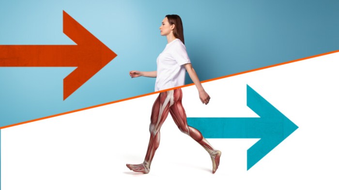
    Forward and reverse walking, while both utilizing gait cycles, exhibit fundamental differences in their biomechanical characteristics. The direction of progression significantly alters the distribution of forces and the recruitment of muscles. In forward walking, the body propels itself forward, relying on a specific sequence of muscle contractions. Reverse walking, conversely, necessitates a reversal of these forces and movements, demanding a distinct set of muscular actions to move the body backward.

    Muscle Activation During Reverse Walking

    The muscle activation patterns in reverse walking are distinct from those in forward walking. Muscles primarily responsible for forward propulsion, such as the gluteus maximus and hamstrings, experience altered roles and activation timings in reverse walking. Instead of pushing the body forward, these muscles now work to counteract the tendency to fall backward. Furthermore, muscles responsible for stabilizing the body, such as the core muscles and hip abductors, become even more crucial in reverse walking to maintain balance and control.

    Muscle Activation Patterns

    Muscle Group Forward Walking Activation Reverse Walking Activation
    Gluteus Maximus Primary extensor of the hip, propelling the body forward Significant activation to stabilize and counteract backward movement; may also be less prominent compared to forward walking
    Hamstrings Assist in hip extension and knee flexion during the swing phase Assist in maintaining balance and controlling the backward movement of the leg, may experience higher activation in the early swing phase to compensate for the opposite movement.
    Quadriceps Femoris Primary extensor of the knee, crucial for maintaining forward momentum. Less prominent compared to forward walking; primarily involved in controlling knee extension to maintain balance.
    Gastrocnemius Assists in plantarflexion, maintaining stability and propelling the body forward Important for maintaining balance during the backward movement; plantarflexion occurs during the stance phase but is modified to stabilize against the backward movement.
    Tibialis Anterior Assists in dorsiflexion, contributing to a smooth stride and forward progression. Crucial for controlling ankle movement to maintain stability during the backward movement.
    Core Muscles Crucial for trunk stability and maintaining posture during the entire gait cycle Even more crucial for maintaining balance and posture; increased activation during the backward movement to counteract the tendency to lean forward.

    Benefits and Applications

    Reverse walking, a seemingly simple exercise, holds significant potential for rehabilitation and improving overall physical function. It offers a unique challenge to the body, promoting balance, strength, and coordination in a controlled environment. This approach can be especially beneficial for patients recovering from injuries or conditions that affect their gait.The exercise’s effectiveness lies in its ability to engage multiple muscle groups, promoting functional strength and balance, crucial for everyday activities.

    Furthermore, the controlled nature of reverse walking allows therapists to adapt the exercise to suit the specific needs and abilities of individual patients.

    Benefits of Reverse Walking Exercises

    Reverse walking provides a multifaceted approach to rehabilitation. It actively challenges the body’s balance mechanisms and strengthens crucial muscle groups, thereby improving stability and gait. The emphasis on controlled movement reduces the risk of falls and injuries, allowing patients to progress safely and effectively. Patients often experience a noticeable improvement in their confidence and ability to perform everyday tasks.

    Role in Improving Balance and Gait

    Reverse walking directly impacts balance and gait by requiring the body to compensate for the atypical movement pattern. This process enhances proprioception, the body’s awareness of its position in space. By practicing reverse walking, patients refine their strategies for maintaining balance, which can translate to improvements in their overall gait pattern during forward walking. Consistent practice strengthens the neural pathways responsible for coordinated movement.

    Role in Strengthening Specific Muscle Groups

    Reverse walking targets a wide array of muscle groups, contributing to overall functional strength. The exercise particularly strengthens muscles in the legs, core, and back, improving stability and reducing the risk of falls. This increased strength also enhances the ability to perform daily activities such as climbing stairs or rising from a chair. By working against gravity in an atypical manner, reverse walking strengthens postural muscles and deep core stabilisers.

    Patient Populations Who Could Benefit

    Reverse walking can be beneficial for a variety of patient populations, including individuals recovering from stroke, neurological conditions, or orthopedic injuries. The controlled environment of a therapy session allows for gradual progression and tailoring of the exercise to meet the individual’s specific needs. It can also be a valuable tool for older adults to maintain or improve their balance and mobility.

    Reverse walking in physical therapy can be a surprisingly effective exercise, but it’s crucial to manage any pain that comes with it. Sometimes, you might need ibuprofen to ease the discomfort, but it’s essential to understand how much is too much. Knowing the safe dosage is key to avoiding potential side effects, and you can find helpful information on that front at how much ibuprofen is too much.

    Remember to listen to your body and adjust your reverse walking routine accordingly, prioritizing pain management and recovery for optimal results.

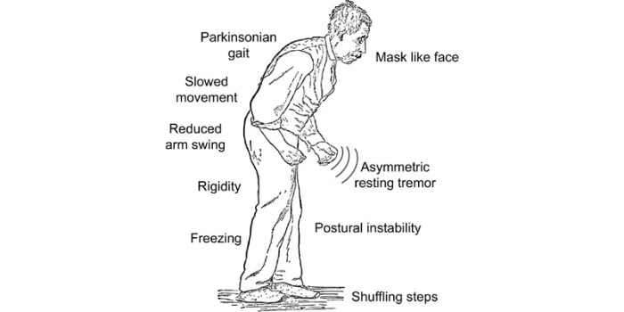
    Moreover, patients with cerebral palsy, multiple sclerosis, or Parkinson’s disease could benefit from reverse walking, given careful consideration and adaptation to their individual conditions.

    Adapting Reverse Walking for Different Levels of Ability

    The exercise’s versatility allows for adaptations to accommodate different levels of ability. Patients with limited mobility can begin with shorter distances or reduced speed, while those with greater capacity can gradually increase the intensity and duration of their training. The environment, support, and guidance provided by a physical therapist are crucial for a safe and effective progression. Furthermore, patients can use assistive devices, such as walkers or canes, to maintain stability and safety during reverse walking.

    Potential Adaptations for Different Patient Needs

    • Reduced Speed and Distance: Starting with short distances and slow speeds allows patients to gradually acclimate to the exercise without overwhelming them. This is particularly important for patients with limited mobility or balance issues.
    • Assisted Walking: Using handrails, walkers, or other assistive devices can help maintain balance and safety, especially for patients with significant balance deficits. The level of assistance should be adjusted as the patient progresses.
    • Varying Terrain: Practicing reverse walking on different surfaces, such as flat ground, inclines, or declines, can progressively challenge balance and coordination. The level of difficulty should be adjusted based on the patient’s capabilities.
    • Resistance Training: Incorporating resistance bands or weights can enhance the strengthening effect of reverse walking, increasing the challenge and promoting functional strength for specific muscle groups.
    • Focus on Balance: During reverse walking, focusing on maintaining balance and control is essential. Visual cues, such as stationary points, can be used to help patients maintain their balance, as well as improve proprioception.

    Exercise Progressions and Modifications

    Reverse walking, while beneficial, requires a structured approach to ensure safety and effectiveness. Progression should be tailored to each patient’s individual needs and abilities, building upon previous successes and accommodating limitations. This careful gradation minimizes risk of injury and maximizes therapeutic gains.

    Exercise Progression Stages

    Gradual progression is crucial in reverse walking exercises. Starting with simple movements and progressively increasing the complexity ensures patients build strength and confidence before tackling more challenging tasks. This approach allows for optimal adaptation and prevents frustration or discouragement.

    Stage Description Equipment Therapeutic Goals
    Stage 1: Initiation Short, controlled steps backward, maintaining a stable stance. Focus on maintaining balance and proper posture. None initially, but potentially a wall or railing for support. Develop balance and proprioception in the reverse direction. Establish a safe starting point for the exercise.
    Stage 2: Controlled Movement Increasing the distance of reverse walking steps, maintaining a steady pace. Maintaining a neutral spine and avoiding excessive hip flexion or extension is important. Potentially light ankle weights. Enhance motor control and coordination in reverse walking. Improve strength and endurance in posterior chain muscles.
    Stage 3: Increased Pace Increasing the speed of reverse walking while maintaining proper form. Gradual increase in speed is essential to prevent falls or injuries. None, or resistance bands for increased challenge. Improve cardiovascular fitness and endurance. Develop dynamic balance and coordination.
    Stage 4: Advanced Challenges Adding variations such as reverse walking with arm movements, or stepping over obstacles. Resistance bands, balance pads, cones for obstacles. Increase functional capacity. Develop more complex motor patterns for functional activities.

    Modifications for Specific Impairments

    Modifications are essential for accommodating patients with diverse impairments, ensuring they can safely participate in reverse walking exercises. Adapting the exercise ensures effectiveness while mitigating risk.

    Impairment Modification Explanation
    Balance Issues Perform reverse walking near a wall or support surface for added stability. Begin with shorter distances and gradually increase the steps. Providing a stable environment reduces the risk of falls and allows the patient to focus on proper form.
    Knee Pain Use a cane or walker for support during reverse walking. Consider using a knee brace for added stability. Minimizing stress on the knee joint through support structures.
    Hip Weakness Use resistance bands around the knees or ankles to increase the resistance and strengthen the hip muscles. Perform reverse walking with a slight bend in the knees to reduce the strain on the hips. Utilizing resistance increases the challenge while focusing on hip strengthening and proper form.
    Foot Drop Use a foot drop brace to maintain proper foot position. Ensure the patient has appropriate footwear for support and stability. Maintaining a functional foot position is critical to maintaining balance and avoiding falls.
    Reduced Range of Motion Use assistive devices to support the affected limb. Modify the exercise to avoid positions that exacerbate pain or stiffness. Avoiding exacerbating the impairment is paramount for effective therapy.

    Safety Considerations and Precautions

    Reverse walking, while beneficial for rehabilitation, necessitates careful consideration of safety protocols. Implementing these precautions minimizes the risk of injury and maximizes the effectiveness of the exercise program. A thorough understanding of potential risks and proactive strategies for prevention are crucial for both therapists and patients.Proper supervision and meticulous patient assessment are paramount to successful reverse walking. An individualized approach, tailored to the patient’s specific needs and limitations, is essential for optimizing safety and progress.

    Importance of Proper Supervision

    Thorough supervision is crucial to ensure patient safety and prevent potential injuries during reverse walking exercises. Qualified physical therapists or trained professionals should closely monitor patients, adjusting the exercise program as needed. Constant observation allows for immediate intervention in case of balance issues, pain, or discomfort. This ensures that the patient remains within their capabilities and avoids overexertion.

    Patient Assessment and Evaluation

    Pre-exercise assessments are vital for identifying potential risks and tailoring the reverse walking program. A comprehensive evaluation should include a thorough medical history, gait analysis, range of motion assessment, strength testing, and balance evaluations. These assessments help determine the patient’s baseline capabilities, identify any pre-existing conditions, and establish appropriate exercise progressions. Identifying and addressing potential issues like joint instability, muscle weakness, or balance problems proactively reduces the risk of injuries.

    Potential Risks and Complications

    Reverse walking, while generally safe, carries potential risks. These include falls, muscle strains, joint pain, and exacerbation of pre-existing conditions. Patients with conditions like osteoporosis, arthritis, or recent injuries are at a higher risk. A careful evaluation and gradual progression are crucial to mitigate these risks. For example, a patient with knee osteoarthritis might experience increased pain if the reverse walking exercises are too vigorous.

    Preventing Common Injuries

    Preventing injuries during reverse walking exercises involves several strategies. First, ensuring appropriate footwear is essential for proper support and balance. Secondly, ensuring the exercise environment is safe and free of obstacles is vital. Thirdly, a gradual increase in intensity and duration is recommended. This gradual progression allows the body to adapt to the new movement pattern and reduces the risk of overuse injuries.

    Mitigation Strategies, Reverse walking in physical therapy

    Several strategies can mitigate risks associated with reverse walking. Using assistive devices, such as canes or walkers, can provide additional support and stability. Modifying the exercise environment, like reducing the surface area, can decrease the risk of falls. Implementing proper warm-up and cool-down routines is essential to prepare the muscles and joints for the exercise and prevent muscle soreness.

    Communication is key, encouraging patients to report any pain or discomfort promptly allows for immediate intervention.

    Reverse walking in physical therapy can be a surprisingly effective exercise, especially for those with specific mobility challenges. While focusing on proper form is key, sometimes underlying health conditions can impact progress. For example, if you’re experiencing hypothyroidism, exploring options like using armour thyroid for hypothyroidism might be beneficial alongside your physical therapy routine. Ultimately, reverse walking, when approached thoughtfully, can lead to improved balance and overall physical well-being.

    Assessment and Evaluation

    Reverse walking, while beneficial, requires careful assessment to ensure patient safety and optimal progress. A thorough evaluation helps determine the appropriate exercise intensity, modifications, and overall program design for each individual. This process allows for personalized treatment plans, maximizing outcomes and minimizing risks.Understanding a patient’s readiness for reverse walking is crucial for successful rehabilitation. This involves evaluating various factors, from their physical capabilities to their cognitive understanding of the exercise.

    A structured approach, combining objective measurements and subjective feedback, ensures a safe and effective rehabilitation journey.

    Patient Readiness Assessment

    A comprehensive assessment prior to introducing reverse walking exercises is paramount. This assessment should not be a cursory check but a detailed evaluation of the patient’s current physical and cognitive state. This will determine their suitability for the exercise, identifying any potential limitations or risks.

    • Medical History Review: A detailed review of the patient’s medical history is essential. This includes any pre-existing conditions, recent surgeries, medications, and any known allergies. This ensures that reverse walking is appropriate and does not exacerbate underlying health issues. For example, a patient with a history of heart conditions might require closer monitoring during the exercise.
    • Range of Motion Assessment: Assessing the patient’s range of motion (ROM) in the hips, knees, ankles, and spine is crucial. Limited ROM in any joint can significantly impact the ability to perform reverse walking. A thorough examination of the patient’s flexibility in these areas is important for successful exercise execution.
    • Balance and Gait Analysis: Evaluating the patient’s balance and gait is fundamental. This involves observing their posture, stance, and walking pattern in a variety of settings, including flat surfaces and potentially uneven terrain. Identifying existing gait abnormalities, such as a tendency to lean, can be addressed through exercise progressions.
    • Strength Assessment: Evaluating the patient’s lower extremity strength is vital. This includes assessing the strength of muscles involved in propulsion and balance during walking. A strength deficit in the lower limbs may necessitate modifications or a slower progression.
    • Cognitive Functioning Evaluation: Assessing the patient’s cognitive ability to understand and follow instructions is critical. Clear communication and proper instruction are essential for patient compliance and safety during exercise.

    Patient Assessment Checklist

    A standardized checklist aids in ensuring a thorough assessment. This provides structure and helps track key data points for each patient.

    Assessment Item Criteria Evaluation
    Medical History Any pre-existing conditions, recent surgeries, medications, allergies Reviewed and documented
    Range of Motion (ROM) Hip, knee, ankle, spine Measured and recorded
    Balance Posture, stance, walking pattern on various surfaces Observed and documented
    Strength Lower extremity muscles (e.g., quadriceps, hamstrings, glutes) Evaluated using standardized tests (e.g., manual muscle testing)
    Cognitive Function Understanding instructions, following commands Observed and documented

    Progress Evaluation Metrics

    Tracking progress is crucial for adapting the exercise program and motivating the patient. Monitoring key metrics provides evidence of improvement.

    • Gait Speed and Distance: Measuring the patient’s walking speed and distance covered in a set time frame allows for objective tracking of improvement in mobility. For example, a patient who initially walks 20 meters in 30 seconds might improve to 30 meters in 30 seconds over time.
    • Balance Metrics: Using tools like the Berg Balance Scale or Timed Up and Go test, allows for objective quantification of balance improvement. Changes in these scores reflect progress in regaining balance control.
    • Gait Analysis: Analyzing gait patterns before and after the intervention can provide detailed insights into improvements in stride length, cadence, and posture. Video recordings, supplemented with gait analysis software, are useful.
    • Patient Reported Outcomes: Collecting patient feedback on their perceived ability to perform reverse walking, pain levels, and any other relevant factors, offers a crucial subjective perspective. This subjective information is vital in evaluating overall improvement.

    Monitoring Exercise Effectiveness

    Monitoring the effectiveness of reverse walking exercises requires a structured approach. Regular evaluation is key to ensuring the exercise program remains appropriate for the patient’s evolving needs.

    • Regular Follow-up Appointments: Scheduling regular follow-up appointments allows for consistent monitoring of progress, adjustments to the exercise program, and identification of any emerging issues.
    • Objective Data Collection: Collecting objective data (e.g., gait speed, balance scores) at each follow-up appointment provides quantifiable evidence of the exercise’s impact.
    • Subjective Feedback: Encouraging patients to provide feedback on their experience with reverse walking exercises helps gauge their comfort level and identify any areas where modifications might be needed.
    • Documentation and Analysis: Thoroughly documenting all assessment findings and progress notes facilitates analysis and adaptation of the rehabilitation plan as needed.

    Equipment and Resources: Reverse Walking In Physical Therapy

    Reverse walking, while a beneficial exercise, can be tailored and supported by various equipment and resources. Understanding the tools available can enhance the safety and effectiveness of the therapy process. Choosing the right equipment is crucial for optimizing patient outcomes.This section details essential equipment for reverse walking exercises, along with valuable supplementary resources. Knowledge of assistive devices can greatly improve the patient experience and therapeutic approach.

    Equipment for Reverse Walking Exercises

    Appropriate equipment can significantly impact the effectiveness and safety of reverse walking exercises. This includes ensuring proper support, stability, and guidance during the exercise.

    • Walking Aids: Canes, walkers, and crutches are crucial for patients with reduced mobility or balance issues. These tools provide additional support, allowing for more controlled and confident reverse walking. The type of assistive device selected will depend on the individual’s specific needs and limitations. For example, a quad cane might be more appropriate for someone with hip or knee pain, while a walker could be better suited for someone with significant lower extremity weakness.

    • Resistance Bands: These bands can be used to increase the difficulty of the reverse walking exercise, particularly for patients who are already able to perform reverse walking without assistance. Resistance bands can be strategically positioned to target specific muscle groups, enhancing the effectiveness of the exercise. For example, a resistance band placed around the ankles can increase the demand on the leg muscles, promoting strength and endurance.

    • Balance Boards: These boards can be used to challenge the patient’s balance and coordination, making reverse walking more challenging. This type of exercise is beneficial for patients with a history of falls or balance issues. For example, a balance board can be incorporated into reverse walking exercises to challenge the stability of the movement, gradually increasing the difficulty over time.

    • Adjustable Height Platforms: These platforms allow for varying degrees of elevation, useful for reverse walking exercises focused on strengthening or stretching specific muscle groups. For instance, by adjusting the height, the exercise can be modified to address different needs or progress to more advanced levels.

    Additional Resources for Learning More

    Several resources are available to expand knowledge about reverse walking in physical therapy. Staying updated with the latest research and techniques is crucial for providing optimal care.

    • Professional Journals: Publications like the Journal of Physical Therapy Science and the Archives of Physical Medicine and Rehabilitation regularly publish research articles on various aspects of physical therapy, including reverse walking. These journals provide valuable insights into the latest research findings and best practices.
    • Online Courses and Workshops: Numerous online platforms offer courses and workshops on physical therapy techniques, including reverse walking. These resources provide practical training and knowledge exchange opportunities.
    • Conferences and Seminars: Attending conferences and seminars dedicated to physical therapy provides an opportunity to learn from leading experts in the field. These events often feature presentations and workshops on topics such as reverse walking, enhancing understanding and skills.
    • Expert Consultations: Consulting with experienced physical therapists specializing in gait rehabilitation can provide personalized guidance and insights into reverse walking techniques for specific patient cases. This individualized approach is particularly important for complex or challenging cases.

    Examples of Assistive Devices

    Assistive devices play a vital role in supporting patients during reverse walking exercises.

    • Walkers: Walkers provide substantial support and stability, enabling individuals with reduced mobility or balance issues to perform reverse walking with greater confidence. Different walker types exist, each with varying levels of support. The choice of walker will depend on the patient’s specific needs.
    • Canes: Canes offer a lighter form of support than walkers, assisting with balance and reducing strain on specific joints during reverse walking. Single-point and quad canes provide different levels of support, aiding the patient in maintaining their balance while performing the exercise.
    • Crutches: Crutches are primarily used for patients with upper body or shoulder impairments, offering support and assisting in weight-bearing during reverse walking exercises. They can be used to reduce stress on the affected limb, facilitating the exercise safely and effectively.

    Last Recap

    Reverse walking in physical therapy

    Reverse walking in physical therapy presents a valuable tool for rehabilitation, offering a nuanced approach to improving gait and balance. By understanding the biomechanics, benefits, and safety precautions, therapists and patients can effectively utilize this technique to achieve optimal outcomes. We’ve covered the key aspects, from initial assessments to advanced progressions, enabling a more comprehensive understanding of this method.

  • Gait Training in Physical Therapy A Comprehensive Guide

    Gait Training in Physical Therapy A Comprehensive Guide

    Gait training in physical therapy focuses on restoring and improving normal walking patterns. It’s crucial for daily life, impacting everything from mobility to overall health. Physical therapists address a wide range of gait abnormalities, tailoring interventions to specific needs. This comprehensive guide explores the entire process, from assessment and evaluation to interventions, patient selection, and measuring outcomes.

    Understanding the historical evolution of gait training techniques and the diverse types of gait abnormalities is essential for therapists. The guide delves into the various methods of assessment, from standardized protocols to the use of advanced tools. It also explores the critical role of patient history and factors contributing to gait impairments. The importance of individualized programs and patient motivation is highlighted.

    Introduction to Gait Training in Physical Therapy

    Gait training in physical therapy

    Gait training in physical therapy is a specialized area focused on improving or restoring an individual’s ability to walk. It involves a comprehensive assessment, intervention, and rehabilitation process tailored to address specific gait impairments. This approach emphasizes restoring normal movement patterns and function, enhancing overall mobility, and improving the quality of life for patients.Gait is fundamental to daily activities, enabling us to perform tasks from simple movements like getting to the kitchen to more complex actions like climbing stairs or navigating challenging terrain.

    A healthy gait is essential for maintaining independence, social participation, and overall well-being. Impairments in gait can significantly impact a person’s ability to perform these activities, potentially leading to isolation, reduced participation in social activities, and increased risk of falls.

    Gait training in physical therapy is crucial for restoring mobility and balance, especially for those with underlying health conditions. Factors like high blood pressure can significantly impact walking patterns, and understanding its causes is key for effective treatment. High blood pressure causes can range from lifestyle choices to underlying health issues, all of which can influence the way a person walks.

    Ultimately, effective gait training programs need to consider these factors and tailor exercises to each individual’s needs.

    Significance of Gait in Daily Activities

    Normal gait is crucial for numerous daily activities. Walking is vital for transportation, social interaction, and participation in recreational activities. It allows individuals to perform tasks ranging from shopping to engaging in hobbies. Without a functional gait, many daily activities become difficult or impossible.

    Types of Gait Abnormalities

    Physical therapists address a wide range of gait abnormalities, each with unique characteristics and underlying causes. These include, but are not limited to, steppage gait, where the foot slaps the ground, and foot drop, where the foot drags. Other gait deviations include compensatory patterns, such as a Trendelenburg gait, which occurs due to hip weakness. A stiff or rigid gait, on the other hand, is often associated with conditions affecting the joints or muscles.

    Goals of Gait Training Programs

    Gait training programs aim to restore normal gait patterns, minimize pain, improve balance, and increase functional mobility. The primary goals are to enable individuals to safely and independently perform essential daily activities, improve their quality of life, and reduce the risk of future falls. A successful program often involves a combination of therapeutic exercises, assistive devices, and patient education.

    Historical Evolution of Gait Training Techniques

    Early gait training methods focused primarily on passive exercises and assistive devices. Over time, a greater understanding of biomechanics and neuromuscular control has led to more sophisticated and targeted approaches. Modern techniques incorporate principles of functional movement, strength training, and balance exercises to achieve better and more durable results. The use of technology, such as gait analysis, has further refined the accuracy and effectiveness of gait training interventions.

    Gait Abnormalities and Treatment Strategies

    Type of Gait Abnormality Common Causes Typical Treatment Strategies
    Steppage Gait Neuromuscular disorders like Charcot-Marie-Tooth disease, peripheral nerve damage, or muscular dystrophy. Strengthening exercises for the lower extremities, proprioceptive training, and potentially orthotics.
    Foot Drop Peripheral nerve damage, stroke, cerebral palsy, or muscular weakness in the anterior tibialis muscle. Strengthening exercises for the anterior tibialis muscle, use of ankle-foot orthoses (AFOs), and gait retraining exercises.
    Trendelenburg Gait Hip abductor weakness, often due to injury or conditions like arthritis. Strengthening exercises for hip abductors, use of assistive devices like canes or walkers, and gait retraining.
    Stiff or Rigid Gait Conditions affecting the joints, such as arthritis, or neurological disorders that affect muscle control. Range-of-motion exercises, stretching, strengthening exercises for affected muscles, and gait retraining.

    Assessment and Evaluation for Gait Training

    Assessing gait is crucial for developing effective physical therapy interventions. A thorough understanding of a patient’s gait patterns, strengths, and weaknesses allows therapists to tailor treatment plans for optimal outcomes. This process involves a combination of subjective observations, objective measurements, and the use of advanced technologies. Accurate gait analysis informs treatment strategies and monitors progress throughout the rehabilitation journey.

    Methods Used to Assess Gait

    Gait assessment employs various methods, ranging from simple observations to sophisticated analyses. Direct observation of the patient’s walking pattern provides valuable initial insights into their gait characteristics, including stride length, cadence, and the presence of any observable compensations. These observations form the basis for more detailed assessments. Qualitative analysis involves careful scrutiny of the patient’s movement, identifying patterns and deviations from normal gait.

    This method allows therapists to pinpoint potential areas for intervention.

    Tools and Technologies in Gait Analysis

    Advanced technologies enhance the accuracy and depth of gait analysis. Force plates measure ground reaction forces during gait, providing quantitative data on forces exerted at different phases of the gait cycle. Video recording, often combined with specialized software, allows for detailed analysis of motion, frame-by-frame, offering insights into joint angles, muscle activation, and movement coordination. 3D gait analysis systems, using multiple cameras, provide comprehensive three-dimensional data, revealing subtle deviations and asymmetries that might be missed with simpler techniques.

    These systems enable a precise understanding of the biomechanics involved in gait.

    Gait training in physical therapy helps patients regain their ability to walk normally after injury or illness. It’s crucial for improving mobility and independence. Understanding the intricate movements involved, like how the body coordinates and balances, is key to effective gait training. Often, this involves working with a physical therapist to practice different walking patterns. For example, learning to control the muscles involved in walking, like the ones that support the eye and regulate pupil function, is important for improved balance and stability.

    To understand the anatomy of the eye, you might find this helpful: what is a pupil. Ultimately, the goal of gait training is to get patients back on their feet and moving freely again.

    Factors Contributing to Gait Impairments

    Numerous factors can contribute to gait impairments. Musculoskeletal conditions, such as osteoarthritis, rheumatoid arthritis, and spinal stenosis, frequently lead to pain and reduced mobility, impacting gait patterns. Neurological disorders, including stroke, multiple sclerosis, and cerebral palsy, can disrupt motor control and coordination, resulting in gait abnormalities. Other factors, such as peripheral neuropathy, can lead to sensory deficits that contribute to gait instability.

    Gait training in physical therapy is crucial for restoring or improving walking ability. A key aspect of this process involves identifying and addressing any underlying issues, which often requires a skilled provider, like a physical therapist, to assess and create a personalized plan. Understanding what a provider is and their role in healthcare is vital for patients seeking this kind of therapy what is a provider.

    Ultimately, gait training empowers individuals to regain their mobility and independence.

    Understanding these contributing factors is essential for developing effective treatment strategies.

    Importance of Patient History and Subjective Data

    Patient history and subjective data play a vital role in the gait assessment process. Information about the patient’s medical history, current symptoms, pain levels, and previous treatments helps in formulating a comprehensive understanding of their condition. Patient reports regarding the onset, duration, and characteristics of gait difficulties are crucial for developing an appropriate treatment plan. Subjective feedback about comfort and perceived functional limitations is also essential.

    This information, combined with objective assessments, creates a holistic picture of the patient’s needs.

    Phases of Gait Analysis

    Gait analysis typically follows a structured process, divided into distinct phases. The initial phase involves gathering patient history and performing a physical examination to identify any obvious musculoskeletal or neurological abnormalities. The next phase involves video recording or motion capture to document the gait pattern. Subsequent phases include data analysis using specialized software to identify specific gait deviations.

    Finally, the data is interpreted to identify contributing factors and formulate treatment strategies.

    Example of a Standardized Gait Assessment Protocol

    A standardized gait assessment protocol might include the following steps:

    1. Gathering patient history and performing a physical examination, including assessment of muscle strength, range of motion, and sensation.
    2. Video recording of the patient walking at their self-selected pace over a specific distance, ensuring a clear view of the entire gait cycle.
    3. Using gait analysis software to measure parameters like stride length, cadence, step time, and velocity.
    4. Identifying any deviations from normal gait, such as asymmetry, excessive pronation, or antalgic gait.
    5. Analyzing the results to formulate a treatment plan tailored to the patient’s specific needs.

    Assessment Tools for Gait Analysis

    A variety of tools are employed in gait analysis. Their application depends on the specific assessment goals.

    Assessment Tool Description Application in Gait Analysis
    Force Plates Measure ground reaction forces during gait. Quantifies forces at different gait phases, identifies asymmetries, and assesses stability.
    Video Recording Captures visual data of the gait cycle. Allows for detailed analysis of joint motion, muscle activation, and compensatory mechanisms.
    3D Gait Analysis Systems Utilizes multiple cameras to capture three-dimensional gait data. Provides comprehensive analysis of spatial and temporal gait parameters, revealing subtle deviations and asymmetries.
    Gait Analysis Software Processes and analyzes data from various assessment tools. Calculates gait parameters, generates reports, and facilitates comparison with normative data.

    Interventions and Techniques in Gait Training

    Gait training interventions are crucial for restoring and improving functional mobility in individuals with gait abnormalities. These interventions are tailored to address the specific impairments and limitations affecting each patient’s gait cycle. A comprehensive approach often involves a combination of therapeutic exercises, assistive devices, and neuromuscular techniques. Successful outcomes depend on a thorough assessment, individualized treatment plans, and patient compliance.

    Types of Interventions for Gait Abnormalities

    Various interventions target different aspects of gait dysfunction. These interventions may include strengthening exercises, stretching, balance training, and gait retraining exercises. They are tailored to the specific cause and nature of the gait impairment, such as weakness, spasticity, or pain. The goal is to improve muscle strength, flexibility, and coordination to facilitate efficient and safe movement.

    Techniques Used in Restoring Normal Gait Patterns

    Restoring normal gait patterns involves a range of techniques. These include: addressing muscle imbalances through targeted strengthening, improving joint mobility through stretching, enhancing proprioception and balance through specific exercises, and correcting gait deviations through biofeedback and visual cues. These techniques work together to re-establish the natural rhythm and efficiency of walking.

    Comparison of Therapeutic Exercise Techniques for Gait

    Different therapeutic exercise techniques for gait restoration offer various approaches. For instance, resistance training can strengthen muscles involved in propulsion and support, while flexibility exercises improve joint range of motion and reduce stiffness. Balance training, utilizing unstable surfaces, enhances postural control and proprioception, which are crucial for smooth gait. Specific gait retraining exercises, such as practicing heel-toe walking, can improve the coordination and timing of movements in the gait cycle.

    Role of Assistive Devices and Adaptive Equipment in Gait Training

    Assistive devices and adaptive equipment play a vital role in gait training, particularly for individuals with significant mobility limitations. Walkers, canes, crutches, and prosthetics can provide support and stability, allowing patients to participate in gait training activities safely and effectively. Adaptive footwear can improve foot support and reduce pain, enhancing the overall gait experience. These tools enable patients to gradually progress towards independent mobility.

    Description of Different Therapeutic Exercise Protocols

    Therapeutic exercise protocols for gait training are developed based on the patient’s specific needs and goals. Protocols might involve progressive resistance training for strengthening, balance exercises on unstable surfaces, and functional gait training using various surfaces and terrains. These protocols can be adjusted based on the patient’s progress and tolerance. For example, a protocol for a patient with knee osteoarthritis might focus on strengthening quadriceps and hamstring muscles, improving joint mobility, and performing balance exercises.

    Application of Neuromuscular Electrical Stimulation in Gait Training

    Neuromuscular electrical stimulation (NMES) can be a valuable tool in gait training. NMES can facilitate muscle contractions, improve muscle strength and endurance, and potentially reduce pain and spasms. It can be particularly beneficial for patients with muscle weakness or neurological impairments. The specific application of NMES would be determined based on the patient’s specific needs and the assessment results.

    For example, NMES might be used to stimulate the quadriceps muscles in a patient with knee weakness, leading to improved strength and gait.

    Therapeutic Exercise Protocol Table

    Intervention Type Description Target Populations Expected Outcomes
    Resistance Training Strengthening exercises using weights, resistance bands, or machines Individuals with muscle weakness, impaired balance, or reduced gait speed Improved muscle strength, increased power, and enhanced functional mobility
    Flexibility Exercises Stretching exercises to improve joint range of motion and reduce stiffness Individuals with limited joint mobility, contractures, or muscle tightness Increased flexibility, reduced pain, and improved range of motion
    Balance Training Exercises performed on unstable surfaces or with eyes closed to improve balance and proprioception Individuals with impaired balance, poor coordination, or history of falls Enhanced balance, improved coordination, and reduced risk of falls
    Gait Retraining Exercises Specific exercises focusing on improving gait patterns and biomechanics Individuals with gait abnormalities, such as deviations or compensations Improved gait efficiency, reduced gait deviations, and enhanced functional mobility
    Assistive Device Use Using canes, walkers, crutches, or prosthetics to support ambulation Individuals with limited mobility or strength Increased stability, improved balance, and enhanced mobility

    Patient Selection and Program Design

    Gait training programs are highly individualized, requiring careful consideration of the patient’s specific needs and limitations. Proper patient selection and program design are crucial for achieving optimal outcomes and preventing setbacks. This section delves into the key factors involved in choosing suitable candidates for gait training and creating effective, tailored programs.Effective gait training hinges on understanding the patient’s unique condition and capabilities.

    This understanding informs the creation of a personalized plan that maximizes progress while minimizing risks.

    Factors to Consider When Selecting Patients

    Patient selection for gait training involves a multifaceted assessment. The decision to include a patient in a gait training program hinges on several key factors. These factors include the patient’s medical history, the nature and severity of their gait impairments, their motivation and willingness to participate, and their overall physical capacity. A comprehensive evaluation is essential to determine the suitability of a patient for gait training.

    • Medical History: Past diagnoses and treatments, particularly those impacting mobility or neurological function, are essential considerations. Conditions like stroke, spinal cord injury, or osteoarthritis require different approaches.
    • Gait Impairments: The severity and nature of the gait dysfunction, such as weakness, pain, balance issues, or sensory deficits, directly influence the design and progression of the program. Assessing the specific areas where the patient is experiencing difficulties is crucial.
    • Motivation and Willingness: Patient motivation and commitment to the program are critical for successful outcomes. Enthusiasm and a positive attitude contribute significantly to adherence and progress.
    • Physical Capacity: The patient’s overall physical condition, including strength, endurance, and flexibility, plays a significant role in determining the appropriate level of intervention and intensity. Consideration should be given to the patient’s ability to tolerate the demands of the training.

    Individualized Program Design, Gait training in physical therapy

    Designing an effective gait training program requires a personalized approach. A customized program addresses the patient’s specific needs, capabilities, and goals. The program should be tailored to the individual, considering their strengths and weaknesses.

    • Initial Assessment: The initial assessment provides a baseline understanding of the patient’s gait characteristics, functional limitations, and overall physical status. This comprehensive evaluation forms the foundation for developing the personalized program.
    • Goal Setting: Collaboratively establishing realistic and measurable goals with the patient is crucial. These goals should be specific, measurable, achievable, relevant, and time-bound (SMART goals). This involves understanding the patient’s desired outcomes, such as improved walking distance or reduced pain.
    • Program Components: The program should include a combination of exercises targeting strength, balance, proprioception, and cardiovascular endurance, tailored to the patient’s specific needs. This might include exercises like strengthening exercises for lower limbs, balance training using wobble boards or balance cushions, and proprioceptive neuromuscular facilitation (PNF) techniques. The exercises should be progressively challenging.

    Program Progressions

    Progressive overload is a key principle in gait training programs. This involves gradually increasing the intensity, duration, or complexity of the exercises to challenge the patient and promote adaptation.

    • Gradual Progression: The program should progress gradually, building upon the patient’s abilities. This prevents frustration and potential injury.
    • Examples: A typical progression might start with short, stationary exercises focusing on specific muscle groups. Later, the exercises could involve walking on flat surfaces, gradually increasing the distance and speed. Finally, the program could incorporate challenging terrain or inclines.

    Patient Motivation and Adherence

    Motivation plays a pivotal role in patient adherence to the gait training program. A positive attitude and understanding of the benefits contribute significantly to the success of the treatment.

    • Encouragement: Providing positive reinforcement and encouragement throughout the training process is essential.
    • Clear Communication: Open communication with the patient regarding their progress, setbacks, and future goals is crucial.
    • Realistic Expectations: Setting realistic expectations helps maintain motivation and avoid frustration. Acknowledging the time commitment required and the gradual nature of improvement is vital.

    Patient Education in Gait Training

    Patient education is an integral component of gait training. Understanding the rationale behind the exercises and the importance of adherence enhances the effectiveness of the treatment.

    • Instruction: Thorough explanation of the exercises, their purpose, and the rationale behind their use.
    • Self-Management: Equipping the patient with the knowledge and skills to continue the exercises at home is vital.
    • Addressing Concerns: Addressing any concerns or questions that the patient may have helps foster trust and confidence.

    Program Examples

    Patient Characteristics Program Components Expected Progress Considerations
    Patient with stroke, moderate weakness in lower extremities, good motivation Strengthening exercises for lower limbs, balance training on wobble board, gait training on flat surfaces, increasing distance gradually Improved gait symmetry, increased walking speed and distance, improved balance Close monitoring for muscle fatigue, encourage rest periods as needed
    Patient with osteoarthritis, mild pain, moderate mobility Range of motion exercises, strengthening exercises for core and lower limbs, low-impact aerobic exercise, pain management techniques, assistive devices as needed Reduced pain, improved range of motion, increased mobility, improved gait efficiency Regular pain assessment, modification of exercises based on pain levels, patient education on proper body mechanics

    Common Gait Disorders and Treatments

    Gait disorders are a diverse range of conditions that affect how we walk. These disorders can significantly impact a person’s mobility, independence, and overall quality of life. Physical therapists play a crucial role in evaluating, diagnosing, and treating these conditions, working closely with patients to restore optimal gait function. Understanding the different types of gait disorders, their underlying causes, and effective treatment approaches is essential for effective physical therapy intervention.

    Common Gait Disorders

    Gait disorders encompass a broad spectrum of conditions, each with unique characteristics and contributing factors. Identifying these specific issues is paramount to creating targeted and effective treatment plans. Recognizing the patterns of abnormal movement is crucial for diagnosis and subsequent treatment.

    • Cerebral Palsy: This group of disorders affects movement and muscle tone due to brain abnormalities during development. Common gait abnormalities include spasticity, ataxia, and weakness. These impairments can cause difficulty with balance, coordination, and walking. The severity varies greatly, impacting the intensity and nature of treatment approaches.
    • Parkinson’s Disease: Characterized by progressive neurodegenerative changes, Parkinson’s disease often manifests as a shuffling gait with reduced arm swing, freezing episodes during gait, and postural instability. The progressive nature of the disease necessitates ongoing adjustments to treatment plans, ensuring interventions remain effective as symptoms evolve.
    • Stroke: Following a stroke, patients often experience weakness, spasticity, or ataxia on one side of the body. These impairments can significantly affect gait, causing problems with balance, coordination, and step length. The specific gait deviations following a stroke depend heavily on the location and extent of the brain damage.
    • Osteoarthritis: This degenerative joint disease affects the hips and knees, causing pain, stiffness, and reduced range of motion. Pain and stiffness can manifest in various ways during walking, impacting gait patterns and potentially causing limping or altered stride length. The degree of gait disturbance correlates with the severity of the arthritis.
    • Multiple Sclerosis: This neurological disease involves damage to the myelin sheath surrounding nerve fibers. This damage leads to a wide array of symptoms, including gait abnormalities. These include issues with balance, coordination, and muscle weakness. The unpredictable nature of MS necessitates a dynamic and individualized approach to gait training.

    Causes and Contributing Factors

    The underlying causes of gait disorders are diverse and complex. Understanding these factors is essential to develop effective treatment strategies.

    • Neurological Conditions: Neurological conditions like cerebral palsy, stroke, and multiple sclerosis can disrupt the signals between the brain and muscles, leading to abnormal gait patterns.
    • Musculoskeletal Conditions: Osteoarthritis, hip dysplasia, and other musculoskeletal problems can cause pain, stiffness, and reduced range of motion, leading to altered gait patterns.
    • Neuromuscular Diseases: Conditions such as muscular dystrophy and Charcot-Marie-Tooth disease affect muscle function and strength, directly impacting gait.
    • Environmental Factors: Conditions like uneven surfaces or poor footwear can exacerbate existing gait problems.

    Treatment Approaches

    Treatment for gait disorders is multifaceted and often involves a combination of approaches.

    • Physical Therapy: A core component of treatment, physical therapy focuses on improving strength, flexibility, balance, and coordination. Specific exercises and techniques are tailored to the individual’s needs and the specific gait disorder.
    • Medications: Medications can play a supportive role in managing pain, spasticity, or other symptoms associated with specific disorders. The use of medications must be integrated within the overall treatment plan and carefully monitored by a medical professional.
    • Assistive Devices: Canes, walkers, and braces can provide support and stability during gait, particularly for individuals with weakened muscles or balance issues.
    • Surgery: In some cases, surgical interventions may be necessary to address underlying structural problems contributing to gait abnormalities.

    Examples of Specific Exercises

    Exercises tailored to the specific gait disorder are essential to enhance mobility and reduce pain.

    • Cerebral Palsy: Stretching exercises, strengthening exercises, and balance exercises are crucial. Specific exercises target affected muscle groups, improving strength and flexibility. Activities like obstacle courses can help improve balance and coordination.
    • Parkinson’s Disease: Exercises focusing on gait initiation, freezing episodes, and maintaining posture are crucial. Examples include heel-toe walking, stepping over objects, and practicing turning movements.
    • Stroke: Exercises to improve strength, range of motion, and balance are critical. Examples include strengthening exercises for affected limbs and balance exercises like standing on one leg.

    Role of Medications

    Medications can be a crucial component of a comprehensive treatment plan.

    • Pain Management: Analgesics or anti-inflammatory medications may be prescribed to manage pain associated with musculoskeletal conditions or neurologic disorders. These medications should be used in conjunction with physical therapy.
    • Muscle Relaxants: These medications may be used to reduce spasticity in individuals with conditions like cerebral palsy or stroke.
    • Other Medications: Medications for specific neurological conditions, such as those for Parkinson’s disease, are essential to manage the progression of the disease and reduce associated symptoms. These medications must be used under strict medical supervision.

    Table of Common Gait Disorders

    Gait Disorder Symptoms Causes Treatment Options
    Cerebral Palsy Spasticity, ataxia, weakness, difficulty with balance and coordination Brain abnormalities during development Physical therapy, medications, assistive devices, surgery
    Parkinson’s Disease Shuffling gait, reduced arm swing, freezing episodes, postural instability Progressive neurodegenerative changes Physical therapy, medications, assistive devices
    Stroke Weakness, spasticity, ataxia on one side of the body, balance and coordination problems Cerebrovascular accident Physical therapy, medications, assistive devices, potential surgery
    Osteoarthritis Pain, stiffness, reduced range of motion, limping, altered stride length Degenerative joint disease Physical therapy, medications, assistive devices, potential surgery
    Multiple Sclerosis Balance problems, coordination issues, muscle weakness, fatigue Neurological disease Physical therapy, medications, assistive devices, ongoing management

    Gait Training in Specific Populations

    Gait training isn’t a one-size-fits-all approach. Understanding the unique needs and challenges of different patient populations is crucial for successful rehabilitation. Tailoring interventions to the specific characteristics of each group ensures optimal outcomes and fosters patient engagement. This section delves into gait training strategies for various populations, highlighting important considerations for each.

    Gait Training for Pediatric Populations

    Pediatric gait training focuses on developing appropriate movement patterns and promoting normal musculoskeletal development. Early intervention is key to address potential gait deviations and ensure proper motor skill acquisition. Therapists need to consider the child’s developmental stage, cognitive abilities, and emotional factors when designing the training program. Games, play-based activities, and adaptive equipment can enhance engagement and motivation.

    • Emphasis on play-based activities and fun, engaging exercises to make the training more enjoyable and motivating for children.
    • Careful consideration of the child’s developmental stage to adapt the exercises and intensity accordingly. Activities should be age-appropriate.
    • Using assistive devices and adaptive equipment, such as walkers or braces, to support the child during gait training, when necessary.
    • Incorporating family education and support to ensure a consistent and comprehensive approach to the child’s rehabilitation.

    Gait Training for Geriatric Patients

    Geriatric gait training addresses the specific needs of older adults, often facing challenges like decreased strength, balance issues, and decreased joint mobility. Interventions should prioritize safety, minimizing the risk of falls, and promoting independence. This approach often emphasizes functional mobility and adapting activities to the individual’s abilities.

    • Prioritizing safety and fall prevention through exercises focusing on balance and proprioception.
    • Utilizing assistive devices, such as walkers or canes, to enhance stability and support during gait training.
    • Tailoring exercises to the patient’s specific physical limitations, adapting intensity and duration as needed.
    • Incorporating functional tasks, such as navigating stairs or getting in and out of chairs, into the training program to improve real-world mobility.

    Gait Training for Individuals with Neurological Conditions

    Individuals with neurological conditions, such as stroke or cerebral palsy, often experience gait impairments due to motor control and coordination issues. Gait training programs should be tailored to address specific neurological deficits, promoting functional independence and improving mobility.

    • Addressing specific impairments like spasticity, weakness, or sensory deficits.
    • Utilizing techniques like constraint-induced movement therapy or task-oriented training to improve motor learning and functional gait.
    • Using visual cues, proprioceptive feedback, and other sensory strategies to enhance gait control.
    • Incorporating neuro-rehabilitation principles and therapies like constraint-induced movement therapy, which involves restricting use of the unaffected limb to promote use of the impaired limb.

    Gait Training for Individuals with Musculoskeletal Impairments

    Musculoskeletal impairments, such as osteoarthritis or amputations, can significantly impact gait. Gait training programs should focus on improving joint function, strength, and flexibility. Adaptive equipment, pain management strategies, and patient education are vital components of these programs.

    • Addressing specific musculoskeletal impairments, such as joint pain, muscle weakness, or limited range of motion.
    • Incorporating pain management techniques and strategies to address pain during movement and improve comfort.
    • Using assistive devices, such as orthotics or prosthetics, to improve gait mechanics and reduce stress on affected joints.
    • Focus on strengthening exercises for affected muscle groups to improve stability and support during ambulation.

    Comparison of Gait Training Programs for Different Populations

    Population Key Considerations Training Focus Example Interventions
    Pediatric Developmental stage, motivation, play-based activities Normal gait development, motor skill acquisition Play-based activities, adaptive equipment, family involvement
    Geriatric Balance, strength, fall prevention, functional mobility Safe and independent mobility, functional tasks Balance exercises, assistive devices, functional training
    Neurological Specific neurological deficits, motor control, coordination Improving motor control, functional mobility, gait patterns Constraint-induced movement therapy, sensory strategies, task-oriented training
    Musculoskeletal Joint pain, strength, flexibility, adaptive equipment Improving joint function, mobility, pain management Orthotics, prosthetics, strengthening exercises, pain management strategies

    Measuring Outcomes and Progress in Gait Training: Gait Training In Physical Therapy

    Tracking progress in gait training is crucial for ensuring effectiveness and tailoring interventions. Accurate measurement allows therapists to assess the impact of treatment, adjust strategies as needed, and ultimately help patients achieve optimal functional outcomes. Monitoring patient responses and evaluating the effectiveness of interventions is key to successful rehabilitation.Precise measurement methods and standardized outcome measures provide a reliable and objective way to quantify changes in gait.

    This allows for a clear understanding of the patient’s progress and facilitates communication between healthcare professionals. Furthermore, objective data supports the development of individualized treatment plans.

    Methods for Measuring Gait Effectiveness

    Various methods are employed to assess the effectiveness of gait training interventions. These include observational assessments, gait analysis using specialized equipment, and functional tests. Observational assessments, though subjective, provide a valuable initial evaluation. They involve the therapist observing the patient’s gait pattern, identifying areas for improvement, and tracking changes over time. Gait analysis, using sophisticated instruments like force plates and motion capture systems, provides detailed quantitative data on gait parameters.

    This data allows for a more comprehensive understanding of the patient’s gait and the impact of interventions. Functional tests, such as timed up and go (TUG) or timed 25-foot walk tests, evaluate the patient’s functional mobility and ability to perform activities of daily living.

    Metrics for Evaluating Gait Progress

    Gait training progress is measured using a variety of metrics. These include measures of gait speed, cadence, stride length, step length, and symmetry. Changes in these metrics reflect the impact of the interventions. For instance, increased gait speed and reduced variability in gait patterns indicate improvement in the patient’s gait. Assessment of balance and postural control are also vital.

    Improved balance and reduced sway are clear indicators of successful training.

    Examples of Tracking and Documenting Gait Changes

    Tracking changes in gait requires careful documentation. This involves recording initial gait parameters and subsequently documenting improvements over time. Using a standardized gait assessment form, clinicians can systematically note changes in gait speed, cadence, stride length, and symmetry. Progress notes should include detailed descriptions of observed changes and specific examples of improvements.

    Outcome Measures in Physical Therapy

    Numerous outcome measures are employed in physical therapy to assess progress. Examples include the Berg Balance Scale, the Timed Up and Go test, the Functional Gait Assessment, and the 6-minute walk test. Each measure targets specific aspects of functional mobility, balance, and gait. These standardized tools provide objective data, aiding in the assessment of progress and facilitating comparison across different patients and treatments.

    Monitoring Patient Response to Interventions

    Monitoring patient response involves a systematic evaluation of the patient’s progress. Regular assessments, including both objective measurements and subjective feedback from the patient, are crucial. Regular review of the patient’s progress allows for adjustments to the intervention plan, ensuring it remains tailored to the individual needs of the patient. Therapists should note any limitations or challenges faced by the patient during the intervention and make necessary modifications.

    Importance of Standardized Outcome Measures

    Standardized outcome measures are essential for reliable and valid assessments. They ensure consistency in measurement across different settings and therapists. This allows for comparison of results, facilitates research, and improves the overall quality of care. Standardized measures provide objective data, minimizing subjective bias and enhancing the accuracy of treatment evaluation.

    Table of Outcome Measures

    Outcome Measure Description Application in Gait Training Expected Improvements
    Berg Balance Scale Assesses balance and mobility Evaluates balance before and after gait training Improved balance, reduced falls, increased confidence in performing daily activities
    Timed Up and Go (TUG) Measures functional mobility Assesses the patient’s ability to get up from a chair and walk a short distance Decreased time to complete the task, improved gait speed, and enhanced functional mobility
    Functional Gait Assessment (FGA) Assesses gait quality and stability Evaluates gait patterns, including initiation, terminal swing, and other aspects of gait Improved gait efficiency, increased stride length, and better postural control
    6-Minute Walk Test Measures the patient’s walking endurance Assesses the patient’s ability to walk for a sustained period Increased walking distance, improved endurance, and increased functional capacity

    Closing Notes

    Gait training in physical therapy

    In conclusion, gait training in physical therapy is a multifaceted process demanding careful assessment, personalized interventions, and continuous monitoring of progress. This guide provides a detailed overview of the entire journey, empowering therapists with the knowledge to effectively address gait abnormalities and improve patients’ quality of life. The diverse techniques, from therapeutic exercises to assistive devices, highlight the multifaceted nature of this critical field.