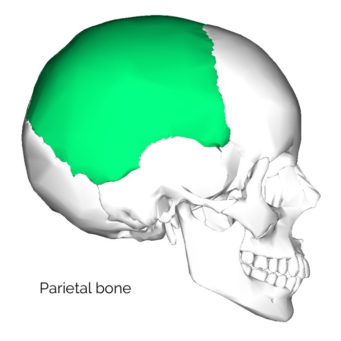
Category: Neurology

  • What is a Myotome A Deep Dive

    What is a Myotome A Deep Dive

    What is a myotome? It’s a crucial concept in understanding how our muscles are controlled by the nervous system. Imagine a specific group of muscles, all working together, coordinated by a particular segment of the spinal cord. That’s essentially a myotome. This isn’t just an academic curiosity; myotome assessment plays a vital role in diagnosing neurological conditions.

    From pinpointing the source of muscle weakness to understanding the intricate pathways of the nervous system, exploring myotomes reveals a fascinating interplay between structure and function.

    This comprehensive guide delves into the world of myotomes, examining their definition, anatomy, function, clinical relevance, and assessment methods. We’ll uncover how these intricate muscle groups are organized, the role of nerve roots, and how evaluating myotomes helps diagnose and understand various neurological issues. Get ready to explore the fascinating science behind your body’s movement!

    Definition and Scope

    A myotome is a group of muscles innervated by a specific spinal nerve root. Understanding myotomes is crucial for clinicians, as they provide a pathway to assess the integrity of the motor pathways of the nervous system. It’s a fundamental concept in neurology, helping pinpoint the location of potential nerve damage.Myotomes and dermatomes, while both associated with spinal nerves, differ in their function.

    Dermatomes map sensory innervation, while myotomes map motor innervation. Damage to a specific nerve root can manifest as weakness or paralysis in the associated myotome, allowing clinicians to pinpoint the affected area. This localized weakness helps determine the level of spinal cord or nerve root compromise.

    Myotome Definition and Relationship to Dermatomes

    A myotome is a group of muscles that are controlled by a single spinal nerve root. This contrasts with dermatomes, which map the sensory areas served by the same nerve root. The relationship between the two is based on the common spinal nerve root origin. Clinical correlation between sensory and motor deficits, observed in dermatomes and myotomes respectively, allows clinicians to pinpoint the affected spinal segment and the extent of the damage.

    A myotome is basically a group of muscles that a single spinal nerve root controls. Understanding these muscle groups is crucial for diagnosing and treating pain, especially when considering active and passive pain management strategies. For example, if you’re experiencing pain in a specific area, evaluating the corresponding myotome can help pinpoint the source of the issue. This kind of assessment can help you determine the best course of action, such as exploring different approaches to pain relief, including active and passive pain management techniques.

    Ultimately, knowing your myotomes can give you a better understanding of your body and how to manage any discomfort effectively.

    This integrative approach is vital in diagnosing and treating various neurological conditions.

    Clinical Significance of Myotomes

    Myotomes are essential in clinical settings for diagnosing nerve root or spinal cord lesions. Weakness or paralysis in a specific myotome can indicate a problem with the corresponding spinal nerve root, often associated with conditions like herniated discs, spinal stenosis, or multiple sclerosis. The pattern of muscle weakness can help determine the precise location and extent of the neurological damage.

    Description of Myotomes

    Myotomes can be described in various ways. An anatomical description focuses on the specific muscles within the group. A functional description emphasizes the movements the myotome enables. Different methods of testing can also provide a detailed description, focusing on the specific movements and muscles involved in the examination. These methods are integral to the clinical evaluation.

    Table of Myotomes

    Myotome Name Associated Muscles Nerve Root Clinical Significance
    Cervical Myotome C5 Deltoid, biceps brachii, brachialis C5 spinal nerve root Weakness in shoulder abduction and elbow flexion can suggest C5 nerve root compression.
    Cervical Myotome C6 Biceps brachii, wrist extensors C6 spinal nerve root Weakness in wrist extension and elbow flexion indicates possible C6 nerve root impingement.
    Lumbar Myotome L2 Hip flexors L2 spinal nerve root Paralysis in hip flexion may point to L2 nerve root compression.
    Lumbar Myotome L4 Knee extensors L4 spinal nerve root Weakness in knee extension often suggests L4 nerve root damage, potentially due to disc herniation.
    Sacral Myotome S1 Plantar flexors, ankle evertors S1 spinal nerve root Weakness in plantar flexion and ankle eversion can indicate S1 nerve root pathology.

    Anatomy and Structure: What Is A Myotome

    What is a myotome

    Myotomes are fascinating segments of the body, directly linked to the intricate network of nerves and muscles. Understanding their anatomical arrangement is crucial for diagnosing and treating neuromuscular disorders. This section delves into the detailed structure of myotomes, illustrating their organization and the specific components that contribute to their function.The arrangement of myotomes reflects the segmental organization of the nervous system.

    They are essentially functional units, each controlled by a specific group of nerve roots. The specific muscles controlled by each myotome are essential in coordinating movement, posture, and other functions. This knowledge is fundamental in assessing neurological integrity.

    Anatomical Arrangement of Myotomes, What is a myotome

    The spinal cord segments and corresponding nerve roots organize myotomes in a precise pattern. Imagine a vertical arrangement of segments, each responsible for controlling muscles in a specific region of the body. This organization allows for precise control and coordination of movement, enabling us to perform a vast array of actions. This arrangement also allows for efficient communication between the brain and different parts of the body.

    Components of a Myotome

    A myotome comprises the nerve root(s) that innervate it and the specific muscle groups it controls. These components are interconnected, forming a functional unit. The nerve roots carry signals from the spinal cord to the muscles, enabling contraction and movement. The muscle groups are the effectors of this signal, performing the intended actions. Together, these elements create a complete functional unit.

    Nerve Root Innervation

    The nerve roots are the essential communication channels connecting the spinal cord to the muscles. Each myotome receives its innervation from specific nerve roots, which originate from the spinal cord. These nerve roots relay signals from the central nervous system to the muscle fibers, triggering contractions. The precise location of these roots is essential in determining the specific myotome.

    Spinal Cord Segments Involved

    The spinal cord is divided into segments, each corresponding to a specific region of the body. These segments are crucial in the formation of myotomes, as they are the source of the nerve roots. Each segment sends signals to the corresponding muscle groups, controlling their actions. The precise location and function of each spinal cord segment are essential in understanding myotome function.

    This information is used to diagnose and treat various neuromuscular conditions.

    Myotome Table

    This table illustrates the relationship between myotome, nerve root(s), and the muscle groups controlled. It highlights the crucial role of nerve roots in activating specific muscle groups, which together create the function of a myotome. This information is critical in assessing neurological function and diagnosing neuromuscular disorders.

    Myotome Nerve Root(s) Innervating It Muscle Groups Controlled
    Cervical Myotomes (C1-C8) C1-C8 Neck, shoulder, arm, hand muscles
    Thoracic Myotomes (T1-T12) T1-T12 Intercostal muscles, back muscles
    Lumbar Myotomes (L1-L5) L1-L5 Hip, thigh, knee muscles
    Sacral Myotomes (S1-S5) S1-S5 Leg, foot muscles

    Neural Pathways Involved in Myotome Function

    The neural pathways involved in myotome function are complex, but they can be described as follows. Sensory input from the muscle and surrounding tissues is conveyed to the spinal cord through sensory nerves. Motor commands are then sent from the spinal cord via motor nerves to the muscle fibers. This two-way communication allows for precise control and coordination of movement.

    The intricate nature of these pathways underscores the importance of a well-functioning nervous system.

    Function and Clinical Relevance

    Myotomes, as we’ve explored, represent a specific group of muscles innervated by a single spinal nerve root. Understanding their function is crucial in evaluating neurological health. This section delves into the practical applications of myotome assessment in clinical settings.Myotome assessment provides valuable insights into the integrity of the peripheral nervous system. Clinicians utilize this information to pinpoint potential nerve root impingements, spinal cord lesions, or peripheral neuropathies.

    The accuracy of this assessment is pivotal in formulating a diagnosis and developing a targeted treatment plan.

    Myotome Function

    Myotomes are responsible for specific movements or actions. Each myotome corresponds to a particular set of muscles, and the coordinated activation of these muscles allows for a range of body movements. For example, the C5 myotome controls shoulder abduction, while the L4 myotome facilitates dorsiflexion of the foot. Understanding the specific movements controlled by each myotome helps clinicians assess the extent of potential neurological damage.

    Myotome Assessment in Clinical Practice

    Clinicians evaluate myotomes through a series of standardized muscle strength tests. These tests involve asking the patient to perform specific movements, and the examiner assesses the strength and range of motion of the corresponding muscles. The examiner grades the strength using a standardized scale, often a 0-5 scale, where 0 indicates no muscle contraction and 5 represents normal strength.

    The precise movements and muscle groups tested depend on the suspected area of neurological impairment. Thorough and careful examination is crucial to ensure accurate assessment.

    Clinical Uses of Myotome Testing

    Myotome testing serves as a vital diagnostic tool in identifying and evaluating neurological conditions. It helps pinpoint the level of spinal cord or nerve root involvement, aiding in differential diagnosis. This testing is particularly useful in cases of suspected nerve root compression, such as from herniated discs, or in conditions like spinal stenosis. Furthermore, myotome assessment can help monitor the progression or response to treatment in various neurological disorders.

    A myotome is a group of muscles that are innervated by a single spinal nerve root. Maintaining good kidney health is crucial for overall well-being, and a balanced diet plays a key role. Eating foods rich in antioxidants and low in sodium, such as those highlighted in this resource on foods for kidney health , can contribute significantly to kidney function.

    Understanding myotomes is important for diagnosing and treating neuromuscular conditions.

    Conditions Where Myotome Assessment is Important

    Myotome assessment is critical in several neurological conditions. The information gathered aids in pinpointing the location and extent of the damage to the nervous system. By evaluating muscle strength, clinicians can identify the specific nerve roots or spinal cord segments that are affected. This allows for a more accurate diagnosis and facilitates appropriate treatment.

    Examples of Conditions and Expected Myotome Weakness

    Condition Expected Myotome Weakness Diagnostic Significance
    Cervical Radiculopathy (C6-C7) Weakness in elbow flexion and wrist extension Indicates nerve root compression at the C6-C7 level, potentially from a herniated disc.
    Lumbar Radiculopathy (L5) Weakness in foot dorsiflexion and great toe extension Suggests nerve root compression at the L5 level, possibly due to spinal stenosis or a herniated disc.
    Spinal Cord Injury (Thoracic Level) Weakness in shoulder abduction, elbow flexion, and wrist extension. Indicates a spinal cord lesion, and the level of the lesion can be determined by the specific myotomes affected.
    Peripheral Neuropathy Gradual, symmetric weakness in multiple myotomes Suggests a diffuse or systemic process affecting the peripheral nerves.

    Assessment and Testing

    Assessing myotome function is crucial for evaluating neuromuscular health and identifying potential nerve or muscle pathologies. Proper myotome testing involves a systematic approach to evaluate the strength and power of specific muscle groups innervated by particular spinal nerve roots. This allows clinicians to pinpoint the location of potential damage or dysfunction.

    Methods for Assessing Myotome Function

    Various methods are employed to assess myotome function, each with its strengths and limitations. These methods range from simple manual muscle testing to more sophisticated electromyographic (EMG) studies. Manual muscle testing is a widely used, cost-effective method that relies on the clinician’s observation of the patient’s ability to perform specific movements against resistance. EMG studies provide a more detailed evaluation of muscle electrical activity, allowing for a deeper understanding of muscle function and identifying any neuromuscular disorders.

    Steps Involved in a Myotome Examination

    A comprehensive myotome examination involves several key steps. First, a thorough patient history and neurological examination are essential to understand the patient’s presenting complaint and potential contributing factors. Second, the examination should focus on identifying the specific muscle groups associated with the relevant myotomes. Third, standardized manual muscle testing procedures should be meticulously followed to ensure accuracy and reliability.

    Lastly, interpretation of the findings should be performed with careful consideration of the patient’s overall clinical presentation.

    Importance of Proper Technique in Myotome Testing

    Proper technique is paramount in myotome testing to ensure accurate and reliable results. Inconsistent or improper techniques can lead to inaccurate assessment and potentially misdiagnosis. Clinicians should adhere to standardized procedures and ensure that the patient is properly positioned and relaxed during the test. Accurate measurement and consistent evaluation criteria are crucial for reliable results. Standardized protocols should be used to maintain consistency and reproducibility.

    Step-by-Step Procedure for Testing Specific Myotomes

    1. Biceps Myotome (C5-C6): Assess the patient’s ability to flex the elbow against resistance. The examiner should position the patient supine or sitting with the elbow flexed at 90 degrees. The examiner applies resistance to the patient’s forearm while the patient attempts to maintain elbow flexion. Note the degree of resistance the patient can overcome. The examiner should observe for any weakness, atrophy, or tremors during the test.

    2. Triceps Myotome (C6-C7): Assess the patient’s ability to extend the elbow against resistance. The examiner should position the patient supine or sitting with the elbow flexed at 90 degrees. The examiner applies resistance to the patient’s forearm while the patient attempts to extend the elbow. Observe the degree of resistance the patient can overcome. Look for any weakness, atrophy, or tremors during the test.

      So, what exactly is a myotome? Basically, it’s a group of muscles that are innervated by a single spinal nerve root. Understanding these muscle groups is crucial for diagnosing nerve damage, and for people exploring natural health options like AHCC supplements, for example, AHCC benefits and safety is an important consideration. Knowing how AHCC might interact with your myotomes is key to potential therapeutic benefits.

      Ultimately, a good understanding of myotomes is essential for anyone working with the body’s complex systems.

      Note that a weak triceps response can suggest damage to the radial nerve.

    3. Wrist Extensors (C6-C7): Assess the patient’s ability to extend the wrist against resistance. The patient should be positioned supine or sitting with the forearm supported and the wrist in a neutral position. The examiner applies resistance to the dorsal aspect of the hand as the patient attempts to extend the wrist. Assess the strength and note any weakness. This test evaluates the integrity of the radial nerve.

    4. Iliopsoas Myotome (L2-L3): Assess hip flexion. The patient should be supine with the knee flexed and the hip at a 90-degree angle. The examiner should apply resistance to the patient’s thigh while they attempt to maintain hip flexion. Record the patient’s ability to maintain the flexion against resistance. Assess for any weakness, atrophy, or other abnormalities.

      This is crucial for assessing the integrity of the lumbar plexus and the associated nerves.

    Clinical Cases and Examples

    Myotome testing plays a crucial role in diagnosing and localizing neurological impairments. By assessing the strength and function of specific muscle groups, clinicians can pinpoint the level of the nervous system affected, whether it’s a peripheral nerve, spinal cord, or brain. Understanding the clinical presentation and interpretation of myotome weakness is vital for developing an accurate diagnosis and appropriate treatment plan.Interpreting myotome testing results requires careful consideration of the patient’s overall neurological presentation.

    The presence of other symptoms, such as sensory changes, reflexes, and cognitive impairments, provides valuable context and helps differentiate between various neurological conditions. These combined findings contribute to a comprehensive picture of the patient’s neurological status.

    Myotome Weakness in Cervical Radiculopathy

    Cervical radiculopathy, often caused by nerve root compression in the neck, frequently leads to specific myotome weakness. The affected myotomes are typically those innervated by the affected nerve roots. For example, C5 and C6 nerve root involvement might result in weakness of the biceps and deltoid muscles, affecting shoulder abduction and flexion.

    Myotome Weakness in Spinal Cord Injury

    Spinal cord injuries can result in varying degrees of myotome weakness, depending on the level and extent of the damage. A complete spinal cord lesion at the C5 level, for example, would likely produce significant weakness in the muscles innervated by the C5 nerve root and below, leading to weakness in shoulder abduction, elbow flexion, wrist extension, and hand movements.

    Myotome Weakness in Peripheral Neuropathy

    Peripheral neuropathies encompass a broad range of conditions affecting peripheral nerves. These conditions can lead to varying degrees of myotome weakness depending on the affected nerves. For instance, a diabetic neuropathy can lead to distal muscle weakness affecting the muscles of the feet and legs, impacting ambulation and balance.

    Interpreting Myotome Testing Results

    Interpreting myotome testing results in conjunction with other neurological findings is crucial for accurate diagnosis. For example, if a patient presents with weakness in the C7 myotome (triceps) but normal reflexes, the cause might be localized to the peripheral nerve rather than the spinal cord. Consideration of the patient’s medical history, symptoms, and other neurological examination findings further enhances diagnostic accuracy.

    Case Studies of Myotome Involvement

    Case Scenario Myotome Affected Symptoms Diagnosis
    Patient with neck pain, radiating down the arm, and weakness in biceps and wrist extensors. C5, C6, C7 Pain, numbness, tingling in the arm and hand, weakness in elbow flexion and wrist extension. Cervical radiculopathy
    Patient with a history of a fall, presenting with decreased sensation in the lower extremities and weakness in hip flexion and knee extension. L2, L3, L4 Numbness and tingling in the lower extremities, weakness in hip flexion, knee extension, and dorsiflexion. Spinal cord injury
    Patient with a history of diabetes, experiencing gradual weakness in the foot and ankle. L4, L5, S1 Gradual weakness and numbness in the feet and ankles, difficulty with ambulation. Diabetic neuropathy

    Variations and Considerations

    Myotomes, while providing a valuable framework for assessing neuromuscular function, aren’t a perfect fit for every individual. Variations in their organization and the influence of various factors can significantly affect how a myotome is assessed and interpreted. Understanding these variations and considerations is crucial for accurate diagnoses and effective treatment plans.The consistency in myotome organization, though a useful guide, is not absolute.

    Individual variations in muscle fiber type, size, and innervation patterns can affect the specific muscles that make up a myotome. This variability in anatomy necessitates a careful and nuanced approach to myotome assessment, always considering the individual’s unique characteristics.

    Variations in Myotome Organization

    Individual differences in muscle anatomy and innervation can lead to variations in myotome boundaries. These variations can sometimes result in a muscle being innervated by a nerve root slightly different from the expected pattern. This can cause a myotome to be slightly larger or smaller in some individuals than in others. Factors like genetic predisposition and previous injuries can also play a role in these anatomical variations.

    Factors Influencing Myotome Assessment

    Several factors can impact the accuracy of myotome testing. Patient cooperation and motivation are critical, as muscle strength is affected by psychological factors like fear and anxiety. Pain, fatigue, and other medical conditions can also influence the ability to perform muscle tests, potentially leading to inaccurate or misleading results. Furthermore, the examiner’s skill and experience in performing the tests directly impacts the reliability of the assessment.

    Careful consideration of these influencing factors is paramount to obtaining meaningful and reliable results.

    Limitations of Myotome Testing

    Myotome testing, despite its usefulness, has limitations. It provides a general indication of nerve root function but doesn’t offer a precise measurement of the extent of the nerve damage. The test’s resolution is limited, and subtle deficits may be missed. Additionally, myotome testing relies on the patient’s ability to cooperate, which can be affected by factors like pain or cognitive impairment.

    It’s important to recognize that myotome testing is a component of a broader neurological assessment, not a stand-alone diagnostic tool.

    Considerations for Assessing Myotomes in Different Age Groups

    Age significantly impacts muscle strength and the ability to perform motor tasks. Children and older adults may have different baseline strength levels, requiring adjusted assessment methods and interpretations. The developmental stages of children, for example, need to be taken into account. In children, the myotome assessment might focus on milestones and developmental norms, rather than precise strength measurements.

    Table: Age Group Considerations for Myotome Assessment

    Age Group Potential Variations Assessment Considerations
    Infants (0-1 year) Immature neuromuscular system, limited voluntary movement Focus on developmental milestones, use of reflexes, parental input
    Children (2-12 years) Varying developmental stages, rapid growth spurts Use age-appropriate tasks, consider developmental norms, assess cooperation
    Adults (18-65 years) Stable muscle mass, generally consistent motor abilities Standard myotome testing methods are generally applicable
    Older Adults (65+) Decreased muscle mass, decreased strength, potential comorbidities Adjust testing to accommodate limitations, assess for confounding factors, use caution

    Imaging and Techniques

    What is a myotome

    Visualizing myotomes, the groups of muscles innervated by a single spinal nerve root, presents unique challenges. While direct visualization of muscle function isn’t possible with standard imaging, these techniques can provide valuable information about the structures involved, aiding in the diagnosis and understanding of neuromuscular conditions. Understanding the limitations of these techniques is equally crucial for accurate interpretation.Imaging modalities offer insights into the anatomy of myotomes, but don’t directly measure their functional capacity.

    This is critical to remember when interpreting results, as imaging findings must be correlated with other assessments like electromyography (EMG) and nerve conduction studies (NCS) to gain a comprehensive picture. Indirect assessments of myotome involvement are possible through visualization of neural pathways and structures related to muscle function.

    MRI and CT Imaging

    Magnetic resonance imaging (MRI) and computed tomography (CT) scans are valuable tools for visualizing the anatomy of the spine, spinal cord, and surrounding tissues. These techniques can depict the spinal nerves, which are crucial for understanding myotome function. MRI excels at providing detailed soft tissue images, highlighting potential spinal cord compression or nerve root impingement, which can affect the associated myotomes.

    CT, on the other hand, provides excellent bone detail, aiding in identifying bony structures that might contribute to nerve root compression.

    Limitations of Imaging Techniques

    Imaging techniques primarily visualize thestructures* involved in myotome function, not the function itself. For example, an MRI might reveal a herniated disc compressing a nerve root, but it doesn’t directly demonstrate the resultant weakness in the muscles of the corresponding myotome. Other factors, like the severity of the compression or the individual’s response to the condition, can also affect the myotome’s function and are not directly measurable by these techniques.

    Visualizing Neural Pathways

    Visualization of neural pathways involves advanced techniques. Advanced MRI techniques, such as diffusion tensor imaging (DTI), can map the pathways of nerve fibers, providing insights into the integrity of neural connections. This is crucial in cases of suspected nerve damage or disease, aiding in identifying the affected myotomes.

    Electrodiagnostic Studies (EMG and NCS)

    Electromyography (EMG) and nerve conduction studies (NCS) are essential for evaluating the electrical activity of muscles and nerves. EMG measures the electrical activity of muscle fibers, while NCS assesses the speed and amplitude of nerve impulses. These studies can directly assess the functional integrity of the myotomes, detecting issues like denervation (loss of nerve supply) or axonal damage.

    For instance, if a specific myotome shows reduced or absent electrical activity, it suggests a potential problem with the nerve root or the muscles it supplies.

    Comparison of Imaging Modalities

    Imaging Modality Strengths Limitations Application in Myotome Assessment
    MRI Excellent soft tissue detail, visualization of spinal cord and nerve roots Limited visualization of bone structures, can be expensive Identifying spinal cord compression, nerve root impingement
    CT Excellent bone detail, rapid acquisition Limited soft tissue contrast, higher radiation exposure Identifying bony structures contributing to nerve root compression
    DTI Visualization of neural pathways Requires specialized software and interpretation, more expensive than standard MRI Assessing nerve fiber integrity, identifying areas of potential damage
    EMG/NCS Direct assessment of muscle and nerve function Invasive procedure, requires specialized expertise for interpretation Evaluating the functional integrity of the myotomes, detecting denervation or axonal damage

    Ending Remarks

    In conclusion, understanding myotomes provides a critical lens through which we can appreciate the intricate relationship between the nervous system and our muscles. From a clinical standpoint, myotome assessment is invaluable for pinpointing the source of neurological issues. This guide has explored the key aspects of myotomes, offering a clear understanding of their definition, anatomy, function, and clinical applications.

    We’ve seen how these muscle groups, coordinated by specific nerve roots, are crucial for our everyday movements and how their assessment is a valuable tool in diagnosing various conditions. Myotomes are more than just anatomical structures; they’re essential for our health and well-being.

  • Hyperpathia, Hyperalgesia, Hypersensitivity A Deep Dive

    Hyperpathia, Hyperalgesia, Hypersensitivity A Deep Dive

    Hyperpathia hyperalgesia and hypersensitivity – Hyperpathia, hyperalgesia, and hypersensitivity: these terms describe heightened pain experiences, but what exactly do they mean? This exploration delves into the intricacies of these conditions, from defining each distinct pain response to understanding the underlying mechanisms and how they affect daily life. We’ll uncover the various causes, diagnostic methods, treatment options, and the profound impact on patients.

    Each condition manifests differently, impacting pain perception, intensity, and duration. Understanding these nuances is crucial for proper diagnosis and management. We’ll examine the intricate interplay of physiological and psychological factors that contribute to these conditions. This exploration will also cover the importance of addressing the psychosocial impact on patients and provide a look into current research and future directions.

    Defining Hyperpathia, Hyperalgesia, and Hypersensitivity: Hyperpathia Hyperalgesia And Hypersensitivity

    Hyperpathia hyperalgesia and hypersensitivity

    Understanding the nuances of pain perception is crucial for effective diagnosis and treatment. This exploration delves into the distinct characteristics of hyperpathia, hyperalgesia, and hypersensitivity, highlighting their physiological mechanisms and the differences in pain perception and response. These conditions, while often intertwined, present unique challenges in clinical settings.These three conditions represent heightened pain responses, characterized by amplified sensitivity to stimuli.

    However, the intensity, duration, and nature of pain differ considerably. This discussion will explore the unique features of each condition, examining the underlying mechanisms that contribute to their development.

    Defining Hyperpathia, Hyperpathia hyperalgesia and hypersensitivity

    Hyperpathia is characterized by an exaggerated response to painful stimuli, often described as a prolonged, intense, and burning sensation beyond the expected pain response. It’s not just an increase in pain intensity, but a qualitative change in the pain experience. Patients frequently report a feeling of heightened sensitivity to touch or pressure, even non-noxious stimuli, in the affected area.

    This condition often coexists with other neurological conditions, indicating a complex interplay of factors in its development.

    Defining Hyperalgesia

    Hyperalgesia signifies an increased sensitivity to painful stimuli in an area surrounding the initial injury. It’s a localized amplification of pain response, rather than a widespread alteration like hyperpathia. The pain is typically more intense than expected for the stimulus. A common example is the heightened sensitivity to touch or pressure in the area surrounding a burn.

    The underlying mechanism involves changes in the nervous system’s processing of pain signals.

    Defining Hypersensitivity

    Hypersensitivity, in the context of pain, refers to an exaggerated response to normally innocuous stimuli. This condition involves an increased perception of pain from non-noxious stimuli. This can manifest as a heightened sensitivity to light touch, temperature changes, or even air currents. The pain experienced is often described as a burning or tingling sensation. The physiological mechanisms underpinning hypersensitivity often involve altered neurotransmitter systems and inflammatory responses.

    Comparing and Contrasting Hyperpathia, Hyperalgesia, and Hypersensitivity

    Characteristic Hyperpathia Hyperalgesia Hypersensitivity
    Pain Quality Prolonged, intense, burning, heightened sensitivity to touch/pressure Increased intensity compared to the stimulus, localized Burning, tingling, heightened sensitivity to normally innocuous stimuli
    Pain Intensity Markedly increased, often disproportionate to the stimulus Increased, but often proportional to the stimulus Increased response to normally innocuous stimuli
    Pain Duration Prolonged, lasting beyond the expected recovery period Can vary depending on the stimulus and injury Can persist even after the stimulus is removed
    Stimulus Usually involves a past injury or neurological condition Localized to the site of injury or damage Normally innocuous stimuli (light touch, temperature, pressure)

    The table above provides a concise comparison of the three conditions. It highlights the distinctions in pain quality, intensity, duration, and the type of stimulus that triggers the heightened response. Further research is needed to fully elucidate the intricate mechanisms underlying these complex conditions.

    Etiology and Causes

    Understanding the root causes of hyperpathia, hyperalgesia, and hypersensitivity is crucial for developing effective treatment strategies. These conditions arise from a complex interplay of factors, ranging from peripheral nerve damage to central nervous system alterations. Delving into these etiological aspects provides a more comprehensive picture of these often debilitating conditions.Peripheral and central nervous systems are intricately connected, with signals flowing between them.

    Disruptions in this intricate network can lead to altered pain processing, resulting in the exaggerated responses seen in hyperpathia, hyperalgesia, and hypersensitivity. This heightened sensitivity to pain stems from various mechanisms, each contributing to the overall experience.

    Potential Causes of Hyperpathia, Hyperalgesia, and Hypersensitivity

    The varied etiologies of these conditions underscore the complex nature of pain processing. They encompass a wide range of factors, from direct nerve injury to systemic illnesses. It is important to understand that multiple factors often interact to produce these conditions.

    Neurological Pathways Involved

    The pathways involved in processing pain signals are intricate and multi-layered. Pain signals initiated at the periphery travel through sensory neurons to the spinal cord, where they are relayed to higher brain centers. Disruptions in any part of this pathway can contribute to the development of these conditions. For example, damage to the peripheral nerves, such as those caused by diabetes or trauma, can lead to altered signal transmission.

    Peripheral and Central Sensitization

    Peripheral sensitization involves heightened responsiveness of nociceptors (pain receptors) in the periphery. This occurs in response to damage, inflammation, or other stimuli. Central sensitization, on the other hand, involves the nervous system’s central processing centers becoming hypersensitive to pain signals. This heightened responsiveness can lead to a more prolonged and intense pain experience. Both peripheral and central sensitization are crucial in the development of chronic pain conditions.

    Genetic and Environmental Factors

    Genetic predisposition plays a role in some cases, influencing the individual’s response to pain stimuli. For example, some individuals might have a genetic susceptibility to nerve damage or inflammatory conditions that can trigger these heightened pain responses. Environmental factors such as exposure to toxins or trauma can also contribute to the development of these conditions.

    Table of Causes and Associated Conditions

    Cause Associated Conditions
    Nerve Damage (e.g., trauma, diabetes, infection) Hyperpathia, Hyperalgesia, Neuropathic pain
    Inflammation (e.g., arthritis, autoimmune diseases) Hyperalgesia, Chronic pain
    Neuropathic Pain Hyperpathia, Hyperalgesia, Hypersensitivity
    Chronic Pain Conditions Hyperpathia, Hyperalgesia, Hypersensitivity
    Systemic Illnesses (e.g., cancer, multiple sclerosis) Hyperpathia, Hyperalgesia, Hypersensitivity
    Substance Abuse Hyperalgesia, Chronic pain

    Diagnosis and Assessment

    Pinpointing hyperpathia, hyperalgesia, and hypersensitivity requires a multi-faceted approach, blending meticulous clinical evaluation with specific diagnostic tools. Accurate diagnosis hinges on identifying the unique constellation of symptoms and signs exhibited by each individual, enabling clinicians to differentiate between these overlapping conditions and pinpoint the underlying cause.A thorough understanding of the patient’s medical history, including past injuries, illnesses, and medications, is crucial.

    This information, combined with a detailed account of the current symptoms, provides valuable context for evaluating the nature and severity of the sensory disturbances.

    Clinical Evaluation Procedures

    Clinical evaluation procedures play a vital role in diagnosing and assessing the severity of hyperpathia, hyperalgesia, and hypersensitivity. These procedures involve a comprehensive examination of the patient’s neurological function, including sensory perception and motor skills. Detailed questioning about the location, intensity, quality, and duration of pain and other sensory symptoms is essential.

    Diagnostic Methods

    Various diagnostic methods are employed to ascertain the presence and extent of these conditions. These methods include a detailed neurological examination, assessing reflexes, muscle strength, and coordination. Furthermore, specific sensory testing, such as pain thresholds and pressure sensitivity, aids in identifying abnormal sensory processing.

    Pain Scales and Assessment Tools

    Pain scales are essential tools for quantifying the intensity and characteristics of pain. Visual Analog Scales (VAS) and Numerical Rating Scales (NRS) allow patients to subjectively report their pain levels. These scales provide a standardized method for assessing pain intensity and help track changes over time. Additionally, other tools like the McGill Pain Questionnaire can provide a more detailed description of the pain experience, encompassing sensory qualities, emotional aspects, and location.

    Neurological Examinations

    Neurological examinations are fundamental in assessing the integrity of the nervous system. These examinations include evaluating reflexes, muscle strength, coordination, and sensory function in various parts of the body. Abnormal findings in these areas may suggest underlying neurological conditions contributing to or exacerbating the pain and sensory issues.

    Differentiating Symptoms and Signs

    Differentiating between hyperpathia, hyperalgesia, and hypersensitivity relies on recognizing subtle yet critical distinctions in the patient’s presentation. For example, hyperpathia is characterized by exaggerated pain responses to normally innocuous stimuli, often accompanied by allodynia (pain from non-painful stimuli). Hyperalgesia, on the other hand, involves heightened pain sensitivity to painful stimuli, while hypersensitivity encompasses a broader range of abnormal sensory responses beyond pain.

    Careful observation of these specific symptoms and signs aids in accurate diagnosis and subsequent treatment planning.

    Summary Table of Diagnostic Criteria and Assessment Methods

    Condition Diagnostic Criteria Assessment Methods
    Hyperpathia Exaggerated pain response to normally innocuous stimuli; allodynia; often associated with neuropathic pain. Pain scales (VAS, NRS); neurological examination (sensory testing); patient history.
    Hyperalgesia Increased sensitivity to painful stimuli; heightened pain perception. Pain scales (VAS, NRS); quantitative sensory testing (QST); patient history.
    Hypersensitivity Abnormal sensory response to various stimuli; may include pain, temperature, touch, or pressure; can involve multiple sensory modalities. QST; neurological examination; patient history; specific sensory tests.

    Management and Treatment

    Managing hyperpathia, hyperalgesia, and hypersensitivity involves a multifaceted approach tailored to individual needs. These conditions often require a combination of pharmacologic and non-pharmacologic interventions to effectively address the underlying causes and symptoms. The goal is not just to reduce pain intensity but also to improve overall quality of life.Effective treatment strategies aim to target both the acute and chronic aspects of these conditions, acknowledging that the approach may need to evolve over time.

    This requires close collaboration between patients, healthcare providers, and other relevant specialists, such as physical therapists or psychologists.

    Pharmacologic Interventions

    Pharmacologic treatments play a crucial role in managing the pain associated with hyperpathia, hyperalgesia, and hypersensitivity. Various classes of medications are used, each with its own mechanism of action and potential side effects. Careful consideration of individual patient factors, such as comorbidities and medication history, is essential when selecting the appropriate pharmacologic intervention.

    Understanding hyperpathia, hyperalgesia, and hypersensitivity can be tricky, but it’s crucial for pinpointing the root cause of discomfort. A rash on the upper inner thigh, in a female, could be a symptom of an underlying issue. Conditions like these might be linked to heightened sensitivity in the area, which could manifest as a rash. For more information on identifying and treating a rash upper inner thigh female, check out this helpful resource: rash upper inner thigh female.

    Ultimately, though, consulting a medical professional is essential to properly diagnose and manage any such sensitivity issues.

    • Opioids: While effective for some patients, opioids carry the risk of addiction and other adverse effects, such as respiratory depression. Careful monitoring and dose titration are crucial. Examples include morphine, oxycodone, and fentanyl.
    • Nonsteroidal anti-inflammatory drugs (NSAIDs): These drugs can reduce inflammation and pain, potentially alleviating some aspects of hyperalgesia. However, they may not be as effective for hyperpathia. Examples include ibuprofen and naproxen.
    • Antidepressants: Certain antidepressants, such as tricyclic antidepressants (TCAs) and serotonin-norepinephrine reuptake inhibitors (SNRIs), can be effective in managing neuropathic pain components of these conditions. They may also help regulate mood and sleep, which can indirectly improve overall well-being.
    • Anticonvulsants: These medications can modulate neuronal activity and are often used to treat neuropathic pain associated with hyperpathia. Examples include gabapentin and pregabalin.

    Non-Pharmacologic Interventions

    Non-pharmacologic interventions are equally important in the management of hyperpathia, hyperalgesia, and hypersensitivity. These strategies can complement pharmacologic treatments and often address the broader impact of these conditions on a patient’s life.

    • Physical Therapy: Exercises, manual therapy, and other physical interventions can improve mobility, reduce muscle tension, and enhance overall physical function. This is particularly helpful in managing pain and improving function in patients with chronic conditions.
    • Cognitive Behavioral Therapy (CBT): CBT can help patients identify and modify negative thought patterns and behaviors related to pain. This can reduce the emotional distress and anxiety associated with chronic pain conditions, which can often exacerbate symptoms.
    • Mindfulness-Based Stress Reduction (MBSR): Practices like meditation and mindfulness can help regulate stress responses and promote relaxation, which can lead to a reduction in pain perception. This approach can be particularly valuable for patients experiencing heightened emotional distress related to their pain.
    • Alternative Therapies: Techniques like acupuncture, massage therapy, and spinal manipulation are sometimes used in conjunction with other treatments to reduce pain and improve overall well-being.

    Efficacy and Side Effects

    The efficacy of various treatment options varies significantly among individuals. Factors like the underlying cause of the condition, the severity of symptoms, and individual patient responses influence treatment outcomes. Close monitoring and adjustments to the treatment plan are essential to optimize efficacy and minimize potential side effects.

    Patient Population Comparisons

    The effectiveness of different treatment approaches may differ across various patient populations, taking into account factors like age, gender, and specific medical conditions. For instance, older adults may have different responses to certain medications compared to younger individuals.

    Hyperpathia, hyperalgesia, and hypersensitivity are fascinating neurological conditions, but they can be quite debilitating. Understanding these conditions is important, but so is knowing when certain eye issues might start to develop, like macular degeneration, which often appears in later life. Research suggests that age plays a significant role in the onset of macular degeneration, and for those interested in learning more, a helpful resource is available here: what age does macular degeneration start.

    While the connection between these conditions isn’t immediately apparent, a deeper understanding of both neurological and ophthalmological conditions is important for better overall health.

    Managing Pain Intensity and Quality of Life

    Management of pain intensity and improvement of quality of life involve a combination of pharmacologic and non-pharmacologic strategies. Tailoring the treatment plan to the individual patient’s needs and preferences is crucial for achieving optimal outcomes. Regular follow-up appointments with healthcare providers are essential to assess treatment effectiveness and make necessary adjustments to the management plan.

    Clinical Presentations and Manifestations

    Understanding the diverse clinical presentations of hyperpathia, hyperalgesia, and hypersensitivity is crucial for accurate diagnosis and effective management. These conditions, while sharing overlapping symptoms, can manifest in vastly different ways depending on the underlying cause. Recognizing the spectrum of presentations allows healthcare providers to tailor treatment strategies to the individual patient’s needs.These conditions are characterized by abnormal pain processing, which can range from heightened sensitivity to specific stimuli to widespread, debilitating pain.

    The specific symptoms and signs observed in patients will depend on the type of condition, its severity, and the underlying cause. Recognizing the nuances in presentation is essential for appropriate diagnostic evaluations and the selection of the most effective interventions.

    Hyperpathia, hyperalgesia, and hypersensitivity are fascinating neurological conditions, often involving heightened pain responses. Understanding these conditions requires a deep dive into the underlying mechanisms, but one crucial factor influencing pain perception is the red blood cell rbc count. Variations in this can significantly impact the body’s ability to deliver oxygen and nutrients, potentially affecting nerve function and contributing to heightened pain sensitivity.

    Ultimately, further research into the intricate interplay between the red blood cell rbc count red blood cell rbc count and the neurological pathways involved in pain processing is essential for developing effective treatments for hyperpathia, hyperalgesia, and hypersensitivity.

    Variability in Clinical Presentation

    The clinical presentations of hyperpathia, hyperalgesia, and hypersensitivity vary significantly due to the diverse etiologies. These conditions are not monolithic; rather, they represent a spectrum of abnormal pain processing. This variability in presentation can be attributed to factors such as the specific location of the affected tissue, the intensity and duration of the stimulus, and the presence of other comorbidities.

    For example, a patient with hyperalgesia resulting from nerve damage may experience extreme sensitivity to touch in the affected area, while a patient with hyperpathia due to a central nervous system disorder might experience prolonged, burning pain after a seemingly minor stimulus.

    Specific Symptoms and Signs

    Patients with these conditions may present with a wide range of symptoms and signs, which can vary in intensity and location. Common symptoms include:

    • Increased sensitivity to stimuli: Patients may report heightened pain responses to normally innocuous stimuli, such as light touch, cold, or pressure. This can range from mild discomfort to severe, debilitating pain. For example, a person with hyperalgesia may experience excruciating pain from a simple touch that would be barely noticeable to a healthy individual.
    • Allodynia: This refers to the experience of pain from a stimulus that does not normally cause pain. A patient with allodynia might experience pain from clothing touching their skin or a light breeze.
    • Hyperpathia: Characterized by a disproportionately intense and prolonged pain response to a stimulus, often accompanied by spontaneous pain episodes. A person experiencing hyperpathia may feel a sharp, intense pain following a minor injury that lasts for hours or days.
    • Spontaneous pain: This is pain that occurs without any apparent external stimulus. Patients may describe it as a burning, throbbing, or aching sensation.
    • Changes in pain perception: Patients may experience altered pain thresholds and pain qualities. This can manifest as an increased sensitivity to certain types of pain or a decreased ability to perceive other types of pain.

    Examples of Different Presentations Based on Underlying Causes

    The clinical presentation of these conditions can vary significantly depending on the underlying cause. For instance, hyperalgesia following a burn injury would likely present with localized pain and increased sensitivity to touch in the affected area. In contrast, hyperpathia associated with a spinal cord injury may manifest as widespread pain, particularly in the extremities, and be accompanied by spontaneous pain episodes.

    Table of Clinical Presentations

    Clinical Presentation Associated Symptoms Possible Causes
    Localized Hyperalgesia Increased sensitivity to touch, pressure, temperature in a specific area; allodynia Trauma, inflammation, nerve damage, burns, post-operative pain
    Widespread Hyperpathia Prolonged, intense pain, spontaneous pain episodes, altered pain perception; possible paresthesia Central nervous system disorders, spinal cord injury, neuropathies
    Diffuse Hypersensitivity Increased sensitivity to various stimuli (touch, temperature, chemicals); allodynia Chronic pain conditions, psychological factors, autoimmune diseases, fibromyalgia

    Impact on Daily Life and Quality of Life

    The chronic pain and heightened sensitivity associated with hyperpathia, hyperalgesia, and hypersensitivity profoundly affect daily life, impacting everything from simple tasks to complex social interactions. These conditions often create significant challenges, diminishing the quality of life and necessitating comprehensive management strategies.These conditions create a multifaceted impact on individuals, extending beyond the physical realm to encompass emotional, social, and vocational aspects.

    Understanding the specific ways these conditions disrupt daily routines, interpersonal relationships, and overall well-being is crucial for developing effective support systems and interventions.

    Impact on Daily Activities

    Daily tasks, often taken for granted, become significant hurdles for individuals experiencing these conditions. Simple actions like walking, dressing, or even handling everyday objects can be excruciatingly painful. This pain can lead to avoidance behaviors, resulting in reduced mobility and isolation.

    • Examples of affected activities: Walking, showering, dressing, using household appliances, or even interacting with certain fabrics or textures. For example, a person with hyperpathia might find the pressure of a bedsheet unbearable, while a person with hyperalgesia might avoid certain foods due to their perceived pain-inducing properties.
    • Reduced participation in social activities: The fear of pain and discomfort can limit participation in social events, hobbies, and recreational activities. This can lead to feelings of isolation and depression.

    Impact on Work and Education

    The fluctuating pain and sensitivity associated with these conditions can severely disrupt work and education. Chronic pain can lead to missed workdays, reduced productivity, and difficulty concentrating. Students may struggle to attend classes, complete assignments, or maintain focus.

    • Impact on work performance: Pain and discomfort can lead to absenteeism, decreased productivity, and difficulty concentrating on tasks. This can have significant financial consequences and impact career advancement.
    • Challenges in education: Students may experience difficulty concentrating in class, completing assignments, and participating in extracurricular activities. This can affect their academic performance and overall well-being.

    Impact on Relationships

    These conditions can strain interpersonal relationships due to the unpredictable nature of pain and the need for adjustments in daily routines. Family members and partners may struggle to understand the challenges faced by the affected individual.

    • Strained communication: Chronic pain and heightened sensitivity can lead to misunderstandings and conflict within relationships. Communication becomes strained as individuals may struggle to express their needs and experiences.
    • Impact on social interactions: Individuals may withdraw from social interactions due to fear of pain or discomfort. This can lead to feelings of isolation and loneliness.

    Impact on Emotional Well-being and Mental Health

    The chronic pain and limitations imposed by hyperpathia, hyperalgesia, and hypersensitivity can take a significant toll on emotional well-being. Anxiety, depression, and feelings of hopelessness are common.

    • Increased risk of depression and anxiety: The chronic pain and limitations associated with these conditions can lead to feelings of helplessness, frustration, and isolation, significantly increasing the risk of developing anxiety and depression.
    • Impact on self-esteem and confidence: The inability to perform everyday tasks and participate in social activities can lead to a decline in self-esteem and confidence.

    Importance of Psychosocial Support

    Addressing the psychosocial impact of these conditions is crucial for effective management. Support groups, counseling, and education programs can help individuals cope with the emotional and social challenges.

    • Importance of psychological support: Providing psychological support, such as counseling or therapy, is essential for managing the emotional distress associated with these conditions.
    • Role of support groups: Support groups can provide a sense of community and shared experience, allowing individuals to connect with others facing similar challenges.

    Research and Future Directions

    Hyperpathia hyperalgesia and hypersensitivity

    Unraveling the complexities of hyperpathia, hyperalgesia, and hypersensitivity requires ongoing research. Current investigations are focusing on understanding the underlying mechanisms and developing effective treatments. This exploration will hopefully lead to improved diagnostic tools, personalized therapies, and a better quality of life for those affected.

    Current Research Focuses

    Current research into these conditions encompasses a broad spectrum of investigations. Researchers are exploring the intricate interplay of neuronal pathways, neurotransmitters, and inflammatory processes. Advanced neuroimaging techniques are providing valuable insights into the structural and functional changes in the brain and nervous system related to these conditions. Genetic studies are identifying potential susceptibility genes and biomarkers for early detection.

    Ongoing Investigations into Potential Treatments

    Numerous studies are evaluating novel therapeutic approaches. These include exploring the efficacy of targeted medications, such as specific analgesics and neuromodulators. Experimental treatments like nerve stimulation techniques, including transcutaneous electrical nerve stimulation (TENS) and spinal cord stimulation, are being examined for their potential in managing symptoms. Furthermore, research into non-pharmacological interventions, such as physical therapy and cognitive behavioral therapy (CBT), is underway to determine their efficacy in alleviating symptoms and improving overall well-being.

    Promising Avenues for Future Research and Development

    Several avenues hold promise for future research and development. A deeper understanding of the specific mechanisms underlying central sensitization is crucial. Further research into the role of the immune system in these conditions is also anticipated. Developing personalized treatment strategies based on individual genetic profiles and symptom presentation is a significant goal. The exploration of novel drug targets and combination therapies is also expected to yield promising results.

    For example, combining pharmacological interventions with non-pharmacological approaches, like mindfulness-based stress reduction, may provide a more comprehensive and effective strategy.

    Need for Further Investigation into the Mechanisms

    A deeper understanding of the complex interplay of factors contributing to these conditions is essential. This includes examining the role of neuroplasticity, the mechanisms of chronic pain, and the interplay between genetic predisposition and environmental factors. Understanding the intricate mechanisms behind these conditions is crucial for developing effective and targeted therapies. For example, identifying the specific molecular pathways involved in central sensitization could pave the way for more precise and effective treatments.

    Potential Areas for Future Research and Clinical Trials

    Future research should focus on developing more sophisticated diagnostic tools, including biomarkers and advanced neuroimaging techniques. Clinical trials should investigate the efficacy of novel treatments, such as gene therapies and regenerative medicine approaches, for managing chronic pain conditions. For example, exploring the possibility of using stem cell therapies to repair damaged nerve tissue warrants further investigation. Further investigation into the role of psychological factors, such as stress and anxiety, in exacerbating symptoms is essential.

    For example, a prospective clinical trial could compare the effectiveness of a new medication with a standard therapy in a group of patients with specific genetic predispositions to hyperpathia. This would require a comprehensive evaluation of patient outcomes, including pain intensity, quality of life, and psychological well-being.

    Summary

    In conclusion, hyperpathia, hyperalgesia, and hypersensitivity are complex conditions that significantly impact patients’ quality of life. From understanding the distinct characteristics of each to exploring potential causes, diagnostic methods, and treatment approaches, this discussion highlights the importance of comprehensive care. Future research is vital to refine our understanding of these conditions and improve treatment strategies.