Category: Medical Conditions

  • Gout in the Fingers Overview and More

    Gout in the Fingers Overview and More

    Gout in the fingers overview and more: Understanding this painful condition is crucial for effective management. This in-depth look explores the causes, symptoms, and treatment options for gout in the fingers, distinguishing it from other similar conditions. We’ll also delve into risk factors, prevention strategies, and the practical aspects of living with gout in the fingers. It’s not just about the physical discomfort; we’ll also address the emotional impact this condition can have.

    Gout attacks in the fingers often present as sudden, intense pain and swelling, typically affecting one joint at a time. This is different from other finger conditions, which may manifest gradually or involve multiple joints. We’ll examine the underlying mechanisms of gout, contrasting it with gout in other body parts like toes and knees. Understanding the differences in symptoms, risk factors, and treatment will equip you with valuable knowledge to better manage this condition.

    Introduction to Gout in the Fingers: Gout In The Fingers Overview And More

    Gout in the fingers overview and more

    Gout is a painful form of inflammatory arthritis characterized by sudden, intense attacks of joint pain, redness, and swelling. It arises from the buildup of uric acid crystals in the joints. These crystals, formed when the body produces or doesn’t excrete enough uric acid, irritate the joint lining, triggering the inflammatory response.Gout attacks typically affect joints in the lower extremities, particularly the big toe.

    However, gout can also affect other joints, including those in the fingers. Symptoms include sharp, throbbing pain, tenderness, swelling, and redness around the affected finger joint. The affected area may also feel warm to the touch. The attacks can be debilitating, often occurring suddenly and lasting for several days or even weeks.

    Underlying Mechanisms of Gout in the Fingers

    Gout in the fingers, like gout in other joints, stems from the precipitation of uric acid crystals. When uric acid levels in the blood become excessively high, the uric acid can crystallize and deposit in the joint spaces. These sharp crystals then trigger an inflammatory response, causing the pain, swelling, and redness characteristic of a gout attack. The specific location of the deposit within the finger joints is often dependent on the individual’s overall health and lifestyle factors.

    Comparison of Gout in Fingers and Other Joints

    Feature Gout in Fingers Gout in Other Joints (e.g., Toes, Knees)
    Location Finger joints, often the metacarpophalangeal (MCP) or proximal interphalangeal (PIP) joints Other joints, such as the metatarsophalangeal (MTP) joint of the big toe, knees, ankles, elbows, or wrists
    Symptoms Severe pain, redness, swelling, tenderness, warmth, and limited mobility in the affected finger. The attacks can often be debilitating. Similar symptoms to finger gout, but the location will vary depending on the joint affected. Pain in the affected joint is often severe and may be accompanied by stiffness.
    Risk Factors Diet high in purines (found in some foods and drinks), excessive alcohol consumption, certain medications, obesity, and a family history of gout. Similar risk factors to finger gout, including a diet high in purines, excessive alcohol consumption, certain medications, obesity, and a family history of gout. Certain medical conditions, like kidney disease, can also contribute to gout.

    Symptoms and Diagnosis

    Gout in the fingers overview and more

    Gout in the fingers, like gout in other joints, is characterized by sudden, intense attacks of pain, swelling, and inflammation. Understanding the specific symptoms and diagnostic processes is crucial for early intervention and effective management. Accurate diagnosis is essential to differentiate gout from other conditions that may present with similar symptoms.

    Symptoms of Gout Attacks in Fingers

    Gout attacks in the fingers, typically affecting the base of the toes, are marked by a constellation of symptoms. Pain is often the most prominent symptom, described as sharp, throbbing, or excruciating. The affected area becomes red, swollen, and extremely tender to the touch. Heat and inflammation are common accompaniments. These symptoms are often accompanied by a fever.

    The pain typically flares up suddenly and peaks within hours, often during the night.

    Gout in the fingers can be a real pain, causing inflammation and swelling. Understanding the different causes and treatments is key. Interestingly, some people find that incorporating apple cider vinegar into their skincare routine might have some positive effects, like reducing inflammation. For example, exploring the potential skin benefits of apple cider vinegar could be helpful apple cider vinegar skin benefits to reduce the inflammation and discomfort associated with gout in the fingers.

    Of course, more research is needed to fully understand this connection, but it’s certainly an intriguing area to explore further.

    Diagnostic Procedures for Gout in Fingers

    Diagnosing gout involves a combination of physical examination, medical history review, and laboratory tests. A thorough physical examination of the affected finger will reveal swelling, redness, and tenderness. The physician will inquire about the patient’s medical history, including any prior episodes of joint pain, medications, and lifestyle factors. Blood tests, specifically urate levels, are crucial. Elevated levels of uric acid in the blood are a strong indicator of gout.

    Joint fluid aspiration and analysis can also be performed to identify urate crystals. These crystals, when observed under a microscope, are definitive confirmation of gout.

    Common Misconceptions About Gout in Fingers

    Several misconceptions surround gout in the fingers. One common misconception is that gout only affects the big toe. In reality, gout can manifest in any joint, including the fingers. Another misconception is that gout is solely a problem of the elderly. While the risk of gout increases with age, younger individuals can also develop the condition.

    It’s important to recognize that gout is a treatable condition, and early diagnosis and intervention are key to preventing long-term joint damage.

    Differences Between Gout and Other Finger Conditions

    Feature Gout Other Finger Conditions
    Symptoms Sudden, intense pain, redness, swelling, tenderness, heat, fever Pain, swelling, stiffness, limited range of motion, gradual onset (e.g., arthritis, tendonitis, infection)
    Causes High levels of uric acid in the blood, leading to crystal formation in joints. Factors such as diet, genetics, and certain medications can contribute. Various factors, including injury, overuse, infection, autoimmune disorders, or metabolic disorders.
    Treatment Medications to lower uric acid levels, anti-inflammatory drugs to manage pain and inflammation, lifestyle modifications (diet, exercise). Treatment depends on the specific condition; this may include rest, physical therapy, medications, surgery, or other interventions.

    This table highlights key distinctions in symptoms, causes, and treatment approaches between gout and other conditions affecting the fingers. Recognizing these differences is crucial for appropriate diagnosis and effective management.

    Risk Factors and Prevention

    Gout in the fingers, like gout in other joints, isn’t simply a matter of bad luck. Understanding the risk factors and implementing preventive measures can significantly reduce your likelihood of experiencing painful gout attacks. This section delves into the key elements contributing to gout in the fingers and actionable steps you can take to protect yourself.

    Factors Increasing Gout Risk

    Several factors increase the risk of developing gout, particularly in the fingers. These are interconnected and often influenced by lifestyle choices. A high level of uric acid in the blood (hyperuricemia) is a primary driver. This excess uric acid can crystallize and deposit in the joints, leading to inflammation and pain, particularly in the smaller joints like those in the fingers.

    Lifestyle Changes for Prevention

    Adopting a healthy lifestyle is crucial in preventing gout attacks. Maintaining a balanced diet, exercising regularly, and managing stress can significantly lower your risk.

    Gout in the fingers, a painful condition, often stems from excess uric acid. Understanding the various triggers, like dehydration, is key to effective management. Sometimes, a seemingly unrelated issue like a headache can actually be linked to dehydration, which can also affect gout flare-ups. For more on how dehydration can be a headache trigger, check out this informative article: understanding dehydration as a headache trigger.

    Ultimately, staying hydrated and managing uric acid levels are crucial for preventing and effectively treating gout in the fingers.

    • Diet: A diet rich in purines, substances that the body breaks down into uric acid, can contribute to higher uric acid levels. Reducing consumption of certain foods can help.
    • Weight Management: Maintaining a healthy weight is vital. Excess weight can contribute to higher uric acid levels and increase the risk of gout.
    • Hydration: Drinking plenty of water helps flush out excess uric acid from the body. Dehydration can worsen gout symptoms.
    • Exercise: Regular physical activity helps maintain a healthy weight, improves overall health, and can contribute to better uric acid management.
    • Stress Management: Stress can exacerbate existing health conditions, including gout. Practicing stress-reducing techniques like meditation, yoga, or deep breathing can be beneficial.

    Dietary Factors Contributing to Gout

    Certain dietary choices can significantly impact uric acid levels. Excessive consumption of specific foods and beverages can contribute to an elevated risk of gout attacks.

    • Purine-Rich Foods: Organ meats (like liver and kidney), red meat, seafood (especially shellfish), and some types of fish are rich in purines. These foods can increase uric acid production.
    • Alcohol Consumption: Alcohol, especially beer and liquor, can increase uric acid levels. While moderate alcohol intake might not be a significant risk factor for all, it’s crucial to be mindful of the impact on individual uric acid levels.
    • Sugary Drinks: Sugary drinks can contribute to weight gain and increase the risk of gout. Excessive consumption of these drinks should be avoided. Their impact on uric acid levels is related to the potential for weight gain and overall metabolic imbalances.

    Dietary Recommendations for Gout Prevention

    This table summarizes key dietary recommendations for preventing gout. Following these guidelines can help maintain healthy uric acid levels and reduce the likelihood of gout attacks.

    Food Type Recommendation
    Red Meat Limit intake
    Alcohol (Beer, Liquor) Moderate intake
    Sugary Drinks (Soda, Juices) Avoid excess
    Organ Meats (Liver, Kidney) Limit intake
    Shellfish Limit intake
    Fatty Foods Limit intake
    Fruits and Vegetables Consume in moderation
    Whole Grains Include in diet
    Low-Fat Dairy Products Consume in moderation

    Treatment and Management

    Gout in the fingers, while often painful and disruptive, is manageable. Early intervention and adherence to a comprehensive treatment plan significantly improve outcomes and prevent long-term complications. Effective treatment addresses both acute attacks and underlying factors that contribute to the condition.A multi-faceted approach is crucial for managing gout. This involves addressing the immediate pain of an attack, as well as identifying and modifying lifestyle choices that may exacerbate the condition.

    The role of medication is paramount, both in treating acute episodes and in preventing future flare-ups. Proper long-term management strategies are vital for preventing chronic joint damage and improving overall well-being.

    Common Treatment Options

    Various treatment approaches are available to manage gout attacks and prevent future occurrences. These options range from over-the-counter pain relievers to prescription medications that target the underlying cause of the condition.

    Role of Medication in Managing Gout Attacks

    Medications play a critical role in managing gout attacks. During an acute attack, nonsteroidal anti-inflammatory drugs (NSAIDs) like ibuprofen or naproxen are often prescribed to reduce inflammation and pain. Colchicine is another option that can effectively shorten the duration of an attack. Corticosteroids, either oral or injected, may be considered for more severe cases or when other medications are not effective.

    Importance of Long-Term Management Strategies

    Long-term management is essential to prevent recurrent gout attacks and the development of chronic joint damage. This involves lifestyle modifications, such as dietary changes to limit purine intake, and adherence to prescribed medication regimens. Maintaining a healthy weight and regular exercise also contribute to overall health and can positively impact gout management.

    Potential Complications if Gout in the Fingers is Left Untreated

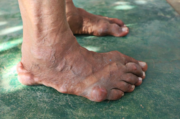
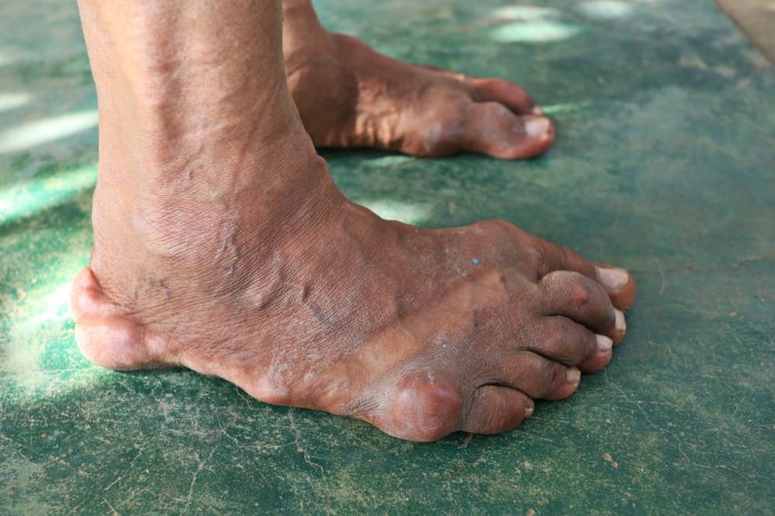
    Untreated gout in the fingers can lead to several serious complications. Chronic inflammation can cause permanent joint damage, leading to deformities and reduced mobility. Tophi, which are deposits of uric acid crystals, can form in and around the joints, causing pain, swelling, and potential infections. In severe cases, kidney stones or kidney damage can occur due to the buildup of uric acid in the body.

    Understanding gout in the fingers involves looking at the underlying causes and potential treatments. Inflammation is a key player, often leading to painful swelling and discomfort. While exploring alternative approaches, it’s worth noting that some research suggests potential links between managing inflammation and the use of CBD oil for diabetes. Further investigation into the effectiveness of CBD oil for these conditions could reveal more insights into the overall picture.

    For a deeper dive into CBD’s role in diabetes management, check out this informative resource on cbd oil for diabetes. Ultimately, a holistic approach to managing gout in the fingers is crucial for finding lasting relief.

    Medication Comparison Table

    Medication Mechanism of Action Potential Side Effects
    Allopurinol Reduces the production of uric acid in the body. Skin rash, nausea, vomiting, diarrhea, liver problems, allergic reactions. Some individuals may experience a temporary increase in gout attacks in the initial phase of treatment.
    Colchicine Reduces inflammation by interfering with the process of white blood cell activity in response to uric acid crystals. Gastrointestinal upset (nausea, vomiting, diarrhea), bone marrow suppression (rare).

    Living with Gout in the Fingers

    Living with gout in the fingers can be challenging, impacting daily life and well-being. The unpredictable nature of attacks and the persistent pain can significantly affect how you go about your day. Understanding how to manage the condition, both during and between attacks, is crucial for maintaining a good quality of life.Effective management involves a multi-faceted approach, encompassing lifestyle adjustments, pain management strategies, and emotional support.

    This section provides practical guidance to help you navigate the complexities of gout in your fingers.

    Managing Daily Activities During a Gout Attack

    Managing daily activities during a gout attack requires careful consideration of your body’s needs. Pain and swelling can limit your range of motion and make simple tasks feel overwhelming.

    • Prioritize rest and avoid strenuous activities. Gentle movements that don’t exacerbate the pain may be beneficial. For example, light stretches, or a few minutes of gentle walking if the pain allows. If you work a job that requires physical exertion, modify tasks as much as possible. Adjusting work tasks or taking breaks as needed is important for preventing further harm or injury.

    • Use assistive devices where appropriate. Simple tools like adaptive utensils, or larger-handled items, can make eating and other self-care tasks easier. This reduces stress on the affected fingers and helps maintain a degree of independence.
    • Modify your home environment. Strategically place frequently used items within easy reach to minimize unnecessary movement. This reduces stress on your fingers and overall discomfort.

    Coping with Pain and Discomfort

    Pain management is essential for coping with gout attacks. A combination of strategies can help you find relief.

    • Apply cold compresses to the affected area. This is a common and effective method for reducing inflammation and pain. Cold compresses constrict blood vessels, decreasing swelling and pain. Applying ice packs directly to the affected area for 15-20 minutes at a time, several times a day, can provide significant relief.
    • Elevate the affected hand. Elevating the hand above your heart can help reduce swelling by promoting fluid drainage. This is often a useful adjunct to cold compresses.
    • Take prescribed medications as directed. Nonsteroidal anti-inflammatory drugs (NSAIDs) or colchicine, often prescribed by a doctor, can effectively reduce pain and inflammation. It is crucial to follow the prescribed dosage and frequency of intake strictly. If you are unsure of how to use a medication or have any questions about potential side effects, consult your healthcare provider.

    Seeking Support from Healthcare Professionals and Support Groups

    Maintaining open communication with your healthcare provider and joining support groups can provide significant support and guidance.

    • Regular check-ups and medication adjustments. Regular appointments with your doctor allow for ongoing monitoring of your condition and necessary adjustments to your treatment plan.
    • Support groups provide a forum for sharing experiences and strategies with others who understand what you are going through. Sharing experiences with others facing similar challenges can foster empathy and support.
    • Open communication with healthcare providers. This allows for personalized advice and treatment plans.

    Emotional Impacts of Gout

    Gout attacks can have a significant emotional impact. The unpredictability and pain can lead to frustration, anxiety, and even depression.

    • Managing stress and anxiety. Stress can exacerbate gout symptoms. Adopting stress-reducing techniques, such as meditation or deep breathing exercises, can be helpful.
    • Seeking emotional support. Talking to a therapist or counselor can provide a safe space to address emotional concerns related to gout.
    • Maintaining a positive outlook. Focusing on manageable aspects of your life and practicing self-care can help maintain a positive outlook.

    Applying Cold Compresses

    Applying cold compresses to affected areas can effectively reduce pain and swelling. Here’s how to do it safely and effectively:

    • Wrap ice in a thin towel or cloth. This prevents direct contact with the skin, which can cause discomfort or damage.
    • Apply the cold compress to the affected area for 15-20 minutes at a time. Repeat as needed.
    • Avoid applying ice directly to the skin. This can lead to frostbite.
    • If the pain persists or worsens, consult a healthcare professional immediately.

    Illustrative Case Studies

    Understanding gout in the fingers requires looking at real-life examples. These case studies highlight the diverse ways the condition manifests, progresses, and responds to treatment. They also illustrate the importance of early diagnosis and proactive management for positive long-term outcomes.

    Case Study: Mr. David, Gout in the fingers overview and more

    Mr. David, a 55-year-old male, presented with severe pain and swelling in his right index finger. The pain began subtly, with intermittent discomfort that progressively worsened over several weeks. Initially, the pain was described as a throbbing ache, localized to the base of the finger. This gradually escalated to a sharp, excruciating pain, particularly at night.

    Progression of the Condition

    The initial symptoms of gout in Mr. David’s finger involved redness, swelling, and extreme tenderness. He reported that the pain intensified with slight pressure or movement of the finger. Over time, the affected joint became visibly enlarged and deformed. The area around the joint developed a characteristic warmth and a purplish-red hue, indicative of inflammation.

    Diagnostic and Treatment Process

    A physical examination, coupled with a detailed medical history, led to a suspected diagnosis of gout. Further investigations, including blood tests to measure uric acid levels, confirmed the diagnosis. Mr. David’s uric acid levels were significantly elevated. Treatment focused on reducing the inflammation and pain.

    Nonsteroidal anti-inflammatory drugs (NSAIDs) were initially prescribed to alleviate the acute pain. Colchicine was subsequently introduced to help prevent further attacks. A long-term strategy involved lifestyle modifications, including dietary changes to reduce purine intake, and medications to control uric acid levels.

    Long-Term Outcomes for Mr. David

    Following a consistent treatment plan and adherence to lifestyle modifications, Mr. David experienced a marked improvement in his symptoms. The frequency and severity of gout attacks decreased substantially. The swelling and inflammation in his finger subsided, and the pain became manageable. He maintained a healthy weight and followed a diet rich in fruits, vegetables, and whole grains, while limiting processed foods and red meat.

    Mr. David’s long-term outcome demonstrated the effectiveness of comprehensive gout management.

    Illustration of a Finger Joint Affected by Gout

    The affected finger joint in Mr. David’s case displayed characteristic changes. The joint capsule, the sac surrounding the joint, was visibly thickened and inflamed. Tiny, needle-like crystals, composed primarily of monosodium urate, were deposited within the joint tissue. These crystals caused further inflammation and irritation.

    The surrounding soft tissues were edematous (swollen), contributing to the overall enlargement of the finger joint. The area displayed a distinctive purplish-red discoloration, indicative of acute inflammation. The joint itself appeared swollen and deformed, with a loss of normal contour.

    Concluding Remarks

    In conclusion, gout in the fingers, while painful, is manageable with the right knowledge and support. We’ve covered the key aspects of this condition, from understanding the mechanisms to coping with the symptoms and living a fulfilling life. Remember, early diagnosis and consistent treatment are crucial for long-term health. If you suspect gout, consulting a healthcare professional is essential.

    With proactive management, you can effectively navigate the challenges and minimize the impact of gout in your daily life.

  • How Graft-Versus-Host Disease GvHD Is Diagnosed

    How Graft-Versus-Host Disease GvHD Is Diagnosed

    How graft versus host disease GvHD is diagnosed is a crucial aspect of patient care. This comprehensive guide delves into the intricacies of identifying GvHD, from understanding its underlying mechanisms to employing various diagnostic tools. We’ll explore the different types of GvHD, their clinical manifestations, and the essential factors to consider when evaluating a patient for potential GvHD.

    From detailed patient histories to advanced imaging techniques, we’ll uncover the diagnostic pathway, examining the diagnostic criteria for acute and chronic GvHD, and highlighting the importance of differential diagnosis to rule out other conditions. We’ll also discuss the role of staging and classification in tailoring treatment strategies.

    Introduction to Graft-versus-Host Disease (GvHD)

    How graft versus host disease gvhd is diagnosed

    Graft-versus-host disease (GvHD) is a serious complication that can arise after a stem cell transplant. It occurs when the immune cells (T cells) in the donated bone marrow or stem cells recognize the recipient’s tissues as foreign and attack them. This attack can damage various organs and tissues, leading to significant health problems. Understanding GvHD’s mechanism is crucial for preventing and managing this potentially life-threatening condition.GvHD results from a complex interplay between donor immune cells and recipient tissues.

    The donor immune cells, initially intended to rebuild the recipient’s immune system, mistakenly identify the recipient’s tissues as hostile. This misidentification triggers an immune response, resulting in inflammation and damage to the recipient’s organs. The severity and location of damage depend on factors such as the type of transplant, the recipient’s health, and the donor’s immune system characteristics.

    Types of GvHD

    GvHD is broadly categorized into two main types: acute and chronic. These types differ in their onset, severity, and organ involvement. Understanding the distinctions between acute and chronic GvHD is vital for effective treatment strategies.

    Acute GvHD

    Acute GvHD typically manifests within the first 100 days after transplantation. Characterized by a rapid onset, acute GvHD often involves multiple organ systems, leading to potentially life-threatening complications. Early detection and prompt treatment are crucial for improving outcomes.

    Chronic GvHD

    Chronic GvHD, on the other hand, usually develops months or years after transplantation. While it may not be as rapid in onset as acute GvHD, chronic GvHD can be just as debilitating, often leading to long-term complications and impacting quality of life. Chronic GvHD can be persistent and challenging to manage.

    Clinical Manifestations of GvHD

    GvHD can affect various organ systems, resulting in a wide range of clinical manifestations. The symptoms can vary depending on the affected organ and the severity of the disease. Identifying these manifestations is vital for prompt diagnosis and treatment.

    Organ System Acute GvHD Symptoms Chronic GvHD Symptoms
    Skin Maculopapular rash, erythema, blistering Skin thickening, atrophy, hyperpigmentation, or hypopigmentation, scarring
    Gastrointestinal Nausea, vomiting, diarrhea, abdominal pain Esophageal strictures, malabsorption, chronic diarrhea, and/or steatorrhea
    Liver Elevated liver enzymes, jaundice, abdominal pain Chronic liver damage, cirrhosis, portal hypertension
    Lung Cough, shortness of breath, respiratory distress Interstitial lung disease, bronchiolitis obliterans
    Eyes Conjunctivitis, corneal involvement Dry eyes, blurred vision, and/or chronic uveitis
    Mouth Oral ulcers, mucositis Oral dryness, and/or difficulty swallowing

    Diagnostic Considerations

    Diagnosing graft-versus-host disease (GvHD) requires a multifaceted approach, combining meticulous patient history, careful physical examination, and supportive laboratory investigations. The process is not straightforward, as GvHD can manifest in various ways, depending on the severity and organ systems involved. Recognizing the subtle symptoms and differentiating them from other conditions is crucial for timely intervention and optimal patient outcomes.

    Crucial Factors in Evaluating Patients

    Accurate diagnosis hinges on recognizing key factors that suggest GvHD. These include a detailed medical history, particularly the timing of the transplant, the type of transplant, and the presence of any pre-existing conditions. Understanding the patient’s immunosuppressive regimen is essential, as it can influence the presentation and severity of GvHD. Also, any recent infections or other complications must be noted.

    Diagnosing graft-versus-host disease (GvHD) involves a multi-faceted approach, looking at symptoms like skin rashes and organ dysfunction. Understanding the role of connective tissue in the body is also crucial; it’s the framework that supports various organs, and its involvement in GvHD can be a key factor in diagnosis. For a deeper dive into what connective tissue is and its importance in the human body, check out this helpful resource: what is connective tissue.

    Ultimately, the diagnosis often hinges on a combination of clinical evaluation, biopsies, and blood tests.

    A thorough physical examination is vital to identify characteristic signs of GvHD, including skin rashes, jaundice, and enlarged lymph nodes. Symptoms like fatigue, fever, and abdominal discomfort should also be considered.

    Importance of Detailed Patient History and Physical Examination

    A detailed patient history, encompassing the timeline of events following the transplant, is critical. This includes the date of the transplant, the type of transplant (e.g., bone marrow, solid organ), the patient’s pre-transplant health, and any prior medical conditions. The immunosuppressive regimen plays a crucial role, as it can influence the development and manifestation of GvHD. The physical examination allows for the identification of clinical features indicative of GvHD, such as skin rashes (e.g., maculopapular rash, erythema), jaundice, and lymphadenopathy.

    Careful observation for any signs of organ dysfunction (e.g., liver, kidney, or lung involvement) is essential.

    Comparison of Diagnostic Criteria for Acute and Chronic GvHD

    Acute and chronic GvHD, though both stemming from the same underlying immune response, differ significantly in their presentation and progression. Acute GvHD typically develops within the first 100 days post-transplant, often manifesting with a rapid onset of symptoms. Chronic GvHD, conversely, typically emerges after the first 100 days and progresses more gradually, with persistent and potentially debilitating symptoms. The diagnostic criteria for each vary.

    Summary Table of Diagnostic Criteria

    Characteristic Acute GvHD Chronic GvHD
    Onset Within 100 days post-transplant After 100 days post-transplant
    Course Rapid Gradual
    Skin Manifestations Maculopapular rash, erythema Skin atrophy, hyperpigmentation, or telangiectasias
    Liver Involvement Elevated liver enzymes, jaundice Chronic liver inflammation, fibrosis, or cirrhosis
    Gastrointestinal Involvement Diarrhea, abdominal pain Malabsorption, strictures, or bowel perforation
    Diagnosis Clinical presentation, biopsy Clinical presentation, biopsy, and exclusion of other conditions

    Diagnostic Tests

    Identifying graft-versus-host disease (GvHD) relies on a combination of clinical evaluation and specific diagnostic tests. A thorough understanding of the patient’s medical history, symptoms, and the timing of their transplant is crucial. Accurate diagnosis allows for prompt and targeted treatment, maximizing chances of successful outcomes.

    Diagnosing graft-versus-host disease (GVHD) involves a multifaceted approach, considering symptoms like skin rashes and organ dysfunction. Blood tests, specifically looking at immune cell counts, are crucial. Biopsies of affected tissues are often necessary to confirm the diagnosis. Interestingly, the financial aspects of medical treatment can be quite daunting, and sometimes, rules exist to wipe medical debt from a credit score, as seen in this article rule wipes medical debt credit score.

    Ultimately, a comprehensive evaluation, including physical exams and lab results, is key to accurate GVHD diagnosis.

    Blood Tests

    Blood tests play a vital role in assessing GvHD, providing insights into various organ systems affected by the immune response. Complete blood counts (CBCs) are frequently ordered to monitor for anemia, leukopenia, and thrombocytopenia, all of which can indicate GvHD involvement. Elevated white blood cell counts, particularly lymphocytes, might suggest ongoing immune activity. Liver function tests (LFTs) are essential for evaluating liver damage, a common manifestation of GvHD.

    Abnormal LFT results, such as elevated bilirubin, transaminases (AST, ALT), and alkaline phosphatase, suggest potential liver involvement. Similarly, kidney function tests can identify any damage to the kidneys.

    Biopsy

    Biopsy remains a critical diagnostic tool in GvHD, offering a direct visualization of the affected tissues. Different biopsy types are used depending on the suspected organ involvement. Skin biopsies are commonly performed to assess the skin manifestations of acute GvHD. Liver biopsies are used to evaluate the extent of liver damage and identify specific patterns of inflammation, often revealing the presence of lymphocytes infiltrating the liver tissue.

    Gastrointestinal biopsies are helpful in assessing the lining of the digestive tract for evidence of inflammation and immune cell infiltration.

    A key aspect of biopsy interpretation is the evaluation of the cellular infiltrate. The presence of characteristic inflammatory cells, like lymphocytes, in the affected tissue provides strong evidence for GvHD.

    Diagnostic Test Summary

    The following table summarizes the diagnostic tests used in GvHD, their expected results in the presence of GvHD, and their limitations:

    Diagnostic Test Expected Result in GvHD Limitations
    Complete Blood Count (CBC) Possible anemia, leukopenia, thrombocytopenia, or elevated white blood cell counts (especially lymphocytes). CBC results alone may not be specific to GvHD and can be influenced by other factors.
    Liver Function Tests (LFTs) Elevated bilirubin, transaminases (AST, ALT), and alkaline phosphatase. Elevated LFTs can be associated with other liver conditions, and the specific pattern of elevation may not be definitively diagnostic.
    Skin Biopsy Evidence of inflammation and infiltration of lymphocytes in the skin tissue. The specific findings may vary depending on the severity and type of GvHD.
    Liver Biopsy Evidence of inflammation, lymphocyte infiltration, and characteristic histopathological changes in liver tissue. Liver biopsy is an invasive procedure with potential complications, and interpretation requires expertise.
    Gastrointestinal Biopsy Evidence of inflammation and infiltration of lymphocytes in the gastrointestinal tract. Results may not always be conclusive, and other conditions can mimic GvHD.

    Imaging Techniques in GvHD Diagnosis

    Imaging plays a crucial role in evaluating organ damage associated with graft-versus-host disease (GvHD). While blood tests and biopsies are essential, imaging modalities can provide a broader view of the extent and severity of the disease, particularly in assessing organ involvement. This allows clinicians to make more informed decisions regarding treatment strategies.

    Role of CT Scans in GvHD Diagnosis

    CT scans, or computed tomography scans, are valuable tools in detecting GvHD-related organ damage. They utilize X-rays to create cross-sectional images of the body, revealing details about the structure and density of organs. In the context of GvHD, CT scans can pinpoint areas of inflammation, edema, or fibrosis within affected organs. For example, a CT scan might reveal enlarged lymph nodes in the chest, a common finding in cases of GvHD.

    Role of MRI Scans in GvHD Diagnosis

    Magnetic resonance imaging (MRI) scans provide detailed anatomical information about soft tissues, which is particularly important in evaluating GvHD in organs like the liver and brain. MRI excels at differentiating between different tissues and identifying subtle changes in organ structure. GvHD often results in inflammation and edema in these areas, which can be visualized with MRI. Furthermore, MRI can reveal subtle changes in the brain, like inflammation or edema, that may not be apparent on CT scans.

    Comparison of Imaging Techniques

    Imaging Technique Advantages Disadvantages Use in GvHD Diagnosis
    CT Scan Relatively quick and inexpensive; good visualization of bony structures and large organs like the lungs and liver; excellent for detecting calcifications. Limited soft tissue detail; may not visualize subtle changes in inflammation or edema as effectively as MRI; radiation exposure. Useful for assessing lung involvement, liver enlargement, and detecting complications like ascites.
    MRI Scan Excellent soft tissue contrast; detailed visualization of the brain, spinal cord, and other organs; no ionizing radiation. Longer scan times; may not be as effective in visualizing bony structures; more expensive than CT. Essential for evaluating brain involvement, liver fibrosis, and spinal cord damage; helpful in differentiating between GvHD and other conditions.

    This table highlights the key advantages and disadvantages of each imaging modality, providing a practical comparison for clinicians considering the most appropriate imaging technique for a specific patient.

    Examples of Imaging Findings in GvHD

    Imaging findings suggestive of GvHD vary depending on the affected organ. For instance, in the liver, GvHD might manifest as diffuse enhancement or focal areas of increased density on CT scans, indicative of inflammation. MRI may reveal diffuse or focal edema and fibrosis. In the lungs, GvHD might be indicated by patchy consolidations or ground-glass opacities. Careful correlation of imaging findings with clinical and laboratory data is essential for accurate diagnosis.

    Differential Diagnosis

    Distinguishing graft-versus-host disease (GvHD) from other conditions is crucial for timely and accurate treatment. Many conditions can mimic GvHD’s symptoms, leading to delays in diagnosis and potentially impacting patient outcomes. This necessitates a thorough evaluation, considering both the patient’s history and presenting symptoms, alongside appropriate diagnostic tests. A careful differential diagnosis process helps to rule out other possibilities, ensuring the right treatment plan is implemented.

    Importance of Ruling Out Other Conditions

    Accurate diagnosis is paramount in managing GvHD. Conditions like infections, autoimmune diseases, and drug reactions can exhibit overlapping symptoms with GvHD. Misdiagnosis can lead to inappropriate treatment, potentially worsening the patient’s condition or delaying the initiation of the correct therapy for GvHD. Careful evaluation, considering the patient’s medical history, clinical presentation, and laboratory findings, is critical in differentiating GvHD from these other possibilities.

    Steps in Distinguishing GvHD from Other Conditions

    A multi-step approach is essential for differentiating GvHD from other potential diagnoses. This involves a comprehensive review of the patient’s medical history, a detailed physical examination, and a series of diagnostic tests. Specific laboratory tests, such as blood counts and liver function tests, help assess organ function and identify potential abnormalities. Biopsies of affected tissues can provide valuable histological information, aiding in the distinction.

    Imaging techniques, while not always definitive, can be helpful in evaluating specific organ involvement and ruling out certain conditions. The key is to gather comprehensive data and compare it to the known characteristics of GvHD.

    Examples of Conditions Mimicking GvHD

    Several conditions can mimic GvHD symptoms, requiring careful differentiation. These include infections (viral, bacterial, or fungal), autoimmune diseases (such as lupus or rheumatoid arthritis), drug reactions, and certain malignancies. Infections can cause skin rashes, fever, and organ dysfunction, similar to GvHD. Autoimmune diseases can present with inflammation and organ involvement, potentially mimicking the inflammatory process of GvHD.

    Drug reactions can cause skin eruptions, fever, and gastrointestinal symptoms, making them challenging to distinguish from GvHD.

    Comparison Table of GvHD and Other Conditions

    Characteristic GvHD Infections Autoimmune Diseases Drug Reactions Malignancies
    Symptoms Skin rash, diarrhea, jaundice, liver dysfunction, fatigue, mouth sores Fever, chills, localized pain, organ-specific symptoms Joint pain, fatigue, skin rashes, organ involvement (e.g., kidney) Skin rash, itching, fever, gastrointestinal symptoms, organ dysfunction Weight loss, fatigue, organ-specific symptoms, new or worsening lumps
    Diagnostic Features Immunological testing, tissue biopsy, blood tests (liver function, complete blood count) Microbiological cultures, serological tests Autoantibody testing, immunological markers Drug history, symptom correlation with drug exposure Imaging, biopsy, blood tests (tumor markers)
    Key Differentiators Donor-recipient relationship, timing post-transplant, immunological markers Specific pathogens, response to antibiotics Presence of autoantibodies, inflammatory markers Drug history, symptom correlation, absence of immunological markers Characteristic symptoms, imaging findings, tumor markers

    Staging and Classification of GvHD

    How graft versus host disease gvhd is diagnosed

    Understanding the severity of graft-versus-host disease (GvHD) is crucial for tailoring effective treatment strategies. Different staging systems provide a framework for categorizing the extent and impact of GvHD, helping clinicians assess prognosis and guide therapeutic interventions. Accurate staging allows for more precise predictions of outcomes and facilitates comparisons of treatment efficacy across different patient populations.

    Staging Systems for GvHD, How graft versus host disease gvhd is diagnosed

    Various staging systems exist for classifying GvHD, each focusing on different organ systems affected and the degree of involvement. The most commonly used systems are based on clinical presentation and laboratory findings. The choice of staging system may influence the specific treatment options.

    Clinical Staging Systems

    Clinical staging systems are widely employed due to their relative ease of implementation and reliance on readily available patient data. These systems typically incorporate the affected organ systems and the severity of involvement. Examples include the National Institutes of Health (NIH) criteria and the modified criteria for acute and chronic GvHD.

    Impact of Staging on Treatment Decisions

    The severity of GvHD, as determined by the chosen staging system, directly influences treatment decisions. For instance, patients with mild GvHD may respond well to supportive care and targeted therapies, whereas those with severe GvHD might require more aggressive interventions, including immunosuppressive medications or experimental therapies. Treatment protocols are often tailored to the specific stage of GvHD.

    Diagnosing graft-versus-host disease (GvHD) involves a multi-faceted approach, including blood tests to check for specific immune markers. Doctors also consider the patient’s symptoms, like skin rashes and digestive issues. And, while not directly related to GvHD diagnosis, a healthy diet can certainly play a role in overall well-being, such as considering foods to help gain weight for those experiencing weight loss.

    Ultimately, the diagnosis is confirmed by a combination of these factors, leading to an accurate and tailored treatment plan.

    GvHD Severity and Prognosis

    The severity of GvHD is a significant predictor of patient prognosis. Patients with mild GvHD often have a favorable outcome with appropriate management. Conversely, those with severe GvHD face higher risks of complications and poorer long-term survival rates. The specific organ systems affected and the extent of involvement also contribute to the overall prognosis.

    Flowchart of GvHD Staging and Classification

    .flowchart font-family: Arial, sans-serif; line-height: 1.5; .flowchart rect fill: #f9f9f9; stroke: #333; stroke-width: 1px; padding: 10px; border-radius: 5px; .flowchart text text-anchor: middle; .decision fill: #ccf; .end fill: #ccf;

    Patient presents with GvHD symptoms Clinical evaluation Assign GvHD stage (e.g., mild, moderate, severe)

    Clinical Presentation and Associated Factors

    Understanding the clinical presentation of graft-versus-host disease (GvHD) is crucial for timely diagnosis and appropriate management. GvHD manifests in various ways, affecting different organs and systems, making it a complex condition to diagnose. Recognizing the patterns and severity of these presentations, as well as the factors influencing them, is paramount in guiding treatment decisions.

    Clinical Manifestations of GvHD

    GvHD typically presents with symptoms affecting the skin, liver, and gastrointestinal tract. Skin involvement often appears as a maculopapular rash, which can progress to blistering or desquamation. Gastrointestinal manifestations range from mild nausea and diarrhea to severe mucositis and bowel perforation. Liver involvement can lead to elevated liver enzymes and jaundice. Other organs, such as the lungs and kidneys, can also be affected.

    The severity of these manifestations can vary greatly, from mild discomfort to life-threatening complications.

    Factors Influencing GvHD Presentation and Severity

    Several factors influence the presentation and severity of GvHD. The type of transplant (e.g., allogeneic vs. autologous), the recipient’s pre-transplant health status, the donor-recipient HLA mismatch, and the intensity of conditioning regimen all play significant roles. Furthermore, the timing of GvHD onset, whether acute or chronic, is critical in determining the course of the disease. The specific organs affected and the extent of involvement influence the overall clinical picture.

    Risk Factors Associated with GvHD Development

    Identifying risk factors for GvHD is crucial for preventive strategies. A strong association exists between the degree of HLA mismatch between donor and recipient and the risk of GvHD. The intensity of the conditioning regimen, prior infections, and the recipient’s age and overall health status can also impact the risk. Furthermore, the donor’s immune status and the presence of pre-existing conditions in the recipient are potential factors.

    Correlation of Risk Factors, Clinical Presentation, and Severity

    Risk Factor Clinical Presentation Severity
    High HLA mismatch More severe skin rash, potentially involving extensive areas, and earlier onset of symptoms High
    Intensive conditioning regimen Increased likelihood of multiple organ involvement, such as skin, liver, and gastrointestinal tract, and more rapid progression High
    Recipient’s pre-existing conditions (e.g., diabetes, chronic lung disease) Potentially more severe presentation, especially if the underlying condition is already compromised Potentially High
    Recipient’s age (young or very old) Variable presentations, ranging from mild to severe. Elderly patients may have a delayed onset or slower progression, but are at risk of severe complications Variable
    Timing of GvHD onset (acute vs. chronic) Acute GvHD often presents rapidly with severe symptoms, while chronic GvHD develops gradually with less severe but persistent symptoms. Acute: High, Chronic: Variable

    The table above provides a general overview. Individual presentations and severity can vary significantly. Consult with a healthcare professional for personalized assessment and management.

    Final Summary: How Graft Versus Host Disease Gvhd Is Diagnosed

    In conclusion, diagnosing GvHD requires a multifaceted approach that combines careful clinical evaluation with sophisticated diagnostic tests. Understanding the nuances of GvHD presentation, coupled with a thorough knowledge of diagnostic criteria and differential considerations, empowers healthcare professionals to make accurate and timely diagnoses. This knowledge ultimately leads to improved patient outcomes.