Category: Human Biology

  • What is Plasma in the Body? A Deep Dive

    What is Plasma in the Body? A Deep Dive

    What is plasma in the body? It’s the liquid part of your blood, making up a significant portion – roughly 55% – of your blood volume. This vital component isn’t just a passive carrier; it plays a crucial role in transporting essential nutrients, hormones, and waste products throughout your body. Understanding plasma’s composition, function, and role in various bodily processes is key to comprehending your overall health.

    Plasma, a yellowish liquid, is primarily composed of water, proteins, and electrolytes. These components work together to maintain osmotic pressure, facilitate immune responses, and enable the transport of crucial substances throughout your body. Let’s explore the intricate details of this fascinating bodily fluid.

    Introduction to Plasma in the Body

    Plasma is the liquid component of blood, making up roughly 55% of its total volume. It’s a pale yellow fluid that acts as a vital transport medium, carrying essential nutrients, hormones, and waste products throughout the body. Understanding plasma’s composition and function is key to comprehending the intricate workings of the circulatory system and overall human physiology.Plasma plays a crucial role in maintaining homeostasis.

    It facilitates the delivery of oxygen from the lungs to the tissues, carries away carbon dioxide waste, and transports vital nutrients absorbed from the digestive system to cells needing them. This constant circulation ensures that cells receive the necessary resources for their metabolic functions and eliminate harmful byproducts.

    Plasma Composition

    Plasma is primarily composed of water, which acts as a solvent, allowing for the transportation of various substances. Dissolved within this watery matrix are a multitude of essential components, each contributing to the diverse functions of plasma.

    Key Components of Plasma

    Plasma’s diverse makeup is crucial for its function. It’s not just a simple fluid; it’s a complex solution of various substances working in concert to maintain the body’s delicate balance. Understanding these components is key to grasping plasma’s significance in human physiology.

    Component Function Example
    Water Acts as a solvent for dissolved substances, facilitating their transport throughout the body. Its high volume allows for efficient distribution of nutrients and removal of waste products.
    Proteins Perform a wide array of functions, including maintaining osmotic pressure, transporting various molecules (like hormones and lipids), and contributing to the immune response. Different types of proteins have specific roles. Albumin, globulins, fibrinogen
    Electrolytes Maintain the body’s fluid balance, facilitate nerve impulse transmission, and regulate muscle contractions. These include sodium, potassium, calcium, and chloride. Sodium, Potassium, Calcium, Chloride
    Nutrients Provide essential building blocks for cell growth and repair. Examples include glucose, amino acids, and fatty acids. Glucose, Amino acids, Fatty acids
    Waste Products Transport metabolic waste products from tissues to the organs responsible for their elimination (e.g., kidneys). Urea, Creatinine
    Gases Transport oxygen from the lungs to the tissues and carbon dioxide from the tissues to the lungs. This crucial exchange is essential for cellular respiration. Oxygen, Carbon dioxide
    Hormones Convey chemical signals throughout the body, regulating various physiological processes, such as growth, metabolism, and reproduction. Insulin, Growth Hormone

    Composition of Plasma: What Is Plasma In The Body

    Plasma, the liquid portion of blood, is a complex solution containing various components crucial for maintaining homeostasis. It’s not just a simple liquid; it’s a dynamic environment teeming with proteins, electrolytes, and other substances that facilitate a multitude of physiological processes. Understanding the composition of plasma is vital for comprehending how the body functions and diagnosing potential health issues.The intricate balance of these components is essential for proper blood function and overall health.

    Imbalances in plasma composition can signify underlying health problems, highlighting the importance of regular health checkups and monitoring of blood parameters.

    Plasma, the liquid part of your blood, plays a crucial role in many bodily functions. It’s essentially the transportation system for nutrients, hormones, and waste products throughout your body. Understanding the difference between conditions like osteoarthritis and rheumatoid arthritis, for example, osteoarthritis vs rheumatoid arthritis , can highlight how important healthy blood flow is to joint health.

    So, next time you think about plasma, remember its vital role in keeping your body running smoothly.

    Plasma Proteins

    Plasma proteins are a diverse group of macromolecules with a wide array of functions. They contribute significantly to the osmotic pressure, transport of substances, and blood clotting. Their presence in plasma plays a vital role in maintaining the body’s equilibrium and facilitating crucial biological processes.

    Protein Type Structure Function
    Albumin Small, globular protein; predominant protein in plasma. Maintains osmotic pressure, transports hormones, fatty acids, and drugs.
    Globulins Larger, diverse group of proteins; subdivided into alpha, beta, and gamma globulins. Transport lipids, hormones, and antibodies; play a role in immunity.
    Fibrinogen Large, fibrous protein. Crucial for blood clotting; converted to fibrin during clot formation.

    Electrolytes in Plasma

    Electrolytes are essential minerals that carry an electrical charge and are vital for numerous bodily functions, including nerve impulse transmission, muscle contraction, and fluid balance. The precise concentration of electrolytes in plasma is critical for maintaining normal physiological processes.

    Electrolyte Normal Range (approximate) Function
    Sodium (Na+) 135-145 mEq/L Maintains fluid balance, nerve impulse transmission, muscle contraction.
    Potassium (K+) 3.5-5.0 mEq/L Crucial for nerve impulse transmission, muscle contraction, and regulating heart function.
    Calcium (Ca2+) 8.5-10.5 mg/dL Essential for bone health, muscle contraction, nerve function, blood clotting.

    Important Note: The normal ranges for electrolytes can vary slightly depending on the laboratory and specific testing methods. Consult with a healthcare professional for personalized interpretations.

    Plasma’s Role in Transport

    Plasma, the liquid component of blood, acts as a vital transportation network throughout the body. It’s not just a passive carrier; it actively participates in delivering essential nutrients, hormones, and oxygen to tissues while removing waste products. This crucial role is essential for maintaining homeostasis and overall health.Plasma’s role extends far beyond simply moving substances. It provides a medium for various chemical reactions and maintains the appropriate osmotic pressure within the circulatory system, ensuring that cells receive the necessary fluids.

    The efficient transport of these diverse materials relies on a complex interplay of factors, including plasma proteins and the specific properties of the transported substances.

    Nutrients Transport

    Plasma plays a critical role in transporting nutrients absorbed from the digestive system to various cells throughout the body. This is essential for cellular function and growth. These nutrients include sugars, amino acids, fatty acids, and vitamins. The specific mechanisms for transporting these substances vary depending on their chemical nature. For example, glucose, a simple sugar, is transported in dissolved form, while larger molecules like proteins are often bound to plasma proteins.

    Hormone Transport

    Hormones, chemical messengers produced by endocrine glands, regulate numerous bodily functions. Plasma serves as a crucial transport medium for hormones, ensuring they reach their target cells and exert their effects. The binding of hormones to specific plasma proteins influences their transport efficiency and duration of action. Different hormones have different binding affinities to these proteins. For example, thyroid hormones primarily bind to thyroxine-binding globulin (TBG), while some peptide hormones circulate freely in plasma.

    Waste Product Removal

    Plasma is also responsible for transporting waste products generated by cellular metabolism. These waste products, such as urea, creatinine, and bilirubin, are toxic to the body and need to be eliminated. Plasma proteins facilitate the transport of these substances to the kidneys, where they are filtered and excreted. Changes in the kidney’s function can directly impact the efficiency of waste product removal.

    Plasma Proteins and Transport

    Plasma proteins play a pivotal role in the transport of various substances. Albumin, the most abundant plasma protein, acts as a carrier for many substances, including hormones, fatty acids, and waste products. Globulins, another group of plasma proteins, also participate in transport, including binding to hormones and transporting lipids. Fibrinogen, crucial for blood clotting, is also a plasma protein.

    These proteins bind to substances, preventing their loss from the blood and ensuring their safe delivery to target tissues.

    Changes in Plasma Composition

    Variations in plasma composition can significantly impact transport efficiency. For example, a decrease in albumin levels can lead to a reduction in the capacity to transport various substances. This can affect the delivery of nutrients, hormones, and waste products. Similarly, imbalances in other plasma proteins can also alter transport efficiency. The liver’s role in synthesizing plasma proteins is crucial in maintaining the overall efficiency of this transport system.

    Plasma and Blood Clotting

    Plasma, the liquid component of blood, plays a crucial role in the intricate process of blood clotting. This vital process prevents excessive blood loss from wounds, ensuring the body’s integrity. The coagulation cascade, a complex series of reactions, is initiated and facilitated by components within the plasma, leading to the formation of a fibrin clot that stops bleeding.The intricate web of plasma proteins and cellular elements orchestrates this cascade, transforming a liquid state into a solid clot.

    Understanding this process is vital for diagnosing and treating bleeding disorders, which can result from deficiencies in these crucial plasma components.

    Plasma, the liquid part of your blood, is essential for transporting nutrients and oxygen throughout your body. Sometimes, though, you might experience cramps without a period, which can be a sign of various underlying issues. If you’re experiencing this, it’s always a good idea to check out resources on cramps but no period to better understand the potential causes and when to seek medical attention.

    Ultimately, understanding plasma’s role in your overall health is key to recognizing when something isn’t quite right.

    Components Contributing to Clot Formation

    Plasma contains a variety of proteins that contribute to the blood clotting process. These proteins, often circulating in an inactive form, are activated in a specific sequence to form a fibrin clot.

    • Fibrinogen: A soluble protein that is converted into insoluble fibrin, the primary structural component of a blood clot. This conversion is a critical step in the coagulation cascade.
    • Clotting Factors: A group of proteins that are activated sequentially. These factors are essential for the cascade’s progression and fibrin formation. Examples include factors like VIII, IX, and X.
    • Platelets: Although not strictly a plasma component, platelets are crucial participants in clot formation. They adhere to damaged blood vessels and release substances that initiate the clotting process.

    The Coagulation Cascade

    The coagulation cascade is a complex sequence of enzymatic reactions, where one activated protein activates the next, culminating in the formation of a fibrin clot. This cascade is crucial for preventing excessive blood loss.

    1. Initiation: The cascade begins with the activation of clotting factors in response to tissue damage or vascular injury. This initial activation can occur via the intrinsic or extrinsic pathways.
    2. Amplification: The initial activation triggers a cascade of further activations, amplifying the response and leading to the formation of a larger clot. Positive feedback loops within the cascade ensure rapid and effective clot formation.
    3. Termination: The coagulation cascade must be regulated to prevent excessive clot formation. Certain proteins, such as antithrombin, work to inhibit the activation of clotting factors, ensuring the clot remains localized to the site of injury.

    Effects of Plasma Protein Deficiencies on Clotting

    Deficiencies in plasma proteins can significantly impact the blood clotting process, potentially leading to bleeding disorders. For example, hemophilia A is a genetic disorder characterized by a deficiency in factor VIII, leading to prolonged bleeding.

    • Hemophilia: In hemophilia, a deficiency in specific clotting factors, most commonly factor VIII or factor IX, results in an inability to form a stable clot, leading to excessive bleeding.
    • Von Willebrand Disease: This condition involves a deficiency or dysfunction of von Willebrand factor, a protein essential for platelet function and the activation of factor VIII. This can result in prolonged bleeding.
    • Vitamin K Deficiency: Vitamin K is essential for the synthesis of several clotting factors. A deficiency can impair clot formation and lead to bleeding.

    Plasma and Immunity
    What is plasma in the body

    Plasma, the liquid component of blood, plays a crucial role in the body’s immune response. It acts as a transport system for immune cells and molecules, delivering them to sites of infection or injury. Understanding the interactions between plasma and the immune system is essential for comprehending how the body fights off disease.

    Plasma Proteins and Immune Defense

    Plasma is not just a simple liquid; it’s a complex solution teeming with proteins vital for various bodily functions, including immunity. These proteins act as messengers, signaling molecules, and effectors in the immune response. Their diverse roles in the immune system are essential for a robust defense mechanism.

    Antibodies and Their Production

    Antibodies, specialized proteins produced by the immune system, are crucial components of the humoral immune response. Plasma, as the primary transport medium, carries these antibodies throughout the body. The presence of antibodies in plasma is a key indicator of the body’s immune response to a specific pathogen. Plasma plays a pivotal role in recognizing and neutralizing pathogens.

    Specific Proteins Involved in Immune Defense, What is plasma in the body

    Several plasma proteins are critical players in the immune system. Complement proteins, for example, act as a cascade of enzymes, enhancing the ability of antibodies to destroy pathogens. Another important class is the acute-phase proteins, which increase in concentration during infection, reflecting the body’s inflammatory response.

    • Complement System: This system comprises a collection of proteins that work together to enhance the ability of antibodies and phagocytes (cells that engulf pathogens) to eliminate pathogens. Activation of the complement system often leads to the lysis (destruction) of pathogen cells, a crucial aspect of the immune response. The complement system plays a vital role in both innate and adaptive immunity.

      Plasma, the liquid part of your blood, is crucial for transporting nutrients and oxygen throughout your body. It’s fascinating how vital this component is for overall health. Interestingly, some believe that remedies like oscillococcinum can help with various health issues, and exploring its potential benefits might be worth your time. health benefits of oscillococcinum are a subject of ongoing discussion, but understanding the role of plasma in supporting bodily functions remains important.

      Regardless of your views on supplements, maintaining a healthy balance of fluids and nutrients in your body is key to overall well-being, and plasma plays a central part in this.

    • Acute-Phase Proteins: These proteins, including C-reactive protein (CRP), are produced in the liver in response to inflammation. Increased levels of acute-phase proteins often indicate an ongoing infection or inflammatory process. Monitoring these proteins can be useful in diagnosing and monitoring diseases.
    • Antibodies (Immunoglobulins): Antibodies, also known as immunoglobulins, are proteins that specifically recognize and bind to foreign substances (antigens). They are essential components of the adaptive immune response. Different classes of antibodies (IgG, IgM, IgA, IgE, IgD) have unique roles in neutralizing pathogens and activating other immune cells. These antibodies circulate in the plasma and play a direct role in pathogen elimination.

    Relationship Between Plasma and Antibody Function

    Plasma provides the transport mechanism for antibodies to reach their targets throughout the body. The concentration of specific antibodies in the plasma reflects the body’s ongoing immune response to a particular pathogen. Measuring antibody levels can be a valuable diagnostic tool.

    Clinical Significance of Plasma
    What is plasma in the body

    Plasma, the liquid component of blood, holds immense clinical significance. Its analysis provides invaluable insights into a patient’s overall health and aids in diagnosing a wide range of conditions. From identifying infections to assessing organ function, plasma tests are essential tools in modern medical practice. Understanding the intricacies of plasma analysis empowers healthcare professionals to make informed decisions and deliver targeted treatments.

    Plasma in Medical Diagnostics

    Plasma serves as a rich source of information regarding a patient’s health status. Numerous substances, including proteins, electrolytes, hormones, and waste products, are dissolved within it. Variations in these components can signal various diseases or conditions. Analyzing plasma composition allows healthcare providers to identify abnormalities and guide treatment strategies.

    Examples of Plasma Analysis in Disease Diagnosis

    Plasma analysis is pivotal in diagnosing a multitude of conditions. Elevated levels of certain proteins, such as C-reactive protein (CRP), can indicate inflammation, often associated with infections or autoimmune diseases. Abnormal liver function tests, often derived from plasma samples, can suggest liver damage or disease. Electrolyte imbalances, detectable through plasma analysis, can point to dehydration, kidney dysfunction, or other underlying issues.

    For instance, a low potassium level in plasma might indicate Addison’s disease.

    Significance of Plasma in Blood Transfusions

    Plasma plays a critical role in blood transfusions, particularly in cases of severe blood loss or specific deficiencies. Fresh frozen plasma (FFP) is a crucial component in treating patients with clotting disorders or those who have experienced significant blood loss. The plasma component in FFP contains clotting factors, which are essential for preventing bleeding. Furthermore, plasma can be used to replenish lost volume and maintain blood pressure in such situations.

    Plasma Collection and Processing

    Plasma is typically collected from blood donors. The blood is first drawn and then separated into its components through a process known as centrifugation. This technique allows the separation of plasma from red blood cells and other cellular components. The separated plasma is then processed and stored under controlled conditions to maintain its quality and effectiveness. The procedure is carefully regulated to ensure safety and quality control.

    Plasma Tests and Their Clinical Applications

    Plasma analysis encompasses a wide range of tests, each designed to assess specific components and their levels. A comprehensive overview of various plasma tests and their clinical applications is provided in the table below.

    Plasma Test Clinical Application Expected Result
    Complete Blood Count (CBC) Assessment of overall blood health; detection of anemia, infection, and other blood disorders. Normal ranges for red blood cells, white blood cells, and platelets.
    Liver Function Tests (LFTs) Evaluation of liver health; detection of liver damage, disease, or dysfunction. Normal levels of enzymes (ALT, AST) and bilirubin.
    Kidney Function Tests (KFTs) Assessment of kidney function; detection of kidney disease or damage. Normal levels of creatinine and blood urea nitrogen (BUN).
    Electrolyte Panel Evaluation of electrolyte balance (sodium, potassium, calcium, etc.); detection of imbalances. Electrolytes within normal ranges, specific to individual needs.
    Lipid Profile Assessment of cholesterol and triglyceride levels; detection of cardiovascular risk factors. Desirable levels of cholesterol and triglycerides.
    Coagulation Studies Evaluation of blood clotting factors; detection of bleeding disorders or clotting problems. Normal clotting factor levels.

    Maintaining Plasma Balance

    Plasma, the liquid component of blood, is a dynamic solution constantly adjusting to maintain a stable composition. This delicate balance is crucial for optimal bodily functions. Maintaining the right concentration of water, electrolytes, and proteins within plasma is essential for transporting nutrients, removing waste products, and regulating various physiological processes. Disruptions in this balance can lead to a range of health problems.Maintaining plasma balance involves a complex interplay of several bodily systems, particularly the kidneys, liver, and the body’s intricate regulatory mechanisms.

    These systems work in concert to maintain the appropriate levels of various substances, ensuring the health and well-being of the body.

    Renal Regulation of Plasma Volume and Electrolyte Concentration

    The kidneys play a pivotal role in regulating plasma volume and electrolyte concentration. They meticulously filter blood, reabsorbing essential substances and excreting waste products. This process allows the kidneys to fine-tune the composition of plasma to maintain homeostasis. For instance, if the body loses significant amounts of water through sweating, the kidneys increase water reabsorption, reducing urine output and restoring plasma volume.

    Conversely, if plasma volume becomes excessive, the kidneys increase urine output to maintain equilibrium. The kidneys also regulate electrolyte concentrations like sodium, potassium, and calcium, crucial for nerve and muscle function.

    Hepatic Contribution to Plasma Protein Levels

    The liver is the primary site for synthesizing many crucial plasma proteins, including albumin, globulins, and clotting factors. Albumin, in particular, is essential for maintaining the osmotic pressure of plasma, preventing fluid leakage from blood vessels. Liver damage or disease can significantly impair the synthesis of these proteins, leading to edema (swelling) and other complications. For example, cirrhosis of the liver often results in decreased albumin production, leading to fluid buildup in the body’s tissues.

    A healthy liver is thus vital for maintaining the correct protein levels within plasma.

    Dietary Importance for Plasma Health

    A balanced diet plays a critical role in maintaining plasma health. A diet rich in essential nutrients, including proteins, vitamins, and minerals, supports the proper function of all the bodily systems involved in plasma regulation. Sufficient protein intake is essential for the liver to synthesize plasma proteins. Adequate hydration is crucial for maintaining plasma volume. A diet deficient in essential nutrients can compromise the liver’s ability to produce plasma proteins, and also lead to electrolyte imbalances, affecting plasma composition and impacting overall health.

    For example, a diet lacking in vitamin K can impair blood clotting, impacting plasma’s ability to form clots.

    Epilogue

    In summary, plasma is a dynamic and essential component of blood, acting as a vital transport system for nutrients, hormones, and waste products. Its composition, including water, proteins, and electrolytes, directly influences its various functions, from maintaining osmotic balance to supporting immune responses and blood clotting. Understanding plasma’s role is crucial for comprehending overall health and the complex interplay of bodily processes.

    The next time you think about your blood, remember the critical role of plasma. It’s the unsung hero behind many of the essential functions that keep you alive and healthy.
  • Understanding the Purpose of Lymph Nodes A Deep Dive

    Understanding the Purpose of Lymph Nodes A Deep Dive

    Understanding the purpose of lymph nodes sets the stage for this enthralling narrative, offering readers a glimpse into the intricate network of these vital structures within the human body. This exploration delves into the anatomical structure of lymph nodes, revealing their role in maintaining a healthy immune system. We’ll examine how these tiny filters work to defend us against disease, and how they can be involved in various health conditions.

    This deep dive will unravel the mechanisms behind lymph node function, highlighting their crucial role in immune responses and disease diagnosis.

    From their microscopic structure to their role in disease detection, we’ll explore the multifaceted nature of lymph nodes. We’ll also uncover the intricate processes involved in lymph node assessment, diagnosis, and imaging. This comprehensive guide is designed to provide a thorough understanding of lymph nodes, making complex information accessible and engaging.

    Lymph Node Structure and Function

    Understanding the purpose of lymph nodes

    Understanding the intricate structure and function of lymph nodes is crucial for grasping their vital role in the immune system. These small, bean-shaped structures act as filters for lymph, a fluid containing immune cells and waste products. They are strategically positioned throughout the body, connecting lymphatic vessels to help maintain fluid balance and identify and neutralize pathogens.

    Anatomical Structure of a Lymph Node

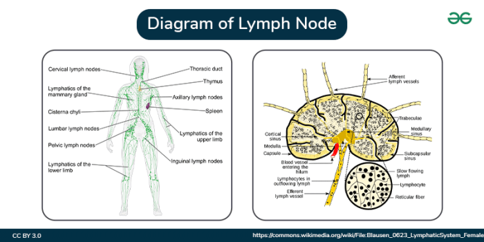
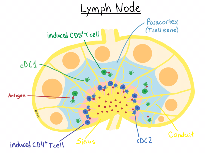
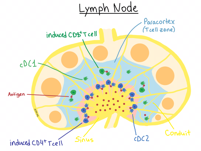
    Lymph nodes possess a characteristic bean-shaped structure, encased in a fibrous capsule. This capsule extends inward, creating partitions that divide the node into distinct regions. The outer region, known as the cortex, is densely populated with lymphocytes, primarily B cells, and dendritic cells. These cells are crucial for initiating immune responses. The inner region, the medulla, contains aggregates of lymphocytes and plasma cells, which are responsible for antibody production.

    Specialized networks of sinuses, spaces filled with lymph, permeate the cortex and medulla, facilitating the flow of lymph and the interaction of immune cells.

    Cellular Components of Lymph Nodes

    A diverse array of immune cells populate lymph nodes. Lymphocytes, specifically B cells and T cells, are paramount. B cells mature into plasma cells, producing antibodies that neutralize pathogens. T cells play a critical role in cell-mediated immunity, directly attacking infected cells. Macrophages and dendritic cells, antigen-presenting cells, capture and process foreign substances, presenting them to T cells for activation.

    Natural killer (NK) cells are also present, providing a rapid response to infected or cancerous cells. These various cell types interact within the lymph node environment, orchestrating an effective immune response.

    Lymphatic Vessels and Lymph Flow

    Lymphatic vessels form a network throughout the body, collecting lymph from tissues. These vessels converge, leading to larger vessels that eventually drain into the bloodstream. Afferent lymphatic vessels carry lymph into the lymph node, while efferent lymphatic vessels transport filtered lymph out. The one-way valves within these vessels ensure unidirectional lymph flow, preventing backflow. Lymph nodes act as checkpoints, allowing immune cells to encounter and respond to pathogens or foreign substances within the lymph.

    Comparison of Lymph Node Regions

    Region Structure Function
    Cortex Outer layer, densely packed with lymphocytes (especially B cells), dendritic cells. Contains germinal centers, where B cells proliferate and differentiate. Initial encounter of antigens with lymphocytes, initiation of immune responses, B cell maturation.
    Medulla Inner layer, contains medullary cords (aggregates of lymphocytes and plasma cells) and medullary sinuses. Antibody production by plasma cells, further filtering of lymph, interaction of immune cells.
    Sinuses Networks of spaces filled with lymph, located throughout the cortex and medulla. Passageway for lymph, allowing interaction between lymphocytes and antigens, facilitating immune cell migration.

    Lymph Node Function in Immunity: Understanding The Purpose Of Lymph Nodes

    Understanding the purpose of lymph nodes

    Lymph nodes, small bean-shaped structures strategically positioned throughout the lymphatic system, are crucial components of the immune system. They act as filters for lymph, a fluid containing immune cells and debris, playing a pivotal role in identifying and eliminating pathogens. Understanding their function in immunity provides insight into how our bodies defend against infections and maintain overall health.Lymph nodes are not just passive filters; they are dynamic hubs where immune cells interact and orchestrate responses to invading pathogens.

    This intricate process involves a complex interplay of cell-to-cell communication, antigen presentation, and activation, ultimately leading to the elimination of threats and the development of immunological memory.

    Filtering Lymph

    Lymph, a clear fluid containing white blood cells, waste products, and cellular debris, circulates throughout the lymphatic system. Lymph nodes act as checkpoints along this network, filtering the lymph as it passes through. Specialized cells within the lymph nodes, including macrophages and dendritic cells, actively scan the lymph for foreign particles, cellular debris, and pathogens. This filtering process ensures that harmful substances are removed from the lymphatic system, preventing them from spreading throughout the body.

    The effectiveness of this filtering process is crucial for maintaining the body’s overall health.

    Antigen Presentation and Immune Cell Activation

    The process of antigen presentation is central to the activation of immune cells within lymph nodes. Macrophages and dendritic cells, acting as professional antigen-presenting cells (APCs), engulf and process foreign antigens. They then present these processed antigens on their surface, combined with major histocompatibility complex (MHC) molecules. This presentation allows T lymphocytes (specifically helper T cells) to recognize the specific antigen.

    Upon recognition, T cells become activated, initiating a cascade of events leading to the activation of other immune cells, such as B cells, to eliminate the pathogen.

    Initiating and Regulating Immune Responses

    Lymph nodes play a vital role in initiating and regulating immune responses. The presence of specific antigens within the lymph node triggers a focused immune response. The organized structure of lymph nodes, with distinct zones for different immune cell types, ensures that the immune response is appropriately targeted and regulated. The interaction between various immune cells, such as T cells, B cells, and macrophages, within the lymph node is carefully orchestrated to maximize the effectiveness of the immune response.

    Different Immune Cell Interactions

    Various immune cells interact within lymph nodes to effectively combat pathogens. B cells, responsible for antibody production, encounter activated T cells presenting the same antigen. This interaction, mediated by specific surface receptors and cytokines, leads to the activation and proliferation of B cells, resulting in the production of antibodies tailored to neutralize the pathogen. T cells, another critical component of the adaptive immune response, play a crucial role in coordinating the entire process.

    Their interaction with other immune cells fine-tunes the immune response and prevents excessive inflammation.

    Understanding the purpose of lymph nodes is crucial for overall health. They’re part of our immune system, filtering out harmful substances and helping our bodies fight off infection. This intricate network plays a vital role in our well-being, much like the complexities surrounding a diagnosis like diabetes. For more information on whether diabetes is considered a disability, check out this resource: is diabetes a disability.

    Ultimately, a strong understanding of the body’s systems, like the lymphatic system, is essential for informed decisions about health and well-being.

    Activation of B and T Lymphocytes in Lymph Nodes

    Step B Lymphocyte Activation T Lymphocyte Activation
    1 Antigen encounter and uptake by B cells Antigen presentation by APC (e.g., dendritic cell) to T cell receptor (TCR)
    2 Processing and presentation of antigen on MHC class II molecules Recognition of antigen-MHC complex by T cell receptor (TCR)
    3 Interaction with helper T cell (Th) that recognizes the same antigen Activation of the T cell
    4 Activation and proliferation of B cells, leading to antibody production Release of cytokines that activate other immune cells

    This table Artikels the key steps in B and T lymphocyte activation within lymph nodes, highlighting the coordinated nature of these processes.

    Lymph Node Involvement in Disease

    Lymph nodes, tiny filters throughout the body’s lymphatic system, play a crucial role in immune responses. They are strategically positioned to encounter and trap pathogens and abnormal cells. When these nodes become involved in disease processes, they often show characteristic changes that provide valuable diagnostic clues for healthcare professionals. This involvement can arise from infections, cancerous growths, or other pathological conditions.

    Understanding these changes is essential for accurate diagnosis and appropriate treatment.Pathological changes in lymph nodes are frequently observed in a wide range of diseases, providing critical information about the underlying cause and severity of the condition. The appearance and consistency of these nodes, along with other clinical findings, contribute to the diagnostic process.

    Lymph Node Involvement in Infections, Understanding the purpose of lymph nodes

    Infections trigger an immune response, often leading to alterations in lymph node structure and function. Lymph nodes swell and become tender, a visible sign of increased activity within. This swelling is often accompanied by inflammation, characterized by redness and warmth in the surrounding tissues. Inflammatory cells, such as lymphocytes and macrophages, accumulate within the lymph node, attempting to eliminate the invading pathogens.

    Understanding the purpose of lymph nodes is crucial for overall health. They’re essentially the body’s filtration system, helping to fight off infection. Factors like blood pressure and heart rate can indirectly affect lymph node function, influencing how effectively they clear out waste products. Monitoring these vital signs, like blood pressure heart rate , can offer insights into the body’s overall health and how well the lymphatic system is functioning.

    Ultimately, understanding lymph node function is key to a healthier immune response.

    Pathological Changes in Lymph Nodes During Infections

    Lymph nodes, when challenged by pathogens, exhibit specific changes. These include an increase in the number of lymphocytes and other immune cells, resulting in enlargement. The nodes become more cellular and less organized. Macrophages, responsible for engulfing pathogens, may accumulate in significant numbers. Suppurative (pus-forming) infections can lead to the formation of pus within the node, causing localized abscesses.

    The presence of these changes can be indicative of the type and severity of the infection.

    Lymph Node Involvement in Cancer Development

    Cancer cells, like pathogens, can spread throughout the body, including the lymphatic system. Metastasis, the spread of cancer from its primary site to distant locations, frequently involves the lymphatic system. Cancer cells can enter the lymphatic vessels, traveling to regional lymph nodes. These nodes can become involved through the process of metastasis, leading to the formation of secondary tumor growths.

    Metastasis Through the Lymphatic System

    Cancer cells detach from the primary tumor and enter the lymphatic capillaries. The cells travel through the lymphatic vessels, arriving at regional lymph nodes. If the cancer cells evade the immune defenses within the nodes, they can continue to proliferate and form new tumors in these nodes. This process, metastasis, is a critical aspect of cancer progression. The extent of lymph node involvement is a key prognostic factor for the patient’s survival and guides treatment decisions.

    Diagnostic Use of Lymph Node Involvement

    Examination of lymph nodes is a critical diagnostic tool. The size, consistency, and appearance of lymph nodes can provide valuable clues about the underlying disease. Biopsies of involved lymph nodes allow pathologists to analyze the cellular composition and identify the specific type of cells present. This analysis is instrumental in diagnosing infections, malignancies, and other diseases. The presence and extent of lymph node involvement are often included in staging systems, which help determine the extent of the disease and guide treatment strategies.

    Summary Table of Diseases Affecting Lymph Nodes

    Disease Type Characteristics
    Infectious Mononucleosis Characterized by enlarged, tender lymph nodes, often in the neck and armpits. Fever and fatigue are common.
    Lymphoma Cancers of the lymphatic system, resulting in the formation of malignant tumors in lymph nodes. Various subtypes exist, each with distinct characteristics.
    Metastatic Cancer Cancer cells spread from a primary tumor site to regional lymph nodes, forming secondary tumors. The extent of metastasis significantly influences treatment strategies.
    Tuberculosis Infectious disease affecting the lungs and other organs. Can lead to granulomatous inflammation in lymph nodes.

    Lymph Node Assessment and Diagnosis

    Understanding the status of lymph nodes is crucial in diagnosing various diseases, particularly those affecting the immune system. Proper assessment methods, including physical examination and imaging techniques, are vital for identifying potential abnormalities and guiding further investigations. Accurate diagnosis hinges on a thorough evaluation of lymph node characteristics and their correlation with clinical symptoms.

    Methods of Lymph Node Assessment

    Assessment of lymph nodes involves a multifaceted approach, combining physical examination with sophisticated imaging techniques. Physical examination, often the first step, relies on palpation to detect abnormalities in size, consistency, and mobility. Imaging modalities, such as ultrasound, CT scans, and MRI, provide detailed visualization of lymph nodes, allowing for more precise evaluation of their morphology and extent of involvement.

    Understanding the purpose of lymph nodes is crucial for overall health. They’re like tiny filtering stations in your body, helping your immune system fight off infections. But sometimes, issues like allergies can lead to fatigue, which can be mistaken for other things. For instance, are you experiencing unusual tiredness? If so, exploring potential links between your allergies and overall health, like the impact on your lymph nodes, might be worthwhile.

    Knowing more about the role of your lymph nodes in your immune system can be key to understanding how your body works in dealing with allergies. Check out this informative article on can allergies make you tired to learn more.

    Lymph Node Biopsy

    A lymph node biopsy is a crucial procedure for obtaining tissue samples for microscopic examination, aiding in the definitive diagnosis of suspected pathologies. This invasive technique allows pathologists to examine the cellular composition and architecture of the lymph node, offering critical insights into the nature of any observed abnormalities.

    Lymph Node Biopsy Procedure

    The procedure for a lymph node biopsy typically involves the following steps:

    • Patient Preparation: The patient is prepped and draped, and the area around the lymph node to be biopsied is cleansed. Local anesthesia is often administered to minimize discomfort during the procedure.
    • Incision: A small incision is made over the targeted lymph node. The incision size is carefully determined based on the anticipated size of the sample needed.
    • Excision: The lymph node, or a portion of it, is carefully excised, maintaining tissue integrity as much as possible. Techniques like fine-needle aspiration (FNA) may be used for smaller or more superficial nodes.
    • Closure: The incision site is meticulously closed with sutures, and a dressing is applied.
    • Pathology Review: The excised tissue is sent to a pathologist for microscopic examination and subsequent interpretation.

    Comparison of Imaging Techniques

    Imaging Technique Advantages Disadvantages
    Ultrasound Real-time imaging, portable, relatively inexpensive Limited depth penetration, operator-dependent, not ideal for deep-seated nodes
    Computed Tomography (CT) Scan Excellent spatial resolution, multiplanar imaging, can evaluate adjacent structures Exposure to ionizing radiation, potential for contrast reactions
    Magnetic Resonance Imaging (MRI) Excellent soft-tissue contrast, allows for detailed visualization of nodal architecture Longer scan times, can be more expensive than CT

    Significance of Lymph Node Characteristics

    The size, consistency, and location of lymph nodes can offer valuable clues in the diagnostic process. Enlarged lymph nodes (lymphadenopathy) often signal an underlying inflammatory or neoplastic process. Hard or firm consistency might suggest malignancy, whereas soft consistency may point to an inflammatory condition. The location of the enlarged nodes can also provide clues about the possible source of the disease.

    Lymph Node Pathologies and Imaging Appearances

    Pathology Typical Imaging Appearance
    Reactive Lymphadenitis Enlarged, sometimes tender, nodes; often with homogeneous appearance on imaging.
    Metastatic Cancer Enlarged, firm, or hard nodes, often with irregular margins on imaging; may show calcification or necrosis.
    Lymphoma Enlarged, sometimes matted nodes; may have heterogeneous appearance, and potentially show a mass effect on surrounding structures on imaging.

    Lymph Node Imaging and Biopsy

    Understanding lymph nodes is crucial for diagnosing various medical conditions. Imaging techniques and biopsies play vital roles in evaluating lymph nodes, helping clinicians determine the presence and nature of abnormalities. This section delves into the specifics of these procedures, their preparation, and potential findings.

    Common Imaging Techniques for Visualizing Lymph Nodes

    Various imaging modalities aid in visualizing lymph nodes, providing crucial information about their size, shape, and internal structure. These techniques are often employed to detect abnormalities before biopsy is necessary.

    • Ultrasound (US): Ultrasound uses high-frequency sound waves to create images of the internal structures. It is a non-invasive, relatively inexpensive, and readily available technique, particularly useful for superficial lymph nodes. Real-time imaging allows for dynamic assessment of the nodes during the procedure. It’s particularly helpful in evaluating the presence of fluid collections (cysts) or masses within the nodes.

    • Computed Tomography (CT): CT scans utilize X-rays and sophisticated computer processing to generate cross-sectional images of the body. CT provides detailed anatomical information, including the size, location, and relationship of lymph nodes to surrounding structures. It’s frequently used to assess deeper lymph nodes and evaluate the extent of disease involvement. Contrast agents may be administered to enhance the visibility of lymph nodes and surrounding tissues.

    • Magnetic Resonance Imaging (MRI): MRI utilizes strong magnetic fields and radio waves to produce detailed images of the body’s internal structures. MRI excels at distinguishing between different soft tissues, providing valuable information about the characteristics of lymph nodes. It’s often employed to evaluate the presence of inflammation, edema, or specific types of tumors within the nodes. The use of contrast agents further enhances the visualization of subtle abnormalities.

    Types of Lymph Node Biopsies

    A biopsy is a procedure to obtain a tissue sample for microscopic examination. Different types of biopsies are performed depending on the location and size of the lymph node, as well as the suspected pathology.

    • Excisional Biopsy: This involves the complete removal of the lymph node. It is often chosen for smaller, superficial nodes where the complete removal is feasible and does not compromise important structures. This approach is typically preferred for suspected benign conditions.
    • Incisional Biopsy: This procedure involves removing a portion of the lymph node. It is a less invasive alternative to an excisional biopsy, particularly useful for larger nodes or those located in areas with complex anatomy. An incisional biopsy provides a representative sample for pathologic evaluation.

    Preparation for Lymph Node Imaging Techniques

    Preparation for imaging procedures varies depending on the specific technique. For ultrasound, no special preparation is usually needed. CT and MRI scans may require fasting or the administration of contrast agents.

    • Ultrasound: No special preparation is generally required, except for removing any loose clothing or jewelry that may interfere with the procedure. The patient may be asked to drink water or other liquids to help with distention of the targeted area.
    • CT: Patients are typically required to fast for a certain period before the scan, depending on the type of contrast agent used. Patients should inform the technician about any allergies or previous reactions to contrast agents. If contrast is required, intravenous access is typically established.
    • MRI: Preparation for MRI is similar to that of CT. Fasting may be required, and patients should inform the technician about any implanted medical devices or metal in the body.

    Preparation for a Lymph Node Biopsy

    Preparation for a lymph node biopsy is dependent on the type of biopsy and the patient’s overall health. The physician will provide specific instructions.

    • General Instructions: Patients are often asked to fast for a few hours before the procedure. Inform the medical team about any medications, allergies, or medical conditions. Any relevant medical history and current medications should be disclosed to the physician.

    Typical Findings on Lymph Node Imaging

    Lymph node imaging findings can vary depending on the underlying pathology.

    Pathology Typical Imaging Findings
    Benign Lymphadenopathy Enlarged lymph nodes, typically symmetrical, with smooth margins, and no internal calcifications.
    Malignant Lymphoma Enlarged lymph nodes, often asymmetrical, with irregular margins and possible internal calcifications or necrosis.
    Metastatic Disease Enlarged lymph nodes, often multiple, with varying sizes and shapes, and possible internal calcifications or masses.

    Interpreting Lymph Node Biopsy Results

    Pathologists meticulously examine the biopsy specimens under a microscope. Microscopic examination helps determine the nature of the cells and tissues within the lymph node.

    • Microscopic Evaluation: The pathologist analyzes the cellular architecture, the presence of atypical cells, and any signs of inflammation or infection. The results of this analysis are crucial in determining the diagnosis and appropriate treatment plan.

    Lymph Node Location and Drainage

    Understanding the intricate network of lymphatic drainage is crucial for comprehending the body’s immune responses and disease processes. This system, a vital component of the circulatory system, plays a critical role in transporting lymph, a fluid containing immune cells, throughout the body. Knowledge of lymphatic drainage patterns is indispensable in diagnosing and staging various illnesses, particularly cancers.

    Major Lymph Node Groups

    The lymphatic system comprises a vast network of vessels and nodes. Major lymph node groups are strategically positioned throughout the body, acting as filters for lymph. These groups are interconnected, ensuring efficient drainage and immune surveillance. Their locations and drainage patterns are essential for interpreting diagnostic findings.

    Drainage Patterns of Different Body Regions

    The lymphatic drainage follows specific pathways, conveying lymph from different body regions to particular lymph node groups. The upper extremities, for instance, drain into axillary lymph nodes, while the lower extremities drain into inguinal lymph nodes. Head and neck regions drain into cervical lymph nodes. This directional flow facilitates the identification of the origin of infections or malignancies.

    Clinical Significance of Lymphatic Drainage Patterns

    Understanding the lymphatic drainage patterns is of paramount importance in clinical settings. Knowing the drainage pathways allows healthcare professionals to accurately identify the region of origin of an infection or tumor. For instance, if swelling or abnormalities are detected in the axillary lymph nodes, it might suggest a problem originating in the upper limb or breast region. This crucial information aids in determining the appropriate diagnostic procedures and treatment strategies.

    Lymph Node Drainage in Cancer Diagnosis and Staging

    In cancer diagnosis and staging, the lymphatic drainage patterns are invaluable. Cancer cells can spread through the lymphatic system, metastasizing to distant lymph nodes. Assessing the presence and extent of metastasis in regional lymph nodes provides critical information for determining the stage of the disease. This knowledge is crucial for planning appropriate treatment strategies and predicting patient outcomes.

    The higher the number of lymph nodes involved, the more advanced the stage of the cancer is often considered.

    Lymphatic Drainage of the Upper Limb

    The lymphatic drainage of the upper limb follows a predictable pattern, originating at the fingertips and progressing upwards.

    Body Region Drainage to Explanation
    Fingers, Palm Anterior axillary lymph nodes Lymphatic vessels ascend along the veins of the hand and arm, converging at the axillary lymph nodes.
    Lateral forearm Lateral axillary lymph nodes The lateral aspect of the forearm drains to the lateral axillary nodes.
    Medial forearm Anterior axillary lymph nodes The medial forearm drains to the anterior axillary lymph nodes.
    Upper arm Apical and central axillary lymph nodes The lymph from the upper arm ultimately flows to the apical and central axillary lymph nodes.

    This detailed drainage system allows for precise identification of the location and extent of potential spread of cancer from the upper limb. This understanding is crucial for staging and treatment planning.

    Last Point

    In conclusion, understanding the purpose of lymph nodes is crucial for comprehending the intricate workings of the immune system. Their function in filtering lymph, initiating immune responses, and involvement in disease processes is undeniable. We’ve explored the structure, function, and clinical implications of lymph nodes, emphasizing their significance in disease diagnosis and treatment. This comprehensive overview should equip readers with a deeper understanding of these essential components of the human body.

  • What is a Sphincter? Bodys Gatekeepers

    What is a Sphincter? Bodys Gatekeepers

    What is a sphincter? These remarkable little muscles act as gatekeepers throughout the human body, controlling the flow of substances. From regulating digestion to managing urination, sphincters play a crucial role in maintaining bodily functions. This exploration delves into the fascinating world of sphincters, examining their diverse types, mechanisms, associated disorders, and specific examples in detail. We’ll also compare their structure and function across various species, providing a comprehensive overview of these essential body components.

    Sphincters are circular muscles that constrict or relax to control the passage of materials. They’re found in various locations throughout the body, including the digestive, urinary, and respiratory tracts. Understanding their intricate workings is key to grasping how our bodies function seamlessly.

    Defining Sphincters

    What is a sphincter

    Sphincters are crucial components of the human body, acting as valves to regulate the passage of substances through various openings. They play a vital role in maintaining homeostasis and preventing unwanted leakage or flow. Understanding their diverse functions and locations is essential for comprehending overall bodily processes.A sphincter is a ring-like muscle that contracts and relaxes to control the opening and closing of a body passage.

    Their ability to regulate flow is paramount in preventing the backflow or leakage of substances, whether that’s food, waste, or other fluids. These muscles are strategically positioned to perform this vital function.

    Types of Sphincters

    Sphincters are classified into different categories based on their location and function. Recognizing these distinctions is crucial to understanding their specific roles.

    • Gastrointestinal Sphincters: These sphincters are situated within the digestive system, regulating the passage of food and other substances along the alimentary canal. Their precise positioning and controlled contractions are vital for efficient digestion and absorption.
    • Urinary Sphincters: Located in the urinary tract, these sphincters manage the flow of urine from the bladder to the outside of the body. Their ability to contract and relax is essential for voluntary and involuntary control of urination.
    • Anal Sphincters: These sphincters, comprising both internal and external components, regulate the passage of feces from the rectum to the anus. The interplay of these muscles is key to maintaining bowel control.
    • Vascular Sphincters: These are found in the circulatory system. They control blood flow within capillaries, adjusting the amount of blood reaching specific tissues. This is vital for temperature regulation and other physiological responses.

    Anatomical Locations of Sphincters

    Understanding the precise location of sphincters is vital for grasping their specific functions. The strategic placement of these muscles is crucial to their roles in regulating the flow of substances through various openings.

    • Gastrointestinal: The esophagus, stomach, small intestine, and large intestine all have sphincters. The esophageal sphincter regulates the passage of food from the esophagus into the stomach, while the pyloric sphincter controls the flow of food from the stomach to the small intestine. The ileocecal sphincter connects the small and large intestines. Numerous others are present throughout the gastrointestinal tract.

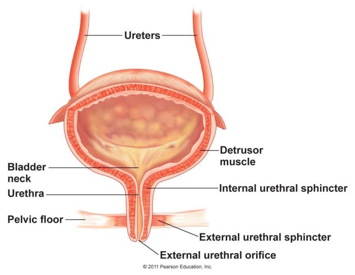
    • Urinary: The internal urethral sphincter is located at the junction of the bladder and the urethra. The external urethral sphincter surrounds the urethra as it passes through the pelvic floor. These sphincters work together to control the release of urine.
    • Anal: The internal anal sphincter is located within the rectum, while the external anal sphincter is located in the pelvic floor muscles. Both are crucial for maintaining continence.
    • Vascular: Vascular sphincters are situated within the walls of arterioles, the smallest arteries, and capillaries.

    Comparative Analysis of Sphincter Types

    This table summarizes the key characteristics of different sphincter types.

    Sphincter Type Location Function Structure
    Gastrointestinal Esophagus, stomach, intestines Regulate food and waste movement Circular muscles
    Urinary Bladder and urethra Control urine flow Circular muscles
    Anal Rectum and anus Control fecal expulsion Circular and longitudinal muscles
    Vascular Arterioles and capillaries Control blood flow Smooth muscle

    Sphincter Mechanisms

    Sphincters, those crucial ring-like muscles, are far more than simple gatekeepers. Their intricate mechanisms ensure controlled flow of substances, from food to waste, through various bodily pathways. Understanding these mechanisms is key to appreciating the sophistication and precision of the human body. The following exploration delves into the muscular, neurophysiological, and hormonal interplay that dictates sphincter function.Sphincter action is a finely tuned dance between muscular contraction and relaxation, orchestrated by complex neurophysiological pathways and subtly influenced by hormones.

    These mechanisms ensure precise control over the passage of materials through the body, safeguarding against leaks and ensuring efficient digestion and waste elimination.

    Muscular Mechanisms in Sphincter Function

    The precise functioning of sphincters hinges on the interplay of various muscle types. These muscles are not monolithic entities but rather intricate networks of contractile fibers. Smooth muscle, with its slow, rhythmic contractions, plays a critical role in maintaining basal tone and responding to stimuli. Skeletal muscle, controlled consciously, allows for voluntary control in some sphincters.

    • Smooth Muscle: This involuntary muscle type, found in the walls of internal organs, exhibits sustained contractions for maintaining a baseline pressure gradient, known as basal tone. This constant tension prevents unwanted passage and is crucial for maintaining the integrity of the canal.
    • Skeletal Muscle: Skeletal muscle, innervated by the somatic nervous system, allows for conscious control over sphincter function. This is particularly important for voluntary actions like urination and defecation.

    Neurophysiological Control of Sphincters

    Neurological control is fundamental to sphincter regulation. A complex interplay of nerves, signals, and pathways governs the intricate dance of contraction and relaxation. The autonomic nervous system, comprised of the sympathetic and parasympathetic branches, plays a pivotal role.

    • Autonomic Nervous System: The autonomic nervous system, divided into sympathetic and parasympathetic branches, regulates the involuntary contractions and relaxations. The sympathetic system generally inhibits the sphincter, while the parasympathetic system promotes relaxation, allowing the passage of substances.
    • Sensory Input: Sensory receptors in the surrounding tissues send signals to the brain, providing feedback on the status of the sphincter. This sensory input is crucial for adjusting the degree of contraction or relaxation, ensuring precise control.

    Role of Hormones in Sphincter Regulation

    Hormones, chemical messengers of the body, can also modulate sphincter activity. Their influence, although often subtle, can be significant in certain circumstances.

    • Hormonal Influences: Hormones, such as those involved in the digestive process, can subtly influence sphincter activity. For example, certain hormones can affect the motility of the gut, thereby impacting the tone and responsiveness of the sphincters along the digestive tract.

    Steps in Sphincter Contraction and Relaxation

    The process of sphincter contraction and relaxation involves a coordinated sequence of events.

    1. Stimulus: A variety of stimuli, ranging from nervous signals to hormonal cues, initiate the process.
    2. Signal Transmission: The signals are transmitted through nerves, triggering the necessary muscular responses.
    3. Muscle Contraction/Relaxation: Smooth muscle contracts to close the sphincter or relaxes to open it, while skeletal muscle responds according to voluntary commands. The exact mechanism depends on the type of muscle involved.
    4. Feedback: The body constantly monitors the state of the sphincter, ensuring proper function.

    Physiological Changes During Sphincter Opening and Closing

    Opening and closing a sphincter are accompanied by noticeable physiological changes. The pressure gradient across the sphincter is the key factor.

    Action Pressure Gradient Physiological Changes
    Sphincter Opening Decreased pressure Relaxation of the sphincter muscles, allowing passage of materials
    Sphincter Closing Increased pressure Contraction of the sphincter muscles, obstructing passage

    Sphincter Disorders

    Sphincters, those crucial gatekeepers of our bodies, can malfunction, leading to a range of uncomfortable and sometimes debilitating conditions. Understanding these disorders is essential for recognizing the symptoms, seeking appropriate treatment, and ultimately improving quality of life for those affected. From the subtle discomfort of occasional leakage to the more severe complications of chronic dysfunction, sphincter disorders impact various aspects of daily life.

    So, what exactly is a sphincter? Basically, it’s a ring of muscle that controls the opening and closing of a passageway in the body. Think about how important these are in everyday functions, like digestion and controlling the flow of waste. And while we’re on the topic of things that control flow, have you ever wondered about the impact of certain foods on hormones?

    For example, Foods with Estrogen Eat or Avoid? can be a real factor in overall well-being. Knowing what we eat can have a surprising impact on these crucial functions, including the ones controlled by sphincters.

    Common Sphincter Malfunctions

    Sphincter malfunctions encompass a spectrum of problems, varying in severity and origin. These issues can affect both the voluntary and involuntary control of sphincter muscles, impacting bowel and bladder function. The diverse range of causes underscores the complexity of these disorders.

    Examples of Sphincter Dysfunction

    Examples of sphincter dysfunction include incontinence, both urinary and fecal, and anal fissures. Urinary incontinence can manifest as a frequent, involuntary leakage of urine, impacting daily routines and social interactions. Fecal incontinence involves involuntary loss of stool, causing significant discomfort and potentially leading to embarrassment. Anal fissures, small tears in the anal lining, can result in painful bowel movements, and are often associated with straining during defecation.

    Sphincters are those amazing little muscles that control things like bowel movements and bladder release. Understanding how they work is crucial, and it’s often linked to the need for proper health insurance. For example, if you’re considering a new job or have a pre-existing condition, knowing What is Minimum Essential Coverage and Why Does It Matter? becomes incredibly important.

    This knowledge can help you ensure you have the right coverage to protect your health, which ultimately influences the health of those important sphincter muscles, too.

    These are just a few examples, highlighting the diverse ways sphincter dysfunction can impact daily life.

    Ever wondered what a sphincter is? Basically, it’s a ring of muscle that controls the opening and closing of certain body passages. Thinking about that, I was curious about how air purifiers affect dust, especially considering their role in keeping things like the airways clear. This article on Do Air Purifiers Help With Dust? A Deep Dive dives into that question, and ultimately, it’s fascinating to see how similar mechanisms work in both the human body and our efforts to control the air around us.

    So, next time you think about a sphincter, remember its job of controlling openings – it’s a pretty crucial function!

    Symptoms of Sphincter-Related Problems

    Symptoms associated with sphincter-related problems can range from mild to severe. Common symptoms include urinary incontinence (frequent, involuntary urination), fecal incontinence (involuntary loss of stool), painful bowel movements (especially with anal fissures), and a sensation of incomplete emptying of the bladder or bowel. These symptoms can significantly impact quality of life, causing anxiety, social isolation, and emotional distress.

    Causes of Sphincter Disorders

    The causes of sphincter disorders are varied and often complex. These can include neurological conditions like multiple sclerosis or spinal cord injuries, which can disrupt the nerve signals controlling sphincter muscles. Chronic conditions like diabetes can also impact sphincter function. Damage to the sphincter muscles from childbirth, trauma, or surgery can also be a factor. Furthermore, certain medications and lifestyle choices, such as prolonged straining during bowel movements or inadequate fluid intake, can exacerbate existing conditions.

    Understanding these diverse factors is crucial for effective diagnosis and treatment.

    Treatment Options for Sphincter Dysfunction

    Treatment options for sphincter dysfunction are tailored to the specific cause and severity of the condition. Conservative approaches, such as dietary modifications, pelvic floor exercises, and biofeedback therapy, can often improve symptoms. Medications, including those that address underlying conditions like diabetes, may also play a role in treatment. In more severe cases, surgical interventions, such as sphincteroplasty (repair or reconstruction of the sphincter), may be necessary.

    The decision of the most appropriate treatment plan is made collaboratively between the patient and healthcare provider.

    Table of Sphincter Disorders and Treatments, What is a sphincter

    Disorder Symptoms Causes Treatment
    Urinary Incontinence Frequent, involuntary urination Neurological conditions, childbirth trauma, medications, aging Pelvic floor exercises, biofeedback, medications, surgery (in severe cases)
    Fecal Incontinence Involuntary loss of stool Neurological conditions, chronic constipation, childbirth trauma, surgery Dietary modifications, pelvic floor exercises, medications, surgery (in severe cases)
    Anal Fissures Painful bowel movements, bleeding Straining during bowel movements, chronic constipation, inflammatory bowel disease Dietary changes, stool softeners, topical creams, surgery (in severe cases)

    Sphincter Examples in Detail

    Sphincters, those ring-like muscles, play a crucial role in regulating the passage of substances through various parts of the body. Understanding their function is key to comprehending how our digestive and urinary systems operate. From controlling food movement to preventing accidental leakage, sphincters are essential components of human physiology.

    Anal Sphincter in Digestion

    The anal sphincter, composed of two major muscles, the internal and external anal sphincters, is a crucial component in the digestive process. The internal sphincter, involuntary, maintains continence during rest. The external sphincter, under voluntary control, allows for conscious control of bowel movements. These muscles work in concert to prevent leakage and regulate the expulsion of feces.

    This coordinated action ensures that bowel movements occur when desired, avoiding accidental soiling.

    Urinary Sphincter in Urination

    The urinary sphincter complex comprises two parts: the internal urethral sphincter and the external urethral sphincter. The internal sphincter, an involuntary muscle, prevents urine leakage while the bladder is filling. The external urethral sphincter, a voluntary muscle, provides conscious control over urination. This intricate interplay of voluntary and involuntary muscles allows for precise control of urination, enabling individuals to urinate when and where it is appropriate.

    Esophageal Sphincter in Swallowing

    The esophageal sphincter, specifically the lower esophageal sphincter (LES), is a critical component in the swallowing process. It acts as a valve, relaxing to allow food to pass into the stomach and contracting to prevent reflux of stomach contents into the esophagus. The rhythmic contractions and relaxation of the LES ensure that food moves unidirectionally, preventing regurgitation. A malfunctioning LES can lead to acid reflux, a common digestive issue.

    Pyloric Sphincter in Digestion

    The pyloric sphincter is a ring of muscle at the junction of the stomach and the duodenum (the first part of the small intestine). Its role is to regulate the passage of partially digested food (chyme) from the stomach into the small intestine. The pyloric sphincter’s controlled opening and closing ensures that chyme is released gradually into the small intestine, allowing for optimal digestion and nutrient absorption.

    Bladder Sphincter in Urinary Retention

    The bladder sphincter, a complex of muscles including the internal and external urethral sphincters, plays a pivotal role in urinary retention. The internal sphincter, involuntary, prevents leakage during bladder filling. The external sphincter, voluntary, allows for conscious control of urination. The coordinated function of these muscles ensures that urine is retained in the bladder until the individual is ready to urinate.

    Structure of the Cardiac Sphincter

    The cardiac sphincter, also known as the lower esophageal sphincter (LES), is not a distinct, separate structure, but rather a specialized region of the esophageal musculature. It is formed by the circular muscles of the esophagus at the junction with the stomach. The LES’s function is to prevent stomach acid from refluxing into the esophagus. Its structure consists of a thickened ring of smooth muscle fibers.

    Illustrations of Sphincters

    Anal Sphincter: A cross-section of the anal canal would show the internal and external anal sphincter muscles encircling the anal canal. The internal sphincter, located deeper, is involuntary. The external sphincter, closer to the surface, is under voluntary control.
    Urinary Sphincter: A diagram of the bladder and the surrounding structures would depict the internal urethral sphincter as a ring of muscle at the bladder neck, and the external urethral sphincter located more distally.

    Comparative Analysis of Sphincters

    What is a sphincter

    Sphincters, those crucial ring-like muscles, play a vital role in controlling the passage of substances throughout the body. Their diverse structures and functions are essential for maintaining homeostasis. This section delves into a comparative analysis, highlighting the similarities and differences in structure, function, mechanisms, evolutionary adaptations, and the roles they play across various species.Different sphincters, despite their shared function, exhibit variations in their structural makeup and functional mechanisms, reflecting the diverse demands of different bodily systems.

    Understanding these variations provides a deeper appreciation for the complexity and elegance of biological design.

    Structure and Function of Different Sphincters

    Various sphincters exhibit distinct structural features tailored to their specific roles. The internal anal sphincter, for instance, is a smooth muscle, whereas the external anal sphincter is composed of skeletal muscle. These differences in muscle type correlate directly with the function and control of each sphincter. The smooth muscle of the internal sphincter provides involuntary control, while the skeletal muscle of the external sphincter allows for voluntary control.

    This dual control system ensures proper bowel function. Similarly, the pyloric sphincter in the stomach has a unique structure that facilitates the regulated release of chyme into the small intestine.

    Mechanisms of Action of Various Sphincters

    The mechanisms by which sphincters regulate passage vary significantly. The pyloric sphincter, for example, uses a complex interplay of smooth muscle contractions and relaxation to regulate the emptying of the stomach. These contractions are coordinated by intricate neural signals and hormones. In contrast, the urinary sphincters, composed of both smooth and skeletal muscles, utilize both involuntary and voluntary control mechanisms.

    The smooth muscle provides continuous tone, while the skeletal muscle allows for conscious control over urination.

    Evolutionary Adaptations of Sphincters

    The evolution of sphincters reflects the adaptation of organisms to different environmental pressures and dietary habits. In herbivores, for example, the sphincters regulating the digestive tract are often more complex and elaborate compared to carnivores, due to the greater complexity of digesting plant matter. This evolutionary pressure resulted in specialized adaptations to accommodate different diets and lifestyles.

    Role of Sphincters in Different Species

    Sphincters are critical for the survival of numerous species. In marine animals, sphincters regulate the expulsion of waste products. In birds, they control the passage of food through the digestive system. The specific roles of sphincters can vary greatly depending on the species and its unique ecological niche.

    Comparative Analysis Table

    Sphincter Structure Function Mechanism
    Pyloric Sphincter Smooth muscle Regulates emptying of stomach contents into small intestine Contractions and relaxation coordinated by neural and hormonal signals
    Internal Anal Sphincter Smooth muscle Provides involuntary control over bowel movements Continuous tonic contraction regulated by autonomic nervous system
    External Anal Sphincter Skeletal muscle Provides voluntary control over bowel movements Conscious control via somatic nervous system
    Urethral Sphincters Smooth and skeletal muscle Regulates the flow of urine Involuntary tone from smooth muscle; voluntary control from skeletal muscle

    Ultimate Conclusion: What Is A Sphincter

    In conclusion, sphincters are vital for maintaining homeostasis within the body. Their precise control over the passage of substances is critical for various bodily processes. We’ve explored their mechanisms, disorders, and diverse roles in the body. The next time you experience a natural bodily function, remember the remarkable sphincters working diligently behind the scenes to ensure smooth operation.

    Understanding their importance can lead to better appreciation of the complexity and efficiency of the human body.