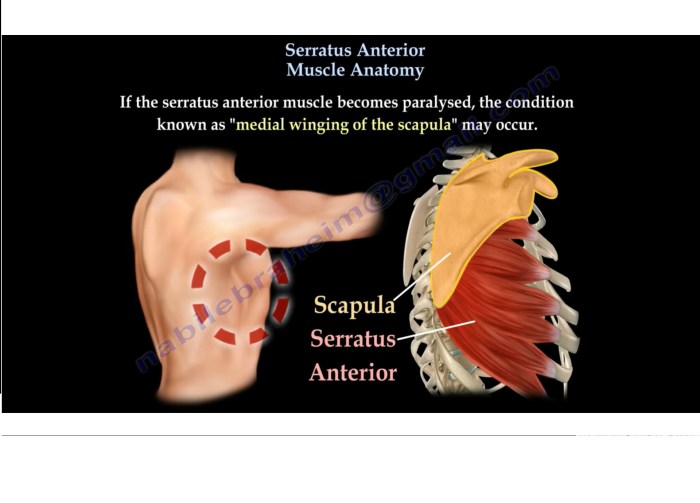
Category: Human Anatomy

  • Internal Jugular Vein Anatomy A Deep Dive

    Internal Jugular Vein Anatomy A Deep Dive

    Internal jugular vein anatomy sets the stage for this enthralling narrative, offering readers a glimpse into the intricate network of vessels that power our bodies. We’ll explore the vein’s position amidst other neck structures, examining its crucial role in venous return and its significance in various clinical scenarios. From anatomical landmarks to surgical considerations, this journey promises to unveil the complexities of this vital vessel.

    This comprehensive exploration delves into the internal jugular vein’s structure, its course through the neck, and its tributaries. We’ll examine potential variations and anomalies, understanding how they might impact surgical procedures. The discussion will also highlight the vein’s clinical significance in venous access and various medical conditions. Further, the role of imaging techniques in visualizing and diagnosing related issues will be covered, providing a holistic understanding of this essential anatomical feature.

    Anatomical Position and Relations

    The internal jugular vein, a crucial component of the neck’s venous drainage system, plays a vital role in returning blood from the brain and head to the heart. Understanding its precise location and relationship to surrounding structures is essential for both anatomical knowledge and clinical applications, particularly in procedures involving the neck region. This section will delve into the anatomical position and relations of the internal jugular vein, highlighting its proximity to other vessels and structures.The internal jugular vein, nestled within the deep cervical region, is intricately associated with the carotid artery.

    Their close proximity is a significant factor to consider, as both structures are susceptible to injury or compression during surgical procedures or trauma. A thorough understanding of these relationships ensures safe and effective interventions.

    Location in Relation to Other Neck Structures

    The internal jugular vein courses vertically along the neck, situated laterally to the trachea and esophagus. It’s situated deep to the sternocleidomastoid muscle, providing a significant landmark for its identification. Crucially, it lies medial to the carotid sheath, which houses the common carotid artery, internal carotid artery, and vagus nerve. This relationship is vital to understand, as damage to one structure can compromise the function of the others.

    Relationship to the Carotid Artery and Other Vessels

    The internal jugular vein and the carotid artery are intimately intertwined within the carotid sheath. This close proximity means potential for compression or injury during procedures or traumatic events. The vagus nerve, another important component of the carotid sheath, is also in close proximity, and its delicate nature must be considered during any surgical intervention in the region.

    Careful attention must be paid to avoid damaging these structures.

    Anatomical Landmarks for Identification

    Several anatomical landmarks facilitate the identification of the internal jugular vein. The sternocleidomastoid muscle provides a superficial reference point. The clavicle, particularly its medial third, offers another significant landmark. Furthermore, the intersection of the anterior border of the sternocleidomastoid muscle and the clavicle often indicates the general region where the internal jugular vein can be found.

    Table of Surrounding Structures

    Structure Proximity Potential Interaction
    Carotid Artery Close Potential for compression or injury during surgical procedures or trauma. Careful dissection is necessary to avoid iatrogenic damage.
    Vagus Nerve Close Potential for damage during surgical procedures, especially in the neck region.
    Trachea Lateral The trachea is a vital airway structure and should be carefully preserved during any procedure.
    Esophagus Posterior The esophagus is a critical part of the digestive system, and its integrity should be maintained.
    Subclavian Vein Superior The junction of the subclavian vein and the internal jugular vein forms the brachiocephalic vein. This juncture should be considered in procedures involving the upper thorax and neck.

    Vessel Structure and Course

    Internal jugular vein anatomy

    The internal jugular vein, a crucial component of the circulatory system, plays a vital role in returning deoxygenated blood from the head and neck back to the heart. Understanding its intricate structure and course is essential for comprehending its function and clinical relevance. This section delves into the layers surrounding the vein, its detailed pathway, and the tributaries that contribute to its flow.The internal jugular vein’s journey is a complex interplay of anatomical structures, ensuring efficient blood drainage.

    Its structure and course are vital to its function, and knowing its tributaries helps in understanding the broader circulatory system.

    Layers Surrounding the Internal Jugular Vein

    The internal jugular vein, like other major vessels, is encased within connective tissue layers that provide support and protection. These layers are crucial for maintaining the vein’s integrity and preventing injury. The layers surrounding the vein include the tunica intima, tunica media, and tunica externa, as well as the surrounding areolar connective tissue. These layers contribute to the vessel’s resilience and ability to withstand internal pressure.

    These tissues are also crucial for the vein’s stability within the neck.

    Course of the Internal Jugular Vein

    The internal jugular vein originates at the base of the skull, more specifically at the junction of the sigmoid sinus and the confluence of sinuses, at the jugular foramen. It descends vertically along the lateral aspect of the neck, passing deep to the sternocleidomastoid muscle. As it progresses, it gradually gains in size, receiving numerous tributaries. It merges with the subclavian vein to form the brachiocephalic vein, and eventually the superior vena cava.

    This journey through the neck involves navigating various structures, highlighting the intricate relationships within the vascular system.

    Major Tributaries of the Internal Jugular Vein

    Several veins contribute to the flow of blood into the internal jugular vein. These tributaries, crucial for overall blood drainage, include the lingual vein, facial vein, pharyngeal veins, and the retromandibular vein. Understanding the tributaries is essential for comprehending the overall venous drainage from the head and neck.

    • Lingual Vein: Drains blood from the tongue.
    • Facial Vein: Drains blood from the face and scalp.
    • Pharyngeal Veins: Drain blood from the pharynx.
    • Retromandibular Vein: Drains blood from the parotid gland, the superficial temporal and maxillary veins.

    Diagram of Internal Jugular Vein

    A diagram illustrating the structure and course of the internal jugular vein is essential for visualization. The diagram should clearly depict the vein’s origin at the jugular foramen, its descent along the neck, and its confluence with the subclavian vein to form the brachiocephalic vein. The diagram should also highlight the major tributaries connecting to the internal jugular vein, emphasizing their contributions to the overall blood drainage.

    Understanding the internal jugular vein’s intricate anatomy is crucial for medical professionals. However, sometimes the most effective therapies for cognitive decline, like therapeutic doll therapy in dementia , can unexpectedly illuminate the intricate neural pathways that affect even the smallest vessels in the body. This fascinating interplay between seemingly disparate fields underscores the complexity of human health and the need for innovative approaches to care.

    The diagram should showcase the relationship of the vein to surrounding structures such as the carotid artery, which is often found close to it. The diagram would emphasize the vein’s descent through the neck and its termination point.

    Variations and Anomalies

    The internal jugular vein, a crucial component of the venous drainage system in the neck, isn’t always a perfectly symmetrical structure. Variations in its course, size, and connections with other vessels are common. Understanding these variations is critical for surgeons performing procedures in the neck, as unexpected anatomical differences can lead to complications. These deviations from the typical anatomy necessitate careful preoperative planning and intraoperative vigilance.

    Common Variations in Course

    Variations in the internal jugular vein’s course can involve changes in its origin, its relation to adjacent structures, and its confluence with other vessels. For example, the vein may originate slightly higher or lower than typical, or it might exhibit a more tortuous or angled path. These alterations can be subtle or more pronounced. Such variations can affect the accessibility of the vein for cannulation during central venous access procedures or affect the precise location of surgical incisions during neck surgeries.

    Variations in Structure

    Variations in the internal jugular vein’s structure encompass a range of anomalies, from minor variations in caliber to significant anatomical differences. The vein’s diameter can be larger or smaller than expected, and its walls might exhibit differing thicknesses. Sometimes, there are variations in the presence or absence of tributaries. These structural differences can impact the vein’s ability to effectively drain blood from the head and neck, and these variances might affect the vein’s ability to withstand pressure changes or be susceptible to injury during procedures.

    Examples of Anomalies and Their Impact

    One example is a duplicated internal jugular vein. This anomaly, where a second, smaller vein runs alongside the main vein, can be a significant challenge during surgical procedures. Incorrect identification of the main vein could lead to accidental injury to the accessory vein or inadequate drainage. Another example is an abnormally high or low origin of the vein.

    This variation could necessitate modifying surgical approaches or cannulation techniques to avoid injuring the vein or its surrounding structures.

    Impact on Surgical Procedures

    Variations in the internal jugular vein’s course and structure can directly impact surgical procedures in the neck region. For instance, an aberrant course might make accessing the vein more challenging, increasing the risk of injury to adjacent structures. Similarly, variations in size or presence of tributaries could affect the ease and safety of cannulation.

    Table Comparing Typical Anatomy with Common Anomalies

    Typical Anatomy Anomaly Impact
    Single internal jugular vein, originating from the confluence of subclavian and brachiocephalic veins Duplicated internal jugular vein Increased risk of injury to the accessory vein during surgical procedures; difficulty in cannulation; requires meticulous anatomical assessment preoperatively.
    Internal jugular vein typically located laterally to the carotid artery Internal jugular vein positioned more medially Potential for increased risk of injury to the carotid artery during surgical procedures; requires more careful surgical planning.
    Internal jugular vein generally of a consistent caliber Significant variation in internal jugular vein caliber Can affect the success of central venous access procedures; necessitates adjustments in cannulation techniques.

    Clinical Significance

    The internal jugular vein, a crucial component of the venous system, plays a vital role in returning deoxygenated blood from the head and neck back to the heart. Its anatomical position, structure, and relationships with surrounding structures make it clinically significant in a wide range of conditions and procedures. Understanding its function and potential pathologies is essential for medical professionals.The internal jugular vein’s importance extends beyond its basic physiological role.

    Its close proximity to other vital structures and its unique course within the neck make it a target for both diagnostic and therapeutic interventions. Its involvement in venous return mechanisms and central venous access makes it a key player in many clinical scenarios.

    Role in Venous Return

    The internal jugular vein, acting as a major venous pathway, efficiently collects blood from the brain, face, and neck. Its confluence with the subclavian vein forms the brachiocephalic vein, which ultimately drains into the superior vena cava, returning the blood to the right atrium of the heart. This direct pathway is critical for maintaining adequate blood circulation and preventing the accumulation of blood in the head and neck regions.

    Understanding the internal jugular vein’s intricate anatomy is crucial for medical professionals. A healthy immune system is essential for overall well-being, and incorporating foods like citrus fruits and leafy greens into your diet can significantly support this. These nutrients can help bolster your immune system, which, in turn, plays a vital role in maintaining the health of your circulatory system, including the internal jugular vein, which is a key component of blood circulation.

    For a more in-depth look at foods that boost immune system function, check out this helpful resource: foods that boost immune system. Knowing how to support your body’s natural defenses is important for overall health and well-being, and understanding the internal jugular vein’s role in that process is equally important.

    This venous return is essential for removing metabolic waste products and delivering fresh oxygenated blood to the tissues.

    Clinical Relevance in Various Conditions

    The internal jugular vein’s position and structure make it susceptible to various pathologies. These conditions can range from benign to life-threatening, highlighting the vein’s critical role in maintaining overall health. Conditions affecting the internal jugular vein often present with symptoms related to impaired venous return, such as swelling, pain, or discoloration. These symptoms can vary in severity and may require immediate medical intervention.

    Clinical Scenarios

    Several clinical scenarios underscore the importance of understanding the internal jugular vein’s anatomy. For example, trauma to the neck can lead to internal jugular vein injuries, resulting in bleeding and potentially life-threatening complications. Thorough assessment and immediate management are critical in such cases. Similarly, infections, such as thrombophlebitis, can affect the internal jugular vein, leading to inflammation and potentially causing complications in the surrounding structures.

    Moreover, certain malignancies can involve or compress the internal jugular vein, leading to venous obstruction and associated symptoms.

    Ever wondered about the intricate internal jugular vein anatomy? It’s a crucial part of our circulatory system, responsible for returning blood from the head and neck to the heart. While we’re on the subject of healthy blood flow, did you know that incorporating more sweet potatoes into your diet, like benefits of sweet potatoes , can contribute to overall cardiovascular health?

    This delicious root vegetable is packed with nutrients, potentially supporting a healthy circulatory system, which ultimately benefits the internal jugular vein’s function.

    Importance in Central Venous Access

    The internal jugular vein is a frequently utilized site for central venous access. Its large size and relatively constant location make it an ideal vessel for inserting central venous catheters. These catheters provide a direct pathway to the central circulation, enabling the administration of medications, fluids, and nutrition in various clinical settings. The internal jugular vein’s ability to provide access to the superior vena cava, makes it a vital pathway for monitoring central venous pressure and providing prolonged access to the vascular system.

    In critical care settings, this access is often crucial for supporting patients with severe illnesses or injuries.

    Surgical Considerations

    Internal jugular vein anatomy

    Surgical interventions involving the internal jugular vein (IJV) necessitate meticulous planning and execution due to its critical role in venous drainage and its proximity to vital structures. Understanding potential complications and employing appropriate surgical techniques are paramount for successful outcomes. Minimizing damage to surrounding nerves and vessels is essential to prevent long-term sequelae.

    Surgical Approaches to the Internal Jugular Vein

    The IJV’s location within the neck, alongside important structures like the carotid artery and nerves, dictates surgical approaches. These approaches are tailored to the specific surgical goal. Direct approaches to the IJV are often used for procedures like cannulation for long-term venous access, or during vascular reconstruction. Minimally invasive techniques are also increasingly employed for their potential to reduce scarring and recovery time.

    Potential Complications During IJV Procedures

    Surgical interventions on the IJV carry a spectrum of potential complications. These complications can range from minor, self-limiting issues to severe, life-threatening events. Bleeding, hematoma formation, and nerve damage are all possibilities. The risk of infection and venous thrombosis is also present. Careful surgical technique and meticulous attention to detail are crucial to minimizing these risks.

    Venous Cannulation Techniques

    Venous cannulation of the IJV is a common procedure in various medical contexts. Precise technique is critical to ensure the safety and efficacy of the procedure. Ultrasound guidance is frequently used to visualize the vein and confirm correct placement of the catheter. Proper hemostasis and meticulous monitoring of the insertion site are essential to prevent complications. Different cannulation techniques, including the Seldinger technique and the direct puncture method, are employed depending on the clinical need.

    Table of Potential Surgical Approaches and Risks

    Surgical Approach Description Risks
    Direct Incision A direct surgical approach to the vein, often used for vascular reconstruction. Bleeding, hematoma formation, infection, nerve damage, injury to adjacent vessels.
    Minimally Invasive Endovascular Techniques Utilizing catheters and guidewires to access and cannulate the vein, often under ultrasound guidance. Bleeding, vascular injury, catheter malposition, infection, thrombosis.
    Percutaneous Cannulation Inserting a catheter into the vein through the skin, often under local anesthesia. Bleeding, hematoma, infection, nerve injury, venous thrombosis, pneumothorax (in cases of incorrect cannulation).

    Imaging and Diagnostic Procedures

    Visualizing the internal jugular vein (IJV) is crucial for diagnosing various pathologies. Different imaging modalities offer unique perspectives, enabling clinicians to assess the vein’s structure, function, and relationship to surrounding tissues. This section delves into the application of ultrasound, computed tomography (CT), and magnetic resonance imaging (MRI) in evaluating the IJV, providing insights into their respective strengths and limitations.Various imaging techniques are used to evaluate the IJV, allowing for a comprehensive assessment of its health and function.

    These techniques provide valuable information about the vein’s size, shape, wall thickness, and blood flow characteristics, which are crucial in diagnosing a wide range of conditions.

    Ultrasound Imaging, Internal jugular vein anatomy

    Ultrasound is a widely used, non-invasive imaging modality for evaluating the IJV. Its real-time nature and ability to visualize blood flow make it an excellent tool for assessing the IJV’s patency and detecting abnormalities.Ultrasound allows for dynamic visualization of blood flow within the vein. This dynamic assessment is invaluable in identifying stenosis, thrombi, and other occlusive processes. By observing the Doppler signals, the direction and velocity of blood flow can be evaluated.

    This helps to identify any potential blockages or irregularities in the flow, providing a clear picture of the vein’s functionality. For instance, a reduced blood flow velocity in a specific segment of the vein might suggest a narrowing, potentially caused by a thrombus or tumor.

    Computed Tomography (CT)

    CT scans offer detailed cross-sectional views of the IJV and surrounding structures. This three-dimensional imaging capability is helpful in evaluating the vein’s relationship to neighboring anatomical structures like the carotid artery and nerves. CT angiography, a specialized CT technique, is particularly useful in visualizing the IJV’s lumen and its relationship to the surrounding vasculature, highlighting potential vascular abnormalities.CT scans are often employed when a more comprehensive assessment of the IJV and surrounding tissues is needed, particularly when evaluating complex cases involving tumors or bony structures.

    For example, a patient presenting with neck pain and suspected tumor involvement would benefit from a CT scan to assess the relationship between the tumor and the IJV, as well as other critical structures.

    Magnetic Resonance Imaging (MRI)

    MRI provides detailed anatomical information, including the soft tissue structures surrounding the IJV. MRI’s ability to differentiate between different soft tissues makes it valuable in identifying inflammatory processes or tumors that might be compressing or infiltrating the vein. Furthermore, MRI can visualize the vein’s relationship to the nerves and other soft tissues with higher resolution than CT.In certain cases, MRI may be preferred over CT.

    For example, in evaluating patients with suspected inflammatory conditions, MRI’s ability to provide detailed soft tissue contrast is particularly helpful in identifying the extent and characteristics of the inflammation around the IJV.

    Interpreting Ultrasound Images of the Internal Jugular Vein

    Proper interpretation of ultrasound images requires a systematic approach. Key elements to consider include:

    • Vessel Size and Shape: Measure the diameter of the IJV at different points along its course. Any significant variations in diameter or an abnormally dilated segment can suggest underlying pathology.
    • Wall Thickness: Evaluate the wall thickness of the IJV. A thickened wall could indicate inflammation, a tumor, or other conditions.
    • Blood Flow: Analyze the blood flow patterns within the vein. Reduced flow velocity or turbulent flow can indicate stenosis, thrombosis, or other occlusive processes. Presence of a thrombus can be indicated by a lack of blood flow in a specific segment. The use of color Doppler imaging can help to visualize the direction and velocity of blood flow within the vein.

    • Presence of Infiltrating Mass: Evaluate for any mass that might be infiltrating or compressing the vein. The presence of a mass can indicate a tumor or other lesions.

    By systematically evaluating these features, clinicians can use ultrasound to accurately assess the health and functionality of the IJV.

    Ending Remarks: Internal Jugular Vein Anatomy

    In conclusion, the internal jugular vein’s anatomy is a fascinating study in precision and complexity. Its position relative to other neck structures, its intricate structure and course, and its crucial clinical significance make it a vital component of the human body. Understanding its potential variations and anomalies is equally important for surgical planning and diagnostic accuracy. This exploration hopefully has provided a comprehensive overview of the internal jugular vein, equipping you with a deeper understanding of this critical anatomical component.

  • Trachea Function and Conditions and Diagram A Deep Dive

    Trachea Function and Conditions and Diagram A Deep Dive

    Trachea function and conditions and diagram provides a comprehensive overview of the windpipe, its crucial role in respiration, and the various conditions that can affect it. We’ll explore its intricate structure, examining how air travels through it, its impact on the overall respiratory system, and the diseases that can impact this vital part of our anatomy. This guide also includes a detailed diagram to visualize the trachea’s position and components within the respiratory system.

    This exploration delves into the mechanics of air flow, highlighting the trachea’s role in warming, humidifying, and filtering the air we breathe. We’ll also analyze the impact of different conditions and diseases on trachea function, discussing the symptoms, causes, and treatment options. A detailed look at the relationship between the trachea and surrounding structures, like the esophagus and blood vessels, will further illuminate its complexity.

    Introduction to the Trachea

    The trachea, commonly known as the windpipe, is a vital component of the respiratory system, situated in the anterior neck and mediastinum of the chest. It acts as a crucial conduit for air to travel between the larynx (voice box) and the lungs. Understanding its structure and function is essential for comprehending the mechanics of breathing and diagnosing respiratory conditions.The trachea’s role in the respiratory system is primarily to facilitate the unimpeded flow of air.

    It’s a flexible tube, allowing for some movement during breathing cycles. This flexibility, combined with its robust structure, ensures that air can pass smoothly, even during shifts in body position. Its position, anterior to the esophagus, also allows for simultaneous passage of food and air.

    Tracheal Structure and Components

    The trachea’s structure is well-suited to its function as an air passage. It’s composed of a series of C-shaped cartilaginous rings that provide structural support and prevent the trachea from collapsing. These rings are incomplete posteriorly, allowing for flexibility and accommodating the esophagus’s expansion during swallowing. The tracheal wall also contains smooth muscle tissue, which further regulates the diameter of the trachea, influencing airflow.

    Understanding the trachea’s function, like its role in breathing and common conditions affecting it, is crucial, as seen in diagrams. However, similar to how the frontal bone’s anatomy, function, and treatment options are vital to understand, as detailed in this article on frontal bone anatomy function and treatment , a healthy airway is fundamental. Knowing about the intricate details of the trachea and its potential issues is key to maintaining overall respiratory health.

    The inner lining, or mucosa, is covered with a ciliated epithelium. These cilia constantly beat, moving mucus and trapped particles upward toward the pharynx, preventing them from entering the lungs.

    Trachea’s Role in Air Passage

    The trachea acts as a critical conduit for air, carrying oxygen-rich air from the larynx to the lungs. The branching of the trachea into the right and left bronchi marks the transition into the lower respiratory tract. This branching ensures that air is directed to each lung. The rhythmic expansion and contraction of the chest, driven by the diaphragm and intercostal muscles, creates pressure differentials that facilitate the movement of air in and out of the lungs.

    The cartilage rings, as previously mentioned, are essential in maintaining the patency of the airway, preventing collapse during inhalation. This structural integrity is crucial for proper ventilation.

    Tracheal Connections and Respiratory Organs

    The trachea connects the larynx superiorly and bifurcates into the left and right main bronchi inferiorly. The main bronchi further branch into smaller bronchioles, eventually reaching the alveoli, the tiny air sacs in the lungs where gas exchange occurs. This branching network ensures efficient delivery of air to all parts of the lungs, maximizing oxygen uptake. The smooth muscle in the tracheal walls and bronchioles allows for adjustments in airflow based on the body’s needs.

    Table: Trachea Anatomy and Function

    Anatomical Structure Function Location Image Description
    Cartilage Rings Provide structural support, prevent collapse, and allow for flexibility. Surrounding the trachea, forming a C-shape. A diagram showing the trachea with clearly visible C-shaped cartilaginous rings. The rings should be depicted as a series of interconnected C’s that encircle the tube.
    Mucosa Line the trachea’s inner surface; cilia move mucus and trapped particles upward. Lines the interior of the trachea. A close-up view of the trachea’s inner lining, highlighting the ciliated epithelial cells and the mucus layer.
    Smooth Muscle Regulates airflow by adjusting the trachea’s diameter. Within the tracheal wall. A diagram illustrating the smooth muscle fibers within the tracheal wall, showing how they surround the cartilage rings.
    Larynx Superior connection to the trachea, conduit for air. Above the trachea in the neck. A diagram showing the trachea with a distinct connection to the larynx. The larynx should be clearly visible and labeled.

    Trachea Function in Respiration

    The trachea, often called the windpipe, plays a crucial role in the respiratory system, enabling the passage of air to and from the lungs. Its structure and function are intricately linked to the efficient exchange of gases vital for life. Understanding the trachea’s function during inhalation and exhalation, as well as its role in conditioning inspired air, is essential to comprehending the overall respiratory process.The trachea acts as a conduit, facilitating the movement of air between the external environment and the lungs.

    This passageway is critical for the continuous supply of oxygen needed for cellular respiration and the removal of carbon dioxide, a waste product. The unique structure of the trachea allows for efficient airflow, warming, humidifying, and filtering the inhaled air before it reaches the delicate lung tissues.

    Mechanics of Airflow

    Air moves through the trachea due to pressure differences between the atmosphere and the lungs. During inhalation, the diaphragm contracts, increasing the volume of the thoracic cavity. This expansion creates a lower pressure within the lungs than in the atmosphere, causing air to flow into the trachea and subsequently into the lungs. Conversely, during exhalation, the diaphragm relaxes, decreasing the volume of the thoracic cavity, and increasing the pressure within the lungs.

    This higher pressure forces air out of the lungs, through the trachea, and into the atmosphere.

    Trachea’s Role in Air Conditioning

    The trachea is lined with specialized cells that play a vital role in preparing the inhaled air for the delicate structures of the lungs. These cells warm the air to body temperature, ensuring the lungs are not subjected to extreme temperature fluctuations. They also humidify the air, adding moisture to prevent the delicate lung tissues from drying out.

    Furthermore, the trachea’s lining contains cilia and mucus, which filter out foreign particles, such as dust and pathogens, preventing them from reaching the sensitive lung tissues.

    Tracheal Structure for Efficient Airflow

    The trachea’s rigid structure, composed of C-shaped cartilaginous rings, provides crucial support and prevents the trachea from collapsing during inhalation and exhalation. This structure allows for flexibility, enabling the trachea to move and adapt to changes in posture and body movement without impeding airflow. The C-shape also allows for flexibility in the esophagus, permitting food passage without obstructing the airway.

    This structure, combined with the smooth muscle lining, ensures efficient and unobstructed air passage.

    Trachea’s Role in Gas Exchange

    The trachea’s role in gas exchange is primarily as a conduit. It does not directly participate in the actual exchange of oxygen and carbon dioxide. The gas exchange occurs in the alveoli, the tiny air sacs within the lungs. The trachea simply provides the pathway for the air to reach these vital gas-exchange sites. The efficient movement of air through the trachea is critical for maintaining the continuous supply of oxygen to the body’s tissues and the removal of carbon dioxide.

    Comparison of Trachea, Bronchi, and Alveoli

    Feature Trachea Bronchi Alveoli
    Location Superior part of the respiratory system Branching from the trachea Terminal air sacs in the lungs
    Structure C-shaped cartilaginous rings Cartilage decreases in size; smooth muscle increases Thin-walled sacs with extensive capillary network
    Function Conduits air to and from lungs; warms, humidifies, filters air Conduits air to the alveoli; regulates airflow Gas exchange; oxygen diffuses into blood; carbon dioxide diffuses out
    Importance in Respiration Essential for initial air passage; crucial for preventing damage Vital for directing air to specific areas of the lungs Site of the critical gas exchange process

    The table above highlights the unique roles of the trachea, bronchi, and alveoli in the respiratory system, emphasizing their distinct structural and functional characteristics. Each component plays a vital part in the overall respiratory process, from initial air intake to the final gas exchange.

    Conditions Affecting Trachea Function

    Trachea function and conditions and diagram

    The trachea, a vital component of the respiratory system, is susceptible to various conditions that can impair its function and, consequently, the overall respiratory process. Understanding these conditions is crucial for recognizing symptoms, seeking timely medical intervention, and ultimately improving patient outcomes. These conditions can range from relatively benign irritations to life-threatening obstructions.The trachea’s role in facilitating airflow is paramount.

    Any disruption to this function can lead to significant respiratory distress. Conditions affecting the trachea can be broadly categorized as obstructive, inflammatory, or neoplastic. Obstructive conditions hinder the passage of air, inflammatory conditions cause swelling and irritation, and neoplastic conditions involve abnormal tissue growth. Recognizing the underlying causes and symptoms is critical for appropriate diagnosis and treatment.

    Obstructive Conditions

    Obstructive conditions impede the flow of air through the trachea. These obstructions can stem from various sources, including foreign bodies, tumors, or narrowing of the airway. Symptoms typically manifest as difficulty breathing, especially during exertion. These conditions can significantly impact the efficiency of the respiratory system, leading to reduced oxygen intake and carbon dioxide elimination. The severity of these conditions can range from mild discomfort to life-threatening situations, demanding immediate medical attention.

    Inflammatory Conditions

    Inflammatory conditions, such as tracheitis, involve swelling and irritation of the tracheal lining. These conditions are frequently triggered by infections, irritants, or allergic reactions. Symptoms often include a persistent cough, chest discomfort, and sometimes fever. The inflammatory response can affect the entire respiratory system, potentially leading to complications such as pneumonia or bronchitis. The severity of inflammatory conditions depends on the underlying cause and the individual’s immune response.

    Neoplastic Conditions

    Neoplastic conditions, including tumors, can develop within or around the trachea. These growths can exert pressure on the airway, leading to narrowing and obstruction. Symptoms may vary depending on the size and location of the tumor. In some cases, the symptoms are subtle, but in others, they can be severe and life-threatening. The impact of neoplastic conditions on the respiratory system can be profound, potentially leading to respiratory failure if left untreated.

    Common Conditions Affecting Trachea Function

    Condition Cause Symptoms Treatment
    Tracheitis Viral or bacterial infections, irritants, allergies Cough, chest discomfort, fever, difficulty breathing Antibiotics (if bacterial), pain relievers, rest, hydration
    Tracheal stenosis Traumatic injury, inflammatory diseases, congenital abnormalities Shortness of breath, wheezing, chronic cough, chest pain Bronchoscopy, dilation, surgery (in severe cases)
    Tracheal tumor Abnormal cell growth (benign or malignant) Cough, hoarseness, dyspnea, hemoptysis, chest pain, dysphagia Surgery, radiation therapy, chemotherapy (depending on tumor type and stage)
    Foreign body aspiration Inhalation of foreign objects (e.g., food, toys) Sudden onset of respiratory distress, coughing, choking, wheezing Immediate medical intervention (e.g., bronchoscopy for removal)

    Trachea-Related Diseases

    The trachea, a vital component of the respiratory system, is susceptible to various diseases that can significantly impact breathing and overall health. Understanding these conditions is crucial for timely diagnosis and effective treatment. These diseases can range from relatively benign irritations to life-threatening obstructions. This section will delve into specific tracheal diseases, exploring their causes, mechanisms, symptoms, and diagnostic approaches.

    Specific Tracheal Diseases

    Various conditions can affect the trachea, impacting its structure and function. These include infections, inflammatory responses, and structural abnormalities. Understanding these diseases helps in identifying the underlying cause and implementing appropriate treatment strategies.

    Inflammatory Tracheal Conditions

    Inflammation of the trachea, often triggered by infections or irritants, can lead to various symptoms. Examples include tracheitis and tracheobronchitis. Tracheitis is characterized by inflammation of the tracheal lining, often associated with acute infections, whereas tracheobronchitis extends the inflammation to involve the bronchi as well.

    • Tracheitis: This condition, typically caused by viral or bacterial infections, presents with symptoms like a cough, hoarseness, and chest discomfort. The inflammation can narrow the airway, leading to breathing difficulties. Pathophysiologically, the infection triggers an inflammatory response in the tracheal mucosa, resulting in edema and cellular infiltration. The clinical presentation includes the aforementioned symptoms, and diagnosis often relies on physical examination, including listening to lung sounds, and possibly imaging studies.

      Treatment involves supportive care, such as rest, fluids, and over-the-counter pain relievers. Antibiotics might be necessary if a bacterial infection is suspected.

    • Tracheobronchitis: This more extensive inflammation encompasses both the trachea and bronchi, often following an upper respiratory infection. It typically presents with a persistent cough, which may produce mucus, and shortness of breath. The pathophysiology involves the spread of infection from the upper respiratory tract to the lower airways. Diagnosis is often made based on clinical presentation and physical examination.

      Treatment, similar to tracheitis, focuses on supportive care and potentially antibiotics if bacterial involvement is suspected. Monitoring for complications is important in both conditions.

    Tracheal Tumors

    Neoplasms, both benign and malignant, can develop within the trachea. These growths can obstruct the airway, leading to breathing difficulties.

    • Benign Tumors: These growths, such as papillomas or fibromas, typically cause a gradual narrowing of the airway. They are often slow-growing and may present with symptoms like persistent cough, wheezing, and a sensation of tightness in the chest. Diagnosis involves imaging studies like CT scans or bronchoscopy. Treatment options often involve surgical removal or other minimally invasive procedures.

    • Malignant Tumors: Malignant tumors, such as squamous cell carcinoma or adenocarcinomas, can grow rapidly and aggressively. They may cause severe airway obstruction, leading to life-threatening respiratory distress. Clinical presentation includes persistent cough, hemoptysis (coughing up blood), and dyspnea. Diagnostic procedures include bronchoscopy with biopsy, CT scans, and PET scans. Treatment strategies often involve surgery, radiation therapy, and chemotherapy, depending on the tumor’s stage and location.

    Diagnostic Procedures

    Diagnosing tracheal diseases involves a combination of methods, including physical examination, imaging studies, and endoscopic procedures.

    • Bronchoscopy: A flexible or rigid tube with a camera is inserted through the nose or mouth into the airways. This allows direct visualization of the trachea and surrounding structures, enabling the identification of abnormalities, like tumors or foreign bodies.
    • Imaging Studies: X-rays, CT scans, and MRI scans provide detailed images of the trachea and surrounding tissues, allowing for the detection of structural abnormalities, masses, or infections. The choice of imaging modality depends on the suspected condition and clinical presentation.

    Comparison of Tracheal Diseases

    | Disease | Description | Symptoms | Treatment Options ||—|—|—|—|| Tracheitis | Inflammation of the tracheal lining | Cough, hoarseness, chest discomfort | Rest, fluids, pain relievers; antibiotics if bacterial || Tracheobronchitis | Inflammation of the trachea and bronchi | Persistent cough, mucus production, shortness of breath | Rest, fluids, pain relievers; antibiotics if bacterial || Benign Tracheal Tumors | Non-cancerous growths in the trachea | Persistent cough, wheezing, chest tightness | Surgical removal, other minimally invasive procedures || Malignant Tracheal Tumors | Cancerous growths in the trachea | Persistent cough, hemoptysis, dyspnea | Surgery, radiation therapy, chemotherapy |

    Diagram of the Trachea: Trachea Function And Conditions And Diagram

    The trachea, often called the windpipe, is a crucial component of the respiratory system. Understanding its structure and position within the body is essential for comprehending how air travels to and from the lungs. This section delves into a visual representation of the trachea, highlighting its key anatomical features and variations.The trachea’s complex structure allows for efficient airflow while also protecting against foreign particles.

    This intricate design is vital for proper respiration and overall health. A clear understanding of its anatomy and position within the respiratory system is key to appreciating the delicate balance of the body’s breathing mechanisms.

    Trachea Structure Diagram

    This diagram will illustrate the trachea’s position within the respiratory system, along with its key anatomical features. It’s crucial to visualize the trachea’s relationship to other vital organs, such as the esophagus and the lungs. This aids in understanding how respiratory ailments can affect other bodily functions.

    Anterior View of the Trachea

    The trachea is a flexible tube situated in the anterior part of the neck and the mediastinum of the thorax. It’s positioned anterior to the esophagus, with its cartilaginous rings providing structural support. The diagram below depicts this anterior view, showing the trachea’s characteristic C-shaped cartilaginous rings and its connection to the larynx and bronchi. This visualization helps identify its placement and relationship with other surrounding structures.

    [Imagine a diagram here. It should show the trachea positioned in the front of the neck and upper chest, connecting the larynx to the bronchi. The C-shaped cartilaginous rings should be clearly visible. Label the larynx, trachea, bronchi, and esophagus.]

    Cross-Sectional View of the Trachea, Trachea function and conditions and diagram

    This view provides a detailed look at the trachea’s internal structure. The diagram below illustrates the trachea’s cross-section, highlighting the mucosa lining, submucosa, hyaline cartilage rings, and adventitia. This detailed representation aids in understanding the trachea’s functional layers and structural integrity.

    [Imagine a diagram here. The diagram should show a cross-section of the trachea, clearly labeling the mucosa, submucosa, hyaline cartilage rings, and adventitia. The diagram should also show the lumen of the trachea.]

    Key Anatomical Features Table

    The table below summarizes the trachea’s key anatomical features, their locations, functions, and descriptions. This structured approach aids in understanding the relationship between structure and function in this vital respiratory organ.

    Structure Location Function Description
    C-shaped Cartilage Rings Surrounding the trachea Provide structural support and prevent collapse These rings maintain the trachea’s open shape, ensuring unobstructed airflow.
    Mucosa Lines the trachea Protects against foreign particles and pathogens The mucosa contains cilia that sweep mucus and trapped particles upward, aiding in the body’s defense mechanism.
    Submucosa Beneath the mucosa Connective tissue layer This layer contains glands that secrete mucus, aiding in moistening the air and trapping foreign particles.
    Adventitia Outermost layer Anchors the trachea to surrounding structures Connective tissue that holds the trachea in place.

    Trachea and Surrounding Structures

    The trachea, or windpipe, is a crucial part of the respiratory system, but it doesn’t operate in isolation. It’s surrounded by vital structures that influence its function and are themselves affected by respiratory movements. Understanding these relationships is key to comprehending how the respiratory system works as a whole.The trachea is nestled within the anterior mediastinum, a region of the chest cavity.

    This anatomical positioning places it close to other essential organs and tissues, creating a complex interplay.

    Learning about the trachea’s function and potential conditions, like tracheitis or tumors, is fascinating, and diagrams really help visualize it all. However, I’ve recently been researching how magnesium might help with period cramps, which is something I’m personally interested in, and the connection to overall bodily function is interesting. Magnesium for period cramps seems to be a promising area of study.

    Ultimately, understanding the trachea’s role in breathing and how it can be affected by various issues is still my primary focus.

    Surrounding Structures and Their Roles

    The trachea is intimately associated with several structures. Its immediate neighbors include the esophagus, major blood vessels (like the aorta and its branches), and the nerves of the mediastinum. These structures interact with the trachea in various ways.

    • Esophagus: The esophagus, a muscular tube that carries food from the mouth to the stomach, lies posterior to the trachea. This proximity necessitates a close relationship and coordinated movement. The esophagus’s rhythmic contractions during swallowing must not impede the trachea’s function during respiration. The cartilaginous rings of the trachea provide structural support, preventing the trachea from collapsing during inhalation.

      The flexible nature of the esophagus allows for the passage of food without obstructing the airway.

    • Blood Vessels: The aorta and its branches, along with other major blood vessels, encircle and run alongside the trachea. These vessels deliver oxygenated blood to the body. The close proximity ensures efficient blood supply to the surrounding tissues, including the trachea itself. The pulsations of the blood vessels are negligible, but the overall blood flow is vital for maintaining the health of the trachea and surrounding tissues.

    • Nerves: Nerves of the mediastinum run near the trachea. These nerves are crucial for regulating various functions, including the trachea’s smooth muscle contractions and reflexes involved in coughing. The presence of these nerves is critical for maintaining the integrity and function of the respiratory system. Damage to these nerves can disrupt normal breathing patterns. These nerves facilitate coordination with other respiratory muscles and regulate the airway’s response to stimuli.

    Interaction During Respiratory Movements

    During inhalation, the expansion of the chest cavity creates a negative pressure within the lungs, drawing air into the respiratory system. Simultaneously, the diaphragm and intercostal muscles contract, increasing the volume of the thoracic cavity. The trachea, supported by its cartilaginous rings, remains open and allows for the unobstructed flow of air.During exhalation, the chest cavity contracts, decreasing the volume of the lungs.

    The elastic recoil of the lungs and the relaxation of the respiratory muscles facilitate the expulsion of air. The surrounding structures, like the esophagus, accommodate these respiratory movements without hindering the airflow through the trachea. The coordination between the trachea and its neighboring structures is critical for efficient respiration.

    Diagram of Trachea and Surrounding Structures

    [Imagine a diagram here showing the trachea in the center, with the esophagus positioned posteriorly, major blood vessels (aorta, etc.) encircling it, and nerves positioned around the trachea. The diagram should illustrate the relative positions of these structures and the spaces between them. Label the trachea, esophagus, blood vessels, and nerves clearly.]The diagram should visually represent the anatomical relationship between the trachea and its neighboring structures, highlighting the close proximity and potential for interaction.

    This visualization emphasizes the importance of coordinated movement between the trachea and its surrounding structures during respiration.

    Understanding the trachea’s role in breathing is crucial, and diagrams help visualize its function and potential conditions. However, it’s also important to be aware of other health concerns, like sexually transmitted infections (STIs). For a comprehensive look at various STI symptoms, check out this helpful resource: std symptoms gallery std pictures. Ultimately, a healthy respiratory system, as represented by the trachea, is essential, and recognizing potential issues is key.

    This knowledge can lead to early detection and appropriate treatment.

    Clinical Significance of Trachea Function

    Trachea function and conditions and diagram

    The trachea, often overlooked, plays a crucial role in maintaining overall health. Understanding its function and the potential for dysfunction is vital for diagnosing and treating various respiratory and systemic issues. The implications of tracheal problems extend beyond the respiratory system, affecting other organs and potentially leading to serious complications.Thorough comprehension of tracheal function and conditions empowers healthcare professionals to intervene effectively, improving patient outcomes and reducing the risk of severe consequences.

    Early detection and appropriate management are paramount in minimizing the impact of tracheal disorders on the body.

    Importance of Diagnosing and Treating Trachea-Related Problems

    Prompt diagnosis and treatment of tracheal issues are critical for preventing severe complications and maintaining respiratory health. Early intervention can mitigate the progression of diseases and improve patient quality of life. Failure to address tracheal problems can lead to a cascade of adverse effects, impacting various organ systems.

    Examples of How Tracheal Dysfunction Can Affect Other Bodily Systems

    Tracheal dysfunction can have a significant impact on other systems due to its role in respiration. For instance, impaired airflow due to tracheal narrowing or obstruction can lead to hypoxia (low oxygen levels) in the blood, affecting oxygen delivery to all tissues and organs. This can cause damage to vital organs like the heart and brain. Further, chronic respiratory distress associated with tracheal conditions can strain the cardiovascular system, potentially leading to heart failure.

    Moreover, impaired ventilation can impact acid-base balance, potentially causing metabolic acidosis or alkalosis.

    Potential Complications of Untreated Trachea Conditions

    Untreated tracheal conditions can lead to a multitude of serious complications, including severe respiratory distress, life-threatening infections, and even death. Failure to address conditions like tracheal stenosis (narrowing) or tumors can lead to complete airway obstruction, requiring urgent intervention. Furthermore, chronic inflammation and infection can lead to permanent lung damage, reducing lung capacity and increasing susceptibility to further infections.

    Clinical Significance Table

    Condition Impact on Other Systems Diagnostic Methods Potential Complications
    Tracheal Stenosis Impaired airflow, hypoxia, strain on cardiovascular system, potential for metabolic imbalances. Bronchoscopy, CT scans, X-rays, and possibly a flexible fiberoptic bronchoscopy. Respiratory failure, heart failure, infections, and permanent lung damage. Potentially requiring surgical intervention, such as a tracheostomy or dilation.
    Tracheal Tumors Partial or complete airway obstruction, compromised oxygenation, and potential for spread to other parts of the body. Bronchoscopy, CT scans, biopsy. May involve imaging techniques such as PET scans. Respiratory distress, lung damage, infections, spread of the tumor to other parts of the body, and even death in severe cases. Surgical removal may be necessary.
    Tracheomalacia Irregular airflow, potentially causing recurrent respiratory infections, and potentially impacting the cardiovascular system with long-term effects. Bronchoscopy, X-rays during respiration, and CT scans. Possibly lung function tests. Respiratory infections, increased susceptibility to lung complications, and difficulty breathing, particularly during exertion. May need supportive measures such as positive airway pressure.
    Tracheitis Inflammation, reduced airflow, potentially leading to pneumonia, and potentially causing an infection to spread to other areas. Physical examination, X-rays, blood tests (CBC and CRP), possibly bronchoscopy. Pneumonia, severe infections, and potential complications of severe respiratory distress, particularly in individuals with compromised immune systems.

    Ultimate Conclusion

    In conclusion, understanding the trachea’s function and potential conditions is crucial for comprehending the intricate workings of the respiratory system. This comprehensive guide has highlighted the trachea’s vital role in respiration, its intricate structure, the impact of various conditions, and the importance of early diagnosis and treatment. By grasping the information presented here, you can gain a deeper appreciation for the remarkable mechanics of the human body.