Category: Health Conditions

  • Chronic Fatigue Syndrome vs Fibromyalgia A Deep Dive

    Chronic Fatigue Syndrome vs Fibromyalgia A Deep Dive

    Chronic fatigue syndrome vs fibromyalgia: Understanding these conditions is crucial for those affected and those supporting them. This exploration delves into the complexities of diagnosing, treating, and living with these often-misunderstood illnesses. We’ll examine their shared and distinct symptoms, explore the challenges in diagnosis, and investigate the potential underlying mechanisms.

    Both conditions significantly impact daily life, affecting work, social activities, and overall well-being. This blog post aims to provide a comprehensive overview, equipping readers with a better understanding of these challenging illnesses.

    Understanding Chronic Fatigue Syndrome and Fibromyalgia

    Chronic Fatigue Syndrome (CFS) and Fibromyalgia (FM) are debilitating conditions characterized by widespread pain and persistent fatigue. While often confused, they have distinct features and diagnostic criteria. This section will clarify the similarities and differences, highlighting common symptoms and the specific diagnostic approaches for each condition.These conditions significantly impact daily life, often leading to limitations in work, social activities, and overall well-being.

    Understanding the nuances of each condition is crucial for effective diagnosis and management.

    Overview of Chronic Fatigue Syndrome (CFS)

    Chronic Fatigue Syndrome, or CFS, is primarily defined by overwhelming fatigue that lasts for at least six months. This fatigue is often not relieved by rest and significantly impacts daily functioning. It’s a complex disorder with a multitude of potential contributing factors.

    Overview of Fibromyalgia (FM)

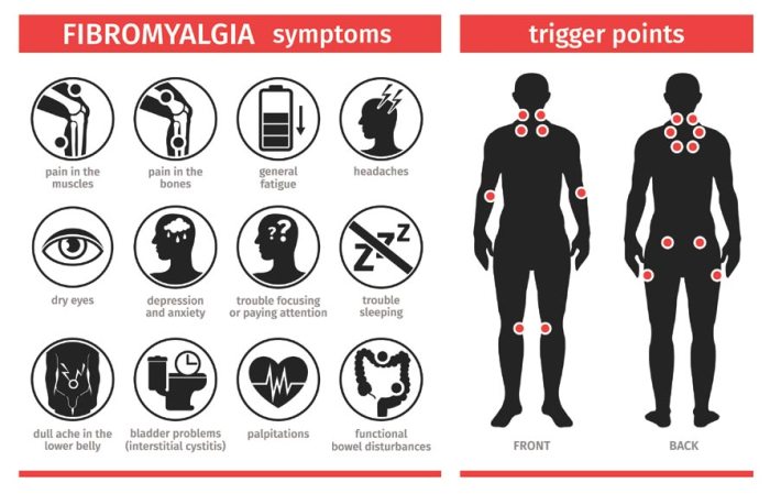
    Fibromyalgia (FM) is characterized by widespread musculoskeletal pain accompanied by fatigue, sleep disturbances, and cognitive difficulties. The pain is often described as aching, burning, or stabbing, and it typically affects multiple areas of the body. Unlike CFS, FM doesn’t focus solely on fatigue but incorporates a constellation of symptoms.

    Similarities Between CFS and FM

    Both CFS and FM share several overlapping symptoms, leading to frequent misdiagnosis. These shared symptoms include:

    • Profound fatigue: This is a hallmark of both conditions, impacting energy levels and the ability to engage in daily activities.
    • Sleep disturbances: Difficulty falling asleep, staying asleep, or experiencing restorative sleep is common to both CFS and FM.
    • Cognitive difficulties: “Brain fog” or difficulties with concentration, memory, and other cognitive functions are often reported by individuals with either condition.

    Differences Between CFS and FM

    Despite the overlapping symptoms, CFS and FM differ in their primary focus and diagnostic criteria. A crucial distinction is the nature of the pain.

    • CFS: Focuses primarily on extreme fatigue, which is often the most prominent symptom. The pain, when present, tends to be less widespread and less prominent than the fatigue.
    • FM: Focuses on widespread pain as a primary symptom. While fatigue is also a common feature, the persistent pain is often the driving force behind the diagnosis.

    Diagnostic Criteria for CFS

    The diagnostic criteria for CFS often rely on a combination of symptoms and a lack of other explanations for the fatigue.

    • Symptom duration: Fatigue must persist for at least six months.
    • Symptom severity: The fatigue must significantly impair daily activities and not be attributable to other medical conditions.
    • Exclusion of other causes: The fatigue should not be easily explained by other conditions, like depression or anxiety.

    Diagnostic Criteria for Fibromyalgia

    The diagnosis of Fibromyalgia (FM) usually requires a combination of widespread pain and other associated symptoms.

    • Widespread pain: Pain must be present in at least multiple body areas, often described as aching, burning, or stabbing.
    • Symptoms of FM: Additional symptoms such as fatigue, sleep disturbances, and cognitive difficulties are often present and contribute to the overall diagnosis.
    • Exclusion of other causes: The symptoms should not be easily explained by other medical conditions.

    Diagnostic Challenges: Chronic Fatigue Syndrome Vs Fibromyalgia

    Navigating the world of chronic illnesses like Chronic Fatigue Syndrome (CFS) and Fibromyalgia (FM) often feels like walking through a dense fog. One of the most significant hurdles is the lack of definitive diagnostic tools. The very nature of these conditions, characterized by a complex interplay of symptoms, makes precise identification and differentiation a challenging endeavor.The absence of objective markers for both CFS and FM often leads to a diagnostic journey filled with uncertainty and potential misdiagnosis.

    Clinicians must rely heavily on the patient’s self-reported symptoms, making the subjective experience of the individual central to the diagnostic process. This reliance on symptom-based criteria necessitates a thorough understanding of the complexities of the conditions and a careful evaluation of the patient’s individual experience.

    Diagnostic Criteria Reliance

    The diagnostic criteria for CFS and FM are primarily symptom-based. This means that doctors assess the presence and severity of specific symptoms to arrive at a diagnosis. This method, while necessary, presents challenges due to the highly subjective nature of many of the symptoms. Patients may experience varying degrees of the same symptom, and the interpretation of these experiences can differ greatly from one individual to another.

    Difficulties in Distinguishing CFS and FM

    The symptoms of CFS and FM can overlap considerably, making differentiation difficult. Both conditions are characterized by fatigue, pain, and cognitive dysfunction, which can make accurate diagnosis challenging for healthcare professionals. The shared symptoms can contribute to diagnostic confusion and delay in receiving the appropriate care.

    Lack of Objective Diagnostic Tests

    Currently, there are no objective diagnostic tests for either CFS or FM. This lack of definitive biomarkers for these conditions creates a significant hurdle for healthcare professionals. The absence of objective tests often necessitates a comprehensive evaluation of the patient’s history, physical examination, and symptom reporting to reach a diagnosis.

    Subjective Nature of Symptoms

    The subjective nature of symptoms in both CFS and FM is a critical factor in the diagnostic process. Factors such as individual variations in pain perception, cognitive function, and fatigue levels can significantly influence the experience and reporting of symptoms. This subjectivity can make comparing and contrasting symptoms between individuals challenging, further complicating the diagnostic process.

    Comparison of Diagnostic Criteria

    Characteristic Chronic Fatigue Syndrome (CFS) Fibromyalgia (FM)
    Primary Symptom Extreme fatigue lasting at least six months, with significant impact on daily activities. Widespread musculoskeletal pain, often accompanied by fatigue, sleep disturbances, and cognitive dysfunction.
    Duration of Symptoms Persistent fatigue for at least six months. Persistent pain for at least three months.
    Symptoms (Key) Post-exertional malaise, sleep disturbances, cognitive dysfunction, and headache. Widespread pain, tenderness to the touch at specific points, and sleep disturbances.
    Other Considerations Exclusion of other conditions with similar symptoms. Exclusion of other conditions with similar symptoms.

    This table highlights some key differences in the diagnostic criteria for CFS and FM. The reliance on symptom reporting and the absence of objective tests necessitate a careful and thorough assessment process for both conditions.

    Symptoms and Manifestations

    Understanding the wide range of symptoms associated with Chronic Fatigue Syndrome (CFS) and Fibromyalgia (FM) is crucial for accurate diagnosis and effective management. These conditions are often characterized by overlapping symptoms, making differentiation challenging. This section delves into the diverse array of physical, cognitive, and emotional manifestations, highlighting the variability between individuals.The symptoms of CFS and FM can significantly impact daily life, affecting work, relationships, and overall well-being.

    Figuring out chronic fatigue syndrome versus fibromyalgia can be tricky, right? Sometimes, seemingly unrelated symptoms like ear drainage can complicate the picture. Understanding the causes and treatment options for ear drainage is crucial, as it can sometimes be a symptom of an underlying health condition, potentially linked to the complex interplay of CFS and fibromyalgia. For a deeper dive into the potential causes and treatment of ear drainage, check out this helpful resource on ear drainage causes and treatment.

    Ultimately, accurately diagnosing and managing these conditions requires careful consideration of all presenting symptoms.

    Recognizing these symptoms is a first step toward seeking appropriate medical attention and support.

    Physical Symptoms

    Physical symptoms are often prominent in both CFS and FM. A wide array of physical discomfort can be experienced, significantly impacting daily activities. Muscle pain, fatigue, and sleep disturbances are key components of both conditions.

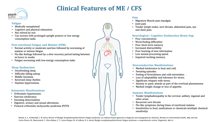
    • Fatigue: This is a hallmark symptom of both CFS and FM, characterized by extreme tiredness and lack of energy. The fatigue in CFS is often described as debilitating, hindering even basic tasks. In FM, the fatigue is frequently accompanied by pain and stiffness.
    • Pain: Widespread pain is a defining feature of FM, often described as aching, throbbing, or burning sensations. CFS may also present with muscle or joint pain, though this is not as consistently a prominent feature as in FM.
    • Sleep Disturbances: Difficulty falling asleep, staying asleep, or experiencing restful sleep is common to both conditions. This can further exacerbate fatigue and other symptoms.
    • Other Physical Symptoms: Other potential physical symptoms include headaches, dizziness, gastrointestinal issues, and musculoskeletal problems. These symptoms can vary significantly in intensity and frequency.

    Cognitive Symptoms

    Cognitive impairments, often referred to as “brain fog,” are significant challenges for individuals with CFS and FM. These impairments can affect concentration, memory, and processing speed.

    Figuring out chronic fatigue syndrome (CFS) versus fibromyalgia can be tricky, as their symptoms overlap. One aspect often overlooked is how physical activity, like running a mile, affects calorie expenditure. Knowing how many calories you burn running a mile how many calories do you burn running a mile might help you better understand your body’s needs when dealing with these conditions.

    Ultimately, a doctor’s diagnosis is crucial for proper management of either CFS or fibromyalgia.

    • Concentration Problems: Difficulty concentrating, focusing on tasks, and maintaining attention are common cognitive symptoms.
    • Memory Issues: Short-term memory problems and difficulty recalling information are frequent complaints.
    • Processing Speed: Slower processing speed and difficulty with mental tasks can affect daily functioning.

    Emotional Symptoms

    The emotional impact of CFS and FM is often underestimated. These conditions can lead to significant emotional distress, impacting mental health and overall well-being.

    • Mood Swings: Fluctuations in mood, including irritability, anxiety, and depression, are common.
    • Anxiety and Depression: The chronic nature of these conditions can contribute to feelings of anxiety and depression, requiring specific mental health support.
    • Emotional Exhaustion: The persistent physical and cognitive symptoms can lead to significant emotional exhaustion and feelings of overwhelm.

    Symptom Severity Comparison

    | Symptom Category | Chronic Fatigue Syndrome (CFS) | Fibromyalgia (FM) ||—|—|—|| Physical | Severe, debilitating fatigue, widespread pain, significant sleep disturbances | Widespread pain, fatigue, varying intensity, sleep disturbances || Cognitive | Significant brain fog, difficulty with concentration and memory | Cognitive impairments, including concentration and memory problems, varying intensity || Emotional | Increased risk of anxiety and depression, emotional exhaustion | Increased risk of anxiety and depression, emotional exhaustion, mood swings |

    Underlying Mechanisms

    Chronic fatigue syndrome vs fibromyalgia

    Unraveling the intricate biological pathways behind chronic fatigue syndrome (CFS) and fibromyalgia (FM) remains a significant challenge for researchers. While definitive causes remain elusive, growing evidence points towards a complex interplay of factors involving inflammation, immune dysfunction, and potential genetic predispositions. This exploration delves into the current understanding of these underlying mechanisms, highlighting similarities and differences between the two conditions.The complex nature of these conditions suggests that a single, isolated cause is unlikely.

    Instead, it is probable that a combination of genetic, environmental, and lifestyle factors contribute to the development and persistence of symptoms. Understanding these contributing factors is crucial for developing effective treatment strategies and improving the lives of those affected.

    Potential Biological Mechanisms

    Current research suggests that both CFS and FM are likely influenced by a combination of biological factors. Inflammation, immune dysfunction, and neuroendocrine imbalances are key areas of investigation. The interplay between these factors is likely different for each individual, adding further complexity to the conditions.

    Role of Inflammation

    Inflammation, a crucial part of the body’s defense mechanism, is often implicated in the development and persistence of both CFS and FM. Chronic low-grade inflammation can lead to various symptoms, including fatigue, pain, and sleep disturbances. Studies have shown elevated levels of inflammatory markers, such as cytokines, in individuals with both conditions. This chronic inflammation may contribute to the persistent pain and fatigue observed in these conditions.

    Immune Dysfunction

    Immune dysfunction is another potential contributor to both CFS and FM. The intricate balance of the immune system may be disrupted, potentially leading to an overreaction or underreaction to various stimuli. This disruption may contribute to the development of chronic fatigue and pain, and the exacerbation of symptoms by external factors. For example, infections or other stressors may trigger or worsen symptoms in individuals with compromised immune function.

    Neuroendocrine Imbalances

    Neuroendocrine imbalances, which affect the intricate interplay between the nervous and endocrine systems, are also suspected to play a role. Hormonal fluctuations and dysfunction in the hypothalamic-pituitary-adrenal (HPA) axis, responsible for stress response, have been linked to the development and exacerbation of symptoms. Disruptions in the neurotransmitter systems, which regulate mood and pain perception, may also be involved.

    Genetic Predisposition

    Genetic factors likely contribute to an individual’s susceptibility to developing CFS and FM. While no single gene has been identified as a definitive cause, studies suggest a complex interplay of genes, potentially influencing immune response, inflammation, and pain perception. Family history of these conditions may indicate a potential genetic predisposition.

    Factors Contributing to Development

    Several factors are thought to contribute to the development of both CFS and FM. These include infections, physical trauma, psychological stressors, and environmental exposures. The specific factors that trigger the conditions vary greatly between individuals. For example, one person might experience a viral infection, while another might experience a traumatic event as a catalyst.

    Potential Interactions Diagram

    Imagine a complex web of interacting factors. At the center, genetic predisposition influences an individual’s susceptibility. External factors like infections, trauma, or stress can trigger a cascade of events. These events, including neuroendocrine imbalances, and immune dysfunction, lead to chronic inflammation. The interplay of these factors leads to the persistent symptoms of fatigue, pain, and cognitive dysfunction.

    This web is highly individualized and not precisely understood.

    Treatment Approaches

    Chronic fatigue syndrome vs fibromyalgia

    Navigating the complex landscapes of Chronic Fatigue Syndrome (CFS) and Fibromyalgia (FM) often feels like trying to thread a needle in a hurricane. The lack of a single, definitive diagnostic test and the highly variable presentation of symptoms make treatment a personalized journey. Finding effective strategies that address the diverse needs of each individual is crucial. This often involves a multifaceted approach that considers both the physical and psychological aspects of the illness.Effective treatment for CFS and FM involves a multifaceted approach tailored to individual needs.

    A personalized plan should address symptoms, manage pain, and improve overall quality of life. This necessitates a deep understanding of the patient’s unique experience and preferences. Crucially, treatment should be ongoing and adaptable to changing symptoms.

    Different Treatment Strategies

    Various strategies are employed in treating CFS and FM. These include lifestyle modifications, medications, and complementary therapies. The selection of specific treatments is heavily influenced by the individual patient’s symptoms, preferences, and medical history.

    Figuring out chronic fatigue syndrome vs. fibromyalgia can be tricky, right? Understanding your health insurance options is just as important, especially when dealing with conditions like these. A gold plan health insurance, like gold plan health insurance what is it , can significantly impact your ability to manage the costs of treatment and ongoing care. Ultimately, knowing your options and how they relate to your specific needs is key to navigating these complex health issues.

    Lifestyle Modifications

    Implementing lifestyle adjustments can significantly impact the management of CFS and FM symptoms. Regular exercise, even in small increments, can improve energy levels and mood. Prioritizing adequate sleep is paramount, as sleep disturbances are frequently reported by patients. Maintaining a balanced diet, avoiding triggers (such as caffeine or stress), and practicing stress-reducing techniques, like mindfulness or meditation, can also contribute to symptom control.

    Medications

    Various medications are used to manage symptoms of CFS and FM. Pain relievers, such as NSAIDs or opioids, may help with pain management. Stimulants can sometimes increase energy levels. Antidepressants can be beneficial in addressing pain, sleep disturbances, and mood symptoms. However, medication responses vary widely, and finding the right medication or combination of medications often requires trial and error.

    “Medication selection should be a collaborative decision between the patient and their healthcare provider.”

    Complementary Therapies

    Complementary therapies, including physical therapy, occupational therapy, and counseling, play an important role in the comprehensive management of CFS and FM. Physical therapy can help improve strength, flexibility, and endurance. Occupational therapy can help patients adapt their daily routines to accommodate their limitations. Counseling can address the psychological distress often associated with these conditions.

    Patient-Centered Care

    Patient-centered care is paramount in managing CFS and FM. This approach prioritizes the patient’s individual needs, preferences, and values. Open communication, active listening, and shared decision-making are essential elements of patient-centered care. Understanding the patient’s perspective is vital for developing an effective treatment plan.

    Multidisciplinary Teams

    Multidisciplinary teams, comprised of specialists like physicians, physical therapists, occupational therapists, psychologists, and social workers, can provide comprehensive care. The collaborative nature of these teams allows for a holistic approach to treatment, addressing the various physical, psychological, and social aspects of the conditions.

    Comparison of Treatment Modalities

    Treatment Modality CFS Effectiveness FM Effectiveness Potential Considerations
    Lifestyle Modifications Often beneficial, improves quality of life Often beneficial, improves quality of life Requires commitment and patient follow-through
    Medications Varied responses, requires careful selection Varied responses, requires careful selection Potential side effects, trial and error approach
    Complementary Therapies May improve symptoms and function May improve symptoms and function Individual responses vary, not a replacement for medical care
    Multidisciplinary Teams Provides comprehensive care, improves outcomes Provides comprehensive care, improves outcomes Requires access to specialized expertise

    Impact on Daily Life

    Chronic Fatigue Syndrome (CFS) and Fibromyalgia (FM) profoundly impact daily life, often making simple tasks feel overwhelming. These conditions aren’t just about feeling tired; they significantly alter how individuals function and interact with the world around them. The impact extends far beyond physical symptoms, affecting emotional well-being, social connections, and professional life.Living with CFS and FM requires constant adaptation and resourcefulness.

    The fluctuating energy levels, pain, and cognitive difficulties can disrupt routines, making it hard to maintain independence and participate in activities once enjoyed. The unpredictable nature of these conditions adds a layer of stress and anxiety to daily life, further hindering overall well-being.

    Impact on Work

    The fluctuating energy levels, cognitive difficulties, and pain associated with CFS and FM can severely impact employment. Many individuals experience reduced productivity, missed workdays, and difficulty concentrating on tasks. These conditions can also lead to job loss or career limitations due to the ongoing challenges of maintaining a consistent work schedule and fulfilling job requirements. For example, a person with CFS or FM might find it challenging to focus on complex tasks or maintain consistent energy levels throughout the workday, impacting their ability to meet deadlines or perform at a satisfactory level.

    Impact on Social Activities

    Social activities can become a significant challenge for individuals with CFS and FM. The fluctuating energy levels, pain, and cognitive difficulties can make it challenging to participate in social events, maintain relationships, and engage in hobbies. The unpredictable nature of these conditions can also lead to social isolation and feelings of loneliness, as individuals may find it difficult to keep up with social commitments or maintain consistent social interactions.

    This can manifest in the form of cancelled plans, missed gatherings, and reduced social contact.

    Impact on Relationships

    The significant impact of CFS and FM on daily life can strain relationships. Changes in energy levels, mood swings, and difficulties with communication can create tension and misunderstandings within personal relationships. The constant need for rest and accommodation can place a burden on family members and partners, potentially leading to frustration and resentment. For instance, a partner might feel frustrated with the limitations imposed by the condition, while the individual with CFS or FM might feel misunderstood or unsupported.

    Impact on Daily Tasks

    Managing daily tasks can be incredibly difficult for individuals with CFS and FM. Simple activities, such as showering, cooking, or cleaning, can become significant hurdles due to pain, fatigue, and cognitive impairments. These conditions can significantly impact the ability to perform basic self-care activities, leading to a decrease in overall independence and quality of life. Individuals may require assistance with daily tasks or make significant adjustments to their routines to accommodate their symptoms.

    For instance, a person with CFS or FM might need to break down large tasks into smaller, more manageable steps or enlist help from others to complete daily chores.

    Impact on Quality of Life

    The chronic nature of CFS and FM can significantly diminish the quality of life for those affected. The constant pain, fatigue, and cognitive difficulties can limit participation in activities, hobbies, and social events. This can lead to feelings of isolation, depression, and anxiety, further impacting overall well-being. The ability to enjoy life’s simple pleasures and engage in meaningful activities is often severely compromised.

    Impact on Social Life

    Social interactions become challenging due to the fluctuating energy levels, pain, and cognitive difficulties. Individuals may find it difficult to maintain friendships, attend social gatherings, or participate in activities that were once enjoyable. This can lead to social isolation and feelings of loneliness. Individuals with CFS and FM might need to limit their social engagements or adjust their social interactions to accommodate their symptoms.

    For example, attending a party might be exhausting and painful, making it necessary to leave early or decline invitations altogether.

    Challenges in the Workplace

    The unpredictable nature of CFS and FM presents significant challenges in the workplace. Fluctuating energy levels, cognitive difficulties, and pain can lead to missed workdays, reduced productivity, and difficulty concentrating on tasks. This can result in difficulties with meeting deadlines, completing projects, and maintaining consistent job performance. Employers may not fully understand the challenges faced by employees with CFS and FM, potentially leading to misunderstandings or discrimination.

    This can manifest in difficulties with scheduling, workload management, and accommodations.

    Research and Future Directions

    The quest to understand and effectively treat chronic fatigue syndrome (CFS) and fibromyalgia (FM) hinges on ongoing research. Current knowledge, while offering insights into these complex conditions, leaves significant gaps in our understanding of their underlying causes and effective treatments. This exploration into the future of research will highlight promising avenues for progress, emphasizing the crucial role of ongoing investigations in improving the lives of those affected.

    Ongoing Research Efforts

    Research into CFS and FM is multifaceted, encompassing various approaches to uncover the intricate mechanisms driving these conditions. Scientists are employing a variety of methodologies, from genetic studies to neuroimaging techniques, to delve into the biological underpinnings of these illnesses. This multifaceted approach is crucial given the complex interplay of factors likely contributing to these conditions.

    Areas of Active Investigation

    Numerous areas are currently under intense investigation. Genetic research is uncovering potential susceptibility genes and pathways linked to the development of CFS and FM. Neuroimaging studies are providing insights into brain function and structure, potentially revealing alterations in brain regions associated with pain processing, fatigue, and cognition. Furthermore, investigations into the role of the immune system and its interactions with the nervous system are being pursued.

    These efforts aim to illuminate the intricate interplay of factors contributing to the symptoms experienced by patients.

    Potential Breakthroughs in Treatment and Diagnosis, Chronic fatigue syndrome vs fibromyalgia

    Advancements in diagnostic tools and therapeutic strategies are crucial for improving the lives of those living with CFS and FM. Researchers are exploring novel biomarkers that could aid in early detection and diagnosis, moving beyond the reliance on subjective symptom reports. Moreover, innovative treatments targeting specific biological pathways implicated in the conditions are being investigated. For example, therapies that modulate the immune system or address neuroinflammation show promise, though further research is necessary to validate these approaches.

    Importance of Research to Improve Lives

    Research efforts play a pivotal role in improving the lives of individuals affected by CFS and FM. By uncovering the underlying causes and mechanisms, scientists pave the way for more precise and effective treatments. Furthermore, improved diagnostic tools can lead to earlier intervention and potentially better management of symptoms. This has profound implications for reducing the debilitating impact of these conditions on daily life.

    Current Research Findings

    Recent research has highlighted potential avenues for treatment and diagnosis. Studies suggest a link between gut microbiota imbalances and CFS/FM symptoms. These findings underscore the potential role of restoring gut health in managing these conditions. Further, research indicates that interventions targeting neuroinflammation, such as certain anti-inflammatory medications, may show promise in alleviating symptoms. It is important to note that these are preliminary findings, and further research is necessary to confirm their validity and translate them into effective treatments.

    Potential Implications of Future Research Directions

    The future of research in CFS and FM holds significant potential. Continued investigations into genetic predispositions, immune system dysregulation, and neuroinflammation could lead to the development of personalized treatment strategies tailored to individual needs. These strategies could improve symptom management and overall well-being. For example, a better understanding of the role of specific immune cells in CFS/FM could lead to the development of targeted therapies that effectively modulate the immune response, thereby reducing the intensity of symptoms.

    This could lead to better outcomes and quality of life for those living with these debilitating conditions.

    Final Summary

    In conclusion, chronic fatigue syndrome and fibromyalgia present unique diagnostic and treatment challenges. While sharing some overlapping symptoms, crucial differences exist in their presentation and underlying mechanisms. The subjective nature of symptoms underscores the importance of patient-centered care and multidisciplinary approaches to treatment. Further research is essential to enhance diagnostic accuracy and develop more effective treatments, ultimately improving the quality of life for those affected.

  • Rheumatoid Arthritis Back Pain Understanding the Impact

    Rheumatoid Arthritis Back Pain Understanding the Impact

    Rheumatoid arthritis back pain is a complex condition that significantly impacts a person’s life. It’s not just about aches and stiffness; it’s about understanding the underlying mechanisms, the various symptoms, and the available treatment options. This comprehensive guide delves into the nuances of RA back pain, providing a detailed overview of its causes, diagnosis, and management strategies.

    From defining the specific characteristics of RA back pain and distinguishing it from other forms of back pain, to exploring the intricate relationship between genetics, lifestyle, and the development of the condition, we aim to shed light on every aspect. This exploration covers the full spectrum, from diagnosis and treatment to managing daily life and potential complications.

    Defining Rheumatoid Arthritis Back Pain

    Rheumatoid arthritis (RA) is a chronic autoimmune disease that primarily affects the joints. While joint inflammation is the hallmark of RA, it can also impact other parts of the body, including the spine and surrounding tissues, leading to back pain. Understanding the specific ways RA affects the back is crucial for accurate diagnosis and effective management.RA can cause inflammation in the structures surrounding the spine, such as ligaments, tendons, and bursae.

    This inflammation can lead to pain, stiffness, and limited movement. The inflammatory process can also directly affect the joints within the spine itself, although this is less common than inflammation in the supporting tissues. This explains why back pain associated with RA can manifest differently from other types of back pain.

    Rheumatoid Arthritis (RA) and its Impact on the Back

    RA’s impact on the back stems from its systemic inflammatory nature. The immune system mistakenly attacks the lining of the joints, causing inflammation and damage. This inflammation can spread to the tissues surrounding the spine, including the facet joints, intervertebral discs, and ligaments. This results in pain, stiffness, and restricted movement. Furthermore, the chronic inflammation can contribute to bone erosion and deformities in the spine over time.

    Mechanisms of RA Back Pain

    Inflammation in the spinal structures is a key mechanism. Inflammation triggers the release of chemicals that cause pain and swelling. These chemicals can also damage the cartilage and other tissues within the spine, further contributing to the pain and stiffness. Additionally, the body’s response to the inflammation can cause muscle spasms, adding to the discomfort. In some cases, the inflammatory process can cause the formation of rheumatoid nodules in the back, adding to the pain.

    Dealing with rheumatoid arthritis back pain can be tough, especially when you’re trying to figure out the best approach. Sometimes, exploring other potential contributing factors can help. For instance, understanding how IUD use in nulliparous women iud use in nulliparous women might offer insights into overall pelvic health, which could indirectly affect back pain. Ultimately, it’s crucial to consult with a healthcare professional for personalized advice and treatment options related to rheumatoid arthritis back pain.

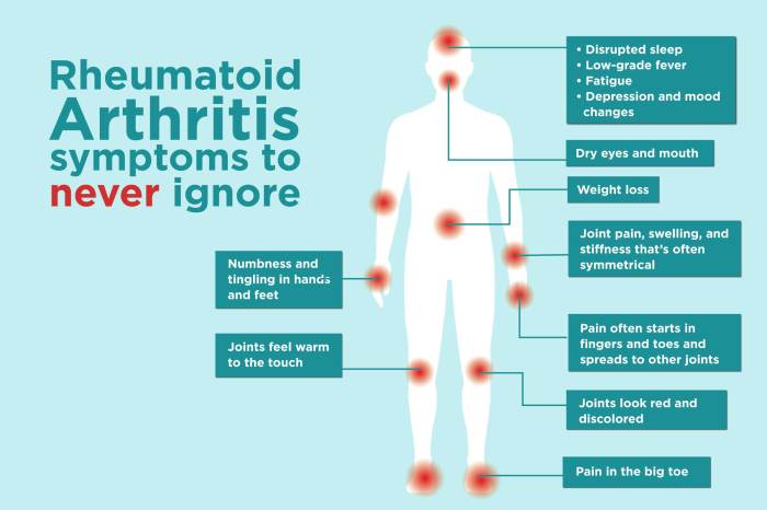
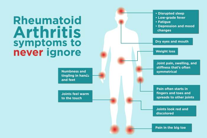
    Common Symptoms of RA Back Pain

    RA back pain often presents with characteristics different from other types of back pain. While mechanical back pain typically involves localized pain, RA back pain can radiate to other areas, particularly the hips or shoulders. Stiffness, especially in the morning, is a common symptom. This stiffness may last for several hours, unlike the transient stiffness often associated with mechanical back pain.

    The pain may be accompanied by swelling, warmth, and redness in the affected areas. Fatigue and fever are also potential symptoms, reflecting the systemic nature of the disease.

    Differentiating RA Back Pain from Other Types of Back Pain

    | Feature | Rheumatoid Arthritis Back Pain | Mechanical Back Pain | Spinal Stenosis ||—|—|—|—|| Pain Location | Often radiating, can involve hips/shoulders | Localized, typically within the back | Often radiating down the legs || Stiffness | Significant morning stiffness, lasting hours | Mild or absent stiffness | Stiffness may occur, but often accompanied by leg pain/numbness || Symptoms | Systemic symptoms (fatigue, fever) possible | No systemic symptoms | Neurological symptoms (numbness, weakness) common || Progression | Gradual, chronic | Acute or chronic, episodic | Progressive, potentially leading to significant disability |

    Progression of RA Back Pain

    The progression of RA back pain can vary significantly depending on the individual’s response to treatment and overall health. Early stages may involve intermittent pain and stiffness. As the disease progresses, pain and stiffness can become more persistent and severe. In some cases, significant spinal deformities can develop, leading to chronic pain and significant functional limitations. These deformities can also increase the risk of fractures.

    Causes and Risk Factors: Rheumatoid Arthritis Back Pain

    Rheumatoid arthritis back pain

    Understanding the causes and risk factors of rheumatoid arthritis (RA) back pain is crucial for early detection and effective management. While the exact cause of RA remains elusive, research suggests a complex interplay of genetic predisposition and environmental triggers. This understanding can empower individuals to take proactive steps to minimize their risk and seek appropriate medical attention when symptoms arise.The precise mechanisms linking RA to back pain are multifaceted.

    Inflammation, a hallmark of RA, can affect the joints, tendons, and ligaments throughout the body, including those in the spine. This inflammation can lead to pain, stiffness, and reduced mobility in the back. Moreover, the autoimmune response that characterizes RA can potentially trigger or exacerbate existing conditions, sometimes manifesting as back pain.

    Potential Causes of RA Back Pain

    RA back pain often stems from the inflammatory response characteristic of the disease. Inflammation in the spinal structures, including ligaments and tendons, can cause pain and stiffness. Additionally, RA can cause erosions and structural damage to the joints, which can contribute to back pain. It’s important to remember that not all individuals with RA experience back pain, and the intensity and frequency can vary greatly.

    Risk Factors for RA Back Pain, Rheumatoid arthritis back pain

    Certain factors can increase an individual’s susceptibility to developing RA back pain. These risk factors can be categorized into genetic predispositions and environmental influences.

    Role of Genetics in RA Back Pain

    Genetic factors play a significant role in determining an individual’s susceptibility to RA. Family history of RA is a well-established risk factor. Individuals with a family history of RA have a higher chance of developing the condition themselves. Specific genes, such as HLA-DRB1, are strongly associated with an increased risk of developing RA. Genetic variations can influence the body’s immune response, making some individuals more prone to developing the autoimmune reaction associated with RA.

    Role of Environmental Factors in RA Back Pain

    Environmental factors can also contribute to the development of RA back pain. Exposure to certain infections, such as Epstein-Barr virus, may potentially trigger or exacerbate the autoimmune response. Smoking is a significant environmental risk factor for developing RA. Individuals who smoke are more likely to experience RA-related back pain and have a higher disease severity. Additionally, certain environmental toxins and occupational exposures might contribute to the condition’s progression.

    Table of Factors Increasing the Likelihood of RA Back Pain

    Factor Category Specific Factors
    Genetics Family history of RA, Specific genes (HLA-DRB1), Genetic variations
    Environment Exposure to certain infections (e.g., Epstein-Barr virus), Smoking, Environmental toxins, Occupational exposures
    Lifestyle Lack of physical activity, Obesity, Poor diet

    Connections Between Other Health Conditions and RA Back Pain

    Several health conditions can be linked to or potentially exacerbate RA back pain. Conditions such as fibromyalgia, osteoarthritis, and lupus can coexist with RA, sometimes causing overlapping symptoms, including back pain. Furthermore, individuals with RA might experience chronic pain conditions, which can complicate diagnosis and treatment.

    Diagnosis and Assessment

    Diagnosing rheumatoid arthritis (RA) back pain requires a meticulous approach, combining various diagnostic tools and assessments. A thorough understanding of the patient’s medical history, symptoms, and physical examination findings is crucial. Accurate diagnosis helps guide treatment strategies and prevent misdiagnosis, which can lead to inappropriate therapies and delayed management of the condition.

    Diagnostic Process for RA Back Pain

    The diagnostic process for RA back pain typically involves a multi-faceted approach. It begins with a detailed history of the patient’s symptoms, including the onset, duration, location, and characteristics of the pain. This information, combined with a thorough physical examination, allows the healthcare professional to narrow down the possible causes of the back pain. Supporting diagnostic tests are then employed to confirm or rule out RA as the underlying cause.

    Relevant Tests and Procedures

    A battery of tests is often employed to confirm or rule out a diagnosis of RA back pain. These tests include blood tests, specifically looking for markers of inflammation, such as elevated erythrocyte sedimentation rate (ESR) and C-reactive protein (CRP). These tests help indicate the presence of an inflammatory process in the body, a hallmark of RA. Other blood tests may be conducted to identify specific antibodies associated with RA, like rheumatoid factor (RF) and anti-cyclic citrullinated peptide (anti-CCP) antibodies.

    These antibody tests can provide further evidence supporting the diagnosis.

    Distinguishing RA Back Pain from Other Conditions

    Differentiating RA back pain from other back pain conditions is essential. Conditions like osteoarthritis, spinal stenosis, and ankylosing spondylitis can all present with back pain, making accurate diagnosis critical. A comprehensive evaluation, including a detailed history, physical examination, and relevant diagnostic tests, helps distinguish between these conditions. The presence of systemic symptoms, such as fatigue, fever, or joint swelling, often suggests RA as a possible cause.

    The pattern of pain, such as its morning stiffness or worsening with rest, can also provide clues to the underlying etiology.

    Role of Imaging Techniques

    Imaging techniques play a crucial role in evaluating the extent of the inflammatory process and potential structural damage in the spine. X-rays are often the initial imaging modality used to assess for bone erosions or joint space narrowing, which can be signs of RA. Magnetic resonance imaging (MRI) scans provide detailed images of soft tissues, including ligaments, tendons, and muscles, which can be useful for evaluating inflammation and damage in the spine.

    MRI is particularly helpful in detecting early signs of inflammation and identifying potential complications of RA, such as spinal cord compression.

    Importance of Physical Examinations

    A thorough physical examination is an integral part of the diagnostic process. The examination assesses the range of motion, tenderness, and presence of inflammation in the back and surrounding joints. Palpation of the spine and surrounding soft tissues, along with assessment of joint swelling and tenderness, aids in evaluating the extent of the inflammatory process. Specific tests for spinal mobility and posture, such as Schober test, can also provide valuable information about spinal involvement.

    Summary of Diagnostic Criteria for RA Back Pain

    Criterion Description
    Symptoms Persistent back pain, often with morning stiffness lasting at least 30 minutes. The pain may be accompanied by other systemic symptoms, such as fatigue or fever.
    Physical Examination Presence of joint swelling, tenderness, and restricted range of motion in the back and surrounding joints. Specific tests like the Schober test can assess spinal mobility.
    Blood Tests Elevated ESR and CRP levels, indicating inflammation. Positive rheumatoid factor (RF) and anti-cyclic citrullinated peptide (anti-CCP) antibodies.
    Imaging Studies X-rays and MRI scans may reveal signs of bone erosion, joint space narrowing, or inflammation in the spine, providing further support for the diagnosis.

    Treatment and Management

    Managing rheumatoid arthritis (RA) back pain requires a multifaceted approach that considers various factors, including the severity of the condition, individual needs, and potential side effects of treatments. A collaborative effort between patients, healthcare professionals, and support systems is crucial for effective pain management and overall well-being. The goal is not just to reduce pain, but also to improve function, maintain quality of life, and prevent further joint damage.Effective RA back pain management involves a combination of pharmacological interventions, therapies, and lifestyle modifications.

    Choosing the right treatment plan depends on individual factors and can evolve over time as the disease progresses or treatment responses change.

    Medication Options

    Various medications are used to treat RA back pain, each with its own mechanism of action, efficacy, and potential side effects. Nonsteroidal anti-inflammatory drugs (NSAIDs) like ibuprofen and naproxen are often the first-line treatment for mild to moderate pain. They help reduce inflammation and pain but may not be suitable for everyone due to potential gastrointestinal issues. Disease-modifying antirheumatic drugs (DMARDs) are more potent and can help slow the progression of the disease.

    Examples include methotrexate, hydroxychloroquine, and sulfasalazine. Biologics are another category of medications that target specific components of the immune system involved in RA inflammation. These may be considered for individuals who don’t respond well to other treatments or experience significant side effects. Each medication has a unique profile, and careful consideration is necessary when selecting the most appropriate option for an individual patient.

    Dealing with rheumatoid arthritis back pain can be tough, impacting everything from daily tasks to overall well-being. Interestingly, some research suggests overlapping symptoms between RA and conditions like autism spectrum disorder, particularly in girls. For example, understanding the subtle signs of autism in girls, such as social communication challenges, can help in early diagnosis and appropriate interventions. Ultimately, recognizing these potential connections is crucial for a holistic approach to managing rheumatoid arthritis back pain and improving quality of life.

    signs of autism in girls

    Role of Physical Therapy

    Physical therapy plays a vital role in managing RA back pain by improving strength, flexibility, and range of motion. Physical therapists design personalized exercise programs tailored to individual needs and capabilities. These programs focus on strengthening core muscles, improving posture, and increasing flexibility. Regular exercise, guided by a physical therapist, can significantly reduce pain and stiffness while improving overall physical function.

    Exercises like swimming, water aerobics, and gentle stretching can be highly beneficial for patients with RA back pain. The therapist can also teach patients how to use assistive devices, such as canes or walkers, to reduce strain on the affected joints.

    Lifestyle Modifications

    Lifestyle modifications are an integral part of managing RA back pain. Adopting healthy habits can help improve overall well-being and reduce the impact of the disease on daily life. A balanced diet rich in fruits, vegetables, and whole grains, coupled with adequate hydration, can contribute to overall health and potentially reduce inflammation. Maintaining a healthy weight can also lessen the strain on joints and improve overall function.

    Regular, moderate-intensity exercise, such as walking or cycling, can improve joint mobility, muscle strength, and reduce stiffness.

    Structured List of Lifestyle Changes

    • Maintaining a healthy weight through a balanced diet and regular exercise. Weight loss, if applicable, can significantly reduce stress on the joints, leading to decreased pain and improved function.
    • Adopting a balanced diet rich in fruits, vegetables, and whole grains. These foods are packed with essential nutrients that support overall health and potentially reduce inflammation.
    • Engaging in regular physical activity, such as walking, swimming, or gentle yoga. Exercise helps improve joint mobility, muscle strength, and overall fitness, thereby alleviating pain and stiffness.
    • Getting sufficient sleep. Adequate rest allows the body to repair and recover, which is crucial for managing RA pain and stiffness.
    • Managing stress through relaxation techniques such as meditation, deep breathing exercises, or spending time in nature. Stress can exacerbate pain and inflammation, so incorporating stress-reducing practices can be highly beneficial.

    Treatment Approaches and Effectiveness

    Treatment Approach Description Effectiveness
    NSAIDs Nonsteroidal anti-inflammatory drugs (NSAIDs) Generally effective for mild to moderate pain, but may not address the underlying disease progression and may have side effects.
    DMARDs Disease-modifying antirheumatic drugs (DMARDs) More potent than NSAIDs, they can slow the progression of the disease and improve long-term outcomes.
    Biologics Target specific components of the immune system involved in inflammation Often highly effective in reducing inflammation and pain, especially for those who haven’t responded well to other treatments.
    Physical Therapy Personalized exercise programs and rehabilitation Proven to improve strength, flexibility, range of motion, and reduce pain and stiffness.
    Lifestyle Modifications Dietary changes, weight management, regular exercise, stress reduction Can significantly improve overall well-being and reduce the impact of RA on daily life.

    Living with Rheumatoid Arthritis Back Pain

    Navigating daily life with rheumatoid arthritis (RA) back pain can be challenging, but it’s not insurmountable. Understanding the impact of RA on daily activities, developing effective pain management strategies, and fostering emotional well-being are crucial for maintaining a fulfilling life. This section provides practical insights and resources to help you cope.Living with RA back pain requires a multifaceted approach that addresses both the physical and emotional aspects of the condition.

    Managing pain, adjusting daily routines, and maintaining a positive outlook are key components of successful RA management. This section explores these strategies in detail.

    Managing Daily Activities with RA Back Pain

    Daily tasks can become significantly harder when RA affects the back. Adapting routines and utilizing assistive devices can make a substantial difference in maintaining independence and quality of life. Planning ahead and prioritizing tasks based on energy levels can be very beneficial. For example, scheduling less physically demanding activities for times when pain is expected to be more intense.

    Pain Management and Symptom Control

    Effective pain management is crucial for managing RA back pain. A combination of strategies, including medication, physical therapy, and alternative therapies, can significantly reduce discomfort and improve overall function. Medications, such as NSAIDs and disease-modifying antirheumatic drugs (DMARDs), are often prescribed to manage inflammation and pain. Physical therapy can help improve posture, strengthen back muscles, and improve flexibility, reducing pain and stiffness.

    Importance of Emotional Well-being for Individuals with RA Back Pain

    The chronic nature of RA and the accompanying pain can take a toll on emotional well-being. Acknowledging and addressing emotional needs is just as important as managing physical symptoms. Maintaining a positive mindset, practicing relaxation techniques, and engaging in activities that bring joy can help to combat feelings of stress, anxiety, and depression. For example, practicing mindfulness, yoga, or spending time in nature can promote emotional well-being.

    Support Groups and Counseling for Coping with RA Back Pain

    Connecting with others who understand the challenges of living with RA back pain can provide invaluable support and encouragement. Support groups offer a safe space to share experiences, gain advice, and build a sense of community. Counseling can provide tools and strategies for coping with the emotional and psychological aspects of the condition. Seeking professional help can be instrumental in developing healthy coping mechanisms and improving overall well-being.

    Practical Techniques for Maintaining a Healthy Lifestyle

    Maintaining a healthy lifestyle, including regular exercise, a balanced diet, and adequate sleep, is vital for managing RA back pain. Gentle exercise can improve strength, flexibility, and reduce pain. A balanced diet rich in anti-inflammatory foods can support overall health and reduce symptoms. Adequate sleep allows the body to recover and repair, which is essential for managing pain and fatigue.

    For example, incorporating activities like swimming, walking, or gentle stretching into the daily routine can help maintain physical well-being.

    Resources and Support Systems

    Category Resources
    Medical Professionals Rheumatologists, Physical Therapists, Pain Management Specialists
    Support Groups Local RA support groups, Online forums, and communities
    Educational Resources Websites of reputable organizations, Patient advocacy groups
    Mental Health Professionals Counsellors, Psychologists, and Therapists
    Assistive Devices Wheelchairs, canes, and other mobility aids

    Potential Complications

    Rheumatoid arthritis (RA) back pain, if left unmanaged, can lead to a cascade of complications impacting overall health and well-being. Ignoring the symptoms or delaying treatment can significantly worsen the condition and restrict daily activities. Understanding these potential consequences is crucial for proactive management and improving quality of life.

    Impact on Mobility and Daily Life

    RA back pain often results in reduced mobility and functional limitations. Stiffness, pain, and inflammation can make simple tasks, like bending, lifting, or walking, challenging. This can lead to decreased participation in social activities, work limitations, and a decline in overall physical fitness. Individuals with severe RA back pain may experience difficulties performing self-care activities, impacting their independence and requiring assistance from others.

    For example, someone with severe RA back pain might struggle to dress themselves or bathe, leading to social isolation and reduced self-esteem.

    Development of Other Health Issues

    Untreated RA back pain can exacerbate existing health conditions or contribute to the development of new ones. The chronic inflammation associated with RA can affect various organs and systems in the body. For example, individuals with RA are at increased risk of cardiovascular disease due to inflammation’s impact on blood vessels. Osteoporosis, a condition characterized by weakened bones, is another common complication linked to RA.

    The sustained inflammation and medication use can also contribute to digestive issues and eye problems. Furthermore, the chronic pain and stress associated with RA can impact mental well-being, increasing the risk of depression and anxiety.

    Importance of Early Intervention

    Early intervention in managing RA back pain is crucial for preventing complications. Prompt diagnosis and appropriate treatment can significantly reduce the severity and progression of the disease. Early intervention allows for better control of inflammation, thereby minimizing the risk of joint damage, bone loss, and other potential complications. Early intervention also helps individuals maintain a higher level of function and independence, enabling them to participate more fully in daily life.

    Dealing with rheumatoid arthritis back pain can be tough, especially when the discomfort feels relentless. It’s important to understand that different medications and treatments have varying timelines for effectiveness. For example, some hormonal birth control methods, like the pill, may take a few weeks to reach full effectiveness, as detailed in this helpful resource on how long does birth control take to work.

    However, even with the right medication, managing rheumatoid arthritis back pain often requires a combination of therapies and a personalized approach.

    Long-Term Consequences of Untreated RA Back Pain

    The table below Artikels some potential long-term consequences of untreated RA back pain:

    Potential Complication Description Impact on Daily Life
    Joint Deformities Progressive damage to the joints, leading to permanent misalignment and loss of function. Severe limitations in mobility, difficulty with self-care tasks, and potential for social isolation.
    Reduced Physical Fitness Chronic pain and inflammation restrict physical activity, leading to decreased strength and endurance. Difficulty with work, decreased participation in social activities, and reduced overall quality of life.
    Cardiovascular Disease Increased risk of heart disease and stroke due to the chronic inflammatory response. Potential for premature death, reduced lifespan, and increased risk of serious health complications.
    Osteoporosis Weakening of bones, increasing the risk of fractures. Increased risk of falls and fractures, resulting in pain, disability, and reduced mobility.
    Mental Health Issues Chronic pain and stress can contribute to depression, anxiety, and other mental health concerns. Negative impact on mood, sleep, concentration, and overall well-being.

    Prevention and Early Detection

    Rheumatoid arthritis back pain

    Preventing rheumatoid arthritis (RA) back pain, or minimizing its severity, is a crucial aspect of managing the condition. While a complete cure isn’t yet available, proactive measures can significantly impact the course of the disease and improve quality of life. Understanding risk factors and adopting preventive strategies is vital for individuals predisposed to RA back pain.

    Strategies to Reduce the Risk of Developing RA Back Pain

    Early intervention and proactive measures are key to reducing the risk of developing RA back pain. Identifying individuals at risk and implementing preventive strategies can help mitigate the development of the condition. These strategies can significantly improve the overall well-being of those susceptible to RA.

    Importance of Regular Check-ups for Individuals at Risk

    Regular check-ups are essential for early detection of RA back pain. Early diagnosis allows for timely intervention and management, potentially slowing disease progression and preventing long-term complications. Monitoring for symptoms and risk factors, coupled with professional assessments, is crucial in identifying and addressing potential issues early.

    Role of Lifestyle Choices in Preventing RA Back Pain

    Adopting a healthy lifestyle significantly impacts the risk of RA back pain. A balanced diet rich in fruits, vegetables, and whole grains, combined with regular exercise, can contribute to overall health and potentially reduce the risk of RA development. Adequate sleep and stress management are also important components of a healthy lifestyle that may influence the risk.

    Detailed List of Preventive Measures

    Implementing a multi-faceted approach to preventive measures can help minimize the risk of RA back pain. These strategies encompass various aspects of health and well-being.

    • Maintaining a healthy weight: Maintaining a healthy weight through a balanced diet and regular exercise can contribute to overall well-being and potentially reduce the risk of developing RA back pain.
    • Regular exercise: Engaging in regular physical activity strengthens muscles, improves joint function, and promotes overall health, potentially reducing the risk of RA back pain.
    • Balanced diet: A diet rich in fruits, vegetables, and whole grains, coupled with adequate protein intake, is crucial for overall health and may reduce the risk of developing RA back pain.
    • Stress management: Chronic stress can negatively impact the immune system, potentially increasing the risk of RA back pain. Practicing stress-reducing techniques like meditation or yoga may be beneficial.
    • Adequate sleep: Sufficient sleep is vital for overall health and well-being. Adequate sleep allows the body to repair and recover, potentially reducing the risk of developing RA back pain.
    • Avoiding smoking and excessive alcohol consumption: Smoking and excessive alcohol consumption can have detrimental effects on overall health and potentially increase the risk of RA back pain.

    Table Outlining Preventive Measures and Their Effectiveness

    The table below provides an overview of preventive measures and their potential effectiveness in reducing the risk of RA back pain. Note that the effectiveness of these measures can vary from person to person, and the specific impact is often complex and not always quantifiable.

    Preventive Measure Potential Effectiveness
    Maintaining a healthy weight May reduce risk by improving overall health and reducing stress on joints.
    Regular exercise May reduce risk by improving joint function and overall health.
    Balanced diet May reduce risk by providing essential nutrients and supporting overall health.
    Stress management May reduce risk by promoting a healthier immune response.
    Adequate sleep May reduce risk by allowing the body to repair and recover.
    Avoiding smoking and excessive alcohol consumption May reduce risk by mitigating negative effects on overall health.

    Outcome Summary

    In conclusion, rheumatoid arthritis back pain demands a multi-faceted approach. Understanding the different stages, potential complications, and the importance of early intervention is key. A holistic approach that considers medical treatment, lifestyle modifications, and emotional well-being is vital for effectively managing the condition and improving the quality of life for those affected. This guide serves as a resource for patients, healthcare providers, and anyone seeking a deeper understanding of this challenging condition.

  • Tuberculous Meningitis Overview and More

    Tuberculous Meningitis Overview and More

    Tuberculous meningitis overview and more delves into the complexities of this often-overlooked neurological disease. From its insidious origins to the intricate diagnostic processes, we’ll explore the multifaceted nature of this infection, highlighting its unique challenges and the critical need for early intervention.

    This comprehensive overview covers everything from the defining symptoms and diagnostic methods to the intricate pathophysiology, treatment approaches, and ultimately, the potential long-term outcomes. We’ll examine the crucial role of a multidisciplinary approach in patient care and offer a glimpse into the challenges of managing such a complex case.

    Introduction to Tuberculous Meningitis

    Tuberculous meningitis (TBM) is a serious and potentially life-threatening infection of the membranes surrounding the brain and spinal cord. It’s a form of meningitis, caused by the bacteriumMycobacterium tuberculosis*, the same organism responsible for tuberculosis. Understanding its causes, symptoms, and diagnosis is crucial for timely intervention and improved patient outcomes.Tuberculous meningitis arises when theMycobacterium tuberculosis* bacteria, typically present in the lungs, spreads to the central nervous system (CNS).

    The exact pathogenesis, or how the disease develops, is complex, involving the body’s immune response to the infection. In essence, the immune system’s attempts to fight off the bacteria can cause inflammation and damage to the meninges, leading to a range of neurological symptoms.

    Definition of Tuberculous Meningitis

    Tuberculous meningitis (TBM) is an inflammation of the meninges (the protective membranes surrounding the brain and spinal cord) caused by the bacteriumMycobacterium tuberculosis*. This inflammatory response leads to a range of neurological symptoms and complications.

    Etiology and Pathogenesis

    Tuberculosis, caused byMycobacterium tuberculosis*, is the underlying cause of TBM. The bacteria often initially infect the lungs, but they can spread to other parts of the body, including the central nervous system. This dissemination happens through the bloodstream, leading to the inflammation of the meninges. The immune system’s response to the infection plays a crucial role in the pathogenesis.

    While the immune system attempts to control the infection, it can also contribute to the damage of the meninges, resulting in the characteristic symptoms of TBM.

    Typical Presentation of Symptoms

    Patients with TBM typically present with a gradual onset of symptoms. Early symptoms may mimic other illnesses, making diagnosis challenging. Common symptoms include:

    • Headache: Often persistent and worsening over time, sometimes described as a throbbing or band-like sensation around the head.
    • Fever: Usually high, and often accompanied by chills and sweating.
    • Nausea and vomiting: Can be severe and occur frequently.
    • Stiff neck (meningismus): A common symptom indicative of meningeal irritation.
    • Neurological dysfunction: Varying symptoms, including confusion, lethargy, seizures, focal neurological deficits (e.g., weakness on one side of the body), and cranial nerve palsies (e.g., impaired vision or hearing).

    These symptoms often develop subtly, with a progression that can be insidious.

    Common Diagnostic Methods

    Diagnosis of TBM relies on a combination of clinical evaluation, laboratory tests, and imaging studies. Key methods include:

    • Lumbar puncture (spinal tap): A crucial diagnostic procedure that involves obtaining cerebrospinal fluid (CSF) for analysis. Examination of the CSF for presence of bacteria, inflammatory cells, and other markers can aid in diagnosis.
    • Chest X-ray: Used to look for evidence of active pulmonary tuberculosis, a common source of the infection.
    • Blood tests: May reveal evidence of an infection, but aren’t specific to TBM.
    • Polymerase Chain Reaction (PCR) testing: A molecular test that can detect the presence of
      -Mycobacterium tuberculosis* DNA in CSF, providing a rapid and highly sensitive method for diagnosis.

    Comparison of Tuberculous Meningitis Symptoms with Other Meningitis Types, Tuberculous meningitis overview and more

    Symptom Tuberculous Meningitis Viral Meningitis Bacterial Meningitis
    Headache Often persistent, worsening May occur, often less severe Sudden, severe
    Fever Often high, gradual onset May occur, often mild Often high, rapid onset
    Neck Stiffness Present May be present Present
    Neurological Dysfunction Common, progressive Less common, generally mild Common, potentially severe and rapidly evolving
    CSF Findings Lymphocytic pleocytosis, elevated protein, low glucose Lymphocytic pleocytosis, normal or slightly elevated protein, normal glucose Neutrophilic pleocytosis, elevated protein, low glucose

    Note that the CSF findings (cerebrospinal fluid) are particularly important in differentiating between different types of meningitis. Variations in cell counts, protein levels, and glucose levels provide clues about the underlying cause of the meningitis.

    Pathophysiology

    Tuberculous meningitis (TBM) arises from the complex interplay between Mycobacterium tuberculosis (Mtb) infection and the host’s immune response within the central nervous system (CNS). The infection’s progression isn’t a straightforward process but involves a cascade of events, ultimately leading to inflammation and neurological damage. Understanding the pathophysiology is crucial for developing effective diagnostic and therapeutic strategies.The immune system’s initial response to Mtb in the CNS is characterized by the activation of macrophages and the release of inflammatory cytokines.

    These initial inflammatory responses, while crucial in containing the infection, can also contribute to the development of the disease. The subsequent immune response within the CNS leads to the formation of granulomas, which can cause localized damage. The presence of Mtb within the CNS triggers a series of events that ultimately lead to the clinical manifestations of TBM.

    Immune Response to Mycobacterium tuberculosis in the CNS

    The immune response to Mtb in the CNS is multifaceted and involves various cell types and mediators. Macrophages, a crucial part of the innate immune system, engulf Mtb and initiate an inflammatory response. This response, while intended to eliminate the pathogen, can lead to significant inflammation and tissue damage within the CNS. The subsequent activation of T cells and the release of cytokines further contribute to the inflammatory cascade.

    Inflammatory Processes in the Development of TBM

    The inflammatory processes in TBM are complex and involve multiple signaling pathways. The release of cytokines like TNF-α, IL-1β, and IFN-γ, crucial components of the immune response, promotes inflammation. These cytokines can cause increased vascular permeability, leading to edema and further tissue damage within the CNS. The presence of inflammatory cells, such as neutrophils and lymphocytes, contributes to the inflammatory infiltrate within the CNS, further exacerbating the damage.

    Mechanisms of Neurological Damage

    Neurological damage in TBM stems from several factors. The inflammatory process itself causes direct damage to neuronal cells and their supporting structures. The accumulation of inflammatory cells and the resulting edema lead to compression of brain structures, causing neurological dysfunction. The presence of granulomas and the formation of fibrous tissue can obstruct cerebrospinal fluid flow and contribute to hydrocephalus.

    In severe cases, this damage can lead to permanent neurological sequelae.

    Role of Granuloma Formation

    Granuloma formation is a key characteristic of TBM. These structures, formed by immune cells and containing Mtb, attempt to contain the infection. However, the presence of granulomas within the CNS can cause mechanical damage, disrupting brain tissue and leading to neurological deficits. The size and location of granulomas significantly influence the severity of the neurological manifestations. Granulomas can also cause compression of vital structures within the brain, potentially resulting in focal neurological deficits.

    Stages of Tuberculous Meningitis Progression

    Stage Characteristics Clinical Manifestations
    Early Stage Initial infection and mild inflammation; often asymptomatic or with nonspecific symptoms Headache, fever, malaise, anorexia, lethargy
    Intermediate Stage Progression of inflammation; increased intracranial pressure Severe headache, stiff neck (meningismus), photophobia, nausea, vomiting, seizures, cranial nerve palsies
    Late Stage Extensive inflammation and neurological damage; potential for complications Focal neurological deficits (e.g., hemiparesis, aphasia), hydrocephalus, coma, death

    The progression of TBM is highly variable, influenced by the host’s immune response, the virulence of the infecting Mtb strain, and the timely initiation of treatment. Monitoring and tracking the disease’s progression are crucial to ensure appropriate therapeutic intervention and minimize long-term neurological sequelae.

    Clinical Manifestations

    Tuberculous meningitis (TBM) presents a complex tapestry of neurological and systemic symptoms, often varying significantly between individuals and even within the same patient over time. Recognizing these manifestations, both subtle and dramatic, is crucial for timely diagnosis and appropriate management. The spectrum of presentations underscores the importance of a high index of suspicion, particularly in individuals with risk factors for TB exposure.

    Early diagnosis and intervention are vital to mitigate long-term neurological sequelae.The clinical picture of TBM is often characterized by insidious onset, with initial symptoms frequently subtle and easily overlooked. This insidious nature often leads to delayed diagnosis. Recognizing the potential for these subtle manifestations and the possibility of TBM in vulnerable populations is critical. Prompt evaluation is crucial to avoid potential neurological complications.

    Neurological Symptoms

    A wide array of neurological symptoms can emerge in TBM. These range from mild headache and fever to more severe manifestations like seizures, cranial nerve palsies, and altered mental status. The progression of neurological dysfunction is often gradual, but can also exhibit sudden exacerbations.

    • Headache: A persistent headache, often described as throbbing or severe, is a frequent initial symptom. It may be accompanied by fever and stiffness of the neck. Its intensity may fluctuate, and its localization may be diffuse or focal, mimicking other neurological conditions.
    • Fever: High fever is a common presenting symptom. Its presence, coupled with other symptoms, may heighten suspicion for TBM.
    • Neck Stiffness (Nuchal Rigidity): This symptom often accompanies meningitis, reflecting inflammation of the meninges. It is usually a late symptom in the progression of TBM.
    • Cranial Nerve Palsies: Inflammation around the cranial nerves can lead to a range of impairments. These can include blurred vision, difficulty with eye movements, facial weakness, and difficulty swallowing. The specific cranial nerve affected can offer clues about the location of the inflammatory process.
    • Seizures: These can be focal or generalized, reflecting the underlying inflammatory process and potential involvement of brain tissue.
    • Altered Mental Status: This can manifest as confusion, drowsiness, lethargy, or even coma. The degree of mental status change can fluctuate, sometimes correlating with the severity of the infection.
    • Focal neurological deficits: These can include weakness, numbness, or sensory loss in specific body parts, indicating localized brain involvement.

    Associated Systemic Symptoms

    Beyond neurological manifestations, TBM can also present with systemic symptoms, which can often mimic other infectious illnesses. These may be present in varying degrees of severity.

    • Weight loss: This can be significant, reflecting the body’s response to the chronic inflammatory process.
    • Fatigue: Persistent fatigue and weakness are common, contributing to the overall decline in function.
    • Anorexia: Loss of appetite is often present, compounding the patient’s overall decline.
    • Night sweats: These can be a prominent symptom, adding to the diagnostic considerations.
    • Cough: In some cases, a cough may be present, possibly reflecting pulmonary involvement in the disease.
    • General malaise: This is a non-specific symptom encompassing a feeling of discomfort and unease, often accompanying various illnesses.

    Variability in Clinical Presentation

    The clinical presentation of TBM can vary considerably, depending on factors such as age, immune status, and the extent of the disease. This variability can complicate early diagnosis.

    • Age: Infants and young children may present with nonspecific symptoms such as irritability, feeding difficulties, and lethargy, making diagnosis even more challenging.
    • Immunocompromised individuals: Patients with weakened immune systems, such as those with HIV/AIDS, may have atypical or muted presentations, making it harder to differentiate TBM from other infections.
    • Co-morbidities: Pre-existing conditions can influence the presentation and severity of TBM symptoms.

    Importance of Early Diagnosis

    Early diagnosis of TBM is paramount. Prompt treatment significantly reduces the risk of long-term neurological complications. Delay in diagnosis can lead to irreversible brain damage and even death.

    • Reduced neurological sequelae: Early treatment can help minimize permanent neurological damage.
    • Improved patient outcomes: Treatment initiated early can significantly enhance patient survival and functional recovery.

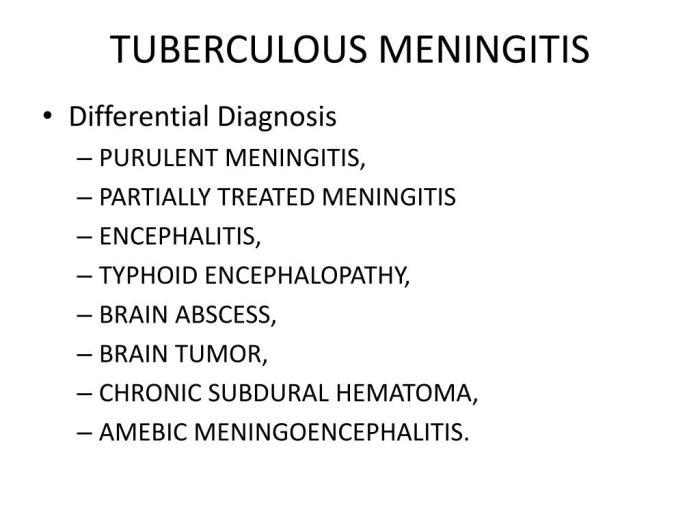
    Differentiating from Other Neurological Conditions

    Differentiating TBM from other neurological conditions requires a comprehensive evaluation, including detailed history, physical examination, and appropriate laboratory investigations.

    • Other types of meningitis: Bacterial meningitis, viral meningitis, and fungal meningitis may present with overlapping symptoms, necessitating careful laboratory investigation.
    • Brain tumors: Some brain tumors can mimic the symptoms of meningitis, but usually present with more persistent and slowly evolving symptoms.
    • Neurodegenerative diseases: Certain neurodegenerative conditions may exhibit symptoms that overlap with those of TBM. However, the progression and presentation are usually different.

    Diagnosis

    Diagnosing tuberculous meningitis (TBM) can be challenging, often requiring a multifaceted approach. The presentation can mimic other types of meningitis, making a definitive diagnosis crucial for timely and appropriate treatment. Accurate diagnosis hinges on a thorough history, physical examination, and a battery of laboratory investigations, particularly cerebrospinal fluid (CSF) analysis.A crucial aspect of the diagnostic process is recognizing the subtle but important clues in the patient’s history and physical findings.

    Early recognition and prompt investigation are essential to reduce morbidity and mortality associated with this condition.

    Diagnostic Criteria

    Tuberculous meningitis diagnosis relies on a combination of clinical and laboratory findings. No single test definitively confirms TBM. The diagnosis often involves ruling out other potential causes of meningitis while accumulating suggestive evidence. The presence of granulomas and acid-fast bacilli (AFB) in the cerebrospinal fluid (CSF) is highly suggestive, though not always present.

    Importance of History and Physical Examination

    A detailed history, focusing on recent or prolonged exposure to individuals with tuberculosis (TB), is crucial. This includes evaluating any potential contact with individuals exhibiting symptoms of pulmonary TB, or even asymptomatic individuals. Physical examination should meticulously assess neurological status, looking for signs of meningeal irritation, such as nuchal rigidity, or focal neurological deficits. These subtle clinical findings, combined with the patient’s history, can significantly aid in the diagnostic process.

    Laboratory Investigations

    Several laboratory investigations are employed to aid in the diagnosis of TBM. A comprehensive blood work-up, including a complete blood count (CBC), erythrocyte sedimentation rate (ESR), and C-reactive protein (CRP), can offer insights into the patient’s overall health and inflammatory state. Importantly, the cerebrospinal fluid (CSF) analysis is paramount.

    CSF Analysis

    Cerebrospinal fluid (CSF) analysis is a cornerstone of TBM diagnosis. It’s crucial to analyze the CSF for various parameters. This includes an examination for cells, protein, glucose, and other biomarkers. The typical CSF findings in TBM often include an elevated white blood cell count (WBC), with a predominance of lymphocytes, an elevated protein level, and a decreased glucose level.

    These findings, when considered alongside the clinical picture, provide valuable clues.

    CSF Findings Comparison Table

    | Feature | Tuberculous Meningitis | Other Meningitis Types ||—|—|—|| Cell Count | Lymphocytic pleocytosis (predominantly lymphocytes) | Variable, can include neutrophils or lymphocytes || Protein | Elevated | Variable, can be elevated || Glucose | Decreased | Normal or slightly decreased (in some cases) || Gram Stain | Negative | Variable, may be positive || Culture | Positive for

    Mycobacterium tuberculosis* | Variable |

    | Acid-Fast Bacilli (AFB) | Positive | Negative |

    Radiological Findings

    Neuroimaging plays a crucial role in evaluating the extent of the disease. Computed tomography (CT) scans and magnetic resonance imaging (MRI) scans are valuable tools in assessing the brain and meninges. CT scans may reveal hydrocephalus, which is often observed in severe cases. MRI scans can depict the presence of inflammatory lesions, which are characteristic of TBM.

    In addition, the location of the lesions within the brain can offer clues to the underlying pathology.

    Treatment and Management

    Tuberculous meningitis, a severe form of infection, necessitates a multifaceted approach to treatment and management. Early diagnosis and aggressive intervention are crucial to minimizing neurological damage and improving patient outcomes. This includes a combination of anti-tubercular drugs, supportive care, and careful monitoring. Successful management relies heavily on adherence to the prescribed treatment regimen.Standard treatment for tuberculous meningitis typically involves a combination of multiple drugs targeting the causative bacteria, Mycobacterium tuberculosis.

    This multi-drug therapy (MDT) approach is vital in combating the development of drug resistance. Supportive care plays a significant role in addressing the symptoms and complications that often accompany this disease.

    Standard Treatment Regimens

    The standard treatment for tuberculous meningitis encompasses a multi-drug regimen, typically lasting for 12 to 24 months. This extended duration reflects the need for thorough eradication of the bacteria from the central nervous system. The specific medications and dosages are carefully tailored to individual patient needs and response. A crucial aspect of treatment is close monitoring of the patient’s condition throughout the duration of therapy.

    Importance of Multi-Drug Therapy

    Multi-drug therapy (MDT) is crucial in the treatment of tuberculous meningitis. It significantly reduces the risk of developing drug resistance in Mycobacterium tuberculosis, a critical factor in the long-term effectiveness of the treatment. This approach utilizes a combination of medications with different mechanisms of action, which hinders the emergence of resistant strains and enhances the overall success rate of the treatment.

    Failure to adhere to MDT can lead to treatment failure and potentially life-threatening complications.

    Role of Supportive Care

    Supportive care is integral to the management of tuberculous meningitis. It addresses the various symptoms and complications that may arise, including fever, headache, seizures, and altered mental status. Close monitoring of vital signs, fluid balance, and nutritional status is essential. Appropriate management of seizures, if they occur, is crucial to prevent further neurological damage. Adequate hydration and nutrition are paramount to supporting the body’s response to treatment.

    Tuberculous meningitis, a serious infection, requires careful medical attention. Understanding the causes and symptoms is crucial for early diagnosis and treatment. While researching this, I stumbled upon an interesting question: can blood donation lower cholesterol levels? The answer, as explored in this resource, can blood donation lower cholesterol levels , is complex and multifaceted. Ultimately, the best approach to managing tuberculous meningitis, and overall health, is a holistic one involving medical guidance and proactive lifestyle choices.

    Potential Complications and Management

    Tuberculous meningitis can lead to a range of complications, including hydrocephalus, cranial nerve palsies, and cerebral infarction. Hydrocephalus, characterized by the accumulation of cerebrospinal fluid in the brain, may necessitate surgical intervention to relieve pressure. Cranial nerve palsies can result in impaired motor or sensory function, requiring close monitoring and supportive care. Cerebral infarction, a blockage of blood flow to the brain, can lead to permanent neurological deficits.

    Treatment focuses on addressing the underlying cause, managing symptoms, and preventing further complications. Early detection and prompt intervention are vital in mitigating the severity of these complications.

    Specific Medications in the Treatment Regimen

    Medication Mechanism of Action Potential Side Effects
    Isoniazid (INH) Inhibits the synthesis of mycolic acids, essential components of the mycobacterial cell wall. Peripheral neuropathy, liver damage
    Rifampicin (RIF) Inhibits RNA synthesis in mycobacteria. Hepatotoxicity, discoloration of body fluids
    Pyrazinamide (PZA) Inhibits bacterial growth at an acidic pH. Hepatotoxicity, hyperuricemia
    Ethambutol (EMB) Disrupts the synthesis of the mycobacterial cell wall. Optic neuritis
    Streptomycin (STR) Inhibits protein synthesis in mycobacteria. Ototoxicity, nephrotoxicity

    Note: This table provides a general overview. Specific dosages and durations of treatment should be determined by a qualified healthcare professional based on individual patient factors and response to therapy.

    Prognosis and Outcomes

    Tuberculous meningitis (TBM) is a serious and potentially devastating infection of the brain and spinal cord. The prognosis for TBM varies significantly depending on several factors, including the speed of diagnosis and treatment initiation, the severity of the initial infection, and the individual’s overall health. Understanding these factors is crucial for developing effective management strategies and improving patient outcomes.

    Factors Influencing Prognosis

    The prognosis of TBM is significantly influenced by several key factors. These include the time taken to diagnose and initiate appropriate treatment, the extent of brain involvement, the presence of complications like hydrocephalus, and the patient’s pre-existing health conditions. Early detection and prompt initiation of therapy are paramount to minimize long-term neurological damage. Patients with more extensive involvement of the meninges or brain parenchyma tend to have a poorer prognosis.

    Furthermore, the presence of hydrocephalus, a buildup of cerebrospinal fluid, often complicates the clinical course and necessitates surgical intervention to reduce pressure on the brain. Pre-existing medical conditions, such as HIV infection or malnutrition, can exacerbate the severity of TBM and negatively impact the prognosis.

    Potential Long-Term Neurological Sequelae

    TBM can lead to a range of long-term neurological sequelae, affecting various cognitive, motor, and sensory functions. These sequelae can include cognitive impairment, such as memory loss, difficulty concentrating, or learning disabilities. Motor deficits, like paralysis or weakness in limbs, are also common. Sensory disturbances, including vision or hearing problems, can also occur. The severity of these sequelae is highly dependent on the extent of brain damage caused by the infection and the effectiveness of treatment.

    For instance, a patient with minimal involvement and prompt treatment might experience only mild cognitive difficulties, whereas someone with widespread infection and delayed treatment could suffer significant, long-lasting neurological impairments.

    Importance of Early Intervention and Appropriate Treatment

    Early diagnosis and treatment are crucial for minimizing the risk of long-term neurological damage. Prompt initiation of anti-tuberculosis therapy, often administered intravenously, is essential to control the infection and prevent further damage to the brain. Early recognition of symptoms, such as headache, fever, stiff neck, and altered mental status, is vital to initiate timely intervention. Proper adherence to the prescribed treatment regimen is equally important to ensure the effectiveness of therapy and prevent relapse.

    A well-structured treatment plan, carefully monitored by medical professionals, is crucial for optimizing patient outcomes.

    Monitoring Procedures for Assessing Treatment Effectiveness and Prognosis

    Monitoring the effectiveness of treatment and prognosis involves a combination of clinical assessments and laboratory tests. Neurological examinations, including assessments of motor function, sensory perception, and cognitive abilities, are routinely performed to evaluate the progression or improvement in the patient’s condition. Serial cerebrospinal fluid (CSF) analyses are essential to monitor the decline in the number of inflammatory cells and the reduction in the presence of bacteria.

    Tuberculous meningitis, a serious infection, often starts subtly like a typical cold. But unlike a common cold, it requires immediate medical attention. Learning the subtle signs your cold is getting better, like a reduced cough or a clearer nose, can be a helpful comparison, but it’s crucial to remember that tuberculous meningitis needs a doctor’s diagnosis. Understanding the overview and more about this condition is vital for early detection and treatment.

    For a detailed guide on recognizing cold improvement, check out this helpful resource: signs your cold is getting better. Ultimately, any unusual symptoms should prompt a visit to your doctor.

    Regular monitoring of vital signs, such as temperature, blood pressure, and heart rate, is also crucial for identifying any complications or deterioration. Radiological imaging, such as CT scans or MRI, may be used to evaluate brain structure and identify any signs of complications.

    Correlation Between Treatment Duration and Recovery Outcomes

    The duration of treatment for TBM significantly correlates with the likelihood of full recovery. Patients who adhere to the complete treatment regimen, typically lasting for several months, have a higher chance of achieving a full recovery. A shorter duration of treatment might be associated with a higher risk of relapse or incomplete recovery. Early intervention, prompt initiation of therapy, and consistent monitoring are critical to maximizing the chances of a successful outcome.

    Treatment Duration (Months) Recovery Outcome
    6-9 Good recovery, minimal sequelae
    9-12 Moderate recovery, some sequelae
    >12 Poor recovery, significant sequelae

    The table above provides a general guideline. Individual responses to treatment vary, and the actual outcomes may differ based on the specific patient’s condition and adherence to treatment.

    Prevention and Public Health

    Tuberculous meningitis, a devastating complication of tuberculosis, underscores the critical need for proactive prevention and robust public health strategies. Effective interventions at individual and societal levels are essential to minimize the risk of transmission and the devastating consequences of this disease. This section will Artikel key preventive measures, emphasizing the importance of early detection and treatment, and highlighting public health strategies for controlling outbreaks.Effective prevention hinges on understanding the transmission pathways and implementing appropriate measures.

    The transmission ofMycobacterium tuberculosis* occurs primarily through respiratory droplets. This means individuals who are infected and coughing or sneezing can spread the bacteria to those nearby. Preventing transmission involves mitigating exposure to these droplets.

    Measures to Prevent Transmission

    Preventing the transmission ofMycobacterium tuberculosis* relies on multiple strategies. These strategies aim to reduce exposure to the bacteria and to ensure prompt treatment of active cases. Individuals with active tuberculosis must take precautions to prevent the spread of the disease. This often involves the use of respiratory protection, such as masks, and isolation to minimize contact with others.

    Role of Vaccination

    Vaccination plays a crucial role in preventing tuberculosis, particularly in high-risk populations. The BCG vaccine, while not completely protective against all forms of tuberculosis, has proven effective in reducing the incidence of severe forms of the disease, including meningitis. This vaccine is frequently used in many parts of the world, especially in children.

    Importance of Early Detection and Treatment

    Early detection and treatment of tuberculosis are critical in preventing the development of tuberculous meningitis. Early diagnosis allows for prompt initiation of treatment, reducing the risk of transmission and the development of severe complications. Regular screening and monitoring of high-risk populations are essential components of this strategy.

    Public Health Strategies for Controlling Tuberculosis Outbreaks

    Public health strategies for controlling tuberculosis outbreaks encompass a wide range of measures. These strategies are multifaceted and involve community engagement, contact tracing, and the provision of appropriate healthcare services. Comprehensive tuberculosis control programs are essential for effective disease management and prevention of outbreaks. Such programs often include:

    • Contact Tracing: Identifying and monitoring individuals who have had close contact with individuals with active tuberculosis is crucial for preventing further transmission. This involves interviewing individuals who were exposed to determine their risk and providing them with necessary preventative measures or testing.
    • Screening and Testing: Regular screening and testing of high-risk populations, such as healthcare workers and individuals with HIV, are important for early detection of tuberculosis and preventing the development of severe complications like meningitis.
    • Treatment Adherence Support: Ensuring patients with tuberculosis complete their treatment regimen is essential to prevent the emergence of drug-resistant strains and the spread of the disease. This includes addressing social and economic barriers that may hinder treatment adherence.
    • Improved Infrastructure and Resources: Strengthening healthcare infrastructure and ensuring adequate resources for tuberculosis diagnosis and treatment are vital for effective disease control. This includes ensuring access to diagnostic tools and trained healthcare personnel.

    Preventive Measures for Individuals at Risk

    Individuals at increased risk of developing tuberculosis, such as those with weakened immune systems or those with close contact to individuals with active tuberculosis, should take proactive measures to minimize their risk of infection. These measures include:

    • Hygiene Practices: Maintaining good hygiene practices, such as frequent handwashing, covering coughs and sneezes, and avoiding close contact with individuals with active tuberculosis, is crucial for preventing the spread of the disease.
    • Healthy Lifestyle Choices: Maintaining a healthy lifestyle, including adequate nutrition and regular exercise, can strengthen the immune system and make individuals less susceptible to tuberculosis infection.
    • Regular Check-ups: Regular check-ups with healthcare providers, particularly for those with underlying health conditions or known exposures, can facilitate early detection of any potential tuberculosis infection.
    • Vaccination (if eligible): Individuals eligible for vaccination, particularly those in high-risk populations, should consider vaccination with BCG.

    Illustrative Case Studies

    Tuberculous meningitis overview and more

    Tuberculous meningitis (TBM) presents a complex diagnostic and therapeutic challenge, often requiring a multidisciplinary approach. Early recognition and prompt treatment are crucial to minimize neurological sequelae. The following case studies illustrate the diverse presentations, diagnostic dilemmas, and management strategies involved in TBM.

    Tuberculous meningitis, a serious infection of the brain’s membranes, requires careful medical attention. Understanding the various symptoms is crucial, but sometimes similar symptoms can arise from other conditions. For instance, back pain and nerve-related issues, like those stemming from herniated discs, what are herniated disc symptoms , can mimic certain aspects of meningitis. This is why a proper diagnosis is essential to ensure the right treatment.

    Further research into the different causes and management strategies for tuberculous meningitis is important to promote awareness and effective care.

    Case Study 1: A Young Adult with Gradual Neurological Deterioration

    This case involves a 25-year-old male presenting with a progressive headache, fever, and gradually worsening confusion over a 3-week period. He denied any recent travel or contact with individuals with known infections. Physical examination revealed meningeal signs, including stiff neck and Brudzinski’s sign. A lumbar puncture yielded cerebrospinal fluid (CSF) analysis showing elevated protein, lymphocytic pleocytosis, and a positive acid-fast bacilli smear.

    PCR for Mycobacterium tuberculosis was also positive. The patient was diagnosed with tuberculous meningitis and started on a four-drug regimen (isoniazid, rifampicin, pyrazinamide, and ethambutol). He was also administered corticosteroids to reduce inflammation.Challenges encountered included the insidious onset of symptoms, which delayed the diagnosis. The absence of travel history or obvious risk factors further complicated the initial assessment.

    The patient’s progressive neurological deterioration required intensive monitoring and supportive care, including respiratory support.A multidisciplinary approach, involving neurologists, infectious disease specialists, and pulmonologists, was crucial for optimal patient management. The team coordinated treatment, monitored for side effects, and addressed the patient’s psychosocial needs.The patient responded well to treatment, showing gradual improvement in neurological function and resolution of symptoms.

    Follow-up care and monitoring were essential to ensure long-term recovery.

    Case Study 2: A Child with Acute Meningitis

    A 6-year-old child presented with high fever, severe headache, and vomiting. The child exhibited stiff neck, altered consciousness, and a positive Kernig’s sign. The CSF analysis revealed elevated protein, lymphocytic pleocytosis, and a positive culture for Mycobacterium tuberculosis. Diagnosis was further confirmed with PCR. The patient received the same four-drug regimen as in Case Study 1, supplemented with close monitoring for complications.

    The child’s condition was initially critical, requiring intensive care and close observation for signs of increased intracranial pressure.The challenges in this case included the difficulty in distinguishing TBM from other forms of acute meningitis, especially in a pediatric population. Early diagnosis was crucial to prevent severe neurological damage. The multidisciplinary approach ensured close collaboration between pediatric specialists, neurologists, and infectious disease specialists.The child responded to treatment with a positive trend in the CSF analysis.

    However, significant neurological sequelae, including cognitive impairment, could not be entirely ruled out, highlighting the importance of long-term rehabilitation.

    Tuberculous meningitis requires a comprehensive approach encompassing early diagnosis, aggressive treatment, and a strong multidisciplinary team. Factors like the patient’s age, underlying health conditions, and adherence to treatment play crucial roles in the outcome. Proper follow-up care is essential to assess and address potential complications.

    Differential Diagnosis: Tuberculous Meningitis Overview And More

    Tuberculous meningitis overview and more

    Tuberculous meningitis (TBM) can mimic other neurological conditions, making accurate diagnosis crucial. The overlapping symptoms and subtle differences in presentation can lead to diagnostic challenges. Early and precise identification of TBM is vital for timely treatment and favorable outcomes. This necessitates a thorough understanding of the differential diagnoses and the specific diagnostic tests used to distinguish TBM from other causes of meningitis.Differentiating TBM from other forms of meningitis and neurological illnesses is essential to provide appropriate and effective treatment.

    Failure to accurately identify TBM can lead to delays in initiating crucial anti-tuberculosis therapy, potentially resulting in poor neurological outcomes. A systematic approach, considering the clinical presentation, epidemiological factors, and specific diagnostic tests, is paramount in arriving at a definitive diagnosis.

    Comparison with Other Meningitis Causes

    Identifying the specific cause of meningitis is a critical aspect of patient management. Different types of meningitis have varying clinical presentations, diagnostic findings, and treatment strategies. Accurate differentiation is crucial for initiating the appropriate management plan.

    • Viral Meningitis: Viral meningitis is frequently characterized by a more benign course compared to TBM. Symptoms are often less severe and typically resolve within a few weeks. Laboratory findings, such as cerebrospinal fluid (CSF) analysis, often show a lymphocytic pleocytosis, with a higher proportion of lymphocytes compared to TBM. The presence of characteristic viral pathogens in the CSF can further aid in diagnosis.

      Viral meningitis is typically self-limiting, and treatment focuses on symptom management.

    • Bacterial Meningitis: Bacterial meningitis, often caused by pathogens like
      -Streptococcus pneumoniae* or
      -Neisseria meningitidis*, presents with a more rapid and severe onset of symptoms compared to TBM. CSF analysis typically reveals a neutrophilic pleocytosis, high protein levels, and low glucose levels. Rapid initiation of broad-spectrum antibiotics is crucial for bacterial meningitis. The clinical presentation, particularly the rapid progression and severe symptoms, helps distinguish it from TBM.

    • Fungal Meningitis: Fungal meningitis, often caused by
      -Cryptococcus neoformans*, may present with a subacute or chronic course. CSF analysis may reveal lymphocytic pleocytosis, but the presence of specific fungal elements can aid in diagnosis. Treatment strategies differ significantly from TBM and involve antifungal agents.
    • Other Neurological Conditions: Conditions such as brain abscesses, stroke, and encephalitis can present with symptoms overlapping with TBM, especially concerning neurological deficits. Imaging studies, like CT scans and MRIs, can aid in distinguishing these conditions from TBM. The presence of focal neurological deficits or specific imaging findings is key to differentiating them from the more diffuse involvement seen in TBM.

    Diagnostic Tests for Differential Diagnosis

    A comprehensive diagnostic approach is crucial in distinguishing TBM from other neurological conditions. This involves a combination of clinical evaluation, laboratory tests, and imaging studies.

    • Cerebrospinal Fluid (CSF) Analysis: CSF analysis is a fundamental diagnostic tool. The presence of lymphocytes, low glucose levels, and elevated protein levels, along with the presence of acid-fast bacilli (AFB) in the CSF, is indicative of TBM. Comparing these findings with other causes of meningitis helps establish a differential diagnosis. Furthermore, the cellular composition and other markers in the CSF help differentiate between the various causes of meningitis.

    • Imaging Studies: Imaging studies, such as CT scans and MRIs, are essential to rule out other neurological conditions. These scans can reveal abnormalities indicative of brain abscesses, stroke, or other focal lesions. CT scans can help identify signs of intracranial pressure elevation, which is common in TBM, and MRIs can further delineate the extent of the pathology and identify specific regions of involvement.

    • Sputum and Tissue Culture: Culture of sputum or tissue samples for
      -Mycobacterium tuberculosis* can confirm the diagnosis of TBM. This is important for accurate identification of the causative agent, which is critical for effective treatment.

    Overlap of Symptoms and Challenges

    The overlapping symptoms between TBM and other neurological conditions can make differentiation challenging. For example, both TBM and viral meningitis can manifest with fever, headache, and altered mental status. A meticulous clinical history, a thorough neurological examination, and the combination of laboratory findings are crucial for distinguishing TBM from other causes.

    Table of Key Differences

    Characteristic Tuberculous Meningitis Viral Meningitis Bacterial Meningitis
    CSF Findings Lymphocytic pleocytosis, low glucose, elevated protein, AFB Lymphocytic pleocytosis, normal glucose, normal or slightly elevated protein Neutrophilic pleocytosis, low glucose, elevated protein
    Course Insidious, subacute Acute, self-limiting Rapid, severe
    Symptoms Headache, fever, altered mental status, neurological deficits Headache, fever, altered mental status, malaise Severe headache, high fever, stiff neck, altered mental status

    Conclusion

    In conclusion, tuberculous meningitis, though a formidable adversary, is not insurmountable. By understanding its multifaceted nature, from its insidious onset to the potential for long-term sequelae, we can better equip ourselves to combat this disease. Early diagnosis and appropriate treatment remain paramount, underscoring the vital role of public health initiatives and research in prevention and improved outcomes.

  • What is Renal Artery Stenosis? A Deep Dive

    What is Renal Artery Stenosis? A Deep Dive

    What is renal artery stenosis? This condition, where the arteries supplying the kidneys narrow, can lead to serious health problems. Understanding the causes, symptoms, and treatment options is crucial for managing this potentially life-altering situation. This post delves into the specifics of renal artery stenosis, exploring the intricacies of this often overlooked health issue.

    Renal artery stenosis (RAS) occurs when the arteries supplying the kidneys become narrowed. This narrowing can disrupt blood flow to the kidneys, potentially leading to a cascade of complications. The condition can be caused by various factors, and early detection is essential for effective management.

    Renal Artery Stenosis (RAS): A Deep Dive

    Renal artery stenosis (RAS) is a condition where the renal arteries, the blood vessels supplying blood to the kidneys, become narrowed or blocked. This narrowing reduces blood flow to the kidneys, potentially leading to serious health problems. Understanding the intricacies of RAS is crucial for early diagnosis and effective treatment.The renal arteries play a vital role in the body’s filtration system.

    They carry oxygenated blood from the heart to the kidneys, where waste products are filtered out and urine is produced. Adequate blood flow through the renal arteries is essential for maintaining healthy kidney function. When these arteries are compromised, the kidneys may not receive the necessary blood supply, resulting in various complications.

    Types of Renal Artery Stenosis

    Renal artery stenosis can be categorized into different types, each with its own underlying causes and clinical implications. This classification is essential for tailored treatment strategies.

    Type of RAS Causes Risk Factors Symptoms
    Atherosclerotic RAS Fatty deposits (plaque) build-up in the artery walls, a common form of arteriosclerosis. High blood pressure, high cholesterol, smoking, diabetes, family history of heart disease, age, and a sedentary lifestyle. Often asymptomatic in early stages. Later stages may present with hypertension, decreased kidney function, and possible symptoms of chronic kidney disease.
    Dissection RAS A tear or split in the inner lining of the artery, often associated with trauma or high blood pressure. History of hypertension, connective tissue disorders (like Marfan syndrome), and trauma to the abdomen. Sudden onset of severe flank pain, hypertension, and possible kidney failure.
    Fibromuscular Dysplasia (FMD) RAS Abnormal thickening and narrowing of the artery walls due to irregular development of the muscle and connective tissues in the arterial wall. More common in women, especially those in their 30s and 40s. Family history of FMD and certain genetic predispositions are possible risk factors. Often asymptomatic in early stages. Symptoms can include hypertension, headaches, and kidney-related problems, like proteinuria (protein in the urine).
    Vasculitis RAS Inflammation of the blood vessel walls, often triggered by autoimmune disorders. Autoimmune diseases like polyarteritis nodosa, giant cell arteritis, and lupus. Symptoms may include fever, fatigue, and other systemic symptoms. Kidney-specific symptoms like blood in the urine or decreased urine output might also appear.

    Atherosclerotic Renal Artery Stenosis (RAS)

    Atherosclerotic RAS is the most common type, characterized by plaque buildup in the renal arteries. This plaque hardens and narrows the arteries, reducing blood flow to the kidneys. Factors like high blood pressure and high cholesterol significantly contribute to the development of this condition.

    Fibromuscular Dysplasia (FMD) RAS

    FMD RAS involves an abnormal thickening and narrowing of the artery walls, impacting the flow of blood to the kidneys. This condition is more prevalent in women and often occurs in younger individuals.

    Causes and Risk Factors

    Renal artery stenosis (RAS) isn’t a single, monolithic condition. It’s a narrowing of the renal arteries, the blood vessels supplying the kidneys, often caused by a variety of factors. Understanding these causes and risk factors is crucial for early diagnosis and effective treatment. This deep dive will explore the common culprits behind RAS, highlighting the differences in prevalence and risk across various populations.The narrowing of the renal arteries can significantly impact kidney function, potentially leading to high blood pressure and kidney damage.

    Identifying the root cause of the stenosis is vital for personalized treatment strategies.

    Common Causes of Renal Artery Stenosis

    Atherosclerosis and fibromuscular dysplasia are the two most prevalent causes of renal artery stenosis. Atherosclerosis, the hardening and narrowing of arteries due to plaque buildup, is a common culprit in individuals with a history of cardiovascular disease. Fibromuscular dysplasia, on the other hand, is a non-atherosclerotic condition characterized by abnormal thickening and narrowing of the artery walls. It’s more prevalent in younger individuals and women.

    Renal artery stenosis, basically, is when the arteries leading to your kidneys narrow. This can lead to high blood pressure and, in some cases, kidney damage. Caring for someone with this condition can be incredibly demanding, often leading to feelings of exhaustion and stress – signs of caregiver burnout, which are important to recognize. Identifying these signs early can help prevent further issues and ensure you’re not neglecting your own well-being.

    Ultimately, understanding renal artery stenosis is crucial for effective management and support for both the patient and their caregiver.

    Conditions Increasing Risk of RAS

    Several conditions increase the likelihood of developing renal artery stenosis. Hypertension, or high blood pressure, is a significant risk factor, as the sustained high pressure can damage the artery walls, contributing to the development of stenosis. Diabetes, a condition affecting blood sugar regulation, can also increase the risk. Smoking and a sedentary lifestyle further compound the risk by contributing to the development of atherosclerosis.

    Prevalence of RAS in Different Populations

    The prevalence of RAS can vary significantly across different populations. Studies indicate a higher incidence in individuals of Caucasian descent compared to those of African or Asian descent. However, this is a complex issue, and the factors contributing to these differences are still being researched. Environmental factors, socioeconomic status, and access to healthcare can all influence the observed differences.

    Role of Genetics and Lifestyle Factors, What is renal artery stenosis

    Genetic predisposition plays a role in the development of some forms of renal artery stenosis, while lifestyle factors like diet, physical activity, and stress levels contribute to the risk of developing atherosclerosis. A diet high in saturated and trans fats, coupled with a lack of physical activity, can accelerate the buildup of plaque in the arteries, increasing the risk of stenosis.

    Conversely, a healthy diet and regular exercise can help mitigate these risks.

    Renal artery stenosis, a narrowing of the arteries leading to the kidneys, can sometimes lead to fatigue, much like how some energy drinks, despite their intended effect, can leave you feeling drained. This can be due to a variety of factors, and if you’re curious about why energy drinks make you tired, check out this insightful article: why do energy drinks make me tired.

    Ultimately, understanding the underlying causes of fatigue is crucial, whether it’s related to renal artery stenosis or something else entirely. Chronic fatigue can be a symptom of many things, including kidney issues.

    Comparison of Causes and Risk Factors for Different Types of RAS

    Cause Description Risk Factor Type Examples
    Atherosclerosis Hardening and narrowing of the arteries due to plaque buildup. Lifestyle, Cardiovascular High cholesterol, high blood pressure, smoking, obesity
    Fibromuscular Dysplasia (FMD) Non-atherosclerotic condition characterized by abnormal thickening and narrowing of the artery walls. Genetic, Hormonal Young adults, particularly women, individuals with a family history of FMD.
    Trauma Physical injury to the renal artery. External Car accidents, blunt force trauma.
    Inflammation Inflammation of the renal artery. Autoimmune, Infections Vasculitis, certain infections.

    Symptoms and Diagnosis

    Renal artery stenosis (RAS) often presents subtly, mimicking other conditions, particularly high blood pressure. Understanding the range of symptoms and diagnostic methods is crucial for early detection and effective management. This can significantly impact the patient’s long-term health.Identifying the specific symptoms and utilizing appropriate diagnostic tools are vital steps in the process of diagnosing renal artery stenosis. A thorough evaluation helps clinicians differentiate RAS from other conditions causing hypertension or kidney dysfunction.

    Early diagnosis is critical for timely intervention and improved patient outcomes.

    Common Symptoms of Renal Artery Stenosis

    The symptoms of renal artery stenosis can vary significantly depending on the severity and the presence of other underlying health conditions. While hypertension is a hallmark symptom, other manifestations can occur. Important to note that some patients may not experience any symptoms at all, particularly in the early stages.

    • Hypertension: Elevated blood pressure is the most frequent symptom, often severe and resistant to typical blood pressure medications. Persistent high blood pressure that doesn’t respond to standard treatment should raise suspicion for underlying conditions like renal artery stenosis.
    • Renal dysfunction: In advanced stages, kidney function may decline, leading to decreased urine output, swelling (edema), and electrolyte imbalances. These symptoms are more evident as the condition progresses.
    • Flank pain: Pain in the flank (side) area of the abdomen, particularly on the affected side, can be a symptom, though it’s not always present.
    • Decreased appetite and fatigue: Some patients experience reduced appetite and feelings of tiredness, which can be attributed to various factors, including the impact of RAS on the kidneys.
    • Other potential symptoms: These may include dizziness, headaches, and blood in the urine. The presence of these symptoms, in combination with other risk factors, should prompt a thorough evaluation.

    Diagnostic Methods for Renal Artery Stenosis

    Various imaging techniques are employed to diagnose renal artery stenosis, each with its own strengths and limitations. These techniques play a crucial role in accurately identifying the condition and guiding appropriate treatment strategies.

    • Imaging Techniques: Several imaging modalities can identify renal artery stenosis. These methods provide crucial insights into the structure and function of the renal arteries.
    Diagnostic Procedure Accuracy Limitations Description
    Ultrasound Generally good for initial screening Limited in complex cases; may not visualize all areas Uses sound waves to create images of the renal arteries. It is non-invasive, relatively inexpensive, and readily available.
    CT Angiography High accuracy for detecting stenosis Exposure to ionizing radiation; contrast agent may cause allergic reactions Combines computed tomography (CT) scans with contrast material to visualize the arteries. It provides detailed images of the blood vessels.
    MRI Angiography Excellent for visualizing blood vessels; good for evaluating the kidney Longer examination time; may not be suitable for patients with certain medical conditions Uses magnetic fields and radio waves to create images of the renal arteries, offering detailed anatomical information without radiation exposure.

    Importance of Timely Diagnosis

    Early diagnosis of renal artery stenosis is critical for managing the condition effectively. Prompt identification allows for the implementation of appropriate treatment strategies to prevent further complications and preserve kidney function. Delay in diagnosis can lead to irreversible damage to the kidneys.

    Treatment Options

    What is renal artery stenosis

    Renal artery stenosis (RAS) treatment aims to improve blood flow to the kidneys, reducing the risk of kidney damage and high blood pressure. The optimal approach depends on the severity of the stenosis, the patient’s overall health, and the presence of any other underlying conditions. Different treatment options offer varying degrees of success and potential side effects.Effective management of RAS often involves a combination of strategies, tailored to the individual patient.

    This can include lifestyle modifications, medical therapy, and potentially surgical intervention. Understanding the various approaches allows patients and healthcare providers to make informed decisions.

    Medical Management

    Medical therapies are often the first line of treatment for RAS. These medications aim to control blood pressure, reduce cholesterol, and prevent blood clots. They also help manage underlying conditions like diabetes or high cholesterol that contribute to the development of RAS.

    • Angiotensin-converting enzyme (ACE) inhibitors and angiotensin receptor blockers (ARBs): These medications help relax blood vessels, reducing blood pressure and improving blood flow to the kidneys. Examples include lisinopril and valsartan.
    • Beta-blockers: These medications can help control blood pressure and heart rate, which is important in managing RAS. Examples include metoprolol and atenolol.
    • Statins: If cholesterol is elevated, statins can help lower it, reducing the risk of further plaque buildup and narrowing of the renal arteries. Examples include atorvastatin and simvastatin.
    • Antiplatelet agents: These medications help prevent blood clots, which can further reduce blood flow to the kidneys. Aspirin is a common example.

    Surgical Interventions

    Surgical interventions are often considered for patients who do not respond adequately to medical therapy or who have severe stenosis. These procedures aim to restore blood flow to the kidneys.

    • Angioplasty and Stenting: This is a minimally invasive procedure. A catheter with a balloon at the tip is inserted into the narrowed renal artery. The balloon is inflated to widen the artery, and a stent, a small mesh tube, is often placed to keep the artery open. This procedure improves blood flow to the kidneys and helps prevent further narrowing.

      It’s considered a relatively safe procedure with a high success rate.

    • Renal Artery Bypass: In cases where the stenosis is extensive or involves a major portion of the renal artery, a bypass surgery may be necessary. A healthy blood vessel, often from another part of the body, is used to create an alternate route for blood flow to the kidney, bypassing the narrowed area. This procedure is more complex and carries a higher risk of complications than angioplasty and stenting.

    Treatment Effectiveness

    The effectiveness of treatment for RAS varies depending on the individual patient and the severity of the condition. Medical therapy can be highly effective in managing milder cases and preventing progression of the disease. Surgical interventions, particularly angioplasty and stenting, often yield excellent results in restoring blood flow and reducing the risk of kidney damage in more severe cases.

    However, the long-term success of treatment can be influenced by factors like adherence to medication regimens, lifestyle choices, and the presence of other health issues.

    Comparison of Treatment Options

    Treatment Option Advantages Disadvantages Description
    Medical Management Non-invasive, generally well-tolerated, cost-effective, first-line approach May not be effective in severe cases, requires long-term commitment to medication, potential side effects Control blood pressure, cholesterol, and other risk factors through medication.
    Angioplasty and Stenting Minimally invasive, high success rate in restoring blood flow, less invasive than bypass Risk of complications such as bleeding, infection, or blood clots; need for follow-up care; potential for restenosis Widening the artery using a balloon and inserting a stent to maintain patency.
    Renal Artery Bypass Restores blood flow to the kidney when other options are not effective More invasive, higher risk of complications, longer recovery time Creating an alternate route for blood flow using a healthy blood vessel.

    Complications and Prognosis: What Is Renal Artery Stenosis

    What is renal artery stenosis

    Renal artery stenosis (RAS) can lead to a range of complications, impacting the long-term health and quality of life for those affected. Understanding these potential issues and the factors influencing prognosis is crucial for effective management and patient care. The severity of stenosis and the chosen treatment significantly affect the outlook.The progression of RAS, and the subsequent complications, is often influenced by the degree of narrowing in the renal arteries, as well as the presence of other underlying health conditions.

    A timely diagnosis and appropriate treatment strategy are essential to mitigate these risks and improve the overall prognosis.

    Potential Complications

    The complications of RAS can significantly impact the patient’s health and well-being. These complications stem from the kidneys’ reduced blood supply and the body’s attempts to compensate for the reduced perfusion.

    • Hypertension: The kidneys play a crucial role in regulating blood pressure. Reduced blood flow to the kidneys can trigger a cascade of hormonal responses, leading to sustained high blood pressure (hypertension). Untreated hypertension significantly increases the risk of cardiovascular disease, stroke, and kidney damage. For example, a patient with severe RAS may require multiple medications to control their blood pressure, demonstrating the impact on their overall health management.

      Renal artery stenosis, a narrowing of the arteries supplying blood to the kidneys, can sometimes be linked to sleep patterns. Interestingly, how you sleep – whether you’re a hot sleeper or a cold sleeper, as discussed in detail in this helpful article about hot sleeper vs cold sleeper – might have surprising connections to your overall health, including kidney health.

      Ultimately, understanding renal artery stenosis involves looking at various factors, not just sleep styles.

    • Chronic Kidney Disease (CKD): Prolonged reduced blood flow to the kidneys can lead to chronic kidney disease (CKD). This progressive condition damages the kidney’s filtering ability, ultimately affecting the body’s ability to remove waste products and maintain electrolyte balance. Patients with CKD often experience fatigue, fluid retention, and other symptoms that impact their daily lives. The severity of CKD depends on the duration and degree of RAS.

    • Heart Disease: The high blood pressure often associated with RAS significantly increases the risk of developing cardiovascular diseases. The constant strain on the heart can lead to conditions like coronary artery disease, heart failure, and stroke. Individuals with RAS often require ongoing monitoring and management of their cardiovascular health.
    • Aneurysms: In some cases, the increased blood pressure in the narrowed artery can lead to the formation of an aneurysm, a bulge or weakening in the artery wall. This can pose a significant risk of rupture, potentially leading to life-threatening complications.

    Long-Term Prognosis

    The long-term prognosis for patients with renal artery stenosis depends on various factors.

    • Severity of Stenosis: The extent of the narrowing in the renal artery significantly influences the prognosis. Mild stenosis may not cause significant complications, whereas severe stenosis may lead to more severe consequences. For instance, patients with severe stenosis might experience faster progression of CKD compared to those with milder stenosis.
    • Treatment Choice: The choice of treatment plays a critical role in the long-term prognosis. Effective treatment options, such as angioplasty with stenting, can significantly improve blood flow to the kidneys, reducing the risk of complications and improving overall outcomes. Surgical intervention is often necessary in cases where medical management fails to effectively control the stenosis. The efficacy of the chosen treatment method determines the success rate of preventing further complications.

    • Overall Health: Pre-existing health conditions, such as diabetes or high cholesterol, can worsen the prognosis for patients with RAS. These factors can exacerbate the complications and lead to faster progression of kidney damage and cardiovascular disease. For example, a patient with pre-existing diabetes is more susceptible to developing severe CKD from RAS compared to a patient without diabetes.

    Impact on Quality of Life

    The complications arising from renal artery stenosis can substantially impact a patient’s quality of life. These complications can lead to a range of symptoms that affect daily activities, social interactions, and overall well-being.

    • Fatigue and Weakness: Kidney dysfunction can lead to fatigue, weakness, and reduced energy levels, making it difficult to perform daily tasks. The accumulation of waste products in the body due to impaired kidney function contributes to this feeling of fatigue.
    • Fluid Retention: Kidney problems can cause fluid retention, leading to swelling in the limbs and other body parts. This can significantly impact mobility and overall comfort.
    • Dietary Restrictions: Patients with renal disease often need to follow strict dietary restrictions to manage their condition, which can be challenging and affect their social life.

    Untreated RAS

    Untreated renal artery stenosis can have severe and potentially life-threatening consequences.

    • Accelerated Progression of CKD: Untreated RAS allows kidney damage to progress more rapidly, leading to a decline in kidney function and an increased risk of kidney failure.
    • Significant Increase in Hypertension: The untreated condition can result in uncontrolled high blood pressure, significantly increasing the risk of cardiovascular events such as heart attack and stroke.
    • Reduced Life Expectancy: The cumulative effect of these complications can significantly reduce life expectancy for patients with untreated RAS. The severe complications can lead to a shorter life expectancy and reduced quality of life. A proactive approach towards diagnosis and treatment can drastically improve the patient’s long-term outlook.

    Prevention Strategies

    Renal artery stenosis (RAS) is a significant health concern, but proactive measures can significantly reduce your risk. By adopting healthy lifestyle choices and diligently managing underlying conditions, you can substantially decrease your chances of developing this potentially serious condition. Early intervention and consistent preventive strategies are key to maintaining long-term cardiovascular health.A combination of lifestyle modifications, consistent medical monitoring, and proactive management of risk factors are crucial for preventing RAS.

    This involves understanding the modifiable factors and actively working towards positive changes in your daily habits. By prioritizing preventive measures, you can significantly lower your risk and improve your overall well-being.

    Lifestyle Modifications for Reduced Risk

    Implementing healthy lifestyle choices plays a vital role in minimizing the risk of RAS. A balanced diet, regular exercise, and stress management techniques can contribute to a healthier cardiovascular system. These strategies, when combined with diligent management of risk factors, significantly reduce the likelihood of developing RAS.

    • Balanced Diet: Maintaining a diet rich in fruits, vegetables, and whole grains, while limiting processed foods, saturated fats, and excessive sodium intake, is essential for overall cardiovascular health. A diet rich in fiber and low in saturated fats can help manage cholesterol levels and blood pressure, reducing the risk of atherosclerosis, a major contributor to RAS.
    • Regular Physical Activity: Engaging in regular physical activity, such as brisk walking, swimming, or cycling, can help maintain a healthy weight, control blood pressure, and improve overall cardiovascular health. Aim for at least 150 minutes of moderate-intensity aerobic activity per week, or 75 minutes of vigorous-intensity activity. Consistency is key to reaping the benefits.
    • Stress Management Techniques: Chronic stress can contribute to elevated blood pressure and other risk factors associated with RAS. Practicing stress-reducing techniques like yoga, meditation, or deep breathing exercises can be beneficial in managing stress levels and promoting cardiovascular health. Finding healthy coping mechanisms for stress is crucial in mitigating the negative effects of chronic stress on the cardiovascular system.

    Importance of Regular Health Checkups

    Regular health checkups are vital for identifying and managing risk factors associated with RAS. These checkups allow for early detection of potential problems, enabling prompt intervention and reducing the severity of the condition. Proactive monitoring allows for early intervention and treatment, which can greatly reduce the likelihood of serious complications.

    • Blood Pressure Monitoring: Regular blood pressure checks are crucial for detecting hypertension, a significant risk factor for RAS. Early detection and management of hypertension can prevent the progression of the disease and reduce the risk of complications.
    • Blood Sugar Monitoring: Monitoring blood sugar levels is essential for individuals with diabetes, as uncontrolled diabetes can damage blood vessels, including those in the kidneys. Maintaining optimal blood sugar levels is critical for reducing the risk of RAS and other related complications.
    • Lipid Panel Checks: Regular lipid panel checks can help identify and manage high cholesterol levels, which are a significant risk factor for atherosclerosis. Monitoring and managing cholesterol levels can help prevent the buildup of plaque in the renal arteries, reducing the risk of RAS.

    Controlling Risk Factors: Hypertension and Diabetes

    Controlling risk factors such as hypertension and diabetes is paramount in preventing RAS. These conditions, if left unmanaged, can significantly increase the risk of developing the disease. Consistent management of these conditions is critical to minimize the risks and maintain overall cardiovascular health.

    • Hypertension Management: Hypertension, or high blood pressure, is a major risk factor for RAS. Adopting lifestyle modifications, adhering to prescribed medications, and following a doctor’s recommendations are essential in controlling blood pressure. A healthy diet, regular exercise, and stress management techniques are often effective in conjunction with medication to control blood pressure.
    • Diabetes Management: Individuals with diabetes should actively manage their blood sugar levels through a combination of medication, diet, and exercise. Strict adherence to a diabetes management plan is crucial for minimizing the risk of RAS and its associated complications. Regular monitoring and close communication with healthcare providers are vital.

    End of Discussion

    In conclusion, renal artery stenosis is a significant health concern requiring careful attention. Understanding the causes, symptoms, and treatment options is vital for timely intervention and managing the potential complications. By grasping the intricacies of this condition, individuals can actively participate in their healthcare journey and work towards a healthier future.

  • Fibromyalgia Primary or Secondary Unveiling the Types

    Fibromyalgia Primary or Secondary Unveiling the Types

    Fibromyalgia primary or secondary is a complex condition that often stumps doctors and patients alike. Understanding the distinctions between primary and secondary forms is crucial for proper diagnosis and effective management. This exploration delves into the characteristics, causes, and treatment approaches for both, offering a comprehensive guide for anyone affected by or seeking to understand this pervasive illness.

    This article will cover the definition of fibromyalgia, including its diagnostic criteria and comparison to other conditions. We will delve into the origins and contributing factors of primary fibromyalgia, contrasting them with the secondary form, which often arises from other underlying health issues. We will explore the diagnostic process, differentiating the symptoms and approaches for each type. Further, we will Artikel management strategies tailored to both primary and secondary forms, emphasizing the crucial role of interdisciplinary care.

    Illustrative case studies will solidify the discussion and highlight the complexity of diagnosis.

    Defining Fibromyalgia

    Fibromyalgia is a chronic condition characterized by widespread musculoskeletal pain, fatigue, and a range of other symptoms. Understanding its intricacies is crucial for effective diagnosis and management. This chronic pain syndrome impacts daily life significantly, demanding a comprehensive approach to care.Fibromyalgia is a complex disorder that involves widespread pain, accompanied by various symptoms impacting a person’s overall well-being.

    Figuring out if your fibromyalgia is primary or secondary can be tricky. It’s a bit like choosing the right doctor for your asthma – you need someone who understands the nuances and complexities of your specific condition. Finding the right specialist, like a pulmonologist for types of asthma doctors and choosing the best one , is key to getting the right diagnosis and treatment plan.

    Ultimately, pinpointing whether your fibromyalgia is primary or secondary is crucial for developing an effective treatment strategy. It’s a personal journey, so don’t hesitate to seek out expert opinions.

    It’s not just about the pain; it encompasses a constellation of issues affecting sleep, mood, and cognitive function. The exact cause of fibromyalgia remains elusive, but research suggests a combination of genetic predisposition, environmental factors, and psychological elements play a role.

    Characteristics of Fibromyalgia

    Fibromyalgia is defined by its pervasive pain and accompanying symptoms. The pain is often described as a deep, aching sensation, affecting multiple areas of the body simultaneously. Fatigue is a common and significant symptom, often impacting energy levels and daily activities. Sleep disturbances are frequently reported, with difficulty falling asleep, staying asleep, or experiencing restorative sleep.

    Symptoms of Fibromyalgia

    The symptoms of fibromyalgia extend beyond pain and fatigue. Cognitive difficulties, such as trouble concentrating or memory problems, are common complaints. Emotional distress, including anxiety and depression, frequently accompanies the condition. Other symptoms include headaches, irritable bowel syndrome, and sleep disturbances.

    Diagnostic Criteria for Fibromyalgia

    The diagnosis of fibromyalgia relies on a combination of clinical evaluation and symptom assessment. The American College of Rheumatology (ACR) criteria are commonly used. These criteria include widespread pain, pain lasting for at least three months, and the presence of tender points. These tender points are specific areas on the body where pressure elicits pain.

    Comparison with Other Conditions

    | Feature | Fibromyalgia | Rheumatoid Arthritis | Osteoarthritis ||—|—|—|—|| Pain | Widespread, deep aching | Joint pain, symmetrical | Joint pain, localized, progressive || Inflammation | Low to absent | High | Low to moderate || Tender Points | Present | Absent | Absent || Fatigue | Often severe | Variable | Variable || Sleep | Disrupted | Variable | Variable || Cognitive Issues | Common | Variable | Variable |
    This table highlights key distinctions between fibromyalgia and other conditions.

    The presence or absence of inflammation, tender points, and specific pain patterns can aid in differentiation. Consulting with a healthcare professional is essential for accurate diagnosis and tailored treatment plans.

    Diagnostic Process Flowchart

    (A visual flowchart would be best presented graphically. Here is a textual representation instead.) Patient presents with symptoms (e.g., widespread pain, fatigue, sleep disturbances).→ Healthcare professional assesses patient history and performs a physical examination.→ Questions about the duration, intensity, and location of pain are asked.→ Examination for tender points according to the ACR criteria.→ Evaluation of other symptoms (fatigue, sleep, cognitive issues).→ Rule out other potential conditions (e.g., rheumatoid arthritis, lupus).→ If criteria for fibromyalgia are met, a diagnosis of fibromyalgia is made.→ Patient is provided with appropriate management strategies and referrals (e.g., physical therapy, counseling).
    This flowchart illustrates a general diagnostic process. The exact steps and evaluations may vary depending on the individual case and healthcare provider’s approach.

    Primary Fibromyalgia

    Fibromyalgia primary or secondary

    Primary fibromyalgia, a perplexing condition, remains a mystery in many ways. While the exact causes are still being investigated, researchers are uncovering potential factors that may play a role in its development. This form of fibromyalgia is not triggered by another underlying medical condition, as is the case with secondary fibromyalgia. Instead, it is believed to originate from intrinsic factors within the individual.Primary fibromyalgia is characterized by widespread pain, fatigue, and a constellation of other symptoms.

    The origins of this condition are often attributed to a complex interplay of genetic predispositions and environmental influences. Understanding these contributing factors can pave the way for improved diagnostic and therapeutic approaches.

    Potential Origins of Primary Fibromyalgia

    The exact origins of primary fibromyalgia remain elusive. Several hypotheses propose potential pathways for its development, highlighting the complexity of the condition. These hypotheses suggest that primary fibromyalgia isn’t simply a single cause but rather an amalgamation of factors interacting in intricate ways.

    Figuring out if your fibromyalgia is primary or secondary can be tricky, right? While exploring natural remedies for pain management, I’ve been researching different approaches, and stumbled upon some interesting natural treatments for endometriosis. Natural treatments for endometriosis are definitely worth looking into for anyone experiencing chronic pain, and it’s a good starting point to help identify possible underlying factors, which could help to understand the roots of fibromyalgia.

    Ultimately, determining whether fibromyalgia is primary or secondary requires a thorough assessment with your doctor, so don’t self-diagnose!

    Contributing Factors to Primary Fibromyalgia

    Several factors are believed to contribute to the development of primary fibromyalgia. These factors may act individually or in combination to increase susceptibility to the condition.

    • Genetic Predisposition: A family history of fibromyalgia or other chronic pain conditions can increase the risk of developing primary fibromyalgia. This suggests a potential genetic component, though the specific genes involved remain to be fully elucidated. Individuals with a family history of mood disorders, anxiety, or other chronic conditions might be at a higher risk as well.

      This correlation needs further investigation.

    • Environmental Factors: Exposure to certain environmental stressors, such as infections, trauma, or significant life events, may play a role in triggering or exacerbating primary fibromyalgia. Stressful events, such as the death of a loved one or a major accident, might trigger or worsen existing pain and symptoms. The precise nature of this relationship remains unclear.
    • Neurobiological Factors: Impairments in the central nervous system’s processing of pain signals might be a key element in primary fibromyalgia. This includes abnormal functioning of neurotransmitters and altered brain activity patterns. These dysfunctions can lead to a heightened sensitivity to pain and a more pronounced response to otherwise benign stimuli.

    Prevalence of Primary vs. Secondary Fibromyalgia

    The prevalence of primary fibromyalgia compared to secondary fibromyalgia is a topic of ongoing research. Estimates vary, and more studies are needed to clarify the exact prevalence of each form. However, primary fibromyalgia is believed to be more prevalent than secondary fibromyalgia in certain patient populations, particularly in those without any other identifiable underlying conditions. This difference in prevalence needs further study to understand its true extent.

    Genetic and Environmental Factors

    Genetic and environmental factors are suspected to play a crucial role in primary fibromyalgia. The interplay of these factors is believed to influence an individual’s susceptibility to the condition. However, the precise mechanisms through which these factors interact remain to be fully understood. Further research is needed to uncover the specific genes and environmental triggers that contribute to the development of primary fibromyalgia.

    Theories Regarding the Cause of Primary Fibromyalgia

    Theory Description
    Neurobiological Dysfunction Suggests abnormal processing of pain signals in the central nervous system, potentially involving altered neurotransmitter function.
    Genetic Predisposition Implies a hereditary component, where individuals with certain genetic markers might be more susceptible to developing the condition.
    Environmental Factors Posits that environmental stressors, such as infections, trauma, or significant life events, can trigger or exacerbate primary fibromyalgia.
    Interaction of Multiple Factors Recognizes that the development of primary fibromyalgia likely involves a complex interplay of genetic, neurobiological, and environmental factors.

    Secondary Fibromyalgia

    Fibromyalgia primary or secondary

    Secondary fibromyalgia isn’t a distinct condition in itself, but rather a manifestation of fibromyalgia symptoms arising from another underlying medical issue. It’s crucial to understand that the symptoms are not caused by the underlying condition directly but are often a complex consequence of the body’s response to it. This response can involve various physiological and psychological mechanisms. Identifying and addressing the root cause is vital for effective management.Secondary fibromyalgia often shares overlapping symptoms with primary fibromyalgia, making diagnosis challenging.

    However, the presence of a clear link to another medical condition is the key differentiator. Differentiating between primary and secondary fibromyalgia necessitates a thorough medical evaluation, including a complete medical history, physical examination, and potentially diagnostic tests to identify the contributing factors.

    Conditions Associated with Secondary Fibromyalgia

    A variety of medical conditions can trigger or exacerbate the development of secondary fibromyalgia. These conditions can impact various systems within the body, from the musculoskeletal to the neurological and immune systems. Understanding these connections is essential for comprehensive care.

    • Chronic Fatigue Syndrome (CFS): CFS often coexists with fibromyalgia, characterized by persistent fatigue, sleep disturbances, and cognitive difficulties. The overlap suggests shared pathophysiological mechanisms potentially involving immune system dysfunction or neurotransmitter imbalances.
    • Rheumatoid Arthritis (RA): Chronic inflammation associated with RA can lead to widespread pain and stiffness, mimicking fibromyalgia symptoms. The shared inflammatory response may contribute to the development of secondary fibromyalgia in some individuals.
    • Lupus: Systemic lupus erythematosus (SLE) is an autoimmune disorder that can cause a range of symptoms, including widespread pain, fatigue, and cognitive dysfunction. The chronic inflammatory nature of lupus may be a factor in the development of secondary fibromyalgia.
    • Multiple Sclerosis (MS): The neurological damage caused by MS can result in pain, fatigue, and sensory disturbances that overlap with fibromyalgia symptoms. The neurological involvement may be a crucial factor in the development of secondary fibromyalgia in some cases.
    • Post-Traumatic Stress Disorder (PTSD): Experiencing a traumatic event can significantly impact the body’s physiological and psychological well-being. The resulting stress response may contribute to the development of secondary fibromyalgia, especially when coupled with other physical or psychological conditions.

    Pathophysiological Mechanisms

    The precise pathophysiological mechanisms linking various conditions to secondary fibromyalgia are not fully understood. However, several potential factors are implicated.

    • Chronic Inflammation: Many conditions associated with secondary fibromyalgia involve chronic inflammation. This inflammation can trigger a cascade of events leading to widespread pain, fatigue, and other symptoms associated with fibromyalgia.
    • Neurotransmitter Imbalances: Some research suggests that neurotransmitter dysregulation, such as serotonin and norepinephrine imbalances, may play a role in both primary and secondary fibromyalgia. The presence of underlying conditions may exacerbate these imbalances, contributing to the development of symptoms.
    • Immune System Dysfunction: Dysfunction in the immune system has been linked to various conditions associated with secondary fibromyalgia. This dysfunction may lead to a heightened inflammatory response, impacting various tissues and organs, potentially contributing to widespread pain and fatigue.
    • Central Sensitization: Conditions that involve chronic pain or nerve damage can lead to central sensitization. This altered processing of pain signals in the central nervous system can result in widespread pain and other symptoms, potentially leading to secondary fibromyalgia.

    Potential Triggers

    Various factors can potentially trigger or exacerbate secondary fibromyalgia in individuals with underlying conditions.

    • Stress: Stressful life events, including physical or emotional trauma, can significantly impact the body’s physiological and psychological responses, possibly contributing to or worsening secondary fibromyalgia symptoms.
    • Medications: Certain medications can have side effects that mimic or worsen fibromyalgia symptoms, particularly those targeting chronic pain or inflammation. Proper medication management is essential.
    • Infection: Infections, especially chronic infections, can trigger or exacerbate underlying conditions and contribute to secondary fibromyalgia.
    • Lifestyle Factors: Unhealthy lifestyle choices, such as poor diet, lack of exercise, and inadequate sleep, can worsen existing conditions and potentially contribute to the development of secondary fibromyalgia.

    Conditions Potentially Causing Secondary Fibromyalgia

    Condition Associated Symptoms
    Chronic Fatigue Syndrome Persistent fatigue, sleep disturbances, cognitive difficulties
    Rheumatoid Arthritis Widespread pain, stiffness, inflammation
    Lupus Widespread pain, fatigue, cognitive dysfunction, skin rashes
    Multiple Sclerosis Neurological symptoms, pain, fatigue, sensory disturbances
    Post-Traumatic Stress Disorder Anxiety, depression, flashbacks, nightmares, sleep disturbances

    Differentiating Primary and Secondary Fibromyalgia

    Understanding the distinctions between primary and secondary fibromyalgia is crucial for accurate diagnosis and tailored treatment. While both conditions share overlapping symptoms, their underlying causes and diagnostic pathways differ significantly. This difference in origins influences the course and prognosis of the illness.Primary fibromyalgia, often seen as an idiopathic condition, is characterized by a complex interplay of genetic and environmental factors that contribute to its development.

    Secondary fibromyalgia, conversely, is frequently linked to another identifiable medical condition or event. Distinguishing these two types is essential for guiding appropriate medical interventions and management strategies.

    Symptom Comparison

    The symptoms of primary and secondary fibromyalgia can overlap, but subtle differences exist. Primary fibromyalgia often presents with widespread pain, fatigue, and sleep disturbances as the initial and dominant symptoms. Secondary fibromyalgia, in contrast, may present with pain and fatigue alongside symptoms specific to the underlying condition. For instance, if the secondary cause is lupus, the patient may exhibit joint inflammation and skin rashes alongside the fibromyalgia symptoms.

    Diagnostic Approaches

    Diagnostic approaches for primary and secondary fibromyalgia differ based on the suspected cause. Primary fibromyalgia relies heavily on a comprehensive medical history, physical examination, and symptom assessment. Clinicians use standardized pain scales and questionnaires to gauge the severity and distribution of pain. In secondary fibromyalgia, the diagnostic process involves identifying the underlying condition first. The assessment then focuses on evaluating the symptoms of fibromyalgia within the context of the primary condition.

    For example, if the patient has rheumatoid arthritis, the rheumatologist would consider both the arthritis-related symptoms and the potential fibromyalgia symptoms to formulate a complete diagnosis.

    Risk Factors

    Identifying potential risk factors is important for both primary and secondary fibromyalgia. For primary fibromyalgia, genetic predisposition, stress, and trauma are frequently implicated. For secondary fibromyalgia, the risk factors are intricately linked to the underlying cause. For instance, individuals with a history of trauma or chronic infections are at higher risk of developing secondary fibromyalgia. It’s important to remember that these are risk factors, not definitive causes.

    Course and Prognosis

    Characteristic Primary Fibromyalgia Secondary Fibromyalgia
    Symptom Onset Often insidious, developing gradually over time. May occur abruptly or gradually, often triggered by a specific event.
    Symptom Progression Generally chronic, with periods of fluctuating severity. May fluctuate alongside the progression of the underlying condition; if the underlying condition is resolved, symptoms may improve.
    Treatment Response Varied; some individuals respond well to medication and lifestyle interventions, while others experience limited relief. Treatment response often depends on the efficacy of treatment for the underlying condition; improvement in the underlying condition often leads to improvement in fibromyalgia symptoms.
    Prognosis Chronic condition requiring ongoing management. Dependent on the prognosis of the underlying condition; if the underlying condition is managed, the secondary fibromyalgia may also improve.

    Typical Presentation

    The typical presentation of primary fibromyalgia often involves a gradual onset of widespread pain, fatigue, and sleep disturbances. The pain may be described as aching, burning, or stabbing, and is typically present for at least three months. In secondary fibromyalgia, the symptom presentation is often influenced by the underlying condition. For instance, a patient with lupus might initially present with joint pain, fatigue, and sleep disturbances characteristic of lupus, with the fibromyalgia symptoms developing over time.

    Management Strategies

    Managing fibromyalgia, whether primary or secondary, requires a multifaceted approach focusing on pain relief, symptom management, and overall well-being. Effective strategies often involve a combination of therapies and lifestyle adjustments tailored to the individual’s specific needs and the underlying cause, if applicable. This personalized approach is crucial for achieving optimal outcomes.Effective management strategies for fibromyalgia address not only the physical symptoms but also the significant impact on mental health and daily functioning.

    Understanding the specific needs of each individual is paramount, as different treatments and combinations of therapies may be more or less effective depending on the person.

    Figuring out if your fibromyalgia is primary or secondary can be tricky, right? While there’s no magic bullet, some folks explore natural remedies like apple cider vinegar shots apple cider vinegar shots for potential symptom relief. Ultimately, accurate diagnosis and treatment plans are crucial for managing fibromyalgia, no matter the type.

    Common Management Strategies for Primary Fibromyalgia

    A variety of strategies are employed to manage primary fibromyalgia, aiming to reduce pain, improve sleep, and enhance overall function. These approaches are typically holistic, recognizing the interconnectedness of physical, emotional, and mental well-being.

    • Pharmacological Interventions: Medications such as antidepressants (e.g., duloxetine, milnacipran) and anticonvulsants (e.g., pregabalin, gabapentin) are often prescribed to help manage pain and other symptoms. These medications can be used individually or in combination, and their effectiveness varies from person to person.
    • Physical Therapy: Physical therapy plays a crucial role in managing pain and improving mobility. Exercises tailored to the individual’s capabilities, such as range-of-motion exercises, stretching, and strengthening exercises, can help alleviate pain and stiffness. Physical therapists can also teach patients proper posture and body mechanics to reduce strain on the body.
    • Occupational Therapy: Occupational therapy focuses on adapting daily activities and environments to accommodate the limitations imposed by fibromyalgia. This may involve modifying work tasks, home routines, and using assistive devices to minimize physical strain and improve overall function. This includes evaluating and modifying activities, adapting workspaces, and selecting assistive devices for easier and less painful task completion.
    • Lifestyle Modifications: Regular exercise, a balanced diet, and stress management techniques are essential components of fibromyalgia management. Consistent exercise, even in short bursts, can improve mood, reduce pain, and increase energy levels. Maintaining a healthy diet rich in nutrients supports overall well-being and can indirectly influence symptom management. Stress reduction techniques, such as yoga, meditation, or deep breathing exercises, can help manage emotional distress and its impact on pain perception.

    Management Strategies for Secondary Fibromyalgia, Fibromyalgia primary or secondary

    When fibromyalgia is secondary to another medical condition, the management strategy must address both the underlying condition and the symptoms of fibromyalgia. This necessitates a comprehensive approach that considers the interplay between the two.

    • Treating the Underlying Condition: Prioritizing the treatment of the underlying cause is crucial. For example, if the secondary fibromyalgia is related to rheumatoid arthritis, the treatment plan would include managing the arthritis alongside the fibromyalgia symptoms. Effective management of the primary condition often leads to improved fibromyalgia symptoms.
    • Symptom-Specific Interventions: Managing the pain and other symptoms associated with both conditions is critical. Treatments may include the same approaches used for primary fibromyalgia, such as medications, physical therapy, and lifestyle modifications, adapted as needed to address the specific needs of the underlying condition.
    • Multidisciplinary Collaboration: Collaboration between specialists treating the underlying condition and fibromyalgia specialists is essential to ensure a holistic and coordinated approach. This ensures that the treatments for both conditions are complementary and avoid any negative interactions.

    Comparison of Treatment Approaches

    While both primary and secondary fibromyalgia management strategies often overlap, key differences arise from the presence or absence of an underlying condition.

    • Primary Fibromyalgia: Treatment focuses on managing the symptoms directly, employing various therapies and lifestyle modifications without the need to address an underlying cause.
    • Secondary Fibromyalgia: Treatment requires a dual approach, focusing on both the underlying condition and the fibromyalgia symptoms. Addressing the underlying cause often significantly impacts fibromyalgia symptom management.

    Interdisciplinary Care

    Interdisciplinary care plays a critical role in managing both primary and secondary fibromyalgia. This collaborative approach brings together specialists from various fields, including physicians, physical therapists, occupational therapists, psychologists, and social workers.

    • Holistic Approach: Interdisciplinary care fosters a holistic approach to patient care, addressing the diverse aspects of fibromyalgia, including physical, emotional, and social needs.
    • Personalized Treatment Plans: Collaborative efforts lead to the development of personalized treatment plans that consider the unique needs and circumstances of each individual.
    • Improved Outcomes: A coordinated approach improves treatment outcomes by ensuring that all relevant factors are considered and addressed.

    Treatment Modalities Table

    Treatment Modality Primary Fibromyalgia Secondary Fibromyalgia
    Pharmacological Interventions Antidepressants, anticonvulsants Antidepressants, anticonvulsants, medications for underlying condition
    Physical Therapy Exercise, stretching, mobility improvement Exercise, stretching, mobility improvement, tailored to underlying condition
    Occupational Therapy Adaptive strategies, assistive devices Adaptive strategies, assistive devices, tailored to underlying condition
    Lifestyle Modifications Regular exercise, balanced diet, stress management Regular exercise, balanced diet, stress management, lifestyle changes related to underlying condition
    Interdisciplinary Care Essential for holistic approach Crucial for coordinated care and addressing both conditions

    Illustrative Case Studies

    Understanding the complexities of fibromyalgia often requires delving into real-world scenarios. These case studies, while hypothetical, aim to highlight the diverse presentations and management approaches for primary and secondary fibromyalgia, emphasizing the nuances in diagnosis and treatment. The distinctions between these two forms are crucial for developing effective and personalized care plans.

    Case Study 1: Primary Fibromyalgia

    This case study focuses on a 35-year-old woman, Sarah, presenting with a gradual onset of widespread pain over the past five years. Her pain is described as a deep, aching sensation, affecting her entire body. Accompanying symptoms include fatigue, sleep disturbances, and cognitive difficulties, which significantly impact her daily life. She reports frequent headaches and irritable bowel syndrome.

    • Medical History: Sarah has a family history of anxiety and depression, but no known autoimmune conditions. She reports no significant past traumas or infections. Her physical examinations are unremarkable, revealing no specific physical abnormalities.
    • Diagnostic Process: A comprehensive evaluation, including a thorough medical history, physical examination, and symptom assessment, is crucial. The diagnostic criteria for fibromyalgia, focusing on widespread pain and accompanying symptoms, are considered. Blood tests and imaging studies are often conducted to rule out other conditions, such as rheumatoid arthritis or lupus. Sarah’s responses to standardized questionnaires for fibromyalgia symptom assessment were suggestive of the condition.

    • Treatment Plan: The treatment plan for Sarah emphasizes a multidisciplinary approach. This includes medication for pain management, such as non-steroidal anti-inflammatory drugs (NSAIDs), antidepressants, or other medications as prescribed by her physician. Alongside medication, stress management techniques, such as yoga, meditation, or counseling, are encouraged to address anxiety and mood issues. Physical therapy and exercise, tailored to her abilities and pain levels, can also improve mobility and function.

    Case Study 2: Secondary Fibromyalgia

    This case study involves a 48-year-old man, David, who developed widespread pain symptoms after experiencing a significant injury, including a whiplash-associated disorder, three years prior. He initially experienced pain in the neck and shoulders but now describes widespread pain, accompanied by muscle spasms, and pronounced fatigue.

    • Medical History: David’s medical history reveals a recent severe motor vehicle accident that led to a whiplash injury. He also reports having a history of chronic back pain due to a previous injury, although the severity has increased significantly after the accident. He has also reported intermittent gastrointestinal issues.
    • Diagnostic Process: The diagnostic process for David involves a thorough evaluation of his symptoms, including a careful examination of his accident history. A neurological evaluation to assess nerve function and spinal integrity is essential, alongside diagnostic imaging (X-rays, MRIs) to identify any physical abnormalities resulting from the accident. Laboratory tests may be performed to rule out other conditions.

      In this case, careful correlation of symptoms with the accident history and subsequent physical evaluations will be critical for diagnosis.

    • Treatment Plan: The treatment plan for David requires a more comprehensive approach, recognizing the underlying cause of his fibromyalgia. Addressing the whiplash-associated disorder is crucial. This may involve physical therapy, pain management techniques, and potentially medication for pain relief. The treatment also focuses on addressing his general health and well-being, including stress management and improving sleep quality. Crucially, the treatment plan must incorporate rehabilitation exercises for his neck and back to restore function and reduce chronic pain.

    Closing Summary: Fibromyalgia Primary Or Secondary

    In conclusion, differentiating between primary and secondary fibromyalgia is essential for personalized treatment plans. While both types share similar symptoms, their underlying causes and management approaches differ significantly. Understanding these distinctions is vital for patients and healthcare professionals to navigate the diagnostic and therapeutic journey effectively. By examining the causes, symptoms, and management strategies for each type, we can better equip ourselves to provide optimal care and support for those living with fibromyalgia.

  • Nasal Polyps Symptoms Dizziness A Deep Dive

    Nasal Polyps Symptoms Dizziness A Deep Dive

    Nasal polyps symptoms dizzy can significantly impact daily life. This comprehensive exploration delves into the various symptoms, potential causes, and available treatment options for this condition. Understanding the interplay between nasal polyps and dizziness is crucial for effective management.

    Nasal polyps are benign growths that develop inside the nasal passages. They often cause nasal congestion, difficulty breathing, and a range of other symptoms. This condition, while not life-threatening, can disrupt daily activities and quality of life. The connection between nasal polyps and dizziness is complex, often stemming from the swelling and inflammation within the nasal passages and their impact on the balance system.

    This article will delve into these connections, examining the symptoms, causes, diagnosis, and treatment strategies.

    Nasal Polyps and Dizziness: A Deeper Look

    Nasal polyps are benign growths that develop within the lining of the nasal passages. These fleshy, often pale-colored tissues can obstruct airflow, leading to various symptoms. While often associated with chronic allergies or other inflammatory conditions, the exact cause of nasal polyps isn’t always clear. Understanding their presence and potential connections to dizziness is important for proper diagnosis and management.The presence of nasal polyps can create a cascade of symptoms, some of which are fairly common and some less so.

    This exploration will delve into the potential link between nasal polyps and dizziness, examining the mechanisms that might explain this association.

    Nasal Polyp Characteristics

    Nasal polyps are noncancerous growths that form in the lining of the nasal passages and sinuses. They are often associated with chronic inflammation, typically from conditions like allergic rhinitis, cystic fibrosis, or asthma. Their development can be gradual, sometimes going unnoticed until they cause significant symptoms. The exact mechanisms leading to their formation remain an active area of research.

    Symptoms of Nasal Polyps

    Nasal polyps present a variety of symptoms, which can range from mild to severe. A key characteristic is the gradual onset and progression of these symptoms, which may initially be subtle and easily overlooked. Recognizing the specific symptoms and their frequency is critical in proper diagnosis and treatment planning.

    Symptom Description Frequency
    Nasal Congestion A feeling of stuffiness or blockage in the nasal passages, often impacting breathing. Common
    Facial Pain/Pressure Discomfort or pain in the facial area, especially around the sinuses. Common
    Runny Nose Discharge, which can be clear, white, or yellowish, and may be accompanied by a postnasal drip. Common
    Loss of Smell/Taste Diminished or absent sense of smell or taste. Common
    Headaches Pain in the head, often localized to the forehead or temples. Common
    Postnasal Drip Mucus dripping down the back of the throat, causing a persistent cough or sore throat. Common
    Hearing Loss Difficulty hearing clearly, sometimes related to Eustachian tube dysfunction. Uncommon
    Dizziness A sensation of lightheadedness, imbalance, or spinning. Potentially linked, but less common as a direct symptom.

    Nasal Polyps and Dizziness: Potential Mechanisms

    While dizziness isn’t a primary symptom of nasal polyps, a connection is possible. Obstruction of the nasal passages can impact airflow and sinus pressure. This altered airflow can potentially affect the inner ear, leading to dizziness. Furthermore, inflammation related to nasal polyps might influence the central nervous system, impacting balance and equilibrium. However, more research is needed to definitively establish a direct causal relationship.

    In some cases, a patient might report dizziness as a symptom alongside other polyp-related issues. It’s important to remember that the presence of nasal polyps doesn’t automatically mean dizziness will occur.

    Symptoms of Nasal Polyps

    Nasal polyps, while often benign growths, can significantly impact quality of life. Understanding the array of symptoms, including those potentially linked to dizziness, is crucial for timely diagnosis and effective management. This section delves into the various manifestations of nasal polyps, emphasizing their potential influence on dizziness.Nasal polyps can present with a wide range of symptoms, ranging from mild discomfort to severe impairment.

    The symptoms often overlap with other conditions, making accurate diagnosis essential. The degree of symptom severity varies greatly depending on the size, location, and number of polyps, as well as the individual’s overall health.

    Symptoms Related to Nasal Obstruction

    Nasal obstruction is a hallmark symptom of nasal polyps. This blockage can lead to difficulty breathing through the nose, causing a shift in how the body processes airflow. This is often a common and early symptom reported by patients. The severity of obstruction can range from mild difficulty breathing to complete nasal blockage, requiring constant use of mouth breathing.

    This constant mouth breathing can further contribute to issues like dry mouth and throat, as well as impacting sleep quality.

    Symptoms Related to Sinus Pressure and Pain

    Sinus pressure and pain are frequently associated with nasal polyps. The growths can press against the sinus cavities, causing discomfort, throbbing pain, or a feeling of fullness in the face. The pain can be localized to the forehead, cheeks, or around the eyes. In some cases, the pressure and pain can radiate to the temples or even the teeth.

    The pain is often described as dull or sharp, and can vary in intensity.

    Symptoms Related to Dizziness and Balance Issues

    While not as prominent as other symptoms, dizziness and balance issues can sometimes be linked to nasal polyps. The nasal blockage caused by polyps can affect the normal airflow to the inner ear, impacting balance mechanisms. In some individuals, this may result in episodes of lightheadedness, vertigo, or unsteadiness. The severity of dizziness can vary, from mild feelings of faintness to more pronounced episodes of vertigo, impacting daily activities.

    This is a less common symptom but should not be disregarded.

    Symptoms Related to Hearing Loss

    Nasal polyps, when situated close to the Eustachian tube, can sometimes lead to hearing loss. The blockage caused by the polyps can impair the tube’s ability to equalize pressure between the middle ear and the nasal passages, which is crucial for clear hearing. This is particularly noticeable in cases where the polyps are extensive. The hearing loss can be subtle or pronounced, and its severity depends on the extent of the blockage.

    Symptoms Related to Smell and Taste Changes

    Nasal polyps can impact the sense of smell and taste. The blockage can prevent odor molecules from reaching the olfactory nerves, resulting in a diminished sense of smell. Similarly, taste perception can also be affected, potentially causing a loss of taste or a change in taste perception. The degree of change can vary, from a slight reduction in sensitivity to a complete loss of smell or taste.

    Experiencing nasal polyps and dizziness can be a real head-scratcher. It’s important to remember that these symptoms aren’t always related, but sometimes they can be. While researching other potential causes, you might find it interesting to learn about the differences between vitiligo and albinism, which are both skin conditions. For a detailed breakdown of vitiligo vs albinism what s the difference, check out this helpful resource: vitiligo vs albinism what s the difference.

    Ultimately, if you’re concerned about these nasal polyp symptoms and dizziness, it’s always best to consult a doctor for proper diagnosis and treatment.

    Symptom Summary Table

    Symptom Description Potential Impact on Dizziness
    Nasal Obstruction Difficulty breathing through the nose, mouth breathing May affect airflow to inner ear, potentially leading to dizziness.
    Sinus Pressure/Pain Discomfort, throbbing, fullness in the face Indirectly, significant pain can lead to dizziness through stress response.
    Dizziness/Balance Issues Lightheadedness, vertigo, unsteadiness Directly related to potential inner ear airflow disruption.
    Hearing Loss Impaired ability to hear May indirectly affect balance if the inner ear is impacted.
    Smell/Taste Changes Diminished sense of smell or taste Indirectly, changes in smell/taste can cause discomfort or anxiety, potentially leading to dizziness.

    Potential Causes of Dizziness with Nasal Polyps

    Nasal polyps, while primarily affecting the nasal passages, can sometimes lead to dizziness. Understanding the potential connections between these seemingly disparate issues is crucial for effective diagnosis and management. This exploration delves into the physiological mechanisms that might link nasal congestion with balance problems.The intricate relationship between the nasal passages and the body’s balance system is not immediately obvious.

    However, the interconnectedness of the head and neck regions plays a significant role in how we perceive balance. Inflammation and swelling, common features of nasal polyps, can impact this intricate system, leading to symptoms beyond the typical nasal discomfort.

    Physiological Connections

    Nasal congestion, a hallmark of nasal polyps, can directly affect the balance system. The pressure changes and restricted airflow within the nasal passages can disrupt the normal functioning of the inner ear structures, including the semicircular canals, which are vital for balance. This disruption can trigger dizziness or vertigo. Additionally, inflammation and swelling can extend beyond the nasal cavity, potentially impacting nearby cranial nerves and blood vessels that influence equilibrium.

    Impact of Nasal Congestion on the Balance System

    Nasal congestion significantly impedes the normal function of the olfactory and trigeminal nerves, which play a crucial role in facial sensations. The obstruction of airflow can lead to reduced oxygenation, which in turn affects the brain’s ability to regulate balance and coordination. This can manifest as dizziness or even lightheadedness.

    Role of Inflammation and Swelling

    Inflammation and swelling associated with nasal polyps can extend to surrounding tissues, including the nerves and blood vessels crucial for maintaining equilibrium. This swelling can exert pressure on these delicate structures, causing disruption to their normal functioning. Chronic inflammation, a key feature of nasal polyps, can lead to long-term effects on the balance system, potentially resulting in persistent dizziness.

    Potential for Other Underlying Medical Conditions

    It’s important to remember that dizziness can stem from various medical conditions beyond nasal polyps. Other underlying conditions, such as inner ear infections, neurological disorders, or even medication side effects, can mimic the symptoms of dizziness. A comprehensive evaluation by a healthcare professional is essential to rule out other potential causes and ensure proper treatment.

    Experiencing dizzy spells alongside nasal polyps can be a confusing mix of symptoms. While the exact connection isn’t fully understood, it’s important to explore potential underlying causes. Learning about drusen meaning and types might offer a clearer picture, as some conditions associated with these tiny deposits can also impact balance. drusen meaning and types could shed light on possible correlations, ultimately helping you understand the complex interplay of symptoms.

    This knowledge could be helpful in discussing your symptoms with a doctor and getting the right diagnosis and treatment for nasal polyps symptoms dizzy.

    Table of Potential Causes of Dizziness with Nasal Polyps

    Potential Cause Explanation Symptoms Associated with Dizziness
    Nasal Polyp-Induced Congestion Pressure changes and airflow restrictions within the nasal passages can disrupt the inner ear structures responsible for balance. Dizziness, vertigo, lightheadedness, imbalance, nausea, and possible vomiting.
    Inflammation and Swelling Inflammation and swelling in the nasal passages and surrounding tissues can exert pressure on nerves and blood vessels that influence equilibrium. Dizziness, fluctuating balance, and episodic vertigo, particularly after periods of exertion or stress.
    Underlying Medical Conditions Other medical conditions, including inner ear infections, neurological disorders, and medication side effects, can present with similar symptoms. Dizziness, accompanied by other symptoms such as tinnitus, hearing loss, headache, or neurological deficits.

    Diagnosis and Treatment: Nasal Polyps Symptoms Dizzy

    Unraveling the connection between nasal polyps and dizziness requires a meticulous diagnostic process. This often involves a combination of physical examinations, imaging techniques, and potentially, specialized tests to pinpoint the specific cause. Understanding the diagnostic steps and available treatments is crucial for effective management of these conditions.Accurate diagnosis hinges on a thorough evaluation of symptoms and medical history.

    This includes a detailed description of the dizziness, its frequency, and potential triggers, alongside a comprehensive assessment of nasal polyp characteristics, duration, and associated symptoms.

    Diagnostic Process for Nasal Polyps and Dizziness

    The diagnostic process for nasal polyps and dizziness involves several steps, starting with a comprehensive medical history and physical examination. Physicians will investigate the nature of the dizziness, its frequency and intensity, and any potential triggers. A thorough nasal examination is crucial to identify the presence and characteristics of nasal polyps.

    Potential Diagnostic Tests, Nasal polyps symptoms dizzy

    Various diagnostic tests can aid in pinpointing the cause of dizziness and evaluating the extent of nasal polyps. These tests are essential to determine the underlying issues contributing to the patient’s symptoms.

    • Nasal Endoscopy: This procedure involves inserting a thin, flexible tube with a camera into the nasal passages to visualize the nasal cavity and identify the presence, size, and location of polyps. This provides crucial information for guiding treatment decisions. Nasal endoscopy is a non-invasive technique, typically well-tolerated, and is highly effective in evaluating the nasal cavity.
    • CT Scan or MRI: Computed tomography (CT) and magnetic resonance imaging (MRI) scans are valuable for providing detailed images of the sinuses and surrounding structures. CT scans are particularly helpful for evaluating bone structures and identifying any potential complications or structural abnormalities associated with nasal polyps. MRI scans are often employed when evaluating soft tissues, offering greater clarity in assessing the extent and potential spread of inflammation or infection.

    • Audiometry: Evaluation of hearing is important as dizziness can sometimes be related to inner ear issues. Audiometry tests assess hearing sensitivity across various frequencies. Hearing loss can be a subtle indicator of an underlying problem that might contribute to the dizziness. For example, inner ear infections can lead to hearing loss and vertigo, often linked to dizziness.

    Treatment Options for Nasal Polyps

    Managing nasal polyps and associated dizziness requires a multifaceted approach. Treatment options vary depending on the severity of the condition and the presence of co-occurring issues.

    • Medications: Nasal corticosteroids, like fluticasone and mometasone, are commonly prescribed to reduce inflammation and shrink the size of nasal polyps. These medications can be effective in alleviating symptoms, but their long-term use requires careful monitoring and consideration of potential side effects. For example, prolonged use of nasal sprays might lead to a decrease in the sense of smell.

    • Surgical Procedures: Surgical removal of nasal polyps (polypectomy) is often considered for cases where medications are ineffective or when significant obstruction is present. This procedure involves carefully removing the polyps under local or general anesthesia. The effectiveness of surgery in alleviating dizziness directly depends on whether the dizziness is a consequence of nasal blockage.
    • Other Interventions: In cases where underlying allergies or environmental factors contribute to nasal polyps, addressing these triggers through allergy management or environmental control can help reduce polyp recurrence. For example, avoidance of allergens and maintaining proper humidity levels can reduce the likelihood of polyp formation and recurrence.

    Treatment Comparison Table

    Treatment Option Effectiveness Potential Side Effects Typical Duration
    Nasal Corticosteroids Generally effective in reducing inflammation and polyp size Potential for nasal dryness, irritation, or epistaxis (nosebleeds). Rarely, systemic side effects. Ongoing, potentially lifelong, depending on the severity of the condition.
    Polypectomy Highly effective in removing polyps, often resulting in significant symptom improvement. Potential for bleeding, infection, or scarring. Possible nasal discomfort or temporary loss of smell. Recovery period varies depending on the extent of the procedure, typically several weeks.
    Allergy Management Effective in reducing polyp recurrence by addressing underlying causes. Potential for allergic reactions to medications or environmental allergens. Ongoing, potentially lifelong, to manage underlying allergies.

    Lifestyle Considerations

    Managing nasal polyps and their associated dizziness requires a multifaceted approach, encompassing lifestyle adjustments that can help alleviate symptoms and promote overall well-being. Understanding how various lifestyle choices impact nasal inflammation and dizziness is crucial for developing a personalized management strategy. This section explores key lifestyle considerations to better understand how they might influence dizziness associated with nasal polyps.

    Dietary Considerations

    Dietary choices play a significant role in managing inflammation throughout the body, including the sinuses. Certain foods and dietary patterns may exacerbate nasal inflammation, potentially worsening dizziness. Conversely, some dietary approaches may help reduce inflammation and improve overall symptom control.A balanced diet rich in fruits, vegetables, and whole grains can contribute to a healthier immune system and potentially lessen inflammation.

    Foods high in antioxidants, such as berries and leafy greens, may offer additional benefits. Avoiding processed foods, sugary drinks, and excessive intake of saturated fats is also recommended.

    Environmental Factors

    Environmental factors can significantly impact nasal inflammation and, consequently, dizziness. Exposure to irritants such as smoke, dust, and strong odors can trigger or worsen nasal congestion and inflammation. Maintaining a clean and well-ventilated environment, avoiding exposure to allergens, and using appropriate air purifiers can contribute to symptom relief.

    Lifestyle Factors Impacting Dizziness

    Lifestyle Factor Description Potential Impact on Dizziness
    Hydration Maintaining adequate fluid intake. Proper hydration can thin mucus, potentially reducing nasal congestion and associated dizziness. Dehydration can worsen nasal congestion, potentially leading to dizziness.
    Stress Management Practicing stress-reducing techniques like meditation, yoga, or deep breathing exercises. Stress can exacerbate inflammation and worsen nasal symptoms. Effective stress management may contribute to improved symptom control and reduce dizziness.
    Exercise Regular physical activity, tailored to individual capabilities. Regular exercise improves overall health, potentially boosting the immune system and reducing inflammation, potentially leading to reduced dizziness. Excessive or strenuous exercise may exacerbate existing symptoms.
    Sleep Hygiene Ensuring sufficient and quality sleep. Adequate sleep is essential for the body’s natural repair and recovery processes, which may help reduce inflammation. Lack of sleep can contribute to increased inflammation and worsen symptoms, including dizziness.
    Air Quality Controlling indoor air quality, avoiding allergens, and using air purifiers. Exposure to irritants like dust, smoke, or allergens can trigger or worsen nasal inflammation and associated dizziness. Maintaining a clean and well-ventilated environment can significantly improve symptoms.

    Case Studies and Examples

    Understanding the connection between nasal polyps and dizziness requires looking at real-life scenarios. Case studies offer valuable insights into the diverse presentations and outcomes of this interplay. They highlight the importance of thorough diagnostic evaluations and personalized treatment plans.

    Hypothetical Case Study 1: A Patient with Persistent Dizziness

    A 35-year-old woman presented with a history of intermittent, worsening nasal congestion and facial pressure. She also reported experiencing frequent spells of dizziness, often accompanied by nausea and a feeling of imbalance. Physical examination revealed nasal polyps and signs of inflammation in the nasal passages. Further investigation, including imaging studies (CT scan or MRI), confirmed the presence of nasal polyps and their potential impact on the Eustachian tubes.

    The patient’s dizziness was likely linked to the inflammation and pressure changes in her head and nasal cavity. The symptoms responded well to a combination of nasal steroid sprays and allergy management.

    Hypothetical Case Study 2: A Different Presentation

    A 60-year-old man with a history of chronic sinusitis presented with a gradual onset of dizziness, particularly during periods of physical exertion. He also reported a progressive loss of smell and taste, along with mild headaches. While nasal polyps were suspected, the presence of concurrent hearing loss and tinnitus prompted a comprehensive evaluation to rule out other possible causes of dizziness.

    Experiencing dizzy spells alongside nasal polyps can be a real concern. While there’s no direct connection between shrinking nasal polyps and bunions, some natural remedies for managing bunions might surprisingly offer relief from the overall discomfort. For instance, exploring methods like those outlined in this guide on how to shrink bunions naturally might help to alleviate some of the general inflammation that could contribute to dizziness associated with nasal polyps.

    Ultimately, it’s crucial to consult a doctor for a proper diagnosis and treatment plan for nasal polyps symptoms dizzy.

    The imaging revealed significant nasal polyp growth and blockage of the Eustachian tubes. The combination of factors suggested a potential link between the nasal polyps, altered pressure within the inner ear, and the patient’s dizziness. Treatment focused on reducing inflammation, improving nasal airflow, and managing the patient’s symptoms.

    Hypothetical Case Study 3: Diagnostic and Treatment Approaches

    A 42-year-old female presented with a combination of symptoms including nasal blockage, postnasal drip, and episodic dizziness. Her medical history included a known allergy to dust mites. The initial evaluation involved a thorough physical examination, nasal endoscopy to visualize the polyps, and a hearing test to rule out inner ear involvement. Blood tests and allergy testing further narrowed down the potential causes.

    The diagnosis confirmed the presence of nasal polyps and suspected connection to the patient’s symptoms. Treatment involved a multifaceted approach including allergy management with medications, nasal steroid sprays, and possible surgical removal of the polyps. Regular follow-up visits were scheduled to monitor the patient’s progress and adjust the treatment plan as needed.

    Summary Table of Case Studies

    Case Study Symptoms Diagnosis Treatment
    1: Persistent Dizziness Nasal congestion, facial pressure, frequent dizziness, nausea, imbalance Nasal polyps, inflammation, potential Eustachian tube impact Nasal steroid sprays, allergy management
    2: Different Presentation Chronic sinusitis, gradual dizziness (exertion-related), loss of smell/taste, mild headaches, hearing loss, tinnitus Nasal polyps, blocked Eustachian tubes, possible inner ear pressure changes Inflammation reduction, improved nasal airflow, symptom management
    3: Diagnostic & Treatment Nasal blockage, postnasal drip, episodic dizziness, dust mite allergy Nasal polyps, suspected connection to symptoms, allergy confirmation Allergy management, nasal steroid sprays, possible polyp removal, follow-up monitoring

    Differentiating Nasal Polyp Symptoms from Other Conditions

    Nasal polyps symptoms dizzy

    Nasal polyps, while often associated with chronic nasal congestion, can sometimes present with dizziness, making accurate diagnosis crucial. Differentiating nasal polyp symptoms from those of other conditions requires careful evaluation of the complete clinical picture. This involves understanding the potential interplay between nasal polyps and other common causes of dizziness.Identifying the precise cause of dizziness is paramount for effective management.

    Many conditions can mimic the symptoms of nasal polyps, including sinus infections, allergies, and even certain neurological issues. A comprehensive evaluation that considers the patient’s medical history, physical examination findings, and diagnostic tests is vital for distinguishing nasal polyps from other potential causes.

    Comparison of Symptoms

    Understanding the nuances of symptom presentation is critical for distinguishing nasal polyps from other conditions. Different conditions often have unique symptom profiles, providing clues for accurate diagnosis.

    • Nasal Polyps: Nasal polyps often manifest as persistent nasal congestion, difficulty breathing through the nose, facial pressure, and a feeling of fullness in the ears. These symptoms can sometimes be accompanied by a postnasal drip and occasional headaches. Dizziness, if present, is often a secondary effect related to the underlying nasal blockage. The onset of symptoms is typically gradual and chronic.

    • Sinus Infections: Sinus infections, commonly known as sinusitis, typically cause facial pain, pressure, and tenderness around the sinuses. Symptoms often include a thick, discolored nasal discharge, fever, and body aches. Dizziness is less common with sinusitis but can occur due to associated inflammation or secondary infections.
    • Allergies: Allergies can trigger a variety of symptoms, including sneezing, runny nose, itchy eyes, and congestion. Dizziness, though possible, is not a typical allergy symptom. Symptoms related to allergies are often triggered by exposure to specific allergens.

    Diagnostic Approaches

    Different diagnostic approaches are employed to distinguish nasal polyps from other conditions. The process typically involves a comprehensive evaluation.

    • Physical Examination: A physical examination of the nose and sinuses is a critical first step. The examination allows for direct visualization of nasal passages and potential polyps. This examination can also help in identifying the presence of any other contributing factors.
    • Imaging Studies: Imaging techniques, such as CT scans or X-rays, can help visualize the structures of the nasal passages and sinuses, enabling the detection of nasal polyps and potential complications. These studies are often crucial for evaluating the extent and location of the nasal polyps.
    • Allergy Testing: Allergy testing may be necessary if allergies are suspected as a contributing factor to the symptoms. These tests help identify specific allergens that might be triggering the symptoms.

    Symptom Comparison Table

    This table summarizes the key differences in symptoms, causes, and treatments for nasal polyps, sinus infections, and allergies, along with potential dizziness associations.

    Condition Symptoms Causes Treatment
    Nasal Polyps Chronic nasal congestion, breathing difficulties, facial pressure, postnasal drip, occasional headaches, possible dizziness Inflammation of the nasal lining, genetic predisposition, environmental factors Surgical removal (polypectomy), medication to reduce inflammation
    Sinus Infections Facial pain, pressure, tenderness around sinuses, thick nasal discharge, fever, body aches, possible dizziness Viral, bacterial, or fungal infections Antibiotics (bacterial infections), pain relievers, nasal sprays
    Allergies Sneezing, runny nose, itchy eyes, congestion, possible dizziness (less common) Exposure to allergens (pollen, dust mites, pet dander) Allergy medications, avoidance of allergens, immunotherapy

    Illustrative Examples and Visualizations

    Nasal polyps symptoms dizzy

    Understanding nasal polyps and their potential link to dizziness requires a visual approach. This section provides illustrative examples to better grasp the physical presence of polyps, their impact on the nasal passages and balance, and the effects of congestion on blood flow. Visualizations will enhance comprehension and help distinguish the symptoms from other conditions.

    Nasal Polyp Appearance and Impact

    Nasal polyps are benign growths that develop within the lining of the nasal passages. They typically appear as smooth, pale, and often teardrop-shaped masses. Their size can vary significantly, from small, barely noticeable protrusions to large, obstructing growths. The size and location of the polyps directly impact the nasal cavity. For instance, large polyps can block the nasal airflow, leading to a sensation of stuffiness, difficulty breathing through the nose, and even sinus pressure.

    This blockage can also cause a reduced sense of smell. Visualize a teardrop-shaped, pale mass growing within the intricate network of the nasal passages, potentially obstructing the airflow through the nostrils.

    Visualizing the Impact on the Balance System

    While nasal polyps themselves do not directly affect the balance system, their presence can indirectly impact balance through the effect of congestion. Nasal congestion can cause pressure changes in the sinuses, which, in turn, may influence the inner ear, potentially affecting the balance organs. This indirect influence can lead to feelings of dizziness or lightheadedness. Imagine a congested nasal cavity, where the air pressure is not consistent throughout the sinus cavity.

    This pressure imbalance may affect the delicate structures within the inner ear, causing a disruption in the balance system.

    Nasal Congestion’s Effect on Blood Flow and Oxygenation

    Nasal congestion significantly reduces airflow and thus oxygen intake through the nose. This can lead to a decrease in oxygenated blood reaching the brain. Imagine a highway (blood vessels) with traffic (blood flow) being slowed or stopped by a construction site (nasal congestion). This reduced oxygenation can lead to symptoms like dizziness, headaches, and a general feeling of fatigue.

    A visual representation of this would show the nasal cavity with reduced air intake, which would result in less oxygenated blood flowing to the brain.

    Diagnostic Process Visualization

    The diagnostic process for nasal polyps involves a series of steps, which can be visualized as a flowchart. First, a medical history is taken, followed by a physical examination, including nasal endoscopy. Nasal endoscopy involves inserting a thin, flexible tube with a camera into the nasal passages to visualize the interior and identify any abnormalities. This is often followed by imaging tests, such as CT scans or MRI scans, to provide detailed images of the nasal structures and surrounding tissues.

    These images can confirm the presence and extent of polyps. A simple diagram showing a patient’s head with a nasal endoscope and subsequent CT/MRI scans would visually illustrate this process.

    End of Discussion

    In conclusion, nasal polyps and dizziness are often interconnected. Understanding the diverse symptoms, potential causes, and diagnostic methods is vital for effective management. This guide offers valuable insights into the connection between these conditions, providing readers with the knowledge needed to navigate this challenging health issue. Remember, this information is for educational purposes only and should not replace professional medical advice.

    Consult with a healthcare provider for proper diagnosis and treatment.

  • Overview of Strep Throat A Comprehensive Guide

    Overview of Strep Throat A Comprehensive Guide

    Overview of strep throat dives deep into the world of this common ailment, exploring its symptoms, causes, diagnosis, treatment, and prevention. From understanding the initial stages to recognizing potential complications, this guide equips you with the knowledge needed to navigate this health concern effectively.

    This comprehensive overview will cover everything from the initial symptoms to the necessary treatments and potential complications. We’ll explore the different ways strep throat can manifest, and the crucial importance of early diagnosis and treatment. Furthermore, we’ll discuss the various preventive measures you can take to safeguard yourself and others.

    Introduction to Strep Throat

    Strep throat, also known as streptococcal pharyngitis, is a common bacterial infection of the throat and tonsils. It’s typically caused by group A Streptococcus bacteria, and while it can affect people of all ages, it’s more prevalent in children and young adults. Understanding the signs, symptoms, and progression of strep throat is crucial for prompt diagnosis and treatment.

    Early intervention is key to preventing potential complications.This condition is characterized by a range of symptoms that can vary in intensity. Identifying these symptoms early can significantly impact the course of treatment and recovery. The infection typically progresses through distinct phases, each marked by specific characteristics. Prompt recognition of these phases is essential for initiating timely treatment and minimizing the risk of complications.

    Recognizing the patterns of symptoms, including their severity and duration, can be instrumental in aiding medical professionals in making accurate diagnoses.

    Defining Strep Throat

    Strep throat is a contagious bacterial infection of the throat and tonsils. The infection is caused by the Streptococcus pyogenes bacterium. It’s characterized by inflammation and soreness in the throat, often accompanied by other symptoms.

    Common Symptoms of Strep Throat

    The symptoms of strep throat often develop suddenly and can include:

    • Sore throat, often described as scratchy or painful, especially when swallowing.
    • Difficulty swallowing.
    • Fever, sometimes reaching high temperatures.
    • Swollen lymph nodes in the neck.
    • Headache.
    • Body aches.
    • Loss of appetite.
    • Nausea and vomiting (more common in children).
    • Red and swollen tonsils, possibly with white patches or streaks of pus.

    These symptoms, while common, can vary in severity and presentation depending on the individual and the stage of the illness.

    Typical Progression of Strep Throat, Overview of strep throat

    Strep throat typically begins with a sudden onset of symptoms. The initial phase is characterized by the rapid development of throat pain and inflammation. The symptoms often peak within a few days and then gradually subside with proper treatment. If left untreated, strep throat can lead to potential complications, including rheumatic fever and kidney inflammation (glomerulonephritis). This is why prompt diagnosis and treatment are critical.

    Importance of Early Diagnosis and Treatment

    Early diagnosis and treatment are essential for preventing complications and promoting a faster recovery. Untreated strep throat can lead to serious complications, such as rheumatic fever, a potentially life-threatening inflammatory disease that can affect the heart, joints, and brain. Prompt medical attention allows for appropriate antibiotic treatment, which can significantly reduce the duration of illness and prevent these complications.

    Overview of Strep Throat Types (if any exist)

    While strep throat is primarily caused by group A Streptococcus, different strains can exhibit varying degrees of severity. However, there are no distinct “types” of strep throat categorized based on different bacterial strains in a clinically significant way. The focus is on the symptoms and prompt treatment rather than variations in bacterial types.

    Stages of Strep Throat

    Symptom Severity Duration
    Sore throat, slight fever Mild 1-3 days
    Severe sore throat, high fever, swollen tonsils Moderate 3-5 days
    Persistent sore throat, lingering fever, possible complications Severe 5+ days (requires medical intervention)

    This table provides a general guideline for the typical progression of strep throat symptoms. Severity and duration can vary considerably depending on individual factors and response to treatment.

    Causes and Transmission

    Strep throat, a common ailment, is caused by a specific type of bacteria, making it a contagious infection. Understanding the causative agents and transmission methods is crucial for prevention and treatment. This section will delve into the factors contributing to strep throat transmission, including the role of bacteria, transmission routes, and associated risk factors.The primary culprit behind strep throat isStreptococcus pyogenes*, a bacterium belonging to the group A streptococci (GAS).

    These bacteria produce toxins that irritate the throat and cause the characteristic symptoms. This specific type of bacteria is responsible for the infection, distinguishing it from other throat irritations.

    Causative Agents

    The primary causative agent of strep throat isStreptococcus pyogenes*. This bacterium, known as group A streptococcus (GAS), is a common inhabitant of the throat and nasal passages. Its presence does not always lead to infection; however, under certain conditions, it can multiply rapidly and cause the symptoms associated with strep throat.

    Transmission Methods

    Strep throat is highly contagious, spreading through close contact with an infected person. The bacteria are transmitted through respiratory droplets expelled when an infected individual coughs, sneezes, or talks. Direct contact with contaminated objects, such as shared utensils or toys, can also facilitate transmission. The virus spreads rapidly in enclosed spaces like schools and dormitories.

    So, I’ve been researching strep throat lately, and it’s surprisingly complex. Understanding the different types and how to treat them is key. This leads me to consider other health challenges, like the progression of time to castration resistant prostate cancer , and the various strategies for managing it. Ultimately, it all boils down to the importance of proactive health care and staying informed.

    Risk Factors

    Several factors increase the susceptibility to strep throat.

    Age

    Children in school-age and preschool years are at higher risk due to close contact and frequent exposure to respiratory droplets. Younger children often have less developed immune systems and are more likely to contract strep throat when exposed. Adults can also be susceptible, but their immune systems generally provide a better defense.

    Environment

    Crowded environments, such as classrooms, daycare centers, and dormitories, significantly increase the risk of strep throat transmission. The close proximity and frequent contact among individuals in these settings facilitate the spread of respiratory droplets containing the bacteria.

    Personal Hygiene

    Poor hygiene practices, such as inadequate handwashing, can contribute to the spread of strep throat. Failure to wash hands after coughing or sneezing can spread the infection to others. Sharing personal items like utensils and drinking glasses can also transmit the bacteria. The importance of frequent handwashing and maintaining hygiene cannot be overstated in preventing the spread of the infection.

    Comparison of Transmission Routes

    Infectious Agent Transmission Route
    *Streptococcus pyogenes* (Strep Throat) Respiratory droplets, direct contact with contaminated objects
    Influenza Virus Respiratory droplets
    Common Cold Virus Respiratory droplets, direct contact with contaminated surfaces

    This table highlights the common transmission routes for various infectious agents. Note the overlap in transmission methods, emphasizing the importance of general hygiene practices in preventing the spread of different infections.

    Prevention Through Hygiene Practices

    Maintaining good hygiene practices is crucial in preventing the spread of strep throat. Frequent handwashing with soap and water, especially after coughing or sneezing, is essential. Avoiding close contact with infected individuals, particularly in enclosed spaces, can significantly reduce the risk of transmission. Disinfecting frequently touched surfaces and objects can also help prevent the spread of the infection.

    Diagnosis and Testing

    Figuring out if you have strep throat often involves a combination of physical examination and specific tests. Doctors use these methods to identify the bacteria responsible for the infection and determine the best course of treatment. A prompt and accurate diagnosis is essential for preventing complications and ensuring a faster recovery.Doctors typically begin with a physical examination, looking for signs like redness and swelling in the throat, white patches or spots on the tonsils, and swollen lymph nodes.

    This initial assessment helps guide the decision on which tests to perform.

    Rapid Antigen Tests

    Rapid antigen tests are a quick and convenient way to detect the presence of strep bacteria. These tests use a sample of fluid from the back of the throat to identify specific proteins produced by the bacteria. Results are usually available within minutes, allowing for a prompt diagnosis and immediate treatment initiation.

    Throat Culture

    A throat culture is a more definitive diagnostic method, although it typically takes longer to produce results compared to rapid antigen tests. For a throat culture, a cotton swab is used to collect a sample from the back of the throat. This sample is then sent to a laboratory where it’s examined under a microscope to identify the presence of streptococcal bacteria.

    Diagnostic Test Comparison

    The accuracy of these tests varies slightly. A table below summarizes the key characteristics and accuracy rates of the diagnostic methods:

    Test Type Accuracy Time to Results Cost
    Rapid Antigen Test Typically 80-95% accurate Minutes Relatively low
    Throat Culture Very high accuracy (95-98%) 24-48 hours Slightly higher

    Note: Accuracy rates can vary depending on the specific test used and the experience of the healthcare provider.

    Interpreting Rapid Strep Test Results

    Understanding the results of a rapid strep test is crucial for appropriate action. A positive result indicates the presence of strep bacteria, while a negative result suggests the absence of strep. However, a negative result does not always rule out strep throat. If the result is negative, a throat culture might be necessary to confirm the diagnosis, especially if clinical symptoms strongly suggest strep throat.

    Importance of Follow-Up Testing

    If a rapid strep test is negative, but symptoms persist or worsen, a follow-up throat culture may be necessary to confirm or rule out the presence of strep throat. This ensures that the most accurate diagnosis is made and the appropriate treatment is given. Additionally, it helps prevent the spread of the infection, especially in close contacts, by providing a clear diagnosis.

    For example, if a child shows symptoms but a rapid strep test is negative, a follow-up throat culture may be necessary to confirm or rule out the infection.

    Treatment and Management

    Overview of strep throat

    Strep throat, while often uncomfortable, is typically treatable and manageable. Prompt and appropriate treatment is crucial to prevent complications and ensure a swift recovery. The cornerstone of treatment is the use of antibiotics, which effectively target the bacterial infection. Understanding the treatment protocols, including the importance of completing the entire course, is vital for successful management.Effective management of strep throat involves a combination of medical interventions and home remedies to alleviate symptoms and promote recovery.

    A comprehensive approach ensures a swift and comfortable return to health.

    Standard Treatment Protocols

    Treatment for strep throat typically involves a course of antibiotics. These medications, specifically designed to combat bacteria, are essential in eradicating the infection. The selection and duration of antibiotics are determined by the patient’s medical history and the severity of the infection.

    Role of Antibiotics in Treating Strep Throat

    Antibiotics are the primary treatment for strep throat. They work by inhibiting the growth and reproduction of Streptococcus pyogenes bacteria, the causative agent of strep throat. This disruption of bacterial activity allows the body’s immune system to effectively clear the infection. By targeting the bacteria directly, antibiotics help to reduce the duration and severity of symptoms.

    Importance of Completing the Entire Course of Antibiotics

    It’s crucial to complete the entire course of antibiotics, even if symptoms subside. Stopping the medication prematurely can lead to the resurgence of the infection. This incomplete treatment allows some bacteria to survive and potentially develop antibiotic resistance. This resistance can make future strep throat infections more challenging to treat. Following the prescribed dosage schedule is essential to achieve the desired therapeutic effect and prevent the development of resistant strains.

    Potential Side Effects of Antibiotic Treatment

    Antibiotic treatment, while effective, can sometimes have side effects. Common side effects include nausea, vomiting, diarrhea, and abdominal pain. In rare cases, more serious allergic reactions can occur. It’s essential to discuss any concerns about potential side effects with your healthcare provider. They can advise on how to manage any discomfort and monitor for any adverse reactions.

    Comparison and Contrast of Different Antibiotic Regimens

    Various antibiotics are effective against strep throat. Penicillin is often a first-line treatment due to its generally good safety profile and effectiveness. Amoxicillin, a penicillin derivative, is also commonly used. Other antibiotics, like cephalosporins, are considered if penicillin allergies exist. The choice of antibiotic depends on several factors, including individual patient sensitivities and potential allergic reactions.

    Consult your physician to determine the most suitable antibiotic regimen for your specific needs.

    Home Remedies for Symptom Relief

    While antibiotics address the bacterial infection, several home remedies can provide symptom relief. Rest, hydration, and a soft diet can help alleviate discomfort. Warm salt water gargles can soothe sore throats. Over-the-counter pain relievers, like acetaminophen or ibuprofen, can help manage fever and pain. Applying a cool compress to the neck can also provide temporary relief.

    These remedies can enhance comfort and well-being during the recovery period.

    So, you’re looking for a quick overview of strep throat? It’s a common bacterial infection, often causing sore throats, fever, and sometimes a rash. Interestingly, recent studies, like the sauerkraut gut health study , are exploring the potential link between gut health and immune function, which might play a role in how our bodies fight off infections like strep throat.

    Hopefully, this overview gives you a good starting point!

    Antibiotic Regimens

    Antibiotic Dosage Potential Side Effects
    Penicillin V 250-500 mg orally every 6-8 hours for 10 days Nausea, vomiting, diarrhea, allergic reactions (rare)
    Amoxicillin 250-500 mg orally every 8 hours for 10 days Nausea, vomiting, diarrhea, allergic reactions (rare)
    Cephalexin 250-500 mg orally every 6-8 hours for 10 days Nausea, vomiting, diarrhea, allergic reactions (rare)

    Complications and Prevention: Overview Of Strep Throat

    Strep throat, while often treatable with antibiotics, can lead to serious complications if left untreated or inadequately addressed. Understanding these potential issues and the measures to prevent them is crucial for maintaining overall health. This section will delve into the complications of strep throat, highlighting both immediate and long-term consequences. It also emphasizes the importance of preventative measures, including vaccination and hygiene practices.Untreated or improperly treated strep throat can lead to a cascade of problems, some of which can significantly impact a person’s well-being.

    Prompt and appropriate medical intervention minimizes these risks. Prevention, therefore, plays a pivotal role in ensuring the health and well-being of individuals and communities.

    Potential Complications of Untreated Strep Throat

    Untreated strep throat can lead to a variety of complications, ranging from mild to severe. These complications arise from the infection spreading to other parts of the body. Acute rheumatic fever is a significant concern, potentially causing long-term heart damage. Post-streptococcal glomerulonephritis, an inflammatory condition affecting the kidneys, is another potential complication. These complications underscore the importance of seeking prompt medical attention for strep throat.

    Long-Term Consequences of Strep Throat

    While most cases of strep throat resolve without long-term issues, the potential for long-term consequences necessitates careful consideration. Untreated strep throat can result in severe conditions such as rheumatic heart disease. This condition, a consequence of rheumatic fever, can lead to permanent heart valve damage, impacting cardiac function over a lifetime. Another consequence is post-streptococcal glomerulonephritis, an inflammatory kidney condition that can cause kidney damage and require long-term medical monitoring.

    Understanding these potential long-term consequences is critical for promoting preventative measures.

    Preventive Measures to Reduce Strep Throat Risk

    Preventing strep throat involves a multifaceted approach focusing on hygiene and reducing exposure to the bacteria. Maintaining good hand hygiene, covering coughs and sneezes, and avoiding close contact with infected individuals are crucial steps. Avoiding sharing personal items like utensils and cups can also help prevent transmission.

    Preventive Strategies Comparison

    | Prevention Strategy | Description | Effectiveness | Considerations ||—|—|—|—|| Hand Hygiene | Frequent and thorough handwashing with soap and water, especially after coughing, sneezing, or touching shared surfaces. | High | Ensuring adequate handwashing duration and proper technique. || Cough and Sneeze Etiquette | Covering coughs and sneezes with a tissue or the crook of the elbow to prevent the spread of droplets.

    | Moderate | Consistent practice and reminding others of the importance. || Avoiding Close Contact | Minimizing close contact with individuals showing symptoms of strep throat. | High | Difficult to completely avoid all close contact in social settings. || Vaccination (if applicable) | Vaccination is not currently available for strep throat, but preventative measures such as avoiding contact with infected individuals can reduce risk.

    | Limited | Focusing on personal hygiene practices and avoiding close contact with symptomatic individuals. || Proper Hygiene | Maintaining cleanliness in personal spaces, including regularly cleaning and disinfecting surfaces. | Moderate | Effectiveness varies based on the environment and frequency of cleaning. |

    Preventing the Spread of Strep Throat

    Preventing the spread of strep throat is vital in limiting the impact of this infection. Practicing good hygiene, particularly frequent handwashing, is paramount. Individuals should cover coughs and sneezes to minimize the transmission of droplets containing the bacteria. Avoiding close contact with infected individuals and promptly treating cases of strep throat are also crucial steps. Community-wide awareness and educational initiatives can further promote preventative measures and minimize the spread of strep throat.

    When to Seek Medical Attention

    Strep throat, while often treatable with antibiotics, can lead to serious complications if left untreated. Knowing when to seek medical attention is crucial for preventing these issues. Early diagnosis and prompt treatment significantly reduce the risk of long-term health problems.Understanding the potential complications and recognizing the warning signs allows for timely intervention, ultimately safeguarding your well-being.

    Symptoms Requiring Immediate Medical Attention

    Recognizing the symptoms that necessitate immediate medical attention is vital for preventing potential complications. These symptoms often indicate a more severe condition that warrants prompt medical evaluation.

    • Difficulty breathing or swallowing: Severe difficulty in breathing or swallowing can signal a life-threatening airway obstruction. This is a medical emergency that requires immediate attention. A person struggling to breathe or swallow might exhibit signs of distress, such as rapid or shallow breathing, wheezing, or bluish discoloration of the skin (cyanosis). Examples include severe swelling in the throat or difficulty opening the mouth, potentially due to inflammation or an abscess.

    • Severe or persistent headache, neck stiffness, or body aches: These symptoms, particularly if accompanied by fever or difficulty swallowing, could indicate a more serious infection spreading to surrounding areas. For instance, a severe headache combined with neck stiffness could suggest meningitis, a serious infection of the membranes surrounding the brain and spinal cord. The urgency arises from the potential for the infection to rapidly spread and cause further complications.

    • High fever (101°F or higher) lasting more than 24-48 hours: A persistent high fever, especially if it doesn’t respond to over-the-counter medications, may indicate a more severe infection or potential complications. The fever is a crucial indicator, as prolonged high temperatures can put undue stress on the body and potentially lead to dehydration. It’s important to consult a medical professional if the fever persists.

    • Drooling or inability to swallow saliva: Inability to swallow saliva, especially if accompanied by other concerning symptoms, suggests a serious blockage or infection in the throat or mouth. This could be a sign of an abscess or a significant swelling that is causing difficulty swallowing and could lead to aspiration if left untreated. Prompt medical attention is needed to evaluate the situation and determine the best course of action.

      Understanding strep throat involves recognizing its symptoms, like a sore throat and fever. While not directly related, knowing how to perform the ac joint compression test can help in differentiating strep throat from other potential causes of upper body pain. This test, the ac joint compression test , can help rule out specific shoulder issues, ultimately leading to a more accurate diagnosis and treatment plan for strep throat or other ailments.

      It’s important to seek professional medical advice for any health concerns.

    • Significant swelling of the neck or face: Rapid swelling of the neck or face, especially if accompanied by difficulty breathing or swallowing, is a serious concern that warrants immediate medical attention. This is a possible sign of an infection spreading rapidly and potentially causing airway obstruction. The urgency stems from the risk of severe complications and potential breathing difficulties.

    Severity of Potential Complications

    Untreated strep throat can lead to a range of serious complications. The severity of these complications varies, depending on the individual’s immune response and the timeliness of treatment. These potential complications can include rheumatic fever, kidney inflammation (glomerulonephritis), and severe complications affecting the heart and kidneys.

    • Rheumatic fever: This serious inflammatory condition can affect the heart valves, potentially causing permanent damage. Untreated strep throat can increase the risk of rheumatic fever, leading to severe heart problems later in life. This is a significant concern as it can impact the cardiovascular system.
    • Glomerulonephritis: This condition involves inflammation of the kidney’s filtering units. Untreated strep throat can increase the risk of kidney damage, leading to long-term kidney problems or even kidney failure. This underlines the importance of prompt treatment for preventing this complication.

    Urgency Associated with Certain Symptoms

    The urgency of seeking medical attention is directly related to the severity and nature of the symptoms. Symptoms like difficulty breathing or swallowing demand immediate action, whereas persistent fever or severe pain might necessitate a quicker consultation.

    • Difficulty breathing or swallowing: This necessitates immediate action due to the potential for airway obstruction or serious complications. Prompt medical intervention is crucial to prevent life-threatening situations.
    • Persistent high fever: Prolonged high fever (above 101°F or 38.3°C) that doesn’t respond to over-the-counter medications requires prompt medical evaluation to rule out more serious infections.
    • Severe pain or swelling: Significant throat pain or swelling, especially if accompanied by other concerning symptoms, demands attention. Prompt diagnosis and treatment can prevent the progression of complications.

    Actions to Take When Symptoms Worsen

    If symptoms worsen, it is critical to contact a healthcare professional promptly. This could involve seeking emergency care or scheduling an appointment. The worsening of symptoms could be an indication of complications.

    • Contact your doctor or seek emergency medical attention if your symptoms worsen, or if you experience any new or concerning symptoms.
    • Provide your doctor with a complete medical history and details about your current symptoms.
    • Follow your doctor’s instructions for monitoring your symptoms and any prescribed medications.

    Decision-Making Flowchart for Seeking Medical Care

    This flowchart Artikels the decision-making process for seeking medical care based on the severity of strep throat symptoms.

    Symptom Action
    Mild symptoms (sore throat, slight fever) Schedule an appointment with your doctor within 24-48 hours.
    Moderate symptoms (severe sore throat, high fever, difficulty swallowing) Contact your doctor immediately or seek urgent care.
    Severe symptoms (difficulty breathing, drooling, significant swelling) Seek emergency medical attention immediately.

    Warning Signs in Order of Increasing Urgency

    Recognizing warning signs in increasing order of urgency can help determine the appropriate course of action.

    1. Mild sore throat, low-grade fever
    2. Severe sore throat, high fever, difficulty swallowing
    3. Difficulty breathing, drooling, significant swelling

    Frequently Asked Questions (FAQ)

    Overview of strep throat

    Strep throat, while often treatable, can be confusing due to its similarities with other sore throat conditions. This FAQ section addresses common questions about strep throat, differentiating it from other infections, and clarifying potential misconceptions. Understanding the answers to these questions can empower you to seek appropriate care and manage your health effectively.

    Understanding Strep Throat Symptoms

    Strep throat is characterized by specific symptoms that distinguish it from other common viral infections. A key aspect of identifying strep throat involves recognizing the unique set of symptoms it presents. A significant indicator is the presence of a sudden onset of sore throat, often accompanied by pain when swallowing. Other frequent symptoms include fever, headache, and body aches.

    A noticeable feature is the development of a white or yellowish coating on the tonsils, which can sometimes appear inflamed and swollen.

    Treatment and Management Strategies

    The appropriate management of strep throat is crucial for ensuring swift recovery and preventing potential complications. Antibiotics are typically prescribed to effectively eliminate the Streptococcus bacteria responsible for strep throat. Adherence to the prescribed antibiotic regimen is vital to prevent the recurrence of strep throat and minimize the risk of complications. Over-the-counter pain relievers and throat lozenges can help alleviate discomfort and facilitate swallowing.

    Rest and hydration are equally important components of the treatment plan, promoting the body’s natural healing processes.

    Prevention and Mitigation Strategies

    Preventing strep throat involves adopting proactive measures to minimize exposure to the bacteria. Frequent handwashing, particularly after touching surfaces in public areas, is a crucial preventative step. Avoiding close contact with individuals exhibiting symptoms of strep throat is another vital preventative strategy. Maintaining a healthy immune system through a balanced diet and sufficient rest can also help strengthen the body’s defense against infection.

    Distinguishing Strep Throat from Other Throat Infections

    Differentiating strep throat from other throat infections can be challenging, as many share overlapping symptoms. While viral infections often cause sore throats, the presence of specific symptoms, such as a white or yellowish coating on the tonsils, along with a sudden onset of fever and pain when swallowing, may suggest strep throat. A rapid strep test or throat culture can definitively diagnose strep throat, enabling the appropriate medical intervention.

    Frequently Asked Questions and Concise Answers

    Question Answer
    What are the key symptoms of strep throat? Sudden onset sore throat, pain when swallowing, fever, headache, body aches, and sometimes a white or yellowish coating on the tonsils.
    How is strep throat treated? Typically treated with antibiotics to eliminate the bacteria.
    How can I prevent strep throat? Frequent handwashing, avoiding close contact with infected individuals, and maintaining a healthy immune system.
    How is strep throat different from other throat infections? While both can cause sore throats, strep throat is caused by bacteria and often features a distinctive coating on the tonsils, along with specific symptoms.

    Wrap-Up

    In conclusion, understanding strep throat is key to managing your health and well-being. By grasping the information presented in this overview, you’ll be better equipped to identify the signs, seek prompt medical attention when needed, and take proactive steps to prevent its spread. Remember, early intervention is critical for a swift recovery. This detailed overview provides the essential knowledge for navigating this health issue effectively.

  • IBS vs Colon Cancer A Crucial Comparison

    IBS vs Colon Cancer A Crucial Comparison

    Kicking off with IBS vs colon cancer, this exploration delves into the often-confusing similarities and differences between these two conditions. Irritable bowel syndrome (IBS) and colon cancer, while both affecting the digestive system, have distinct characteristics. Understanding these differences is vital for accurate diagnosis and effective management. This comprehensive guide will explore the symptoms, diagnostic challenges, treatment approaches, and long-term impacts of each, equipping you with a better understanding of the complexities involved.

    From the initial symptoms to the diagnostic process, the comparison highlights the nuances that can easily lead to misdiagnosis. We’ll break down the key distinctions between IBS and colon cancer, examining the specific symptoms, typical progression, and response to treatment. This in-depth analysis will help you navigate the complexities of these conditions and make informed decisions about your health.

    Introduction to IBS and Colon Cancer

    Irritable bowel syndrome (IBS) and colon cancer are two distinct conditions affecting the digestive system, yet they share some overlapping symptoms that can lead to confusion. Understanding the differences in their causes, symptoms, and diagnostic processes is crucial for proper healthcare. This section will Artikel the key characteristics of both conditions to help clarify their unique features.IBS is a common functional disorder of the digestive tract, while colon cancer is a serious malignancy.

    While both can cause significant discomfort and disrupt daily life, the underlying mechanisms and potential outcomes are vastly different. Early detection and appropriate treatment are essential for both conditions, and recognizing the distinguishing features is paramount for effective medical intervention.

    Irritable Bowel Syndrome (IBS)

    Irritable bowel syndrome (IBS) is a chronic functional disorder of the digestive system characterized by recurring abdominal pain, changes in bowel habits, and other associated symptoms. It’s important to note that IBS does not cause damage to the bowel tissue, unlike some other conditions.Common symptoms of IBS include abdominal pain, bloating, gas, diarrhea, and/or constipation. The pain is often described as cramping and can be influenced by factors like stress, diet, and hormonal changes.

    Symptoms may fluctuate in severity and frequency, and periods of remission are possible.

    Types of IBS

    IBS is categorized into different subtypes based on predominant bowel habits. The most common types are IBS with constipation (IBS-C), IBS with diarrhea (IBS-D), and IBS with mixed bowel habits (IBS-M). The classification helps in tailoring treatment strategies.

    Diagnostic Process for IBS

    Diagnosis of IBS typically involves a comprehensive evaluation by a healthcare professional. This includes a thorough medical history, physical examination, and often, a review of symptoms and dietary patterns. Specific tests like stool analysis and colonoscopies may be used to rule out other conditions. It’s crucial to note that the diagnostic process for IBS often focuses on excluding other conditions with similar symptoms.

    Mechanisms of Colon Cancer Development

    Colon cancer arises from the uncontrolled growth of cells lining the colon. This process typically involves several stages, including the development of polyps, which are abnormal growths on the colon lining. Over time, some polyps can develop into cancerous tumors. Genetic mutations and environmental factors can play a role in the development of these polyps and the progression to cancer.

    “Early detection of polyps through screening is crucial in preventing the development of colon cancer.”

    Figuring out if your gut issues are IBS or something more serious like colon cancer can be tricky. Sometimes, symptoms can overlap, but a key difference lies in the potential for complications. Distal intestinal obstruction syndrome, for instance, can arise from certain conditions, highlighting the importance of seeking medical advice. Ultimately, accurate diagnosis requires a thorough examination, and comparing symptoms of IBS against the risks of colon cancer remains crucial in getting the right care.

    Risk Factors for Colon Cancer

    Several factors can increase the risk of developing colon cancer. These include age (risk increases with age), family history of colon cancer, a personal history of inflammatory bowel disease (IBD), certain dietary habits, and lifestyle factors like smoking and lack of physical activity.

    Comparison of IBS and Colon Cancer Symptoms

    Symptom IBS Description Colon Cancer Description Severity
    Abdominal Pain Cramping, intermittent pain, often relieved by bowel movements Persistent pain, possibly radiating, may worsen over time Mild to Moderate
    Changes in Bowel Habits Diarrhea, constipation, or alternating patterns Changes in bowel habits, including blood in stool, can be a sign of advanced stages Mild to Moderate
    Bloating and Gas Common, often associated with pain Bloating and gas can occur but are not primary symptoms Mild to Moderate
    Fatigue May occur but is not a primary symptom Can be a symptom of advanced stages or related to anemia Mild to Moderate

    Similarities and Differences

    Ibs vs colon cancer

    Understanding the nuances between Irritable Bowel Syndrome (IBS) and colon cancer is crucial for accurate diagnosis and effective treatment. Both conditions can manifest with similar gastrointestinal symptoms, leading to potential misdiagnosis. However, critical differences exist in their underlying causes, symptom progression, and response to treatment. This section will delve into the overlapping and distinct characteristics of these conditions.Differentiating IBS from colon cancer requires careful consideration of symptom patterns, duration, and accompanying factors.

    A thorough medical evaluation, including physical examinations and diagnostic tests, is essential to reach a precise diagnosis.

    Symptom Overlap and Potential Misdiagnosis

    The shared symptoms between IBS and colon cancer can often lead to confusion and misdiagnosis. Both conditions can present with abdominal pain, changes in bowel habits (constipation or diarrhea), bloating, and nausea. This overlap in symptoms underscores the importance of a comprehensive evaluation by a healthcare professional. For example, a patient experiencing intermittent abdominal pain and altered bowel movements might initially be suspected of having IBS, but a deeper investigation might reveal signs of colon cancer.

    This emphasizes the critical need for further testing to rule out more serious conditions.

    Distinguishing Symptoms

    While some symptoms overlap, key differences exist in the nature and progression of symptoms. In IBS, symptoms are typically intermittent and fluctuate in intensity. They often respond to dietary changes and stress management techniques. In contrast, colon cancer symptoms frequently progress over time, and symptoms may become more persistent and severe. An individual experiencing persistent and worsening abdominal pain, blood in the stool, unexplained weight loss, or a significant change in bowel habits warrants immediate medical attention to rule out colon cancer.

    Symptom Progression

    The progression of symptoms differs significantly between IBS and colon cancer. IBS symptoms often fluctuate, coming and going with periods of remission. Colon cancer, on the other hand, usually develops gradually, with symptoms often worsening over time. Early detection is critical, as the longer colon cancer goes undetected, the more advanced it becomes, and the more severe the symptoms and potential complications become.

    Response to Treatment

    The response to treatment also varies. IBS is typically managed with lifestyle modifications, dietary changes, stress reduction techniques, and medications to alleviate symptoms. Colon cancer, however, necessitates a multifaceted approach, including surgery, chemotherapy, radiation therapy, and targeted therapies, depending on the stage of the disease.

    Key Differences Table

    Feature IBS Colon Cancer Implications
    Symptoms Intermittent abdominal pain, altered bowel habits (constipation or diarrhea), bloating, gas Persistent abdominal pain, blood in the stool, unexplained weight loss, change in bowel habits, fatigue, anemia Symptoms can overlap, necessitating careful evaluation.
    Symptom Progression Symptoms fluctuate, periods of remission Symptoms gradually worsen over time Early detection is crucial for colon cancer.
    Treatment Lifestyle modifications, dietary changes, stress management, medications Surgery, chemotherapy, radiation therapy, targeted therapies Treatment approaches differ dramatically.
    Underlying Cause Complex interplay of factors including genetics, diet, and stress Uncontrolled cell growth in the colon Different root causes lead to different treatment strategies.

    Diagnostic Challenges

    Ibs vs colon cancer

    Distinguishing between Irritable Bowel Syndrome (IBS) and colon cancer can be notoriously difficult. Both conditions can present with similar symptoms, making accurate diagnosis a crucial step in appropriate treatment. This complexity often necessitates a multi-faceted approach, combining detailed medical history, physical examinations, and various diagnostic tests.Early and precise diagnosis is paramount in cases of colon cancer, as early detection significantly improves treatment outcomes.

    Figuring out if your tummy troubles are IBS or something more serious like colon cancer can be tricky. While diet plays a role in both, understanding if foods like strawberries are good for your gut health can be helpful. A recent study I’ve been looking into suggests that certain fruits, like are strawberries good for you and their impact on gut bacteria, might be key to preventing inflammation.

    Ultimately, if you’re concerned, consulting a doctor is the best way to differentiate between these conditions.

    Conversely, misdiagnosis of colon cancer as IBS can lead to delayed intervention and potentially more severe health consequences. Therefore, healthcare providers must carefully consider all available information to arrive at an accurate diagnosis.

    Difficulties in Distinguishing Symptoms

    The overlapping symptoms between IBS and colon cancer pose significant challenges for clinicians. Abdominal pain, bloating, changes in bowel habits, and blood in the stool can be present in both conditions. This similarity often necessitates further investigation to determine the underlying cause. Careful symptom analysis and detailed medical history are essential in differentiating between the two.

    Figuring out if your tummy troubles are IBS or something more serious like colon cancer can be tricky. Understanding the different symptoms is key, but sometimes it’s the subtle things, like what foods make you feel full, that can help. Knowing which foods fill you up longer and are easy on your digestive system could help pinpoint potential issues.

    For example, discovering foods that promote satiety and comfort can be a crucial step in managing IBS symptoms and preventing any potential discomfort or problems. Learning more about these foods can be a good starting point to better understand your body and its needs, helping you make informed choices about your diet and potentially differentiating IBS from colon cancer.

    You can find some great ideas on foods that make you feel full that might be helpful.

    Limitations of Current Diagnostic Methods

    Currently available diagnostic methods for differentiating IBS from colon cancer are not foolproof. Some tests may yield inconclusive results or show non-specific findings. For instance, some blood tests may indicate inflammation, but inflammation can be associated with various conditions, including both IBS and colon cancer. This ambiguity underscores the importance of a comprehensive approach to diagnosis.

    Importance of Thorough Medical History

    A comprehensive medical history plays a critical role in the diagnostic process. A detailed account of symptoms, including their onset, duration, frequency, and any associated factors (such as stress or diet), can provide valuable clues. A detailed family history, including instances of colon cancer or other relevant gastrointestinal conditions, is equally important. The clinician should meticulously document the patient’s history to better understand the nuances of their symptoms and potentially identify risk factors.

    Role of Imaging Techniques

    Imaging techniques, such as CT scans, colonoscopies, and MRI scans, play a significant role in the diagnostic process. A CT scan can reveal abnormalities in the colon or abdomen, while colonoscopies allow direct visualization of the colon’s lining, potentially identifying cancerous polyps or tumors. These imaging modalities can provide crucial anatomical information, aiding in the differentiation between IBS and colon cancer.

    However, imaging alone may not always be conclusive and must be interpreted alongside other diagnostic data.

    Tests to Rule Out Colon Cancer in IBS Patients

    Several tests are commonly employed to rule out colon cancer in patients presenting with IBS symptoms. These tests help identify potential indicators of cancerous growths or other concerning abnormalities. A key goal is to avoid unnecessary anxiety while still ensuring thorough evaluation.

    • Colonoscopy: A colonoscopy is a procedure that allows direct visualization of the colon’s lining. This is a vital tool for identifying polyps, tumors, or other abnormalities that might be indicative of colon cancer.
    • Barium Enema: This X-ray procedure involves injecting barium into the colon to visualize its structure and identify any structural abnormalities. While less common than colonoscopy, it can still be a valuable tool in the diagnostic process.
    • Stool Tests: Various stool tests, including occult blood tests, can detect the presence of blood in the stool, a potential sign of colon cancer. These tests are frequently used as a screening tool.
    • Blood Tests: Certain blood tests can help assess inflammation or other markers that might be indicative of a more serious condition like colon cancer.
    • CT Scan: A CT scan can provide cross-sectional images of the abdomen and pelvis, allowing visualization of the colon and surrounding tissues. This imaging technique can help identify potential abnormalities or masses.

    Diagnostic Tests Table

    Test Purpose IBS Relevance Colon Cancer Relevance
    Colonoscopy Visualize the colon lining Can detect IBS-related inflammation or abnormalities Identifies polyps, tumors, and cancerous lesions
    Barium Enema Visualize the colon structure Can identify IBS-related structural abnormalities Can reveal tumors or masses
    Stool Tests (Occult Blood) Detect blood in the stool May be positive due to inflammation Crucial for detecting hidden bleeding, a potential sign of cancer
    Blood Tests (e.g., CRP) Assess inflammation May indicate inflammation associated with IBS May indicate inflammation associated with cancer
    CT Scan Provide cross-sectional images of the abdomen Can show abnormalities in the colon or abdomen Can detect tumors, masses, and metastasis

    Treatment Approaches

    Understanding the treatment approaches for both IBS and colon cancer is crucial for effective management and improved quality of life. Different strategies are employed depending on the severity and individual characteristics of the disease. Proper diagnosis and tailored treatment plans are essential to achieving the best possible outcomes.

    Standard Treatment Approaches for IBS

    IBS, a functional disorder, often responds well to lifestyle modifications and symptom-management strategies. Medications are typically reserved for persistent or severe symptoms. Dietary changes, stress reduction techniques, and regular exercise are frequently recommended.

    • Dietary Modifications: A key aspect of IBS management involves identifying trigger foods and implementing dietary strategies. Elimination diets, where certain foods are temporarily removed from the diet, can help pinpoint culprits. Low-FODMAP diets, limiting fermentable oligosaccharides, disaccharides, monosaccharides, and polyols, are often beneficial for reducing symptom severity. Regular meals and avoiding large meals can also be helpful.

    • Stress Management Techniques: Stress and anxiety can significantly exacerbate IBS symptoms. Techniques like yoga, meditation, and mindfulness exercises can be valuable tools for managing stress and improving overall well-being. Sufficient sleep and regular relaxation practices are also crucial.
    • Medications: Various medications can be prescribed to address specific IBS symptoms. Antispasmodics can help reduce abdominal cramps and pain. Laxatives may be used to manage constipation. Antidiarrheals can be effective in controlling diarrhea. Antidepressants can also be beneficial in some cases, especially for managing pain and anxiety associated with IBS.

    Treatment Plans for Colon Cancer

    Colon cancer treatment is highly individualized and depends on the stage and extent of the disease. A multidisciplinary approach, often involving surgeons, oncologists, and gastroenterologists, is commonly employed.

    • Surgical Procedures: Surgical resection, the removal of the cancerous tumor and surrounding tissue, is a cornerstone of colon cancer treatment. The specific surgical approach varies based on the location and extent of the tumor. Colectomy, the surgical removal of part or all of the colon, is a common procedure. The decision of whether to perform a partial or total colectomy is made on a case-by-case basis.

      Surgical options include laparoscopic procedures, which minimize incisions and promote faster recovery.

    • Chemotherapy: Chemotherapy drugs target rapidly dividing cancer cells, often used in combination with other treatments to reduce tumor size and prevent recurrence. The specific chemotherapy regimen is tailored to the individual patient’s needs.
    • Radiation Therapy: Radiation therapy uses high-energy beams to damage cancer cells, often used in conjunction with surgery or chemotherapy to eliminate remaining cancer cells and reduce the risk of recurrence. The type and dosage of radiation therapy are carefully planned to minimize damage to healthy tissues.
    • Targeted Therapies: Targeted therapies are designed to specifically attack cancer cells, often used to treat advanced colon cancer or when other treatments have not been effective. These therapies often focus on specific genetic mutations or molecular pathways involved in cancer growth.

    Lifestyle Modifications for IBS Management

    Lifestyle modifications play a vital role in managing IBS symptoms. Maintaining a healthy diet, regular exercise, and stress reduction techniques can significantly improve quality of life for individuals with IBS.

    • Dietary Strategies: Maintaining a balanced diet with adequate fiber intake, avoiding trigger foods, and managing portion sizes are important dietary strategies for managing IBS symptoms.
    • Regular Exercise: Regular physical activity is beneficial for IBS management, promoting overall well-being and potentially reducing symptom severity.
    • Stress Reduction Techniques: Stress reduction techniques, including yoga, meditation, and mindfulness, can help manage the impact of stress on IBS symptoms.

    Comparison of Treatment Efficacy

    The efficacy of treatments for IBS and colon cancer differs significantly. IBS treatments primarily focus on symptom management, while colon cancer treatments aim to eradicate the disease and prevent recurrence. The success rates of colon cancer treatments are highly dependent on the stage of the cancer at diagnosis. Early detection and prompt treatment significantly improve survival rates.

    Treatment IBS Effect Colon Cancer Effect Success Rate
    Dietary Modifications Symptom reduction Improved gut health Variable
    Stress Management Symptom reduction Improved overall health Variable
    Medications (IBS) Symptom relief N/A Variable
    Surgical Resection (Colon Cancer) N/A Tumor removal High (early stage)
    Chemotherapy (Colon Cancer) N/A Cancer cell destruction Variable (depends on stage)

    Prevention and Management

    Navigating the complexities of both Irritable Bowel Syndrome (IBS) and colon cancer requires a multifaceted approach encompassing preventative measures, symptom management, and early detection strategies. Understanding the interplay between these conditions allows for proactive steps to minimize risk and enhance overall well-being. Proactive measures can significantly impact the trajectory of both diseases.Effective management of these conditions involves a commitment to a holistic lifestyle that integrates dietary adjustments, stress reduction techniques, and regular check-ups.

    The key is to identify and address potential triggers for both conditions, fostering a healthier and more balanced life.

    Preventative Measures for Colon Cancer

    Colon cancer prevention centers on lifestyle choices and regular screenings. A diet rich in fruits, vegetables, and whole grains, coupled with regular physical activity, plays a crucial role in reducing the risk. Maintaining a healthy weight and avoiding excessive alcohol consumption are also important factors. Regular screening, such as colonoscopies, is essential for early detection, as early intervention significantly improves treatment outcomes.

    Genetic predispositions can also increase the risk, necessitating close monitoring and potentially earlier screening in at-risk individuals.

    Strategies for Managing IBS Symptoms, Ibs vs colon cancer

    Effective IBS symptom management often involves a personalized approach tailored to individual needs. Dietary modifications are crucial, focusing on identifying and eliminating trigger foods. Stress management techniques, such as meditation or yoga, can help mitigate the impact of stress on IBS symptoms. Over-the-counter medications or prescription therapies, when appropriate, can aid in symptom control. Keeping a detailed symptom diary can help track patterns and identify potential triggers, facilitating more effective management strategies.

    Importance of Early Detection for Colon Cancer

    Early detection of colon cancer is critical. Early-stage cancers are often easier to treat, with higher rates of successful outcomes. Regular screenings, such as colonoscopies, can detect precancerous polyps, allowing for their removal before they develop into cancerous tumors. This proactive approach significantly improves the chances of successful treatment and a positive prognosis.

    Role of Regular Check-ups and Screenings

    Regular check-ups and screenings are vital for both overall health and early disease detection. They provide opportunities for preventive care, including discussions about lifestyle modifications and risk factors. For colon cancer, regular screenings, such as colonoscopies, are recommended for individuals at average risk starting at age 45. For those with a family history or other risk factors, earlier and more frequent screenings might be necessary.

    This proactive approach can identify potential issues early, allowing for timely intervention and potentially preventing the development of serious conditions.

    Managing IBS Triggers

    Identifying and managing IBS triggers is paramount for effective symptom control. Keeping a detailed food diary can help pinpoint specific foods or dietary patterns that exacerbate symptoms. Stress management techniques, such as relaxation exercises or mindfulness practices, can help mitigate the impact of stress on IBS symptoms. Furthermore, regular physical activity and a healthy sleep schedule contribute to overall well-being and can reduce the intensity of IBS symptoms.

    Working with a healthcare professional to develop a personalized management plan can be beneficial.

    Lifestyle Recommendations for Preventing Both IBS and Colon Cancer

    Recommendation IBS Benefit Colon Cancer Benefit Difficulty Level
    High Fiber Diet Improves bowel regularity, reduces bloating and gas Reduces risk of colon cancer by promoting healthy digestion and preventing constipation Medium
    Regular Exercise Reduces stress, improves digestion, and increases energy levels Maintains a healthy weight, reduces inflammation, and improves overall health Medium
    Stress Management Techniques Reduces anxiety and stress-related IBS symptoms Reduces inflammation and stress, which are linked to cancer risk High
    Hydration Improves bowel movements, reduces constipation and bloating Supports overall bodily functions, crucial for cellular health Low
    Limit Processed Foods Reduces potential IBS triggers Reduces intake of unhealthy fats and additives Medium
    Maintain a Healthy Weight Reduces abdominal discomfort and bloating Reduces risk factors for both IBS and colon cancer High

    Long-Term Impact: Ibs Vs Colon Cancer

    Living with IBS or colon cancer significantly impacts a person’s life, extending beyond the immediate diagnosis and treatment. The long-term effects can be physical, emotional, and social, requiring patients to adapt and navigate a new normal. Understanding these impacts is crucial for providing comprehensive care and support.

    Long-Term Effects of IBS on Quality of Life

    IBS can lead to chronic discomfort and unpredictable symptoms, impacting daily activities and relationships. The fluctuating nature of symptoms can cause anxiety and stress, potentially affecting sleep patterns and overall well-being. Some individuals may experience a reduced ability to participate in social activities due to the unpredictability of their symptoms. Furthermore, the need for frequent bathroom trips can disrupt work and social schedules.

    Dietary restrictions and lifestyle modifications can also create adjustments and challenges. Managing IBS effectively often requires ongoing adjustments to diet, stress management techniques, and medical interventions.

    Long-Term Effects of Colon Cancer Treatment

    Colon cancer treatment, including surgery, chemotherapy, and radiation, can have lasting physical consequences. Fatigue, nausea, and pain are common side effects that can persist for weeks or months after treatment. Some patients experience long-term digestive issues, such as bowel incontinence or changes in bowel habits. Furthermore, treatment can affect the patient’s ability to perform everyday tasks. The emotional toll of a cancer diagnosis and treatment can be significant, leading to anxiety, depression, and feelings of isolation.

    Reconstructing a sense of normalcy and adjusting to a new body image can be long and challenging processes.

    Psychological Impact of IBS or Colon Cancer Diagnosis

    A diagnosis of IBS or colon cancer can trigger a range of psychological responses. Anxiety, fear, and depression are common, often exacerbated by the uncertainty surrounding the conditions. The potential for long-term health issues and the disruption of daily life can lead to feelings of hopelessness and isolation. Patients may also experience body image issues, particularly following cancer treatment.

    Maintaining a positive outlook and adapting to a changed lifestyle requires considerable emotional strength and resilience. Support groups and counseling can play a vital role in helping patients cope with these emotions.

    Support Systems for Patients

    Access to strong support systems is crucial for patients with either condition. Family and friends can offer invaluable emotional support and practical assistance. Support groups provide a safe space for patients to share experiences, offer encouragement, and learn coping strategies from others facing similar challenges. Professional counselors and therapists can offer guidance and strategies to manage stress, anxiety, and other psychological impacts.

    Healthcare providers play a critical role in connecting patients with appropriate support services.

    Coping Mechanisms

    Developing effective coping mechanisms is essential for managing the long-term impacts of IBS or colon cancer. These can include stress-reduction techniques such as meditation, yoga, or deep breathing exercises. Maintaining a healthy diet and regular exercise routine can help improve overall well-being. Connecting with others who understand the challenges can offer invaluable emotional support. Journaling, creative outlets, and maintaining a sense of humor can also contribute to coping strategies.

    Potential Long-Term Effects

    Condition Physical Impact Psychological Impact Social Impact
    IBS Chronic abdominal pain, bloating, diarrhea, constipation, fatigue, sleep disturbances, potential for reduced quality of life. Anxiety, depression, stress, feelings of isolation, difficulty maintaining relationships due to unpredictable symptoms. Potential limitations in social activities, work, and travel due to symptom unpredictability. Dietary restrictions may affect social gatherings.
    Colon Cancer Fatigue, nausea, pain, digestive issues (bowel incontinence, changes in bowel habits), potential for long-term physical limitations, body image issues. Anxiety, depression, fear, feelings of isolation, emotional distress, body image issues, potential for financial strain. Potential impact on social activities, relationships, and work due to treatment and potential physical limitations. Changes in daily routines and interactions with others.

    Final Conclusion

    In conclusion, while both IBS and colon cancer can cause significant discomfort and concern, their underlying causes, symptoms, and treatment paths differ significantly. This comparison underscores the critical importance of accurate diagnosis and emphasizes the role of early detection and proactive management in both conditions. Armed with a better understanding of the key distinctions, individuals can take informed steps toward improving their health and well-being.

    Ultimately, early diagnosis and proactive management are paramount to optimizing outcomes in both IBS and colon cancer.

  • Congenital Adrenal Hyperplasia Symptoms A Deep Dive

    Congenital Adrenal Hyperplasia Symptoms A Deep Dive

    Congenital adrenal hyperplasia symptoms present a complex interplay of hormonal imbalances and their effects on various aspects of development and health. This intricate condition arises from genetic factors affecting the adrenal glands, impacting individuals differently throughout their lives. We’ll explore the diverse symptoms, from newborn manifestations to those observed in children, adolescents, and adults. Understanding these varied presentations is crucial for early diagnosis and appropriate management.

    This comprehensive guide will delve into the specific symptoms associated with congenital adrenal hyperplasia, covering everything from the underlying genetic causes and the role of the adrenal glands to the different types of CAH. We’ll also examine how these symptoms vary depending on factors like age and sex, highlighting the unique challenges faced by affected individuals. We’ll provide clear explanations of the hormone imbalances and salt loss issues, and the associated complications.

    Finally, we’ll discuss the diagnostic process and the importance of differentiating CAH from similar conditions.

    Introduction to Congenital Adrenal Hyperplasia (CAH)

    Congenital adrenal hyperplasia (CAH) is a group of inherited disorders affecting the adrenal glands, leading to an imbalance in hormone production. These disorders are present from birth and can impact various bodily functions, requiring careful management. Early diagnosis and treatment are crucial to minimize long-term complications.The underlying cause of CAH lies in genetic mutations affecting enzymes involved in the production of steroid hormones, particularly cortisol and aldosterone.

    These enzymes are essential for maintaining a healthy balance of hormones in the body. Defects in these enzymes disrupt the normal hormonal pathway, resulting in the overproduction of certain hormones and underproduction of others.The adrenal glands, small, triangular-shaped structures located atop each kidney, are responsible for producing a variety of hormones crucial for many bodily functions. These hormones regulate metabolism, blood pressure, and response to stress, among other functions.

    The hormones produced by the adrenal glands are essential for various processes, including regulating blood sugar levels, salt balance, and the body’s response to stress.Several different types of CAH exist, each characterized by specific hormonal imbalances. These variations arise from different enzyme deficiencies, leading to distinct clinical presentations. Understanding the different types of CAH is critical for appropriate diagnosis and tailored treatment strategies.

    Types of Congenital Adrenal Hyperplasia

    Different types of CAH result from varying degrees of enzyme deficiencies in the adrenal cortex. The severity of the deficiency directly impacts the types and degrees of hormonal imbalances. This, in turn, dictates the specific symptoms experienced by the affected individual.

    Type Cause Key Characteristics
    Classical CAH (most severe) Deficiency in 21-hydroxylase enzyme, crucial for cortisol and aldosterone production. High levels of androgens (male hormones) are produced, leading to ambiguous genitalia in females and accelerated growth in males. Often accompanied by salt loss and dehydration.
    Non-classical CAH (less severe) Partial deficiency in 21-hydroxylase enzyme, typically impacting cortisol production. Usually diagnosed later in life. Symptoms may include excessive hair growth, acne, menstrual irregularities, and infertility. Often, the individual has a normal salt balance.
    11β-hydroxylase deficiency Deficiency in 11β-hydroxylase enzyme, impacting cortisol production. High levels of androgens are also produced, leading to similar characteristics as 21-hydroxylase deficiency, like ambiguous genitalia or early puberty. Elevated blood pressure may also be present.
    17α-hydroxylase deficiency Deficiency in 17α-hydroxylase enzyme, affecting cortisol and androgen production. Characterized by high levels of mineralocorticoids, leading to salt retention and hypertension. May also present with ambiguous genitalia in females.

    Common Symptoms in Infants

    Congenital adrenal hyperplasia (CAH) can manifest with a range of symptoms in infants, often appearing soon after birth. Early detection is crucial for timely intervention and management of the condition. Understanding these symptoms is vital for parents and healthcare professionals to ensure the best possible outcome for affected newborns.

    Frequent Symptoms in Newborns

    Newborn infants with CAH often exhibit several noticeable signs. These symptoms arise from the hormonal imbalances inherent in the condition, leading to various physiological responses. It’s important to remember that not all infants with CAH will exhibit all of these symptoms, and their severity can vary.

    • Salt Wasting Crisis: This is a severe, potentially life-threatening condition, characterized by excessive loss of sodium and chloride through the urine. Newborns might experience extreme dehydration, vomiting, diarrhea, lethargy, and poor feeding. This can lead to dangerously low blood pressure and electrolyte imbalances, necessitating immediate medical attention.
    • Poor Weight Gain: Infants with CAH may struggle to gain weight appropriately. The combination of poor feeding and metabolic disturbances can lead to significant weight loss in the first few weeks of life. This symptom often accompanies other signs and is a red flag for potential CAH.
    • Dehydration: As mentioned, the salt-wasting form of CAH can cause severe dehydration. This manifests as dry skin, sunken eyes, and decreased urine output. It’s essential to recognize the signs of dehydration in newborns as it can quickly become critical.
    • Electrolyte Imbalances: CAH can disrupt the body’s electrolyte balance, causing a range of symptoms. Low levels of sodium and potassium can result in muscle weakness, seizures, and other neurological problems. These imbalances are often detected through blood tests.
    • Unusual Genital Development (in females): In females, CAH can sometimes lead to ambiguous or partially masculinized genitalia. This can range from mild changes to more significant alterations, requiring careful examination and assessment by medical professionals. This symptom is particularly relevant when considering CAH in newborns.

    Symptoms Comparison: Newborns vs. Older Children

    The presentation of CAH symptoms can vary significantly between newborns and older children. This difference reflects the evolving hormonal imbalances and the body’s adaptation mechanisms over time.

    Congenital adrenal hyperplasia (CAH) can manifest in various ways, from subtle symptoms to more severe issues. One aspect of managing CAH symptoms often involves looking at environmental factors. While I’m not a doctor, it’s interesting to see how things like humidity might play a role in overall health, especially if you’re dealing with allergies, which can sometimes be connected to CAH.

    For instance, you might want to research how humidifiers can help with allergies by checking out this helpful resource on do humidifiers help with allergies. Ultimately, it’s always important to consult a healthcare professional for personalized advice on managing CAH symptoms.

    Symptom Newborn Older Child
    Salt Wasting Crisis Frequent vomiting, diarrhea, dehydration, lethargy, poor feeding, low blood pressure. Less frequent but can still occur, especially with stress or illness.
    Poor Weight Gain Failure to thrive, significant weight loss. Growth retardation, delayed puberty.
    Electrolyte Imbalances Often present, detected through blood tests. Less prominent but can still affect growth and development.
    Genital Ambiguity (in females) Possible ambiguous or partially masculinized genitalia. No changes in external genitalia. However, internal reproductive structures may still be affected.
    Hirsutism (in both sexes) Less likely to be noticeable. Excessive hair growth, especially in areas like the face and chest.

    Symptoms in Children and Adolescents

    Congenital adrenal hyperplasia (CAH) can manifest differently in children compared to infants. While some symptoms overlap, the presentation often evolves as children grow and their bodies mature. Understanding these age-related variations is crucial for accurate diagnosis and appropriate management. This section delves into the symptoms typically seen in older children and adolescents, highlighting the differences from infant presentations, and examining the potential long-term health implications.

    Symptoms in Older Children, Congenital adrenal hyperplasia symptoms

    The symptoms of CAH in children and adolescents can be diverse and vary in severity. Unlike the often dramatic presentations in infants, the symptoms in older children may be more subtle, making early diagnosis more challenging. Common symptoms in older children can include signs of puberty that begin too early or too late, or signs of hormonal imbalances.

    The presence of these symptoms often depends on the specific type and severity of CAH.

    Differences in Symptom Presentation Between Infants and Older Children

    Infants with CAH often present with ambiguous genitalia, salt-wasting crises, and other dramatic symptoms. Older children, however, may not show these obvious indicators. Instead, symptoms might manifest as changes in growth patterns, delayed or precocious puberty, or subtle signs of hormone imbalance. The subtle nature of these symptoms emphasizes the importance of ongoing monitoring and awareness of potential CAH in children.

    Comparison of Symptoms in Pre-Puberty and Post-Puberty

    Pre-pubertal children may experience slower growth compared to their peers, leading to shorter stature. They might also exhibit delayed or absent secondary sexual characteristics. Post-puberty, issues related to irregular or absent menstrual cycles in girls and abnormal growth patterns in both boys and girls could emerge. In some cases, post-pubertal symptoms might mimic other hormonal disorders, making accurate diagnosis more complex.

    Long-Term Health Implications

    Long-term health implications of CAH in children are significant and require ongoing monitoring and management. These can include complications in bone development, an increased risk of osteoporosis, and difficulties with fertility. Additionally, ongoing hormonal imbalances can affect overall well-being and lead to emotional challenges. Regular medical checkups are vital to ensure appropriate management and mitigate long-term risks.

    Symptoms by Age Group and Severity

    Age Group Symptom Mild Moderate Severe
    Childhood (Pre-Puberty) Growth Slightly slower than peers Significantly slower growth, short stature Significant growth retardation
    Puberty Delayed puberty Delayed or absent secondary sexual characteristics Absent secondary sexual characteristics
    Skin Normal skin appearance Increased acne or oily skin Severe acne, oily skin, excessive hair growth
    Mood Normal mood Mood swings, irritability Significant mood swings, anxiety, depression
    Adolescence (Post-Puberty) Menstruation (Girls) Irregular periods Absent periods Absent periods, painful periods
    Bone health Normal bone density Decreased bone density Significant decrease in bone density
    Fertility Normal fertility potential Potential fertility challenges Reduced fertility potential
    Mood/Mental Health Normal mood Anxiety, depression, irritability Severe anxiety, depression, mood swings

    Note: The severity of symptoms can vary significantly between individuals and depends on the specific type and degree of CAH. This table provides a general overview and is not a definitive diagnostic tool. Consult with a healthcare professional for accurate assessment and management.

    Symptoms Related to Hormone Imbalance

    Congenital adrenal hyperplasia (CAH) is primarily characterized by an imbalance in the production of steroid hormones, particularly cortisol and aldosterone. This hormonal disruption has cascading effects throughout the body, leading to a variety of symptoms. Understanding these hormonal imbalances is crucial for recognizing and managing CAH effectively.Hormonal imbalances in CAH stem from deficiencies in enzymes crucial for the synthesis of these steroid hormones.

    The lack or reduced activity of these enzymes leads to a buildup of certain hormones, like androgens, and a deficiency in others, like cortisol and aldosterone. This disruption significantly impacts various bodily functions.

    Effects of Excess Androgens

    Excess androgens, a class of male hormones, have significant effects on the body. In individuals with CAH, these elevated levels can lead to masculinization, especially in females. The effects can vary in severity and often manifest differently depending on the age of the affected individual.

    Symptoms in Children and Adolescents

    Elevated androgen levels in children and adolescents with CAH can lead to various symptoms. These include premature development of pubertal characteristics in girls, such as early breast development, pubic hair growth, and deepened voice. Boys may experience accelerated growth and development of secondary sexual characteristics. In some cases, ambiguous genitalia in infants may also be present. The degree of androgen excess and the associated symptoms vary widely depending on the specific type of CAH.

    Comparison of Hormone Levels in Different CAH Types

    CAH Type Cortisol Levels Aldosterone Levels Androgen Levels
    21-hydroxylase deficiency (most common) Low Low (in some cases) High
    11β-hydroxylase deficiency Low Normal or slightly elevated High
    3β-hydroxysteroid dehydrogenase deficiency Low Low High

    This table provides a general overview of hormone levels in different types of CAH. The specific hormone levels can vary considerably among individuals and even within the same type of CAH.

    Congenital adrenal hyperplasia (CAH) can manifest with a variety of symptoms, impacting hormone balance and overall health. Finding the right exercise routine is crucial for managing symptoms and overall well-being. For instance, considering the pros and cons of different cardio equipment, like an exercise bike versus a treadmill, can be important in maintaining a healthy lifestyle. Ultimately, understanding CAH symptoms and working with a healthcare professional is key to managing the condition effectively.

    Choosing the right equipment for exercise, like exercise bike vs treadmill , can be a significant factor in overall health and fitness. This will also help in managing potential symptoms of CAH.

    Symptoms Related to Salt Loss: Congenital Adrenal Hyperplasia Symptoms

    Congenital adrenal hyperplasia symptoms

    Congenital adrenal hyperplasia (CAH) can manifest in various ways, and one significant aspect is the potential for salt loss. This salt loss, or more accurately, a failure to retain sodium and proper electrolyte balance, can lead to severe and even life-threatening complications if not recognized and managed promptly. Understanding the symptoms of salt-wasting CAH is crucial for early diagnosis and intervention.Salt loss in CAH arises from a deficiency in the production of certain hormones crucial for regulating electrolyte levels, particularly sodium.

    This disruption in hormone production affects the body’s ability to retain sodium, a vital component of maintaining fluid balance. As a result, sodium levels in the blood drop (hyponatremia), while potassium levels can rise (hyperkalemia). This imbalance creates a cascade of symptoms that can vary in severity, depending on the specific type and severity of CAH.

    Symptoms of Salt Loss in CAH

    The symptoms associated with salt loss in CAH often present in infancy, but they can also develop in childhood and adolescence. These symptoms stem from the body’s struggle to maintain appropriate electrolyte levels.

    Dehydration and Electrolyte Imbalances

    Dehydration is a key symptom, as the body loses essential fluids along with sodium. This can manifest as decreased urine output, dry mouth and skin, and lethargy. Electrolyte imbalances are a significant concern, impacting numerous bodily functions. Symptoms associated with these imbalances can include muscle weakness, cramping, and even paralysis in severe cases.

    Specific Signs of Dehydration and Electrolyte Imbalances

    A crucial aspect of recognizing salt-wasting CAH is identifying the signs of dehydration and electrolyte imbalances. These signs can include:

    • Decreased urine output: Reduced urine production is a clear indication of dehydration, as the kidneys struggle to excrete excess fluids when sodium levels are low.
    • Dry mouth and skin: The body’s inability to retain fluids leads to dryness in the mouth and skin, a noticeable sign of dehydration.
    • Lethargy and weakness: A general lack of energy and muscle weakness are common symptoms, directly related to the electrolyte imbalances.
    • Vomiting and diarrhea: These symptoms can exacerbate dehydration and electrolyte imbalances by further depleting the body’s sodium and fluid stores.
    • Poor feeding and weight loss: In infants, poor feeding and weight loss are frequent symptoms due to the combination of dehydration, electrolyte imbalances, and difficulty absorbing nutrients.

    Symptoms Related to Hyponatremia

    Hyponatremia, characterized by low sodium levels, presents with several specific symptoms.

    • Muscle cramps and weakness: These symptoms are common due to the disruption in nerve and muscle function caused by low sodium levels.
    • Headache and confusion: The brain, reliant on proper electrolyte balance, can experience dysfunction leading to headaches and disorientation.
    • Seizures: In severe cases, hyponatremia can trigger seizures, a serious neurological event that requires immediate medical attention.

    Symptoms Related to Hyperkalemia

    Hyperkalemia, characterized by elevated potassium levels, can cause several symptoms.

    • Cardiac arrhythmias: Elevated potassium levels can disrupt the electrical activity of the heart, potentially leading to irregular heartbeats.
    • Muscle weakness and paralysis: Excessive potassium can interfere with nerve and muscle function, causing weakness and even paralysis.
    • Nausea and vomiting: These symptoms can result from the disruption in various bodily systems caused by high potassium levels.

    Comparison of Salt-Wasting and Non-Salt-Wasting CAH

    The following table highlights the differences in symptoms between salt-wasting and non-salt-wasting CAH, emphasizing the importance of distinguishing between the two types for appropriate management.

    Symptom Salt-Wasting CAH Non-Salt-Wasting CAH
    Sodium Levels Low Normal or slightly elevated
    Potassium Levels High Normal
    Symptoms Dehydration, electrolyte imbalances, vomiting, diarrhea, poor feeding, weakness, and potentially life-threatening complications. Symptoms predominantly related to hormonal imbalance (e.g., ambiguous genitalia, early puberty). Salt loss is less prominent.

    Symptoms in Females

    Congenital adrenal hyperplasia (CAH) can manifest differently in females compared to males, often impacting their reproductive health and development. Understanding these specific symptoms is crucial for early diagnosis and appropriate management. The hormonal imbalances associated with CAH can lead to various physical and developmental changes.

    Specific Symptoms in Females

    The symptoms in females with CAH can vary depending on the specific type and severity of the condition. Some common symptoms may include ambiguous genitalia at birth, or the development of virilization, which involves the development of male-like characteristics. This can manifest as the development of facial hair, deeper voice, or an increase in muscle mass. It’s important to note that not all females with CAH will experience virilization.

    Effects on Reproductive Development

    The hormonal imbalances in CAH can significantly affect reproductive development. In some cases, females may experience difficulties with normal menstrual cycles or may experience amenorrhea, the absence of menstruation. This can lead to concerns regarding fertility and overall reproductive health. The effects can range from subtle to more pronounced, and the specific impact is dependent on the individual and the severity of the condition.

    Impact on Puberty and Menstruation

    Puberty in females with CAH can be delayed or altered compared to healthy females. The development of secondary sexual characteristics may also be affected. Menstruation may be irregular or absent, and the timing and progression of puberty may differ. This variability is due to the unpredictable nature of the hormonal fluctuations. For example, some females may experience early puberty, while others may experience delayed or absent puberty.

    Long-Term Implications for Women with CAH

    The long-term implications of CAH for women can vary. Some women may experience difficulties with pregnancy, including problems with carrying the pregnancy to term. Others may face issues with fertility or difficulties conceiving. Regular monitoring and appropriate medical interventions are crucial to mitigate these long-term risks. For example, a woman with CAH may need to undergo hormone replacement therapy or other specialized treatments during pregnancy.

    Comparison of Symptoms in Females with CAH and Healthy Females

    Characteristic Healthy Females Females with CAH
    Genitalia at Birth Typically clearly female May be ambiguous or show signs of virilization
    Puberty Usually on schedule May be delayed, accelerated, or irregular
    Menstruation Regular menstrual cycles Irregular or absent menstrual cycles
    Secondary Sexual Characteristics Develop as expected May develop differently or at an unexpected pace
    Reproductive Health Normal reproductive function May experience difficulties with fertility or pregnancy

    Symptoms in Males

    Congenital adrenal hyperplasia symptoms

    Congenital adrenal hyperplasia (CAH) affects males differently than females, primarily due to variations in hormone production and the interplay of androgens and other hormones crucial for development. Understanding these distinctions is vital for proper diagnosis and management. The impact of CAH on male development often manifests in subtle yet significant ways, ranging from ambiguous genitalia to difficulties with puberty.CAH in males can lead to a spectrum of symptoms, influenced by the specific enzyme deficiency and the severity of the hormonal imbalance.

    These symptoms may become evident at various stages of life, from infancy to adulthood. Early diagnosis and intervention can significantly mitigate long-term consequences.

    Specific Symptoms in Males

    Early detection of CAH in males is often linked to ambiguous genitalia at birth. However, symptoms can also emerge later in childhood or adolescence, depending on the type of CAH and the individual’s hormonal profile. In some cases, there may be no noticeable physical manifestations during infancy or childhood, and the condition only becomes apparent during puberty. Some of these symptoms include:

    • Delayed or incomplete puberty. This may manifest as a lack of growth spurts, the absence of secondary sexual characteristics, or a slower progression of typical pubertal milestones.
    • Reduced or absent facial hair. This is a common indicator of hormonal imbalance in males, as androgens are crucial for promoting hair growth.
    • Small testes. The size of the testes can be an indicator of androgen production and can be a sign of hormonal imbalance.
    • Infertility. The hormonal disruption caused by CAH can affect sperm production and function, leading to infertility issues in some cases.

    Impact on Male Development

    The hormone imbalances associated with CAH can significantly impact male development, influencing everything from physical characteristics to reproductive function. The imbalance can affect the production of testosterone, which plays a critical role in the development of male secondary sexual characteristics. The absence of adequate androgen levels leads to underdeveloped or delayed development of male features.

    Effects on Puberty and Sexual Development

    CAH can cause delayed or incomplete puberty, leading to a slower progression of secondary sexual characteristics. This can include a lack of facial hair growth, changes in voice pitch, and diminished muscle mass. Furthermore, CAH can impact sexual function, including the ability to achieve and maintain an erection.

    Congenital adrenal hyperplasia can manifest in various ways, impacting hormone balance and potentially causing a range of symptoms. While dealing with these symptoms can be challenging, remember that over-the-counter dental pain relief options like otc dental pain relief can provide temporary comfort for different kinds of discomfort. Ultimately, it’s crucial to consult a doctor to properly diagnose and manage any suspected cases of congenital adrenal hyperplasia.

    Long-Term Implications for Men with CAH

    The long-term implications of CAH for men can vary, depending on the severity of the condition, the type of CAH, and the effectiveness of treatment. Men with CAH may experience challenges with fertility, and may require medical intervention to address these issues. Furthermore, they may face issues related to body image and self-esteem due to differences in physical development compared to their peers.

    Comparison of Symptoms in Males with CAH and Healthy Males

    Characteristic Healthy Males Males with CAH
    Puberty onset Typically around 12-14 years Delayed or incomplete puberty
    Secondary sexual characteristics Develop progressively May be underdeveloped or delayed
    Facial hair growth Develops typically May be reduced or absent
    Testes size Normal size May be smaller than normal
    Sperm production Normal May be affected, potentially leading to infertility

    Diagnosis and Testing

    Unveiling the presence of Congenital Adrenal Hyperplasia (CAH) hinges on accurate diagnosis. Early detection is crucial for prompt treatment and management of the condition, mitigating potential complications. The diagnostic process involves a combination of clinical evaluation, blood tests, and sometimes imaging studies, enabling healthcare providers to pinpoint the specific type of CAH and the severity of the hormonal imbalance.The diagnostic approach for CAH begins with a thorough evaluation of the patient’s medical history and symptoms.

    This includes inquiring about the presence of any unusual symptoms, such as those related to hormone imbalances, salt loss, or specific physical characteristics. A physical examination is also essential, looking for signs that might suggest CAH.

    Diagnostic Tests for CAH

    A range of laboratory tests play a critical role in confirming the diagnosis and determining the specific type of CAH. These tests help measure hormone levels, providing valuable insights into the functioning of the adrenal glands. Accurate hormone measurements are essential for identifying the underlying hormonal imbalance.

    Hormone Tests and Their Significance

    Several hormone tests are used to assess hormone levels and pinpoint the specific cause of CAH. These tests measure the levels of various hormones in the blood, helping to determine if the adrenal glands are producing the correct amounts of cortisol, aldosterone, and other hormones.

    • Cortisol: Cortisol is a crucial steroid hormone produced by the adrenal glands. Measuring cortisol levels helps assess the body’s ability to produce and regulate this hormone. Low cortisol levels can indicate a problem with the adrenal glands’ function, while elevated levels may indicate other conditions.
    • Aldosterone: Aldosterone, another crucial adrenal hormone, regulates sodium and potassium balance in the body. Tests for aldosterone levels help evaluate the function of the adrenal glands in maintaining this balance. Low levels might suggest a problem with aldosterone production. High levels can also signal underlying issues.
    • 17-hydroxyprogesterone: Elevated levels of 17-hydroxyprogesterone are a hallmark sign of some types of CAH. This hormone is a precursor to cortisol and other hormones. Testing for 17-hydroxyprogesterone levels helps pinpoint the specific type of CAH and guide treatment strategies.
    • Androgens: Androgen levels are often elevated in CAH, particularly in females. Elevated androgen levels can cause various symptoms, including masculinization. Assessing androgen levels helps evaluate the severity of the hormonal imbalance and guide appropriate management.

    Common Diagnostic Tests and Their Purposes

    The following table Artikels some common diagnostic tests and their purposes in the diagnosis of CAH:

    Test Purpose
    17-hydroxyprogesterone level measurement To detect elevated levels, a key indicator of CAH.
    Cortisol and aldosterone levels measurement To assess adrenal gland function and determine the specific type of CAH.
    Salt (sodium and potassium) levels To evaluate electrolyte imbalances, a common symptom of CAH.
    Genetic testing To identify the specific gene mutation responsible for CAH. This allows for accurate diagnosis and helps predict the severity and type of CAH.

    Illustrative Case Studies (Example Scenarios)

    Understanding congenital adrenal hyperplasia (CAH) requires exploring how its various forms manifest in different individuals. These illustrative case studies highlight the spectrum of symptoms, emphasizing the importance of early diagnosis and tailored treatment plans. Each scenario aims to showcase the diverse ways CAH can affect individuals across different age groups.

    Case Study 1: Infant with Classic CAH

    A newborn, Lily, presents with ambiguous genitalia. She also exhibits excessive thirst and frequent urination, indicative of salt loss. Further evaluation reveals low blood sodium and high blood potassium levels. The characteristic features suggest classic CAH, specifically the salt-wasting type. Early diagnosis and appropriate hormone replacement therapy are crucial to prevent potentially life-threatening complications.

    This example demonstrates the importance of newborn screening for CAH.

    Case Study 2: Child with Non-Classic CAH

    A 10-year-old boy, Ethan, experiences excessive growth spurts and early puberty. He also suffers from acne and facial hair development. These symptoms point towards non-classic CAH. Although milder than the classic form, non-classic CAH can still significantly impact the child’s physical and emotional development. Proper management with medication can help regulate hormone levels and prevent long-term complications.

    Case Study 3: Adolescent Female with CAH

    A 16-year-old girl, Maya, experiences irregular menstrual cycles and hirsutism (excessive hair growth). She also reports fatigue and depression. These symptoms, along with a family history of CAH, suggest the possibility of non-classic CAH. In addition to hormonal treatment, addressing the psychological and emotional well-being of adolescents with CAH is critical for their overall health and quality of life.

    Case Study 4: Adult with CAH

    A 30-year-old woman, Sarah, has a history of infertility and recurrent miscarriages. Her medical records also reveal a history of irregular periods. Symptoms like these, presenting in adulthood, often point to undiagnosed CAH, particularly the non-classic form. Early diagnosis is critical to enable successful pregnancy and prevent potential complications. In this case, proper treatment can potentially restore hormonal balance and improve reproductive outcomes.

    Summary Table of Case Studies

    Age Type Symptoms Diagnosis
    Newborn Classic (salt-wasting) Ambiguous genitalia, excessive thirst, frequent urination, low blood sodium, high blood potassium Newborn screening, hormone testing, genetic analysis
    Child (10) Non-classic Excessive growth spurts, early puberty, acne, facial hair Hormone testing, family history, physical examination
    Adolescent (16) Non-classic Irregular menstrual cycles, hirsutism, fatigue, depression Hormone testing, physical examination, imaging studies
    Adult (30) Non-classic Infertility, recurrent miscarriages, irregular periods Hormone testing, physical examination, detailed medical history

    Differentiating Symptoms from Other Conditions

    Understanding congenital adrenal hyperplasia (CAH) involves recognizing its symptoms and distinguishing them from those of other conditions. This crucial step in diagnosis ensures accurate treatment and management. Mimicking symptoms can lead to misdiagnosis and delayed intervention, which can significantly impact a child’s health and well-being. Careful consideration of presenting symptoms, coupled with a comprehensive medical history and appropriate testing, is essential for a precise diagnosis.Precisely identifying CAH requires differentiating it from other conditions that might share similar symptoms.

    A variety of endocrine and metabolic disorders can manifest with overlapping signs, making accurate diagnosis challenging. For instance, certain types of adrenal insufficiency, pituitary disorders, and even some genetic conditions can present with symptoms akin to CAH. Thus, a thorough evaluation is necessary to distinguish between various possibilities.

    Conditions Presenting with Similar Symptoms

    Several conditions can mimic the symptoms of CAH, highlighting the importance of a comprehensive diagnostic approach. These conditions often involve hormonal imbalances, impacting growth, development, and electrolyte balance. Understanding the potential overlap is crucial for accurate diagnosis.

    Comparison of CAH with Other Adrenal Disorders

    Adrenal disorders, including congenital adrenal hyperplasia (CAH), can present with similar symptoms. However, specific patterns and associated features can help differentiate them. For example, differences in the degree of hormone imbalance and the presence of specific electrolyte abnormalities can provide valuable clues.

    Examples of Conditions to Differentiate from CAH

    Various conditions may share some clinical features with CAH. These include other forms of adrenal insufficiency, such as those stemming from autoimmune disorders or infections. Also, disorders of the pituitary gland, which regulates hormone production in the body, can sometimes present with symptoms similar to CAH. Moreover, certain genetic disorders can manifest with hormonal imbalances and electrolyte disturbances.

    An accurate differential diagnosis is vital to ensure appropriate treatment.

    Distinguishing Factors Between CAH and Similar Conditions

    | Feature | Congenital Adrenal Hyperplasia (CAH) | Other Adrenal Disorders | Other Endocrine Disorders ||—|—|—|—|| Hormonal Imbalance | Predominantly cortisol deficiency, often with elevated androgens | Varying hormonal deficiencies; may be specific to glucocorticoids or mineralocorticoids | Varying hormone deficiencies, depending on the specific disorder; may not be limited to adrenal hormones || Electrolyte Imbalance | Often hyponatremia (low sodium) and hyperkalemia (high potassium), depending on the type | Variable; can be similar to CAH or different | Variable; may or may not involve electrolyte abnormalities || Physical Characteristics | Possible early puberty in females; ambiguous genitalia in some types | Varies; may not be present or might differ depending on the disorder | Varies depending on the specific disorder, potentially affecting growth, development, and physical characteristics || Family History | Often a genetic predisposition | May or may not have a family history | May or may not have a family history || Symptoms in Infants | Difficult to differentiate without specific testing; often presents with salt loss, dehydration, or ambiguous genitalia | May present with similar symptoms depending on the disorder | May present with similar symptoms depending on the disorder |

    Closure

    In conclusion, congenital adrenal hyperplasia symptoms manifest in a range of ways, impacting individuals differently depending on the specific type, age, and sex. Understanding these diverse presentations, from infancy to adulthood, is vital for early diagnosis and effective management. This exploration underscores the importance of recognizing the signs and symptoms, differentiating them from other conditions, and seeking medical attention for appropriate intervention.

  • Acid Reflux and Shortness of Breath A Deep Dive

    Acid Reflux and Shortness of Breath A Deep Dive

    Acid reflux and shortness of breath are often intertwined, creating a complex medical puzzle. Understanding the connection between these seemingly disparate symptoms is crucial for proper diagnosis and effective treatment. This exploration delves into the physiological mechanisms, potential underlying conditions, diagnostic procedures, management strategies, and ultimately, the prognosis for individuals experiencing both acid reflux and shortness of breath.

    This detailed look at acid reflux and shortness of breath will examine how these conditions can overlap and the crucial role of proper diagnosis in creating a personalized treatment plan. We’ll cover everything from the common symptoms to the more subtle, potentially serious underlying issues that can mimic these symptoms. The discussion will also emphasize the importance of differentiating acid reflux from other respiratory conditions, highlighting the need for a comprehensive approach to care.

    Introduction to Acid Reflux and Shortness of Breath

    Acid reflux and shortness of breath

    Acid reflux, also known as gastroesophageal reflux disease (GERD), occurs when stomach acid flows back into the esophagus. This backward flow can irritate the esophageal lining, causing a range of symptoms. Common symptoms include heartburn, a burning sensation in the chest, regurgitation of stomach contents, and a sour taste in the mouth. While often uncomfortable, these symptoms are usually manageable with lifestyle changes and medications.

    However, some individuals experience shortness of breath alongside acid reflux, raising concerns about potential underlying mechanisms and the need for careful medical evaluation.The connection between acid reflux and shortness of breath stems from the physiological effects of stomach acid escaping into the esophagus. This irritation can trigger a cascade of responses, including spasms in the esophageal muscles, which can affect the surrounding structures.

    Furthermore, the inflammatory response to the acid reflux can lead to swelling and pressure in the chest, potentially compressing the airways. In severe cases, this can contribute to difficulty breathing or shortness of breath. The presentation of acid reflux-related shortness of breath is often subtle and may not be immediately recognized as a consequence of reflux.

    Symptoms of Acid Reflux-Related Shortness of Breath

    Individuals experiencing shortness of breath alongside acid reflux may report a variety of symptoms. These symptoms are often described as a feeling of tightness or constriction in the chest, accompanied by difficulty taking deep breaths. The shortness of breath may be intermittent, occurring after meals or during periods of increased stomach acid production. Some patients may experience a cough, wheezing, or a sensation of tightness in the throat that can be mistaken for other respiratory conditions.

    Important to note is that these symptoms are not exclusive to acid reflux; other conditions can cause similar presentations. Thus, careful evaluation by a healthcare professional is essential for accurate diagnosis.

    Differentiating Acid Reflux Symptoms from Shortness of Breath

    The following table highlights the differences between typical acid reflux symptoms and shortness of breath, providing a basic overview. Understanding these distinctions can aid in self-assessment and communication with healthcare providers.

    Symptom Acid Reflux Shortness of Breath
    Burning sensation in the chest Common, often described as heartburn Rare, though chest tightness can be present
    Regurgitation Acidic stomach contents returning to the mouth Not a typical symptom of shortness of breath
    Sour taste in the mouth Common after reflux episodes Not a typical symptom of shortness of breath
    Difficulty swallowing Can occur due to esophageal irritation Less likely to be a direct symptom, though chest tightness might affect swallowing
    Cough Possible, but not a defining symptom A common symptom of respiratory conditions
    Wheezing Less common Common in respiratory issues

    Underlying Medical Conditions

    Acid reflux and shortness of breath, while seemingly disparate, can sometimes arise from shared underlying medical conditions. Pinpointing the root cause can be challenging, demanding a thorough diagnostic approach that considers both digestive and respiratory systems. The difficulty lies in distinguishing between symptoms stemming from acid reflux, those arising from respiratory issues, and those indicative of a more complex underlying condition.Identifying the specific cause is crucial for effective treatment.

    Ignoring potential underlying conditions can lead to ineffective management of symptoms and, in some cases, delay or misdiagnosis of potentially serious diseases. For example, a patient experiencing both acid reflux and shortness of breath might be misdiagnosed with a simple acid reflux disorder, leading to delayed treatment for a more serious condition like a heart condition. Therefore, meticulous evaluation of symptoms and a comprehensive medical history are essential.

    Potential Underlying Medical Conditions

    Several conditions can simultaneously manifest as acid reflux and shortness of breath. These include conditions affecting the esophagus, heart, lungs, and even the diaphragm. Conditions like esophageal spasms, hiatal hernias, and certain types of tumors can cause both acid reflux and respiratory discomfort. Additionally, some heart conditions can produce both symptoms.

    Diagnostic Challenges

    Diagnosing the underlying cause can be complex. Symptoms can overlap, making it challenging to pinpoint the primary source. For instance, chest pain associated with acid reflux can mimic angina, a symptom of heart disease. This overlap necessitates careful consideration of a patient’s complete medical history, including family history, lifestyle factors, and current medications. Thorough examinations, including physical assessments, imaging tests (like X-rays and endoscopies), and potentially blood tests, are often necessary to rule out various possibilities.

    Distinguishing Acid Reflux from Respiratory Conditions

    Accurate diagnosis requires careful differentiation between acid reflux-related symptoms and those stemming from respiratory issues. While acid reflux can sometimes present with chest discomfort, it typically does not involve the same type of respiratory distress as conditions like asthma or chronic obstructive pulmonary disease (COPD). For example, acid reflux is often associated with heartburn and a sour taste in the mouth, while respiratory conditions might involve wheezing, coughing, and mucus production.

    This difference in symptom presentation is key to the initial diagnostic process.

    Mimicking Conditions

    Certain conditions can mimic the symptoms of acid reflux-related shortness of breath. For instance, anxiety and panic attacks can lead to shortness of breath and chest tightness, sometimes alongside heartburn-like sensations. Similarly, some cardiac conditions can present with symptoms resembling both acid reflux and respiratory distress. It is crucial to rule out these potential causes to ensure accurate diagnosis and treatment.

    Comparison of Symptoms

    Condition Acid Reflux Respiratory Condition (e.g., Asthma)
    Symptom Onset Often after meals, lying down, or during specific times of day Often triggered by allergens, exercise, or stress; can be intermittent or chronic
    Chest Pain Burning sensation, often behind the breastbone Tightness, pressure, or sharp pain; location may vary
    Shortness of Breath Can occur due to reflux-induced irritation, but not a primary symptom A defining symptom, often accompanied by wheezing or coughing
    Other Symptoms Heartburn, regurgitation, sour taste in mouth, throat clearing Wheezing, coughing, mucus production, chest tightness

    Diagnostic Considerations

    Pinpointing the cause of acid reflux and shortness of breath requires a careful evaluation. A comprehensive diagnostic approach considers patient history, physical examination, and various diagnostic tests. The goal is to distinguish between potentially serious conditions and more common, manageable issues. Accurate diagnosis enables appropriate treatment and management strategies.The diagnostic process begins with gathering a detailed patient history, including symptoms, duration, frequency, and any potential triggers.

    This information, combined with a thorough physical examination, provides valuable clues about the underlying cause. The physician will assess vital signs, listen to the lungs and heart, and examine the abdomen for any abnormalities. These initial steps lay the groundwork for further investigation and help to narrow down the possible causes.

    Patient History and Physical Examination

    A detailed patient history is crucial in diagnosing acid reflux and shortness of breath. The history should encompass symptom onset, duration, frequency, and any potential triggers. Factors like diet, medications, and lifestyle habits can provide insights into the possible causes. For example, a history of recent travel or exposure to potential infections can suggest a possible infectious origin for the symptoms.

    Physical examination involves assessing vital signs, listening to the lungs and heart, and examining the abdomen for any abnormalities. Abdominal tenderness or specific lung sounds can point towards specific conditions.

    Experiencing acid reflux and shortness of breath can be really unsettling. It’s important to remember that sometimes, these symptoms can be linked to various underlying issues, and sometimes, they are just a symptom of something else entirely, like stress. For example, if you’re wondering if those pesky hot flashes might be a sign of pregnancy, checking out this helpful resource on are hot flashes a sign of pregnancy could offer some clarity.

    But, regardless of the potential connection to pregnancy, if your acid reflux and shortness of breath are persistent, it’s always best to consult a doctor for a proper diagnosis and treatment plan.

    Diagnostic Tests, Acid reflux and shortness of breath

    Various diagnostic tests are used to confirm the suspected diagnosis and rule out other potential conditions. These tests help determine the severity and nature of the underlying problem. The choice of tests depends on the specific clinical presentation and suspicion of underlying conditions.

    • Esophagogastroduodenoscopy (EGD): This procedure involves inserting a flexible tube with a camera (endoscope) into the esophagus, stomach, and duodenum. It allows visualization of the lining of these organs, enabling direct observation of any abnormalities like inflammation, ulcers, or tumors. An EGD can be useful in diagnosing acid reflux disease (GERD), peptic ulcers, and esophageal strictures. For example, a patient with chronic heartburn and difficulty swallowing might undergo an EGD to confirm the presence of GERD or other esophageal issues.

    • Chest X-ray: A chest X-ray provides images of the structures within the chest, including the lungs, heart, and major blood vessels. It can reveal potential issues like pneumonia, pulmonary edema, or tumors, which can contribute to shortness of breath. An abnormal X-ray might necessitate further investigation. For example, a patient experiencing sudden shortness of breath and chest pain could benefit from a chest X-ray to assess for possible lung or heart problems.

      Experiencing acid reflux and shortness of breath can be really unsettling. Sometimes, these symptoms can be linked to underlying heart issues, like premature atrial complexes PACs, which are extra heartbeats originating in the atria. Understanding the connection between these issues is crucial for proper diagnosis and treatment. If you’re concerned about acid reflux and shortness of breath, it’s vital to discuss your symptoms with a doctor to explore all potential causes, including premature atrial complexes PACs.

      premature atrial complexes pacs can sometimes mimic the symptoms of acid reflux and shortness of breath, making proper diagnosis even more important. Ultimately, getting to the root of the problem is key to effectively managing acid reflux and shortness of breath.

    • Pulmonary Function Tests (PFTs): These tests evaluate the function of the lungs, measuring lung capacity, airflow, and gas exchange. PFTs can help diagnose conditions like asthma, chronic obstructive pulmonary disease (COPD), or restrictive lung diseases, which can cause shortness of breath. For instance, a patient with a persistent cough and wheezing could undergo PFTs to assess lung function and rule out respiratory conditions.

      Experiencing acid reflux and shortness of breath can be really frustrating. It’s often a complex issue, and sometimes, seemingly unrelated symptoms like itchy skin without a rash can be a clue. This often gets overlooked, but it might be a sign of an underlying condition contributing to both the acid reflux and shortness of breath. You might want to check out this article on itchy skin no rash to learn more about possible connections.

      Ultimately, if you’re dealing with both acid reflux and shortness of breath, consulting a doctor is crucial for proper diagnosis and treatment.

    • Upper Endoscopy with pH Monitoring: This advanced test combines the visual examination of the upper digestive tract (endoscopy) with continuous monitoring of the pH levels in the esophagus. This procedure provides a detailed assessment of acid exposure in the esophagus, which is crucial in diagnosing GERD. This test can identify the extent and duration of acid reflux episodes, leading to more accurate diagnosis and treatment planning.

    Summary of Diagnostic Tests

    Test Description Significance
    Esophagogastroduodenoscopy (EGD) Visual examination of the esophagus, stomach, and duodenum using an endoscope. Identifies abnormalities like inflammation, ulcers, or tumors; helpful in diagnosing GERD, peptic ulcers, and esophageal strictures.
    Chest X-ray Imaging of the chest structures. Reveals potential lung or heart problems, such as pneumonia, pulmonary edema, or tumors, contributing to shortness of breath.
    Pulmonary Function Tests (PFTs) Evaluation of lung function, including capacity, airflow, and gas exchange. Diagnoses conditions like asthma, COPD, or restrictive lung diseases causing shortness of breath.
    Upper Endoscopy with pH Monitoring Combines endoscopic visualization with continuous pH monitoring in the esophagus. Precisely assesses acid exposure in the esophagus, crucial for accurate GERD diagnosis and treatment planning.

    Management Strategies

    Managing acid reflux-related shortness of breath requires a multifaceted approach, encompassing lifestyle adjustments, medication, and, in some cases, surgical intervention. Effective management aims to reduce acid reflux episodes, alleviate symptoms, and improve overall respiratory function. A personalized strategy tailored to the individual’s specific needs is crucial for optimal outcomes.Addressing the underlying causes and triggers is paramount. Identifying and eliminating these triggers can significantly reduce the frequency and severity of both acid reflux and shortness of breath.

    This often involves a collaborative effort between the patient, physician, and other healthcare professionals.

    Lifestyle Modifications

    Lifestyle changes play a significant role in managing acid reflux-related shortness of breath. Dietary modifications are crucial, as certain foods and drinks can exacerbate acid reflux symptoms. Reducing or eliminating trigger foods, such as fatty or fried foods, caffeine, alcohol, and acidic beverages, can significantly lessen reflux episodes. Maintaining a healthy weight and avoiding tight clothing around the abdomen can also contribute to symptom improvement.

    Elevating the head of the bed by 6-8 inches can help prevent nighttime reflux. Quitting smoking and limiting alcohol intake are also essential for overall health and symptom management.

    Medications

    Medications are often necessary to manage acid reflux and its associated symptoms, including shortness of breath. Antacids, such as calcium carbonate and magnesium hydroxide, provide temporary relief by neutralizing stomach acid. H2 blockers, like cimetidine and famotidine, reduce the production of stomach acid, offering more sustained relief. Proton pump inhibitors (PPIs), such as omeprazole and esomeprazole, are highly effective in suppressing stomach acid production, often considered the gold standard for acid reflux treatment.

    Medication Mechanism of Action Potential Side Effects
    Calcium Carbonate Neutralizes stomach acid Constipation, gas, abdominal bloating, interaction with certain medications
    Omeprazole Inhibits the enzyme that produces stomach acid Headache, diarrhea, abdominal pain, nausea, vitamin B12 deficiency (long-term use)
    Cimetidine Reduces stomach acid production Drowsiness, dizziness, headache, nausea, gynecomastia (rare)
    Esomeprazole Inhibits the enzyme that produces stomach acid Headache, abdominal pain, diarrhea, nausea, constipation

    Treatment Approaches

    Various treatment approaches are available, each with its own advantages and disadvantages. The choice of treatment often depends on the severity of symptoms, the individual’s response to different medications, and potential side effects. A physician can guide patients through the decision-making process, balancing efficacy and safety. Regular follow-up appointments are crucial to monitor treatment effectiveness and adjust strategies as needed.

    Prevention and Prognosis: Acid Reflux And Shortness Of Breath

    Acid reflux and shortness of breath

    Simultaneous acid reflux and shortness of breath can significantly impact a person’s quality of life. Understanding how to prevent these conditions and what to expect in the long term is crucial for effective management and well-being. This section delves into strategies for avoiding future episodes and the factors influencing long-term health outcomes.Effective management involves a proactive approach, focusing on both lifestyle modifications and medical interventions.

    A clear understanding of the potential long-term consequences and the importance of regular check-ups are also key to maintaining good health.

    Preventing Acid Reflux

    Lifestyle adjustments play a vital role in mitigating acid reflux. Dietary modifications are often the first line of defense. Avoiding trigger foods, like fatty and spicy meals, chocolate, caffeine, and alcohol, can greatly reduce the frequency and severity of acid reflux episodes. Smaller, more frequent meals can also be helpful. Elevating the head of the bed by 6-8 inches can help keep stomach acid from flowing back up into the esophagus.

    Maintaining a healthy weight and avoiding tight-fitting clothing around the abdomen are also beneficial.

    Preventing Shortness of Breath

    Preventing shortness of breath often involves addressing the underlying cause. If shortness of breath is linked to acid reflux, lifestyle changes to manage acid reflux will also help. In cases of other causes, such as asthma or heart conditions, ongoing medical care and adherence to prescribed medications are crucial. Avoiding known triggers for shortness of breath, such as allergens or strenuous exercise when symptoms are present, is also vital.

    Prognosis Factors

    Several factors influence the long-term prognosis for individuals experiencing both acid reflux and shortness of breath. The severity and duration of both conditions are key indicators. A person experiencing mild, infrequent acid reflux with occasional shortness of breath will likely have a better prognosis than someone with severe, persistent symptoms. Underlying medical conditions, such as asthma, GERD, or cardiovascular issues, can significantly impact the long-term outlook.

    Adherence to prescribed treatments and lifestyle modifications is another critical factor. Individuals who actively participate in managing their conditions and regularly attend follow-up appointments tend to have better outcomes.

    Importance of Regular Checkups

    Regular checkups with a healthcare provider are essential for monitoring the progression of both acid reflux and shortness of breath. This allows for early detection of complications and adjustments to treatment plans as needed. Routine blood tests, endoscopies, or other diagnostic procedures can help track the condition’s evolution and ensure appropriate management. Early intervention can often prevent more serious health issues from developing.

    For example, early detection of esophageal inflammation can prevent the progression to Barrett’s esophagus.

    Long-Term Prognosis

    The long-term prognosis for individuals experiencing both acid reflux and shortness of breath is generally favorable, particularly if the conditions are managed proactively. Individuals who take a proactive approach to their health and consistently follow their healthcare provider’s recommendations have a better chance of maintaining good quality of life. However, the specific prognosis depends on the individual’s overall health, the severity of the conditions, and the presence of any underlying medical conditions.

    For instance, a person with a pre-existing heart condition might experience more frequent or severe shortness of breath episodes, potentially affecting their long-term prognosis. A patient with a history of acid reflux who promptly addresses the issue with lifestyle modifications and medications is more likely to have a positive long-term outcome.

    Case Studies

    Understanding the interplay between acid reflux and shortness of breath requires careful examination of individual cases. Analyzing specific patient experiences provides valuable insights into diagnostic processes, treatment efficacy, and the importance of personalized care. These case studies illustrate the spectrum of presentations and highlight the crucial role of a thorough medical history.

    Fictional Case Study: Diagnostic Process and Management

    A 45-year-old female, Sarah, presented with a persistent history of heartburn, acid regurgitation, and occasional shortness of breath, particularly after meals. Her symptoms had progressively worsened over the past six months. A comprehensive medical history revealed a family history of gastroesophageal reflux disease (GERD) and a sedentary lifestyle. Initial diagnostic tests included an upper endoscopy, which confirmed the presence of GERD with mild esophagitis.

    Further investigation, including a pulmonary function test and echocardiogram, ruled out significant underlying cardiac or pulmonary issues. Given the combination of GERD symptoms and shortness of breath, a correlation between the two was suspected. Management strategies focused on lifestyle modifications, including dietary changes, weight reduction, and elevating the head of her bed. Pharmacological intervention involved proton pump inhibitors (PPIs) to control acid reflux.

    Regular follow-up appointments and symptom monitoring were crucial in optimizing treatment response.

    Lifestyle Interventions in Acid Reflux and Shortness of Breath

    Lifestyle modifications play a pivotal role in managing acid reflux and shortness of breath, especially in cases where underlying medical conditions are not severe. These interventions address the interplay between lifestyle factors and the symptoms.

    • Dietary Modifications: Sarah, a patient experiencing both acid reflux and shortness of breath, found that reducing her intake of fatty and spicy foods significantly improved her symptoms. She also made a conscious effort to avoid consuming large meals close to bedtime.
    • Weight Management: Obesity often exacerbates GERD. A 32-year-old male patient, David, experienced marked improvement in his acid reflux and shortness of breath symptoms after losing 20 pounds through a combination of diet and exercise. Weight loss alleviated the pressure on the diaphragm, reducing the likelihood of acid reflux and improving breathing.
    • Stress Reduction: Chronic stress can exacerbate acid reflux symptoms. Stress management techniques, such as yoga, meditation, and deep breathing exercises, are essential in mitigating symptoms. A 50-year-old patient, Emily, noticed a reduction in both acid reflux and shortness of breath after incorporating stress-reduction strategies into her daily routine.

    Case Study: Treatment Process, Outcomes, and Considerations

    This case study Artikels the comprehensive approach to treating a patient experiencing acid reflux and shortness of breath.

    Phase Interventions Outcomes Considerations
    Initial Evaluation Detailed medical history, physical examination, and diagnostic tests (e.g., endoscopy, pulmonary function tests). Diagnosis of GERD and ruled out other underlying conditions. Accurate diagnosis is crucial for appropriate treatment.
    Pharmacological Management Prescription of proton pump inhibitors (PPIs) to reduce acid production. Significant reduction in heartburn and acid regurgitation. Some improvement in shortness of breath. Individualized PPI selection and dosage are essential.
    Lifestyle Modifications Dietary changes, weight management, stress reduction techniques, and avoidance of triggers. Continued improvement in GERD symptoms and a noticeable decrease in shortness of breath. Sustaining lifestyle changes is crucial for long-term management.
    Follow-up and Monitoring Regular follow-up appointments, symptom monitoring, and adjustment of treatment plan as needed. Long-term symptom control and improved quality of life. Addressing potential side effects of medications and ongoing monitoring.

    Closing Summary

    In conclusion, navigating the complexities of acid reflux and shortness of breath requires a multifaceted approach, combining meticulous diagnostic evaluations with personalized management strategies. From lifestyle modifications to medication options, understanding the interplay between these symptoms is essential for optimal patient care. This comprehensive exploration has provided valuable insights into the often-overlooked connections between these conditions, encouraging a deeper understanding and a proactive approach to treatment and well-being.