Category: Eye Health

  • Dry Eyes After Cataract Surgery A Guide

    Dry Eyes After Cataract Surgery A Guide

    Dry eyes after cataract surgery is a common issue, and understanding the reasons behind it, the symptoms, and effective treatments is crucial for a smooth recovery. This guide delves into the various aspects of dry eye syndrome following cataract surgery, from the initial onset to long-term management.

    Cataract surgery, while improving vision, can disrupt the delicate balance of tear production and drainage, leading to dry eye symptoms. This article examines the prevalence, typical timeline, and underlying mechanisms of this post-surgical complication.

    Dry Eyes After Cataract Surgery

    Post-cataract surgery, many patients experience dry eye syndrome. This is a common complication, and understanding its prevalence, timeline, and contributing factors is crucial for proactive management. Proper knowledge empowers both patients and surgeons to address this issue effectively.Dry eye, a significant post-surgical concern, can significantly impact a patient’s quality of life. The discomfort and potential for vision impairment necessitate careful attention to the factors that influence its development and management strategies.

    Prevalence of Dry Eye After Cataract Surgery

    Dry eye syndrome is a frequent post-cataract surgery complication. Studies indicate that a substantial percentage of patients experience some degree of dry eye symptoms following the procedure. This isn’t a universal experience, but it is a noteworthy consideration for surgical planning and patient expectations.

    Timeline of Dry Eye Development

    Dry eye symptoms can manifest at various points after cataract surgery. Initial symptoms might be subtle and appear within days of the procedure, while more pronounced symptoms may develop over weeks or even months. It’s important to recognize that the timeline can vary from patient to patient.

    Mechanisms Behind Increased Dry Eye Risk

    Several factors contribute to the increased risk of dry eye following cataract surgery. The surgical procedure itself can disrupt the delicate tear film mechanism. Additionally, medications used during or after surgery may also influence tear production and quality. Post-surgical inflammation can further exacerbate the problem. Finally, the use of intraocular lenses (IOLs) can also contribute.

    Symptom Onset, Duration, and Severity

    Symptom Onset Duration Severity
    Days to weeks post-surgery Variable, from a few days to several months Mild, moderate, or severe, depending on individual factors
    Examples Examples include initial discomfort to prolonged, severe irritation Examples include slight discomfort to significant pain that interferes with daily activities

    The table above provides a general overview. Individual experiences can vary significantly. This information should not be considered medical advice, and consultation with an ophthalmologist is always recommended.

    Symptoms and Diagnosis

    Dry eyes after cataract surgery

    Cataract surgery, while a life-changing procedure, can sometimes lead to a range of complications, one of which is dry eye. Understanding the symptoms and diagnostic processes is crucial for timely intervention and effective management. This section will delve into the specific symptoms associated with dry eye after cataract surgery, differentiating them from other causes of dry eye and outlining the diagnostic procedures ophthalmologists use.

    Common Symptoms of Dry Eye Post-Surgery

    Dry eye after cataract surgery presents a variety of symptoms, often subtle initially. These symptoms can range from mild discomfort to significant visual impairment, impacting the patient’s quality of life. Recognizing these symptoms is vital for prompt diagnosis and treatment.

    • Visual Disturbances: Blurred vision, fluctuating vision, glare, and halos around lights are common. These can vary in severity and are often transient, but can also persist, necessitating further investigation.
    • Discomfort: A persistent, gritty, or sandy feeling in the eye is frequently reported. This discomfort can range from mild irritation to severe pain, often exacerbated by activities such as reading or using a computer.
    • Foreign Body Sensation: Patients may experience a sensation as if a foreign object is lodged in the eye, accompanied by a feeling of dryness or irritation. This sensation can be a significant source of discomfort and is often associated with reduced tear production.
    • Other Symptoms: Other symptoms can include redness, itching, and excessive tearing. While tearing might seem counterintuitive in dry eye, it’s a body’s attempt to compensate for insufficient lubrication. This excessive tearing can lead to a further feeling of discomfort.

    Differentiating Symptoms from Other Dry Eye Causes

    Dry eye is a common condition, and its symptoms can overlap with those of other eye conditions. Understanding the specific characteristics of dry eye symptoms after cataract surgery is key to accurate diagnosis.

    • Post-Surgical Dry Eye: Symptoms often present immediately or shortly after the surgery. The reduced tear production is directly related to the surgical procedure and the subsequent changes in the ocular surface.
    • Non-Surgical Dry Eye: Symptoms associated with other causes, such as environmental factors (e.g., air conditioning, dry climate), systemic conditions (e.g., autoimmune diseases), or medication side effects, may manifest gradually over time. Symptoms in these cases are not necessarily linked to a specific event like cataract surgery.

    Diagnostic Procedures for Post-Surgical Dry Eye

    Accurate diagnosis of post-surgical dry eye involves a multi-faceted approach, combining subjective patient reports with objective measurements.

    Diagnostic Procedure Description
    Tear Film Evaluation This involves assessing the quantity, quality, and stability of the tear film. Measurements include tear breakup time (TBUT), which indicates how quickly the tear film evaporates. This is a common and valuable diagnostic tool.
    Ocular Surface Examinations Detailed examination of the ocular surface using slit lamp microscopy and other advanced techniques can reveal signs of inflammation, damage, or dryness. Findings from this step aid in understanding the severity and type of dry eye.
    Slit Lamp Biomicroscopy A specialized microscope allows the ophthalmologist to examine the cornea and conjunctiva, revealing potential damage or inflammation.
    Patient History Thorough questioning about symptoms, medications, and any pre-existing conditions is crucial. A complete medical history helps identify potential contributing factors.

    Risk Factors and Contributing Factors

    Post-cataract surgery dry eye is a complex issue, influenced by a multitude of factors. Understanding these factors is crucial for preventative measures and personalized management strategies. This section delves into the various risk factors and contributing elements, aiming to shed light on their relative significance in the development of dry eye after cataract surgery.While cataract surgery itself is a generally safe procedure, certain predispositions and circumstances can increase the likelihood of post-operative dry eye.

    This heightened susceptibility stems from a combination of surgical techniques, patient characteristics, and underlying health conditions.

    Surgical Techniques

    Surgical factors play a significant role in the development of post-operative dry eye. Different surgical approaches and the use of certain instruments can potentially impact the tear film stability and overall ocular surface health. The incision size, the type of lens implanted, and the extent of manipulation of the ocular structures can all influence the risk of dry eye.

    For instance, a larger incision or complex lens implantation might increase the likelihood of disrupting the tear film’s natural balance.

    Patient Demographics

    Patient-related characteristics also contribute to the development of dry eye after cataract surgery. Age, gender, and pre-existing conditions are all factors to consider. Age is often linked to a decrease in tear production, making older individuals more susceptible. Similarly, certain pre-existing medical conditions, such as autoimmune diseases, can exacerbate the risk of dry eye.

    Pre-existing Conditions

    Pre-existing conditions can significantly influence the development of dry eye post-cataract surgery. These conditions might affect tear production, tear film quality, or the overall health of the ocular surface. Conditions like Sjögren’s syndrome, rheumatoid arthritis, and certain autoimmune diseases are known to increase the risk of dry eye. Furthermore, pre-existing dry eye conditions, even if mild, can be exacerbated by the surgical procedure.

    Relative Importance of Risk Factors

    It is challenging to definitively quantify the relative importance of each risk factor. The impact of each factor is often interwoven and dependent on individual circumstances. For example, a patient with pre-existing dry eye and a history of autoimmune diseases will likely be at a higher risk compared to someone with no pre-existing conditions undergoing a standard cataract procedure.

    Summary Table of Risk Factors

    Risk Factor Potential Impact
    Surgical Technique (e.g., incision size, lens implantation) Disruption of tear film balance, potential for increased surface irritation.
    Patient Age Decreased tear production, increased susceptibility to dry eye.
    Gender Potential variations in tear production and composition.
    Pre-existing Dry Eye Exacerbation of existing condition.
    Autoimmune Diseases (e.g., Sjögren’s syndrome) Significant reduction in tear production and overall ocular surface health.
    Medications Potential for side effects impacting tear production.

    Management and Treatment Strategies

    Managing dry eye after cataract surgery requires a multifaceted approach tailored to the individual’s specific needs and the severity of the condition. Early intervention and proactive management are crucial to alleviate discomfort and prevent potential complications. A combination of different treatments often proves most effective.Effective treatment strategies for dry eye after cataract surgery focus on restoring tear film stability and improving tear production.

    This can involve addressing underlying causes, supplementing natural tear production, and promoting tear film health. Various treatment options are available, and choosing the right combination often requires consultation with an ophthalmologist.

    Dealing with dry eyes after cataract surgery can be tricky. It’s a common side effect, but did you know that the body’s overall health, including proper digestion, can play a role? For example, understanding what digestive enzymes do can help you better manage post-surgery eye health. What are digestive enzymes are crucial for breaking down food, and a healthy digestive system could indirectly impact your body’s ability to produce moisture for your eyes.

    So, while dry eyes are often treated with eye drops, maintaining a balanced digestive system might be another important step in recovery.

    Artificial Tears

    Artificial tears are a cornerstone of dry eye management. These over-the-counter eye drops provide a lubricating layer on the surface of the eye, mimicking the natural tear film. They come in various formulations, including preservative-free options for increased comfort, especially for long-term use. Frequent application throughout the day is often necessary to maintain a sufficient moisture level.

    The immediate relief they offer makes them a valuable first-line treatment for mild to moderate dry eye.

    Punctal Plugs

    Punctal plugs are small, temporary or permanent devices inserted into the tear drainage channels (puncta). These plugs block the outflow of tears, increasing the amount of moisture on the eye’s surface. This leads to a longer-lasting moisturizing effect compared to artificial tears alone. Temporary plugs are often used initially to assess their effectiveness, while permanent plugs may be considered for more severe or chronic cases.

    The procedure is relatively simple, but individual responses to plugs can vary.

    Prescription Eye Drops

    Prescription eye drops can be used to address specific underlying causes of dry eye or to augment the effects of artificial tears. These drops can include cyclosporine, lifitegrast, or other medications designed to improve tear production or reduce inflammation. While these drops often show positive results, they may have side effects such as mild stinging or burning, and careful monitoring by an ophthalmologist is essential.

    The efficacy and potential side effects of each prescription medication differ.

    Comparison of Treatment Options

    Type Mechanism of Action Potential Side Effects
    Artificial Tears Provide a lubricating layer on the eye surface, mimicking natural tears. Minimal, some individuals may experience mild stinging or irritation.
    Punctal Plugs Block tear drainage, increasing tear retention on the eye surface. Potential for discomfort during insertion, slight redness, or infrequent tearing. Temporary plugs are generally well-tolerated.
    Prescription Eye Drops (e.g., Cyclosporine) Reduce inflammation and promote tear production. Potential for mild stinging, burning, or other minor discomfort; possible systemic effects (less common).

    Careful consideration of individual needs and responses is paramount in selecting the most suitable treatment plan for dry eye after cataract surgery. Consulting with an ophthalmologist is essential to determine the most effective and appropriate combination of therapies.

    Prognosis and Long-Term Outcomes

    Dry eyes after cataract surgery

    Post-cataract surgery dry eye can vary significantly in its impact on patients. While many individuals experience temporary discomfort, some face more persistent issues. Understanding the potential long-term outcomes and factors influencing them is crucial for effective management and patient expectations.The prognosis for dry eye after cataract surgery depends on several factors, including the individual’s pre-existing eye health, the surgical technique employed, and the post-operative care followed.

    Early detection and prompt intervention are essential in minimizing the risk of long-term complications.

    Factors Influencing Long-Term Outcomes

    Several factors contribute to the long-term outcomes of dry eye management. Individual responses to treatment, adherence to prescribed regimens, and proactive management of contributing factors play a vital role. For instance, patients who actively participate in eye drops and lifestyle adjustments are more likely to experience positive outcomes compared to those who do not. Consistent follow-up appointments with ophthalmologists are also key to monitoring the condition and adapting treatment plans as needed.

    Potential Long-Term Outcomes

    The long-term effects of dry eye after cataract surgery can range from mild discomfort to more severe conditions. Persistent dryness, accompanied by symptoms like burning, stinging, and foreign body sensation, can significantly impact quality of life. In some cases, dry eye can lead to corneal damage, such as superficial punctate keratitis, which may manifest as blurred vision or pain.

    Vision Impairment

    Vision impairment is a potential, though not inevitable, consequence of persistent dry eye. The severity of vision impairment is directly linked to the degree of corneal damage. In mild cases, vision changes might be subtle, causing slight blurring or reduced clarity. In more severe instances, vision impairment can be more pronounced, potentially leading to significant discomfort and difficulty with daily tasks.

    For example, a patient with advanced corneal damage might experience difficulty driving or reading. The severity of vision impairment can vary based on individual factors and the response to treatment.

    Dealing with dry eyes after cataract surgery can be a real pain, and understanding the medical procedures involved can be helpful. For instance, learning about CPT codes, which are used to report medical procedures like cataract surgery to insurance companies, can give you a better understanding of the billing process. Knowing what these codes mean can help you navigate the paperwork and ensure you’re getting the appropriate care for your dry eye symptoms after cataract surgery.

    what are cpt codes It’s all part of the recovery journey.

    Persistent Discomfort

    Persistent discomfort is another possible long-term outcome. This can involve chronic burning, itching, or foreign body sensation. These symptoms can significantly affect a patient’s daily activities and overall well-being. For example, someone experiencing constant discomfort might find it difficult to concentrate at work or enjoy social activities. Such persistent discomfort highlights the importance of proactive management and close collaboration with an ophthalmologist to address the underlying causes and implement effective treatment strategies.

    Severity and Duration of Dry Eye

    The severity and duration of dry eye symptoms can significantly influence the long-term outcomes.

    Severity and Duration of Dry Eye Potential Outcomes
    Mild, short-term Generally good prognosis; symptoms often resolve with appropriate treatment.
    Moderate, short-term Potential for some residual symptoms; close monitoring and management needed.
    Mild, long-term Potential for long-term discomfort, but vision impairment is less likely.
    Moderate, long-term Increased risk of vision impairment; requires aggressive management.
    Severe, long-term High risk of significant vision impairment and persistent discomfort; may necessitate specialized interventions.

    “Early intervention and consistent management of dry eye symptoms are crucial for achieving positive long-term outcomes and minimizing the risk of vision impairment.”

    Prevention Strategies

    Preventing dry eye after cataract surgery is crucial for a smooth recovery and optimal visual outcomes. Taking proactive steps both before and after the procedure can significantly reduce the risk of developing this common complication. By understanding the contributing factors and implementing preventative measures, patients can significantly improve their chances of a comfortable and successful post-operative experience.Implementing preventive strategies is key to minimizing the risk of post-operative dry eye.

    This proactive approach involves careful consideration of pre-operative factors, diligent adherence to post-operative instructions, and a comprehensive understanding of the condition itself. By equipping themselves with knowledge and taking the necessary precautions, patients can significantly reduce the likelihood of dry eye and ensure a more comfortable recovery.

    Pre-Operative Considerations

    A thorough pre-operative evaluation plays a critical role in mitigating the risk of dry eye. This evaluation should include an assessment of the patient’s existing eye health, including any pre-existing dry eye conditions or contributing factors. Identifying and addressing these issues before surgery allows for targeted management and proactive strategies to minimize the risk of complications. For example, if a patient has a history of mild dry eye, the ophthalmologist can discuss potential pre-operative eye drops to prepare the eyes for the procedure.

    Post-Operative Care, Dry eyes after cataract surgery

    Adherence to post-operative instructions is essential for preventing dry eye. This includes consistent use of prescribed eye drops as directed by the ophthalmologist. Regular lubrication of the eyes helps maintain tear film stability, preventing dryness and promoting healing. The frequency and type of eye drops will depend on individual needs and the surgeon’s recommendations. Patients should follow their surgeon’s instructions meticulously, ensuring proper use of the prescribed medications to prevent dry eye and support recovery.

    Dealing with dry eyes after cataract surgery can be a real pain. It’s a common side effect, but sometimes other health issues can contribute to the problem, like inflammatory bowel conditions. For example, understanding what indeterminate colitis is can be helpful in identifying potential underlying causes for persistent dry eyes. What is indeterminate colitis is a type of inflammatory bowel disease that can affect the digestive system.

    Even though dry eyes seem unrelated, it’s worth considering these potential connections to get a more complete picture of your health. Fortunately, managing dry eyes post-surgery is usually straightforward with eye drops and other treatments.

    Patient Education

    Patient education is a cornerstone of successful dry eye prevention. Educating patients about the causes, symptoms, and management strategies of dry eye is crucial for proactive management and better outcomes. This education empowers patients to take an active role in their recovery and proactively address any emerging symptoms. For instance, a detailed explanation of the different types of eye drops, their purpose, and potential side effects, will aid the patient in making informed decisions and adhering to their treatment plan.

    Preventive Measures

    • Regular Eye Lubrication: Maintaining a healthy tear film is paramount. Regular use of artificial tears, as prescribed by the ophthalmologist, helps to replace lost tears and keep the eyes moist. This can include using lubricating eye drops before and after activities that may dry the eyes, such as prolonged computer use, or exposure to wind or air conditioning. This preventative step can significantly reduce the likelihood of dry eye.

    • Maintaining Proper Hydration: Adequate hydration is essential for overall health, including eye health. Drinking sufficient amounts of water helps to maintain the proper balance of fluids in the body, which can indirectly affect tear production. Maintaining good hydration can be a simple, yet effective preventative measure.
    • Avoiding Irritants: Exposure to environmental irritants, such as smoke, wind, or air conditioning, can exacerbate dry eye symptoms. Minimizing exposure to these irritants can be a crucial preventative step.
    • Managing Underlying Conditions: Certain medical conditions, such as autoimmune diseases, can contribute to dry eye. Managing any underlying conditions effectively can help prevent or minimize dry eye symptoms. This involves consulting with primary care physicians and ophthalmologists for comprehensive care.
    • Avoiding Overuse of Contact Lenses: Frequent or prolonged use of contact lenses can lead to dryness. If possible, patients should consider temporarily switching to glasses to reduce strain and potential dryness.

    Illustrations and Visual Aids: Dry Eyes After Cataract Surgery

    Understanding dry eye after cataract surgery requires visualizing the intricate interplay of eye anatomy, tear production, and surgical techniques. Visual aids help clarify the complex mechanisms involved and the impact of surgical interventions on the delicate tear film. This section will provide detailed illustrations and descriptions to aid in comprehension.

    Eye Anatomy and Tear Production

    The eye’s anatomy is crucial for understanding tear production and drainage. The tear film, a crucial component for eye health, is composed of three layers: an oily layer (lipid layer), an aqueous layer, and a mucous layer. The lacrimal gland produces the aqueous layer, providing lubrication and nutrients. Meibomian glands, located along the eyelids, produce the lipid layer, which prevents tear evaporation.

    The conjunctiva, a thin membrane lining the eyelids and eye surface, secretes the mucous layer, promoting tear film stability. The tear drainage system, including the puncta, canaliculi, lacrimal sac, and nasolacrimal duct, removes excess tears. Illustration of Eye Anatomy

    This illustration depicts the anatomy of the eye, highlighting the lacrimal gland, meibomian glands, conjunctiva, and tear drainage system. It emphasizes the relationship between these structures and tear production and drainage.

    Tear Film Evaporation Mechanisms

    Tear film evaporation is a natural process, but factors can accelerate it. The lipid layer plays a crucial role in preventing evaporation.

    Defects or insufficient production of the lipid layer can lead to increased evaporation. Environmental conditions, such as dry air, wind, and low humidity, also contribute to tear film evaporation. Additionally, blinking frequency and eyelid movements influence tear film stability. Reduced blinking can exacerbate evaporation. Illustration of Tear Film Evaporation

    This diagram visually represents the layers of the tear film and illustrates how the lipid layer acts as a barrier against evaporation. It also depicts the effect of environmental factors on tear film stability.

    Impact of Surgical Techniques on Tear Film

    Cataract surgery, while improving vision, can sometimes impact the tear film.

    Phacoemulsification, a common technique, may disrupt the meibomian glands, potentially affecting lipid layer production. Intraocular lens (IOL) placement and incision size can also influence tear film dynamics. Different types of IOLs may have varying effects on the tear film. Illustration of Surgical Techniques

    This illustration demonstrates how different surgical techniques, like phacoemulsification, can affect the meibomian glands and the tear film. It highlights potential points of disruption and impact.

    Dry Eye Symptoms

    Dry eye symptoms vary in severity and presentation. Common symptoms include a gritty or sandy feeling in the eyes, burning, stinging, and excessive tearing.

    Other symptoms include blurry vision, especially in fluctuating light conditions, and discomfort when wearing contact lenses. The severity of symptoms can vary considerably among individuals. Illustration of Dry Eye Symptoms

    This image illustrates various dry eye symptoms, ranging from mild discomfort to more severe issues, helping patients recognize potential signs.

    Final Wrap-Up

    In conclusion, dry eyes after cataract surgery is a treatable condition. Understanding the risk factors, recognizing the symptoms, and implementing appropriate management strategies are key to achieving a successful recovery and maintaining good eye health.

    Preventive measures and proactive communication with your ophthalmologist are vital in minimizing the risk and maximizing positive outcomes.

  • What Causes Watery Eyes? A Comprehensive Guide

    What Causes Watery Eyes? A Comprehensive Guide

    What causes watery eyes? This insightful exploration delves into the myriad reasons behind those persistent, uncomfortable tears. From common allergies and infections to underlying medical conditions and environmental factors, we’ll uncover the root causes and provide a comprehensive understanding of this prevalent eye issue. This detailed guide will explore potential triggers, symptoms, diagnostic procedures, treatment options, and preventive measures to help you navigate this condition.

    This guide provides a detailed overview of the different factors that can contribute to watery eyes. We’ll explore the common culprits, such as allergies and infections, and examine less obvious causes, including certain medical conditions and environmental stimuli. Understanding the potential causes of watery eyes empowers individuals to seek appropriate care and adopt preventive measures.

    Causes of Watery Eyes

    Watery eyes, a common complaint, can stem from a variety of underlying issues. Understanding the potential causes, ranging from allergies to infections, can help in identifying the appropriate treatment. This detailed exploration will delve into the different factors contributing to this often-irritating symptom.

    Potential Causes of Watery Eyes

    Watery eyes can be a symptom of various conditions, and the specific cause often dictates the best course of action. Different triggers can lead to excessive tear production, and understanding these distinctions is crucial for accurate diagnosis.

    • Allergies: Allergic reactions, triggered by substances like pollen, dust mites, or pet dander, frequently lead to watery eyes. This is a common response as the body releases histamine, a chemical mediator, in an attempt to combat the perceived threat.
    • Infections: Viral or bacterial infections of the eye, such as conjunctivitis (pinkeye), can cause significant discomfort, including excessive tearing. The body’s immune response to the infection often results in inflammation and increased tear production.
    • Irritants: Environmental irritants like smoke, dust, wind, or even certain chemicals can cause the eyes to produce more tears as a protective mechanism.
    • Dry Eye Syndrome: Ironically, a lack of tears can sometimes lead to watery eyes. The eyes may overcompensate for insufficient lubrication, leading to excessive tear production.
    • Eye Irritation: Foreign bodies, like dust particles or small insects, lodged in the eye can stimulate a reflex to produce more tears as a defense mechanism.
    • Medications: Certain medications, including some allergy medications and some blood pressure drugs, can have watery eyes as a side effect.
    • Medical Conditions: Some underlying medical conditions, such as thyroid problems or certain neurological disorders, can also cause watery eyes.

    Symptoms of Different Causes

    Recognizing the specific symptoms can help narrow down the potential cause of watery eyes. The symptoms often vary depending on the underlying issue.

    • Allergies: Symptoms typically include itchy, watery eyes, redness, and possibly a runny nose. These symptoms often flare up seasonally or when exposed to allergens.
    • Infections: Infections, like conjunctivitis, often manifest with redness, swelling, and discharge, in addition to the watery eyes. The discharge may be clear, white, or yellow, depending on the type of infection.
    • Irritants: Exposure to irritants may result in watery eyes, accompanied by redness and stinging or burning sensations. The symptoms often subside when the irritant is removed.

    Comparing Causes of Watery Eyes

    This table summarizes the key differences between the various causes of watery eyes.

    Cause Symptoms Potential Triggers Treatment Options
    Allergies Itchy, watery eyes, redness, runny nose Pollen, dust mites, pet dander, mold Antihistamines, eye drops, allergy shots
    Infections (e.g., Conjunctivitis) Redness, swelling, discharge, pain Bacterial or viral exposure Antibiotics (for bacterial infections), antiviral medications, eye drops
    Irritants (e.g., Smoke, Dust) Watery eyes, redness, stinging Smoke, dust, wind, chemicals Removing irritant, eye drops, artificial tears
    Dry Eye Syndrome Watery eyes, scratchiness, discomfort Insufficient tear production, environmental factors Artificial tears, eye lubricants, warm compresses

    Common Allergens Causing Watery Eyes

    Certain substances are more prone to triggering allergic reactions and subsequent watery eyes.

    Allergen Source
    Pollen Flowers, trees, grasses
    Dust Mites Dust, bedding
    Pet Dander Pets
    Mold Damp environments
    Foods (e.g., Nuts, Shellfish) Dietary items

    Eye Infections Causing Watery Eyes

    Several types of infections can result in watery eyes. Recognizing the specific type can help in the appropriate treatment.

    • Conjunctivitis (Pinkeye): This is a common inflammation of the conjunctiva, the membrane lining the eyelids and eye. Symptoms often include redness, swelling, discharge (sometimes pus-like), and watery eyes. Conjunctivitis can be viral or bacterial.
    • Keratitis: This is an infection of the cornea, the clear front part of the eye. Symptoms can range from mild discomfort and tearing to severe pain, light sensitivity, and vision changes.

    Irritants and Watery Eyes

    Exposure to irritants can trigger a reflex tear response. This is a natural defense mechanism.

    Substances like smoke, dust, and wind can irritate the eyes, leading to excessive tearing. The body’s response is to produce more tears to flush out the irritant and protect the delicate eye structures.

    Underlying Medical Conditions

    What causes watery eyes

    Watery eyes can be a symptom of a multitude of underlying medical conditions, ranging from relatively benign issues to serious systemic diseases. Understanding these connections is crucial for accurate diagnosis and appropriate treatment. Identifying the root cause is essential to effectively manage the symptom and address the potential underlying health problem.Many medical conditions can affect the delicate balance of tear production and drainage, leading to persistent or excessive watering.

    These conditions can range from simple allergies to more complex autoimmune disorders. Recognizing these connections is important for proper medical care and treatment.

    Systemic Diseases and Disorders

    Several systemic diseases and disorders can contribute to watery eyes. These conditions often affect multiple organ systems, and watery eyes can be a subtle but significant indicator of a larger issue. For example, certain inflammatory conditions, such as rheumatoid arthritis, can lead to eye irritation and increased tear production.

    • Autoimmune Diseases: Autoimmune disorders, where the body’s immune system mistakenly attacks healthy tissues, can manifest with watery eyes. Examples include rheumatoid arthritis, lupus, and Sjögren’s syndrome. These conditions often involve inflammation, which can affect tear glands and ducts, disrupting the normal tear film production and causing watery eyes.
    • Hormonal Imbalances: Hormonal fluctuations can influence tear production. For instance, thyroid disorders, particularly hypothyroidism, can lead to reduced tear production, while other hormonal imbalances can cause increased tear production.
    • Infections: Certain infections, such as conjunctivitis (pink eye), can cause inflammation and irritation of the eye, leading to watery eyes as a symptom.

    Medications and Side Effects

    Certain medications can have watery eyes as a side effect. Understanding the potential link between medications and eye symptoms is important for patient safety and effective management.

    • Antihistamines: Some antihistamines can cause dryness of the eyes, while others might cause increased tear production. This is a complex relationship, and the specific effect depends on the individual medication.
    • Antidepressants: Some antidepressants, especially those affecting the nervous system, can potentially cause watery eyes as a side effect. Individual responses vary significantly.
    • Diuretics: Diuretics, used to treat fluid retention, can sometimes lead to dryness or increased tear production. This effect varies based on the type of diuretic and individual sensitivity.

    Hormonal Influence on Tear Production

    Hormones play a critical role in regulating tear production. Imbalances in these hormones can disrupt the delicate balance of tear production and lead to watery eyes.

    • Thyroid Hormones: Thyroid hormones significantly impact various bodily functions, including tear production. Hypothyroidism, characterized by insufficient thyroid hormone production, can lead to dry eyes, while hyperthyroidism might cause watery eyes. This demonstrates the complex interaction between hormones and eye health.

    Autoimmune Diseases and Watery Eyes

    Autoimmune diseases can impact tear production and lead to watery eyes. These conditions often involve inflammation and immune system dysregulation.

    • Sjögren’s Syndrome: This autoimmune disorder attacks the moisture-producing glands in the body, including those in the eyes. The resulting dryness can manifest as dry eyes and, in some cases, watery eyes. Other symptoms include dry mouth, dry skin, and fatigue.

    Table of Conditions Related to Watery Eyes

    Condition Potential Symptoms Differentiation
    Allergies Itching, redness, watery eyes, sneezing Often accompanied by other allergy symptoms
    Conjunctivitis Redness, swelling, discharge, watery eyes Can be caused by various pathogens
    Sjögren’s Syndrome Dry eyes, dry mouth, fatigue, joint pain, watery eyes Systemic autoimmune disorder
    Thyroid Disorders Varying symptoms, including watery eyes or dry eyes Related to hormonal imbalance

    Environmental Factors

    Environmental factors play a significant role in triggering watery eyes. From drastic shifts in weather patterns to the air we breathe, various environmental elements can irritate the delicate lining of our eyes, leading to increased tear production. Understanding these influences can help us proactively manage eye discomfort and maintain optimal eye health.Environmental irritants often interact with the delicate tear film, disrupting its natural balance and causing excess tear production.

    This can manifest as a constant or intermittent watery eye condition. Recognizing these triggers can empower individuals to take preventative measures and mitigate discomfort.

    Climate Changes

    Fluctuations in temperature and humidity can disrupt the delicate tear film. Sudden shifts in climate, such as rapid temperature drops or increases, can irritate the eyes, leading to increased tear production. Extreme heat can cause the eyes to produce more tears to cool and lubricate. Conversely, extremely low temperatures can lead to dry air, which in turn can exacerbate existing eye dryness, causing the eyes to produce more tears in an attempt to maintain moisture.

    Pollution

    Air pollution, a significant environmental concern, has a profound impact on eye health. Various pollutants, including particulate matter, smog, and industrial emissions, can irritate the delicate surface of the eye. Prolonged exposure to these pollutants can lead to inflammation and increased tear production. Different types of pollutants exert varying effects on the eyes.

    Bright Light and Wind

    Prolonged exposure to bright sunlight, especially intense ultraviolet (UV) radiation, can trigger watery eyes. The eyes respond to this excessive light by producing more tears to protect the delicate corneal surface. Similarly, strong winds can dry out the eyes, causing them to produce more tears as a compensatory mechanism.

    Summary of Environmental Factors Affecting Tear Production

    Environmental Factor Effect on Tear Production
    Extreme Heat Increased tear production to cool and lubricate the eyes.
    Extreme Cold Increased tear production to compensate for dry air.
    Air Pollution (Particulate Matter, Smog) Increased tear production due to irritation and inflammation.
    Bright Sunlight (High UV Radiation) Increased tear production to protect the cornea.
    Strong Winds Increased tear production to maintain moisture.

    Air Quality and Eye Health

    Air quality directly correlates with eye health. Poor air quality, often characterized by high levels of pollutants, can lead to increased tear production, irritation, and potentially more serious eye conditions. Studies have shown a strong link between exposure to polluted air and eye-related issues.

    Comparison of Pollution Types and Eye Effects

    Different types of pollutants exert varying effects on the eyes. For instance, particulate matter, often found in smog and industrial emissions, can physically irritate the eye surface, leading to inflammation and increased tear production. Chemical pollutants, on the other hand, may cause a different type of irritation, potentially involving a more intense inflammatory response. Understanding these differences is crucial for developing effective preventative measures and treatments.

    Diagnostic Procedures

    Understanding the cause of watery eyes often involves a multi-faceted approach. A thorough evaluation considers both the patient’s medical history and a physical examination, alongside specific tests to identify potential infections or underlying conditions. Accurate diagnosis is crucial for implementing appropriate treatment strategies.

    Diagnostic Tests for Infections and Underlying Conditions

    A variety of tests can help pinpoint the specific cause of watery eyes. These tests aim to detect infections, allergies, or underlying medical conditions contributing to the issue. Cultures and sensitivity tests, for example, are vital for identifying the causative organism in suspected infections. These tests involve growing bacteria or fungi from a sample to determine the most effective antibiotic or antifungal treatment.

    • Cultures and Sensitivity Tests: These tests are essential for identifying the specific bacteria or fungi causing an infection. A sample of the eye discharge or other affected area is collected and grown in a laboratory. The results then determine the most appropriate antibiotic or antifungal medication.
    • Allergy Testing: If allergies are suspected, allergy testing can confirm the presence and type of allergens causing the irritation. These tests can involve skin prick tests or blood tests to identify specific allergens.
    • Blood Tests: Blood tests are sometimes necessary to assess overall health and identify potential underlying medical conditions, such as thyroid problems or autoimmune disorders, that can contribute to watery eyes. The presence of certain antibodies or inflammatory markers can point towards these conditions.

    Physical Examination

    A comprehensive physical examination plays a critical role in diagnosing the cause of watery eyes. The examination includes a thorough inspection of the eyes, surrounding tissues, and other relevant areas. The eye doctor meticulously examines the eyelids, conjunctiva, and cornea, looking for signs of inflammation, redness, or discharge. They also evaluate the tear production and drainage pathways.

    Ever wondered what causes those annoying watery eyes? It could be allergies, dry air, or even something more serious like an infection. Sometimes, though, it might be a sign of something else entirely, like a stress fracture in the eye socket, which can also cause discomfort and swelling. If you’re experiencing persistent watery eyes, it’s best to check with a doctor to rule out any underlying conditions.

    To learn more about stress fractures, check out this helpful resource: what is a stress fracture. Regardless of the cause, getting those watery eyes sorted is key to a comfortable day.

    Careful observation of the patient’s overall health can also reveal clues about potential underlying medical conditions.

    Watery eyes can be a real pain, right? It could be allergies, or even something more serious. Sometimes, medications like ibuprofen or celebrex can cause this side effect, so knowing how to choose between them is crucial. For example, if you’re considering celebrex vs ibuprofen, this helpful guide can help you navigate the differences and make the best decision for your health.

    Ultimately, figuring out the underlying cause is key to finding relief, and that could involve a doctor’s visit.

    Comparison of Diagnostic Methods

    Diagnostic Method Accuracy in Determining Cause Advantages Disadvantages
    Physical Examination High Non-invasive, relatively quick, cost-effective Subjectivity in interpretation, may not identify all causes
    Cultures and Sensitivity Tests High Identifies specific infection, guides treatment Requires time for results, may not detect all infections
    Allergy Testing Moderate to High Identifies specific allergens Can be uncomfortable, results may not always be definitive
    Blood Tests Moderate Assesses overall health, identifies potential underlying conditions Invasive, may not directly diagnose the cause of watery eyes

    Detailed Patient History

    A detailed patient history is paramount in the diagnostic process. The history encompasses the duration and characteristics of the watery eyes, any associated symptoms (e.g., pain, redness, itching), and potential triggers (e.g., environmental factors, medications, recent illnesses). The history also includes past medical conditions and any current medications the patient is taking. This information provides crucial context and helps the doctor narrow down potential causes.

    Evaluation of Tear Production

    Evaluating tear production is an important diagnostic step. This involves assessing the quantity and quality of tears produced by the eye. The doctor can evaluate tear production through various methods. For instance, they may observe the presence or absence of tearing during the blink cycle or use special tests to measure tear volume and composition. These methods can help determine if the watery eyes result from insufficient tear production, excessive tear production, or a blockage in tear drainage.

    A common method is the Schirmer’s test, which measures tear production by placing a special strip of paper in the lower eyelid. The amount of moisture absorbed by the paper provides an indication of tear production.

    Treatment Options

    Dealing with watery eyes can be frustrating, but the good news is that effective treatments are available. The best approach depends heavily on the underlying cause. Whether it’s a simple irritant or a more serious condition, proper diagnosis and tailored treatment are key to alleviating discomfort and restoring eye health.Understanding the cause of your watery eyes is crucial for choosing the right treatment.

    Watery eyes can be a real pain, right? Sometimes it’s just allergies or a bit of dryness, but other times, it could be something more complex. Understanding the role of genes, DNA, and chromosomes in our bodies is key to understanding how our bodies function, including why we might have watery eyes. For example, certain genetic predispositions can affect the production and drainage of tears.

    Knowing more about what are genes, DNA, and chromosomes can help us better understand the complex interplay of factors involved in watery eyes. Ultimately, figuring out the underlying cause is crucial for finding the right treatment.

    A thorough examination by an eye care professional will help determine the appropriate course of action, from over-the-counter remedies to prescription medications and potential surgical interventions.

    Over-the-Counter Remedies
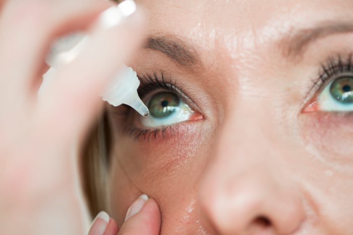
 for Mild Cases

    Many mild cases of watery eyes can be effectively managed with over-the-counter remedies. These options are often sufficient for temporary relief and can help reduce irritation and discomfort.

    • Artificial Tears: These lubricating eye drops help to moisturize the eyes, reducing dryness and associated watering. They are a safe and effective first-line treatment for mild cases of watery eyes caused by dryness, environmental irritants, or even minor allergies.
    • Cool Compresses: Applying a cool compress to the eyes can soothe inflammation and reduce irritation, which can contribute to watery eyes. This is a simple, inexpensive, and readily available method for providing temporary relief.
    • Antihistamines: If allergies are suspected as a cause, over-the-counter antihistamines can help alleviate the symptoms, including watery eyes. Be mindful of potential side effects and consult a doctor if symptoms persist or worsen.

    Prescription Medications for Underlying Conditions

    For watery eyes stemming from more serious conditions, prescription medications may be necessary. These medications target the root cause of the problem.

    • Anti-inflammatory Medications: In cases of inflammation or infection, anti-inflammatory medications can help reduce swelling and promote healing, thereby addressing the source of the watery eyes. Examples include topical corticosteroids, often prescribed for conditions like allergic conjunctivitis.
    • Antibiotics: Bacterial infections can cause significant eye irritation and watering. Antibiotics, in topical or oral forms, are essential to eliminate the infection and restore eye health. This is crucial to prevent complications and ensure proper healing.
    • Immunosuppressants: For autoimmune diseases or conditions where the immune system is overactive, causing chronic inflammation and watery eyes, immunosuppressants can help to regulate the immune response. This approach is usually considered for more severe or persistent cases.

    Eye Drops and Ointments

    Eye drops and ointments play a crucial role in treating watery eyes, providing targeted relief and addressing specific needs.

    • Purpose-Specific Eye Drops: Different types of eye drops address different causes. For example, those containing antihistamines are effective against allergic reactions, while those with lubricating properties are beneficial for dryness. Proper selection is vital.
    • Ointments: Eye ointments provide a thicker layer of lubrication and can be beneficial for conditions where more sustained moisture is needed. They are often used at bedtime for overnight relief.

    When Professional Medical Intervention Is Necessary

    While many cases of watery eyes can be managed at home, some situations warrant immediate professional medical attention. This is particularly important for persistent or severe cases, or those accompanied by other symptoms.

    • Severe Pain or Discomfort: Intense pain or discomfort in the eyes, along with watery discharge, requires immediate medical evaluation.
    • Vision Changes: Sudden changes in vision, even accompanied by watery eyes, are a significant cause for concern and require immediate professional attention.
    • Redness and Swelling: Persistent redness and swelling in the eye, often associated with watering, could indicate a serious underlying condition. A doctor’s examination is essential.

    Comparing Treatment Options

    Different treatment options vary in their effectiveness and suitability depending on the cause of the watery eyes.

    Treatment Option Effectiveness Suitability
    Over-the-counter remedies Effective for mild cases, temporary relief Dryness, minor irritations, allergies
    Prescription medications Effective for underlying conditions, long-term relief Infections, autoimmune diseases, inflammation
    Eye drops/ointments Provide lubrication, address specific causes Dryness, allergies, infections

    Careful consideration of the specific cause of the watery eyes is crucial for choosing the most effective treatment strategy. Consulting an eye care professional is always recommended for proper diagnosis and personalized treatment plans.

    Preventive Measures: What Causes Watery Eyes

    Watery eyes, while often a temporary discomfort, can sometimes indicate underlying health issues. Taking proactive steps to prevent watery eyes is crucial for maintaining eye health and overall well-being. By understanding the factors contributing to watery eyes and implementing preventive measures, you can significantly reduce the risk of this annoying condition.

    Protecting Eyes from Environmental Irritants, What causes watery eyes

    Environmental factors significantly impact eye health. Exposure to allergens, pollutants, and harsh weather conditions can trigger excessive tearing. Implementing strategies to shield your eyes from these irritants is essential. Protecting your eyes from wind, dust, and smoke is vital, especially during outdoor activities. Wearing sunglasses during bright sunlight is a crucial preventive measure to minimize the impact of UV rays on the delicate eye surface.

    • Wear protective eyewear, such as goggles or safety glasses, when engaging in activities that may expose your eyes to dust, chemicals, or other irritants.
    • Use air purifiers or filters in your home or workplace to reduce indoor air pollution.
    • Avoid environments with excessive smoke, dust, or allergens.
    • Rinse your eyes with cool water if exposed to irritants.

    Maintaining Good Hygiene

    Maintaining good hygiene practices is paramount for preventing eye infections and reducing the risk of watery eyes. Regularly washing your hands and avoiding touching your eyes is critical. Proper hygiene around contact lenses is especially important for preventing infections if you wear them.

    • Wash your hands thoroughly with soap and water before and after touching your eyes.
    • Avoid rubbing or touching your eyes.
    • If you wear contact lenses, follow the manufacturer’s instructions carefully for cleaning and disinfection.
    • Use clean towels or tissues to avoid spreading germs.

    Lifestyle Adjustments

    Certain lifestyle factors can contribute to watery eyes. Adjusting these factors can help reduce the risk. For instance, maintaining proper hydration by drinking enough water helps keep the eye’s moisture levels balanced. A balanced diet rich in nutrients, including antioxidants, supports overall eye health. Adequate rest is essential for allowing the body to repair and maintain optimal functioning.

    • Maintain proper hydration by drinking plenty of water throughout the day.
    • Consume a balanced diet rich in fruits, vegetables, and omega-3 fatty acids.
    • Ensure sufficient sleep to allow your body to rest and repair.
    • Manage stress levels through relaxation techniques like meditation or yoga.

    Regular Eye Exams

    Regular eye exams are crucial for detecting potential problems early on. Early detection allows for timely intervention and management of underlying conditions that can lead to watery eyes. These exams can identify and address issues such as dry eye syndrome, infections, or other abnormalities.

    • Schedule regular eye exams with an ophthalmologist or optometrist, at least once a year, or as recommended by your eye care professional.
    • Discuss any concerns about your vision or eye health with your eye care provider.

    Preventive Measures Effectiveness

    Preventive Measure Effectiveness
    Wearing protective eyewear High – shields eyes from irritants
    Maintaining good hygiene High – reduces risk of infections
    Adjusting lifestyle factors Moderate – supports overall eye health
    Regular eye exams High – detects potential problems early

    Closing Notes

    What causes watery eyes

    In conclusion, watery eyes can stem from a multitude of sources, ranging from straightforward irritants to complex medical conditions. By understanding the diverse causes, from environmental triggers to underlying health issues, individuals can take proactive steps towards diagnosis and effective management. This guide provides a comprehensive overview, equipping readers with the knowledge needed to navigate this common eye concern.

    Remember, if your watery eyes persist or worsen, consulting an eye care professional is crucial for accurate diagnosis and tailored treatment.

  • How to Cure Dry Eyes A Comprehensive Guide

    How to Cure Dry Eyes A Comprehensive Guide

    How to cure dry eyes? This comprehensive guide dives deep into understanding, diagnosing, and treating dry eye syndrome. We’ll explore the various causes, symptoms, and stages of this common condition, and equip you with effective treatment options, from over-the-counter remedies to lifestyle changes.

    From understanding the intricate tear production process to exploring preventative measures and long-term management strategies, this guide provides a thorough roadmap to conquering dry eyes. We’ll cover everything from home remedies to prescription medications, and offer practical tips to maintain eye health and comfort.

    Understanding Dry Eye Syndrome

    Dry eye syndrome, a common condition affecting millions globally, is characterized by a persistent lack of sufficient tears to lubricate and protect the eyes. This discomfort can significantly impact daily activities and quality of life. Understanding the causes, types, symptoms, and risk factors is crucial for effective management and prevention. Proper diagnosis and treatment can help alleviate symptoms and improve eye health.Dry eye isn’t just an inconvenience; it can lead to more serious complications if left untreated.

    Chronic dry eye can cause damage to the corneal surface, potentially leading to blurry vision, pain, and even scarring. Early intervention and a tailored approach to management are vital for preventing long-term eye health problems.

    Dealing with dry eyes can be a real pain, but thankfully there are plenty of remedies. A crucial aspect of eye health is understanding the factors affecting your eyes. For example, if you’ve recently had ACL tear surgery, you’ll want to know how long the rehabilitation process takes. acl tear surgery rehab how long does does it take is a great resource for that.

    Ultimately, staying hydrated and using lubricating eye drops are often the most effective ways to combat dry eyes.

    Causes of Dry Eye

    Tears are essential for maintaining healthy eyes, and disruptions in tear production or quality can result in dry eye. Several factors can contribute to this imbalance. Reduced tear production is a primary culprit, often linked to aging, certain medications, and underlying medical conditions. Another significant factor is the evaporation of tears, which can be accelerated by environmental factors like wind, air conditioning, and prolonged screen time.

    Meibomian gland dysfunction, a common cause of dry eye, involves the disruption of oil production in the eyelids, which normally prevents tear evaporation.

    Types of Dry Eye

    Dry eye can be broadly categorized into two main types: aqueous-deficient dry eye and evaporative dry eye. Aqueous-deficient dry eye is characterized by a decrease in the production of the watery component of tears. Evaporative dry eye, on the other hand, is primarily caused by the rapid evaporation of tears, often due to issues with the oily component produced by the meibomian glands.

    Understanding the specific type can help tailor treatment strategies for optimal results.

    Symptoms of Dry Eye

    Dry eye presents with a variety of symptoms, ranging from mild discomfort to significant impairment. Common symptoms include a persistent burning or stinging sensation in the eyes, a gritty feeling, redness, and blurry vision. These symptoms can vary in intensity and frequency depending on the individual and the severity of the dry eye. Some individuals may experience excessive tearing as a compensatory mechanism, while others may experience no tearing at all.

    Risk Factors for Dry Eye

    Several factors increase the risk of developing dry eye. Age is a significant risk factor, as tear production naturally decreases with age. Certain medical conditions, such as rheumatoid arthritis and Sjögren’s syndrome, can also contribute to dry eye. Medications, including some antidepressants and antihistamines, can reduce tear production. Environmental factors, like exposure to wind or smoke, and lifestyle factors, like prolonged screen time, also play a role.

    The interplay of these factors can create a higher risk profile for individuals.

    Stages of Dry Eye Severity

    Dry eye severity can be classified into different stages, each characterized by a unique set of symptoms and associated impact on daily life. Mild dry eye often presents with occasional discomfort and mild symptoms, such as a slight burning sensation. Moderate dry eye demonstrates more frequent and intense symptoms, including significant discomfort and potential interference with activities. Severe dry eye can lead to significant impairment in vision and daily activities, and may require more aggressive intervention.

    Understanding these stages is critical for appropriate treatment and management.

    Anatomical Structures Involved in Tear Production and Dry Eye

    The eye’s tear production system is complex, involving several anatomical structures. The lacrimal glands, located above the eye, produce the aqueous component of tears. The meibomian glands, located in the eyelids, secrete an oily substance that helps to prevent tear evaporation. The conjunctiva, a thin membrane lining the inner surface of the eyelids and covering the eye’s surface, plays a role in tear distribution.

    Dysfunction in any of these structures can contribute to dry eye. Proper functioning of the tear film is essential for maintaining eye health and preventing dry eye.

    Diagnostic Methods

    How to cure dry eyes

    Pinpointing the specific cause of dry eye syndrome is crucial for effective treatment. A thorough diagnostic process involves a combination of patient history, physical examination, and specialized tests. This multifaceted approach helps ophthalmologists differentiate dry eye from other eye conditions and tailor the most appropriate management strategy.

    Common Diagnostic Tests

    Several tests are used to evaluate the tear film and identify potential underlying causes of dry eye. These tests help determine the quantity, quality, and stability of the tear film, crucial factors in maintaining healthy eye comfort. Ophthalmologists typically use a combination of these methods to form a comprehensive understanding of the patient’s condition.

    Steps in a Comprehensive Dry Eye Evaluation

    A comprehensive evaluation typically begins with a detailed patient history, which includes questions about the patient’s symptoms, such as frequency, severity, and triggering factors. The ophthalmologist will also perform a thorough examination of the eyes, assessing the overall health of the tear film, and looking for signs of inflammation or damage. This includes checking for any visible abnormalities in the eyelids, conjunctiva, or cornea.

    Tear Film Analysis

    Tear film analysis plays a vital role in diagnosing dry eye. It measures various aspects of the tear film, including its lipid, aqueous, and mucin layers. The results provide insights into the tear film’s composition and stability, which can help determine the type of dry eye present. This data is then used to tailor the treatment approach.

    For instance, if the analysis reveals a deficiency in the lipid layer, a specific type of artificial tear could be prescribed to address the deficiency.

    Potential Complications of Undiagnosed Dry Eye

    Failing to diagnose and treat dry eye syndrome can lead to several complications. Left untreated, dry eye can exacerbate existing eye conditions, potentially impacting vision. Chronic irritation and inflammation can lead to corneal damage, including erosion and ulceration. The discomfort associated with dry eye can significantly impact a person’s quality of life, impacting daily activities and overall well-being.

    Dealing with dry eyes can be a real pain, and sometimes, hormone replacement therapies like hormone pellets might be suggested. It’s important to weigh the pros and cons of hormone pellets, which you can explore further in this insightful article about pros and cons of hormone pellets. While considering these options, remember that lifestyle changes like regular eye drops and a healthy diet can also significantly improve dry eye symptoms.

    In severe cases, vision impairment could result.

    • Corneal Damage: Chronic dry eye can lead to inflammation and corneal damage, potentially affecting vision.
    • Increased Risk of Infection: The compromised tear film can increase the risk of bacterial or viral eye infections.
    • Vision Impairment: Persistent dry eye can lead to blurred vision and reduced clarity, affecting daily activities.
    • Eye Pain and Discomfort: The persistent dryness and irritation can cause significant discomfort and pain, impacting quality of life.

    Comparison of Diagnostic Tools

    The table below summarizes the strengths and weaknesses of different diagnostic tools used in evaluating dry eye.

    Diagnostic Tool Description Strengths Weaknesses
    Schirmer Test Measures tear production. Simple, quick, and inexpensive. Doesn’t assess tear quality or lipid layer. Can be affected by patient factors.
    Tear Osmolarity Test Measures the concentration of salts in tears. Identifies evaporative dry eye. Requires specialized equipment and expertise.
    Lipifilm Test Evaluates the lipid layer of the tear film. Directly assesses the lipid layer. Can be expensive and not widely available.
    Corneal Staining Evaluates corneal surface for damage. Identifies corneal damage and inflammation. Doesn’t quantify the severity of dry eye.

    Treatment Options

    Treating dry eye syndrome often involves a multifaceted approach, combining various strategies to alleviate symptoms and improve tear film health. This involves understanding the underlying causes of dry eye and tailoring a personalized treatment plan. Different approaches target various aspects of tear production, quality, and evaporation.Effective treatment plans typically incorporate a combination of lifestyle modifications, over-the-counter remedies, and potentially prescription medications, all guided by a healthcare professional.

    The goal is to restore a healthy tear film balance and reduce the frequency and severity of dry eye symptoms.

    Types of Eye Drops

    Eye drops are a cornerstone of dry eye treatment. They come in a variety of formulations, each designed to address different aspects of tear deficiency. Understanding the mechanism of action of each type of drop can help patients choose the most suitable option.

    • Artificial Tears: These drops primarily replenish the tear film’s water content, providing immediate relief from dryness and irritation. They often contain lubricants like hyaluronic acid, glycerin, or polymers to enhance tear film viscosity and stability. This helps prevent rapid evaporation of tears and keeps the eyes moist. Examples include Refresh Tears, Systane, and Hypotears.
    • Preservative-Free Eye Drops: Some individuals are sensitive to preservatives commonly found in many eye drops. Preservative-free formulations are designed to minimize this risk, reducing the possibility of allergic reactions or other adverse effects. These drops are typically more expensive and may require more frequent use to maintain adequate hydration. They are often recommended for individuals with sensitive eyes or a history of allergic reactions.

    • Lubricating Eye Drops: These drops contain substances that coat the surface of the eye, reducing friction and promoting comfort. They can contain ingredients like cyclosporine, or other substances that provide long-lasting lubrication and reduce inflammation. These are particularly beneficial for individuals with chronic dry eye.
    • Restorative Eye Drops: These eye drops are designed to promote the production of tears. They may contain ingredients like cyclosporine, which can help to stimulate tear production. This can be especially helpful for individuals with severe dry eye conditions. Examples include Restasis, which contains cyclosporine A, an immunomodulator that can help decrease inflammation and stimulate tear production.

    Over-the-Counter Treatments

    Over-the-counter treatments are often a good starting point for managing mild to moderate dry eye. These options provide relief from symptoms without the need for a prescription.

    • Eye Lubricants: Many over-the-counter eye lubricants can provide temporary relief from dryness and irritation. These are generally well-tolerated and affordable. However, they often provide only short-term relief and may not be effective for severe cases.
    • Warm Compresses: Applying warm compresses to the eyes can help increase tear production and relieve discomfort. This simple method can be used in conjunction with other treatments to enhance overall relief.

    Prescription Medications

    Prescription medications are sometimes necessary for more severe or chronic cases of dry eye. These medications can address the underlying causes of the condition, leading to more significant and lasting improvements.

    • Immunomodulators: Prescription medications like cyclosporine (Restasis) are immunomodulators. They help to reduce inflammation in the eye and stimulate tear production. These medications can be effective for those who have significant inflammation contributing to their dry eye. Their use is usually reserved for moderate to severe cases.

    Comparison of Eye Drops

    Type of Eye Drop Mechanism of Action Pros Cons
    Artificial Tears Replenish water content, lubricate Affordable, readily available, immediate relief Short-term relief, may not be sufficient for severe cases
    Preservative-Free Reduce preservative exposure Suitable for sensitive eyes, potentially reduces allergic reactions More expensive, may need more frequent use
    Lubricating Coat eye surface, reduce friction Long-lasting lubrication, improved comfort May not be as effective for tear production issues
    Restorative (e.g., Cyclosporine) Stimulate tear production, reduce inflammation Potentially addresses underlying cause, more lasting relief Prescription required, potential side effects, more expensive

    Potential Side Effects of Treatments

    Treatment Potential Side Effects
    Eye Drops (all types) Burning, stinging, redness, temporary blurred vision, allergic reactions (rare)
    Prescription Medications (e.g., Cyclosporine) Eye irritation, blurred vision, infection, allergic reactions, increased risk of cataracts or glaucoma (in rare cases)

    Lifestyle Modifications

    Lifestyle modifications play a crucial role in managing dry eye. These adjustments can complement other treatments and contribute to overall eye health.

    • Environmental Controls: Controlling environmental factors like air dryness, smoke, or dust can significantly reduce irritation and dryness. Using a humidifier can help maintain appropriate humidity levels in the home and office.
    • Nutrition: A balanced diet rich in essential nutrients can support overall health, including eye health. Omega-3 fatty acids, found in fish oil, are believed to support healthy tear production.
    • Proper Hydration: Drinking sufficient water is crucial for maintaining overall hydration, which indirectly benefits eye health.

    Home Remedies and Self-Care

    How to cure dry eyes

    Managing dry eye symptoms often involves a multifaceted approach that combines professional treatment with self-care strategies. This section explores effective home remedies, emphasizing good eye hygiene, nutrition, and creating a supportive environment to alleviate discomfort and promote overall eye health. Understanding these techniques can significantly enhance your ability to manage dry eye syndrome effectively.Effective dry eye management goes beyond prescription eye drops.

    By incorporating simple home remedies and self-care practices into your daily routine, you can significantly improve comfort and reduce the frequency of dry eye symptoms. This proactive approach allows you to take control of your eye health and contribute to a more comfortable lifestyle.

    Effective Home Remedies

    Several home remedies can provide temporary relief from dry eye symptoms. Warm compresses, for instance, can help increase tear production by gently warming the eyelids and surrounding tissues. Also, gentle eyelid scrubs can help remove debris and promote healthy tear film production. A simple, warm compress and a gentle massage can effectively reduce inflammation and ease discomfort.

    These techniques can provide temporary relief, but it’s important to remember that they are not a substitute for professional medical advice or treatment.

    Importance of Eye Hygiene

    Maintaining good eye hygiene plays a crucial role in preventing and managing dry eye. Regularly cleaning your eyelids with a gentle, warm washcloth and a mild cleanser can help remove debris and prevent infections. This simple practice helps maintain a healthy tear film and minimizes irritation. Furthermore, avoiding harsh chemicals, smoke, and dust can significantly reduce the risk of exacerbating dry eye symptoms.

    Nutrition for Eye Health

    Proper nutrition is vital for overall eye health, including dry eye management. Consuming a diet rich in omega-3 fatty acids, found in fatty fish like salmon and flaxseeds, can help maintain a healthy tear film. Furthermore, incorporating antioxidants, found in fruits and vegetables, can protect the eyes from damage and promote overall eye health. These nutrients are essential for maintaining the integrity of the delicate tissues in the eye and for overall well-being.

    Creating a Supportive Environment

    Creating a supportive environment for dry eye management involves several key elements. Minimizing exposure to environmental irritants like wind, smoke, and air conditioning can help prevent dry eye symptoms. Taking breaks from prolonged screen time and ensuring adequate hydration are also essential. These measures can greatly reduce the frequency and severity of dry eye episodes.

    Dealing with dry eyes can be a real drag, but luckily, there are some simple remedies. Staying hydrated is key, and you might want to consider eye drops. If you’re also dealing with a persistent cough, checking out some helpful tips on what to do about a cough might be worthwhile, as sometimes coughs can be a symptom of other underlying issues that can affect your eyes.

    Ultimately, focusing on hydration and good eye care practices is the best way to get those dry eyes under control.

    Daily Habits Exacerbating Dry Eye

    Certain daily habits can exacerbate dry eye symptoms. Prolonged screen time, air travel, and use of certain medications can all contribute to dry eye. Furthermore, not blinking frequently enough when using screens or in dry environments can lead to increased dryness. It’s important to be aware of these factors to implement strategies for mitigating their impact.

    • Prolonged screen time.
    • Air travel.
    • Certain medications.
    • Insufficient blinking.
    • Dry environments.
    • Use of air conditioning or heating.
    • Exposure to smoke, dust, or allergens.

    Correct Application of Eye Drops

    Proper application of eye drops is crucial for their effectiveness and to avoid introducing infections. First, thoroughly wash your hands with soap and water to prevent contamination. Next, gently tilt your head back and pull down the lower eyelid to create a small pouch. Carefully place the prescribed number of drops into the pouch, avoiding contact with your eye or eyelashes.

    Finally, gently close your eyes and gently press on the inner corner of your eye to help the drops absorb. This simple procedure can ensure optimal absorption and minimize the risk of contamination.

    Prevention Strategies

    Preventing dry eye syndrome involves proactive measures to maintain healthy tear film function and reduce environmental irritants. A multifaceted approach, combining lifestyle adjustments, environmental controls, and regular eye care, is key to minimizing the risk of developing or exacerbating dry eye. Understanding the factors that contribute to dry eye is essential for implementing effective preventive strategies.

    Environmental Factors and Dry Eye

    Environmental factors play a significant role in the development and progression of dry eye. Exposure to dry air, particularly in heated or air-conditioned environments, can lead to tear evaporation. Wind, smoke, and dust can also irritate the eyes and disrupt the delicate tear film. Prolonged computer use or other activities requiring focused near-vision can lead to decreased blinking frequency, further contributing to tear film instability.

    Individuals living in arid climates or those who spend significant time in dry environments are particularly susceptible to dry eye.

    Importance of Regular Eye Check-ups

    Regular eye check-ups are crucial for early detection and management of dry eye. Comprehensive eye exams can identify underlying conditions or contributing factors that may not be apparent without professional evaluation. Early intervention can prevent the progression of dry eye symptoms and improve overall eye health. Furthermore, these exams allow for personalized recommendations and strategies tailored to the individual’s specific needs.

    Proper Hydration and Eye Health

    Proper hydration is essential for overall health, including eye health. Adequate fluid intake helps maintain the volume and quality of tears. Dehydration can lead to decreased tear production and increased tear evaporation, making individuals more susceptible to dry eye. Maintaining a healthy fluid balance throughout the day, including sufficient water intake and consumption of hydrating foods, is critical for optimal eye health.

    Drinking plenty of water, especially during periods of increased activity or environmental dryness, can help to support tear production.

    Preventative Measures for Dry Eye

    Implementing preventative measures can significantly reduce the risk of dry eye. These measures focus on protecting the eyes from environmental stressors and promoting optimal tear film health. A combination of strategies often proves most effective.

    • Maintaining adequate hydration: Drinking sufficient fluids throughout the day is critical for overall health and tear production. Aim for a daily intake of 8 glasses of water or more, adjusting based on individual needs and activity levels.
    • Improving blinking frequency: Consciously increase the frequency of blinking, especially during prolonged near-work tasks like computer use or reading. This helps distribute tears evenly across the eye surface and reduce tear evaporation.
    • Using humidifiers: In dry environments, humidifiers can help increase the moisture content of the air, reducing tear evaporation and promoting comfort for the eyes.
    • Avoiding irritants: Exposure to smoke, dust, wind, and other environmental irritants can cause eye irritation and contribute to dry eye. Take precautions to minimize exposure, such as wearing protective eyewear in dusty or windy conditions.
    • Protecting eyes from sun exposure: Prolonged sun exposure can contribute to dry eye. Using sunglasses with UV protection can help minimize this effect.

    Protective Eyewear

    Protective eyewear plays a critical role in preventing dry eye, especially in high-risk environments. Specialized eyewear, such as goggles or safety glasses, can shield the eyes from dust, debris, wind, and other irritants. This protection reduces the risk of tear evaporation and eye irritation, thus mitigating the development of dry eye. Consider the specific environment and potential hazards when choosing appropriate eyewear.

    Summary of Preventative Measures

    Preventative Measure Description
    Adequate Hydration Maintaining sufficient fluid intake to support tear production and prevent dehydration.
    Increased Blinking Frequency Consciously increasing the rate of blinking to distribute tears and reduce evaporation.
    Use of Humidifiers Increasing the moisture content of the air to reduce tear evaporation.
    Avoiding Irritants Minimizing exposure to smoke, dust, wind, and other environmental irritants.
    Sun Protection Using sunglasses with UV protection to shield the eyes from the sun’s harmful rays.
    Protective Eyewear Using appropriate goggles or safety glasses to protect the eyes from environmental hazards.

    Long-Term Management

    Managing dry eye syndrome effectively requires a long-term approach that goes beyond initial treatment. It’s crucial to understand that dry eye is often a chronic condition, meaning it persists over time. This necessitates ongoing vigilance, proactive adjustments to your treatment plan, and a strong partnership with your eye care professional. This comprehensive approach ensures that you can maintain comfort and visual clarity in the long run.Maintaining a stable condition often involves a combination of lifestyle modifications, consistent medication use, and regular follow-up care.

    Understanding how to monitor your symptoms and adjust treatments accordingly is vital for optimal management. Proactive steps, like understanding the triggers and contributing factors, help in preventing exacerbations and improving long-term comfort.

    Strategies for Long-Term Management

    Consistent adherence to your prescribed treatment regimen is essential for managing dry eye effectively. This includes using prescribed eye drops, ointments, or other medications as directed by your eye care professional. Understanding the specific needs of your eyes and the reasons for your condition is crucial. Consistency with treatment, as well as a thorough understanding of the triggers, will ensure that your symptoms are managed.

    If you experience changes in your symptoms, such as increased dryness or discomfort, it is important to communicate this to your eye care professional.

    Importance of Monitoring Symptoms

    Regularly monitoring your symptoms is critical for long-term dry eye management. This involves noting the frequency, intensity, and duration of dryness, discomfort, and any other associated symptoms. A detailed log, including the time of day, activities performed, and environmental conditions, can be helpful in identifying patterns and triggers. This data can aid in adjusting treatment strategies and understanding your individual responses.

    This systematic monitoring can reveal factors that contribute to your dry eye, like certain environmental conditions or particular activities.

    Role of Follow-Up Appointments

    Regular follow-up appointments with your eye care professional are essential for ongoing management. These appointments allow for adjustments to your treatment plan as needed, based on the observed changes in your symptoms. Your eye care professional can assess the effectiveness of the current treatment and make necessary modifications to maintain optimal eye health. They can also detect any underlying conditions or complications that may arise over time.

    During these appointments, you can discuss any concerns or changes in your symptoms, which helps ensure the treatment plan is continuously tailored to your needs.

    Importance of Compliance with Treatment Plans, How to cure dry eyes

    Adherence to your prescribed treatment plan is paramount for long-term dry eye management. This involves using eye drops, ointments, or other medications as directed, and following any lifestyle modifications suggested by your eye care professional. Consistent compliance ensures that your eyes receive the necessary treatment to maintain a healthy condition. Failure to adhere to the treatment plan may result in a worsening of symptoms and a reduced quality of life.

    Regular compliance can lead to better outcomes.

    Flowchart for Managing Dry Eye Over Time

    Step Action
    1 Initial Evaluation: Consult with an ophthalmologist for a comprehensive eye examination and diagnosis.
    2 Treatment Plan: Receive a personalized treatment plan, including medication, lifestyle adjustments, and follow-up appointments.
    3 Symptom Monitoring: Regularly track your dry eye symptoms, noting frequency, intensity, and triggers.
    4 Regular Follow-up: Attend scheduled follow-up appointments with your ophthalmologist to discuss symptom progression and adjust the treatment plan as needed.
    5 Lifestyle Modifications: Implement any lifestyle changes suggested by your ophthalmologist, such as environmental adjustments or dietary changes.
    6 Compliance: Adhere strictly to your treatment plan and follow-up appointments.
    7 Feedback & Adjustments: Communicate any changes in symptoms or concerns to your ophthalmologist for adjustments to the treatment plan.

    Resources for Further Information on Dry Eye

    • American Academy of Ophthalmology: A comprehensive resource for information on eye health, including dry eye syndrome.
    • National Eye Institute (NEI): Provides valuable information and research on various eye conditions, including dry eye.
    • Mayo Clinic: Offers detailed explanations and advice on managing dry eye syndrome.
    • Dry Eye Support Groups: Online forums or local support groups can provide valuable peer-to-peer support and information exchange.

    Visual Aids and Accommodations: How To Cure Dry Eyes

    Dry eye syndrome can significantly impact vision and daily activities. Fortunately, various visual aids and accommodations can help alleviate discomfort and improve quality of life for those affected. These adjustments can range from simple lighting modifications to more complex magnification tools, allowing individuals to maintain optimal visual function and participate fully in their lives.Effective management of dry eye often involves a multi-faceted approach, including both medical treatments and lifestyle modifications.

    Visual aids and accommodations play a crucial role in this process by enhancing comfort and improving visual clarity. These tools can be particularly helpful in situations where dry eye symptoms are aggravated, such as extended reading or computer use.

    Magnification Devices

    Magnification devices are instrumental in improving vision for individuals with dry eye, especially those experiencing reduced visual acuity. These tools come in various forms, from handheld magnifiers to specialized computer software with zoom capabilities. Proper magnification reduces the strain on the eyes by allowing for clearer viewing of fine details, which is often helpful when dealing with the discomfort associated with dry eye.

    By providing magnified images, magnification devices reduce the need for close-up focusing, thus minimizing the strain on the eye muscles. This can significantly improve visual comfort and overall well-being.

    Lighting Adjustments

    Proper lighting plays a significant role in minimizing eye strain and reducing the severity of dry eye symptoms. Adjusting the brightness and type of light can have a positive impact on visual comfort. For example, using soft, indirect lighting rather than harsh overhead lighting can be very helpful. Avoid glare from reflective surfaces, as this can exacerbate eye strain and dryness.

    Dimming the screen brightness of electronic devices can also reduce eye strain. Using task lighting directly on the area of focus, rather than general room lighting, is often beneficial for reducing glare and improving clarity.

    Comparison of Visual Aids

    Visual Aid Description Benefits for Dry Eye Considerations
    Handheld Magnifier Portable magnifying glass for close-up tasks. Improves clarity for reading, fine detail work. Limited magnification range, may not be suitable for all tasks.
    Desktop Magnifier Larger magnifier with adjustable stand for stationary tasks. Provides greater magnification and stability. May be less portable than handheld magnifiers.
    Computer Magnification Software Software that zooms in on computer screens. Improves readability of computer screens, reducing eye strain. Requires computer access and software installation.
    Adaptive Lighting Systems Adjusts light intensity and color temperature to optimize vision. Reduces glare and eye strain, particularly beneficial in reducing discomfort associated with dry eye. May require professional installation and cost considerations.

    Workplace Accommodations

    Employers have a responsibility to provide reasonable accommodations for employees with dry eye syndrome. These accommodations can significantly improve workplace productivity and overall well-being. Examples of workplace accommodations include:

    • Adjusting computer screen brightness and resolution: This can minimize glare and eye strain. Adjusting the brightness and contrast of the monitor, along with appropriate font size, can help minimize the symptoms of dry eye.
    • Providing ergonomic workstation setup: Ensuring proper seating, monitor placement, and lighting configuration can minimize eye strain and promote good posture.
    • Scheduling breaks: Regular breaks to rest the eyes can prevent prolonged eye strain and dry eye. Breaks should be timed and incorporated into the workday to promote rest and relaxation.
    • Allowing for adjustable lighting: Adjusting the lighting in the workplace to avoid glare and harsh lighting can help minimize discomfort for those with dry eye.
    • Providing eye drops or humidifiers: In some cases, employers may provide eye drops or humidifiers to help alleviate symptoms.

    Wrap-Up

    In conclusion, managing dry eyes effectively involves a multi-faceted approach. By understanding the causes, seeking proper diagnosis, and implementing the right treatment strategies, you can effectively control symptoms and prevent long-term complications. Remember, consistency in treatment and lifestyle modifications play a vital role in achieving sustained relief and maintaining eye health.

  • Silicone Hydrogel Contact Lenses A Deep Dive

    Silicone Hydrogel Contact Lenses A Deep Dive

    Silicone hydrogel contact lenses have revolutionized vision correction, offering unparalleled comfort and enhanced eye health. These lenses, meticulously crafted from a blend of silicone and hydrogel, boast superior oxygen permeability compared to traditional materials. This allows for extended wear and reduces the risk of dry eyes, a common concern with other types of contact lenses. This comprehensive exploration delves into the science behind their development, the benefits they provide, and the considerations for fitting, care, and maintenance.

    The evolution of silicone hydrogel contact lenses has been remarkable. From initial designs focused on oxygen permeability to advancements incorporating advanced features and customizable designs, the technology continues to improve. This in-depth look will examine the key components, manufacturing processes, and clinical applications, providing a complete picture of this fascinating field.

    Introduction to Silicone Hydrogel Contact Lenses

    Silicone hydrogel contact lenses represent a significant advancement in the field of ophthalmic lenses. They are a type of soft contact lens that combines the desirable properties of both silicone and hydrogel materials, leading to improved comfort and vision quality for wearers. Their unique composition allows for greater oxygen permeability compared to traditional hydrogel lenses, crucial for maintaining healthy corneal tissue.These lenses have revolutionized contact lens technology, enabling extended wear and improved comfort levels, and have become the dominant type of soft contact lens available today.

    This evolution stems from the desire for more breathable and comfortable contact lenses, directly addressing the limitations of previous designs. Silicone hydrogel lenses have become a standard of care for many contact lens wearers.

    Key Components and Functionalities

    Silicone hydrogel contact lenses are composed of two primary components: a silicone component and a hydrogel component. The silicone component provides exceptional oxygen permeability, allowing more oxygen to reach the cornea. The hydrogel component, conversely, contributes to the lens’s flexibility, comfort, and ability to hold moisture. This balanced composition is crucial for maintaining corneal health during extended wear.

    Historical Development and Evolution

    The development of silicone hydrogel lenses was a gradual process, driven by advancements in polymer science and materials engineering. Early attempts to combine silicone and hydrogel materials faced challenges in achieving a balance of oxygen permeability, water content, and mechanical properties. The development of advanced polymerization techniques allowed for the creation of lenses with superior oxygen permeability and increased comfort for wearers.

    These advancements have dramatically altered the landscape of contact lens technology, with notable improvements in the safety and comfort of wear.

    Types and Variations of Silicone Hydrogel Lenses

    Silicone hydrogel lenses come in a wide variety of types, tailored to meet the specific needs of different wearers. These variations include differing base curves, diameters, and lens designs. These variations account for different prescriptions and corneal shapes, ensuring a comfortable and visually optimal fit. For example, some lenses are designed for extended wear, while others are optimized for daily wear.

    Comparison of Silicone Hydrogel Lens Types

    Lens Type Oxygen Permeability Water Content Comfort Rating
    Daily Disposable High Moderate Excellent
    Extended Wear Very High Low Good to Excellent (with proper hygiene and care)
    Multifocal High Moderate Good, varies depending on individual adaptation
    Toric High Moderate Good, depending on the specific lens design

    The table above presents a general overview. Specific values for oxygen permeability, water content, and comfort ratings can vary significantly between different brands and lens models. Individual experiences with a particular lens type can also influence comfort ratings. Careful consideration of these factors is crucial when choosing a silicone hydrogel lens.

    Benefits and Advantages

    Silicone hydrogel contact lenses have revolutionized the world of vision correction, offering a significant leap forward from traditional contact lens materials. Their unique composition provides substantial improvements in comfort, extended wear options, and overall eye health, making them a preferred choice for many contact lens wearers.The key advantages stem from the innovative blend of silicone and hydrogel in these lenses.

    This combination allows for superior oxygen permeability, crucial for healthy corneal function, and contributes to enhanced comfort and extended wear times, which are significant improvements over traditional materials. This improved comfort and extended wear translates to greater convenience and a better overall user experience for individuals.

    Improved Comfort and Extended Wear, Silicone hydrogel contact lenses

    Silicone hydrogel lenses are designed to conform more closely to the shape of the eye, resulting in a smoother and more comfortable fit. This enhanced comfort is directly linked to the lower friction and better moisture retention capabilities of the material. The reduced friction translates into less irritation and dryness, which are common complaints with traditional contact lenses.

    Extended wear options offer greater freedom and flexibility in daily activities, minimizing the need for frequent lens removal and cleaning. This allows for a more convenient lifestyle for those who prefer extended wear or have busy schedules.

    Enhanced Oxygen Permeability

    A critical advantage of silicone hydrogel lenses is their superior oxygen permeability compared to traditional materials. This higher oxygen transmission rate (OTR) allows more oxygen to reach the cornea, which is vital for maintaining healthy corneal tissue. Adequate oxygen supply prevents hypoxia, a condition where insufficient oxygen supply can damage the cornea. This improvement is crucial for extended wear, as it minimizes the risk of complications associated with reduced oxygen to the eye.

    This is particularly beneficial for individuals who wear contact lenses for extended periods or those with conditions that make traditional lens materials less suitable. For example, individuals with dry eye syndrome or certain corneal conditions may experience significant relief with the enhanced oxygen permeability of silicone hydrogels.

    Silicone hydrogel contact lenses are fantastic for extended wear, but maintaining eye health is key. Proper nutrition plays a vital role, and supplementing with the right dosage of magnesium L-threonate ( magnesium l threonate dosage ) might just be the extra boost you need. This can help support overall eye health, which is especially important when wearing lenses for extended periods.

    Ultimately, the comfort and longevity of your silicone hydrogel contact lenses depend on a holistic approach to eye care.

    Benefits for Individuals with Specific Eye Conditions

    Individuals with specific eye conditions can significantly benefit from the properties of silicone hydrogel contact lenses. For instance, those with dry eye syndrome often find that the enhanced moisture retention and improved oxygen permeability of these lenses can alleviate discomfort and dryness. Similarly, individuals with certain corneal conditions may find that the superior oxygen permeability minimizes the risk of complications.

    Furthermore, individuals with astigmatism or other refractive errors may find that the improved comfort and fit of these lenses lead to a better visual experience.

    Comparison of Silicone Hydrogel to Other Lens Materials

    Feature Silicone Hydrogel Other Lens Material
    Oxygen Permeability Higher OTR, allowing more oxygen to reach the cornea. Lower OTR, potentially leading to reduced oxygen supply to the cornea.
    Comfort Generally more comfortable due to better fit and moisture retention. May be less comfortable, leading to dryness or irritation.
    Extended Wear More options for extended wear periods. Limited extended wear options due to lower oxygen permeability.
    Potential for Eye Health Minimizes the risk of complications associated with reduced oxygen supply. May increase the risk of complications with extended wear due to lower oxygen permeability.

    Material Science and Manufacturing

    Silicone hydrogel contact lenses, a marvel of modern materials science, offer a comfortable and breathable alternative to traditional contact lenses. Their unique properties are a direct result of the meticulous material selection and intricate manufacturing processes employed. Understanding the science behind these lenses unveils the complexity and precision involved in creating these crucial vision aids.

    Material Composition

    The core of silicone hydrogel lenses lies in their polymer matrix. These polymers are meticulously engineered to combine the desirable attributes of both silicone and hydrogel materials. Silicone imparts high oxygen permeability, crucial for healthy corneal function. Hydrogel, on the other hand, provides the necessary water content for comfort and lens shape retention. The specific ratios of these components are precisely controlled during manufacturing, influencing the lens’s ultimate properties.

    The key to a comfortable and effective lens lies in the optimal balance of these materials.

    Chemical Processes

    The chemical processes involved in producing silicone hydrogel lenses are highly sophisticated. A common method involves a step-wise process of polymerization, where monomers (small molecules) combine to form long chains (polymers). This polymerization is typically initiated by catalysts and controlled by precise temperature and pressure conditions. These controlled reactions dictate the molecular structure and thus the final properties of the lens material.

    Manufacturing Techniques

    Various manufacturing techniques are used to produce silicone hydrogel lenses, each with its own advantages and limitations. One widely used approach involves a casting process where a precise mold is used to shape the liquid polymer. Subsequent treatments, like cross-linking, are applied to improve the mechanical properties of the lens. Another technique involves the use of a laser to precisely shape the lens, allowing for greater customization potential.

    The selection of a specific technique often depends on the desired characteristics of the finished product.

    Customization Strategies

    Silicone hydrogel lenses are increasingly customizable to cater to individual needs. This customization extends to various aspects like lens power, diameter, and curve. Advanced manufacturing methods, like laser ablation, enable highly precise shaping, ensuring a perfect fit and optimal comfort. Advanced designs also consider factors like the user’s tear film and eye health.

    Silicone hydrogel contact lenses are fantastic for extended wear, but did you know they can subtly affect your overall health? Properly managing your blood pressure heart rate is crucial for maintaining good eye health, and, conversely, good eye health is essential for a well-rounded lifestyle. If you’re considering these lenses, be sure to consult with an eye doctor, as they can help you understand how these lenses might interact with your existing health conditions, such as blood pressure heart rate management.

    Ultimately, silicone hydrogel lenses offer a comfortable and convenient solution for many, but proactive health checks are always key.

    Manufacturing Process Flowchart

    Manufacturing Process Flowchart

    The above image (placeholder) would depict a flowchart starting with the raw materials and showing the various steps: from polymerization, mixing, molding, cross-linking, to the final lens shaping and packaging.

    Examples of Customization

    • Toric Lenses: Designed to correct astigmatism, toric lenses have a specific curvature to compensate for the irregular shape of the cornea.
    • Multifocal Lenses: These lenses correct for both near and far vision, typically combining different lens powers in a single lens.
    • Colored Lenses: The incorporation of pigments alters the appearance of the lens, allowing for cosmetic modifications.

    The incorporation of specialized features, like toric designs for astigmatism correction, or multifocal designs for presbyopia, highlights the advancements in tailoring silicone hydrogel lenses to specific patient needs.

    Fitting, Care, and Maintenance

    Silicone hydrogel contact lenses offer significant advantages in comfort and oxygen permeability, but proper fitting, care, and maintenance are crucial for optimal performance and eye health. These lenses, while advanced, require meticulous attention to detail in handling and cleaning to prevent complications. Understanding the fitting process, care regimen, and maintenance procedures will help ensure a safe and comfortable contact lens experience.

    Fitting Process

    The fitting process for silicone hydrogel contact lenses is a precise procedure involving careful evaluation of the eye’s unique characteristics. An optometrist or ophthalmologist will meticulously measure the cornea’s curvature, determine the appropriate lens parameters, and assess the overall health of the eye. This includes taking detailed measurements, such as keratometry, and using specialized instruments to ensure a snug yet comfortable fit.

    Accurate fitting is critical for preventing dryness, irritation, and potential vision distortion. A well-fitted silicone hydrogel lens will allow for maximum oxygen permeability, promoting healthy corneal tissue and overall comfort.

    Importance of Proper Lens Care and Hygiene

    Maintaining proper lens care and hygiene is paramount to prevent eye infections and maintain optimal vision. Contamination from bacteria, fungi, or other microorganisms can lead to serious eye infections, requiring prompt medical attention. Adhering to a strict cleaning and disinfection routine, combined with proper lens storage, minimizes the risk of infection. The routine should include thorough handwashing, lens removal, and careful cleaning, to ensure that the lenses are free of debris and pathogens.

    Cleaning and Disinfection Procedures

    Proper cleaning and disinfection of silicone hydrogel lenses is essential for preventing eye infections. The process typically involves rinsing the lenses with a sterile solution, followed by disinfection in a designated cleaning solution. Different methods exist, and a thorough understanding of the recommended procedures is critical. Always follow the specific instructions provided by your eye care professional for your chosen lens and cleaning solution.

    Lens Storage

    Proper lens storage is just as important as cleaning. Storing lenses in a clean, sterile case with fresh solution is vital for preventing contamination. Cases should be thoroughly cleaned and disinfected regularly, as recommended by the manufacturer. Empty cases should be avoided. The storage solution should be replaced according to the manufacturer’s instructions.

    This will help maintain the lenses’ integrity and prevent microbial growth.

    Methods for Cleaning and Disinfecting

    Several methods can be employed for cleaning and disinfecting silicone hydrogel contact lenses. A common method involves using a multi-purpose solution, which often combines cleaning and disinfection in a single step. The multi-purpose solution is usually recommended by the lens manufacturer and will ensure proper cleaning and disinfection. Thoroughly rinsing the lenses with sterile solution after cleaning and disinfection is important.

    Always refer to the instructions provided by the lens manufacturer and eye care professional.

    Recommended Care and Maintenance Solutions

    Step Action Materials
    1 Rinse the lens Sterile contact lens solution
    2 Soak the lens in cleaning solution Contact lens cleaning solution (follow manufacturer’s instructions)
    3 Thoroughly rinse the lens with fresh sterile solution Sterile contact lens solution
    4 Place the lens in a clean, disinfected case Clean contact lens case, fresh sterile solution
    5 Replace the solution in the case daily Fresh sterile contact lens solution

    Remember to consult your eye care professional for personalized guidance on the specific care and maintenance requirements for your silicone hydrogel contact lenses. They can provide tailored recommendations based on your individual needs and eye health.

    Clinical Applications and Research

    Silicone hydrogel contact lenses

    Silicone hydrogel contact lenses have revolutionized vision correction, and ongoing research continues to refine their applications and safety profiles. This evolution is driven by a need for improved comfort, enhanced oxygen permeability, and a wider range of corrective options for diverse patient populations. Understanding the clinical applications, ongoing research, and potential risks is crucial for both practitioners and patients.This section delves into the specific clinical uses of these lenses, the advancements in material science driving these innovations, and the benefits and potential complications for different patient types.

    We’ll examine the safety and efficacy of silicone hydrogel lenses through the lens of current research findings.

    Specific Clinical Applications

    Silicone hydrogel lenses offer a wide range of applications beyond standard myopia correction. Their superior oxygen permeability makes them suitable for extended wear, allowing for prolonged comfort and reduced risk of complications. They are particularly valuable for patients with certain eye conditions that require extended lens wear or high levels of oxygen transmission. For instance, they are often preferred in cases of keratoconus or other conditions where traditional soft lenses might not provide adequate comfort or oxygenation.

    Further, the ability to tailor lens designs and materials to specific needs makes them a powerful tool in the management of various corneal conditions.

    Advancements in Material Science

    Ongoing research focuses on refining the properties of silicone hydrogel materials. Scientists are exploring ways to improve biocompatibility, further enhance oxygen permeability, and create novel lens designs for enhanced comfort and visual acuity. For example, studies are investigating the use of advanced polymers and incorporating bioactive agents into the lens material to potentially improve healing processes or reduce inflammation.

    Silicone hydrogel contact lenses are a game-changer for comfort and clarity, but proper hygiene is key. To keep your lenses sparkling clean, remember to follow the instructions for using mouthwash correctly. how to use mouthwash is a crucial step in maintaining the health of your eyes and ensuring your lenses are effectively disinfected. This will ultimately lead to a more comfortable and enjoyable experience with your silicone hydrogel contact lenses.

    This advancement leads to lenses with a greater tolerance for the individual needs of the patient. Researchers are continually striving to create a more comfortable and effective vision correction experience.

    Benefits for Different Patient Populations

    Silicone hydrogel lenses offer significant advantages for diverse patient groups. Extended wear capabilities are a major benefit for patients who require prolonged lens wear for professional or lifestyle reasons, such as athletes or medical professionals. The superior oxygen permeability reduces the risk of complications associated with prolonged wear, such as corneal hypoxia. Additionally, the material properties enable custom-fitting solutions for patients with irregular corneas or other eye conditions.

    This customization improves comfort and visual acuity in a broader spectrum of patient needs.

    Potential Complications and Risks

    While generally safe, silicone hydrogel lenses, like all contact lenses, carry potential risks. Corneal complications such as dryness, infection, and inflammation are possible, though less frequent with these lenses compared to traditional materials. Careful lens selection and proper fitting procedures minimize these risks. Proper lens care and hygiene practices are crucial in preventing infections and maintaining the health of the cornea.

    Furthermore, the possibility of allergic reactions to the lens materials must be considered, although less common with silicone hydrogel lenses.

    Summary of Research Findings

    Extensive research consistently supports the safety and efficacy of silicone hydrogel contact lenses. Studies have demonstrated a lower incidence of corneal complications compared to traditional soft lenses, particularly for extended wear. The increased oxygen permeability correlates with a reduced risk of corneal hypoxia, a key factor in long-term eye health. Research continues to explore the long-term effects of these lenses, confirming their value in improving patient outcomes and comfort.

    The results from studies, in general, are positive and point to a significant improvement in vision correction compared to previous generations of contact lenses.

    Future Trends and Innovations: Silicone Hydrogel Contact Lenses

    Silicone hydrogel contact lenses have revolutionized vision correction, and the future promises even more advanced designs and features. Continuous research and development are pushing the boundaries of comfort, performance, and functionality, leading to exciting possibilities for patients. This evolution is driven by a desire to improve lens longevity, enhance patient comfort, and potentially incorporate advanced technologies into the lenses themselves.

    Potential Advancements in Lens Design

    Improvements in lens design are critical to enhancing patient experience. Future lenses may feature optimized geometries, incorporating sophisticated curves and surfaces to provide enhanced comfort and reduce the risk of complications like dry eye. This could involve the use of 3D printing technologies to create highly customized lens shapes tailored to individual corneal topography, allowing for a more precise fit and reduced pressure points.

    Advanced computational modeling will also play a crucial role in designing lenses that provide superior optical performance and reduce the risk of astigmatism.

    Material Science Innovations

    Material science advancements are vital for achieving next-generation silicone hydrogel lenses. The pursuit of novel polymers with improved oxygen permeability and water content will lead to more comfortable and stable lenses. Researchers are exploring the integration of advanced materials like biocompatible hydrogels and nanomaterials, potentially enhancing lens longevity and bioactivity. The goal is to create lenses that adapt to the wearer’s physiology over time, further increasing comfort and reducing the need for frequent lens replacements.

    Incorporating Advanced Features

    Future silicone hydrogel lenses may incorporate advanced features beyond simple vision correction. For example, incorporating sensors to monitor tear film health, providing real-time feedback to the wearer about their ocular environment, could be a significant advancement. These sensors could also transmit data to healthcare professionals for personalized treatment plans. Furthermore, the integration of drug delivery systems into the lenses could potentially treat specific eye conditions, opening doors to novel therapies.

    Consider the possibility of lenses that adapt to changing light conditions, providing dynamic vision correction tailored to the environment.

    Potential Impact on Patient Care

    These advancements have the potential to dramatically impact patient care. Customizable lenses, fitted to individual corneal shapes, could lead to improved comfort and reduced incidence of complications. Advanced features like tear film monitoring and drug delivery systems could significantly improve treatment efficacy for various eye conditions. This could lead to fewer visits to eye care professionals and improved overall eye health.

    Possible Future Features for Silicone Hydrogel Contact Lenses

    • Customizable Lens Designs: Lenses that adjust to individual corneal shapes, using 3D printing techniques, would dramatically improve fit and comfort. This is already beginning to be utilized in the current market with personalized lens design.
    • Smart Sensors: Incorporating sensors that monitor tear film health and transmit data to healthcare providers could allow for early detection and treatment of eye conditions. The potential of early diagnosis through monitoring is a significant advancement.
    • Drug Delivery Systems: Lenses incorporating drug delivery systems could treat various eye conditions directly, leading to more effective and convenient therapies. This is a promising area with significant potential for clinical trials and future applications.
    • Adaptive Optics: Lenses capable of adapting to changing light conditions could provide dynamic vision correction tailored to the environment, particularly useful for outdoor activities or those working in variable light settings.
    • Enhanced Oxygen Permeability: Further improvements in material science could lead to lenses with even higher oxygen permeability, enabling extended wear times and reducing the risk of discomfort and complications.

    Wrap-Up

    Silicone hydrogel contact lenses

    In conclusion, silicone hydrogel contact lenses represent a significant advancement in vision correction technology. Their superior oxygen permeability, enhanced comfort, and extended wear capabilities make them a valuable option for many individuals. While specific care and maintenance are essential, these lenses have proven to be a safe and effective solution for improving visual acuity and overall eye health.

    The future of these lenses is promising, with ongoing research and development poised to introduce even more innovative features and designs.

  • Macular Telangiectasia Eye Disease A Deep Dive

    Macular Telangiectasia Eye Disease A Deep Dive

    Macular telangiectasia eye disease is a condition affecting the macula, a crucial part of the retina responsible for sharp central vision. This comprehensive look explores the various types, symptoms, causes, and potential impacts on daily life. We’ll delve into the complexities of diagnosis, treatment options, and the vital role of research in improving outcomes for those affected.

    Understanding the intricacies of this eye disease requires an exploration of its different forms, such as types 1 and 2, each with its own unique characteristics. This detailed examination will reveal the nuances in symptoms and progression, equipping you with the knowledge needed to grasp the challenges faced by those affected.

    Introduction to Macular Telangiectasia

    Macular telangiectasia is a chronic eye condition affecting the macula, the central part of the retina responsible for sharp, detailed vision. This delicate area of the eye is highly susceptible to damage, and telangiectasia involves the abnormal widening and development of tiny blood vessels. The resulting changes in the macula can significantly impair vision, ranging from subtle blurring to profound loss of central vision.The core characteristic of macular telangiectasia is the development of these abnormal blood vessels in the macula.

    This structural alteration leads to leakage of fluid and blood, potentially causing inflammation and scarring that compromise the macula’s function. The progression of the disease can vary, affecting each individual differently. Understanding the different types and symptoms is crucial for timely diagnosis and management.

    Types of Macular Telangiectasia

    Macular telangiectasia presents in two primary forms, each with its own set of characteristics: type 1 and type 2. Type 1, also known as “classic” macular telangiectasia, is characterized by the development of small, dilated blood vessels within the macula. Type 2, on the other hand, exhibits larger, tortuous blood vessels in the macula, frequently accompanied by macular edema.

    Symptoms of Macular Telangiectasia

    The symptoms associated with macular telangiectasia typically manifest gradually. Common symptoms include blurring or distortion of central vision, difficulty reading or recognizing faces, and a gradual loss of fine details. The perception of straight lines appearing wavy or distorted is also frequently reported. The initial symptoms are often subtle and may not be immediately recognized as a sign of an underlying disease.

    Progression of Macular Telangiectasia

    The progression of macular telangiectasia varies significantly depending on the type. Early stages might be marked by mild visual changes, while later stages can lead to considerable loss of central vision. The rate of progression can also differ between individuals. Factors such as age, genetics, and lifestyle choices might influence the speed at which the disease progresses.

    Progression Stages by Type

    Type Symptoms Progression Stages
    Type 1 Initially, patients may experience mild blurring or distortion of central vision. Gradually, the symptoms worsen, with increasing difficulty in tasks requiring fine visual acuity. Early: Subtle vision changes. Middle: Progressive loss of central vision, difficulty reading and recognizing faces. Late: Significant loss of central vision, potential for complete loss of vision in severe cases.
    Type 2 Patients typically experience more pronounced symptoms from the outset, with noticeable blurring and distortion of central vision. Fluid accumulation in the macula may contribute to a rapid decline in vision. Early: Significant vision changes, with symptoms noticeable from the start. Middle: Rapid loss of central vision, pronounced distortion of straight lines. Late: Severe loss of central vision, potential for complete loss of vision in advanced stages.

    Causes and Risk Factors

    Understanding the causes and risk factors of macular telangiectasia is crucial for early detection and management. While the precise mechanisms behind the development of this eye disease are still under investigation, several contributing factors have been identified. This knowledge helps in identifying individuals at higher risk and potentially influencing preventative strategies.The exact causes of macular telangiectasia remain largely unknown.

    However, a combination of genetic predisposition and environmental factors appears to play a significant role in the development of the disease. Research continues to unravel the complex interplay between these elements, leading to a deeper understanding of the condition.

    Genetic Predisposition, Macular telangiectasia eye disease

    Genetic factors contribute significantly to the risk of developing macular telangiectasia. Certain genetic variations are associated with an increased likelihood of developing the disease. Individuals with a family history of the condition are more susceptible, suggesting a hereditary component. Studies are ongoing to identify specific genes involved, which could lead to more accurate risk assessment and potentially targeted therapies in the future.

    Environmental Factors

    Environmental factors also influence the development of macular telangiectasia. Smoking, for instance, is a known risk factor, potentially contributing to the development or progression of the disease. Prolonged sun exposure, particularly without adequate eye protection, may also play a role, although more research is needed to fully understand the relationship. Lifestyle choices, including diet and overall health, may also influence the risk, but further investigation is required to establish clear correlations.

    Macular telangiectasia, a frustrating eye condition, can sometimes make you feel like your vision is playing tricks on you. While focusing on healthy eating is crucial for overall well-being, especially when dealing with eye health, it’s important to be mindful of what you eat if you’re experiencing digestive issues like diarrhea. For example, some foods can irritate your gut and worsen the problem, so check out this article on Foods to Avoid When You Have Diarrhea for helpful tips.

    Ultimately, maintaining a balanced diet and focusing on nutritious foods is essential for managing both digestive health and eye health in the long run, particularly with macular telangiectasia.

    Risk Factors Summary

    Several factors have been identified as potentially increasing the risk of macular telangiectasia. It’s important to remember that these are not definitive causes, but rather factors associated with an elevated risk.

    • Family history of macular telangiectasia
    • Smoking
    • Prolonged sun exposure without eye protection
    • Age (the risk generally increases with age)
    • Certain underlying health conditions, such as hypertension or diabetes

    Comparison of Risk Factors for Different Types

    While the exact causes remain unclear, some differences exist in risk factors between the different types of macular telangiectasia. A table below summarizes these observed variations. Note that the data is based on current research and more definitive comparisons may emerge as studies progress.

    Macular telangiectasia, a condition affecting the eye’s macula, can cause vision problems. While seemingly unrelated, similar vascular issues can also lead to hematuria, or blood in the urine, as explored in this insightful article about Hematuria Blood in Urine Possible Causes in Men. Understanding these vascular connections is crucial for comprehensive health awareness, highlighting the importance of seeking expert advice for both conditions like macular telangiectasia.

    Risk Factor Macular Telangiectasia Type 1 Macular Telangiectasia Type 2
    Family history High Moderate
    Smoking Moderate to High Moderate
    Sun exposure Moderate Low
    Age High High
    Underlying health conditions Moderate Moderate

    Diagnosis and Evaluation

    Diagnosing macular telangiectasia requires a careful evaluation by an ophthalmologist. The process involves a series of tests designed to identify the specific type of macular telangiectasia and assess its severity. Accurate diagnosis is crucial for determining the appropriate treatment plan and monitoring disease progression.

    Diagnostic Procedures

    Macular telangiectasia is diagnosed through a combination of clinical examinations and specialized imaging techniques. Ophthalmologists use a systematic approach to gather information and ensure a precise diagnosis.

    Ophthalmoscopy

    A crucial initial step is ophthalmoscopy, which involves using an ophthalmoscope to examine the retina. This instrument allows the doctor to visualize the blood vessels in the macula. The presence of dilated, tortuous, and/or clustered blood vessels is a key indicator of macular telangiectasia. The ophthalmologist carefully assesses the extent and characteristics of the telangiectasia. Variations in the appearance of the telangiectasia, including their size and distribution, help in determining the specific type of the disease.

    Specialized Imaging Techniques

    Advanced imaging techniques further refine the diagnostic process. Fundus photography provides detailed still images of the retina, allowing for comparison over time. Fluorescein angiography (FA) is a valuable tool. It involves injecting a fluorescent dye into the bloodstream. The dye highlights the blood vessels in the retina, revealing any abnormalities in their structure and function.

    This method is especially helpful in identifying the leakage of fluid and blood. Optical coherence tomography (OCT) is another crucial technique. It produces high-resolution cross-sectional images of the retina, providing detailed views of the macula’s layers. OCT can help distinguish between macular telangiectasia and other macular conditions with similar appearances. These advanced imaging tools are crucial for comprehensive assessment.

    Visual Field Tests

    Visual field tests assess the patient’s peripheral vision. As macular telangiectasia progresses, it can lead to vision loss. These tests provide valuable information about the extent of the vision loss and its impact on daily activities. Various types of visual field testing are used, such as static perimetry and kinetic perimetry. These tests help determine the specific areas of vision loss.

    The visual field tests are critical in evaluating the impact of the disease on the patient’s overall visual function.

    Comprehensive Eye Exam Procedure

    A comprehensive eye exam for macular telangiectasia typically involves these steps:

    • Patient history: The ophthalmologist gathers information about the patient’s symptoms, medical history, and any family history of eye diseases.
    • Visual acuity testing: This measures the sharpness of the patient’s vision using an eye chart.
    • Ophthalmoscopy: The ophthalmologist examines the retina, looking for characteristic signs of macular telangiectasia, such as dilated and tortuous blood vessels.
    • Specialized imaging techniques: Fluorescein angiography (FA) and optical coherence tomography (OCT) provide detailed images of the macula and surrounding tissues.
    • Visual field testing: This assesses the extent of vision loss and identifies the specific areas affected.

    Summary of Diagnostic Tests

    Diagnostic Test Purpose
    Ophthalmoscopy Visual examination of the retina to detect characteristic blood vessel changes.
    Fundus Photography Provides detailed still images of the retina for comparison over time.
    Fluorescein Angiography (FA) Highlights blood vessels, revealing abnormalities in structure and function, especially leakage.
    Optical Coherence Tomography (OCT) Produces high-resolution images of the macula, aiding in identifying structural changes.
    Visual Field Tests Assesses the extent and location of visual loss.

    Management and Treatment Options: Macular Telangiectasia Eye Disease

    Managing macular telangiectasia involves a multifaceted approach focusing on slowing disease progression and mitigating vision loss. Current strategies emphasize a combination of lifestyle modifications, monitoring, and, in some cases, specific treatments. Understanding the nuances of these approaches is crucial for patients and their healthcare providers.

    Current Management Strategies

    Current management strategies for macular telangiectasia are primarily centered on preventing further damage and preserving existing vision. These strategies include regular eye examinations to monitor disease progression and promptly address any changes. Early intervention can often slow the rate of vision loss and potentially improve outcomes.

    Lifestyle Modifications

    Lifestyle modifications play a significant role in managing macular telangiectasia. Adopting a healthy lifestyle can potentially reduce the risk factors associated with the disease and mitigate its progression. A diet rich in antioxidants and vitamins, such as vitamins C and E, may help protect the delicate tissues of the eye. Regular exercise and maintaining a healthy weight can also positively influence overall health and reduce cardiovascular risk, which may indirectly affect the progression of the disease.

    Efficacy of Various Treatments

    Various treatments are available for managing macular telangiectasia, although there’s no cure. The efficacy of each treatment varies depending on the specific subtype and stage of the disease. Laser photocoagulation is a common treatment that involves using a laser to seal off the abnormal blood vessels in the macula. This procedure aims to reduce leakage and prevent further damage.

    While effective in some cases, it may not be suitable for all patients or stages of the disease.

    Comparison of Treatment Options

    Comparing the effectiveness of different treatment options requires careful consideration of individual patient factors. Factors such as the severity of the disease, the specific subtype, and the patient’s overall health must be taken into account when deciding on a course of treatment. While laser photocoagulation is a frequently used procedure, other options, such as anti-VEGF injections, may be considered for certain cases.

    Summary Table of Treatment Options

    Treatment Option Potential Benefits Potential Drawbacks
    Laser Photocoagulation Can reduce leakage from abnormal vessels, potentially slowing vision loss. May not be suitable for all stages or subtypes. May not completely halt progression. Potential for side effects, including scarring.
    Anti-VEGF Injections May reduce leakage from abnormal vessels, potentially slowing vision loss in some cases. Requires frequent injections, potentially leading to discomfort or complications. Not effective for all patients.
    Vitamin Supplements May reduce risk factors, potentially slowing disease progression. Limited clinical evidence of significant effect on disease progression. Should be taken under medical supervision.
    Lifestyle Modifications May reduce risk factors and support overall health, potentially slowing disease progression. Does not directly treat the disease, but supports overall health. Requires sustained commitment.

    Impact on Vision and Daily Life

    Macular telangiectasia eye disease

    Living with macular telangiectasia can significantly impact vision quality and daily life. While the disease doesn’t typically cause complete blindness, it gradually affects central vision, making tasks that require sharp focus challenging. This progressive nature of the condition demands understanding and proactive strategies for maintaining independence and quality of life.The effects of macular telangiectasia on vision extend beyond simple blurring.

    Individuals experience varying degrees of distortion, blurring, and loss of central vision. This impacts their ability to perform tasks requiring precise visual acuity. The consequences extend to numerous aspects of daily living, requiring adjustments and adaptations to maintain a fulfilling life.

    Effects on Vision Quality

    Macular telangiectasia primarily affects central vision, impacting the ability to see fine details, read, recognize faces, and drive. Individuals may notice blurring, distortion, and gradual loss of detail in the central part of their visual field. This can lead to difficulties in tasks that require clear vision, such as reading, writing, and recognizing faces. The gradual nature of the vision loss can make it challenging to adjust to the changing visual demands of daily life.

    Impact on Daily Activities and Tasks

    The impact on daily activities can range from minor inconveniences to significant challenges. Reading becomes more difficult, and navigating complex environments can be hazardous. Everyday tasks like cooking, driving, and using electronic devices can become frustrating or even impossible without assistance. Individuals may experience a decrease in their independence as they rely more on others for help with everyday activities.

    Need for Assistive Technologies and Adaptations

    Assistive technologies and adaptations are crucial for maintaining independence and quality of life. These tools can compensate for vision loss and help individuals continue participating in their desired activities. Examples include large-print materials, screen magnifiers, and voice-activated software. Properly selected assistive devices and techniques can help individuals maintain a sense of control and participation in their daily lives.

    Macular telangiectasia, a frustrating eye condition, can sometimes be linked to underlying systemic issues. For instance, a deeper dive into the connection between eye health and hormonal imbalances like hypothyroidism and adrenal insufficiency is explored in detail in this insightful article: Hypothyroidism and Adrenal Insufficiency A Deep Dive. While the exact mechanisms aren’t fully understood, exploring these potential correlations can help in better understanding and managing macular telangiectasia.

    Strategies for Maintaining Independence and Quality of Life

    Maintaining independence and quality of life is paramount for individuals with macular telangiectasia. Regular eye care visits, open communication with healthcare professionals, and proactive management of the condition are essential. Learning about available assistive technologies, exploring support groups, and seeking professional guidance can provide essential tools and encouragement for adjusting to the changes in vision. Adapting daily routines and embracing assistive technologies are crucial for maintaining independence.

    Potential Accommodations and Assistive Devices

    • Large-print materials: Books, newspapers, and other printed materials in larger font sizes can significantly improve readability. Many libraries and bookstores offer large-print options.
    • Screen magnifiers: These devices magnify computer screens, making it easier to read text and use electronic devices. Various types are available, ranging from handheld magnifiers to specialized software.
    • Voice-activated software: Software that reads text aloud can assist with tasks like reading emails, documents, and websites. This can greatly improve access to information and communication.
    • Assistive technology for daily living: Assistive devices for daily tasks, such as button-hookers or tools for reaching items, can help maintain independence. Adaptive devices for driving can assist with safely navigating roads and traffic.
    • Visual aids: Enhancing visual aids like brightly colored objects or contrasting backgrounds can increase visibility and help with spatial awareness.

    Patient Resources and Support

    Navigating a macular telangiectasia diagnosis can be challenging. Having access to reliable resources and supportive communities is crucial for patients to understand their condition, manage their expectations, and live fulfilling lives. This section Artikels valuable resources and emphasizes the importance of patient education and self-management.

    Reliable Resources for Patients

    Understanding macular telangiectasia involves accessing accurate and trustworthy information. Numerous organizations offer comprehensive details on the disease, its progression, and available management options. Patient-centered resources are essential for empowering individuals with the knowledge they need to actively participate in their care.

    • National Institutes of Health (NIH): The NIH provides extensive information on various health conditions, including macular telangiectasia. Their website offers detailed descriptions of the disease, research updates, and links to relevant studies. This is a cornerstone resource for patients seeking in-depth information and the latest advancements in research.
    • American Academy of Ophthalmology (AAO): The AAO is a reputable professional organization dedicated to eye health. Their website features informative articles, educational materials, and details on different eye conditions, including macular telangiectasia. This is a valuable resource for those seeking expert perspectives and professional guidance.
    • Mayo Clinic: The Mayo Clinic is a renowned medical institution known for its comprehensive medical information. Their website provides detailed explanations of various eye diseases and treatment options, making it a reliable resource for patients seeking expert insights.

    Support Groups and Online Communities

    Connecting with others facing similar challenges can be incredibly beneficial. Support groups and online communities offer a safe space for sharing experiences, exchanging advice, and receiving emotional support.

    • Online Forums and Support Groups: Many online forums and support groups cater to individuals with specific health conditions, including macular telangiectasia. These platforms offer a chance to connect with peers who understand the unique challenges of the disease. Sharing experiences and strategies with others facing similar challenges can be highly valuable.
    • Local Support Groups: Check with local eye care centers, hospitals, and community organizations to see if support groups exist for individuals with macular telangiectasia. These in-person groups can provide opportunities for direct interaction and personal connection with others who understand the specific concerns related to the condition.

    Importance of Patient Education and Self-Management

    Active participation in managing macular telangiectasia is essential for maintaining the best possible quality of life.

    • Patient Education: Understanding the condition and its potential impact is crucial for proactive self-management. Educating oneself about treatment options, lifestyle adjustments, and potential complications empowers individuals to make informed decisions regarding their care.
    • Self-Management Strategies: Implementing strategies for managing symptoms, including regular eye exams, medication adherence, and healthy lifestyle choices, can significantly impact the course of the disease and overall well-being.

    Role of Ophthalmologists and Other Healthcare Professionals

    Ophthalmologists play a vital role in the diagnosis and management of macular telangiectasia. Collaborating with other healthcare professionals is also important for comprehensive care.

    • Ophthalmologists: Ophthalmologists are eye specialists who diagnose and manage macular telangiectasia. They are essential for monitoring the disease’s progression, recommending appropriate treatments, and providing guidance on managing vision loss.
    • Other Healthcare Professionals: Collaboration with other healthcare professionals, such as optometrists, nutritionists, and therapists, can provide additional support and resources for managing the broader impacts of the condition on daily life.

    Patient Resources and Support Organizations

    This table provides links to organizations that offer resources and support for patients with macular telangiectasia.

    Organization Website
    American Academy of Ophthalmology [Link to AAO website]
    National Eye Institute (NEI) [Link to NEI website]
    Mayo Clinic [Link to Mayo Clinic website]
    [Add more organizations as appropriate] [Add links]

    Final Conclusion

    Right-angled vessels in macular telangiectasia type 2 | British Journal of Ophthalmology

    In conclusion, macular telangiectasia eye disease presents a multifaceted challenge impacting vision and daily life. Fortunately, proactive diagnosis, appropriate management strategies, and ongoing research offer hope for improving outcomes. This discussion highlights the importance of patient education and support, empowering individuals to navigate this condition effectively.

  • Bulls Eye Maculopathy Overview and More

    Bulls Eye Maculopathy Overview and More

    Bull s eye maculopathy overview and more – Bull’s eye maculopathy overview and more delves into the intricacies of this eye condition. It explores the defining characteristics, underlying mechanisms, and visual field impact. We’ll also examine different types, diagnostic methods, risk factors, management strategies, prognosis, visual rehabilitation, and case studies. Get a comprehensive understanding of this complex eye disorder.

    This in-depth look at bull’s eye maculopathy provides a clear picture of the challenges and opportunities for those affected. We’ll explore the science behind the condition, the various approaches to diagnosis and treatment, and the crucial role of support systems in managing this condition. This isn’t just a medical overview; it’s a journey through the experiences of those living with bull’s eye maculopathy and the ongoing quest for better outcomes.

    Introduction to Bull’s Eye Maculopathy: Bull S Eye Maculopathy Overview And More

    Bull’s eye maculopathy is a condition affecting the macula, the central part of the retina responsible for sharp, central vision. Its defining characteristic is a characteristic pattern of light and dark rings or areas within the macula, resembling a bull’s eye. This distinctive appearance arises from specific changes in the retinal tissues, impacting the way light is processed and ultimately perceived.

    Understanding the underlying mechanisms and visual field impact of this condition is crucial for accurate diagnosis and appropriate management.

    Defining Characteristics of Bull’s Eye Maculopathy

    The hallmark of bull’s eye maculopathy is the distinctive “bull’s eye” pattern visible on optical coherence tomography (OCT) scans. This pattern typically presents as alternating areas of hyper-reflectivity (brighter areas) and hypo-reflectivity (darker areas) within the macular region. The hyper-reflective areas often correspond to increased retinal thickness, while the hypo-reflective regions reflect thinning or atrophy of retinal tissues.

    This interplay of different retinal layers contributes to the characteristic appearance.

    Underlying Mechanisms

    The specific mechanisms leading to the bull’s eye pattern are complex and not fully understood. However, several factors are implicated in the development of this condition. Commonly, drusen, tiny deposits beneath the retina, are frequently observed in patients with bull’s eye maculopathy. These drusen can disrupt the normal functioning of the retinal tissues, potentially leading to the characteristic changes in reflectivity.

    Furthermore, changes in the retinal pigment epithelium (RPE) and choroid, layers beneath the retina, also contribute to the observed pattern. These combined factors result in the specific distribution of reflectivity seen in bull’s eye maculopathy.

    Visual Field Impact

    Bull’s eye maculopathy typically affects central vision. Patients may experience blurring or distortion of objects in the center of their visual field. This can manifest as difficulty reading, recognizing faces, or performing other tasks that require clear central vision. The extent of visual impairment varies depending on the severity and location of the macular changes. In more advanced cases, the central vision loss can be substantial, significantly impacting daily activities.

    Table of Bull’s Eye Maculopathy Characteristics

    Disease Name Defining Characteristics Common Symptoms Associated Conditions
    Bull’s Eye Maculopathy Alternating hyper- and hypo-reflective areas in the macula on OCT, resembling a bull’s eye pattern. Blurred central vision, distortion of objects, difficulty reading, recognizing faces. Age-related macular degeneration (AMD), diabetic retinopathy, and certain medications.

    Types and Subtypes of Bull’s Eye Maculopathy

    Bull’s eye maculopathy, a condition characterized by a distinctive bull’s-eye pattern of retinal pigment changes, presents in various forms. While the core feature remains consistent, the underlying causes and specific characteristics can differ. Understanding these variations is crucial for appropriate diagnosis and treatment planning.While often categorized as a single entity, recent research and clinical observations suggest the possibility of subtypes within bull’s eye maculopathy.

    These variations stem from different etiologies, influencing the disease’s progression and prognosis. Recognizing these nuances is essential for personalized care.

    Different Etiological Factors

    Various factors contribute to the development of bull’s eye maculopathy. These include genetic predisposition, systemic conditions, and even environmental exposures. Precisely pinpointing the specific cause is crucial for tailoring treatment and management strategies. Different etiologies lead to different clinical presentations and prognoses, hence the importance of comprehensive assessment.

    Clinical Presentations

    The clinical presentations of bull’s eye maculopathy often vary depending on the underlying cause. Patients might experience varying degrees of visual impairment, ranging from mild blurring to significant vision loss. The speed of progression can also fluctuate, impacting the overall prognosis. It’s essential to understand that the precise presentation can vary significantly based on the specific subtype and underlying etiology.

    Learning about bull’s eye maculopathy is fascinating, but sometimes unexpected connections pop up. For example, while researching the condition, I stumbled upon the topic of calcium deposits on teeth. It’s surprising how seemingly unrelated health issues can intertwine, and understanding these connections can lead to a more holistic view of overall health. Hopefully, this deeper look into bull’s eye maculopathy overview and more will be beneficial to readers.

    calcium deposits on teeth are often a result of poor oral hygiene, but it’s worth investigating further. I’ll delve deeper into this fascinating topic of bull’s eye maculopathy overview and more soon.

    Prognostic Factors

    Prognosis in bull’s eye maculopathy is multifaceted, influenced by the severity of the initial presentation, the rate of disease progression, and the effectiveness of treatment. The underlying cause plays a significant role in determining the potential for visual recovery. Factors such as age, overall health, and the patient’s response to treatment all contribute to the individual prognosis.

    Potential Overlaps and Distinctions

    There can be overlaps in the clinical presentations of different subtypes of bull’s eye maculopathy. However, distinguishing factors such as the presence of other retinal abnormalities, associated systemic diseases, and the family history can help in differentiation. Accurate diagnosis often hinges on a comprehensive evaluation that considers all these contributing factors.

    Summary Table of Types

    Type Defining Features Associated Risk Factors Prognosis
    Genetic Bull’s Eye Maculopathy Inherited genetic mutations leading to retinal pigment abnormalities. Often presents at a younger age. Family history of eye diseases, specific genetic mutations. Variable, depending on the specific genetic mutation. Can range from mild to severe vision loss, with potential for rapid progression.
    Age-Related Macular Degeneration (AMD) Related Bull’s Eye Maculopathy Associated with the development of drusen and other macular changes in the context of AMD. Often occurs in older individuals. Advanced age, smoking, family history of AMD, high cholesterol levels. Variable, influenced by the degree of AMD severity and the effectiveness of treatment options. Often a slower progression compared to genetic forms.
    Drug-Induced Bull’s Eye Maculopathy Resulting from the toxic effects of certain medications on the retina. Exposure to specific medications, pre-existing eye conditions. Potentially reversible if the medication is discontinued, or the underlying condition is effectively managed. The prognosis depends on the duration and severity of medication exposure and the patient’s response to treatment.

    Diagnostic Methods and Procedures

    Pinpointing bull’s eye maculopathy requires a meticulous approach, combining various eye examinations and specialized tests. A thorough understanding of the patient’s medical history and symptoms is crucial, as these factors can significantly influence the diagnostic process. This section will Artikel the key diagnostic procedures employed in identifying and confirming the presence of bull’s eye maculopathy.Accurate diagnosis of bull’s eye maculopathy necessitates a comprehensive evaluation, integrating different diagnostic tools to pinpoint the specific characteristics of the condition.

    This multifaceted approach helps differentiate it from other macular diseases, ensuring appropriate and timely treatment.

    Visual Acuity and Field Tests

    Visual acuity testing measures the sharpness of vision, providing a baseline for assessing the impact of the macular condition. A reduction in visual acuity, particularly in central vision, often signals the presence of a macular disorder. Perimetry tests, such as visual field tests, help map the extent of vision loss, identifying the specific areas affected by the bull’s eye pattern.

    These tests are essential for evaluating the functional consequences of the condition and monitoring its progression.

    Ophthalmoscopy and Fundus Photography

    Ophthalmoscopy allows direct visualization of the retina, enabling the ophthalmologist to observe the characteristic bull’s eye pattern of retinal pigment epithelium (RPE) changes. The appearance of the macular region, with its distinctive ring-like structure, is a crucial element in the diagnostic process. Fundus photography, a non-invasive technique, captures detailed images of the retina, which can be analyzed and compared over time to monitor the disease progression.

    So, I’ve been researching bull’s eye maculopathy, and it’s quite a complex eye condition. Understanding the causes and symptoms is key, and it’s fascinating how different things can affect our eyes. It got me thinking about other health issues, like coughing after running in cold weather, which can be quite disruptive. For more info on that, check out this article on coughing after running in cold.

    Ultimately, it’s all connected, and I’m finding it really interesting to explore these different health topics together. Back to bull’s eye maculopathy overview and more.

    These images provide a crucial record for future reference and comparison.

    Optical Coherence Tomography (OCT)

    OCT is a non-invasive imaging technique that provides high-resolution cross-sectional images of the retina. It allows detailed visualization of the retinal layers, including the macula, enabling the identification of structural changes characteristic of bull’s eye maculopathy. Specific OCT findings, such as thinning of the retinal layers in the central area and the presence of drusenoid deposits, may be seen in patients with this condition.

    The detailed anatomical information obtained from OCT is invaluable for diagnosing and monitoring the disease.

    Amsler Grid

    The Amsler grid is a simple, self-administered test that helps detect distortions in central vision. Patients look through the grid, identifying any straight lines that appear wavy or distorted. A characteristic finding in bull’s eye maculopathy is a central scotoma with a ring of preserved vision. This test is used to determine the pattern of visual impairment, aiding in the differential diagnosis.

    Table of Diagnostic Tests, Bull s eye maculopathy overview and more

    Diagnostic Test Purpose Limitations
    Visual Acuity Testing Measures the sharpness of vision Provides a general assessment; does not directly visualize the macula.
    Perimetry Tests Maps the extent of vision loss May not be sensitive to subtle changes in vision.
    Ophthalmoscopy Directly visualizes the retina Subjectivity in interpretation; may not capture all details.
    Fundus Photography Captures detailed images of the retina Requires skilled interpretation; does not provide detailed structural information.
    Optical Coherence Tomography (OCT) Provides high-resolution images of the retina Can be expensive; requires specialized equipment and expertise.
    Amsler Grid Detects distortions in central vision Subjective; not a definitive diagnostic tool on its own.

    Risk Factors and Predisposing Conditions

    Bull s eye maculopathy overview and more

    Understanding the factors that contribute to bull’s eye maculopathy is crucial for prevention and early intervention. While the exact cause remains elusive, various elements can increase the risk of developing this complex eye condition. These risk factors can range from lifestyle choices to underlying medical conditions. Pinpointing these influences can help individuals take proactive steps to safeguard their eye health.

    Genetic Predisposition

    Genetic factors play a significant role in the development of bull’s eye maculopathy. Individuals with a family history of macular degeneration or other inherited eye diseases may have a higher likelihood of developing the condition. This inherited susceptibility suggests a genetic component in the disease’s pathogenesis. Research continues to investigate specific genes that might increase the risk, paving the way for future diagnostic and preventative strategies.

    Age

    Age is a substantial risk factor for many age-related macular degenerations. The risk of developing bull’s eye maculopathy, like other age-related macular conditions, generally increases with advancing years. The natural aging process and cumulative environmental influences over time are likely contributors to the increased incidence in older populations.

    Lifestyle Choices

    Certain lifestyle choices can influence the risk of bull’s eye maculopathy. A diet deficient in essential nutrients, such as antioxidants and vitamins, may contribute to the condition’s development. Smoking is another significant lifestyle factor that has been linked to an increased risk of macular degeneration and potentially bull’s eye maculopathy. Maintaining a healthy diet, avoiding smoking, and regular exercise are crucial for eye health.

    Underlying Medical Conditions

    Some underlying medical conditions can increase the susceptibility to bull’s eye maculopathy. Conditions like diabetes, high blood pressure, and cardiovascular disease are often associated with an elevated risk of various eye conditions, including macular degeneration. Managing these underlying health conditions effectively can significantly reduce the risk of complications, including the development of bull’s eye maculopathy.

    Environmental Factors

    Environmental exposures, such as prolonged sun exposure without adequate protection, can potentially contribute to the development of bull’s eye maculopathy. Exposure to certain environmental toxins and pollutants may also increase the risk. Proactive measures, such as wearing sunglasses with UV protection and minimizing exposure to environmental hazards, can help mitigate the risk.

    Table of Potential Risk Factors

    Potential Risk Factor Association with Bull’s Eye Maculopathy
    Family history of macular degeneration Increased risk due to genetic predisposition
    Age Risk increases with advancing years
    Poor diet (low in antioxidants and vitamins) Potential contribution to disease development
    Smoking Significant correlation with increased risk
    Diabetes Elevated risk due to systemic effects
    High blood pressure Increased risk due to systemic effects
    Cardiovascular disease Increased risk due to systemic effects
    Prolonged sun exposure Potential contribution due to UV radiation
    Environmental toxins/pollutants Potential contribution to disease development

    Management and Treatment Strategies

    Unfortunately, there’s currently no cure for bull’s eye maculopathy, focusing on management strategies to slow its progression and preserve vision. Treatment approaches aim to address underlying causes, if known, and to manage symptoms effectively. The best course of action depends on the individual’s specific condition, including the severity of the maculopathy, the underlying cause, and the patient’s overall health.Effective management involves a multidisciplinary approach, combining medical interventions with lifestyle adjustments.

    This approach acknowledges the complex nature of the condition and its impact on visual function.

    Current Management Strategies

    Current management strategies for bull’s eye maculopathy revolve around addressing the underlying cause, if known, and managing symptoms to prevent further vision loss. Strategies focus on slowing the progression of the condition, improving quality of life, and providing appropriate support.

    Role of Treatment Options

    Various treatment options are available for bull’s eye maculopathy, encompassing both medical and surgical interventions. The effectiveness of these options varies significantly depending on the specific case and the individual’s response. Crucially, the chosen treatment must be tailored to the unique circumstances of the patient.

    Medication

    Medications play a crucial role in managing the underlying causes of bull’s eye maculopathy, such as diabetes or hypertension, which often contribute to the condition’s development.

    Learning about bull’s eye maculopathy is fascinating, but did you know that understanding inflammatory bowel diseases like ulcerative colitis facts about ulcerative colitis can offer valuable context? While seemingly disparate, both conditions highlight the complex interplay of the immune system and its potential impact on our health. Further research into bull’s eye maculopathy’s underlying causes and treatment options will be crucial.

    Managing systemic conditions is often essential. For example, tight blood sugar control in individuals with diabetes can slow the progression of macular edema. Similarly, controlling blood pressure can reduce the risk of further damage to the macula. While these medications don’t directly treat the bull’s eye maculopathy itself, they significantly impact its progression and prevent further vision loss.

    The effectiveness of these medications depends on their ability to control the underlying condition, and potential side effects are generally related to the specific medication used.

    Surgical Interventions

    Surgical interventions for bull’s eye maculopathy are generally reserved for specific cases, particularly when macular edema is present.

    In cases of significant macular edema, surgical procedures like vitrectomy may be considered. Vitrectomy involves removing the vitreous gel in the eye, potentially reducing pressure on the macula. However, these procedures carry risks, including potential complications like cataracts, retinal detachment, and infection. The decision to pursue surgical intervention is a complex one, weighing the potential benefits against the inherent risks.

    Individual circumstances, such as the extent of macular edema and the patient’s overall health, play a vital role in the decision-making process.

    Table Summarizing Treatment Options

    Treatment Option Effectiveness Potential Side Effects
    Medication (e.g., blood pressure control, blood sugar management) Can slow progression by addressing underlying conditions Possible side effects related to the specific medication
    Surgical Interventions (e.g., vitrectomy) Potentially reduces macular edema in severe cases Risks of cataracts, retinal detachment, infection

    Prognosis and Long-Term Outcomes

    Bull s eye maculopathy overview and more

    Bull’s eye maculopathy, while a significant condition affecting central vision, doesn’t always follow a predictable path. The prognosis, or anticipated outcome, varies considerably depending on several factors. Understanding these factors, along with the potential long-term impacts, is crucial for individuals and their families. It’s essential to remember that every case is unique and requires careful monitoring and individualized management.The long-term outcome of bull’s eye maculopathy is influenced by the severity of the condition, the underlying cause, and the effectiveness of treatment strategies.

    Some individuals may experience significant vision loss, while others may maintain relatively stable vision or even show improvement over time. A thorough understanding of these variables empowers individuals and their healthcare providers to anticipate possible outcomes and make informed decisions about management.

    Overview of Prognosis

    The prognosis for bull’s eye maculopathy varies significantly, influenced by the specific type of the condition and the speed of progression. Some individuals may experience gradual vision loss, while others may experience more rapid deterioration. The presence of other contributing factors, such as diabetes or hypertension, can affect the rate of progression. Careful monitoring and prompt intervention can help to mitigate the severity of the condition and improve long-term outcomes.

    Factors Influencing Long-Term Outcomes

    Several factors play a role in determining the long-term outcome of bull’s eye maculopathy. These factors include the specific type of bull’s eye maculopathy, the underlying cause (e.g., age-related macular degeneration, diabetes), the severity of the initial damage, and the effectiveness of treatment strategies. Individuals with a history of other eye conditions or systemic diseases may experience a more aggressive progression of the condition.

    Importance of Regular Monitoring

    Regular ophthalmological examinations are critical in monitoring the progression of bull’s eye maculopathy. These examinations help track any changes in vision loss, evaluate the effectiveness of treatments, and detect any signs of complications. Early detection of progression allows for timely interventions and adjustments to treatment plans. The goal is to maintain the best possible visual acuity and quality of life for the affected individual.

    This involves a partnership between the patient, ophthalmologist, and any other specialists involved in their care.

    Impact on Daily Life and Quality of Life

    Bull’s eye maculopathy can significantly impact an individual’s daily life and quality of life. Reduced central vision can affect activities such as reading, driving, recognizing faces, and performing other tasks that require detailed vision. The impact can range from mild inconvenience to significant limitations depending on the degree of vision loss. Support systems, adaptive strategies, and assistive technologies can help individuals maintain independence and a good quality of life.

    Visual Rehabilitation and Support

    Navigating the challenges of bull’s eye maculopathy requires a multi-faceted approach encompassing not just medical treatment but also comprehensive visual rehabilitation and support systems. This crucial aspect empowers individuals to maintain their independence and quality of life while adapting to their changing visual circumstances. Effective strategies, coupled with appropriate resources, are essential in maximizing their remaining vision and promoting overall well-being.

    Strategies for Visual Rehabilitation

    Visual rehabilitation programs tailored to individual needs are critical for optimizing remaining vision and enhancing daily functioning. These programs incorporate various techniques, exercises, and strategies to help patients adapt to their visual impairments. Training in strategies like visual scanning, peripheral awareness, and compensatory techniques are often part of the program. This training can be provided by ophthalmologists, optometrists, and vision rehabilitation therapists.

    Available Resources and Support Systems

    A robust support network is crucial for individuals and families coping with bull’s eye maculopathy. This network includes support groups, educational resources, and counseling services. Support groups offer a platform for sharing experiences, providing emotional support, and learning from others facing similar challenges. Online forums and community support groups can also connect individuals with a sense of community.

    Additionally, organizations specializing in visual impairments often provide crucial information and guidance. Counseling services can offer emotional support to individuals and their families as they adjust to the impact of the condition.

    Importance of Assistive Technologies and Adaptive Strategies

    Assistive technologies and adaptive strategies play a pivotal role in enhancing daily activities and independence for individuals with bull’s eye maculopathy. They are tools that allow for effective communication, navigation, and participation in daily tasks. Choosing appropriate assistive technology is a collaborative process between the individual, their family, and rehabilitation professionals.

    Assistive Technologies

    Assistive Technology Potential Benefits
    Large-print materials and software Improved readability for tasks like reading, writing, and completing forms.
    Magnifiers (handheld, desktop, and electronic) Enhance visual detail for tasks requiring precise vision, such as working with small objects or reading small print.
    Screen readers and screen magnification software Enable individuals to access digital information and operate computers with ease.
    Closed-captioned videos and audio descriptions Provide access to media content and enhance understanding of visual cues.
    Adaptive switches and communication devices Assist with communication and control of electronic devices, making daily activities more independent.
    Specialized vehicles and equipment Improve mobility and safety, whether navigating public spaces or using specialized equipment for tasks.
    Navigation aids (GPS apps, voice-guided maps) Assist with navigating unfamiliar environments, increasing independence and confidence in unfamiliar settings.

    Illustrative Case Studies

    Understanding bull’s eye maculopathy requires examining real-world examples. The following case studies highlight the diverse presentations of this condition, illustrating the variability in clinical features, diagnostic journeys, and treatment responses. These accounts underscore the importance of individualized care in managing this complex eye disease.

    Case Study 1: Progressive Central Vision Loss

    This patient, a 65-year-old woman with a history of hypertension and diabetes, presented with progressive central vision loss over a six-month period. Initial symptoms included blurring of central vision, making reading and recognizing faces challenging. Visual acuity was significantly reduced in both eyes, particularly noticeable in the central visual field. Fundus examination revealed characteristic bull’s eye maculopathy lesions in both eyes.

    Optical coherence tomography (OCT) scans confirmed the presence of central retinal thinning and drusenoid deposits. The patient underwent thorough metabolic testing, ruling out other potential causes of macular damage. Given the progressive nature of the disease, a combination of anti-VEGF injections and meticulous monitoring was initiated. The response to treatment was initially promising, with some stabilization of vision loss, although the condition remained stable rather than improving significantly.

    Case Study 2: Acute Onset of Visual Distortion

    A 42-year-old male presented with an acute onset of visual distortion, described as a “wavy” or “blurred” appearance in central vision. The patient reported no prior eye problems or systemic conditions. His visual acuity was slightly reduced in both eyes, with a noticeable metamorphopsia (distortion of shapes). The fundus examination showed distinct bull’s eye maculopathy in both eyes, without significant exudates or neovascularization.

    OCT scans demonstrated focal areas of macular atrophy and drusen-like deposits. Given the relatively acute onset and lack of severe inflammation, initial management focused on close monitoring and regular OCT scans. The patient’s visual distortion improved minimally over time, and the treatment remained primarily supportive, aiming to prevent further progression.

    Summary of Case Studies

    Case Study Age/Sex Presenting Symptoms Diagnostic Findings Treatment Response
    1 65-year-old female Progressive central vision loss Bull’s eye maculopathy, central retinal thinning, drusenoid deposits Initial stabilization of vision loss with anti-VEGF injections
    2 42-year-old male Acute onset of visual distortion Bull’s eye maculopathy, focal macular atrophy, drusen-like deposits Minimal improvement in visual distortion with close monitoring

    Final Wrap-Up

    In conclusion, bull’s eye maculopathy presents a complex challenge for both patients and healthcare professionals. While the condition can significantly impact vision, the availability of diagnostic tools, treatment options, and support resources can greatly enhance the quality of life for affected individuals. The ongoing research and development in this area hold promise for improving outcomes and offering hope for a brighter future.

    We’ve covered a lot of ground in this overview, from the fundamental causes to the practical strategies for living with this condition. Hopefully, this has been an informative and engaging exploration into this complex topic.