Category: Eye Care

  • LASIK Side Effects What to Expect

    LASIK Side Effects What to Expect

    What side effects can I expect after LASIK? This comprehensive guide dives into the potential short-term and long-term effects of laser eye surgery. From the initial recovery period to possible complications, we’ll explore the factors influencing outcomes and strategies for managing any issues that may arise. Understanding the risks and benefits of LASIK is crucial for informed decision-making.

    LASIK, a popular vision correction procedure, involves reshaping the cornea to improve eyesight. While generally safe and effective, potential side effects exist, varying in severity and duration. This detailed exploration will help you navigate the complexities of LASIK recovery and make well-informed choices about your eye health.

    Introduction to LASIK Side Effects

    LASIK, or Laser-Assisted In Situ Keratomileusis, is a common refractive surgery procedure designed to correct nearsightedness, farsightedness, and astigmatism. It involves reshaping the cornea, the clear front part of the eye, using a laser to improve the eye’s focusing ability. This precise reshaping allows individuals to potentially reduce or eliminate their dependence on eyeglasses or contact lenses.The LASIK procedure typically involves several steps.

    First, a small flap of corneal tissue is created. Then, the laser precisely removes corneal tissue to adjust its shape. Finally, the flap is repositioned to its original position. The procedure aims to precisely reshape the cornea to better focus light onto the retina, thereby improving vision. Variations in procedure types and techniques exist, each with its own nuances in the recovery process.

    Types of LASIK Procedures

    Different LASIK procedures are available, each with potential variations in the laser used, the flap creation method, and the amount of corneal tissue removed. The choice of procedure depends on individual eye characteristics and the specific correction needed. Understanding the nuances of these variations is important for anticipating potential side effects.

    Potential Risks and Benefits of LASIK

    LASIK, like any surgical procedure, carries potential risks and benefits. The benefits include improved vision, potentially eliminating the need for eyeglasses or contact lenses, and an overall enhancement of lifestyle. However, there are potential side effects that can range from mild to more severe, impacting the recovery period and potentially the long-term visual outcome.

    Comparison of LASIK Procedure Types

    Procedure Type Potential Benefits Potential Risks Recovery Time
    Standard LASIK Improved vision, reduced dependence on glasses/contacts, enhanced lifestyle Dry eye, glare, halos, astigmatism, fluctuating vision, infection, flap complications Typically 1-2 weeks, with full recovery within several months
    Custom LASIK Potential for more precise vision correction, potentially reducing the need for additional procedures Similar to standard LASIK, but with potential for more pronounced side effects if not carefully performed Similar to standard LASIK, though some individual experiences may vary
    Epi-LASIK Preserves the corneal epithelium, which can potentially reduce the risk of some side effects Longer recovery time, potential for dry eye, and potentially higher risk of flap complications Typically 2-4 weeks, with full recovery within several months
    SMILE (Small Incision Lenticule Extraction) Minimally invasive, potentially faster recovery, less risk of flap-related complications Potential for dry eye, glare, halos, and fluctuating vision. Potential for complications like infection or irregular astigmatism. Generally faster recovery compared to LASIK, with full recovery typically within a few weeks.

    Common Short-Term Side Effects

    LASIK, while a transformative procedure, can bring about a range of temporary side effects. Understanding these common post-operative symptoms empowers patients to proactively manage them and ensures a smoother recovery journey. This section delves into the most frequent short-term effects, their causes, duration, and management strategies.

    Common Symptoms and Their Causes

    Many patients experience temporary discomfort following LASIK surgery. These symptoms, while often mild and temporary, are a natural response to the surgical procedure and the healing process. The most common causes include the incisions made during the procedure, the subsequent inflammation, and the body’s natural healing response. Dry eyes, for example, are a result of the disruption of the tear film and the subsequent reduction in tear production.

    Duration of Short-Term Effects

    The duration of these short-term side effects varies considerably, depending on the individual and the specific issue. Some symptoms, such as mild discomfort and blurry vision, may persist for a few days or weeks. Others, like dry eyes, might linger for a month or two. A doctor’s guidance on expected recovery times and individual symptom duration is crucial.

    Severity of Short-Term Effects

    Symptom Description Severity Level (1-5, 1 being mildest)
    Mild Eye Discomfort Slight ache or pressure in the eyes 2
    Blurry Vision Reduced clarity of vision 3
    Dry Eyes Feeling of dryness, irritation, or foreign body sensation 2-3
    Photophobia Sensitivity to light 2-4
    Eye Pain Moderate to severe pain in the eyes 4-5

    This table offers a general guide to the severity of common short-term side effects. The severity levels are subjective and may vary between individuals. The severity of each effect should be assessed and monitored by an ophthalmologist.

    Strategies for Managing Short-Term Effects

    Managing these short-term effects involves a combination of prescribed eye drops, lifestyle adjustments, and, in some cases, over-the-counter pain relievers. Following your surgeon’s post-operative instructions precisely is vital. Taking regular breaks, maintaining proper hydration, and using prescribed eye drops as directed are essential strategies for mitigating discomfort.

    List of Common Short-Term Effects and Duration

    • Eye Discomfort: This may include a slight ache, pressure, or burning sensation. The discomfort usually subsides within a few days.
    • Blurred Vision: Patients may experience blurry vision that gradually improves over a week or two.
    • Dry Eyes: Dry eyes, characterized by dryness, irritation, and a foreign body sensation, often persist for a few weeks or even a month. This is a common complaint, and appropriate eye drops can help alleviate this.
    • Photophobia: Sensitivity to light, or photophobia, can last for a few days or a couple of weeks. Protective eyewear might be needed during this time.
    • Slight Eye Pain: Mild pain, often described as a dull ache, is a common occurrence and typically resolves within a few days. Using prescribed eye drops as directed is important.

    Potential Long-Term Side Effects

    While LASIK is generally a safe and effective procedure, potential long-term complications can arise. Understanding these possibilities is crucial for informed decision-making. This section delves into the potential long-term complications associated with LASIK, providing insight into their causes, likelihood, and variations across different surgical techniques.The long-term effects of LASIK can vary significantly from person to person. Factors such as individual eye health, the specific LASIK procedure, and post-operative care all contribute to the potential for complications.

    It’s essential to remember that while these complications are possible, they don’t necessarily mean they will occur. Open communication with your eye surgeon about potential risks is vital.

    Dry Eye Syndrome

    Dry eye syndrome is a common post-LASIK complication. The procedure can alter the tear film’s production and distribution, leading to dryness, irritation, and discomfort. This condition often presents as a persistent feeling of dryness, itching, or a gritty sensation in the eyes.

    Blurry Vision

    Blurred vision, though typically temporary after LASIK, can sometimes persist or worsen over time. This can result from issues with the corneal flap or irregularities in the corneal surface post-surgery. Some individuals might experience blurry vision in certain lighting conditions or after extended periods of focusing on a task.

    Post-LASIK, some temporary side effects like blurry vision and eye dryness are common. While I’m not a doctor, I’ve heard that similar discomfort can sometimes be related to underlying heart health issues, such as those detectable through coronary artery calcium scans. Thankfully, these are usually mild and short-lived. The good news is, most people experience a significant improvement in vision after the initial healing period.

    Refractive Error Changes

    Refractive errors like myopia (nearsightedness), hyperopia (farsightedness), and astigmatism can sometimes recur or change in severity after LASIK. These changes are often subtle, but they can necessitate further corrective procedures, such as additional LASIK surgery or the use of glasses or contact lenses. The reasons for this can vary from residual irregularities in the cornea to natural changes in the eye’s shape over time.

    Potential Complications Related to Different LASIK Procedures

    The specific LASIK procedure employed can influence the likelihood of experiencing certain long-term complications. For example, different techniques for creating the corneal flap or the use of different lasers might lead to varying outcomes.

    Likelihood of Long-Term Complications

    The likelihood of experiencing long-term side effects from LASIK varies depending on the individual and the procedure. Unfortunately, precise figures for these probabilities are difficult to provide. This is due to the wide range of factors that can influence the outcome, including the patient’s specific circumstances, the surgeon’s expertise, and the choice of surgical technique.

    Table Comparing Likelihood of Long-Term Complications Across Different LASIK Procedures

    Long-Term Complication Likelihood (Approximate)

    Standard LASIK

    Likelihood (Approximate)

    Advanced LASIK

    Dry Eye Moderate Moderate
    Blurry Vision Low Very Low
    Refractive Error Changes Low Very Low

    Note: This table provides a general comparison. Specific likelihoods can vary greatly depending on individual factors and surgical technique. Consult with an ophthalmologist for personalized information.

    Factors Influencing Side Effects

    LASIK, while a highly effective procedure, isn’t without potential side effects. Understanding the factors that can influence the likelihood and severity of these outcomes is crucial for both patients and surgeons. These factors range from pre-existing eye conditions to lifestyle choices and even the surgeon’s experience. This section delves into these influential aspects to provide a more comprehensive picture of the LASIK journey.Factors like patient health, age, lifestyle, surgeon skill, and even demographics play significant roles in shaping the LASIK experience.

    A thorough understanding of these factors can help patients and surgeons anticipate potential issues and tailor the procedure to individual needs.

    Patient Health Conditions

    Pre-existing eye conditions significantly impact LASIK outcomes. Conditions like dry eye syndrome, corneal diseases, and glaucoma can either increase the risk of side effects or complicate the recovery process. For example, a patient with a history of severe dry eye may experience more pronounced post-operative discomfort and a slower healing time. Furthermore, certain medical conditions, such as diabetes, can affect the healing process.

    It’s crucial for patients to disclose all pre-existing conditions to their surgeon to allow for personalized strategies and risk assessment.

    Age and Lifestyle Choices

    Age and lifestyle choices can also influence the success and safety of LASIK. Younger patients generally exhibit quicker healing times and greater adaptability to changes in vision. Conversely, older patients may experience slower recovery periods and a slightly increased risk of certain side effects. Lifestyle choices, including smoking, excessive alcohol consumption, and poor sleep habits, can also affect the healing process and overall outcome.

    A patient’s commitment to a healthy lifestyle post-surgery can dramatically impact their recovery.

    Surgeon Experience and Skill

    The surgeon’s experience and skill level play a pivotal role in the outcome of LASIK. Experienced surgeons with a proven track record of successful procedures are more likely to deliver optimal results with fewer complications. Surgical techniques, the precision of the laser, and the surgeon’s ability to adapt to individual patient needs all contribute to the overall success of the procedure.

    Choosing a qualified and experienced surgeon is a crucial decision for a positive LASIK experience.

    So, you’re considering LASIK? While it’s generally a safe procedure, some temporary side effects are common, like blurry vision or dry eyes. Interestingly, maintaining good overall health, including adequate vitamin D intake, might play a role in the healing process. Knowing the differences between vitamin D2 and vitamin D3 could be beneficial for your recovery. For a deeper dive into the specifics of vitamin D2 versus vitamin D3, check out this resource: vitamin d two vs d three.

    Ultimately, proper post-LASIK care, including following your eye doctor’s instructions, will significantly impact your recovery and minimize any lingering side effects.

    Impact on Different Demographics

    Different patient demographics may experience varying degrees of side effects. For instance, patients with certain ethnic backgrounds or those with specific eye shapes might be more prone to certain types of complications. Understanding these potential variations is essential for both the patient and the surgeon. Careful consideration of the individual needs and characteristics of each patient is key.

    Impact of Various Factors on Side Effects

    Factor Potential Influence on Side Effects Examples
    Pre-existing eye conditions (e.g., dry eye, glaucoma) Increased risk of complications, prolonged recovery, or less favorable outcomes. A patient with severe dry eye may experience more post-operative discomfort.
    Age Younger patients generally recover faster; older patients might experience slower healing. A 20-year-old will likely recover quicker than a 50-year-old.
    Lifestyle choices (e.g., smoking, alcohol, sleep) May hinder the healing process and increase the risk of complications. Smoking can impede the healing process.
    Surgeon experience Experienced surgeons are associated with lower complication rates. Surgeons with extensive experience and proven track records.
    Patient demographics (e.g., ethnicity, eye shape) May influence the potential for certain side effects. Variations in eye shape can impact the procedure’s success.

    Management and Treatment of Side Effects

    What side effects can i expect after lasik

    Navigating the post-LASIK journey often involves managing various side effects. Understanding how to handle these issues, from mild discomfort to more serious concerns, is crucial for a smooth recovery and optimal visual outcomes. This section will provide practical strategies for managing both short-term and long-term side effects, outlining when professional medical attention is warranted.Successfully managing post-LASIK side effects involves a proactive approach.

    Early intervention and adherence to post-operative instructions are vital for a positive outcome. This proactive strategy will ensure that you’re equipped to handle any potential challenges and achieve a successful vision restoration.

    Strategies for Managing Common Short-Term Side Effects

    Short-term side effects, often experienced in the days following LASIK, typically resolve on their own. Implementing effective management strategies can expedite the recovery process and minimize discomfort.

    • Eye Drops: Following prescribed regimens for eye drops is essential. These drops help reduce inflammation, lubricate the eyes, and prevent infection. Examples include artificial tears, steroid eye drops, and antibiotic eye drops. The specific type and frequency of drops will vary depending on the individual’s needs and the severity of the side effects.
    • Pain Management: Mild discomfort, such as eye pain or itching, can be managed with over-the-counter pain relievers like acetaminophen. However, consult your ophthalmologist if pain is severe or persistent. Applying cool compresses to the affected area can also provide relief.
    • Rest and Avoiding Strenuous Activities: Giving your eyes ample rest and avoiding strenuous activities is crucial. This includes limiting activities that could strain your eyes, such as prolonged screen time or intense physical exertion.
    • Protecting Your Eyes: Avoid rubbing your eyes, and wear protective eyewear when outdoors to shield them from dust and debris. This is particularly important in the initial recovery phase.

    Methods for Addressing Long-Term Side Effects

    While most LASIK side effects are temporary, some long-term issues can arise. Addressing these requires a more nuanced approach, often involving further medical intervention.

    • Dry Eye Syndrome: If dry eye syndrome persists or worsens, your ophthalmologist may prescribe different types of eye drops, such as punctal plugs, or recommend other therapies, including humidifiers or lifestyle adjustments.
    • Astigmatism or Blurred Vision: In some cases, astigmatism or persistent blurred vision can be addressed with a touch-up LASIK procedure or other corrective measures. It is crucial to schedule follow-up appointments to assess the effectiveness of the treatment.
    • Persistent Pain or Discomfort: If persistent pain or discomfort continues despite initial management strategies, it’s crucial to seek medical attention immediately. This is essential to rule out any underlying issues and determine the most appropriate course of action.

    When to Seek Medical Attention

    Recognizing when to seek medical attention is critical for managing potential complications. Prompt intervention can prevent the worsening of side effects.

    • Severe Pain or Increased Sensitivity to Light: If you experience intense pain, increased sensitivity to light, or a sudden change in vision, consult your ophthalmologist immediately. These symptoms could indicate a more serious problem requiring immediate attention.
    • Vision Deterioration: Any significant deterioration in vision, including persistent blurring or double vision, should prompt a prompt visit to your ophthalmologist.
    • Signs of Infection: Symptoms such as redness, swelling, excessive tearing, or discharge from the eyes could signal an infection. Prompt medical attention is crucial in these situations.

    Available Treatments for Specific Side Effects

    Various treatments are available to address specific LASIK side effects.

    Side Effect Potential Treatments
    Dry Eye Prescription eye drops, punctal plugs, humidifiers, lifestyle modifications
    Persistent Blurred Vision Retreatment, or other vision correction procedures
    Infection Antibiotic eye drops, oral antibiotics

    Post-Operative Care

    Adhering to post-operative care instructions is essential for a smooth recovery.

    • Follow-up Appointments: Regular follow-up appointments are crucial for monitoring your recovery and addressing any concerns.
    • Hygiene Practices: Maintaining good hygiene practices, such as avoiding touching your eyes and washing your hands frequently, is vital.
    • Medication Adherence: Strictly follow the prescribed medication regimen to manage inflammation and prevent infection.

    Flowchart for Managing Side Effects

    This flowchart illustrates the steps to take if side effects arise:[Diagram of a flowchart illustrating the steps to take if side effects arise. The flowchart should include steps such as assessing the severity of the side effect, contacting the ophthalmologist, following prescribed treatments, and scheduling follow-up appointments. The flowchart should be visually clear and easy to understand. The flowchart should also clearly indicate when to seek emergency medical attention.]

    Illustrative Cases of Side Effects

    LASIK, while a highly effective procedure, can sometimes result in unexpected side effects. Understanding these potential complications is crucial for both patients and surgeons. This section delves into real-world examples of side effects, highlighting the initial symptoms, treatment approaches, and ultimate outcomes. These case studies aim to provide a realistic perspective on the recovery process and the management of LASIK complications.

    Case Study: Persistent Dry Eye

    Persistent dry eye is a common post-LASIK complaint. Patients often experience a gritty or sandy sensation in their eyes, along with increased tearing and discomfort. The discomfort can significantly impact daily activities, especially those requiring prolonged periods of screen time.A patient named Sarah, following her LASIK procedure, reported persistent dry eye symptoms. Initially, she used over-the-counter lubricating eye drops, but the discomfort persisted.

    So, you’re considering LASIK? It’s a pretty cool procedure, but what about potential side effects? Well, temporary blurry vision, dry eyes, and even some discomfort are common. But, hey, maybe you could burn off some of those recovery jitters with some unusual ways to burn calories! Check out some creative ideas on how to stay active and healthy while you recover here.

    Ultimately, though, remember that the specifics of post-LASIK side effects vary from person to person, so it’s always best to talk to your doctor.

    Her ophthalmologist then prescribed a combination of artificial tears and a punctal plug, a small device inserted into the tear duct to reduce tear evaporation. The punctal plug proved effective, and Sarah’s dry eye symptoms gradually subsided within a few months.

    Case Study: Astigmatism Recurrence

    Post-LASIK, some patients may experience a recurrence of astigmatism, an imperfection in the eye’s shape that causes blurred vision. This recurrence can manifest as a gradual or sudden change in vision quality.One patient, David, experienced a slight increase in astigmatism after his LASIK procedure. His initial vision improvement was noticeable, but over several months, he began experiencing a progressive blurring of vision.

    David consulted his ophthalmologist, who performed a comprehensive eye exam and determined the need for a corrective lens procedure to address the recurred astigmatism. Following the additional procedure, David regained clear vision and experienced long-term stability.

    Case Study: Rare Corneal Scarring

    Although uncommon, corneal scarring can sometimes occur after LASIK. This condition may present as persistent blurry vision, pain, and light sensitivity. Early diagnosis and appropriate intervention are crucial for preventing vision loss.A patient, Emily, experienced a rare case of corneal scarring after LASIK. Initial symptoms included persistent blurry vision, discomfort, and increased sensitivity to light. The ophthalmologist immediately initiated a course of topical medications and closely monitored Emily’s eye condition.

    Fortunately, with careful management and follow-up care, the scarring resolved, and Emily’s vision gradually improved.

    Table of Case Studies Demonstrating Various LASIK Side Effects

    Patient Side Effect Initial Symptoms Treatment Resolution/Management
    Sarah Persistent Dry Eye Gritty, sandy sensation; increased tearing Artificial tears, punctal plug Symptoms subsided within months
    David Astigmatism Recurrence Gradual blurring of vision Corrective lens procedure Regained clear vision
    Emily Corneal Scarring Blurry vision, discomfort, light sensitivity Topical medications, close monitoring Scarring resolved, vision improved

    Comparison with Other Vision Correction Procedures: What Side Effects Can I Expect After Lasik

    Choosing the right vision correction procedure can be daunting, as each method comes with its own set of potential benefits and drawbacks. Understanding the nuances of different techniques, like LASIK, PRK, and LASEK, is crucial for making an informed decision. This comparison delves into the potential side effects, recovery times, and overall advantages and disadvantages of each procedure.

    LASIK vs. PRK vs. LASEK: A Side-by-Side Look

    LASIK, PRK, and LASEK are all refractive surgical procedures designed to improve vision. However, they differ significantly in their approach, which translates to variations in potential side effects and recovery times. Understanding these differences can help you decide which procedure is most appropriate for your individual needs.

    Similarities and Differences in Potential Side Effects

    While all three procedures aim to reshape the cornea, the method of doing so varies. This difference in approach leads to variations in the types and severity of potential side effects. For instance, LASIK involves a flap creation, which might lead to some flap-related complications. PRK, on the other hand, removes the surface layer of the cornea directly, potentially causing more discomfort during the healing process.

    LASEK uses an epithelial shield, potentially reducing some of the pain associated with PRK.

    Recovery Time Considerations

    The recovery period for each procedure also differs significantly. LASIK typically involves a quicker recovery compared to PRK and LASEK. The timeframe for complete recovery and achieving optimal vision varies, depending on the individual and the procedure. PRK generally requires a longer healing period than LASIK, and LASEK often falls somewhere in between. Patients should expect some degree of discomfort and potential visual disturbances during the recovery period.

    Advantages and Disadvantages Table

    Procedure Potential Side Effects (Summary) Recovery Time (General Estimate) Advantages Disadvantages
    LASIK Dry eye, glare, halos, fluctuating vision, flap complications Generally quicker (few days to weeks) Faster recovery, potentially less discomfort during healing Risk of flap complications, slightly higher risk of dry eye
    PRK Significant pain and discomfort during healing, potential for infection, blurred vision, dry eye Longer recovery (several weeks) Potential for better long-term vision outcomes in some cases More intense pain and discomfort, longer recovery period
    LASEK Less pain than PRK, some risk of infection, dry eye, blurry vision, halos Intermediate recovery time (several weeks) Less intense pain than PRK, potentially quicker recovery than PRK Still requires a healing period, risk of infection

    Important Note: This table is for general guidance only. Individual experiences and outcomes may vary. Consult with a qualified ophthalmologist for a personalized assessment and to discuss the best course of action for your specific needs.

    Prevention Strategies
    What side effects can i expect after lasik

    Minimizing the risk of LASIK side effects is paramount. Proactive steps, from meticulous pre-operative preparation to vigilant post-operative care, significantly influence the likelihood and severity of complications. A proactive approach, coupled with a well-informed decision about choosing an experienced surgeon, dramatically enhances the chances of a smooth recovery and optimal vision outcomes.

    Selecting a Qualified Surgeon

    Choosing an experienced and qualified LASIK surgeon is crucial. Researching a surgeon’s credentials, experience, and track record is essential. Look for surgeons with board certifications, extensive experience performing LASIK procedures, and a proven history of positive patient outcomes. A surgeon’s familiarity with the specific technology used in the procedure is also critical. Consult with multiple surgeons, discuss your concerns, and select the professional who best aligns with your needs and expectations.

    Reviews from other patients can also offer valuable insights.

    Maintaining Good Eye Health

    Prioritizing good eye health before and after LASIK surgery is essential. Maintaining a healthy lifestyle with a balanced diet, regular exercise, and sufficient sleep can contribute to a quicker recovery and better outcomes. Proper hydration, reducing exposure to irritants like smoke and allergens, and avoiding eye rubbing are crucial. Consistent eye examinations, even after surgery, are vital for early detection and treatment of any potential issues.

    Managing underlying eye conditions, such as dry eye, before and after surgery, is a critical aspect of pre and post-operative care.

    Pre- and Post-Operative Care Checklist, What side effects can i expect after lasik

    Adherence to pre- and post-operative instructions is critical for a successful recovery. This includes diligently following the surgeon’s pre-operative guidelines regarding medications, eye drops, and lifestyle adjustments. Strict adherence to post-operative care instructions, such as the use of prescribed eye drops, avoiding strenuous activities, and attending follow-up appointments, are equally important. A thorough understanding and meticulous adherence to these instructions can significantly minimize the risk of complications.

    • Pre-operative Care: This involves abstaining from certain medications, maintaining good hydration, and ensuring a comprehensive eye examination to identify any pre-existing conditions.
    • Post-operative Care: This includes using prescribed eye drops meticulously, protecting the eyes from dust and irritants, and avoiding strenuous activities as directed by the surgeon.

    Stress Management

    Stress and anxiety can negatively impact the healing process. Employing stress-reduction techniques like meditation, yoga, or deep breathing exercises can significantly contribute to a smoother recovery. Adequate rest and a positive mindset are essential for optimal healing. Identifying and managing stress triggers before and after surgery can be a significant factor in a successful recovery.

    • Techniques for Stress Management: Employing stress-reducing techniques like meditation, yoga, or deep breathing exercises can promote a smoother recovery.
    • Importance of a Positive Mindset: A positive and proactive approach to the procedure can facilitate a better healing process and minimize stress and anxiety.

    Preventative Measures Checklist

    This checklist Artikels proactive measures to reduce LASIK side effects:

    Category Action
    Pre-operative
    • Comprehensive eye examination
    • Review of medical history
    • Discussion of expectations with surgeon
    • Adherence to pre-operative instructions
    Post-operative
    • Strict adherence to post-operative care instructions
    • Regular follow-up appointments
    • Proper use of eye drops
    • Protection from dust and irritants
    • Avoidance of strenuous activities
    Lifestyle
    • Healthy diet
    • Regular exercise
    • Adequate sleep
    • Stress management techniques

    Closing Summary

    In conclusion, LASIK, while a remarkable procedure, carries potential side effects that vary significantly between individuals. Understanding the possible short-term and long-term complications, along with factors influencing them, empowers you to make informed decisions and choose the best course of action. Careful consideration of these aspects, alongside the expertise of a qualified surgeon, will significantly enhance your chances of a successful and satisfying LASIK experience.
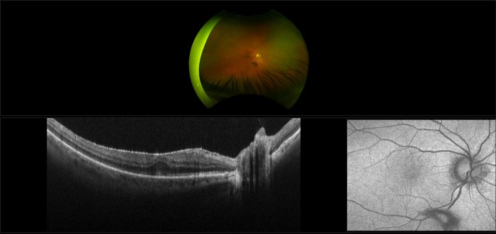
  • Epiretinal Membrane Retinal Scar Tissue A Deep Dive

    Epiretinal Membrane Retinal Scar Tissue A Deep Dive

    Epiretinal membrane retinal scar tissue forms a delicate yet significant threat to vision. This intricate web of scar tissue, often developing on the retina, can lead to a range of visual disturbances. Understanding its formation, diagnosis, and treatment options is crucial for effective management and preserving sight. This post will explore the specifics of this condition, from its definition and symptoms to treatment options and prognosis.

    The formation of epiretinal membrane (ERM) is often linked to the underlying cause of the retinal scar tissue, which could stem from injury, inflammation, or age-related factors. This scar tissue, when located on the retina, can distort vision, leading to significant impairment if not addressed. We’ll explore the different ways this condition is diagnosed and the various treatment approaches available, ranging from medications to surgical procedures.

    This comprehensive look at ERM and retinal scar tissue will be invaluable for patients, families, and healthcare professionals alike.

    Defining Epiretinal Membrane and Retinal Scar Tissue

    Epiretinal membranes (ERMs) and retinal scar tissue are common concerns in eye care, often arising from various eye conditions. Understanding their formation, characteristics, and relationship to the retina is crucial for diagnosis and treatment. This discussion delves into the specifics of ERMs, their connection to scar tissue development, and how they differ from other retinal pathologies.Epiretinal membranes are abnormal layers of tissue that grow over the surface of the retina.

    These membranes, composed of cells and extracellular matrix, can lead to the development of scar tissue. The precise mechanisms driving the formation of this scar tissue are complex, involving inflammatory responses and abnormal cell proliferation.

    Definition of Epiretinal Membrane (ERM)

    An epiretinal membrane (ERM) is a thin, translucent layer of scar tissue that grows over the surface of the retina, typically in the macula, which is the central part of the retina responsible for sharp central vision. This membrane can cause distortion and blurring of central vision.

    Dealing with epiretinal membrane retinal scar tissue can be tough, but focusing on overall health can help. While I’m no doctor, I’ve heard that certain herbal remedies can help with general cold symptoms, and best teas for a cold might offer some soothing relief. Of course, this is no substitute for professional medical advice, but a healthy lifestyle, including good hydration and rest, could potentially aid in the healing process for epiretinal membrane retinal scar tissue.

    Formation of Retinal Scar Tissue in ERM

    The development of scar tissue in ERM is a consequence of the inflammatory response and abnormal cell proliferation associated with the membrane’s formation. Inflammation triggers the release of growth factors that promote the growth and accumulation of extracellular matrix proteins, which contribute to the formation of the scar tissue. This process can lead to significant visual impairment if not addressed.

    Anatomical Location and Relationship to the Retina, Epiretinal membrane retinal scar tissue

    ERMs typically originate from the retinal pigment epithelium (RPE) and other adjacent tissues. They grow over the neurosensory retina, often in the macula. This location, directly over the light-sensitive photoreceptor cells, creates the potential for significant visual impairment as the membrane distorts the normal retinal structure.

    Comparison with Other Retinal Pathologies

    While ERMs and other retinal pathologies can lead to visual impairment, they have distinct characteristics. Macular holes, for example, are focal defects in the macula, resulting in a small, dark area in the central vision, while ERMs are more widespread, growing over the surface of the retina. Age-related macular degeneration (AMD) presents with different visual symptoms and underlying mechanisms compared to ERMs.

    Furthermore, diabetic retinopathy often involves blood vessel abnormalities rather than the membrane growth that defines ERMs.

    Key Differences Between ERM and Macular Hole

    Feature Epiretinal Membrane (ERM) Macular Hole
    Location Over the surface of the retina, often in the macula. A focal defect in the macula.
    Appearance Thin, translucent layer. A small, dark area in the central vision.
    Cause Inflammation, abnormal cell proliferation. Unknown, but likely related to structural weakness in the macula.
    Visual Impact Distortion and blurring of central vision. Loss of central vision.

    Symptoms and Diagnosis

    Epiretinal membrane retinal scar tissue

    Epiretinal membrane (ERM) and retinal scar tissue, though often subtle in their early stages, can lead to significant vision impairment if left untreated. Understanding the symptoms and diagnostic procedures is crucial for timely intervention and effective management. This section delves into the common visual complaints associated with ERM and retinal scar tissue, and Artikels the various diagnostic methods used to identify and characterize these conditions.Identifying the specific symptoms and accurately diagnosing epiretinal membranes and retinal scar tissue is crucial for timely intervention and effective treatment.

    Early detection allows for appropriate management and potentially better outcomes. The visual symptoms, often subtle initially, can progressively worsen over time. Diagnostic procedures are designed to not only confirm the presence of ERM or retinal scar tissue but also to evaluate its extent and impact on vision.

    Visual Symptoms

    Patients with ERM and retinal scar tissue often experience a range of visual symptoms. These symptoms can include blurred vision, distortion of straight lines (metamorphopsia), difficulty with night vision, and reduced visual acuity. The presence of floaters, or small specks or shadows that drift in the field of vision, is also sometimes reported. The severity of these symptoms can vary significantly depending on the size, location, and density of the membrane or scar tissue.

    Diagnostic Procedures

    Several diagnostic procedures are employed to identify and characterize epiretinal membranes and retinal scar tissue. These methods help ophthalmologists assess the extent of the condition and its impact on the patient’s vision. A comprehensive eye examination is crucial, incorporating a thorough evaluation of the patient’s medical history and visual symptoms.

    Ophthalmoscopy

    Ophthalmoscopy, a fundamental diagnostic tool, allows visualization of the interior structures of the eye, including the retina. During ophthalmoscopy, a specialized instrument is used to illuminate the retina, enabling the ophthalmologist to observe the surface of the retina for the presence of abnormalities. Direct ophthalmoscopy, utilizing a direct lens, provides a magnified view of the retina, which is especially helpful in identifying the presence of a membrane or scar tissue.

    Epiretinal membrane retinal scar tissue can be a tricky condition, affecting vision. While researching the complexities of this eye issue, I stumbled across some fascinating historical context about diseases. For example, the history of breast cancer, with its long and complex journey of diagnosis and treatment, offers a valuable comparison to the challenges faced with understanding and managing epiretinal membrane retinal scar tissue.

    Ultimately, understanding the history of such diseases can shed light on the progress made in healthcare and the long road ahead in finding effective treatments for eye conditions like epiretinal membrane retinal scar tissue. history of breast cancer

    Indirect ophthalmoscopy, employing an indirect lens, provides a broader field of view, useful for evaluating the entire retina. The presence of a pearly-white membrane or scar tissue with a slightly elevated appearance is often apparent during ophthalmoscopic examination.

    Optical Coherence Tomography (OCT)

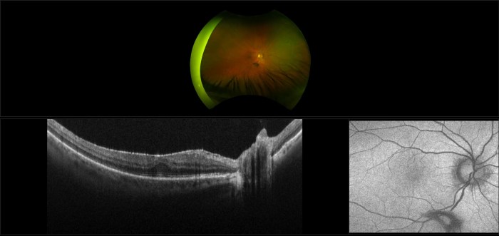
    Optical coherence tomography (OCT) is a non-invasive imaging technique that provides detailed cross-sectional images of the retina. OCT scans are particularly valuable in diagnosing ERM and retinal scar tissue because they offer high-resolution images of the retinal layers. The images generated by OCT reveal the precise location, thickness, and extent of the membrane or scar tissue, helping to distinguish it from other retinal conditions.

    The OCT scan will reveal a distinct elevation of the retina where the epiretinal membrane is attached.

    Summary of Diagnostic Tests

    Diagnostic Test Procedure Expected Result
    Ophthalmoscopy Visual examination of the retina using a specialized instrument. Presence of a pearly-white, elevated membrane or scar tissue.
    Optical Coherence Tomography (OCT) Non-invasive imaging technique that provides detailed cross-sectional images of the retina. Distinct elevation of the retina where the membrane or scar tissue is attached; precise measurement of the membrane’s thickness.

    Treatment Options

    Epiretinal membrane (ERM) and retinal scar tissue can significantly impact vision. Fortunately, various treatment options are available, ranging from observation to surgical interventions. The best approach depends on the severity of the condition, the patient’s overall health, and the location of the ERM within the retina.Understanding the mechanisms of action of these treatments is crucial for patients and healthcare providers to make informed decisions about the best course of action.

    Different modalities target different aspects of the disease, from reducing inflammation to removing scar tissue. This section will explore the diverse array of treatment strategies available for managing ERM and retinal scar tissue.

    Available Treatment Options

    A range of treatment options are available, from watchful waiting to surgical procedures, and anti-VEGF therapy. The optimal choice depends on the individual patient’s situation.

    • Observation: In some cases, especially for early-stage or mild ERM, a period of watchful waiting may be appropriate. This approach involves regular eye examinations to monitor the progression of the condition. This is often a suitable option for patients who do not experience significant symptoms and whose ERM is not causing significant macular edema.
    • Anti-VEGF Therapy: Vascular endothelial growth factor (VEGF) is a protein that plays a role in the development of new blood vessels. In ERM-related macular edema, VEGF can contribute to fluid buildup. Anti-VEGF medications, such as ranibizumab (Lucentis) or aflibercept (Eylea), work by blocking VEGF, thus reducing fluid leakage and inflammation in the macula, which is the central part of the retina.

      This can help improve vision and slow the progression of the disease.

    • Surgical Intervention: Surgical removal of the ERM is an option for patients who experience significant vision loss due to the ERM and macular edema. Surgical techniques involve carefully separating the ERM from the retina, often using specialized instruments. The procedure is typically performed by ophthalmologists experienced in vitreoretinal surgery.

    Mechanism of Action

    The specific mechanisms of action for each treatment modality vary depending on the method chosen.

    • Anti-VEGF therapy targets the overproduction of VEGF, a key player in the formation of new blood vessels. By inhibiting VEGF, these medications reduce the fluid leakage that contributes to macular edema and retinal inflammation. This, in turn, can improve vision and slow the progression of the condition.
    • Surgical intervention aims to physically remove the epiretinal membrane, reducing the pressure on the retina. The surgical approach can involve techniques like peeling the membrane off the retina, using specialized instruments and precise techniques. The goal is to improve retinal function and reduce macular edema by relieving the mechanical stress on the retina caused by the ERM.

    Surgical Procedures for ERM Removal

    Surgical procedures for ERM removal are typically performed by experienced ophthalmologists specializing in vitreoretinal surgery.

    • Vitrectomy: This surgical procedure involves the removal of the vitreous humor, the gel-like substance that fills the eye. The removal of the vitreous creates space, making it easier to access and separate the ERM from the retina. Once the vitreous is removed, the ERM is carefully peeled away from the retina using specialized instruments. This procedure allows for better visualization and manipulation of the ERM and surrounding tissues, improving the likelihood of successful removal.

    Role of Anti-VEGF Therapies

    Anti-VEGF therapies play a crucial role in managing macular edema associated with ERM. The effectiveness of anti-VEGF therapy is often evaluated by the degree of improvement in visual acuity and the reduction in macular edema.

    Treatment Approach Mechanism of Action Risks Benefits
    Observation Monitor condition without intervention Potential for vision loss if condition progresses Avoids potential risks of more invasive procedures
    Anti-VEGF Therapy Blocks VEGF, reducing fluid leakage Potential for side effects (e.g., eye infections, retinal detachment), injections are required Can improve vision, slow progression, and reduce macular edema
    Surgical Intervention Remove ERM from retina Potential for complications (e.g., retinal detachment, infection), longer recovery time Can significantly improve vision, particularly when macular edema is present

    Prognosis and Management: Epiretinal Membrane Retinal Scar Tissue

    Epiretinal membrane (ERM) and associated retinal scar tissue can significantly impact vision. Understanding the potential outcomes and proactive management strategies is crucial for patients and their eye care professionals. This section will delve into the prognosis, emphasizing the importance of regular follow-ups and potential long-term complications. We will also explore strategies for managing the visual consequences.The prognosis for ERM varies greatly depending on several factors.

    These include the extent of the membrane, its location on the retina, and the presence of other eye conditions. While some individuals experience minimal visual impairment, others may face more substantial reductions in vision quality.

    Typical Prognosis

    The typical prognosis for ERM depends on the extent of the membrane’s growth and its location on the retina. Early detection and prompt intervention are crucial for optimizing visual outcomes. In many cases, ERM does not cause significant vision loss. However, if the membrane is extensive or involves critical retinal areas, it can result in noticeable visual disturbances.

    Importance of Regular Follow-up Appointments

    Regular follow-up appointments are essential for monitoring the progression of ERM and associated retinal scarring. These appointments allow eye care professionals to track any changes in the membrane’s size or location, as well as assess the impact on vision. Early intervention can often prevent further vision loss. The frequency of follow-ups will depend on the individual’s specific case and the rate of membrane progression.

    Potential Long-Term Complications

    Potential long-term complications associated with ERM and retinal scar tissue include further vision loss, particularly if the membrane continues to grow or contract abnormally. In some instances, the membrane can cause distortion of the retinal image, leading to problems with central vision. The risk of these complications can be reduced with appropriate management and consistent follow-up care. Another potential complication is the development of macular edema, a swelling of the macula (the central part of the retina responsible for sharp vision).

    This swelling can worsen existing vision problems.

    Visual Outcomes Based on Stage and Treatment

    Stage of ERM Treatment Options Potential Visual Outcome
    Early stage, minimal membrane Observation, close monitoring Minimal to no significant vision loss
    Moderate stage, membrane affecting central vision Laser photocoagulation, vitrectomy Potential for some vision improvement or stabilization, some vision loss possible
    Advanced stage, extensive membrane and significant retinal scarring Vitrectomy with membrane peeling, potential for multiple treatments Potential for partial or significant vision loss, depending on the extent of the membrane and scar tissue. Improvement is less likely.

    Note

    * This table provides a general overview. Individual outcomes may vary based on several factors including the individual’s overall eye health and the response to treatment.

    Epiretinal membrane retinal scar tissue can significantly impact vision, often causing distortions and blurry images. Understanding the intricate pathways of the ophthalmic nerve, including its anatomy, function, and significance, like this insightful article , is crucial to comprehending how these changes affect the transmission of visual information. Ultimately, a thorough knowledge of the ophthalmic nerve’s role is essential to effectively treating and managing epiretinal membrane retinal scar tissue.

    Strategies for Managing Visual Consequences

    Managing the visual consequences of ERM and retinal scar tissue involves a multi-faceted approach. This often includes:

    • Close monitoring: Regular eye exams to track the progression of the condition and any changes in vision.
    • Treatment options: Using laser therapy, vitrectomy, or other procedures to reduce the size or impact of the membrane and associated scar tissue.
    • Management of other eye conditions: Addressing any other underlying eye conditions that may contribute to the progression of ERM or retinal scarring.
    • Addressing symptoms: Managing any symptoms experienced, such as blurry vision or floaters, with appropriate measures.
    • Rehabilitation strategies: Adapting to any changes in vision with assistive technologies and techniques.

    The goal is to slow or stop the progression of the condition and improve or stabilize vision, whenever possible.

    Illustrative Cases

    Epiretinal membrane (ERM) can significantly impact vision, and understanding its progression and response to treatment is crucial. Real-world cases provide valuable insights into the complexities of ERM management and the variability in outcomes. These illustrative examples demonstrate the challenges and triumphs in managing ERM, highlighting both the potential for positive visual recovery and the need for careful patient-specific strategies.

    Progression of ERM in a Patient Case Study

    A 65-year-old female patient presented with a gradual blurring of vision over the past two years. Initial ophthalmic examination revealed an epiretinal membrane (ERM) with a mild degree of macular thickening. The patient’s visual acuity was 20/40 in the affected eye. Over the next year, the ERM gradually thickened, and the patient’s visual acuity deteriorated to 20/60.

    This gradual progression is common in ERM cases, and careful monitoring is vital to detect and address potential exacerbations. Addressing this progression with appropriate intervention could potentially mitigate further decline in visual function.

    Impact of Treatment on Visual Outcomes in a Specific Patient

    A 72-year-old male patient presented with a moderate ERM and visual acuity of 20/80 in his right eye. He underwent a vitrectomy procedure to remove the ERM. Post-operative visual acuity improved to 20/50. This case illustrates how timely and appropriate intervention can lead to tangible improvements in visual function. The specific results will vary based on the severity of the ERM, the patient’s underlying health, and the success of the surgical procedure.

    Visual Acuity and Other Parameters Before, During, and After Treatment

    Parameter Before Treatment During Treatment After Treatment
    Visual Acuity (Snellen) 20/80 20/80 (Pre-vitrectomy) 20/50
    ERM Thickness (µm) 150 150 (Pre-vitrectomy) 100
    Macular Oedema (Grading) Moderate Moderate (Pre-vitrectomy) Mild
    Intraocular Pressure (mmHg) 16 16 16
    Comorbidities Hypertension Hypertension Hypertension

    This table demonstrates the pre-treatment, during-treatment, and post-treatment parameters of a patient undergoing vitrectomy for ERM. These parameters are essential in assessing the effectiveness of the treatment. It is crucial to monitor these parameters in each patient to provide personalized and comprehensive care.

    Management of ERM and Retinal Scar Tissue in a Patient with Comorbidities

    A 55-year-old patient with diabetes and hypertension presented with an ERM and accompanying retinal scar tissue. The management plan prioritized meticulous monitoring of blood sugar and blood pressure to mitigate potential complications. The patient’s overall health status influenced the choice of treatment, and the surgical approach was tailored to minimize the risk of complications. Comprehensive care involving ophthalmologists and other specialists, including diabetologists and cardiologists, is crucial in cases with comorbidities.

    This multidisciplinary approach ensures the patient receives holistic care, addressing the ERM and the associated systemic health concerns.

    Case with Severe Visual Impairment due to ERM

    A 78-year-old patient presented with a severe ERM causing significant visual impairment, with visual acuity of 20/200. The patient’s age and the severity of the ERM presented unique challenges in the treatment approach. Careful consideration was given to the potential risks and benefits of different surgical options. In such cases, a detailed discussion with the patient about the potential outcomes, risks, and realistic expectations is paramount.

    This patient’s journey emphasizes the importance of individualized care plans and open communication between the healthcare team and the patient.

    Research and Future Directions

    Epiretinal membrane (ERM) and retinal scar tissue research is actively pursuing innovative strategies for improved diagnosis, treatment, and prevention. Understanding the intricate mechanisms behind ERM formation is crucial for developing more effective therapies and potentially preventing the condition altogether. This section explores current research areas, potential advancements, and the ongoing need for further investigation.

    Current Research Areas

    Current research into ERM and retinal scar tissue focuses on several key areas. Scientists are exploring the molecular mechanisms driving ERM formation, identifying specific cellular pathways and genetic factors involved. This includes investigating the role of inflammation, apoptosis, and extracellular matrix remodeling in the development of the scar tissue. Furthermore, research is examining the efficacy of various therapeutic approaches, including novel drug delivery systems and gene therapy techniques.

    The ultimate goal is to develop personalized treatment strategies tailored to individual patient needs.

    Potential Advancements in Treatment and Prevention

    Several advancements hold promise for improving treatment and potentially preventing ERM. One promising avenue is the development of targeted therapies that directly address the underlying causes of ERM formation. This could involve inhibiting specific enzymes or signaling pathways that contribute to the disease process. Moreover, novel drug delivery systems may enhance the efficacy of existing treatments by increasing the concentration of active ingredients at the site of the ERM, minimizing systemic side effects.

    Need for Further Research on ERM and Associated Pathologies

    Despite significant progress, further research is critical to fully understand the complexities of ERM and its associated pathologies. A deeper understanding of the intricate interplay between genetic predisposition, environmental factors, and lifestyle choices in ERM development is needed. This would allow for more precise risk stratification and the development of preventive measures. Additionally, ongoing research into the long-term effects of existing treatments and the development of novel diagnostic tools is essential for optimizing patient outcomes.

    Innovative Approaches to Diagnosing ERM and Retinal Scar Tissue

    Innovative approaches to diagnosing ERM and retinal scar tissue include utilizing advanced imaging techniques. Optical coherence tomography (OCT) angiography is increasingly employed to visualize the retinal vasculature with greater precision, providing detailed information about the microcirculation and enabling early detection of subtle changes associated with ERM formation. Furthermore, artificial intelligence (AI) algorithms are being developed to analyze OCT images, potentially accelerating the diagnostic process and improving accuracy.

    Potential Future Areas of Study on Mechanisms of ERM Formation

    Future research should explore the role of specific cellular pathways and genetic factors in ERM formation. One potential area of investigation involves the study of cellular interactions between retinal pigment epithelial cells, photoreceptor cells, and glial cells. This could reveal crucial information about the precise sequence of events that lead to ERM development. Additionally, investigating the influence of environmental factors, such as oxidative stress and inflammation, on ERM pathogenesis will be critical for developing preventative strategies.

    Conclusion

    Epiretinal membrane retinal scar tissue

    In conclusion, epiretinal membrane retinal scar tissue presents a complex challenge requiring a multifaceted approach to diagnosis and treatment. While the prognosis varies depending on the severity and stage of the condition, proactive management, including regular follow-up appointments and appropriate treatment, can significantly improve outcomes. Understanding the potential complications and long-term implications is crucial for effective patient care and informed decision-making.

    This overview provides a solid foundation for understanding this often-overlooked retinal condition.

  • Allergy Eye Drops and Contact Lenses A Guide

    Allergy Eye Drops and Contact Lenses A Guide

    Allergy eye drops and contact lenses: a delicate dance. This post explores the intricate relationship between these two common solutions for itchy, watery eyes, highlighting the potential impacts, management strategies, and safety considerations for contact lens wearers.

    From understanding how different formulations affect various contact lens materials to exploring alternative solutions and expert opinions, we’ll delve into a comprehensive guide to help you navigate this often-confusing territory. Knowing the right approach can significantly improve your comfort and well-being, especially if you suffer from seasonal allergies.

    Impact on Contact Lens Wearers

    Allergy eye drops and contact lenses

    Allergy eye drops, while beneficial for relieving itchy, watery eyes, can pose challenges for contact lens wearers. Understanding the potential interactions between these drops and contact lenses is crucial for maintaining comfortable and safe vision correction. This discussion delves into the specific ways allergy eye drops can affect contact lens wear, from the impact of different formulations to the importance of proper care procedures.Contact lens wearers often experience discomfort or complications when using allergy eye drops.

    The active ingredients in these drops can affect the contact lens’s surface, leading to issues like blurred vision, irritation, or even damage to the lens material. Furthermore, improper handling and cleaning can contribute to infection risk.

    Effects on Contact Lens Wear

    Allergy eye drops can alter the surface tension of the tear film, which can impact the way a contact lens sits on the eye. This disruption can lead to dryness, discomfort, and a feeling of the lens being loose or unstable. Additionally, some ingredients in allergy eye drops can interact with the materials used in contact lenses, potentially causing staining, deposits, or even affecting the lens’s overall integrity.

    Potential Side Effects

    Using allergy eye drops with contact lenses can lead to a variety of potential side effects. These can range from mild discomfort and blurred vision to more severe problems such as eye irritation, redness, and infection. Long-term use of incompatible drops can also contribute to the development of complications in the delicate tissues of the eye. Careful consideration of the specific ingredients in both the allergy drops and the contact lens material is necessary to minimize these risks.

    Types of Contact Lenses and Susceptibility

    Different types of contact lenses have varying levels of susceptibility to issues with allergy eye drops. Soft contact lenses, particularly those made from hydrogel materials, are more prone to absorbing or being affected by the chemicals in allergy eye drops compared to rigid gas permeable lenses. The exact nature of the interaction depends on the specific ingredients in the drops.

    Impact of Different Formulations

    The formulation of the allergy eye drop significantly influences its interaction with contact lenses. Preservative-containing drops, for example, can leave deposits on the lens surface, potentially leading to discomfort or even damage over time. Preservative-free drops, while often preferred, may not be as effective in some cases. The viscosity and pH of the drop also play a role in the interaction.

    Dealing with itchy, watery eyes from allergies can be a real drag, especially when you wear contact lenses. Sometimes, the discomfort from allergy eye drops can be quite significant. Fortunately, there are various ways to manage the pain, including the use of adjuvant analgesics, which can help reduce discomfort. Understanding these types of medications, like those detailed in this helpful resource on what are adjuvant analgesics , could offer relief from the discomfort often associated with allergy eye drops and contact lenses.

    The right combination of treatment strategies might be the key to finding lasting relief.

    Proper Rinsing and Cleaning Procedures

    Thorough rinsing and cleaning procedures are critical after using allergy eye drops with contact lenses. This step helps remove any lingering deposits or residue from the drops that might otherwise interact with the lens material. Contact lens wearers should follow the manufacturer’s instructions for their specific lenses and cleaning solutions. Using appropriate cleaning solutions is vital in preventing eye infections and maintaining lens hygiene.

    Safe Removal and Insertion Procedures

    Carefully following the steps for removing and inserting contact lenses after using allergy eye drops is essential to avoid introducing any contaminants into the eye. Contact lens wearers should thoroughly rinse their lenses with fresh saline solution to remove any residue. The lens should be inspected for any signs of damage or discoloration before insertion.

    Compatibility Table

    Contact Lens Type Soft Hydrogel Silicone Hydrogel Rigid Gas Permeable Allergy Eye Drop Brand A Allergy Eye Drop Brand B Allergy Eye Drop Brand C
    Soft Hydrogel Compatible Generally Compatible Generally Compatible May cause some irritation Compatible Possible Staining
    Silicone Hydrogel Generally Compatible Compatible Generally Compatible Compatible Compatible Compatible
    Rigid Gas Permeable Generally Compatible Generally Compatible Compatible Compatible Compatible Compatible

    Note: This table is for illustrative purposes only. Specific compatibility may vary based on the exact formulation of the allergy eye drop and the individual’s eye sensitivity. Consult with an eye care professional for personalized recommendations.

    Management Strategies

    Dealing with allergy-induced eye irritation while wearing contact lenses can be tricky, but with the right strategies, you can significantly reduce discomfort and maintain a comfortable lens-wearing experience. Effective management requires a multi-faceted approach, combining proactive preventive measures, appropriate eye drop choices, and a consistent eye care routine.Effective management of allergy eye symptoms while wearing contact lenses involves a combination of preventative measures, strategic eye drop selection, and consistent eye care routines.

    This comprehensive approach aims to minimize irritation and maintain comfort throughout the day.

    Different Management Strategies

    Managing allergy eye symptoms while wearing contact lenses necessitates a personalized approach. Understanding the different types of allergy eye drops, their potential effects on contact lenses, and the importance of a consistent eye care routine are crucial for optimal comfort.

    Preventive Measures to Minimize Discomfort

    Implementing preventive measures can significantly reduce the likelihood of allergy-related discomfort while wearing contact lenses. These proactive steps help minimize the need for frequent eye drops and associated issues.

    • Avoiding triggers:
    • Identifying and avoiding environmental allergens like pollen, dust, and mold can dramatically lessen allergy symptoms. This proactive approach often reduces the need for frequent eye drop use.

    • Maintaining good hygiene:
    • Regularly cleaning and disinfecting contact lenses, as well as practicing good hand hygiene, is crucial for preventing infections and reducing the risk of exacerbating allergies. This diligent practice helps minimize discomfort and maintain overall eye health.

    • Using lubricating eye drops before inserting lenses:
    • Pre-soaking lenses with a lubricating eye drop solution can help to prevent friction and discomfort when inserting the lenses. This proactive step can significantly reduce the need for subsequent allergy eye drops.

    Types of Allergy Eye Drops and Effectiveness

    Various allergy eye drops are available, each with different mechanisms of action and potential effects on contact lens wear.

    • Mast cell stabilizers:
    • These drops work by preventing the release of histamine, a key player in allergic reactions. They are often used as preventative measures and can be particularly helpful for long-term management.

    • Antihistamines:
    • These drops directly target histamine, reducing itching and redness. They are often effective in providing rapid relief from allergic symptoms.

    • Decongestants:
    • These drops shrink blood vessels, reducing inflammation and swelling. They can be effective for relieving symptoms like redness and puffiness.

    Advantages and Disadvantages of Different Eye Drops

    Each type of allergy eye drop presents unique advantages and disadvantages when used with contact lenses.

    • Advantages of mast cell stabilizers:
    • These drops often provide long-term relief and can reduce the need for frequent use, minimizing the potential impact on contact lenses.

    • Disadvantages of antihistamines:
    • Some antihistamine eye drops can cause stinging or burning, which can be uncomfortable with contact lenses.

    • Advantages of decongestants:
    • These drops can provide rapid relief from redness and swelling, which can be particularly helpful in managing acute allergy symptoms.

    • Disadvantages of decongestants:
    • Prolonged use can lead to rebound effect, where symptoms worsen after the medication wears off.

    Role of Consistent Eye Care Routines

    A consistent eye care routine plays a crucial role in managing allergy symptoms while wearing contact lenses. Regular cleaning, disinfection, and proper lens care significantly reduce the risk of infection and irritation.

    Dealing with itchy, watery eyes from allergies can be a real pain, especially if you wear contact lenses. It’s fascinating how genetics plays a huge role in determining eye color, a topic that’s been researched extensively, as you can see in this article on genetics of eye color. But back to the issue at hand, finding the right allergy eye drops and contact lens solution is key for comfortable vision, and hopefully, these tips will help you in your journey.

    Consulting an Eye Care Professional

    Consulting an eye care professional is essential for personalized recommendations regarding allergy eye drops and contact lens wear. A comprehensive eye exam and discussion of individual needs will guide the selection of appropriate products and strategies.

    Table of Allergy Eye Drop Brands and Potential Effects on Contact Lens Wear

    Brand Type Potential Effects on Contact Lenses
    Brand A Antihistamine Possible stinging or burning, potentially impacting lens comfort.
    Brand B Mast Cell Stabilizer Generally well-tolerated, minimal impact on lens wear.
    Brand C Decongestant Potential for rebound effect, possible temporary discomfort with lenses.

    Product Information and Safety

    Allergy eye drops and contact lenses

    Knowing the specifics of allergy eye drops and their interaction with contact lenses is crucial for safe and effective use. Understanding the ingredients, potential side effects, and necessary precautions can prevent discomfort and complications. This section will delve into the critical information surrounding allergy eye drop compatibility with contact lenses.

    Dealing with itchy, watery eyes from allergies while wearing contact lenses can be a real drag. Thankfully, allergy eye drops can provide some relief. But if you’re also battling a cold, keeping an eye out for signs your cold is getting better, like reduced congestion or a lessening cough, signs your cold is getting better can help you adjust your allergy eye drop routine.

    Ultimately, finding the right balance of eye drops and contact lens care is key to managing both conditions.

    Product Information: Allergy Eye Drop Brands

    Different allergy eye drop brands employ various active ingredients to combat allergic reactions. These ingredients can vary significantly in their chemical makeup and potential impact on contact lenses. Understanding these variations is essential to ensure compatibility and prevent complications.

    • Common Active Ingredients: Many allergy eye drops contain mast cell stabilizers, antihistamines, or decongestants. Mast cell stabilizers prevent the release of histamine, a key player in allergic reactions. Antihistamines counteract the effects of histamine. Decongestants constrict blood vessels in the eyes, reducing inflammation and redness. Examples of these ingredients include cromolyn sodium, olopatadine, and naphazoline hydrochloride.

    • Brand-Specific Information: Each brand will have specific instructions on their packaging regarding contact lens wear. Some brands might recommend removing contact lenses before applying the drops, while others may advise waiting a certain amount of time after application. Always consult the specific product information for your chosen brand.

    Active Ingredients and Potential Impact on Contact Lenses

    The active ingredients in allergy eye drops can potentially affect contact lenses in several ways. Some ingredients might cause the lenses to become excessively hydrated, leading to potential discomfort or even lens distortion. Conversely, other ingredients could lead to lens dehydration or staining, which could affect clarity and comfort.

    Safety Precautions

    Careful consideration of safety precautions is vital when using allergy eye drops with contact lenses. These precautions minimize the potential for complications and ensure a safe experience.

    • Consult Your Eye Care Professional: Always consult with your eye care professional before using allergy eye drops while wearing contact lenses. They can provide personalized recommendations based on your specific eye health and contact lens type.
    • Follow Manufacturer Instructions: Adherence to the manufacturer’s instructions is critical. The instructions often specify whether contact lenses should be removed before application and how long to wait before reinserting them. Failure to adhere to these guidelines may increase the risk of adverse reactions.
    • Proper Application Technique: Employ the recommended application technique to minimize the potential for contamination or irritation. This often involves gently placing the drops in the affected eye, avoiding contact with the eyelids or contact lenses, and ensuring complete absorption.

    Potential Risks and Complications

    Improper use of allergy eye drops with contact lenses can lead to several potential risks and complications. These include discomfort, blurred vision, and, in rare cases, more severe reactions.

    • Discomfort and Irritation: Irritation and discomfort are possible if the active ingredients in the allergy eye drops interact with the contact lenses or the eye itself. This might manifest as stinging, burning, or itching sensations.
    • Blurred Vision: Changes in the hydration of the contact lens or the eye itself could lead to blurred vision. This effect might be temporary or persistent, depending on the severity of the interaction.
    • Lens Staining or Distortion: Some ingredients might cause staining or distortion of the contact lens material. This can negatively affect clarity and visual acuity.

    Comparison Table of Allergy Eye Drop Brands

    A comparative analysis of different allergy eye drop brands helps understand their active ingredients and potential side effects.

    Brand Active Ingredients Potential Side Effects
    Brand A Cromolyn Sodium Mild stinging, temporary blurred vision
    Brand B Olopatadine Mild itching, slight redness
    Brand C Naphazoline Hydrochloride Temporary dryness, slight rebound effect
    Brand D Phenylephrine Hydrochloride Burning sensation, potential for rebound congestion

    Note: This is a simplified table; complete information should be obtained from the manufacturer’s packaging.

    Importance of Following Manufacturer’s Instructions

    Adherence to the manufacturer’s instructions is paramount when using allergy eye drops with contact lenses. These instructions provide critical information about the interaction between the drops and contact lenses, helping minimize potential risks and maximize effectiveness. Ignoring these instructions can lead to adverse effects and compromise the safety and efficacy of the treatment.

    Alternative Solutions and Considerations

    Dealing with allergy eye symptoms can be frustrating, especially when wearing contact lenses. Fortunately, several alternative solutions and lifestyle adjustments can significantly improve comfort and prevent irritation without compromising your contact lens routine. This section explores these options, highlighting their benefits and potential drawbacks.

    Alternative Treatments for Allergy Eye Symptoms

    Many effective treatments exist for allergy-related eye irritation that don’t interfere with contact lens wear. These options can provide symptom relief and contribute to a healthier eye environment. Consider exploring these methods alongside or instead of your current allergy eye drop regimen.

    Benefits of Lubricating Eye Drops

    Using lubricating eye drops alongside allergy eye drops can significantly enhance comfort and reduce the drying effects often associated with allergy treatments. These drops help maintain a healthy tear film, preventing the stinging and discomfort that can arise from prolonged use of allergy medications. This supplementary approach can lead to a more comfortable and effective management strategy.

    Maintaining a Healthy Eye Environment

    Maintaining a healthy eye environment is crucial for preventing allergy symptoms. Proper hygiene practices, such as regular handwashing and avoiding touching your eyes, can minimize the introduction of irritants. A well-hydrated eye surface is less susceptible to allergy triggers. Additionally, ensuring sufficient rest and managing stress can contribute to a more robust immune response.

    Lifestyle Changes for Allergy Management, Allergy eye drops and contact lenses

    Several lifestyle changes can mitigate allergy symptoms. Avoiding known allergens, such as pollen or pet dander, can significantly reduce exposure. Regular use of air purifiers and maintaining a clean home environment can help minimize irritants. Furthermore, a balanced diet and regular exercise can support overall health and potentially reduce allergic reactions.

    • Identifying and Avoiding Allergens: Keep a diary to track potential triggers like pollen counts, specific foods, or environmental conditions. This proactive approach can help you identify patterns and avoid triggers as much as possible. For example, if you notice your allergies worsen on days with high pollen counts, consider adjusting your outdoor activities accordingly.
    • Improving Indoor Air Quality: Using air purifiers, particularly those with HEPA filters, can effectively reduce airborne allergens. Regularly cleaning and dusting your home can also minimize allergen accumulation. This can create a more comfortable environment for those with allergies.
    • Managing Stress Levels: Stress can exacerbate allergic reactions. Incorporating stress-reducing techniques, such as meditation, yoga, or spending time in nature, can be beneficial. Stress management strategies can contribute to a more stable immune response.

    Comparison of Allergy Eye Drop Types

    Different types of allergy eye drops vary in their active ingredients and mechanisms of action. Some common types include antihistamines, mast cell stabilizers, and decongestants. The effectiveness of each type can differ depending on the individual and the specific allergen. For example, antihistamine eye drops are generally effective in reducing itching and redness, while mast cell stabilizers may be more effective in preventing future reactions.

    Role of Environmental Factors

    Environmental factors play a significant role in triggering allergy symptoms. Pollen counts, mold spores, and pet dander are common allergens. Exposure to these allergens, often exacerbated by specific weather conditions or time of year, can lead to allergic reactions. Understanding these factors can help individuals develop proactive strategies to manage their symptoms.

    Alternative Solutions Comparison Table

    Alternative Solution Potential Benefits
    Using lubricating eye drops Reduces dryness, enhances comfort, and complements allergy treatments
    Avoiding known allergens Reduces exposure to triggers, minimizing allergic reactions
    Improving indoor air quality Minimizes allergen accumulation, promoting a healthier environment
    Stress management techniques Supports a more stable immune response, potentially reducing allergy severity

    Clinical Considerations and Expert Opinions

    Understanding the interplay between allergy eye drops and contact lens wear requires a nuanced perspective from clinical studies and expert opinions. This section delves into the specific concerns ophthalmologists address, the potential interactions, and the long-term effects of this combined usage. We’ll explore the research behind these interactions, highlighting the importance of consulting an eye care professional for personalized advice.

    Clinical Studies on Allergy Eye Drops and Contact Lenses

    Numerous studies have investigated the effects of allergy eye drops on contact lens wearers. These studies often focus on the potential for irritation, discomfort, and the impact on the health of the ocular surface. Some studies have demonstrated that certain preservative-free allergy eye drops can be well-tolerated with contact lenses, while others highlight potential issues with preservative-containing drops.

    The results often vary depending on the specific formulation of the eye drops and the individual’s sensitivity. Furthermore, the duration of use and frequency of application are key factors that influence the outcome. Research continues to explore the long-term effects of consistent use, focusing on the potential for corneal complications.

    Ophthalmologist Approaches to Allergies and Contact Lenses

    Ophthalmologists typically address allergy-related issues in contact lens wearers through a comprehensive approach. This includes a thorough evaluation of the patient’s symptoms, including the type and severity of their allergies, the type of contact lenses worn, and the specific allergy eye drops used. A detailed history of the patient’s contact lens care routine and the frequency of eye drop use is also crucial.

    This allows ophthalmologists to identify potential triggers and recommend appropriate management strategies. They frequently recommend preservative-free eye drops as a first-line approach to minimize potential interactions. Furthermore, they assess the impact of the allergy eye drops on the lens itself and its effect on the lens solution.

    Expert Opinions on Specific Allergy Eye Drops and Contact Lenses

    Expert opinions vary on the suitability of certain allergy eye drops with contact lenses. Some ophthalmologists strongly advise against using preservative-containing eye drops with contact lenses, citing the potential for irritation and corneal complications. Others suggest that, if necessary, preservative-containing drops can be used cautiously, with the understanding that the patient may experience mild discomfort or a temporary disruption in the contact lens fitting.

    Specific brand names and formulations are frequently discussed, with ophthalmologists emphasizing the importance of individual patient assessment.

    Potential Interactions Between Allergy Eye Drops and Contact Lens Solutions

    The interaction between allergy eye drops and contact lens solutions can be significant. Preservatives in some allergy eye drops can interact with the disinfectants or preservatives in contact lens solutions, potentially altering their effectiveness or leading to the buildup of deposits on the lens surface. This can lead to increased discomfort and potentially affect the lens’s fit and longevity.

    The precise nature of the interaction varies depending on the specific components of both the eye drops and the contact lens solution. Ophthalmologists often recommend specific lens solutions that are compatible with the chosen allergy eye drops.

    Long-Term Effects of Using Allergy Eye Drops with Contact Lenses

    The long-term effects of using allergy eye drops with contact lenses are still being studied. While short-term irritation is a common concern, the potential for long-term corneal complications, such as persistent dryness or damage to the corneal epithelium, remains a focus of ongoing research. The cumulative effect of frequent use over an extended period is a key area of investigation.

    In some cases, chronic use of certain allergy eye drops can lead to discomfort and reduced quality of life.

    Table Comparing Expert Opinions on Allergy Eye Drops and Contact Lenses

    Expert Opinion Preservative-Free Eye Drops Preservative-Containing Eye Drops Contact Lens Solution Compatibility
    Dr. Smith Preferred choice; minimal risk Use cautiously, monitor for irritation Select solutions compatible with chosen eye drops
    Dr. Jones Generally safe, but individual variations exist Avoid if possible; significant risk of complications Compatibility testing crucial
    Dr. Garcia Recommended for most patients Potentially acceptable with careful monitoring and specific solutions Thorough assessment of the entire system is paramount

    Last Recap: Allergy Eye Drops And Contact Lenses

    In conclusion, managing allergy eye symptoms while wearing contact lenses requires careful consideration of various factors, from drop formulations and lens types to personalized management strategies. This guide provides a detailed overview, encouraging readers to consult with eye care professionals for personalized advice. Remember, proactive care and understanding are key to experiencing minimal discomfort and maximizing comfort. By understanding the intricate relationship between allergy eye drops and contact lenses, you can take control of your eye health and enjoy a clear vision, even during allergy season.