Category: Biology

  • Blood Flow Through the Heart A Deep Dive

    Blood Flow Through the Heart A Deep Dive

    Blood flow through the heart sets the stage for understanding the intricate workings of the human circulatory system. This fascinating journey, from the body’s periphery to the heart and back again, is crucial for maintaining life. It’s a complex process involving chambers, valves, and vessels, all orchestrated to deliver oxygen and nutrients to our cells.

    This detailed exploration will trace the path of blood through the heart, highlighting the roles of different structures. We’ll delve into the mechanisms that regulate this vital process, including the impact of exercise and disease. The heart’s remarkable ability to adapt to changing demands and the potential disruptions from disease will be key elements.

    Introduction to Cardiovascular System

    12 Interesting Facts About Blood

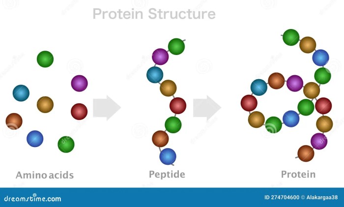
    The human cardiovascular system is a complex network of interconnected vessels and organs that is vital for maintaining life. It efficiently transports oxygen, nutrients, hormones, and waste products throughout the body, ensuring the proper functioning of all tissues and organs. This intricate system is essential for maintaining homeostasis, the body’s internal balance, and its proper functioning. Understanding the components and their roles is crucial for comprehending the importance of this system in overall health.

    Fundamental Components of the Circulatory System

    The circulatory system consists of the heart, blood vessels, and blood. These components work in concert to ensure the continuous movement of blood, facilitating the delivery of essential substances and the removal of waste products. The heart acts as the pump, driving blood through the vessels, while the blood vessels serve as conduits, carrying the blood to all parts of the body.

    Blood, composed of various cells and plasma, carries vital substances, maintaining the body’s overall health.

    Component Function Location within the Circulatory System
    Heart The heart is the muscular pump that propels blood throughout the circulatory system. It rhythmically contracts and relaxes, creating pressure gradients that move blood through the arteries, capillaries, and veins. Located in the thoracic cavity, between the lungs.
    Blood Vessels (Arteries, Veins, Capillaries) Blood vessels form a network of tubes that transport blood throughout the body. Arteries carry oxygenated blood away from the heart, veins return deoxygenated blood to the heart, and capillaries facilitate the exchange of nutrients and waste products between the blood and tissues. Arteries branch from the heart, forming a network of vessels throughout the body. Veins collect blood from tissues and return it to the heart. Capillaries form a dense network in tissues, enabling the exchange of substances between blood and cells.
    Blood Blood is the fluid that circulates throughout the body, carrying oxygen, nutrients, hormones, and waste products. It is composed of red blood cells, white blood cells, platelets, and plasma. Blood is constantly flowing through the heart and blood vessels, circulating throughout the body.

    Significance of Blood Flow in Maintaining Homeostasis

    The continuous movement of blood is critical for maintaining homeostasis. Blood carries oxygen and nutrients essential for cellular respiration, providing energy for various bodily functions. It also transports hormones, which regulate various bodily processes, and removes waste products, preventing their accumulation and potential toxicity. Disruptions in blood flow can lead to a range of health issues, emphasizing the critical role of this continuous process in maintaining optimal health.

    Structure of the Heart: Blood Flow Through The Heart

    The heart, a marvel of biological engineering, is a muscular pump that tirelessly circulates blood throughout our bodies. Understanding its intricate structure is crucial to grasping how it performs this vital function. Its chambers, valves, and associated vessels work in concert to ensure efficient and unidirectional blood flow. This meticulous design is essential for maintaining life.The heart’s anatomy, while complex, can be broken down into manageable components.

    Each part plays a specific role in the overall process of pumping blood, and understanding these components is key to comprehending the heart’s function.

    Chambers of the Heart

    The heart is divided into four chambers: two atria and two ventricles. The atria are the receiving chambers, collecting blood returning to the heart from the body and lungs. The ventricles are the pumping chambers, responsible for propelling blood out of the heart to the lungs and the rest of the body. This distinct separation of function is vital for maintaining the proper flow of blood.

    Valves of the Heart

    The heart’s valves act as one-way doors, ensuring that blood flows in the correct direction. Four valves—the mitral, tricuspid, pulmonary, and aortic valves—prevent backflow of blood, crucial for maintaining the unidirectional blood flow. Their precise function is essential for the heart’s efficient operation.

    Major Vessels

    The heart is connected to the rest of the body through a network of major vessels. The superior and inferior venae cavae return deoxygenated blood from the body to the right atrium. The pulmonary arteries carry deoxygenated blood to the lungs, and the pulmonary veins return oxygenated blood to the left atrium. The aorta carries oxygenated blood from the left ventricle to the rest of the body.

    These vessels form a critical part of the circulatory system, ensuring a continuous flow of blood.

    Structural Features for Unidirectional Blood Flow

    The heart’s structure is meticulously designed to facilitate unidirectional blood flow. The valves, as previously mentioned, prevent backflow, and the arrangement of the chambers ensures that blood progresses through the heart in a consistent, controlled manner. This unidirectional flow is critical for efficient oxygen delivery and waste removal throughout the body.

    Illustration of the Heart’s Internal Structure

    Imagine a three-dimensional model of the heart. The right atrium, located on the heart’s right side, is the first chamber encountered. Blood enters from the superior and inferior venae cavae. A muscular wall, the interatrial septum, separates the right and left atria. The tricuspid valve connects the right atrium to the right ventricle, ensuring that blood flows only from the atrium to the ventricle.

    Next is the right ventricle, which pumps blood through the pulmonary valve to the pulmonary arteries, bound for the lungs. The left atrium, located on the heart’s left side, receives oxygen-rich blood from the pulmonary veins. The mitral valve, similar to the tricuspid valve in function, connects the left atrium to the left ventricle. The left ventricle, the heart’s most muscular chamber, pumps oxygenated blood through the aortic valve into the aorta, the body’s main artery.

    The interventricular septum, another muscular wall, divides the ventricles. The intricate arrangement of these components ensures that blood always flows in one direction.

    Comparison of Atria and Ventricles

    Feature Atria Ventricles
    Structure Thin-walled, receiving chambers Thick-walled, pumping chambers
    Function Collect blood returning to the heart Pump blood out of the heart
    Muscle Thickness Thinner Thicker
    Valve Connection Connected to the ventricles via atrioventricular valves (tricuspid and mitral) Connected to major arteries via semilunar valves (pulmonary and aortic)

    This table highlights the key structural and functional differences between the atria and ventricles, emphasizing their specialized roles in the cardiac cycle.

    Understanding how blood flows through your heart is crucial for overall health. Think about it like a powerful pump, constantly circulating life-giving oxygen. And while a healthy heart is essential, did you know that supporting healthy hair growth might also play a role? Certain nutrients can contribute to healthy hair growth, such as those found in chia seed oil for hair growth , which can promote strong and lustrous locks.

    Ultimately, maintaining a healthy circulatory system, like the blood flow through the heart, is key to overall well-being.

    Pathways of Blood Flow

    Blood flows through the heart in a precise, cyclical manner, ensuring the efficient delivery of oxygen and nutrients to the body’s tissues. This intricate system of vessels and valves allows for the separation of oxygenated and deoxygenated blood, preventing mixing and maintaining a unidirectional flow. Understanding these pathways is crucial for grasping the heart’s vital role in circulation.

    Understanding blood flow through the heart is crucial for overall health. It’s a fascinating process, but sometimes other factors can affect our bodies, like wondering if magnesium supplements could be causing digestive issues like diarrhea. For more information on whether magnesium can cause diarrhea, check out this helpful resource: can magnesium cause diarrhea. Regardless of magnesium intake, a healthy heart and consistent blood flow remain vital for our well-being.

    Tracing Blood Flow Through the Heart

    The heart acts as a pump, pushing blood through a complex network of vessels. Oxygen-poor blood returns to the heart and is pumped to the lungs for oxygenation, while oxygen-rich blood is circulated to the rest of the body. This process is continuous and ensures a constant supply of oxygen and nutrients to the tissues.

    Role of Heart Valves

    Heart valves are crucial components of the cardiovascular system, ensuring one-way blood flow. They act as gates, opening and closing in response to pressure changes within the heart chambers. This regulated opening and closing prevents backflow, maintaining the efficient circulation of blood. The four heart valves—tricuspid, pulmonary, mitral, and aortic—play distinct roles in this process.

    Step-by-Step Blood Flow Through the Heart

    The following sequence Artikels the path of blood through the heart:

    • Deoxygenated blood enters the right atrium from the superior and inferior vena cava.
    • The right atrium contracts, pushing blood through the tricuspid valve into the right ventricle.
    • The right ventricle contracts, forcing blood through the pulmonary valve into the pulmonary artery.
    • The pulmonary artery carries the deoxygenated blood to the lungs for oxygenation.
    • Oxygenated blood returns from the lungs via the pulmonary veins to the left atrium.
    • The left atrium contracts, pushing blood through the mitral valve into the left ventricle.
    • The left ventricle contracts, forcing blood through the aortic valve into the aorta.
    • The aorta distributes the oxygenated blood to the rest of the body.

    Relationship Between Cardiac Cycle and Blood Flow

    The cardiac cycle, encompassing the rhythmic contraction and relaxation of the heart, is intricately linked to blood flow. Each phase of the cycle—atrial systole, ventricular systole, and diastole—contributes to the forward movement of blood. The coordinated action of the heart chambers, regulated by electrical impulses, ensures a continuous and efficient blood flow. The rhythmic contraction and relaxation of the heart chambers, driven by the cardiac cycle, create the pressure gradient necessary to move blood through the circulatory system.

    Regulation of Blood Flow

    The delicate dance of blood flow through our cardiovascular system isn’t a random process. Precise mechanisms govern heart rate, blood pressure, and overall blood flow to ensure the body’s needs are met. This intricate regulation is crucial for maintaining homeostasis and responding to changes in demand, from a leisurely stroll to intense exercise. Understanding these control mechanisms allows us to appreciate the complexity and efficiency of our circulatory system.The regulation of blood flow is a dynamic interplay between various factors, primarily the autonomic nervous system and hormones.

    Understanding how blood flows through the heart is fascinating, right? But did you know that sometimes, dealing with menstrual cramps can throw a wrench into your body’s systems? It’s a common question: do bananas help with cramps? Checking out this resource might offer some insights: do bananas help with cramps. Regardless of the answer, the intricate network of blood vessels and chambers within the heart continues its vital work, keeping everything running smoothly.

    These systems work in concert to adjust heart rate and blood pressure, ensuring an adequate supply of oxygen and nutrients to all tissues. The precise control allows the body to respond rapidly to changing needs, like increased physical activity, and maintain a stable internal environment.

    Heart Rate Control Mechanisms

    The heart’s rhythmic contractions, or heart rate, are meticulously controlled by the autonomic nervous system. This system, comprised of the sympathetic and parasympathetic branches, acts as a fine-tuning mechanism for cardiovascular function.

    Autonomic Nervous System Regulation of Cardiac Output

    The autonomic nervous system plays a critical role in modulating cardiac output, the volume of blood pumped by the heart per minute. The sympathetic nervous system, known for its “fight-or-flight” response, increases heart rate and contractility, boosting cardiac output. Conversely, the parasympathetic nervous system, associated with “rest-and-digest” functions, slows heart rate, decreasing cardiac output. This balanced interplay ensures that heart rate adapts to the body’s changing needs.

    Hormonal Influences on Blood Flow

    Hormones, chemical messengers in the body, also influence blood flow. Epinephrine (adrenaline) and norepinephrine, released during stress or exercise, stimulate the sympathetic nervous system, leading to increased heart rate and blood pressure. Other hormones, such as thyroid hormones, can influence metabolic rate, which, in turn, impacts the body’s need for blood flow. For example, increased metabolic rate might require an increase in blood flow to supply the body’s increased energy needs.

    Sympathetic vs. Parasympathetic Nervous System Effects

    Characteristic Sympathetic Nervous System Parasympathetic Nervous System
    Effect on Heart Rate Increases heart rate Decreases heart rate
    Effect on Blood Vessels (generally) Causes vasoconstriction (narrowing) in most blood vessels, except those supplying skeletal muscles Causes vasodilation (widening) in some blood vessels
    Effect on Blood Pressure Increases blood pressure Decreases blood pressure
    Effect on Bronchioles (airways) Causes bronchodilation (widening) Causes bronchoconstriction (narrowing)
    Dominant State Fight-or-flight response Rest-and-digest response

    These opposing effects of the sympathetic and parasympathetic systems create a dynamic balance, allowing for rapid adjustments in cardiovascular function based on the body’s needs. The interplay between these two systems ensures homeostasis, enabling the body to respond appropriately to changing demands, from a quiet moment of rest to a period of intense physical exertion. This complex regulatory system is crucial for maintaining blood pressure and ensuring an adequate blood supply to all tissues and organs.

    Clinical Considerations

    The intricate dance of blood flow through the heart is crucial for life. Disruptions to this delicate system can lead to a range of serious health problems. Understanding these conditions, the factors that cause them, and the diagnostic and treatment options available is vital for maintaining cardiovascular health.Common heart conditions that disrupt blood flow are numerous and diverse.

    Some involve structural abnormalities, while others stem from functional impairments. These conditions can manifest in various ways, impacting the heart’s ability to pump blood efficiently and consistently.

    Common Heart Conditions Affecting Blood Flow

    Numerous conditions can disrupt the normal flow of blood through the heart. These conditions can lead to reduced cardiac output, impaired oxygen delivery, and a range of other complications. Understanding the underlying mechanisms of these conditions is key to developing effective diagnostic and therapeutic strategies.

    • Coronary Artery Disease (CAD): CAD is characterized by the buildup of plaque in the coronary arteries, the blood vessels that supply the heart muscle with oxygen. This plaque buildup narrows the arteries, reducing blood flow to the heart muscle, potentially leading to angina (chest pain) or a heart attack if the blood supply is completely blocked. This condition is often associated with risk factors such as high blood pressure, high cholesterol, smoking, and diabetes.

      Many individuals lead lives free of symptoms, yet experience significant cardiac complications due to plaque buildup in the coronary arteries.

    • Valvular Heart Disease: Valvular heart disease occurs when one or more of the heart valves do not function properly. This can be due to stenosis (narrowing of the valve) or regurgitation (leaking of the valve). These conditions can disrupt the normal flow of blood through the heart, leading to symptoms such as shortness of breath, fatigue, and palpitations. Valvular heart disease is often treated with medication, surgery, or a combination of both.

    • Congenital Heart Defects: These are structural abnormalities in the heart that are present at birth. They can range from minor issues to severe conditions that require immediate intervention. Examples include septal defects (holes in the heart walls), valve abnormalities, and narrowing of blood vessels. These conditions can disrupt the normal mixing of oxygenated and deoxygenated blood, leading to various circulatory problems.

      Proper diagnosis and intervention during infancy and childhood are essential to improve long-term health outcomes.

    Factors Disrupting Normal Blood Flow Patterns

    Various factors can disrupt the normal flow of blood through the heart. These factors can contribute to the development of various cardiovascular conditions. Understanding these factors is crucial for preventative measures and early intervention.

    • High Blood Pressure: Sustained high blood pressure, or hypertension, puts extra strain on the heart and blood vessels. This increased pressure can damage the arteries over time, leading to atherosclerosis (plaque buildup) and potentially causing heart attacks or strokes. Hypertension is a significant risk factor for cardiovascular disease, and lifestyle modifications and medication are often employed to manage it.

    • High Cholesterol: High levels of cholesterol in the blood can contribute to the formation of plaque in the arteries, reducing blood flow. This condition can also lead to atherosclerosis and increase the risk of cardiovascular events. Dietary changes, lifestyle adjustments, and medications are often used to manage high cholesterol.
    • Smoking: Smoking damages blood vessels, increases blood clotting, and raises blood pressure. These effects increase the risk of developing various cardiovascular conditions, including CAD and stroke. Quitting smoking is crucial for improving cardiovascular health.

    Diagnostic Methods for Assessing Blood Flow

    Various diagnostic techniques are used to assess blood flow through the heart. These methods provide crucial information about the structure and function of the heart, enabling accurate diagnosis and treatment planning.

    • Electrocardiogram (ECG): An ECG measures the electrical activity of the heart. It can detect abnormal rhythms, heart damage, and the presence of certain conditions. It is a non-invasive and relatively quick test.
    • Echocardiogram (Echo): An echocardiogram uses ultrasound waves to create images of the heart. It allows visualization of the heart’s structures, valves, and overall function, including blood flow patterns. It’s a valuable tool for diagnosing various heart conditions.
    • Cardiac Catheterization: Cardiac catheterization involves inserting a thin tube (catheter) into an artery or vein and guiding it to the heart. It allows direct visualization of the coronary arteries, measurement of blood pressure, and assessment of blood flow. This method provides detailed information about the heart’s structure and function, particularly concerning coronary arteries.

    Treatment Options for Conditions Affecting Blood Flow

    Various treatment options are available for conditions affecting blood flow through the heart. The chosen approach depends on the specific condition, its severity, and the patient’s overall health.

    • Lifestyle Modifications: Dietary changes, regular exercise, and stress management can significantly improve cardiovascular health and reduce the risk of various heart conditions. These modifications are often a crucial component of treatment, particularly for preventing the progression of existing conditions.
    • Pharmacological Interventions: Medications such as blood pressure medications, cholesterol-lowering drugs, and anti-platelet agents are commonly used to manage conditions affecting blood flow. These medications help control risk factors and improve overall cardiovascular health.
    • Surgical Interventions: Procedures like angioplasty, coronary artery bypass grafting (CABG), and valve replacement are used to address structural abnormalities or blockages affecting blood flow. Surgical interventions are often employed to restore normal blood flow when other methods are ineffective.

    Blood Flow and Exercise

    Blood flow through the heart

    Exercise significantly impacts blood flow throughout the body, placing a heightened demand on the cardiovascular system. The heart, as the primary pump, must adapt to deliver increased oxygen and nutrients to working muscles. This adaptation involves a complex interplay of physiological responses, ensuring efficient blood delivery and oxygen utilization.

    Effects of Exercise on Heart Rate

    Heart rate increases substantially during exercise to meet the elevated oxygen demands of the body. This increase is a direct result of the sympathetic nervous system’s activation, which stimulates the sinoatrial (SA) node, the heart’s natural pacemaker. Higher heart rates facilitate faster blood circulation, delivering more oxygenated blood to the working muscles. For example, during intense aerobic exercise, heart rates can reach 180-200 beats per minute in trained athletes, whereas resting heart rates typically range from 60-80 beats per minute.

    Effects of Exercise on Stroke Volume

    Stroke volume, the amount of blood pumped by the heart with each beat, also increases during exercise. This is primarily due to enhanced ventricular filling during diastole and a stronger ventricular contraction during systole. Increased venous return, facilitated by muscle contractions and respiratory movements, leads to greater preload, stretching the ventricles and improving their contractile force (Frank-Starling mechanism).

    Additionally, sympathetic stimulation strengthens ventricular contractions, further augmenting stroke volume.

    Effects of Exercise on Cardiac Output

    Cardiac output, the total amount of blood pumped by the heart per minute, is the product of heart rate and stroke volume. Consequently, exercise dramatically elevates cardiac output to match the increased oxygen needs of active tissues. For instance, during vigorous exercise, cardiac output can increase four to seven times its resting value, ensuring adequate oxygen delivery to the working muscles.

    Changes in Cardiovascular Parameters During Exercise

    The following table Artikels the typical changes in heart rate, stroke volume, and cardiac output during various intensities of exercise:

    Exercise Intensity Heart Rate (bpm) Stroke Volume (mL/beat) Cardiac Output (L/min)
    Light 70-120 80-100 5.6-12
    Moderate 120-150 100-120 12-18
    Vigorous 150-180+ 120-150+ 18-35+

    Long-Term Effects of Exercise on Cardiovascular Health

    Regular exercise has numerous beneficial long-term effects on cardiovascular health. It strengthens the heart muscle, making it more efficient at pumping blood. This enhanced contractility lowers the resting heart rate and reduces the risk of hypertension and other cardiovascular diseases. Furthermore, exercise promotes the development of a more robust vascular system, with improved blood vessel elasticity and reduced plaque buildup.

    For example, individuals who consistently engage in aerobic exercise demonstrate a lower incidence of coronary artery disease and a reduced risk of stroke compared to those who are sedentary.

    Blood Flow and Disease

    Heart disease, a broad category encompassing various conditions affecting the heart’s structure and function, significantly impacts blood flow. These conditions can lead to reduced blood supply to vital organs, potentially causing serious complications. Understanding the interplay between heart diseases and blood flow is crucial for effective prevention and treatment strategies.

    Coronary Artery Disease (CAD), Blood flow through the heart

    Coronary artery disease, a leading cause of heart disease, is characterized by the buildup of plaque within the coronary arteries, the blood vessels that supply blood to the heart muscle. This plaque buildup narrows the arteries, reducing blood flow to the heart. This reduced blood flow can lead to angina (chest pain), heart attack, or even sudden cardiac death.

    The extent of the narrowing and the location of the blockage directly influence the severity of the symptoms.

    Valvular Heart Disease

    Valvular heart disease affects the heart’s valves, which regulate the flow of blood through the heart. Damaged or diseased valves can cause blood to leak backward (regurgitation) or obstruct blood flow (stenosis). These issues can strain the heart and lead to symptoms like shortness of breath, fatigue, and swelling in the legs. The specific valve affected and the severity of the dysfunction determine the impact on blood flow.

    Impact on the Heart’s Pumping Ability

    Heart diseases significantly impair the heart’s ability to pump blood effectively. In CAD, the reduced blood supply deprives the heart muscle of oxygen and nutrients, leading to weakened contractions. Valvular heart disease puts extra strain on the heart as it works harder to overcome the obstructed or leaking blood flow. These impairments can result in reduced cardiac output, meaning less blood is circulated throughout the body.

    Physiological Changes

    The physiological changes resulting from heart disease are varied and complex. In CAD, the heart muscle can experience ischemia (lack of blood flow), leading to damage and scarring. Valvular disease can cause hypertrophy (enlargement) of the heart chambers as the heart compensates for the increased workload. The degree of these changes correlates with the severity and duration of the disease.

    Comparison of Heart Diseases and Their Effects

    Heart Disease Mechanism of Reduced Blood Flow Impact on Pumping Ability Physiological Changes Symptoms
    Coronary Artery Disease (CAD) Plaque buildup narrows coronary arteries, reducing blood supply to the heart muscle. Reduced cardiac output due to impaired heart muscle function. Ischemia, damage, and scarring of the heart muscle; possible hypertrophy. Angina, shortness of breath, fatigue, heart attack, sudden cardiac death.
    Valvular Heart Disease Damaged valves cause leakage (regurgitation) or obstruction (stenosis) of blood flow through the heart. Increased workload on the heart, potentially leading to reduced cardiac output and heart failure. Hypertrophy of affected heart chambers, valve damage. Shortness of breath, fatigue, swelling in the legs, palpitations.

    Concluding Remarks

    In conclusion, blood flow through the heart is a remarkable feat of biological engineering. From the intricate structure of the heart to the regulatory mechanisms that govern its function, this intricate process is essential for life. Understanding this process is vital for appreciating the delicate balance of the human body and the importance of preventative measures against disease.

  • What is a Peptide A Deep Dive

    What is a Peptide A Deep Dive

    What is a peptide? This exploration delves into the fascinating world of peptides, those short chains of amino acids that play crucial roles in various biological processes. From their fundamental structure to their diverse functions in nature and medicine, we’ll uncover the secrets behind these remarkable molecules. Prepare to embark on a journey into the captivating realm of peptides!

    Peptides are essentially small proteins, often acting as signaling molecules or components of larger protein structures. Their relatively short length, compared to proteins, allows for greater flexibility and diverse functions. Understanding their formation, properties, and applications is key to appreciating their significance in biology and medicine.

    Defining Peptides

    Peptides are short chains of amino acids, playing crucial roles in various biological processes. Understanding their structure and function is fundamental to comprehending the intricate workings of life. They differ from proteins, which are typically longer chains of amino acids with more complex structures and functions. This section will delve into the specifics of peptide definition, structure, naming, and examples.

    Peptide Definition

    A peptide is a short chain of amino acids linked together by peptide bonds. These bonds form between the carboxyl group of one amino acid and the amino group of another, resulting in the elimination of a water molecule. This process is known as dehydration synthesis. Peptides are typically composed of fewer than 50 amino acids, whereas proteins are longer chains.

    Difference Between Peptides and Proteins

    The primary difference between peptides and proteins lies in their length. Peptides are shorter chains of amino acids, while proteins are significantly longer. This difference in length directly impacts their structural complexity and consequently, their functional diversity. Proteins often fold into complex three-dimensional structures, enabling them to perform a wider array of tasks within biological systems. Peptides, being shorter, tend to have more localized, specific functions.

    General Structure of a Peptide

    A peptide’s structure involves a linear arrangement of amino acids. Each amino acid consists of a central carbon atom (alpha carbon) bonded to an amino group (-NH2), a carboxyl group (-COOH), a hydrogen atom, and a variable side chain (R group). The sequence of these amino acids, determined by the genetic code, dictates the peptide’s unique properties and function.

    The peptide bonds connecting the amino acids are amide linkages.

    Peptide bond: A covalent bond formed between the carboxyl group of one amino acid and the amino group of another.

    So, what exactly is a peptide? It’s basically a short chain of amino acids, the building blocks of proteins. While peptides play a vital role in various bodily functions, did you know that certain essential oils, like lavender and peppermint, are also used to soothe headaches and migraines? For example, exploring the benefits of lavender and peppermint essential oils for relieving headaches and migraines can be really interesting.

    lavender and peppermint essential oils for headaches and migraines could offer some insights into this. Regardless of the use case, understanding the basic structure of peptides is still crucial for understanding how our bodies function.

    Naming Conventions for Peptides

    Peptide names typically indicate the sequence of amino acids. The names are written using the three-letter abbreviations for the amino acids, in the order they appear in the chain. For example, Gly-Ala-Ser denotes a peptide containing glycine, alanine, and serine in that order. Sometimes, the one-letter abbreviations are used, for instance, Gly-Ala-Ser could also be written as G-A-S.

    Examples of Common Peptides

    Peptides play a wide range of roles in biological systems. Here are some examples of common peptides and their functions.

    Peptide Name Source Function
    Oxytocin Posterior pituitary gland Stimulates uterine contractions during childbirth and milk ejection during breastfeeding.
    Vasopressin Posterior pituitary gland Regulates water reabsorption in the kidneys.
    Glucagon Pancreas Raises blood glucose levels.
    Insulin Pancreas Lowers blood glucose levels.
    Bradykinin Blood Causes vasodilation and pain.
    Substance P Nervous system Transmits pain signals.
    Enkephalins Brain Act as natural painkillers.

    Peptide Formation

    What is a peptide

    Peptides, those essential building blocks of proteins, are formed through a fascinating process called peptide bond formation. This crucial step links amino acids together, creating the intricate structures that underpin biological functions. Understanding this process is key to appreciating the complexity and versatility of proteins.The peptide bond formation is a dehydration reaction, a process where a molecule of water is eliminated to join two molecules.

    In this case, the carboxyl group (-COOH) of one amino acid reacts with the amino group (-NH2) of another amino acid. This reaction results in the formation of a peptide bond (–CONH–) between the two amino acids and the release of a water molecule.

    Peptide Bond Formation Mechanism

    The formation of a peptide bond involves a nucleophilic attack. The nitrogen atom of the amino group acts as a nucleophile, attacking the carbonyl carbon of the carboxyl group. This attack leads to the formation of a tetrahedral intermediate. The tetrahedral intermediate then collapses, expelling a water molecule and creating the peptide bond. This process is facilitated by enzymes, crucial players in the peptide synthesis within living organisms.

    Role of Enzymes in Peptide Synthesis, What is a peptide

    Enzymes play a critical role in accelerating and directing peptide bond formation within living systems. They provide a highly controlled environment, precisely positioning the reactants and lowering the activation energy required for the reaction. This precise control ensures that peptide bonds are formed in the correct sequence, leading to the production of the specific protein needed. Without enzymes, these reactions would occur at a negligible rate, hindering the construction of complex proteins.

    Laboratory Peptide Synthesis Methods

    Various methods exist to synthesize peptides in a laboratory setting, each with its advantages and disadvantages. These methods range from simple to complex, depending on the desired peptide length and complexity. The choice of method is dictated by the factors like desired length, purity, and the availability of reagents.

    • Solid-phase peptide synthesis (SPPS): This method is commonly used for the synthesis of longer peptides. It involves attaching one amino acid at a time to a solid support, allowing for the stepwise addition of amino acids in a predefined sequence. This technique offers a high degree of control over the reaction and allows for the purification of the product at each step, enhancing the efficiency of the process.

      The solid support typically consists of a resin bead to which the growing peptide chain is attached.

    • Solution-phase peptide synthesis: This method is typically employed for smaller peptides. In this method, amino acids are linked together in solution. This method, though simpler than SPPS, may lead to lower yields and increased purification challenges. Solution-phase synthesis often relies on protecting groups to prevent unwanted side reactions. Careful control of reaction conditions is critical.

      So, what exactly is a peptide? Basically, it’s a short chain of amino acids, the building blocks of proteins. Interestingly, peptides play a crucial role in various bodily functions, including the complex process of continuous glucose monitoring, like with the new Dexcom 5 continuous glucose monitoring the arrival of Dexcom 5. Understanding these tiny chains helps us appreciate the intricate ways our bodies work, and how advancements like this new technology are impacting healthcare.

    Importance of Reaction Conditions in Peptide Synthesis

    Precise reaction conditions are paramount for successful peptide synthesis. Factors such as pH, temperature, and the presence of protecting groups directly influence the outcome of the reaction. These conditions must be carefully controlled to prevent unwanted side reactions and ensure the formation of the desired peptide.

    • Protecting groups: Protecting groups are essential in preventing unwanted side reactions. These groups block specific functional groups on amino acids, protecting them from unwanted reactions during the synthesis process. These groups are then removed after the peptide bond formation is complete. This is critical to ensuring the desired peptide sequence is produced.
    • Coupling reagents: Coupling reagents facilitate the formation of the peptide bond. Different coupling reagents have varying degrees of efficiency and compatibility with different amino acids. The selection of an appropriate coupling reagent is crucial to optimize peptide synthesis.

    Comparison of Peptide Synthesis Methods

    Method Description Advantages Disadvantages
    Solid-phase peptide synthesis (SPPS) Amino acids added sequentially to a solid support High control, stepwise purification, suitable for longer peptides Can be complex, equipment requirements, potentially lower yields for very short peptides
    Solution-phase peptide synthesis Amino acids linked in solution Simple setup, potentially higher yields for very short peptides Lower control, purification challenges, difficult for longer peptides

    Peptide Properties: What Is A Peptide

    Peptides, the building blocks of proteins, exhibit a fascinating array of physical and chemical properties. Understanding these properties is crucial for comprehending their diverse roles in biological systems and their potential applications in various fields, including medicine and materials science. Their behavior is dictated by the unique combination of their amino acid sequence, the interactions between amino acids, and the surrounding environment.These properties significantly impact the function of peptides.

    For instance, the solubility of a peptide directly influences its ability to interact with other molecules in solution. The stability of a peptide determines its ability to maintain its structure and function under various conditions, while the melting point reveals the energy required to disrupt its secondary and tertiary structures. These characteristics are not uniform across all peptides; they are highly dependent on the specific sequence and the chemical environment.

    Solubility

    Peptide solubility is a crucial factor determining their behavior in biological systems. The solubility of a peptide is largely dictated by the hydrophilic and hydrophobic amino acid residues present in its sequence. Hydrophilic residues, such as serine, threonine, and asparagine, tend to interact favorably with water molecules, increasing the peptide’s solubility. Conversely, hydrophobic residues, such as leucine, isoleucine, and valine, tend to cluster together, minimizing their interaction with water and decreasing solubility.

    The overall balance of these residues influences the peptide’s ability to dissolve in various solvents. For example, a peptide rich in polar amino acids will likely be more soluble in water than a peptide predominantly composed of non-polar amino acids.

    Melting Point

    The melting point of a peptide represents the temperature at which the peptide transitions from an ordered, folded state to a disordered, unfolded state. This transition is often accompanied by significant changes in the peptide’s physical properties, such as solubility and activity. The precise melting point is influenced by the specific amino acid sequence, the strength of the intermolecular forces holding the peptide in its folded conformation, and the presence of any stabilizing factors like metal ions or other molecules.

    Factors like hydrogen bonding, hydrophobic interactions, and disulfide bonds significantly impact the stability of the folded structure and, consequently, the melting point. Higher melting points often indicate greater stability and a more robust structure.

    Stability

    Peptide stability is a critical characteristic that determines their ability to maintain their structure and function under various conditions. This stability is dependent on the interplay of various factors. For example, the presence of disulfide bonds significantly enhances the stability of a peptide by creating strong covalent linkages between cysteine residues. These covalent bonds help to maintain the peptide’s folded structure, even in harsh conditions.

    The surrounding environment also plays a critical role. High temperatures, extreme pH values, and the presence of proteolytic enzymes can all negatively impact peptide stability. Furthermore, the presence of stabilizing elements, such as metal ions, can contribute to the overall stability of the peptide.

    Factors Influencing Folding and Conformation

    Peptide folding and conformation are crucial for determining their function. The specific amino acid sequence dictates the potential folding patterns, and the surrounding environment can influence these patterns. Hydrogen bonding, hydrophobic interactions, and electrostatic interactions between amino acid residues drive the folding process. The precise arrangement of these interactions shapes the unique three-dimensional structure of the peptide, which is crucial for its function.

    Specific examples include alpha-helices, beta-sheets, and random coils, each with different properties and functions. External factors such as temperature, pH, and the presence of other molecules can also alter the peptide’s conformation, influencing its activity.

    Peptide Sequence and Properties

    The amino acid sequence of a peptide directly dictates its physical and chemical properties. A change in even a single amino acid can significantly alter the peptide’s solubility, stability, and overall function. For instance, replacing a hydrophilic amino acid with a hydrophobic one can drastically reduce the peptide’s water solubility. Similarly, alterations in the sequence can affect the formation of secondary structures, impacting the peptide’s stability and overall conformation.

    This highlights the profound impact of sequence on function.

    Comparison of Peptide Properties

    Peptide Type Solubility Melting Point Stability
    Short, hydrophilic peptides High Low Low
    Long, hydrophobic peptides Low High High
    Cyclized peptides Variable High High
    Peptides with disulfide bonds Variable High High

    Peptide Functions

    Peptides, those short chains of amino acids, are not just building blocks; they are active players in a multitude of biological processes. Their diverse functions stem from their unique structures and the specific arrangements of amino acids within their sequences. Understanding these functions is crucial to appreciating the intricate workings of life itself.

    Examples of Peptide Functions in Biological Systems

    Peptides play diverse roles in biological systems, from coordinating cellular responses to catalyzing biochemical reactions. Their specific functions depend on the unique sequence and structure of the peptide. Some peptides act as hormones, regulating physiological processes, while others act as neurotransmitters, transmitting signals between nerve cells. Still others are integral components of the immune system, defending the body against pathogens.

    • Hormones: Many hormones are peptides, such as insulin, glucagon, and growth hormone. These peptides regulate various metabolic processes, including blood sugar levels and growth. Insulin, for example, lowers blood glucose levels by promoting glucose uptake into cells. Glucagon, on the other hand, raises blood glucose levels by stimulating the release of glucose from the liver.

    • Neurotransmitters: Some peptides act as neurotransmitters, carrying signals between nerve cells. Examples include substance P, involved in pain transmission, and endorphins, which act as natural pain relievers. These peptides play critical roles in modulating neuronal activity and influencing behavior.
    • Immune System Peptides: The immune system employs peptides to defend against pathogens. Cytokines, a diverse group of signaling peptides, regulate immune responses. Interleukins and interferons are examples of cytokines that orchestrate the inflammatory response and antiviral defenses.

    Roles of Peptides in Signaling Pathways

    Peptides are essential components in signaling pathways, mediating communication between cells. These peptides act as ligands, binding to specific receptors on target cells, triggering intracellular cascades that lead to various cellular responses. The specific sequence of amino acids dictates the type of signal transmitted and the cellular response.

    • Receptor Binding: Peptides bind to specific receptors on the surface of target cells. This binding initiates a cascade of intracellular events. The interaction between the peptide and the receptor is highly specific, ensuring the precise delivery of the signal.
    • Intracellular Signaling: Upon binding, the receptor undergoes conformational changes, initiating a series of events within the cell. These intracellular signaling cascades involve a complex interplay of proteins, enzymes, and second messengers, ultimately leading to the desired cellular response. The specificity of the peptide-receptor interaction ensures the precise regulation of cellular processes.

    Role of Peptides in Enzyme Activity

    Peptides can influence enzyme activity in various ways. Some peptides act as inhibitors, blocking enzyme function, while others act as activators, enhancing enzyme activity. These peptides can bind to the active site of the enzyme, directly affecting its catalytic ability.

    Peptides are essentially short chains of amino acids, playing crucial roles in various bodily functions. Understanding these small molecules is fascinating, especially when considering how they might be related to conditions like premenstrual dysphoria. Recent research is exploring the potential for innovative therapies like surgical management of premenstrual dysphoria disorder , which could impact the hormonal imbalances that contribute to this condition.

    Ultimately, a deeper understanding of peptides is key to unraveling these complexities and developing effective treatments.

    • Enzyme Inhibition: Certain peptides act as potent inhibitors of specific enzymes. These peptides bind to the active site of the enzyme, preventing substrate binding and hindering the catalytic reaction. This inhibitory mechanism is crucial in regulating enzyme activity and preventing unwanted biochemical processes.
    • Enzyme Activation: Other peptides act as activators, enhancing the activity of specific enzymes. These peptides bind to the enzyme, altering its conformation and increasing its catalytic efficiency. This activation process can be essential in regulating various metabolic pathways.

    Examples of Peptides Used in Medicine and Pharmaceuticals

    Peptides find widespread applications in medicine and pharmaceuticals. Their ability to target specific receptors and regulate cellular processes makes them valuable tools for treating various diseases. Examples include various therapeutic peptides used in treating diabetes, hypertension, and other conditions.

    • Therapeutic Peptides: Many peptides are used as therapeutic agents in medicine. Examples include insulin, used in treating diabetes, and various growth factors used in wound healing and tissue regeneration. These peptides are administered to modulate specific physiological processes, thereby mitigating the effects of diseases.

    Classification of Peptides Based on Biological Functions

    Peptide Function Examples Mechanism
    Hormones Insulin, Glucagon, Growth Hormone Regulate metabolic processes
    Neurotransmitters Substance P, Endorphins Transmit signals between nerve cells
    Immune System Regulators Cytokines (Interleukins, Interferons) Modulate immune responses
    Enzyme Inhibitors/Activators Specific peptide inhibitors/activators Regulate enzyme activity

    Peptide Analysis

    Unraveling the secrets of peptides often hinges on meticulous analysis. Understanding their composition, sequence, and presence within complex mixtures is crucial for research in diverse fields, from drug discovery to clinical diagnostics. This crucial step enables researchers to identify and quantify peptides, providing insights into their biological roles and functions. Detailed analysis allows for a deeper understanding of peptide behavior and their interactions within biological systems.Precise determination of a peptide’s sequence is paramount.

    Various sophisticated techniques have been developed for this purpose, offering high resolution and accuracy. Effective identification of peptides within complex mixtures also presents a significant challenge. A wide array of analytical tools are employed to separate and characterize these molecules, ensuring accurate and reliable results. The importance of this analytical process in various research areas and clinical applications is undeniable.

    Determining Peptide Sequence

    Determining the sequence of a peptide is a fundamental aspect of peptide analysis. Several powerful methods are available, each with its own strengths and limitations. These methods are crucial for elucidating the structure-function relationships of peptides and for validating their predicted function.

    • Edman Degradation: This technique is a classic method for sequencing peptides. It sequentially removes amino acid residues from the N-terminus of the peptide, allowing for the identification of each amino acid in the chain. The process involves chemical modification and cleavage of the N-terminal amino acid, followed by its identification through various chromatographic techniques. This method is particularly useful for smaller peptides.

      However, it is less suitable for very large peptides or those with repetitive sequences.

    • Mass Spectrometry: Mass spectrometry (MS) is a highly versatile technique for peptide sequencing. It measures the mass-to-charge ratio of ionized peptides. By fragmenting the peptide ions, MS provides information about the amino acid sequence. Different types of MS experiments, such as tandem mass spectrometry (MS/MS), are particularly effective in determining the sequence of peptides. Sophisticated software algorithms analyze the fragmentation patterns to deduce the amino acid sequence.

      MS is widely used for sequencing peptides from complex mixtures, making it a powerful tool for proteomics research.

    Identifying Peptides in Complex Mixtures

    Analyzing peptides in complex biological samples, such as blood serum or tissue extracts, is a significant challenge. Sophisticated separation techniques are essential to isolate individual peptides before their characterization. The use of these methods allows researchers to understand the role of peptides in various biological processes.

    • High-Performance Liquid Chromatography (HPLC): HPLC is a widely used technique for separating peptides based on their different properties, such as hydrophobicity or charge. The separation is achieved by passing the peptide mixture through a column packed with a stationary phase. Different peptides will elute at different times, allowing for their isolation and subsequent analysis by MS. This technique is particularly useful when dealing with mixtures containing peptides with similar properties.

      It allows for the separation and isolation of individual peptides from complex mixtures.

    • Capillary Electrophoresis (CE): CE is another powerful separation technique that utilizes an electric field to separate charged molecules based on their size and charge. It offers high resolution and speed compared to HPLC. CE is particularly advantageous when dealing with peptides that are difficult to separate by HPLC. It is commonly used in conjunction with MS for peptide identification and quantification in complex biological samples.

    Common Analytical Tools

    Various analytical tools are employed in peptide analysis, each offering unique capabilities. These tools are essential for researchers to gain a comprehensive understanding of peptide behavior and their roles in biological systems.

    • Mass Spectrometers: These instruments are crucial for determining the mass-to-charge ratio of ionized peptides, leading to sequence elucidation and quantification. They play a vital role in proteomics and peptide-based diagnostics.
    • Chromatographs (HPLC and CE): These instruments enable the separation of peptides based on their physical and chemical properties. They are crucial for isolating individual peptides for detailed analysis by MS.

    Importance of Peptide Analysis

    Peptide analysis is crucial in various research and clinical settings. It plays a significant role in drug discovery, diagnostics, and understanding fundamental biological processes. Analyzing peptides allows researchers to investigate their function and interactions with other molecules.

    Method Principle Applications
    Edman Degradation Sequential removal of N-terminal amino acids Sequencing smaller peptides
    Mass Spectrometry (MS) Measurement of mass-to-charge ratio of ionized peptides Sequencing peptides in complex mixtures, proteomics
    High-Performance Liquid Chromatography (HPLC) Separation of peptides based on properties like hydrophobicity or charge Isolating peptides from complex mixtures
    Capillary Electrophoresis (CE) Separation of charged molecules based on size and charge High-resolution separation of peptides, particularly those difficult to separate by HPLC

    Peptide Applications

    Peptides, those short chains of amino acids, are proving to be remarkably versatile molecules with a wide range of applications. Their specific structures and properties allow them to interact with various biological systems and materials, leading to exciting possibilities in diverse industries. From enhancing food products to revolutionizing personal care, peptides are making significant strides in various fields.

    Peptide Applications in Food Science

    Peptides contribute significantly to the advancement of food science. Their ability to influence taste, texture, and nutritional value makes them valuable ingredients. Hydrolyzed proteins, rich in bioactive peptides, can improve the nutritional profile of foods, potentially increasing the bioavailability of essential nutrients. Furthermore, certain peptides exhibit antimicrobial or antioxidant properties, enhancing the shelf life and safety of food products.

    The use of specific peptides can even modify the mouthfeel of foods, contributing to a more enjoyable eating experience.

    Peptide Applications in Agriculture

    Peptides are finding applications in agricultural practices as well. Their use as biostimulants and plant growth regulators has shown promise. Specific peptides can promote plant development by enhancing nutrient uptake or improving stress tolerance. Additionally, some peptides exhibit insecticidal or fungicidal properties, offering sustainable alternatives to traditional chemical pesticides. This environmentally friendly approach to pest control could have a positive impact on agricultural practices.

    Peptide Applications in Cosmetics and Personal Care

    Peptides are increasingly incorporated into cosmetics and personal care products. Their ability to stimulate collagen production or improve skin elasticity makes them popular ingredients in anti-aging products. Certain peptides may also possess antioxidant properties, helping to protect the skin from environmental damage. Moreover, some peptides can promote wound healing or reduce inflammation, expanding their application in various personal care products.

    Peptide Applications in Materials Science

    Peptides are demonstrating potential in materials science. Their unique structural features can be exploited to create novel materials with tailored properties. Specific peptide sequences can self-assemble into ordered structures, leading to the development of bio-inspired materials with enhanced strength, flexibility, or biocompatibility. This opens doors to applications in areas like tissue engineering and biocompatible coatings.

    Table of Peptide Applications in Different Industries

    Industry Application Example
    Food Science Enhancing flavor, texture, nutritional value, and shelf life Hydrolyzed whey protein peptides improving protein digestibility and increasing the nutritional value of dairy products.
    Agriculture Biostimulants, plant growth regulators, pest control Peptides promoting plant growth and stress tolerance, or inhibiting fungal infections.
    Cosmetics & Personal Care Anti-aging, skin elasticity, wound healing, reducing inflammation Peptides stimulating collagen production in anti-aging creams, or promoting wound healing in topical treatments.
    Materials Science Bio-inspired materials with tailored properties Peptides self-assembling into nanofibers for tissue engineering scaffolds or biocompatible coatings for implants.

    Illustrative Examples

    Peptides, the humble building blocks of proteins, play a vast array of roles in biological systems. From orchestrating intricate signaling pathways to forming structural components, peptides’ diverse functions are crucial for life. Understanding specific examples illuminates the breadth of their impact. This section delves into illustrative cases, highlighting naturally occurring peptides and their synthetic counterparts.Exploring concrete examples of peptides offers a tangible grasp of their diverse roles.

    The structural intricacies and biological functions of peptides, both natural and synthetic, provide valuable insights into their significance.

    Naturally Occurring Peptides with Biological Roles

    Peptides are abundant in nature, each with specialized functions. Understanding their structural properties and biological roles unveils the intricate workings of biological systems. These naturally occurring peptides are often involved in crucial processes, including hormone regulation, neurotransmission, and immune responses.

    • Oxytocin (OT): A neuropeptide with nine amino acids, oxytocin is synthesized in the hypothalamus and plays a vital role in social bonding and reproduction. It stimulates uterine contractions during childbirth and milk ejection during breastfeeding. Oxytocin’s effects extend beyond these physiological processes, influencing social behaviors like trust and empathy. It’s a crucial hormone for maternal care and social interactions.

    • Vasopressin (AVP): Another crucial neuropeptide, vasopressin, is also synthesized in the hypothalamus. Similar to oxytocin, it plays a significant role in regulating social behaviors. Vasopressin influences blood pressure regulation, water retention, and stress responses. Its actions are critical for maintaining homeostasis.
    • Enkephalins: These opioid peptides, composed of five or six amino acids, act as natural pain relievers in the central nervous system. They bind to opioid receptors, modulating pain signals and influencing mood. Enkephalins’ role in pain management and their connection to emotional states make them important for understanding these complex processes.

    Synthetic Peptides in Research and Drug Development

    Synthetic peptides have become indispensable tools in research and drug development. Their precise structure allows for targeted investigation of specific biological processes. Researchers can manipulate amino acid sequences to explore peptide function and design novel therapeutics.

    • Angiotensin II: A crucial peptide involved in blood pressure regulation. Synthetic analogs of angiotensin II are frequently used in research to study the effects of different sequences on blood pressure and cardiovascular function. This aids in the development of potential treatments for hypertension.
    • Growth hormone-releasing hormone (GHRH): Synthetic peptides mimic natural hormones and have applications in various medical conditions. For example, GHRH analogs are used to stimulate growth hormone secretion in specific cases, such as in children with growth hormone deficiency. This highlights the therapeutic potential of peptide-based therapies.

    A Detailed Description of a Specific Peptide

    Gramicidin S is a cyclic decapeptide with a unique structure. Its hydrophobic amino acid residues create a channel across cell membranes. This unique property allows it to permeabilize cell membranes, disrupting cellular function. This characteristic makes it an intriguing subject for research, potentially useful in antimicrobial applications.

    • Gramicidin S: A cyclic decapeptide, gramicidin S, possesses a unique structure that enables it to form channels across cell membranes. This property makes it a promising candidate for antimicrobial applications. The hydrophobic amino acid residues in its structure create a pathway through the cell membrane, disrupting cellular function and potentially leading to bacterial cell death. Its structure and mechanism of action offer a fascinating insight into the intricate interactions between peptides and cell membranes.

    Last Word

    What is a peptide

    In summary, peptides are multifaceted molecules with profound biological significance. From their simple yet intricate structures to their varied roles in signaling, enzyme activity, and medicine, they are crucial to life as we know it. This exploration has highlighted the importance of understanding peptide formation, properties, analysis, and applications across various industries. Further research promises to unveil even more of the mysteries hidden within these essential building blocks.

  • What are Squamous Cells A Comprehensive Guide

    What are Squamous Cells A Comprehensive Guide

    What are squamous cells? These flat, scale-like cells play crucial roles throughout the body, from lining our respiratory tracts to protecting our skin. Understanding their diverse functions, locations, and potential abnormalities is key to grasping their significance in health and disease.

    This in-depth look at squamous cells will cover their defining characteristics, highlighting their structural features and contrasting them with other cell types. We’ll explore their diverse locations in the body, examining their specific roles in various tissues and organs. Furthermore, we’ll delve into their formation, development, and significance in both health and disease. This includes an exploration of squamous cell abnormalities, associated diseases, and the crucial role they play in clinical diagnostics.

    Definition and Characteristics

    Squamous cells, characterized by their flattened, scale-like appearance, are a fundamental component of various tissues throughout the body. Understanding their diverse forms and functions is crucial for comprehending human physiology and disease processes. These cells play critical roles in protection, secretion, and absorption, depending on their location and specialization.The shape and structure of squamous cells directly relate to their function.

    Their thin, flat morphology allows for efficient diffusion of substances across their surfaces. This is particularly important in areas where gas exchange or absorption is paramount. This adaptability in structure contributes to their widespread distribution in the body.

    Types of Squamous Cells

    Squamous cells exhibit variations in their structure and function, categorized into keratinized and non-keratinized types. These differences stem from the presence or absence of keratin, a tough protein that provides a protective barrier. Keratinization is a crucial process in areas subjected to high wear and tear.

    • Keratinized Squamous Cells: These cells are found in areas exposed to the external environment, such as the epidermis of the skin. The presence of keratin provides a robust barrier against pathogens and mechanical stress. This layer of keratinized cells acts as a shield, protecting the underlying tissues from dehydration, abrasion, and infection. Examples include the skin of the palms and soles.

    • Non-keratinized Squamous Cells: These cells line internal surfaces, such as the lining of the mouth, esophagus, and vagina. The absence of keratin contributes to a moist, flexible surface that facilitates various functions, including protection from friction and allowing for ease of movement within the internal environment. These cells maintain a moist environment crucial for various physiological processes.

    Structural Features

    The defining characteristic of squamous cells is their flattened, scale-like shape. This structure is optimized for various functions, particularly in areas where diffusion is critical. The thinness allows for rapid movement of molecules across the cell layer.

    • Shape and Size: Squamous cells are typically thin and flat, resembling irregular, polygonal scales. Their size varies depending on their location and function.
    • Nucleus: The nucleus is typically centrally located and flattened in accordance with the shape of the cell.
    • Cytoplasm: The cytoplasm is thin and sparse, with a minimal amount of organelles. This streamlined structure allows for efficient movement of molecules.

    Comparison with Other Cell Types

    Squamous cells differ significantly from other cell types in terms of shape, structure, and function. Their flattened morphology and thin cytoplasm are distinct characteristics that set them apart from cuboidal and columnar cells. This adaptability allows them to perform a range of functions in different parts of the body.

    Cell Type Description Location Function
    Squamous Epithelial Cells (Keratinized) Flattened, scale-like cells with keratin Epidermis of skin (palms, soles) Protection against dehydration, abrasion, and infection
    Squamous Epithelial Cells (Non-keratinized) Flattened, scale-like cells without keratin Lining of mouth, esophagus, vagina Protection from friction, maintaining a moist environment
    Cuboidal Epithelial Cells Cube-shaped cells Glands, kidney tubules Secretion and absorption
    Columnar Epithelial Cells Column-shaped cells Intestinal lining Absorption and secretion

    Location and Function

    What are squamous cells

    Squamous cells, those flat, scale-like cells, are a crucial part of many bodily systems. Their thin, flattened shape allows for efficient diffusion of substances across their surface. This feature is vital for various functions, from gas exchange in the lungs to absorption in the intestines. Understanding their location and specific roles within different tissues provides a deeper appreciation for their importance in maintaining overall health.

    Locations of Squamous Cells

    Squamous cells are not found in isolation but rather form the lining of various surfaces throughout the body. Their prevalence in these locations highlights their vital role in covering and protecting underlying structures.

    • Respiratory System: In the lungs, squamous cells, also known as type I alveolar cells, form the walls of the alveoli, the tiny air sacs where gas exchange occurs. This location is critical for oxygen uptake and carbon dioxide release, essential for respiration. The thinness of these cells facilitates the rapid diffusion of gases across their surface.
    • Integumentary System: The epidermis, the outermost layer of skin, is primarily composed of squamous cells. This layer acts as a protective barrier against pathogens, dehydration, and physical trauma. The constant turnover of these cells is crucial for maintaining skin integrity.
    • Cardiovascular System: Endothelium, the lining of blood vessels, is composed of squamous cells. These cells regulate blood flow, prevent blood clotting, and facilitate the exchange of nutrients and waste products between blood and surrounding tissues. The smooth surface of the endothelium minimizes friction, promoting efficient blood circulation.
    • Digestive System: Parts of the digestive tract, including the mouth, esophagus, and vagina, contain squamous cells. These cells provide a protective barrier against the harsh conditions within the digestive system, preventing damage and facilitating absorption of nutrients.

    Functions of Squamous Cells

    The varied functions of squamous cells directly correlate with their location. Their primary function is to facilitate the diffusion of substances, whether gases, nutrients, or waste products.

    • Gas Exchange: In the alveoli of the lungs, squamous cells enable the rapid diffusion of oxygen from the air into the bloodstream and the diffusion of carbon dioxide from the bloodstream into the air. This efficient exchange is critical for respiration.
    • Protection: The squamous cells of the epidermis form a protective barrier against pathogens, preventing infection and protecting underlying tissues from environmental damage. This is essential for overall health and well-being.
    • Absorption and Secretion: In the digestive system, squamous cells in the lining of the mouth and esophagus protect these delicate tissues from the harsh environment and facilitate the absorption of nutrients. In some locations, squamous cells also play a role in secretion of mucus.
    • Blood Vessel Regulation: Squamous cells lining blood vessels (endothelium) regulate blood flow, prevent blood clotting, and facilitate the exchange of essential nutrients and waste products. These functions are vital for maintaining circulatory health.

    Importance of Squamous Cell Health

    Maintaining the health of squamous cells is crucial for optimal bodily function. Any disruption to these cells, such as damage from infection or trauma, can negatively impact the delicate balance of these processes. This can lead to serious consequences for health, ranging from respiratory problems to skin infections to cardiovascular issues. Healthy squamous cells are essential for overall well-being.

    Squamous cells are a type of flat, scale-like cell that line various parts of your body, including your mouth. Knowing what these cells look like is important, as they often play a crucial role in healing. If you happen to get a cut inside your mouth, proper care is essential. Following the advice in this guide on how to treat a cut inside your mouth can speed up the healing process and help prevent complications.

    Ultimately, understanding squamous cells is key to maintaining overall oral health.

    Location Function
    Alveoli (Lungs) Gas exchange (oxygen and carbon dioxide)
    Epidermis (Skin) Protection against pathogens and environmental factors
    Endothelium (Blood Vessels) Blood flow regulation, nutrient/waste exchange
    Digestive Tract (Mouth, Esophagus, Vagina) Protection, absorption (in some areas)

    Formation and Development: What Are Squamous Cells

    What are squamous cells

    Squamous cells, those flat, scale-like cells, don’t just appear fully formed. Their development is a fascinating process, influenced by various factors and progressing through distinct stages. Understanding this process is crucial for comprehending their role in tissue health and disease. This journey from precursor cells to mature squamous cells is key to maintaining the integrity of surfaces throughout the body.The formation and development of squamous cells are tightly regulated.

    This intricate process ensures that the cells are appropriately differentiated and functional. Factors like genetic instructions, local environment, and external signals all play a part in guiding the cells through their developmental path. A precise sequence of events and cellular changes determines the final form and function of the squamous cells.

    Stages of Squamous Cell Differentiation

    The transformation of precursor cells into mature squamous cells is a multi-step process. Each stage involves specific changes in cell shape, structure, and function. These changes reflect the increasing specialization of the cells.

    • Proliferation: In this initial stage, precursor cells undergo rapid division and multiplication. This increase in cell number is essential to replenish existing cells and support tissue growth. The environment plays a significant role in stimulating this rapid cell growth, ensuring proper cell replenishment.
    • Differentiation: As the cells progress, they begin to acquire specialized characteristics. This involves changes in cell morphology, with the cells becoming increasingly flattened and acquiring their characteristic squamous shape. Specific genes are activated and deactivated, guiding the cells down a particular developmental pathway.
    • Maturation: During this stage, the cells reach their full functional capacity. Their structural components, like keratin filaments, become more prominent, contributing to the cell’s protective properties. The production of specific proteins, vital for cell function and interactions with surrounding tissues, increases.

    Factors Influencing Squamous Cell Growth and Maturation

    Several factors influence the growth and maturation of squamous cells. These include both intrinsic factors, inherent to the cells themselves, and extrinsic factors, originating from the surrounding environment.

    • Genetic factors: The cell’s genetic makeup dictates the sequence of events and the characteristics of the developing squamous cells. Variations in the genetic code can affect the timing and efficiency of these developmental stages, potentially leading to abnormalities.
    • Growth factors: These signaling molecules stimulate cell proliferation and differentiation. The presence and concentration of growth factors in the microenvironment significantly impact the rate and direction of squamous cell development. For example, epidermal growth factor (EGF) is known to promote squamous cell growth in the skin.
    • Hormones: Hormonal influences can also play a significant role in squamous cell development. For instance, hormones can affect the rate of cell division and the production of specific proteins in the cells.

    Visual Representation of Squamous Cell Development

    Stage Description
    Proliferation Precursor cells rapidly divide and multiply, increasing cell numbers.
    Differentiation Cells acquire specialized characteristics, becoming flattened and adopting the squamous shape. Cellular organelles adapt to the new function.
    Maturation Cells achieve their full functional potential. Keratin production increases, enhancing protective qualities.

    Significance in Health and Disease

    Squamous cells, crucial components of our skin and lining of various organs, play a vital role in maintaining the integrity and function of these tissues. Their flat, scale-like structure provides a protective barrier against external insults and pathogens. However, abnormalities in squamous cells can lead to serious health concerns. Understanding these abnormalities and the associated diseases is critical for effective diagnosis and treatment.Squamous cell abnormalities represent a significant area of concern in medicine.

    These deviations from the normal structure and function of squamous cells can manifest as precancerous lesions or, in more advanced stages, as invasive cancers. The causes and risk factors behind these abnormalities are varied, but often include genetic predisposition, environmental exposures, and chronic irritations. Recognizing these factors is essential for prevention and early intervention.

    Squamous Cell Abnormalities

    Squamous cell abnormalities encompass a spectrum of conditions, ranging from benign lesions to cancerous growths. Identifying the specific type of abnormality is crucial for appropriate treatment and prognosis. These abnormalities can be characterized by changes in cell shape, size, and arrangement, often visible under a microscope.

    Causes and Risk Factors

    Numerous factors contribute to the development of squamous cell disorders. Genetic predispositions, such as certain inherited syndromes, can increase the risk. Environmental exposures, including prolonged sun exposure, certain chemical agents, and infections, also play a significant role. Chronic irritations, such as those caused by smoking, chewing tobacco, or chronic skin conditions, can further increase the risk. In some cases, the exact cause remains unknown.

    Examples of Squamous Cell Diseases

    Squamous cell disorders range from relatively benign conditions to life-threatening cancers. Understanding these conditions is critical for early detection and treatment.

    Table of Squamous Cell Diseases, What are squamous cells

    Disease Symptoms Causes Treatment Options
    Actinic Keratosis Rough, scaly patches on sun-exposed skin; may be slightly raised or discolored. Prolonged sun exposure, cumulative UV radiation, aging. Cryotherapy, topical medications (5-fluorouracil, imiquimod), surgical excision.
    Squamous Cell Carcinoma (SCC) Scaly, red, or flesh-colored lesions that may ulcerate, bleed, or crust over; may be firm or hard. Prolonged sun exposure, smoking, chewing tobacco, chronic skin irritation, certain genetic syndromes. Surgical excision, radiation therapy, chemotherapy (in advanced cases), targeted therapies.
    Bowen’s Disease Red, scaly patch that appears on skin; may be itchy or painful. Exact cause unknown, but possibly related to chronic irritation or viral infections. Topical medications, phototherapy, surgical excision.

    Microscopy and Imaging

    Peeking into the microscopic world of squamous cells reveals a wealth of information crucial for understanding their structure and behavior, both in health and disease. Different microscopy techniques offer unique perspectives, enabling us to visualize cellular details that are otherwise invisible to the naked eye. This detailed exploration will illuminate how these techniques provide invaluable insights into squamous cell morphology and their role in various clinical scenarios.Advanced imaging techniques play a vital role in the diagnostic and monitoring processes for squamous cell conditions.

    Analyzing images obtained through microscopy allows clinicians to identify specific features, detect abnormalities, and assess the progression of the disease. These techniques provide a precise picture of cellular structure, facilitating early diagnosis and targeted treatment strategies.

    Appearance of Squamous Cells Under Different Microscopy Techniques

    Visualizing squamous cells under various microscopy techniques provides a comprehensive understanding of their morphology. Light microscopy, with its relatively simple setup and affordability, is a fundamental tool for initial examination. High-quality light microscopy images can clearly reveal the flattened, scale-like appearance of squamous cells, along with their characteristic nuclei. Electron microscopy, including transmission electron microscopy (TEM) and scanning electron microscopy (SEM), offers significantly higher resolution.

    TEM images allow visualization of internal cellular structures like the nucleus, cytoplasm, and organelles, revealing details about cell function. SEM provides a three-dimensional perspective of the cell surface, showcasing surface features and textures.

    Use of Imaging Techniques in Diagnosis and Monitoring

    Imaging techniques are indispensable tools in the diagnosis and monitoring of squamous cell conditions. Microscopy images, particularly those obtained from biopsies or cytological samples, are crucial for identifying squamous cell carcinoma (SCC). The presence of abnormal cell shapes, sizes, and nuclear features can be indicative of malignant transformation. Furthermore, monitoring the progression of the disease, response to treatment, and recurrence can be assessed through serial microscopy images.

    This allows for adjustments in treatment plans and facilitates proactive intervention.

    Key Features Observed in Microscopy Images of Squamous Cells

    Microscopy images of squamous cells showcase a range of key features. The characteristic flattened shape and scale-like appearance are readily apparent. The presence and size of the nucleus, often centrally located, are also important indicators. Variations in these features, such as increased nuclear size, abnormal nuclear shape, or loss of cellular organization, can be indicative of squamous cell abnormalities.

    The cytoplasm surrounding the nucleus may exhibit different staining characteristics or cellular structures. In malignant squamous cells, these features are often significantly altered.

    Microscopy Techniques and Their Application in Studying Squamous Cells

    The following table summarizes various microscopy techniques and their applications in studying squamous cells:

    Microscopy Technique Application in Studying Squamous Cells
    Light Microscopy Initial examination; identification of general morphology, cell size, and shape; screening for abnormalities.
    Transmission Electron Microscopy (TEM) Detailed visualization of internal cellular structures, including the nucleus, cytoplasm, and organelles; identification of structural alterations associated with disease.
    Scanning Electron Microscopy (SEM) Three-dimensional visualization of the cell surface; examination of surface features, textures, and the presence of abnormalities or markers of infection.
    Fluorescence Microscopy Visualization of specific cellular components or markers using fluorescent dyes; identification of specific proteins, DNA, or other molecules relevant to the study of squamous cells.
    Confocal Microscopy Three-dimensional imaging of cells; acquisition of high-resolution images of thick specimens.

    Clinical Relevance

    Squamous cells, a ubiquitous component of various tissues, play a crucial role in clinical diagnostics. Their presence, morphology, and abnormalities can provide valuable insights into the health status of an individual and the potential presence of disease. Understanding the clinical relevance of squamous cells extends beyond basic identification to encompass the intricate relationship between their characteristics and a wide spectrum of pathological conditions.Detailed analysis of squamous cells is often integral to diagnostic pathways.

    Techniques such as cytology and histology, which examine the cellular components and tissue structures, rely heavily on the characteristics of squamous cells. The ability to identify and interpret squamous cell variations is essential for accurately assessing potential health concerns.

    Importance in Diagnostic Procedures

    The analysis of squamous cells is instrumental in various medical procedures. From routine pap smears to specialized biopsies, squamous cell evaluation provides critical information for early detection and management of diseases. This is particularly crucial in the context of cancers, where squamous cell abnormalities can be early indicators of malignant transformation.

    Squamous cells are flat, scale-like cells found throughout the body, playing a crucial role in lining various organs. Understanding these cells is vital in various medical contexts, including medical underwriting processes, which assess an applicant’s health risk before insurance coverage. This process, explained further in this insightful guide on what is medical underwriting , often looks at factors related to squamous cell health, like any potential abnormalities or conditions.

    In short, knowing what squamous cells are is fundamental to understanding medical underwriting and overall health assessment.

    Role in Disease Diagnosis

    Squamous cells play a pivotal role in the diagnosis of various diseases. Their presence, distribution, and cellular features can provide valuable clues about the underlying pathology. For instance, the presence of abnormal squamous cells in respiratory samples can suggest infections or malignancies. Similarly, the identification of atypical squamous cells in cervical smears is a significant indicator for the early detection of cervical cancer.

    Examples of Squamous Cell Abnormalities and Associated Diseases

    Several squamous cell abnormalities are strongly linked to specific diseases. One prominent example is the presence of dysplastic squamous cells in cervical tissue, which is a significant precursor to cervical cancer. Similarly, the identification of squamous cell carcinoma in situ (SCCIS) in the skin or oral cavity can indicate a high-risk of developing invasive squamous cell carcinoma. Other diseases that may involve squamous cell abnormalities include lung cancer, oral cancer, and various skin conditions.

    Clinical Applications of Squamous Cell Analysis

    Clinical Application Testing Method Results Interpretation
    Cervical Cancer Screening Pap smear Identification of abnormal squamous cells (e.g., dyskaryosis) suggests the possibility of precancerous or cancerous changes, requiring further investigation.
    Lung Cancer Diagnosis Bronchoscopy with cytology Presence of squamous cells with atypical features in sputum or bronchoalveolar lavage fluid can suggest lung cancer or other respiratory diseases.
    Oral Cancer Screening Oral cytology Identification of abnormal squamous cells in oral mucosa biopsies can indicate precancerous lesions or oral cancer.
    Skin Cancer Diagnosis Skin biopsy Presence of atypical squamous cells in skin biopsies may suggest skin cancer, requiring further evaluation and potentially biopsy.
    Vaginal/Vulvar Cancer Screening Cytology or biopsy Presence of abnormal squamous cells in vaginal or vulvar samples may suggest precancerous lesions or cancer.

    Related Cell Types

    Squamous cells, with their flattened, scale-like shapes, are a common cell type found throughout the body. Understanding their relationship to other cell types provides crucial insight into their function and role in maintaining health. This exploration delves into the similarities and differences between squamous cells and related cell types, highlighting the cellular mechanisms that distinguish them and the functional implications of these distinctions.The comparison of squamous cells with other cell types emphasizes the intricate interplay of cellular structures and functions within the body.

    The differences in morphology and function directly impact the roles these cells play in various tissues and organs, contributing to the overall homeostasis of the organism.

    Squamous cells are a type of flat, scale-like cell found throughout the body, including the cervix. Sometimes, changes in these cells can lead to unusual discharge, like brown discharge before your period. Understanding these changes can help you better interpret your body’s signals, and if you’re concerned, consulting a healthcare professional like the ones at brown discharge before period is always a good idea.

    Learning more about these cells and their role in reproductive health can be incredibly helpful.

    Comparison of Squamous Cells with Related Cell Types

    This comparison Artikels the key characteristics distinguishing squamous cells from their related counterparts, emphasizing the importance of understanding these differences for accurate diagnosis and treatment.

    Feature Squamous Cells Cuboidal Cells Columnar Cells Transitional Cells
    Shape Flattened, scale-like Cube-shaped Column-shaped Variable, depending on the degree of stretching
    Nucleus Flattened, centrally located Round, centrally located Oval, located basally Variable, depending on the degree of stretching
    Location Epithelial linings, alveoli, endothelium Glandular tissue, kidney tubules Gastrointestinal tract, respiratory tract Urinary bladder, ureters
    Function Protection, diffusion, filtration Secretion, absorption Secretion, absorption, protection Distensibility, protection

    Cellular Mechanisms Distinguishing Squamous Cells

    The unique characteristics of squamous cells arise from specific cellular mechanisms. Their thin, flattened shape maximizes surface area for diffusion and filtration. This is critical in areas like the lungs where oxygen uptake and carbon dioxide release are paramount. The structural simplicity also facilitates rapid cell turnover, crucial for tissue repair.

    Functional Implications of Differences

    The differences in shape, location, and function between squamous cells and other cell types have significant implications. For instance, the flattened structure of squamous cells in the alveoli of the lungs is essential for efficient gas exchange. In contrast, the columnar cells lining the intestines have a larger surface area and specialized structures (microvilli) for maximizing nutrient absorption.

    Understanding these differences is fundamental to comprehending the intricate workings of various organ systems.

    Illustrations and Visualizations

    Squamous cells, crucial components of various tissues, are best understood through visual representations. Illustrations and microscopic images provide critical insights into their structure, shape, and arrangement within different contexts. These visualizations bridge the gap between abstract descriptions and concrete understanding, enhancing comprehension of their roles in health and disease.

    Illustration of a Squamous Cell

    A squamous cell is typically depicted as a thin, flattened cell, resembling a fried egg. The cell’s nucleus is centrally located and oval or slightly irregular in shape. The cytoplasm surrounding the nucleus is thin and translucent, exhibiting minimal cellular organelles. A simple drawing of this flattened structure can be easily envisioned, with the nucleus prominently featured within the cell’s flattened body.

    Microscopic Structure of a Squamous Cell

    Under a microscope, a squamous cell displays a characteristically thin, flattened morphology. The cell’s nucleus, usually centrally located, is a prominent feature. Its chromatin appears finely dispersed within the nucleus, reflecting active cellular processes. The cytoplasm, being extremely thin, may not be easily distinguished from the surrounding extracellular matrix. The absence of prominent organelles, apart from the nucleus, further accentuates the cell’s flattened shape.

    Appearance of Squamous Cells in Various Tissues

    The appearance of squamous cells varies depending on the tissue in which they reside. In the epidermis (outer layer of skin), they form a continuous layer, protecting underlying tissues. In the lining of the respiratory tract, squamous cells are often found in the transitional zones between different tissue types, displaying slight variations in their shape and size depending on the specific location within the tract.

    In the lining of the esophagus, squamous cells form a protective barrier against harsh environmental conditions. This varied appearance reflects the diverse functions of squamous cells in different tissues.

    Table of Squamous Cell Illustrations and Descriptions

    Tissue Context Illustration Description
    Epidermis (Skin) A flat, mosaic-like arrangement of cells forming a continuous sheet Tightly packed, thin cells forming a protective barrier. The nucleus is relatively flat and centrally positioned, and the cytoplasm is extremely thin.
    Lining of the Respiratory Tract Cells varying in shape and size, transitioning between stratified and simple squamous epithelia Cells transitioning between cuboidal and squamous shapes, reflecting the dynamic nature of respiratory tissue. Nuclei vary in size and shape, and cytoplasm is visible but less extensive.
    Lining of the Esophagus Layered squamous cells A stratified squamous epithelium, with multiple layers of cells. The cells near the surface are flat, while those deeper are more cuboidal. This layered arrangement provides protection against the harsh environment.
    Inner lining of blood vessels (Endothelium) A single layer of thin cells lining the blood vessel lumen A single layer of squamous cells forming a smooth surface that facilitates blood flow. The nucleus is centrally located and the cytoplasm is very thin.

    Wrap-Up

    In conclusion, squamous cells are essential components of our bodies, performing a multitude of vital functions. From their fundamental structure to their complex roles in health and disease, their significance is undeniable. This exploration has provided a comprehensive overview of these cells, from their formation and development to their clinical relevance in diagnostics and disease understanding. Hopefully, this has given you a deeper appreciation for the intricate role squamous cells play in the overall health and function of the human body.

  • Does the Liver Regenerate? A Deep Dive

    Does the Liver Regenerate? A Deep Dive

    Does the liver regenerate? Absolutely! This fascinating organ possesses an incredible capacity for self-repair, a process crucial for its function and vital for human health. We’ll explore the intricate cellular mechanisms, the impact of various factors, and the clinical significance of this remarkable ability. From the molecular level to the broader implications for medicine, this journey promises a deep understanding of liver regeneration.

    This detailed exploration will uncover the secrets behind liver regeneration, examining the specific processes involved in healing and regrowth. We’ll investigate how different factors like age, nutrition, and pre-existing conditions affect this process. The implications for liver transplantation and treatment of liver diseases will also be examined.

    Liver Regeneration Overview

    The liver, a vital organ responsible for a myriad of functions, possesses an astonishing capacity for regeneration. This remarkable ability allows it to repair and rebuild tissue after injury or damage, a process crucial for maintaining overall health. This regenerative capacity is not uniform across all species, and understanding the underlying mechanisms is key to developing effective therapeutic strategies for liver diseases.The liver’s remarkable regenerative response involves a complex interplay of cellular mechanisms, growth factors, and signaling pathways.

    Ever wondered if the liver can regenerate? It’s a pretty amazing organ, capable of remarkable self-repair. This fascinating ability is connected to overall health, and a crucial component for liver function is sufficient vitamin D levels. Interestingly, a recent study suggests a link between vitamin D deficiency and increased asthma and allergy risks. This fascinating connection highlights the importance of vitamin D in maintaining overall health, and could potentially affect liver function.

    Understanding how vitamin D impacts the body’s systems, including the liver’s regenerative capacity, is an ongoing area of research. To learn more about the relationship between vitamin D deficiency and asthma/allergies, check out this helpful article: vitamin d deficiency causes asthma and allergies. Ultimately, the liver’s regenerative powers are still a remarkable feat of biology.

    This multifaceted process is orchestrated to ensure efficient and precise restoration of liver structure and function. The intricate dance of cellular communication and signaling is essential for the liver to effectively regenerate and restore its functional capacity after damage.

    Cellular Mechanisms of Liver Regeneration

    The liver’s regenerative response is driven by a complex interplay of cellular events. Hepatocytes, the primary functional cells of the liver, play a central role. They proliferate and differentiate, restoring the damaged liver tissue. Non-parenchymal cells, such as hepatic stellate cells and Kupffer cells, also contribute to the process by secreting growth factors and modulating the inflammatory response.

    The coordinated activity of these different cell types is essential for successful regeneration.

    Growth Factors and Signaling Pathways in Liver Regeneration

    A multitude of growth factors and signaling pathways regulate the liver’s regenerative response. Key players include hepatocyte growth factor (HGF), transforming growth factor-beta (TGF-β), epidermal growth factor (EGF), and insulin-like growth factor-1 (IGF-1). These factors act in concert to stimulate hepatocyte proliferation and differentiation, promoting the formation of new liver tissue. Specific signaling pathways, such as the Wnt and Notch pathways, also play crucial roles in orchestrating the cellular responses necessary for successful regeneration.

    These pathways ensure that the liver regenerates in a controlled and organized manner.

    Comparison of Liver Regeneration Across Animal Models

    Animal Model Regeneration Process Key Factors Timeline
    Rat Rats exhibit a robust regenerative response, with hepatocytes rapidly proliferating and restoring liver mass. This regeneration is characterized by a significant increase in cell division. HGF, TGF-β, IGF-1, and other growth factors play key roles in stimulating hepatocyte proliferation and differentiation. Within 2-3 weeks, liver mass is often restored to pre-injury levels.
    Mouse Mice also display substantial regenerative capacity, with hepatocyte proliferation being a prominent feature. The process involves a similar array of growth factors as in rats. HGF, TGF-β, and IGF-1 are key factors in stimulating hepatocyte proliferation and differentiation. Wnt and Notch signaling pathways are also crucial. Mouse liver regeneration usually takes several weeks, but the timeline can vary based on the extent of the injury.
    Dog Dogs, similar to other mammals, exhibit liver regeneration, although the process might differ in terms of speed and efficiency compared to rodents. The regeneration process involves a similar interplay of cellular and molecular mechanisms as observed in other animal models. Similar growth factors (HGF, TGF-β, IGF-1) are involved, but the specific roles and relative contributions of these factors might vary. Regeneration in dogs can take a variable period, depending on the size of the injury and the overall health of the animal.

    Factors Affecting Regeneration

    Liver regeneration, a remarkable process, is not a uniform response. Various factors influence the extent and success of this cellular renewal. Understanding these factors is crucial for developing strategies to enhance regeneration and treat liver diseases effectively. These influences can be categorized from intrinsic factors like age and genetics to extrinsic factors like nutrition and pre-existing conditions.The liver’s remarkable regenerative capacity is a complex interplay of cellular mechanisms and external factors.

    Different types and degrees of liver injury trigger varying responses in regeneration. For instance, a small surgical resection may stimulate a robust regenerative response, whereas chronic, severe damage may hinder or even prevent regeneration. Age, genetics, and overall health play significant roles in determining the liver’s ability to respond to injury and initiate regeneration.

    Impact of Different Liver Injuries on Regeneration

    The type and severity of liver injury directly impact its regenerative potential. Acute, localized injuries, like a surgical resection, often stimulate a robust regenerative response. In contrast, chronic, diffuse damage, as seen in cirrhosis, significantly impairs the regenerative capacity. Chronic inflammation, for example, can create an environment less conducive to cell proliferation and repair, ultimately hindering the process.

    Necrosis, the death of liver cells, can also create an obstacle to regeneration depending on the extent and duration of the damage.

    Role of Age, Genetics, and Overall Health

    Age is a significant factor influencing liver regeneration. Younger individuals generally exhibit a more robust regenerative response compared to older individuals. This difference is likely due to variations in cellular function and proliferation rates across different age groups. Genetic predisposition can also influence regeneration. Certain genetic variations may affect the expression of genes crucial for cell growth and repair.

    Ever wondered if the liver can truly regenerate? Amazingly, it can! This remarkable organ’s ability to repair itself is pretty impressive, and it’s a fascinating aspect of human biology. Learning about the liver’s regenerative properties helps us understand the intricate workings of our bodies, especially when we consider the complex anatomy and function of the integumentary system.

    For a deeper dive into the fascinating structure and role of skin, hair, and nails, check out this insightful article on the integumentary system anatomy and function. Ultimately, understanding the liver’s regenerative capacity is just one piece of the puzzle, and it ties in with other bodily functions in complex ways.

    Moreover, overall health conditions, such as malnutrition, diabetes, or cardiovascular disease, can significantly impact the regenerative process. These conditions can create an environment that is less favorable for cell repair and regeneration.

    So, the liver’s amazing regenerative power is pretty cool, right? It’s truly remarkable how it can repair itself after damage. Thinking about that, it got me wondering about other fascinating bodily functions. Like, what exactly is a pupil? To learn more, check out this helpful resource on what is a pupil.

    Turns out, the pupil’s size adjusts to light, just like the liver’s amazing capacity to heal itself. This intricate balance in our bodies is truly something to behold, isn’t it? And, hey, back to the liver – it’s pretty incredible.

    Impact of Pre-existing Liver Diseases

    Pre-existing liver diseases significantly impair the liver’s regenerative capacity. Conditions like cirrhosis, characterized by the replacement of healthy liver tissue with scar tissue, substantially hinder the regenerative process. The scar tissue impedes the growth and proliferation of hepatocytes, the primary liver cells involved in regeneration. Chronic hepatitis, a persistent inflammation of the liver, also impairs regeneration by creating an environment hostile to cell repair.

    The presence of inflammation and scar tissue creates barriers to the regeneration process.

    Influence of Nutritional Status

    Nutritional status plays a critical role in liver regeneration. Adequate nutrition provides the essential building blocks for new cells and supports the cellular processes necessary for repair. Malnutrition, characterized by deficiencies in protein, vitamins, and minerals, can significantly impair the regenerative response. Specific nutrient deficiencies, like vitamin A and zinc deficiency, are known to hinder liver regeneration.

    These deficiencies can limit the availability of essential nutrients needed for cell growth and repair.

    Factors Influencing Liver Regeneration

    Factor Description Impact on Regeneration
    Age Generally, younger individuals exhibit a more robust regenerative response compared to older individuals. Younger individuals often have a more effective regenerative response than older ones.
    Nutrition Adequate nutrition provides essential building blocks for new cells and supports cellular repair processes. Adequate nutrition is essential for a successful regenerative response.
    Type of Injury Acute, localized injuries stimulate a robust response; chronic, diffuse damage impairs regeneration. The nature of the injury significantly impacts the regenerative process.
    Pre-existing Liver Diseases Conditions like cirrhosis and chronic hepatitis create an environment hostile to regeneration. Pre-existing liver diseases hinder regeneration.
    Genetics Genetic variations may affect the expression of genes crucial for cell growth and repair. Genetic factors can influence the effectiveness of regeneration.
    Overall Health Conditions like malnutrition, diabetes, or cardiovascular disease negatively impact the regenerative process. Overall health significantly influences the liver’s regenerative capacity.

    Clinical Significance of Liver Regeneration

    Does the liver regenerate

    Liver regeneration is a remarkable biological process that plays a crucial role in maintaining liver health and function. This natural ability to repair and regenerate damaged tissue is vital for survival and is critical in various clinical settings. Understanding the mechanisms and factors influencing liver regeneration is paramount for developing effective therapeutic strategies for liver diseases.The clinical significance of liver regeneration extends far beyond basic biological processes.

    It has profound implications for liver transplantation, treatment of liver diseases, and the development of novel regenerative medicine approaches. Effectively harnessing this natural capacity can lead to improved outcomes for patients with various liver pathologies.

    Liver Regeneration in Liver Transplantation, Does the liver regenerate

    Liver transplantation is a life-saving procedure for patients with end-stage liver disease. The remarkable regenerative capacity of the liver is essential for the success of this procedure. The transplanted liver undergoes a complex process of regeneration, adapting to the recipient’s needs. The size and functional capacity of the donor liver often fall short of the recipient’s needs.

    However, the regenerative capabilities of the liver are activated after transplantation, leading to the restoration of normal liver function. This regenerative response is essential for ensuring long-term graft survival and patient well-being.

    Role of Liver Regeneration in Treating Liver Diseases

    Liver regeneration is not only crucial in transplantation but also in managing various liver diseases. Chronic liver diseases, such as hepatitis and cirrhosis, often involve progressive liver damage. Understanding and promoting the liver’s regenerative response can help slow the progression of these diseases and improve patient outcomes. In some cases, regenerative therapies are employed to stimulate the liver’s natural repair mechanisms, potentially leading to partial or complete restoration of liver function.

    This is particularly relevant in chronic hepatitis C where regenerative processes may be compromised.

    Potential of Utilizing Regenerative Medicine for Liver Diseases

    Regenerative medicine holds immense promise for treating liver diseases. The development of novel therapies that stimulate or enhance liver regeneration offers a promising avenue for treating various liver conditions. For instance, stem cell therapies are being investigated as a potential strategy to replace damaged liver cells and promote regeneration. Researchers are also exploring the use of growth factors and other bioactive molecules to enhance liver regeneration.

    These regenerative therapies have the potential to revolutionize the treatment of liver diseases, potentially replacing the need for transplantation in certain cases.

    Enhancing the Regenerative Capacity of the Liver

    Various strategies are being explored to enhance the liver’s natural regenerative capacity. Nutritional support, pharmacological interventions, and gene therapy are all avenues under investigation. Nutritional strategies, such as a balanced diet rich in specific nutrients, may support liver regeneration. Furthermore, certain pharmacological agents can stimulate the production of growth factors or other molecules crucial for liver repair.

    Gene therapy holds the potential to directly modify liver cells to enhance their regenerative capacity. These strategies are still under development, but they show promise in enhancing the natural regenerative capacity of the liver.

    Clinical Applications of Liver Regeneration

    Clinical Application Mechanism Potential Benefits Challenges
    Liver Transplantation Regeneration of the transplanted liver to meet the recipient’s needs. Improved long-term graft survival and patient outcomes. Immunosuppression required to prevent rejection, potential complications of the regenerative process.
    Treatment of Chronic Liver Diseases Stimulating the liver’s natural regenerative processes to slow disease progression. Reduced disease progression, improved liver function, and potentially reduced need for transplantation. Effectiveness of therapies varies, need for long-term treatment, and potential side effects of therapeutic agents.
    Regenerative Medicine Therapies Using stem cells, growth factors, or gene therapy to promote liver regeneration. Potential for complete or partial restoration of liver function in severe cases, reduced need for transplantation. Safety and efficacy of these therapies need further evaluation, high cost of treatments.

    Liver Regeneration in Different Species

    Does the liver regenerate

    The liver’s remarkable ability to regenerate is a fascinating biological phenomenon. This capacity varies significantly across different species, influenced by evolutionary pressures and intrinsic biological mechanisms. Understanding these differences provides valuable insights into the fundamental processes governing liver regeneration and potential applications in human medicine. From the robust regeneration seen in rodents to the more limited capacity in humans, the spectrum of liver regeneration highlights the diversity of adaptation in the animal kingdom.

    Comparative Regeneration Capacity

    The regenerative capacity of the liver demonstrates significant variation across species. Rodents, for example, exhibit an exceptional ability to regenerate lost liver tissue, while humans possess a more limited but still remarkable capacity. This difference reflects evolutionary pressures and the unique physiological demands of each species. Birds, although exhibiting liver regeneration, generally display a regenerative capacity that falls between that of humans and rodents.

    Cellular Mechanisms in Different Species

    The underlying cellular mechanisms driving liver regeneration also differ across species. In rodents, a rapid proliferation of hepatocytes (liver cells) and activation of specific growth factors are key. In contrast, human liver regeneration relies on a more intricate interplay of cellular signaling pathways, including those related to cell cycle regulation and apoptosis (programmed cell death). While the specific mechanisms are not fully understood, these differences highlight the complexity of regenerative processes across different species.

    Unique Features and Evolutionary Implications

    Several unique features contribute to the species-specific regenerative capacities. Rodents’ rapid regeneration, for example, might be attributed to their faster metabolic rates and higher cellular turnover. In contrast, the slower regeneration observed in humans might be linked to their complex social structures and longer lifespans, necessitating a more controlled and nuanced regenerative response. The evolutionary implications of these variations extend beyond mere biological differences.

    The comparative study of liver regeneration across species offers insights into the potential for developing novel therapeutic strategies for liver diseases in humans.

    Table of Comparative Data

    Species Regeneration Capacity Cellular Mechanisms Unique Features
    Human Moderate; partial regeneration is possible following injury or partial resection. Hepatocyte proliferation, activation of growth factors (e.g., TGF-β, HGF), and coordinated cell cycle regulation. Limited regenerative capacity compared to rodents; complex regulatory mechanisms; slower regeneration rate.
    Rat High; complete regeneration of lost liver tissue is possible. Rapid hepatocyte proliferation; activation of multiple growth factors and signaling pathways; enhanced cell cycle progression. Faster regeneration rate; robust activation of compensatory mechanisms; well-studied model organism.
    Chicken Moderate; partial regeneration is possible, but not as robust as in rodents. Hepatocyte proliferation and activation of growth factors; distinct signaling pathways compared to mammals. Potential adaptation to different dietary and metabolic demands.
    Dog Moderate; regenerative capacity is significant, but not as high as rodents. Hepatocyte proliferation, growth factor activation, and complex interplay of immune response. Similar regenerative capacity to humans but more robust than in some other mammals.

    Illustrative Examples of Liver Regeneration: Does The Liver Regenerate

    The liver’s remarkable capacity for regeneration is a testament to its sophisticated cellular mechanisms. This ability to repair and rebuild itself is crucial for maintaining health and function, especially after significant damage. Understanding the process is vital for developing effective treatments for liver diseases.The liver’s regenerative response is highly dynamic and involves intricate interplay between various signaling pathways and cellular components.

    It’s not a simple, uniform process but rather a carefully orchestrated series of events, culminating in the restoration of liver structure and function.

    Partial Hepatectomy

    Partial hepatectomy, the surgical removal of a portion of the liver, is a prime example of liver regeneration. The remaining liver tissue undergoes a remarkable transformation, increasing its mass to compensate for the loss. This process is driven by a complex cascade of molecular events, crucial for restoring the organ’s functionality.

    • Initial Phase: The immediate response involves the activation of signaling pathways, primarily the growth factor pathways. Key players include growth factors like hepatocyte growth factor (HGF) and transforming growth factor-alpha (TGF-α). These factors stimulate the proliferation of surviving hepatocytes, the liver’s primary functional cells.
    • Proliferation Phase: Stimulated hepatocytes begin to divide and multiply, increasing their number to fill the void left by the removed tissue. This is a rapid process, driven by intricate cellular communication and controlled by checkpoints to ensure proper tissue regeneration.
    • Maturation Phase: The newly generated hepatocytes undergo maturation, differentiating and acquiring the specialized functions of the liver. This includes re-establishing the complex architecture of the liver tissue, with proper organization of the bile ducts and vascular system. Specialized cells like Kupffer cells, stellate cells, and endothelial cells also contribute to the regeneration process, maintaining the liver’s microenvironment.

    Molecular Mechanisms

    The molecular mechanisms underpinning liver regeneration are remarkably sophisticated. Key molecules include:

    • Growth Factors: Hepatocyte Growth Factor (HGF) and transforming growth factor-alpha (TGF-α) are crucial in stimulating hepatocyte proliferation.
    • Cytokines: Various cytokines play a role in modulating the inflammatory response and cell growth.
    • Transcription Factors: Specific transcription factors, such as HNF4α and Foxa3, control the expression of genes involved in liver cell differentiation and function.

    Precise regulation of these molecules is essential for ensuring that regeneration occurs efficiently and without excessive scarring or inflammation.

    Visual Representations

    Visual Representation 1: A microscopic view of the regenerating liver reveals an increase in the number of hepatocytes, demonstrating active cell division. The cytoplasm of the hepatocytes appears slightly larger, reflecting increased metabolic activity. Nucleoli within the nuclei are often more prominent, indicating increased protein synthesis. The surrounding connective tissue shows minimal inflammation, suggesting a controlled regeneration process.

    Visual Representation 2: A macroscopic view of the regenerating liver, post-partial hepatectomy, shows a gradual restoration of the liver’s original size and shape. The cut surface of the liver may initially appear uneven but progressively smooths out as the regenerating tissue fills the gap. The overall consistency of the liver tissue will return to normal as regeneration progresses.

    Outcome Summary

    In conclusion, the liver’s regenerative capabilities are a testament to the body’s remarkable ability to heal. This intricate process, influenced by a multitude of factors, plays a pivotal role in both maintaining health and treating disease. Understanding liver regeneration is key to advancements in medicine, particularly in liver transplantation and disease management. Further research promises to unlock even more insights into this extraordinary process.

  • Tour the Digestive System A Journey Inside

    Tour the Digestive System A Journey Inside

    Tour the digestive system, a fascinating journey through the complex machinery of your body! This exploration dives into the intricate process of breaking down food, highlighting the vital roles of each organ from mouth to anus. We’ll uncover the amazing chemical and mechanical processes that transform a meal into usable energy, and explore the fascinating interplay between the digestive system and other bodily functions.

    Prepare to be amazed by the wonders within!

    This comprehensive tour will guide you through the stages of digestion, from ingestion to elimination. We’ll examine the structure and function of key organs like the stomach, small intestine, and liver, and discover how they work together in a symphony of biological processes. We’ll also explore common digestive disorders and ways to maintain a healthy digestive system through lifestyle choices.

    Introduction to the Digestive System: Tour The Digestive System

    The human digestive system is a complex network of organs working together to break down food into nutrients that the body can absorb and use for energy, growth, and repair. This intricate process, starting from the moment we ingest food, involves a coordinated effort from various organs, each playing a specific role in the overall digestive journey. Understanding this process allows us to appreciate the remarkable efficiency of our bodies and the importance of a healthy diet.The digestive system’s primary function is to transform ingested food into absorbable molecules.

    This transformation involves mechanical and chemical processes that break down large food particles into smaller components that can be transported across the intestinal lining and into the bloodstream. The resulting nutrients then fuel various bodily functions, supporting everything from basic metabolism to complex bodily processes.

    Key Organs and Their Functions

    The digestive system is a continuous tube, starting from the mouth and ending at the anus, with various specialized organs along the way. Each organ plays a crucial role in the digestive process.

    • Mouth: The mouth is the entry point for food. Mechanical digestion begins here through chewing (mastication), which breaks down food into smaller pieces. Saliva, secreted by salivary glands, contains enzymes that initiate the chemical breakdown of carbohydrates. This initial breakdown is crucial for efficient digestion later in the process.
    • Esophagus: The esophagus is a muscular tube connecting the mouth to the stomach. Peristalsis, wave-like muscle contractions, propels food down the esophagus, ensuring its uninterrupted journey to the stomach.
    • Stomach: The stomach is a muscular sac where food is further broken down. The stomach’s muscular walls churn the food, mixing it with gastric juices containing enzymes (like pepsin) that begin protein digestion. The acidic environment of the stomach also helps to kill harmful bacteria.
    • Small Intestine: The small intestine is the primary site of nutrient absorption. Its long, folded structure maximizes the surface area available for absorption. Enzymes secreted by the pancreas and bile from the liver further break down carbohydrates, proteins, and fats into their smallest components. Nutrients are then absorbed into the bloodstream through the intestinal lining.
    • Large Intestine: The large intestine absorbs water and electrolytes from the remaining indigestible food matter. Bacteria in the large intestine ferment any remaining undigested material, producing some vitamins. The waste products are then compacted and stored until elimination.
    • Rectum: The rectum is a short, muscular tube that stores feces until elimination.
    • Anus: The anus is the final part of the digestive tract. It controls the release of feces from the body.

    Stages of Digestion

    Digestion involves several key stages.

    1. Ingestion: Ingestion is the intake of food into the body. It’s the initial step in the digestive process, starting the journey of food through the system.
    2. Digestion: Digestion involves the mechanical and chemical breakdown of food. This process transforms large food molecules into smaller, absorbable units. Mechanical digestion involves physical breakdown (chewing, churning), while chemical digestion uses enzymes to break down food into simpler molecules.
    3. Absorption: Absorption is the process by which the body takes up nutrients from digested food. The small intestine is the primary site of absorption, with its vast surface area facilitating the uptake of nutrients into the bloodstream.
    4. Elimination: Elimination is the removal of undigested food and waste products from the body. Feces, the solid waste product of digestion, is expelled through the anus.

    Diagram of the Digestive System

    Organ Description
    Mouth Food enters the system
    Esophagus Tube connecting mouth to stomach
    Stomach Churns and mixes food with enzymes
    Small Intestine Main site of nutrient absorption
    Large Intestine Absorbs water and electrolytes
    Rectum Stores waste before elimination
    Anus Waste is expelled from the body

    Organs of the Digestive System

    The digestive system is a complex network of organs working together to break down food into absorbable nutrients. This process, essential for energy and growth, involves mechanical and chemical breakdown. Understanding the structure and function of each organ is crucial to comprehending the intricate dance of digestion.

    Mouth

    The mouth, the initial entry point for food, plays a vital role in both mechanical and chemical digestion. Teeth mechanically break down food into smaller pieces, while saliva, containing enzymes like amylase, begins the chemical breakdown of carbohydrates. The tongue mixes the food with saliva, forming a bolus that is ready for swallowing.

    Esophagus

    The esophagus, a muscular tube connecting the mouth to the stomach, propels the bolus through peristalsis, a series of coordinated muscle contractions. This involuntary process ensures that food moves unidirectionally, preventing backflow. The esophagus’s smooth muscle lining facilitates this controlled transport.

    Stomach

    The stomach, a muscular sac, continues the mechanical breakdown of food. Its churning action mixes food with gastric juices, which contain hydrochloric acid and enzymes like pepsin. Hydrochloric acid creates an acidic environment essential for pepsin’s activity in protein digestion. This acidic environment also helps kill harmful bacteria that may be present in the food.

    Small Intestine, Tour the digestive system

    The small intestine, a long, coiled tube, is the primary site of nutrient absorption. Its large surface area, due to villi and microvilli, maximizes contact with digested food. Enzymes from the pancreas and the small intestine itself further break down carbohydrates, proteins, and fats. The absorbed nutrients are then transported into the bloodstream for use by the body.

    Large Intestine

    The large intestine, a shorter but wider tube than the small intestine, absorbs water and electrolytes from the remaining undigested material. Bacteria in the large intestine ferment some undigested material, producing vitamins like vitamin K. The remaining waste, now solidified into feces, is stored in the rectum before elimination.

    Liver

    The liver, a vital accessory organ, plays a crucial role in many bodily functions, including digestion. It produces bile, a fluid crucial for fat emulsification, breaking down large fat globules into smaller droplets, which increases the surface area for enzyme action. The liver also processes absorbed nutrients, detoxifies harmful substances, and synthesizes essential proteins.

    Pancreas

    The pancreas, another accessory organ, produces a variety of enzymes crucial for digestion. These enzymes, released into the small intestine, break down carbohydrates, proteins, and fats. The pancreas also regulates blood sugar levels by producing insulin and glucagon.

    Gallbladder

    The gallbladder, a small, pear-shaped sac, stores and concentrates bile produced by the liver. When food containing fat enters the small intestine, the gallbladder releases bile to aid in fat digestion. This ensures that the body has a readily available supply of bile when needed.

    Ever wanted to tour the digestive system? It’s a fascinating journey, isn’t it? Learning how our bodies process food is truly incredible. And for those looking to maintain a healthy weight, a successful weight-loss story like zepbound keeps weight off 3 years might provide some inspiration. Understanding the intricate processes within our bodies, like the digestive system, can be a powerful tool in our wellness journeys.

    This knowledge can be really valuable when trying to make healthy lifestyle choices.

    Enzymes in Digestion

    Enzymes are biological catalysts that speed up chemical reactions in the body, including those involved in digestion. Different enzymes target different types of nutrients. Amylase breaks down carbohydrates, pepsin breaks down proteins, and lipase breaks down fats. The specific environment (pH) and the precise substrate dictate which enzyme is most effective.

    Accessory Organ Comparison

    The liver, pancreas, and gallbladder, though distinct organs, work in concert to support digestion. The liver produces bile, the pancreas secretes digestive enzymes, and the gallbladder stores and releases bile. These accessory organs, acting in synergy, are essential for complete and efficient nutrient breakdown and absorption.

    Ever wanted to tour the digestive system? It’s fascinating how our bodies process food, isn’t it? Knowing how your body breaks down nutrients is key to making smart food choices, like selecting snacks for weight loss that support your goals. Learning about the digestive process can help you understand the impact of different foods, ultimately leading to a more informed approach to overall health.

    A better understanding of the digestive system can empower you to make more thoughtful choices, and ultimately improve your well-being.

    Organ Summary

    Organ Location Function Key Features
    Mouth Oral cavity Mechanical and chemical digestion begins Teeth, tongue, saliva
    Esophagus Connects mouth to stomach Transports food to stomach Peristalsis
    Stomach Upper abdomen Mechanical and chemical digestion of proteins Gastric juices, churning
    Small Intestine Lower abdomen Nutrient absorption Villi, microvilli, enzymes
    Large Intestine Lower abdomen Water absorption, waste elimination Bacteria fermentation
    Liver Upper right abdomen Bile production, nutrient processing Detoxification
    Pancreas Behind stomach Enzyme production, blood sugar regulation Insulin, glucagon
    Gallbladder Below liver Bile storage and release Concentrates bile

    Digestive Processes

    The journey of food through our bodies is a complex interplay of mechanical and chemical processes. From the initial chewing in the mouth to the final absorption in the small intestine, each step is carefully orchestrated to extract nutrients and eliminate waste. This intricate process is crucial for maintaining our overall health and well-being.The digestive system’s efficiency depends on the precise coordination of various mechanisms.

    These include physical breakdown of food, enzymatic reactions that break down complex molecules, and the intricate absorption of nutrients into the bloodstream. Understanding these processes allows us to appreciate the remarkable complexity of our own bodies.

    Ever wanted to tour the digestive system? It’s a fascinating journey, but did you know that sometimes, even our bodies’ internal pathways can be confused about the difference between a simple cold sore and herpes? Learning about the intricacies of the digestive system can help you better understand how your body works, including the importance of proper hygiene to avoid potential infections, like those that cause cold sores or herpes.

    For a deeper dive into the world of oral health and differentiating between these conditions, check out this helpful resource on cold sore vs herpes. Understanding these nuances is key to maintaining overall wellness, and the digestive system plays a significant role in this.

    Mechanical Digestion

    Mechanical digestion involves physically breaking down food into smaller pieces. This process increases the surface area exposed to digestive enzymes, facilitating the chemical breakdown that follows. The act of chewing, for instance, mechanically reduces large pieces of food into smaller particles. This process begins in the mouth and continues throughout the digestive tract.

    Chemical Digestion

    Chemical digestion involves the use of enzymes to break down complex food molecules into simpler absorbable forms. Enzymes are biological catalysts that speed up chemical reactions. Different enzymes target different types of molecules, such as carbohydrates, proteins, and fats. This process is crucial for extracting usable nutrients from the ingested food.

    Peristalsis and Segmentation

    Peristalsis is a series of rhythmic muscular contractions that propel food through the digestive tract. These wave-like movements occur in the esophagus, stomach, and intestines, ensuring that food moves unidirectionally from the mouth to the anus. Segmentation, on the other hand, involves the contraction and relaxation of smooth muscles in the intestines. This process mixes the food with digestive enzymes and further enhances the exposure of food particles to enzymes.

    These coordinated movements facilitate efficient digestion and nutrient absorption.

    Nutrient Absorption in the Small Intestine

    The small intestine is the primary site of nutrient absorption. Its highly folded inner lining, called the villi and microvilli, dramatically increases the surface area available for absorption. Nutrients, such as glucose, amino acids, and fatty acids, are absorbed across the intestinal lining and transported into the bloodstream for distribution throughout the body. This process is vital for providing the body with the building blocks needed for energy, growth, and repair.

    Digestive System Interactions

    The digestive system doesn’t operate in isolation. It interacts with other systems in the body, such as the circulatory system and the endocrine system. The circulatory system transports absorbed nutrients to various tissues and organs. The endocrine system regulates digestive processes through the release of hormones, such as gastrin and secretin. These hormonal signals control the release of digestive enzymes and the movement of food through the digestive tract.

    This interconnectedness highlights the intricate and sophisticated nature of human physiology.

    Comparison of Mechanical and Chemical Digestion

    Characteristic Mechanical Digestion Chemical Digestion
    Mechanism Physical breakdown of food into smaller pieces Breakdown of food molecules into simpler forms using enzymes
    Location Mouth, stomach, intestines Mouth, stomach, intestines
    Examples Chewing, churning in the stomach, segmentation Enzymatic breakdown of carbohydrates (amylase), proteins (proteases), and fats (lipases)
    Effect on food Increases surface area for chemical digestion Breaks down complex molecules into absorbable forms

    Health and Disorders of the Digestive System

    Tour the digestive system

    Our digestive system, a complex network of organs, is vital for our overall health. However, various disorders can disrupt its normal functioning, leading to discomfort and even serious health complications. Understanding these disorders, their causes, and available treatments is crucial for maintaining a healthy digestive system.

    Common Digestive Disorders

    Numerous conditions can affect the digestive system, ranging from mild inconveniences to more severe illnesses. Recognizing the signs and symptoms of these disorders is the first step towards seeking appropriate medical care.

    Heartburn

    Heartburn, a common digestive ailment, is characterized by a burning sensation in the chest, often radiating to the throat. This discomfort typically arises from stomach acid refluxing into the esophagus. Factors such as eating large meals, lying down soon after eating, and certain foods (e.g., fatty foods, citrus fruits, chocolate) can trigger heartburn. Lifestyle modifications, such as avoiding trigger foods, maintaining a healthy weight, and elevating the head of the bed, often provide relief.

    Over-the-counter medications can also effectively manage symptoms.

    Peptic Ulcers

    Peptic ulcers are sores that develop in the lining of the stomach or the upper portion of the small intestine. These ulcers are often caused by a bacterial infection (Helicobacter pylori) or the long-term use of nonsteroidal anti-inflammatory drugs (NSAIDs). Symptoms may include abdominal pain, nausea, and vomiting. Treatment typically involves antibiotics to eliminate the bacteria, medications to reduce stomach acid production, and lifestyle changes to minimize triggers.

    Irritable Bowel Syndrome (IBS)

    Irritable bowel syndrome (IBS) is a chronic condition affecting the large intestine. The exact cause of IBS remains unknown, but factors such as stress, diet, and hormonal changes may play a role. Symptoms of IBS can vary but often include abdominal pain, bloating, gas, diarrhea, and/or constipation. Management of IBS typically involves dietary modifications, stress reduction techniques, and medications to alleviate specific symptoms.

    Other Digestive Disorders

    • Gastroesophageal Reflux Disease (GERD): A more severe form of heartburn, GERD involves frequent and persistent acid reflux, potentially causing esophageal damage. Treatment options include medication, lifestyle changes, and in some cases, surgery.
    • Crohn’s Disease and Ulcerative Colitis: These are inflammatory bowel diseases (IBDs) that cause chronic inflammation in the digestive tract. Symptoms can range from mild to severe and include abdominal pain, diarrhea, weight loss, and fatigue. Treatment focuses on reducing inflammation and managing symptoms.
    • Celiac Disease: An autoimmune disorder triggered by gluten, a protein found in wheat, barley, and rye. Symptoms can vary but often include abdominal pain, diarrhea, and nutrient deficiencies. Treatment involves strict adherence to a gluten-free diet.

    Preventing Digestive Problems

    A healthy diet and lifestyle play a crucial role in preventing digestive issues. Regular exercise, maintaining a healthy weight, and managing stress are all important preventative measures. Choosing nutrient-rich foods, avoiding processed foods and excessive caffeine, and drinking plenty of water can also contribute to digestive health.

    Healthy Dietary Habits

    Adopting healthy dietary habits is key to supporting a healthy digestive system.

    • Fiber-rich foods: Include fruits, vegetables, and whole grains in your diet. Fiber promotes regular bowel movements and aids digestion.
    • Probiotics: These beneficial bacteria can support gut health. They are found in fermented foods such as yogurt, kefir, and sauerkraut.
    • Hydration: Drinking plenty of water helps maintain proper bowel function and prevents constipation.
    • Portion control: Avoiding large meals can help prevent digestive discomfort.

    Table of Common Digestive Disorders and Symptoms

    Disorder Symptoms
    Heartburn Burning sensation in the chest, possibly radiating to the throat; indigestion
    Peptic Ulcers Abdominal pain, nausea, vomiting, bloating, indigestion
    Irritable Bowel Syndrome (IBS) Abdominal pain, bloating, gas, diarrhea, constipation
    Gastroesophageal Reflux Disease (GERD) Frequent or persistent heartburn, acid reflux, potential esophageal damage
    Crohn’s Disease and Ulcerative Colitis Abdominal pain, diarrhea, weight loss, fatigue, fever, bloody stools
    Celiac Disease Abdominal pain, diarrhea, bloating, fatigue, nutrient deficiencies

    Illustrations and Visualizations

    Tour the digestive system

    Delving deeper into the fascinating world of the digestive system, we now move towards visualizing its intricate mechanisms. Understanding the structure and function of key organs through detailed imagery is crucial for grasping the complex processes involved in nutrient breakdown and absorption. These visualizations will serve as valuable tools for a better comprehension of the digestive system’s inner workings.

    Stomach Muscular Structure

    The stomach’s muscular structure is essential for its churning action, crucial for breaking down food. Imagine a thick-walled sac, lined with three layers of smooth muscle tissue: the longitudinal, circular, and oblique layers. These layers work in coordinated contractions, creating a churning motion that mixes food with gastric juices. The oblique layer, in particular, plays a vital role in the forceful mixing action.

    This coordinated contraction efficiently breaks down food particles into smaller components.

    • Longitudinal layer: These muscle fibers run along the length of the stomach, contributing to the overall churning and mixing of food contents.
    • Circular layer: These muscle fibers encircle the stomach, further aiding in the mixing and grinding of food.
    • Oblique layer: These muscle fibers run diagonally across the stomach, adding another dimension to the churning and compressing action, intensifying the mixing process.

    Villi in the Small Intestine

    The small intestine, the primary site of nutrient absorption, is lined with tiny finger-like projections called villi. These structures significantly increase the surface area for absorption. Think of them as miniature trees lining the intestinal wall, greatly expanding the space for nutrients to be absorbed into the bloodstream. Each villus is further covered in even smaller projections called microvilli, creating a brush border effect, which further multiplies the absorption area.

    • Structure: Villi are small, finger-like protrusions that project into the lumen of the small intestine.
    • Function: They dramatically increase the surface area for nutrient absorption, facilitating the efficient uptake of digested food molecules.
    • Microvilli: These microscopic hair-like structures on the surface of each villus further increase the surface area, maximizing the efficiency of nutrient absorption.

    Liver’s Role in Bile Production

    The liver plays a crucial role in producing bile, a substance essential for the digestion and absorption of fats. Imagine the liver as a chemical factory producing this important digestive fluid. Bile is stored in the gallbladder and released into the small intestine when needed. The liver continuously produces bile, which aids in the emulsification of fats, breaking them down into smaller droplets, making them easier for enzymes to digest.

    • Bile Production: The liver continuously manufactures bile, a greenish-yellow fluid.
    • Emulsification of Fats: Bile salts within bile break down large fat globules into smaller droplets, making them more accessible to digestive enzymes.
    • Absorption: This process enhances the absorption of fatty acids and fat-soluble vitamins.

    Nutrient Absorption in the Small Intestine

    The small intestine is the primary site for nutrient absorption, a complex process involving the movement of digested nutrients from the lumen of the intestine into the bloodstream. Nutrients are transported across the intestinal lining through a combination of passive and active transport mechanisms. Think of this as a sophisticated conveyor belt system where nutrients are carefully selected and transported into the circulatory system for distribution throughout the body.

    • Passive Transport: Some nutrients, like water and certain small molecules, can passively diffuse across the intestinal lining.
    • Active Transport: Other nutrients, like glucose and amino acids, require energy to be transported against their concentration gradient, emphasizing the small intestine’s active role in nutrient uptake.

    Large Intestine’s Function in Water Absorption

    The large intestine’s primary function is water absorption. Imagine the large intestine as a water reclamation center, reabsorbing water from the indigestible food residue. This process is critical for maintaining fluid balance in the body. Undigested materials are compacted and prepared for elimination.

    • Water Reabsorption: The large intestine absorbs water from the remaining undigested food matter.
    • Waste Compaction: The remaining waste material is compacted into solid feces.
    • Elimination: The feces are expelled from the body.

    Interactive Elements

    Embarking on a virtual journey through the fascinating world of the digestive system requires engaging tools that go beyond static information. Interactive elements breathe life into the learning experience, allowing for active participation and deeper comprehension. Interactive elements transform passive reading into an active learning process, making the subject matter more memorable and relatable.

    Interactive Quiz

    A well-designed quiz is an effective way to test knowledge retention and pinpoint areas needing further clarification. This section presents a sample quiz designed to assess comprehension of the digestive system.

    Question Options Correct Answer
    Which organ is responsible for the majority of nutrient absorption? a) Stomach
    b) Small Intestine
    c) Large Intestine
    d) Esophagus
    b) Small Intestine
    What is the primary function of the liver? a) Mechanical digestion
    b) Chemical digestion
    c) Nutrient absorption
    d) Bile production
    d) Bile production
    What is the role of the pancreas in digestion? a) Stores bile
    b) Produces enzymes for digestion
    c) Absorbs water
    d) Transports food
    b) Produces enzymes for digestion

    Digestive Process Flowchart

    Visualizing the intricate process of digestion can be challenging. A flowchart provides a clear and concise representation of the steps involved, enabling a better understanding of the sequential nature of digestion. This is essential for comprehending how different parts of the system work together.

    Food Ingestion --> Mechanical Digestion (Mouth) --> Chemical Digestion (Stomach) --> Movement through Small Intestine --> Nutrient Absorption --> Movement through Large Intestine --> Waste Elimination
    

    Animated Journey Through the Digestive Tract

    An animated depiction of food’s journey through the digestive tract offers a dynamic and engaging way to illustrate the process. This visualization brings the abstract concepts to life. Imagine seeing the food being broken down and absorbed as it travels through the various organs.

    Stage Description
    Mouth Food is chewed and mixed with saliva, initiating the mechanical and chemical breakdown.
    Esophagus Food is transported from the mouth to the stomach via peristaltic waves.
    Stomach Food is churned and mixed with stomach acid and enzymes.
    Small Intestine Nutrients are absorbed into the bloodstream.
    Large Intestine Water is absorbed, and waste is prepared for elimination.

    Interactive Organ Matching Game

    A matching game allows users to actively engage with the information and solidify their understanding of the relationship between digestive organs and their functions. This approach is particularly effective for learners who benefit from hands-on activities.

    Organ Function
    Stomach Mechanical and chemical breakdown of food
    Small Intestine Nutrient absorption
    Liver Produces bile for fat digestion
    Pancreas Produces enzymes for digestion

    Virtual Tour Design

    Creating a virtual tour of the digestive system necessitates the integration of interactive elements to enhance engagement. This involves incorporating interactive 3D models of organs, allowing users to explore the anatomy in detail. Interactive elements like clickable labels and pop-up information can also provide context.

    Element Description
    Interactive 3D Models Allow users to rotate, zoom, and explore the structure of organs.
    Clickable Labels Provide detailed information about each organ when clicked.
    Pop-up Information Offer detailed descriptions of functions and processes.
    Animation of Processes Show the journey of food through the system and the actions of various organs.

    Closing Notes

    In conclusion, our tour of the digestive system has unveiled a truly remarkable biological marvel. From the initial breakdown of food in the mouth to the final absorption of nutrients in the intestines, the digestive system’s intricate workings are essential for life. We’ve explored the organs, processes, and potential health concerns associated with this vital system. Understanding the digestive system is crucial for overall well-being, emphasizing the importance of a healthy diet and lifestyle choices for optimal function.

    Hopefully, this tour has sparked your curiosity and left you with a greater appreciation for the complexity and beauty of the human body.