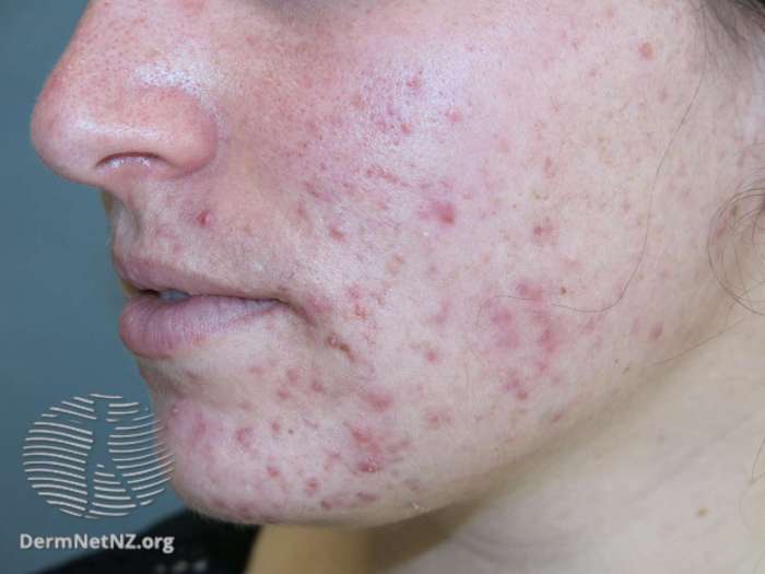
Author: Jonatan Bergnaum

  • Skin Boils Picture Gallery Visual Guide

    Skin Boils Picture Gallery Visual Guide

    Skin boils picture gallery sets the stage for a comprehensive visual guide to understanding these common skin conditions. This resource will provide clear images and descriptions of skin boils at various stages, helping you identify them accurately. We’ll explore different types, potential causes, and even treatment options. Learn how to distinguish skin boils from similar conditions and what preventative measures you can take to minimize their occurrence.

    The gallery presents a collection of images illustrating various appearances of skin boils, from small, red bumps to more advanced, pus-filled lesions. Each image is accompanied by detailed descriptions, helping you differentiate between different stages of boil development and identify potential causes. This visual approach will enhance your understanding and enable you to recognize these conditions promptly.

    Introduction to Skin Boils

    Skin boils, also known as furuncles, are common skin infections that typically present as painful, pus-filled bumps. They are caused by bacterial infections, most frequently Staphylococcus aureus, and often develop in areas with hair follicles. Understanding the causes, symptoms, and types of skin boils is crucial for early recognition and appropriate treatment. Prompt attention to boils can prevent complications and ensure a smoother recovery.Skin boils arise when bacteria, primarily Staphylococcus aureus, infect hair follicles or oil glands in the skin.

    Factors contributing to boil formation include poor hygiene, friction or irritation, cuts or abrasions, and weakened immune systems. The bacteria multiply rapidly, causing inflammation and the formation of a pus-filled lesion. Maintaining good hygiene practices, avoiding excessive friction, and treating any cuts or abrasions promptly can significantly reduce the risk of developing skin boils.

    Common Causes of Skin Boils

    Several factors can contribute to the development of skin boils. Poor hygiene, especially in areas with excessive moisture or friction, can create an environment conducive to bacterial growth. Friction or irritation from clothing, sports equipment, or other sources can damage the skin, leading to an opening for bacterial invasion. Cuts or abrasions, even small ones, provide entry points for bacteria to penetrate the skin.

    Weakened immune systems can make individuals more susceptible to skin infections, including boils.

    Typical Symptoms of Skin Boils

    The characteristic symptoms of a skin boil include a localized, painful, red swelling. The area surrounding the boil may be warm to the touch, and the affected area may be tender to the pressure. A boil usually starts as a small, tender bump that gradually enlarges and becomes increasingly painful. A white or yellow pus-filled head may develop, indicating the presence of infection.

    In some cases, fever and malaise may accompany the boil.

    Types of Skin Boils

    While the term “skin boil” often refers to a furuncle, there are other related skin infections. These infections can present with similar symptoms but may differ in appearance and location.

    Classification of Skin Boils

    Boil Type Typical Appearance Common Location
    Furuncle (Skin Boil) A localized, red, painful swelling containing pus. Face, neck, back, buttocks, legs.
    Carbuncle A cluster of interconnected boils, often larger and deeper than a furuncle. Multiple boils in close proximity, with significant inflammation. Neck, back, buttocks.
    Hidradenitis Suppurativa Multiple, painful, pus-filled boils, often occurring in areas with apocrine sweat glands. Deep inflammation, with chronic recurring nature. Armpits, groin, buttocks.

    A carbuncle, for example, is a cluster of interconnected boils, characterized by significant inflammation and pain. Hidradenitis suppurativa is a chronic condition marked by recurring boils in areas with apocrine sweat glands, often requiring prolonged treatment.

    Visual Identification of Skin Boils

    Skin boils picture gallery

    Identifying skin boils accurately is crucial for proper treatment and preventing complications. Knowing the different stages of boil development and the various appearances they can take can significantly aid in self-diagnosis and prompt medical intervention if needed. This section delves into the visual characteristics of boils, helping you recognize potential problems early on.Visual identification is a critical step in managing skin boils effectively.

    By understanding the typical progression of a boil and the variations in its appearance, you can assess the severity and guide your approach to care.

    Different Stages of Boil Development

    Understanding the stages of a boil’s development is essential for appropriate management. This progression, from initial inflammation to eventual healing, reveals critical information about the boil’s nature and the potential need for professional medical attention.

    Stage Description Image Description
    Early Stage (Inflammation) The skin around the affected area appears red, swollen, and tender. A small, hard bump may be noticeable, often resembling a pimple. Imagine a small, raised, red area of skin, slightly swollen and noticeably warmer than the surrounding skin.
    Maturation Stage (Pus Formation) The boil continues to enlarge and become more inflamed. A white or yellowish head, containing pus, begins to form in the center. The area around the boil remains painful and swollen. Visualize a larger, more prominent red bump, with a noticeable, small, white or yellowish center. The area around the bump will be significantly warmer and tender to the touch.
    Maturation/Suppuration Stage The boil continues to enlarge and the white or yellowish head, filled with pus, becomes more prominent. The surrounding skin remains inflamed and painful. Envision a noticeable, larger, and even more prominent red bump. The white or yellowish center containing pus is clearly visible and more significant than in previous stages. The surrounding skin shows increased redness and swelling.
    Resolution Stage (Healing) The boil’s head may rupture or be drained. The area will gradually decrease in size and inflammation. The surrounding skin begins to heal. Imagine a boil with a diminished size and reduced redness. The central area may show signs of drainage, and the surrounding inflammation is subsiding. The skin around the boil is beginning to return to normal.

    Visual Characteristics of Skin Boils

    Boils can vary in appearance, size, and location. Recognizing these variations helps in determining the severity and appropriate course of action.

    • Small, Red, and Tender: These boils are often in the early stages. The affected area appears as a small, raised red spot that feels warm and tender to the touch. This stage typically precedes pus formation.
    • Pus-Filled: As the boil matures, a white or yellowish, pus-filled head often develops. This is a clear indication that the boil has progressed beyond the initial inflammatory stage.
    • Inflamed: The surrounding skin around the boil will often show signs of inflammation. This includes redness, swelling, and tenderness. The inflammation typically increases as the boil develops.

    Distinguishing Characteristics of Different Types of Skin Boils

    Different types of boils may exhibit slightly different characteristics, though these are usually subtle differences. The distinguishing features can help to determine the type of boil and guide appropriate treatment.

    Image Description Description Potential Cause Treatment
    A small, superficial boil, often resembling a pimple, with minimal surrounding redness. Superficial boil, often appearing similar to a pimple, with limited surrounding redness. Bacterial infection, often caused by Staphylococcus aureus. Gentle cleaning and warm compresses. If it doesn’t improve or shows signs of spreading, consult a doctor.
    A larger, deeper boil with a well-defined, pus-filled head. Deep boil, featuring a noticeable pus-filled head. Bacterial infection, likely caused by Staphylococcus aureus. Possible need for incision and drainage, depending on size and location. Follow your doctor’s recommendations.

    Comparison with Similar Skin Conditions

    Skin boils picture gallery

    Skin boils, while often easily identifiable, can sometimes be confused with other skin infections. Understanding the key differences in appearance and symptoms is crucial for accurate diagnosis and appropriate treatment. Differentiating boils from similar conditions like carbuncles and folliculitis is vital to prevent misdiagnosis and ensure effective care.Differentiating skin conditions requires careful observation of both the physical presentation and accompanying symptoms.

    This comparison will highlight the distinguishing characteristics of boils, carbuncles, and folliculitis, providing practical methods for accurate identification.

    Comparison of Skin Boils with Other Conditions

    Accurate diagnosis relies on recognizing the unique characteristics of each condition. Boils, carbuncles, and folliculitis, while sharing some superficial similarities, present distinct differences in appearance and progression. Understanding these differences is crucial for appropriate medical intervention.

    Characteristic Skin Boil Carbuncle Folliculitis
    Appearance A localized, pus-filled bump, typically round or oval, ranging from a few millimeters to 2-3 centimeters in diameter. Often red and inflamed. Larger, deeper, and more extensive infection involving multiple hair follicles. Characterized by a cluster of boils that may merge, creating a larger, inflamed area. Small, red, inflamed bumps or papules that appear around hair follicles. May be accompanied by small pustules (pus-filled blisters). Often appears as a rash.
    Size Generally smaller, typically under 2-3 cm in diameter. Larger, potentially several centimeters in diameter. Very small, typically less than 1 cm in diameter.
    Symptoms Localized pain, tenderness, swelling, redness, and sometimes warmth. Pus may be visible if the boil is opened. Severe pain, swelling, redness, and warmth extending over a larger area. Multiple boils may be present, and the affected area might be hard and painful. Mild to moderate itching, burning, or discomfort. Possible redness and small, inflamed bumps around hair follicles.
    Location Can occur anywhere on the skin, but common locations include the face, neck, back, and buttocks. Commonly found on the back of the neck, the back, or the buttocks. Can occur anywhere on the body where hair follicles are present. Areas of friction are often affected.
    Duration Typically resolves within 1-3 weeks with proper care. Usually takes longer to heal than a boil, potentially needing several weeks or more. May resolve in a few days to several weeks depending on the severity.

    Differentiating Methods

    Accurate identification of skin conditions requires a multi-faceted approach. Beyond visual inspection, consulting a healthcare professional for diagnosis and treatment is paramount. Physical examination, patient history, and potentially laboratory tests (e.g., culture) are often necessary to differentiate between these conditions.Careful observation of the lesion’s size, location, appearance, and duration, coupled with the patient’s reported symptoms, is essential in the initial assessment.

    Ever wondered what skin boils look like? A quick search for “skin boils picture gallery” can be surprisingly helpful in identifying these pesky little infections. But, have you considered that sometimes body aches might be linked to allergies? For example, can allergies cause body aches ? While a picture gallery can help you visualize potential boils, remember to always consult a doctor for proper diagnosis and treatment.

    So, if you’re looking for a visual guide, a “skin boils picture gallery” is a good starting point.

    The presence of multiple boils or a larger, more extensive area of inflammation strongly suggests a carbuncle.

    Ever wondered what skin boils look like? A quick search for “skin boils picture gallery” can be helpful for visual identification. However, if you’re experiencing persistent muscle and joint pain, it could be related to an underlying condition like thyroid disease. Learning more about the connection between these symptoms and thyroid issues can be really beneficial, and resources like this one on muscle and joint pain with thyroid disease can provide valuable insights.

    Ultimately, a picture gallery can help you identify the potential cause of skin boils, but a doctor’s visit is always best for a proper diagnosis.

    Treatment and Management

    Dealing with a skin boil can be frustrating, but understanding the appropriate treatment and management strategies is key to healing effectively and preventing complications. Prompt and targeted intervention can minimize discomfort and promote faster recovery. This section will Artikel common treatment options, home remedies, and when professional medical attention is crucial.

    Ever wondered what skin boils look like? A quick search for “skin boils picture gallery” can be a good starting point, but it’s important to remember that visual resources are only part of the story. Understanding the underlying causes and potential connections to cellular aging, like in the cellular aging cancer study , could offer a more complete picture.

    Ultimately, a picture gallery can help identify potential issues, but it’s not a substitute for a professional diagnosis.

    Common Treatment Options

    Various treatment methods are available for managing skin boils, ranging from simple home remedies to more involved professional interventions. Choosing the right approach depends on the severity and location of the boil, as well as individual factors. Effective treatment often involves a combination of approaches.

    • Hot Compresses: Applying warm, moist compresses to the affected area can help soften the boil and promote drainage. This facilitates the natural process of healing. The warmth encourages blood flow to the area, aiding in the body’s ability to fight infection.
    • Antibiotics: In cases of infected boils, antibiotics may be prescribed to combat the bacteria causing the infection. Oral antibiotics are often effective in treating the underlying infection and preventing further spread. The type and dosage of antibiotics depend on the specific bacteria and the severity of the infection.
    • Incision and Drainage: A healthcare professional may need to surgically incise and drain the boil, especially if it is large, deep, or painful. This procedure helps to release pus and other accumulated material, alleviating pressure and promoting healing. Proper aseptic techniques are essential to prevent further infection.

    Home Remedies for Managing Skin Boils

    While home remedies can sometimes alleviate symptoms and support the healing process, they should not replace professional medical advice, especially for more severe or persistent boils. Consult a doctor before relying solely on home remedies.

    • Gentle Cleansing: Keeping the affected area clean is crucial to prevent further infection. Gently wash the area with mild soap and water to remove any debris or bacteria. Avoid scrubbing vigorously, as this could irritate the skin and potentially worsen the condition.
    • Avoid Squeezing or Popping: Resist the urge to squeeze or pop the boil. This can spread the infection to surrounding tissues and increase the risk of scarring. If the boil is causing significant pain or discomfort, consult a healthcare professional.
    • Over-the-Counter Pain Relief: Over-the-counter pain relievers like ibuprofen or acetaminophen can help manage pain and discomfort associated with the boil. Follow the recommended dosage instructions carefully.

    When Professional Medical Attention is Necessary

    Certain situations require immediate medical attention for skin boils. Ignoring these signs can lead to serious complications.

    • Signs of Severe Infection: Symptoms such as increasing redness, swelling, intense pain, fever, or pus drainage that is foul-smelling should prompt immediate medical attention. These symptoms may indicate a spreading infection.
    • Boils that Don’t Respond to Home Care: If a boil does not improve or shows signs of worsening after a few days of home care, a doctor’s evaluation is crucial. This could indicate a more serious underlying issue requiring professional intervention.
    • Boils in Sensitive Areas: Boils located in areas like the face, groin, or joints require careful monitoring and professional management to prevent complications and scarring.

    Treatment Options Table

    Treatment Option Description
    Hot Compress Applying warm, moist compresses to the affected area to soften the boil and promote drainage.
    Antibiotics Oral antibiotics prescribed to combat bacteria causing the infection.
    Incision and Drainage Surgical procedure to release pus and other accumulated material, alleviating pressure and promoting healing.

    Prevention and Care

    Skin boils, while often treatable, can be frustrating and uncomfortable. Preventing their recurrence involves a multifaceted approach that combines good hygiene, mindful lifestyle choices, and proper wound care. Understanding these factors can significantly reduce the risk of developing boils and help ensure a speedy recovery.Maintaining a proactive approach to hygiene and overall well-being is crucial in preventing boils.

    This includes understanding the various contributing factors, from minor irritations to underlying health conditions, and the role they play in boil development. A preventive strategy empowers individuals to take control of their skin health and reduce the chance of future outbreaks.

    Preventive Measures to Reduce Boil Risk

    Preventing boils requires a comprehensive approach encompassing both lifestyle choices and diligent hygiene practices. A strong immune system, proper hydration, and a balanced diet are essential for overall well-being, reducing the body’s susceptibility to infections that can lead to boils.

    • Maintaining Good Hygiene: Regular cleansing of the skin, particularly in areas prone to friction or moisture buildup, is essential. This includes showering or bathing daily, and thoroughly cleaning any cuts, scrapes, or abrasions. Use mild, fragrance-free soaps and avoid harsh scrubbing. Frequent handwashing is vital, especially after touching potentially contaminated surfaces.
    • Avoiding Tight-Fitting Clothing: Restricting blood flow and causing friction can increase the risk of skin irritation. Wearing loose-fitting, breathable clothing, particularly in hot and humid environments, is recommended.
    • Treating Minor Skin Irritations Promptly: Any minor cuts, scrapes, or abrasions should be cleaned thoroughly and covered with a sterile bandage to prevent infection. Early intervention minimizes the risk of a minor irritation developing into a more serious boil.

    Proper Hygiene Practices for Minimizing Boil Development

    A consistent routine of proper hygiene practices plays a vital role in reducing the risk of boil formation. This includes daily skin cleansing, avoiding tight clothing, and promptly treating any minor skin irritations.

    • Regular Skin Cleansing: Daily showering or bathing, using gentle cleansers, and avoiding harsh scrubbing is important. This helps remove dirt, debris, and bacteria that can contribute to boil development.
    • Avoiding Tight-Fitting Clothing: Clothing that restricts blood flow, particularly in areas prone to sweating or friction, can increase the risk of skin irritation and subsequent boil formation. Opt for loose-fitting, breathable fabrics.
    • Managing Moisture: Maintaining good skin hygiene, especially in areas prone to moisture buildup, can prevent the growth of bacteria and the subsequent formation of boils.

    Wound Care for Preventing Infection

    Proper wound care is crucial for preventing infection and minimizing scarring. Cleaning, covering, and monitoring the wound are essential steps in the process.

    • Cleaning the Wound: Use clean water and mild soap to gently cleanse the wound. Avoid harsh scrubbing, which can damage the skin.
    • Applying a Sterile Dressing: Cover the wound with a sterile bandage to protect it from further contamination and promote healing.
    • Monitoring for Signs of Infection: Watch for signs of infection, such as increased pain, swelling, redness, or pus. Seek medical attention if these symptoms develop.

    Diet and Lifestyle for Boil Prevention

    A balanced diet and healthy lifestyle can contribute significantly to overall well-being, strengthening the immune system and reducing the risk of boil formation.

    • Balanced Diet: A diet rich in fruits, vegetables, and whole grains provides essential nutrients to support a healthy immune system. Adequate protein intake is also important for tissue repair.
    • Regular Exercise: Physical activity boosts the immune system and promotes overall health.
    • Adequate Hydration: Drinking plenty of water helps maintain healthy skin and bodily functions.

    Proper Wound Care Techniques to Prevent Infection and Scarring

    Proper wound care minimizes the risk of infection and promotes optimal healing. Techniques include cleaning, dressing, and monitoring the wound.

    • Gentle Cleansing: Use mild soap and clean water to gently cleanse the wound. Avoid harsh scrubbing.
    • Appropriate Dressing: Use sterile dressings to cover the wound and protect it from contamination. Change dressings regularly.
    • Monitoring for Infection: Regularly monitor the wound for signs of infection. If any concerns arise, seek medical attention.

    Potential Complications

    Untreated skin boils can lead to a range of complications, from localized infections to systemic issues. Understanding these potential problems is crucial for timely intervention and preventing severe outcomes. Prompt medical attention is vital for managing complications effectively and preventing long-term health impacts.Complications from untreated skin boils can vary in severity, from localized discomfort to life-threatening conditions. The severity of these complications often depends on several factors, including the location of the boil, the individual’s overall health, and the presence of underlying conditions.

    It’s essential to recognize the warning signs of escalating issues and seek appropriate medical care promptly.

    Serious Infection, Skin boils picture gallery

    Untreated boils can spread infection beyond the initial site. Bacteria from the boil can enter the bloodstream, leading to a systemic infection known as bacteremia. This condition can cause fever, chills, and other serious symptoms, requiring prompt medical attention and potentially intravenous antibiotics. Examples of such infections include cellulitis, which spreads into the surrounding tissue, and even more severe cases like sepsis, a life-threatening response to infection.

    Abscess Formation

    A boil can develop into a deep abscess, a collection of pus that can cause significant pain and swelling. This can lead to skin damage and scarring if not treated properly. A spreading abscess can necessitate surgical drainage to prevent further tissue damage and systemic infection. This often involves lancing the boil to release the pus, followed by appropriate wound care.

    Scarring

    While boils typically heal without lasting scarring, deep or persistent boils can result in noticeable scars. The extent of scarring depends on the severity and duration of the boil. Scarring can be minimized by proper treatment and care, including keeping the affected area clean and protected. Early medical intervention is often crucial in reducing the risk of severe scarring.

    Lymphadenitis

    If the infection spreads to nearby lymph nodes, it can cause lymphadenitis, characterized by swollen and painful lymph nodes. This can be a sign of a more extensive infection, requiring prompt medical evaluation. The affected lymph nodes may become warm to the touch and tender to the press. Antibiotics and sometimes surgical drainage may be required.

    Risk Factors for Severe Complications

    Certain factors increase the risk of severe boil complications. These include a weakened immune system, underlying health conditions, such as diabetes or autoimmune diseases, and certain medications that can compromise the body’s immune response. Additionally, the location of the boil plays a role, with boils on the face or neck posing a higher risk of spreading infection. Individuals with these risk factors should be especially vigilant about monitoring their boil and seeking medical attention if symptoms worsen.

    Signs of Infection Worsening

    Several signs indicate a boil is becoming infected. These include increased pain, swelling, redness, warmth, and fever. The area surrounding the boil may also show signs of redness and inflammation. Pus or a foul odor from the boil are further indicators that the infection is worsening. Any increase in these symptoms should prompt a visit to a healthcare provider.

    Managing Complications

    Managing boil complications involves a combination of medical treatments and self-care. Early diagnosis and treatment are essential for preventing severe outcomes. Medical professionals may prescribe antibiotics to combat infection, surgical drainage to remove pus, and pain relievers to manage discomfort. Proper wound care and hygiene practices are crucial for healing and preventing further complications. Following prescribed medication and maintaining good hygiene will greatly assist in the healing process.

    Summary: Skin Boils Picture Gallery

    In conclusion, this skin boils picture gallery provides a valuable resource for recognizing and understanding skin boils. By examining the images and descriptions, you can develop a clearer picture of the various stages, types, and potential complications. This knowledge empowers you to seek appropriate medical care and take preventative steps to manage skin boils effectively. Remember, accurate identification is crucial for proper treatment and management.

    Early intervention often leads to better outcomes.

  • Ehlers-Danlos Syndrome Overview Understanding the Condition

    Ehlers-Danlos Syndrome Overview Understanding the Condition

    Ehlers danlos syndrome overview – Ehlers-Danlos syndrome overview explores this complex group of connective tissue disorders. These conditions, often inherited, affect the body’s structure and function, impacting everything from skin elasticity to joint stability. This overview delves into the different types of EDS, their unique symptoms, and the challenges faced by those living with the condition. We’ll also explore the current understanding of diagnosis, treatment, and ongoing research.

    The diversity of EDS types and symptoms is a key component. From hypermobile EDS to vascular EDS, each presents a unique set of challenges. Understanding the genetic basis and comparing symptoms across types provides crucial insight for individuals and healthcare professionals.

    Introduction to Ehlers-Danlos Syndrome

    Ehlers danlos syndrome overview

    Ehlers-Danlos syndrome (EDS) is a group of inherited connective tissue disorders characterized by hypermobility of joints, skin fragility, and often, a range of other symptoms. These disorders affect the body’s connective tissues, which provide support and structure to various parts of the body, impacting everything from skin elasticity to joint stability. Understanding the diverse types of EDS is crucial for proper diagnosis and management of the condition.The genetic basis of EDS lies in mutations within genes responsible for producing proteins crucial for connective tissue function.

    These proteins, like collagen and elastin, are vital components of tissues that provide strength, flexibility, and resilience. Variations in these genes can lead to a spectrum of symptoms, ranging from mild to severe, highlighting the complex interplay between genetics and phenotype.

    Types of Ehlers-Danlos Syndrome

    EDS encompasses several distinct types, each with its own set of characteristics and genetic underpinnings. Understanding these variations is vital for personalized treatment plans.

    Genetic Basis of EDS

    The genetic foundation of EDS involves mutations in various genes that encode proteins essential for connective tissue formation. These proteins play critical roles in maintaining the integrity and functionality of different tissues throughout the body. The specific genes implicated in each type of EDS can vary significantly.

    Ehlers-Danlos syndrome (EDS) is a fascinating genetic disorder affecting connective tissues. It can cause a wide range of symptoms, and one often overlooked aspect is how it affects skeletal development. While some people wonder when their feet stop growing, for those with EDS, the answer isn’t always straightforward, and it can impact growth patterns in unexpected ways. Knowing more about typical foot growth patterns can help you understand the intricacies of EDS, and how individual cases may vary.

    More information on when feet typically stop growing can be found here: when do feet stop growing. Ultimately, a deeper understanding of EDS requires a holistic approach, considering the range of potential impacts on physical development.

    Comparison of EDS Types

    Type Symptoms Genetic Basis Common Characteristics
    Classical EDS Hypermobile joints, skin hyperextensibility, easy bruising, and tissue fragility. Some individuals may also experience joint dislocations, scoliosis, and dental problems. Mutations in the COL5A1 and COL5A2 genes, which encode proteins crucial for collagen type V synthesis. Characterized by stretchy, fragile skin, easy bruising, and significant joint hypermobility. The severity of symptoms can vary greatly.
    Hypermobile EDS (hEDS) Joint hypermobility, often presenting with a range of symptoms, including chronic pain, fatigue, and autonomic dysfunction. Individuals with hEDS may experience frequent dislocations, headaches, and gastrointestinal issues. Mutations in genes responsible for collagen production or related connective tissue components are often not identified, indicating a complex genetic basis that is still being researched. This lack of a specific genetic marker underscores the difficulty in diagnosing this particular type. While joint hypermobility is a hallmark, the broader spectrum of symptoms, including chronic pain, fatigue, and autonomic dysfunction, distinguishes this type.
    Vascular EDS Fragile blood vessels, easy bruising, skin fragility, and potential for life-threatening complications like aneurysms and arterial tears. Individuals may experience unusual bleeding or bruising. Mutations in the COL3A1 gene, which is responsible for collagen type III production. Characterized by a heightened risk of blood vessel abnormalities, requiring careful monitoring and management to prevent life-threatening complications.
    Kyphoscoliosis type Kyphoscoliosis (abnormal curvature of the spine), joint hypermobility, and skin fragility. This type is often associated with other connective tissue disorders. Mutations in genes involved in collagen synthesis or related processes. A precise genetic basis may not be identified in all cases. Kyphoscoliosis is a defining feature, along with the common symptoms of joint hypermobility and skin fragility.

    Symptoms and Diagnosis

    Understanding Ehlers-Danlos Syndrome (EDS) involves recognizing the wide range of symptoms and the often-complex diagnostic process. This isn’t a one-size-fits-all condition; the presentation varies significantly between individuals and even within families. Accurate diagnosis hinges on careful consideration of symptoms, a thorough medical history, and often, a multidisciplinary approach.

    Common Symptoms

    EDS manifests in a variety of ways, affecting multiple body systems. Recognizing these diverse symptoms is crucial for early diagnosis and appropriate management. The symptoms can range from mild to severe, impacting daily life in varying degrees.

    • Musculoskeletal System: Joint hypermobility, characterized by excessive movement and range of motion in the joints, is a hallmark of many EDS types. This can lead to frequent dislocations, subluxations, and pain. Muscle weakness and fatigue are also common, sometimes contributing to difficulty with physical activities. Individuals may experience chronic pain, often described as aching or throbbing, and sometimes associated with trigger points or myofascial restrictions.

    • Skin: Skin fragility is another defining feature of EDS. The skin may be unusually stretchy, and easily bruised or damaged. This can range from a mild tendency towards bruising to significant skin fragility, making even minor trauma lead to significant injuries. Individuals may also experience skin laxity, with loose or redundant skin folds.
    • Gastrointestinal System: Digestive issues are frequently reported in individuals with EDS. These may include problems with motility (movement of food through the digestive tract), leading to bloating, constipation, or diarrhea. Chronic pain and inflammation within the gastrointestinal tract can also contribute to discomfort and digestive challenges.
    • Cardiovascular System: Some types of EDS can affect the cardiovascular system, leading to conditions like aortic aneurysms or other vascular issues. The severity of these complications varies widely. Individuals with EDS may require ongoing monitoring of cardiovascular health.
    • Neurological System: Neurological symptoms, such as headaches, dizziness, and balance problems, can be present in some individuals with EDS. These can stem from the interplay of joint hypermobility, pain, and potential autonomic nervous system dysfunction.

    Diagnostic Criteria and Procedures

    Diagnosing EDS requires a careful evaluation of the individual’s medical history, physical examination, and sometimes, genetic testing. There’s no single test for EDS, making a thorough assessment critical.

    • Medical History: A comprehensive medical history, including details about family history of connective tissue disorders, personal symptoms, and past injuries, is essential. The history should include details about the onset and progression of symptoms, including the severity and frequency.
    • Physical Examination: A physical examination focuses on assessing joint hypermobility, skin characteristics, and other relevant features. The examination may include range-of-motion assessments and evaluation of skin elasticity and fragility.
    • Genetic Testing: In some cases, genetic testing can confirm a diagnosis of a specific type of EDS. This is especially useful when the clinical presentation is suggestive of a particular subtype or when family history strongly suggests a genetic link. Genetic testing is not always necessary and may not always be conclusive. The information from genetic testing should be interpreted within the context of the individual’s medical history and physical examination findings.

    Importance of a Multidisciplinary Approach

    Given the multifaceted nature of EDS, a multidisciplinary approach is often necessary for optimal diagnosis and management. This involves collaboration among various healthcare professionals.

    • Collaboration with specialists: Collaboration between rheumatologists, geneticists, pain management specialists, physical therapists, and other healthcare professionals can provide a more comprehensive understanding of the individual’s needs and provide the most effective treatment plan. The expertise of multiple specialists is crucial in navigating the complexities of EDS.

    Symptom Severity Table

    Symptom Body System Description Severity Level
    Joint Hypermobility Musculoskeletal Excessive range of motion in joints Mild, Moderate, Severe
    Skin Bruising Skin Easy bruising, tendency to bleed easily Mild, Moderate, Severe
    Gastrointestinal Issues Gastrointestinal Constipation, diarrhea, bloating Mild, Moderate, Severe
    Fatigue Musculoskeletal Chronic tiredness, lack of energy Mild, Moderate, Severe

    Misdiagnosis Scenarios and Early Diagnosis

    Misdiagnosis is unfortunately a concern in EDS cases, often due to the overlap of symptoms with other conditions. Early diagnosis is crucial for timely interventions and improved quality of life.

    • Common Misdiagnoses: Conditions such as fibromyalgia, chronic fatigue syndrome, or other musculoskeletal disorders can be misdiagnosed in individuals with EDS. The presence of overlapping symptoms can make accurate diagnosis challenging.
    • Importance of Early Diagnosis: Early diagnosis allows for prompt implementation of appropriate management strategies, including physical therapy, pain management, and potentially, genetic counseling. This leads to better outcomes in terms of long-term health and quality of life.

    Impact on Daily Life: Ehlers Danlos Syndrome Overview

    Living with Ehlers-Danlos Syndrome (EDS) significantly impacts daily life, requiring adjustments and accommodations across various aspects. The unpredictable nature of symptoms, from joint pain and hypermobility to fatigue and digestive issues, can create substantial challenges in managing everyday tasks and maintaining a fulfilling lifestyle. This section explores the multifaceted impact of EDS on daily activities, personal relationships, and overall well-being.

    Challenges in Daily Activities

    The chronic pain, fatigue, and physical limitations associated with EDS can make seemingly simple tasks feel overwhelming. Individuals may experience difficulty with basic movements like walking, standing, or even dressing themselves. This often leads to a reduction in physical activity, impacting overall health and potentially contributing to further complications. Maintaining personal hygiene, preparing meals, and performing household chores can also be demanding and require significant planning and support.

    The unpredictable nature of symptoms necessitates flexibility and adaptability in daily routines, and often necessitates frequent breaks and rest periods.

    Educational and Employment Challenges

    EDS can present significant obstacles in both education and employment settings. Students may experience difficulties attending classes regularly, concentrating in lectures, and completing assignments due to pain, fatigue, and the need for frequent medical appointments. Similarly, individuals in the workforce may face challenges maintaining consistent employment due to unpredictable absences, reduced productivity, and the need for flexible work arrangements.

    Finding accommodations that balance the demands of work or study with the needs of EDS is crucial for maintaining a sense of normalcy and independence. Examples of accommodations include flexible work hours, modified work tasks, and accessible work environments.

    Social Interactions and Relationships

    Maintaining social connections and relationships can be challenging for individuals with EDS. Fatigue, pain, and the need for frequent rest can limit social activities and make it difficult to participate in social gatherings or maintain close friendships. Understanding and acceptance from friends and family are crucial for managing the emotional and social aspects of living with EDS. Building a support system of understanding individuals and creating a safe and supportive social environment is vital.

    Psychological and Emotional Impact

    Living with a chronic illness like EDS can have a profound psychological and emotional toll. The unpredictability of symptoms, the limitations on physical activity, and the challenges in managing daily tasks can lead to feelings of frustration, anxiety, and depression. Experiencing pain, fatigue, and reduced physical ability can impact self-esteem and body image. The emotional burden of managing the condition can create stress and strain on personal relationships.

    Seeking professional support from therapists or counselors can provide valuable coping mechanisms and strategies to manage emotional well-being.

    Coping Strategies and Support Systems

    Developing effective coping strategies is essential for managing the challenges of EDS. These strategies may include time management techniques, stress reduction methods, and strategies for pain management. Creating a supportive network of family, friends, and healthcare professionals is crucial for navigating the complexities of the condition. Utilizing support groups and online communities provides valuable avenues for connection and shared experiences.

    Seeking professional guidance from therapists and counselors can help individuals develop coping mechanisms and strategies for managing emotional well-being.

    Examples of Accommodations and Support Systems

    Numerous accommodations and support systems are available to individuals with EDS. These may include assistive devices, modified work environments, and flexible educational plans. Support groups, online forums, and advocacy organizations can provide valuable resources and a sense of community. Financial assistance programs and disability benefits can provide financial support to individuals and families managing EDS. Understanding and implementing these supports can significantly improve the quality of life for those living with EDS.

    Treatment and Management

    Ehlers danlos syndrome overview

    Managing Ehlers-Danlos Syndrome (EDS) is a multifaceted process focusing on symptom relief and improving quality of life. A personalized approach is crucial, as EDS presents differently in each individual. Treatment often involves a combination of therapies, medications, and lifestyle adjustments. Holistic care acknowledges the interconnectedness of physical, emotional, and social well-being, creating a comprehensive strategy for managing the challenges associated with EDS.Effective management of EDS requires a strong partnership between the patient, healthcare providers, and support networks.

    Understanding the unique needs of each individual with EDS and tailoring interventions to address those needs is paramount. A proactive and ongoing approach to treatment and management is vital to maximizing functional capacity and overall well-being.

    Current Treatment Options

    Current EDS treatments focus on managing symptoms rather than curing the underlying condition. This includes a wide range of strategies, such as physical therapy, occupational therapy, and medication. Regular medical checkups and monitoring are essential for early detection and management of potential complications.

    Physical Therapy

    Physical therapy plays a vital role in managing EDS. It helps improve joint stability, muscle strength, and flexibility, thereby reducing pain and improving mobility. Specific exercises and techniques, tailored to the individual’s needs, can enhance functional capacity and prevent further joint damage. A physical therapist can develop a personalized exercise program that addresses specific needs, such as strengthening weakened muscles or improving range of motion.

    Occupational Therapy, Ehlers danlos syndrome overview

    Occupational therapy focuses on adapting daily activities and environments to accommodate the limitations imposed by EDS. This may involve modifications to workspaces, home environments, or daily routines. Occupational therapists can also provide strategies for managing pain and fatigue, and teach coping mechanisms for everyday tasks. This can include assistive devices, ergonomic adjustments, and adaptive techniques for various activities.

    Medications and Supplements

    Several medications and supplements may help manage specific EDS symptoms. Pain relievers, such as nonsteroidal anti-inflammatory drugs (NSAIDs) and certain types of analgesics, can provide temporary relief from pain. Medications for anxiety or depression may be helpful if these conditions are present. While supplements may offer potential benefits, consulting with a doctor before taking any supplements is essential to avoid interactions with other medications or potential adverse effects.

    Importance of Regular Medical Checkups and Monitoring

    Regular medical checkups and monitoring are critical for individuals with EDS. This allows for early detection and management of potential complications, such as joint dislocations, cardiovascular issues, or digestive problems. Early intervention can significantly improve outcomes and prevent more severe conditions. Monitoring progress and adjusting treatment plans as needed is vital for managing the fluctuating nature of EDS symptoms.

    Step-by-Step Guide for Managing Pain and Discomfort

    Managing pain and discomfort related to EDS requires a structured approach. This involves recognizing triggers, developing coping mechanisms, and seeking support.

    Understanding Ehlers-Danlos Syndrome (EDS) involves recognizing the diverse range of symptoms, from hypermobile joints to stretchy skin. Proper crutch positioning and sizing is crucial for managing EDS-related mobility limitations. Learning about the correct techniques for crutch usage can significantly improve daily function and prevent further injury. For detailed guidance on crutch positioning and sizing, check out this helpful resource: crutch positioning and sizing.

    Ultimately, a holistic approach to managing EDS requires considering various support strategies, like correctly fitting crutches, for optimal well-being.

    • Identify Pain Triggers: Keep a pain journal to note activities, foods, environmental factors, or other potential triggers that exacerbate pain. This helps to identify patterns and develop strategies to avoid or mitigate these triggers.
    • Develop Coping Mechanisms: Learn relaxation techniques such as deep breathing exercises, mindfulness, or progressive muscle relaxation. These can help manage pain and stress. Consider incorporating stress-reducing activities, such as yoga, meditation, or spending time in nature. This is vital in controlling pain.
    • Seek Support: Connect with support groups or online communities for individuals with EDS. Sharing experiences and coping strategies with others facing similar challenges can provide invaluable emotional support. Support networks are important for both emotional and practical support.
    • Communicate with Healthcare Providers: Regularly communicate with healthcare providers about pain levels, medication effectiveness, and any changes in symptoms. This ensures that the treatment plan remains effective and addresses evolving needs.

    Research and Future Directions

    Unraveling the complexities of Ehlers-Danlos Syndrome (EDS) requires ongoing research to understand its diverse presentations, develop effective treatments, and ultimately improve the lives of those affected. Current research efforts are multifaceted, exploring genetic factors, physiological mechanisms, and potential therapeutic avenues. This exploration into the future of EDS management promises innovative solutions and a better understanding of this challenging condition.The quest for breakthroughs in EDS treatment is fueled by the dedication of researchers, clinicians, and patient advocates.

    Ehlers-Danlos syndrome (EDS) is a fascinating but complex group of inherited connective tissue disorders. It often impacts multiple body systems, which can make managing symptoms challenging. Learning about new treatments like new high blood pressure drug tryvio for other conditions, like hypertension, is crucial for understanding how different medical advancements might affect overall well-being, even when addressing a seemingly unrelated health issue.

    Ultimately, a holistic approach to managing EDS, incorporating the latest research and treatment options, is key.

    A deep dive into the existing research landscape and potential future directions is crucial to inform and guide the pursuit of effective therapies. This knowledge can also facilitate personalized approaches to managing the symptoms and improving the overall quality of life for individuals with EDS.

    Current Research Efforts

    Research into EDS encompasses a broad range of approaches, including genetic studies to identify specific gene mutations associated with different EDS subtypes, and physiological investigations into the underlying mechanisms of connective tissue dysfunction. These efforts aim to enhance our understanding of the specific pathways affected in EDS and how these dysfunctions manifest as diverse symptoms. Further studies are examining the impact of environmental factors and lifestyle choices on symptom presentation and disease progression.

    Potential New Treatments and Therapies

    Several promising avenues for new treatments and therapies are being explored. These include targeted therapies that aim to address specific genetic defects or physiological imbalances, potentially ameliorating symptoms and improving overall health. For example, research is focusing on potential therapies that could mitigate joint pain, promote tissue repair, or improve autonomic nervous system regulation. Personalized medicine approaches tailored to individual genetic profiles and symptom presentations hold significant promise.

    Clinicians are also exploring the use of non-pharmacological therapies like physical therapy and occupational therapy, tailored to each patient’s specific needs.

    Areas Where More Research Is Needed

    Despite significant progress, critical areas require further investigation. Developing standardized diagnostic criteria for various EDS subtypes is still a priority. Improving diagnostic tools, including genetic testing and non-invasive imaging techniques, is essential for timely and accurate diagnoses. Understanding the long-term effects of EDS on different organ systems is another crucial area. This includes better understanding of cardiovascular and neurological implications.

    Additionally, research into the impact of psychological factors on the experience of EDS is needed, enabling comprehensive management strategies.

    Importance of Patient Advocacy and Participation in Research

    Patient advocacy plays a critical role in advancing research efforts and ensuring that research aligns with the needs of the EDS community. Direct input from individuals with EDS is essential in shaping research priorities, methodologies, and outcome measures. Participation in clinical trials and research studies is vital to gathering data, understanding disease progression, and evaluating the efficacy of new treatments.

    This active engagement of the patient community is critical to accelerate the translation of research findings into tangible improvements in care and outcomes.

    Potential Breakthroughs in Understanding and Treating EDS

    Future breakthroughs in understanding and treating EDS could include the development of disease-modifying therapies. These therapies, if successful, could potentially slow or halt the progression of the disease, minimizing the long-term impact on affected individuals. The development of new diagnostic tools could lead to earlier and more accurate diagnoses, enabling timely interventions and better management strategies. The combination of these advancements, supported by continued research and patient engagement, holds the potential to reshape the future of EDS care and create a more hopeful outlook for individuals living with this complex condition.

    Living with Ehlers-Danlos Syndrome

    Navigating life with Ehlers-Danlos Syndrome (EDS) is a unique journey, filled with both challenges and triumphs. Living with EDS requires a multifaceted approach to managing symptoms, maintaining a positive outlook, and fostering a strong support system. Understanding the complexities of the condition and developing effective coping strategies are key to achieving a high quality of life.Individuals with EDS face a range of physical and emotional challenges.

    These can include chronic pain, fatigue, joint instability, and limited mobility. Furthermore, the unpredictable nature of EDS symptoms can lead to anxiety and frustration. However, with the right resources and support, individuals with EDS can lead fulfilling lives.

    Building a Supportive Network

    A strong support network is crucial for individuals living with EDS. It provides emotional comfort, practical assistance, and shared experiences that can significantly impact well-being. The journey with EDS is often better navigated when surrounded by understanding individuals who can provide encouragement and practical support.

    Role of Family, Friends, and Support Groups

    Family and friends play a vital role in supporting individuals with EDS. Their understanding, patience, and willingness to learn about the condition are essential. Support groups offer a unique platform for connecting with others who understand the daily struggles and triumphs of living with EDS. Sharing experiences, finding solutions, and receiving encouragement from peers who understand the unique challenges is a vital part of the process.

    This can be particularly helpful in addressing the emotional impact of the condition.

    Communicating with Healthcare Professionals

    Effective communication with healthcare professionals is paramount. Individuals with EDS should clearly articulate their symptoms, concerns, and treatment goals. Maintaining detailed records of symptoms, treatments, and responses is beneficial. This detailed information aids in diagnosis, treatment planning, and adjustments to therapies. Open communication facilitates a collaborative relationship between the patient and their healthcare team, ensuring optimal management of the condition.

    Clear and concise communication helps to avoid misunderstandings and ensure that healthcare professionals fully understand the individual’s needs.

    Self-Advocacy for Individuals with EDS

    Self-advocacy is essential for individuals with EDS. This involves actively participating in their healthcare decisions, understanding their condition, and expressing their needs and preferences. Learning about EDS and related therapies empowers individuals to make informed choices about their care. Educating others about EDS is a vital part of self-advocacy. It helps to foster understanding and create a more supportive environment.

    Maintaining a Positive Outlook and Quality of Life

    Despite the challenges, maintaining a positive outlook is essential for individuals with EDS. This can involve setting realistic goals, celebrating small victories, and focusing on personal strengths. Finding healthy coping mechanisms, such as engaging in hobbies, connecting with nature, or pursuing creative outlets, can be beneficial. Establishing routines that support physical and emotional well-being can significantly enhance quality of life.

    Prioritizing self-care, including adequate rest, nutrition, and social connections, can help to manage symptoms and maintain a positive perspective.

    Wrap-Up

    In conclusion, Ehlers-Danlos syndrome overview reveals a complex tapestry of challenges and adaptations. While diagnosis can be challenging, early intervention and supportive care are crucial for improving quality of life. The ongoing research into EDS offers hope for future advancements in treatment and understanding, making it vital to stay informed about this condition. Ultimately, fostering a supportive environment for individuals with EDS is paramount to their well-being.

  • How Myasthenia Gravis Is Diagnosed A Comprehensive Guide

    How Myasthenia Gravis Is Diagnosed A Comprehensive Guide

    How myasthenia gravis is diagnosed is a crucial process, often involving a multifaceted approach. This journey begins with recognizing the subtle and sometimes overlooked initial symptoms, progressing through neurological examinations, and culminating in a series of diagnostic tests. Understanding the various methods used in the diagnostic process can help patients and their families navigate this complex health challenge with greater awareness and confidence.

    This comprehensive guide explores the different steps involved in diagnosing myasthenia gravis, from initial symptom assessment to advanced laboratory tests and imaging techniques. We’ll delve into the nuances of muscle weakness, neurological evaluations, and the crucial role of diagnostic tests such as the Edrophonium test and repetitive nerve stimulation. A detailed understanding of these processes can help those affected better navigate the diagnostic pathway.

    Initial Symptoms and Presenting Complaints

    How myasthenia gravis is diagnosed

    Myasthenia gravis (MG) is a chronic autoimmune neuromuscular disease characterized by fluctuating muscle weakness. Understanding the initial symptoms and presenting complaints is crucial for early diagnosis and effective management. Recognizing the diverse ways MG manifests is essential to avoid delayed diagnosis, which can lead to a poorer prognosis. The symptoms vary widely, depending on the specific muscles affected.

    Common Initial Symptoms

    Initial symptoms of myasthenia gravis often begin subtly, with gradual worsening of muscle weakness over time. The hallmark feature is fluctuating muscle weakness, meaning the strength of the affected muscles can change throughout the day, and sometimes even within hours. This fluctuation can make it challenging for patients to accurately describe their symptoms. Common initial complaints often involve muscles used for repetitive or sustained activity.

    Presenting Complaints by Muscle Group

    The presenting complaints in myasthenia gravis can vary greatly depending on the muscle groups affected. Patients might experience difficulty with various tasks due to the varying weakness.

    • Ocular Symptoms: Double vision (diplopia) is a frequent and often the first symptom. It typically affects one or both eyes, often worse at the end of the day or after prolonged use. Ptosis (drooping eyelids) is another common ocular symptom, often occurring in conjunction with diplopia.
    • Facial Symptoms: Difficulty smiling, chewing, or making facial expressions is common. Weakness of facial muscles can lead to a characteristic “mask-like” appearance. Difficulty with swallowing (dysphagia) can also present as an early symptom.
    • Bulbar Symptoms: Difficulty speaking, swallowing, or chewing (bulbar weakness) can indicate involvement of the cranial nerves controlling these functions. These symptoms often manifest as slurred speech, difficulty with mastication, and progressive dysphagia.
    • Limb Symptoms: Weakness in the arms and legs, often affecting both sides equally, can occur. This weakness can present as difficulty with tasks like lifting objects, climbing stairs, or walking.

    Typical Onset and Progression of Different MG Subtypes

    The onset and progression of myasthenia gravis can vary significantly, even within the same patient. This variability necessitates a comprehensive understanding of the disease.

    Subtype Typical Onset Progression Notes
    Ocular MG Often insidious, starting with ptosis or diplopia Slowly progressive, may not affect other muscle groups Frequently the first subtype to appear
    Generalized MG Can start in any muscle group Rapid progression affecting multiple muscle groups Symptoms often more severe and widespread
    Bulbar MG Often starts with dysphagia, dysarthria, or both Can lead to significant swallowing and speaking difficulties May progress to respiratory compromise

    Triggers and Exacerbating Factors

    Several factors can exacerbate symptoms of myasthenia gravis. These factors are important to consider when assessing a patient’s condition.

    • Fatigue: Physical or mental exertion can significantly worsen muscle weakness. This is often a major factor for patients, leading to reduced functional capacity.
    • Stress: Emotional stress and anxiety can trigger or worsen symptoms. This can be an important consideration for managing symptoms.
    • Infection: Infections, especially viral infections, can temporarily worsen the symptoms of myasthenia gravis.
    • Certain Medications: Some medications, including antibiotics, can temporarily exacerbate symptoms.

    Indicators of Myasthenia Gravis in General Weakness

    General weakness can be a symptom of many conditions, but several indicators can suggest myasthenia gravis.

    • Fluctuating Weakness: Weakness that comes and goes, improving with rest and worsening with activity, is a strong indicator. This is a key differentiating feature from other causes of weakness.
    • Presence of Ocular Symptoms: The presence of ptosis or diplopia, even in the absence of other symptoms, should raise suspicion for myasthenia gravis.
    • Family History: A family history of autoimmune disorders may increase the likelihood of myasthenia gravis.
    • Exacerbation with Repetitive Activity: If the weakness worsens with repeated muscle use, it might suggest a neuromuscular junction problem like myasthenia gravis.

    Neurological Examination Procedures

    Unraveling the complexities of Myasthenia Gravis often hinges on meticulous neurological examinations. These procedures provide crucial insights into the patient’s muscle strength, function, and the specific involvement of the neuromuscular junction. By identifying characteristic patterns of weakness and evaluating cranial nerve function, clinicians can narrow down the diagnosis and tailor treatment strategies.

    Muscle Strength and Function Assessment

    Neurological examinations for myasthenia gravis (MG) focus on assessing muscle strength and fatiguability. Clinicians utilize various techniques to evaluate the extent and pattern of weakness. These techniques are designed to identify the characteristic fatigability patterns, a key diagnostic feature of MG. Precise documentation of these findings is vital for monitoring disease progression and treatment efficacy.

    Neuromuscular Junction Evaluation

    Specific tests are employed to evaluate the neuromuscular junction (NMJ), the site where nerves communicate with muscles. These tests aim to assess the efficiency of nerve impulse transmission. The rationale behind each procedure is to pinpoint any disruptions in the NMJ, which may be indicative of MG.

    Key Neurological Findings in Myasthenia Gravis

    Neurological Finding Description Clinical Significance Example
    Muscle Weakness Varying degrees of muscle weakness, particularly in muscles controlling eye movements, facial expression, swallowing, and limb movements. Weakness often worsens with repetitive use or prolonged activity. A hallmark symptom of MG, indicating impaired neuromuscular transmission. Difficulty raising eyelids, drooping eyelids (ptosis), double vision (diplopia), facial weakness, difficulty chewing, or swallowing.
    Cranial Nerve Dysfunction Impairment in cranial nerves, most commonly affecting those controlling eye movement (III, IV, VI), facial expression (VII), and swallowing (IX, X). Suggests the presence of neuromuscular dysfunction affecting specific nerve pathways. Ocular palsies, dysarthria, dysphagia, and facial weakness.
    Fatiguability Progressive weakening of muscles with sustained or repetitive use. A crucial diagnostic sign in MG, reflecting the impaired ability of the NMJ to maintain the necessary neurotransmitter release. Weakness worsens after sustained activity or repetition, like sustained eye movements.
    Response to Ice Packs Temporary improvement in muscle strength after applying ice packs to affected muscles. Can indicate the presence of a myasthenic crisis. Temporary improvement in eye movement after ice application.

    Detecting Characteristic Patterns of Muscle Weakness

    Identifying the characteristic patterns of muscle weakness in MG involves careful observation and testing. Clinicians evaluate the extent and location of weakness, noting whether it affects specific muscle groups or cranial nerves. They observe how weakness changes with rest and activity, specifically if the weakness increases with repeated use or activity. Furthermore, the presence of fatiguability during specific tasks like sustained eye movements is crucial for diagnosis.

    Importance of Cranial Nerve Function Evaluation

    Evaluating cranial nerve function is essential in a patient suspected of having myasthenia gravis. Cranial nerves innervate vital muscles involved in vision, facial expression, chewing, swallowing, and speech. Any impairment in these functions can significantly impact a patient’s quality of life. The presence of specific cranial nerve palsies, particularly those affecting ocular muscles, strongly suggests the possibility of MG.

    Thorough testing and documentation of cranial nerve function provide vital clues for diagnosis and management.

    Diagnostic Tests

    Navigating the diagnostic journey for Myasthenia Gravis (MG) often involves a combination of clinical assessments and specific tests. A thorough understanding of these tests is crucial for accurate diagnosis and effective management of the condition. The goal is to pinpoint the underlying neuromuscular dysfunction that characterizes MG, enabling the right treatment plan to be implemented.

    Diagnosing myasthenia gravis often involves a combination of physical exams, neurological assessments, and blood tests. Doctors look for specific symptoms, like muscle weakness that worships throughout the day, and often, try to rule out other conditions. Switching to healthier alternatives to full-fat dairy products, like healthy alternatives to full fat dairy products , can be a beneficial part of a comprehensive treatment plan, though it’s crucial to remember that this is a complex disease and dietary changes alone won’t diagnose or cure it.

    Ultimately, a team of specialists and thorough testing are key to accurate diagnosis.

    Edrophonium (Tensilon) Test, How myasthenia gravis is diagnosed

    This test is a crucial diagnostic tool in evaluating suspected myasthenia gravis. It involves administering edrophonium, a cholinesterase inhibitor, intravenously. The rationale behind this test is that cholinesterase inhibitors increase the amount of acetylcholine available at the neuromuscular junction. This, in turn, can temporarily improve muscle strength in individuals with MG.

    The mechanism of action is straightforward: edrophonium inhibits acetylcholinesterase, the enzyme that breaks down acetylcholine. This leads to an increased concentration of acetylcholine in the synaptic cleft, stimulating muscle contraction more effectively.

    During the test, muscle strength is meticulously observed before and after the injection. A positive response, characterized by a marked improvement in muscle strength within minutes of the injection, strongly suggests MG. Conversely, a lack of improvement or a minimal response typically indicates that MG is not the primary cause of the symptoms. Important considerations include potential side effects such as bradycardia, nausea, or muscle fasciculations.

    The test should only be performed under strict medical supervision.

    Diagnosing myasthenia gravis often involves a combination of physical exams, neurological tests, and sometimes even blood tests. It’s a complex process, much like figuring out how to manage a stuffy nose at night, which can often be a real struggle. Fortunately, there are some simple strategies to try, like using a saline nasal spray, which can help clear up congestion and make breathing easier.

    This process of elimination and targeted approaches, similar to figuring out the right combination of strategies for a stuffy nose, is important in diagnosing myasthenia gravis accurately. Ultimately, a doctor will need to consider various factors to make a precise diagnosis. how to reduce a stuffy nose at night can help find relief.

    Repetitive Nerve Stimulation (RNS) Testing

    RNS testing assesses the neuromuscular junction’s response to repeated nerve stimulation. The rationale is that in MG, repeated nerve stimulation leads to a quicker decline in muscle strength compared to healthy individuals. This decline is a reflection of the fatigued neuromuscular junction’s reduced ability to produce acetylcholine effectively.

    The procedure involves electrically stimulating a nerve repeatedly, measuring the strength of the resulting muscle contractions. The test identifies the pattern of muscle weakness as a result of repeated stimulation. A progressive decrease in the amplitude of muscle action potentials is a significant finding suggestive of MG.

    Electromyography (EMG)

    EMG is a diagnostic procedure that evaluates the electrical activity of muscles. It plays a crucial role in assessing neuromuscular transmission, the process by which nerve impulses trigger muscle contractions. The rationale for using EMG is that it can reveal subtle abnormalities in the electrical signals produced by muscles and nerves, helping to distinguish between different neuromuscular disorders.

    Diagnosing myasthenia gravis often involves a combination of physical exams, neurological tests, and sometimes even blood tests. It’s a tricky diagnosis, and sometimes, symptoms can overlap with other conditions like, for example, what is tmj disorder , making accurate identification crucial. Ultimately, a thorough evaluation by a neurologist is key to pinpointing the cause and receiving the right treatment for myasthenia gravis.

    During EMG, electrodes are inserted into specific muscles to record their electrical activity. Normal muscle activity is characterized by specific patterns of electrical signals. EMG can reveal characteristic abnormalities in MG, such as reduced amplitude of muscle action potentials and prolonged relaxation times. It’s important to note that EMG findings alone are not always definitive for MG and may require correlation with other clinical and test results.

    Comparison of Diagnostic Tests

    Test Strengths Limitations Typical Findings in MG
    Edrophonium (Tensilon) Test Quick, relatively simple, and can provide immediate diagnostic clues. Can cause adverse reactions, may not be conclusive in all cases, and is not suitable for everyone. Marked improvement in muscle strength after edrophonium administration.
    Repetitive Nerve Stimulation (RNS) Objective measure of neuromuscular junction fatigue, highly sensitive to early signs of MG. Requires specialized equipment and interpretation by trained personnel. Progressive decrease in muscle action potential amplitude with repeated stimulation.
    Electromyography (EMG) Provides detailed information about muscle and nerve function, helpful in ruling out other conditions. Can be invasive, requires specialized interpretation, and may not always directly pinpoint MG. Reduced amplitude of muscle action potentials and prolonged relaxation times.

    Laboratory Tests: How Myasthenia Gravis Is Diagnosed

    Beyond the crucial neurological examination, laboratory tests play a vital role in confirming and characterizing myasthenia gravis (MG). These tests help identify specific immune system responses and rule out other conditions with similar symptoms. They provide critical supporting evidence to aid in the diagnosis and management of MG.Laboratory tests are invaluable tools in the diagnostic process for myasthenia gravis, supplementing clinical findings and neurological examination results.

    These tests help differentiate MG from other neuromuscular disorders and provide insights into the underlying immune mechanisms.

    Serum Acetylcholine Receptor (AChR) Antibody Testing

    A crucial laboratory test in MG diagnosis is the serum AChR antibody test. AChR antibodies are autoantibodies that target the acetylcholine receptors (AChRs) at the neuromuscular junction. The presence of these antibodies strongly suggests MG.The procedure involves drawing a blood sample and analyzing it for the presence and level of AChR antibodies. Various methods exist for detecting these antibodies, including enzyme-linked immunosorbent assays (ELISAs).

    The interpretation of the results is critical. A positive result, indicating the presence of AChR antibodies, significantly strengthens the suspicion of MG, especially when coupled with clinical symptoms and a neurological examination. The level of antibodies can also provide information about the severity of the disease.

    Muscle-Specific Kinase (MuSK) Antibody Testing

    MuSK antibodies are another type of autoantibody implicated in MG. These antibodies target the muscle-specific kinase (MuSK) protein, also crucial at the neuromuscular junction. MuSK antibodies are present in a subset of MG patients, often those who do not have detectable AChR antibodies. This finding highlights the complexity of MG and the diverse mechanisms driving the disease.The procedure for MuSK antibody testing is similar to AChR antibody testing, involving blood draws and specialized laboratory analysis.

    Interpretation of MuSK antibody test results is essential. A positive result, indicating the presence of MuSK antibodies, suggests a form of MG, particularly when clinical symptoms and neurological examination support the diagnosis.

    Other Relevant Laboratory Tests

    Thyroid function tests are routinely included in the laboratory workup for MG, even though they are not directly related to the disease. This is because thyroid disorders can sometimes mimic MG symptoms. Other laboratory tests, such as complete blood counts (CBCs), electrolytes, and creatine kinase (CK) levels, might be ordered to rule out other conditions or assess overall health.

    These tests are vital for a comprehensive evaluation.

    Summary of Laboratory Test Results in Myasthenia Gravis

    Test Typical Finding in MG Deviation from Normal Clinical Significance
    Serum AChR Antibodies Positive Elevated levels Strong indication of MG; level may correlate with disease severity
    Serum MuSK Antibodies Positive (in some cases) Elevated levels Suggests a subtype of MG; often seen in patients negative for AChR antibodies
    Thyroid Function Tests Normal Abnormal thyroid hormone levels (hypothyroidism or hyperthyroidism) May mimic MG symptoms; necessitates further investigation
    Complete Blood Count (CBC) Normal Anemia, lymphopenia, or other abnormalities Could suggest other conditions or complications
    Electrolytes Normal Imbalances May indicate underlying issues or complications
    Creatine Kinase (CK) Normal Elevated levels Suggests muscle damage; may indicate a complication or other underlying conditions

    Imaging Techniques

    How myasthenia gravis is diagnosed

    Imaging techniques play a limited role in the diagnosis of myasthenia gravis (MG). While they can be helpful in excluding other conditions with similar symptoms, they don’t typically provide definitive evidence of MG. Their utility is most often in ruling out structural abnormalities in the neuromuscular junction or other neurological conditions that might mimic MG.Imaging studies are typically not the first-line diagnostic tools for MG.

    A comprehensive neurological examination, along with a detailed medical history and laboratory tests, are usually more critical in reaching a diagnosis. Imaging is often employed only when other tests are inconclusive or when there’s a strong suspicion of an underlying structural problem that might be contributing to the symptoms.

    Role of Imaging in MG Diagnosis

    Imaging plays a supportive role in the diagnostic process. It can help rule out alternative diagnoses or reveal structural abnormalities that might be affecting neuromuscular transmission, potentially mimicking the symptoms of MG. However, imaging alone is insufficient to establish a definitive diagnosis of MG.

    Specific Imaging Modalities

    Several imaging techniques may be employed in the evaluation of patients suspected of having MG. The choice depends on the specific clinical questions and the suspected location of any potential abnormality.

    Neuroimaging

    Neuroimaging techniques, including magnetic resonance imaging (MRI) and computed tomography (CT), are commonly used to evaluate the brain, spinal cord, and other neural structures. These studies can reveal the presence of masses, lesions, or other abnormalities that might be affecting the nervous system. While these techniques are valuable for ruling out other neurological conditions, they rarely show specific findings directly related to MG.

    Muscle Imaging

    Muscle imaging, using techniques like magnetic resonance imaging (MRI), can be utilized in certain situations. This is typically done when there’s a strong suspicion of muscle involvement that isn’t easily explained by other tests. Muscle MRI can potentially show evidence of muscle atrophy or other structural changes. However, these findings are often not pathognomonic for MG.

    Table of Imaging Modalities in MG

    Imaging Modality Usefulness in Identifying Associated Abnormalities Relevant Image Findings (Possible but not Specific) Limitations
    MRI of Brain and Spinal Cord Useful for ruling out other neurological conditions (e.g., tumors, multiple sclerosis). May show lesions or masses in the brain or spinal cord. Does not directly visualize neuromuscular junction.
    MRI of Muscles May reveal muscle atrophy or structural changes in cases of suspected myopathic involvement. Potential for showing atrophy or inflammation of specific muscle groups. Less sensitive for detecting the subtle neuromuscular changes of MG.
    CT Scan Less frequently used than MRI for evaluating the nervous system. May be helpful for detecting certain types of masses or bony abnormalities. May show bony abnormalities or lesions. Lower resolution compared to MRI for soft tissue structures.

    Differential Diagnosis

    Myasthenia gravis (MG) can sometimes masquerade as other neurological disorders, making accurate diagnosis crucial. A thorough differential diagnosis is essential to distinguish MG from conditions with overlapping symptoms, ensuring the correct treatment plan is implemented. This process involves ruling out various possibilities that might cause similar symptoms, such as muscle weakness or fatigue. Failure to identify and exclude these conditions can lead to inappropriate therapies and potentially delay the appropriate treatment for MG.Careful consideration of the patient’s medical history, physical examination findings, and results from various diagnostic tests are vital to differentiate MG from other neuromuscular conditions.

    Precise identification of the underlying cause of the symptoms is essential for appropriate management and to avoid potential misdiagnosis.

    Conditions Mimicking Myasthenia Gravis

    Many neurological and other medical conditions can present with symptoms similar to those of myasthenia gravis, including muscle weakness and fatigue. A comprehensive differential diagnosis considers these possibilities to ensure an accurate assessment of the patient’s condition.

    • Other Neuromuscular Disorders: Conditions such as Lambert-Eaton myasthenic syndrome (LEMS), botulism, and other neuromuscular junction disorders share overlapping symptoms with MG. The distinct characteristics of each condition, such as the presence or absence of specific antibodies or responses to different medications, are crucial for differentiation. For instance, LEMS often presents with muscle weakness that improves with repeated use, while MG typically worsens with repeated use.

    • Autoimmune Diseases: Systemic lupus erythematosus (SLE), rheumatoid arthritis, and other autoimmune disorders can sometimes cause muscle weakness as a secondary effect. The presence of characteristic symptoms and associated laboratory findings helps in distinguishing these conditions from MG.
    • Infectious Diseases: Infections like Lyme disease, poliomyelitis, and others can manifest with muscle weakness. Careful evaluation of the patient’s medical history and laboratory tests, such as serological testing, aid in distinguishing these conditions from MG.
    • Neurodegenerative Diseases: Amyotrophic lateral sclerosis (ALS) and other neurodegenerative diseases can cause progressive muscle weakness. The progressive nature and specific neurological findings associated with these diseases help differentiate them from the fluctuating weakness often seen in MG.
    • Endocrine Disorders: Hypothyroidism, hyperthyroidism, and other endocrine disorders can cause muscle weakness. Appropriate laboratory tests for thyroid function and other relevant endocrine parameters can aid in distinguishing these conditions.
    • Medication Side Effects: Certain medications can cause muscle weakness as a side effect. Carefully reviewing the patient’s medication list and considering the timing of symptom onset in relation to medication use is essential.
    • Structural Neuromuscular Diseases: Conditions like muscular dystrophy, myopathies, and peripheral neuropathies can mimic MG. Detailed neurological examination and specific muscle biopsy results help differentiate these from MG.

    Distinguishing Features

    Accurate diagnosis relies on a thorough understanding of the key differentiating characteristics between MG and other neuromuscular conditions. Precise identification of the distinctive features of each condition helps clinicians arrive at the correct diagnosis.

    Characteristic Myasthenia Gravis Lambert-Eaton Myasthenic Syndrome (LEMS) Amyotrophic Lateral Sclerosis (ALS)
    Muscle Weakness Fluctuating weakness, typically worse with repeated use; improved with rest. Often affecting ocular, bulbar, and limb muscles. Weakness improves with repeated use; characteristically affects proximal muscles first. Progressive, symmetrical muscle weakness affecting both upper and lower extremities.
    Response to Edrophonium (Tensilon) Test Improved muscle strength following edrophonium administration. Muscle strength worsens following edrophonium administration. No significant change in muscle strength with edrophonium.
    EMG Findings May show decremental response to repetitive nerve stimulation. May show incremental response to repetitive nerve stimulation. May show denervation and fibrillation potentials.
    Autoantibodies Presence of acetylcholine receptor antibodies (AChR-Abs) or muscle-specific kinase antibodies (MuSK-Abs). Presence of presynaptic calcium channel antibodies. Absence of specific autoantibodies.

    Closing Summary

    In conclusion, diagnosing myasthenia gravis requires a careful evaluation of symptoms, neurological findings, and a battery of diagnostic tests. This multifaceted approach ensures a more accurate and timely diagnosis, paving the way for appropriate treatment and management strategies. The key is early detection and intervention, allowing individuals to live more fulfilling lives despite the challenges presented by this condition.

  • How Often Should I Wash My Face? Your Guide

    How Often Should I Wash My Face? Your Guide

    How often should I wash my face? This question plagues many, but the answer isn’t one-size-fits-all. Your skin type, daily activities, and even the environment play a crucial role in determining the ideal washing frequency. This guide delves into the factors affecting your face-washing routine, offering personalized insights for healthy skin.

    We’ll explore various skin types, activities, and cleansing methods to help you find the perfect balance for your unique needs. Understanding how to select the right products, establish a consistent routine, and recognize signs of over- or under-washing will empower you to achieve optimal skin health.

    Skin Type Considerations

    Washing your face is a crucial part of a healthy skincare routine. However, the frequency and method of cleansing should be tailored to your individual skin type and any specific skin conditions you might have. Understanding these factors will help you maintain a balanced and effective routine that promotes healthy skin.Skin type plays a significant role in determining how often you should wash your face.

    Oily skin, for example, requires more frequent cleansing than dry skin, as it tends to produce more sebum. Different skin types react differently to various cleansers, leading to the need for specialized products.

    Skin Type Cleansing Frequency

    Different skin types respond differently to cleansing frequency. Understanding these variations is key to a successful skincare routine.

    Skin Type Washing Frequency (General Recommendation) Reasoning Example Cleanser
    Oily 2-3 times daily Oily skin produces more sebum, leading to buildup. Frequent washing helps remove excess oil and prevent clogged pores. Gel cleansers, foaming cleansers, or oil-based cleansers for deep pore cleansing.
    Dry 1-2 times daily Dry skin is prone to dryness and irritation. Over-washing can strip away natural oils, leading to dryness and discomfort. A gentler approach is crucial. Cream cleansers, hydrating cleansers, or milk cleansers.
    Combination 1-2 times daily, focusing on oily areas Combination skin has oily and dry areas. Concentrate on cleansing the oily zones more frequently to remove excess oil without over-drying the dry areas. A combination of cleansers, such as a gel cleanser for oily areas and a cream cleanser for dry areas, or a cleanser formulated for combination skin.
    Sensitive 1 time daily, or as needed Sensitive skin is easily irritated. Excessive washing can trigger inflammation and redness. Gentle cleansing and minimal exposure to harsh ingredients are essential. Gentle cleansers, fragrance-free cleansers, or cleansers specifically formulated for sensitive skin.

    Individual Skin Conditions, How often should i wash my face

    Skin conditions like acne or eczema significantly impact the optimal washing frequency. These conditions require specific considerations.Acne-prone skin often benefits from gentle cleansing to avoid irritating existing breakouts. Using harsh cleansers can worsen inflammation. Eczema, on the other hand, requires a very gentle approach, as even mild irritation can exacerbate the condition.

    Skin Condition Washing Frequency Considerations Cleansing Method Recommendations
    Acne Focus on cleansing twice daily, focusing on affected areas. Use a gentle cleanser formulated for acne-prone skin. Consider incorporating spot treatments or topical medications as prescribed by a dermatologist.
    Eczema Wash only when necessary, using lukewarm water and a very gentle cleanser. Avoid harsh scrubbing or harsh cleansers. Consider using fragrance-free cleansers and gentle cleansers.

    Cleansing Methods and Frequency

    Different cleansing methods cater to varying needs. It’s crucial to understand how different cleansing methods impact your skin.

    • Gentle Cleansing: Suitable for dry or sensitive skin, this method involves using mild cleansers and avoiding harsh scrubbing. This should be used daily or as needed to maintain a clean and comfortable feeling, but not more than twice daily. This method minimizes skin irritation.
    • Deep Cleansing: Used to remove excess oil and impurities, this method typically involves more vigorous scrubbing and/or specialized cleansers. Deep cleansing is best performed 1-2 times a week to address buildup and maintain a clear complexion, but not more often, as it can potentially lead to irritation.

    It is important to avoid over-washing, as this can disrupt the skin’s natural barrier, leading to dryness, irritation, and increased sensitivity. Choosing the right cleansing method and frequency is crucial for healthy skin.

    Washing Frequency Based on Activities

    How often should i wash my face

    Knowing your skin type and its needs is crucial, but understanding how your daily activities impact your skin’s cleanliness is equally important. This section dives into how different activities influence the frequency of face washing, allowing you to tailor your routine for optimal skin health.Washing frequency isn’t a one-size-fits-all rule. Factors like the intensity and duration of an activity, the environment you’re in, and your personal skin sensitivities play a significant role in determining how often you need to cleanse.

    By understanding these factors, you can adjust your routine effectively, maintaining a healthy and radiant complexion.

    Washing Frequency for Different Activities

    Understanding how activities affect your skin’s condition helps in developing a personalized cleansing routine. The table below provides a comparative overview of washing frequency based on different activities.

    Activity Washing Frequency Recommendation Explanation
    Exercise Before and after Sweating and exposure to dirt and pollution during exercise can clog pores and lead to breakouts. Washing before removes excess oil and sweat, while washing after removes accumulated grime.
    Makeup Wear Twice daily Makeup can trap dirt and oil, leading to clogged pores and breakouts. Washing your face both in the morning and evening helps remove makeup and prevent these issues.
    Outdoor Activities (e.g., hiking, gardening) After activity Outdoor activities often expose the skin to dust, pollen, and other environmental elements. Washing after helps remove these particles and prevent irritation or breakouts.
    Exposure to pollution Increase washing frequency High pollution levels can lead to increased dirt and grime on the skin. Washing your face more often, especially after being in a polluted environment, helps remove these pollutants.
    Sun Exposure Wash immediately after sun exposure The sun’s UV rays can damage the skin. Washing immediately after sun exposure helps remove sweat and dirt that have accumulated during sun exposure, preventing skin irritation.

    Adjusting Washing Frequency for Environmental Factors

    Environmental factors, such as pollution and sun exposure, can significantly impact your skin’s condition and, consequently, the frequency of your face washing routine.Adjusting your washing routine to environmental factors is essential. Increased pollution, for example, necessitates a higher frequency of washing to effectively remove the pollutants and prevent skin irritation or breakouts. Similarly, after sun exposure, it is important to wash your face to remove sweat, dirt, and potentially harmful UV particles.

    Consider using gentle cleansers during these times to avoid further irritation.

    Importance of Washing Before and After Activities

    Washing your face before and after certain activities is crucial for maintaining healthy skin. The rationale behind this practice is that these activities often introduce elements that can lead to clogged pores, breakouts, and other skin issues.Washing your face before exercise helps remove excess oil and sweat that can clog pores and lead to breakouts. Washing your face after exercise ensures that the accumulated grime and sweat are removed, preventing skin irritation and minimizing the risk of acne.

    Similarly, washing your face before and after wearing makeup prevents the build-up of dirt, oil, and makeup residue that can clog pores. In addition, washing your face after engaging in outdoor activities helps remove the dirt and grime that can be picked up from the environment.

    Washing Methods and Techniques

    Choosing the right face washing method is crucial for a healthy and radiant complexion. Different techniques cater to various skin types and concerns, so understanding the pros and cons of each is essential. This section delves into various methods, emphasizing proper technique and the importance of using lukewarm water.Effective cleansing is a cornerstone of a healthy skincare routine.

    The method you choose should be gentle and effective, minimizing irritation and maximizing the benefits of your chosen products.

    Comparison of Face Washing Methods

    Understanding the nuances of different washing methods can help you tailor your routine to your specific skin type and needs. This table compares common methods, highlighting their advantages and drawbacks.

    Method Pros Cons
    Using a Washcloth Can help exfoliate gently. Can be abrasive if not used carefully, potentially leading to micro-tears and irritation. Risk of pulling on sensitive skin.
    Using Hands Generally gentler than a washcloth, suitable for most skin types. Can be less effective at removing makeup and impurities, potentially leaving residue. May not reach all areas of the face as thoroughly.
    Using a Facial Cleansing Brush Can effectively remove makeup and impurities, potentially stimulating blood flow. Can be harsh if not used properly, leading to irritation, redness, and micro-tears. Can exacerbate existing skin conditions like acne or rosacea.

    Proper Facial Cleansing Brush Use

    Facial cleansing brushes can be beneficial for deep cleansing, but their use requires a mindful approach to avoid potential damage.Using a cleansing brush correctly minimizes the risk of irritation. Begin by applying a small amount of cleanser to wet skin. Gently move the brush in small, circular motions, avoiding harsh scrubbing. Focus on problem areas, but avoid applying too much pressure.

    Always rinse thoroughly with lukewarm water.Different skin types react differently to cleansing brushes. For sensitive skin, use a soft-bristled brush and minimal pressure. For oily skin, a medium-bristled brush can be effective, but still prioritize gentle use. Dry skin types may benefit from a soft brush, used only on areas where necessary.

    Importance of Lukewarm Water

    The temperature of the water you use for face washing significantly impacts your skin’s health. Lukewarm water is ideal as it effectively cleanses without stripping the skin’s natural oils.Hot water can cause the skin’s protective barrier to become compromised, leading to dryness, irritation, and redness. Cold water, while not as damaging, may not effectively remove all impurities and makeup residue.

    Lukewarm water strikes a balance, gently cleansing without disrupting the skin’s delicate equilibrium.

    Product Selection and Usage

    Choosing the right cleanser is crucial for maintaining healthy skin. It’s not just about washing; it’s about selecting a product that workswith* your skin type and concerns, not against it. This involves understanding your skin’s unique needs and selecting a cleanser that effectively removes impurities without disrupting the skin’s natural barrier. A well-chosen cleanser can be a powerful ally in your skincare routine.

    Selecting Cleansers for Different Skin Types

    Selecting a cleanser tailored to your skin type is essential for optimal results. Different skin types require different formulations to maintain hydration and prevent irritation. Understanding your skin type will allow you to choose a cleanser that meets its specific needs.

    Skin Type Characteristics Suitable Cleanser Type Benefits
    Oily Skin Excess sebum production, often leading to shine and breakouts. Gel-based cleansers, foaming cleansers (with a low-sudsing formula), or water-based cleansers. Effective at removing excess oil and preventing clogged pores. Formulations that are non-comedogenic are particularly important for oily skin.
    Dry Skin Lack of moisture, often resulting in tightness and flakiness. Cream cleansers, oil-based cleansers, or gentle foaming cleansers. Hydrate and nourish the skin, leaving it feeling soft and comfortable.
    Combination Skin A mix of oily and dry areas. Gel-based cleansers or water-based cleansers that are formulated to target oily areas without over-drying. Provide a balanced approach, addressing both oily and dry areas.
    Sensitive Skin Easily irritated and prone to redness, itching, or burning. Cream cleansers, mild foaming cleansers, or fragrance-free cleansers. Gentle and non-irritating, minimizing the risk of skin reactions.

    Choosing the Right Cleanser Based on Skin Concerns

    Specific skin concerns, such as acne, eczema, or rosacea, necessitate the selection of cleansers designed to address these conditions. Knowing the specific ingredients and their potential effects will help you select the most suitable cleanser for your particular skin type and concerns.

    Figuring out how often to wash your face can be tricky, especially when stress factors like anxiety come into play. Stress can really affect your skin, and understanding the different types of anxiety disorders, like generalized anxiety disorder or panic disorder, types of anxiety disorders , can help you better manage your skin routine. Ultimately, the best approach to face washing depends on your skin type and individual needs, so don’t be afraid to experiment and find what works best for you.

    • Acne-prone skin: Look for cleansers specifically formulated for acne. These often contain ingredients like salicylic acid or benzoyl peroxide to help unclog pores and reduce inflammation. For example, a cleanser with salicylic acid can exfoliate the skin, removing dead skin cells that can contribute to breakouts.
    • Eczema-prone skin: Cleansers formulated for eczema often feature soothing ingredients like ceramides or oatmeal to help hydrate and calm irritated skin. Avoid harsh cleansers that may exacerbate dryness and inflammation.
    • Rosacea-prone skin: Choose cleansers that are fragrance-free and contain ingredients like aloe vera or chamomile to soothe the skin and reduce redness. Look for cleansers specifically designed for rosacea.

    Avoiding Harsh Chemicals and Ingredients

    Selecting a cleanser free of harsh chemicals and irritants is paramount for maintaining healthy skin. Harmful ingredients can cause long-term damage and lead to various skin issues. Being aware of potentially harmful ingredients will help you choose the right product.

    Potentially Harmful Ingredient Potential Effects
    Sulfates (SLS, SLES) Can strip the skin of its natural oils, leading to dryness, irritation, and redness.
    Fragrances Can trigger allergic reactions, causing skin irritation, rashes, and itching in sensitive individuals.
    Harsh Acids (e.g., high concentrations of AHAs/BHAs) Can cause excessive dryness, irritation, and potentially damage the skin barrier.
    Alcohol Can dehydrate the skin, making it prone to irritation and dryness.

    Choosing a gentle, suitable cleanser is key for a healthy skincare routine. Avoid harsh chemicals and ingredients that can irritate your skin.

    Figuring out how often to wash your face can be tricky, right? It’s a balancing act between keeping your skin clean and healthy, and potentially irritating it. Recent research on cellular aging and its connection to cancer, like this cellular aging cancer study , suggests that excessive cleansing might actually be counterproductive. So, while maintaining a consistent routine is key, finding the sweet spot for your skin type is crucial for optimal health.

    Less is definitely more when it comes to facial washing.

    Daily Routines and Schedules

    Creating a consistent skincare routine is key to healthy skin, but it’s not a one-size-fits-all approach. Your daily schedule, lifestyle, and even travel plans should all be factored into your face washing routine. Tailoring your routine to fit your unique needs will ensure you’re giving your skin the best possible care.

    Sample Daily Skincare Routines

    Below are examples of daily skincare routines tailored to different lifestyles. These routines emphasize the importance of consistency and adjust to various demands.

    Lifestyle Morning Routine Evening Routine
    Busy Professional
    • Quick cleanse with a foaming wash, focusing on the T-zone and problem areas. This routine should take approximately 2-3 minutes.
    • Apply a lightweight moisturizer suitable for everyday use.
    • Thorough cleanse with a gentle cleanser to remove makeup and impurities. This should take 4-5 minutes, ensuring thorough removal of makeup.
    • Apply a serum targeting specific skin concerns, such as hydration or anti-aging.
    • Apply a night cream to lock in moisture and support overnight skin repair.
    Student
    • Simple cleanse with a gentle foaming wash, focusing on the face, to remove overnight sebum and impurities. This routine should take approximately 1-2 minutes.
    • Apply a light moisturizer to maintain hydration.
    • Gentle cleanse with a mild cleanser, ensuring the removal of any dirt and grime accumulated throughout the day.
    • Apply a targeted treatment for acne or other skin issues, if necessary.
    • Moisturize to maintain hydration and lock in beneficial ingredients.
    Athlete
    • Cleanse with a water-based foaming wash to remove sweat and impurities. This routine should take 2-3 minutes, focusing on the areas most prone to sweat and dirt build-up.
    • Apply a lightweight moisturizer with SPF to protect the skin from sun exposure.
    • Thorough cleanse with a gentle cleanser to remove sweat and dirt from the day’s activities. This routine should take approximately 3-4 minutes, focusing on areas affected by sweat.
    • Apply a hydrating mask or serum to replenish lost moisture.
    • Moisturize to support skin repair and recovery after intense workouts.

    Importance of Consistency

    Maintaining a consistent face washing schedule is crucial for healthy skin. This consistency helps regulate skin’s natural oil production, leading to a more balanced complexion. Consistency also helps your skin get used to the products you’re using, making them more effective. A consistent routine, like any other healthy habit, promotes better skin health and a more balanced complexion over time.

    Washing your face too often can be detrimental to your skin’s natural oils. Finding the right frequency depends on your skin type and lifestyle, but generally, twice a day is sufficient. However, if you’re taking medications for atrial fibrillation, like those mentioned in this informative article about medications for atrial fibrillation , it’s crucial to discuss the best skin care routine with your doctor.

    They can advise on how to adjust your face-washing schedule to minimize any potential interactions between your medication and your skin. Ultimately, a balanced approach is key for healthy skin.

    Incorporating Face Washing into Different Schedules

    Adapting your face washing routine to accommodate busy schedules, travel, or other changes is essential. Consider packing travel-sized products for quick and easy use during trips. Prioritize using products and methods that minimize time commitment. Using a quick, gentle cleanser and a lightweight moisturizer can significantly shorten the routine.

    Adjusting Routines for Travel

    Travel requires adjustments to skincare routines. Pack travel-sized versions of your essential products and choose lightweight, easy-to-use cleansers and moisturizers. You may also consider a hydrating facial mist to help maintain moisture during flights or extended periods in air-conditioned environments. This simple adjustment can significantly enhance your skincare regimen while traveling.

    Signs of Overwashing and Underwashing

    Finding the perfect face-washing routine can feel like a delicate balancing act. Just as underwashing can lead to problems, overwashing can also wreak havoc on your skin. Understanding the subtle signs of both extremes is key to maintaining healthy, radiant skin. This section delves into recognizing the telltale signs of overwashing and underwashing, empowering you to adjust your routine and achieve optimal skin health.

    Signs of Overwashing

    Overwashing, unfortunately, is a common culprit behind skin issues. Regular, excessive cleansing can strip away the skin’s natural oils, leading to dryness, irritation, and discomfort. Recognizing these signs is the first step toward correcting your routine.

    • Dryness and Tightness: Skin that feels excessively dry and tight after cleansing, almost as if a film is on the surface, often indicates overwashing. This is a significant sign, as your skin’s natural moisturizing barrier is being compromised. Your skin might even feel rough or flaky.
    • Redness and Irritation: Persistent redness, inflammation, or a burning sensation after washing can be a clear sign of overwashing. This irritation is your skin’s way of signaling that it’s being stripped of its protective oils. The redness might appear patchy or concentrated in certain areas.
    • Sensitivity: Increased sensitivity to touch or even to normal environmental factors (like wind or cold) can point to overwashing. Your skin becomes more vulnerable to irritants, potentially leading to discomfort or discomfort-inducing reactions.

    Signs of Underwashing

    Conversely, underwashing can also contribute to skin problems. A lack of proper cleansing can lead to the buildup of dirt, oil, and dead skin cells, resulting in clogged pores and breakouts.

    • Breakouts and Acne: Recurring breakouts, especially in areas prone to acne, can be a strong indicator of insufficient cleansing. The buildup of impurities creates an environment conducive to bacterial growth, triggering blemishes and acne.
    • Oily Skin: While some people naturally have oily skin, if your skin appears excessively oily, even after cleansing, it might suggest insufficient washing. The excess oil is a result of accumulated impurities not being removed effectively.
    • Blackheads and Whiteheads: The presence of blackheads and whiteheads, particularly in areas like the nose and forehead, is often linked to underwashing. These are a clear indication of clogged pores, and proper cleansing is necessary to address them.

    Adjusting Washing Frequency

    Recognizing the signs of both overwashing and underwashing is crucial for maintaining a healthy skin routine. Once you’ve identified the problem, adjust your washing frequency and method accordingly.

    • Addressing Overwashing: If you notice signs of overwashing, reduce the frequency of washing. Switching to a gentler cleanser and using lukewarm water instead of hot water can also be beneficial. Consider a gentler cleanser with a moisturizing formula, and avoid harsh scrubbing.
    • Addressing Underwashing: If you’re experiencing breakouts or clogged pores, increase the frequency of washing, but focus on a gentle, effective approach. Use a cleanser suitable for your skin type, and pay attention to the product’s instructions.

    Long-Term Skin Health Considerations: How Often Should I Wash My Face

    Consistent face washing, when done correctly, isn’t just about removing dirt and oil; it plays a crucial role in maintaining long-term skin health. Proper cleansing helps prevent breakouts, premature aging, and other skin concerns. By understanding the relationship between your cleansing routine and your skin’s well-being, you can proactively safeguard your skin for years to come.Effective face washing creates a healthy skin barrier, minimizing the risk of irritation and damage.

    This healthy barrier acts as a protective shield against environmental aggressors and helps to maintain optimal moisture levels, crucial for preventing dryness and premature aging.

    Effects of Proper Washing on Skin’s Overall Well-being

    Proper face washing, when tailored to your skin type and needs, promotes a healthy skin environment. This leads to reduced inflammation, improved skin texture, and a more even complexion. A balanced cleansing routine fosters the natural skin barrier function, preventing excessive moisture loss and maintaining a healthy skin microbiome.

    Preventing Future Skin Problems

    Maintaining a consistent, appropriate face washing routine can effectively prevent future skin problems. By addressing the root causes of skin issues, like clogged pores and inflammation, you proactively avoid potential flare-ups and maintain a clear, healthy complexion. Examples include preventing acne breakouts by regularly removing excess oil and preventing premature aging by protecting the skin’s moisture barrier.

    Long-Term Benefits of Consistent Face Washing Habits

    Consistent and appropriate face washing provides several long-term benefits for skin health. These habits are foundational to a healthy complexion and help prevent a range of skin issues.

    • Reduced Risk of Acne: Regular cleansing removes excess oil and dirt, preventing clogged pores and the development of acne. This proactive approach helps prevent future breakouts and minimizes scarring.
    • Improved Skin Texture and Tone: Consistent face washing helps to remove dead skin cells, revealing a smoother, more radiant complexion. This gradual improvement contributes to a healthier and more even skin tone.
    • Enhanced Skin Barrier Function: Proper cleansing, aligned with your skin type, supports the skin’s natural barrier function. This healthy barrier protects against environmental irritants and moisture loss, promoting long-term skin health.
    • Minimized Signs of Aging: Maintaining a healthy skin barrier through consistent, appropriate cleansing can help delay the appearance of wrinkles and fine lines. This contributes to a more youthful and vibrant complexion.
    • Reduced Risk of Skin Infections: By removing dirt and bacteria, regular cleansing helps to prevent the development of skin infections. This preventive measure safeguards your skin from potential irritations and infections.

    Last Word

    How often should i wash my face

    In conclusion, washing your face is a personal journey that requires understanding your unique skin and lifestyle. Finding the right frequency, method, and products is key to maintaining healthy, glowing skin. Remember, consistency is key, and adjusting your routine based on your skin’s needs and external factors will yield the best results. This guide provides the tools you need to create a customized routine that works for you.

  • What is a Double Bypass Heart Surgery? A Comprehensive Guide

    What is a Double Bypass Heart Surgery? A Comprehensive Guide

    What is a double bypass heart surgery? This procedure, a significant step in cardiovascular care, aims to improve blood flow to the heart. It involves rerouting blood flow around blocked coronary arteries, often caused by atherosclerosis. Understanding the historical context, different types of bypasses, and the meticulous preparation and recovery process is crucial for patients and their families.

    This guide delves into the details, from the initial evaluation to long-term implications, ensuring a clear picture of this complex surgical intervention.

    The surgical process itself involves carefully assessing the patient’s condition and identifying the most appropriate approach for the double bypass. Different types of bypasses cater to various anatomical areas affected by blockages, and the selection process considers the specific needs of each patient. This detailed examination of the procedure also encompasses the critical pre-operative and post-operative phases, including patient counseling, recovery timelines, and potential complications.

    Furthermore, the potential alternatives and long-term outcomes are thoroughly explored, equipping the reader with a complete picture of this life-altering surgery.

    Introduction to Double Bypass Heart Surgery

    What is a double bypass heart surgery

    A double bypass heart surgery is a complex cardiovascular procedure where two blocked coronary arteries are treated by rerouting blood flow around the blockages. This is achieved by using healthy blood vessels from other parts of the body to create new pathways for blood to reach the heart muscle. Essentially, the surgeon creates detours for blood to bypass the obstructed areas, ensuring a continuous and adequate blood supply to the heart.

    The primary goal is to restore normal blood flow and prevent further damage to the heart.The fundamental purpose of a double bypass heart surgery is to address coronary artery disease (CAD). CAD occurs when plaque builds up inside the coronary arteries, the blood vessels that supply blood to the heart. This buildup narrows the arteries, reducing blood flow and potentially leading to a heart attack.

    A double bypass heart surgery reroutes blood flow around blocked arteries, improving heart function. While the focus is on heart health, it’s important to consider the impact of serious illnesses like ALS, Lou Gehrig’s disease, on life expectancy. Understanding life expectancy in cases like this can help patients and families make informed decisions about treatment options, such as a double bypass surgery, als lou gehrigs disease life expectancy.

    Ultimately, the surgery aims to enhance the quality of life for patients by improving their cardiovascular health.

    By creating new pathways for blood, the surgery aims to alleviate the blockage and improve blood flow to the heart muscle. This prevents further damage and improves the patient’s overall health.The overall goal of the surgery is to improve the patient’s quality of life and prevent serious complications from heart disease. This means a significant reduction in chest pain (angina), improvement in energy levels, and the ability to perform daily activities without discomfort.

    Ultimately, the surgery aims to extend the patient’s lifespan and prevent life-threatening events such as heart attacks and strokes.The historical context of this surgical technique is rooted in the evolution of cardiovascular surgery. Early attempts at coronary artery bypass grafting (CABG) emerged in the mid-20th century. Dr. Robert Goetz is often credited with pioneering this technique. Over time, surgical techniques and materials have significantly improved, leading to greater success rates and lower complication rates for patients.

    Improvements in anesthesia and post-operative care also played crucial roles in enhancing the safety and efficacy of the procedure.

    A double bypass heart surgery essentially reroutes blood flow around blocked arteries in the heart. It’s a significant procedure, often necessary for serious heart conditions. Interestingly, the age of onset for certain neurological conditions, like ALS, can sometimes be a factor in considering treatment options, such as als age of onset , alongside the patient’s overall health and the severity of the heart blockage.

    Ultimately, the decision for a double bypass is a complex one, involving careful evaluation by medical professionals.

    Surgical Procedure Details

    A detailed understanding of the procedure itself helps to understand the procedure better. This section will explore the steps involved in a double bypass heart surgery. It will also provide insight into the various considerations for choosing this type of procedure.

    Procedure Name Description Benefits Risks
    Double Coronary Artery Bypass Graft (CABG) The surgeon uses healthy blood vessels (typically from the leg or chest) to create new pathways for blood to flow around blocked sections of the coronary arteries. Improved blood flow to the heart muscle, reduced chest pain, and reduced risk of heart attack or stroke. Bleeding, infection, stroke, blood clots, and damage to surrounding tissues.

    Types of Double Bypass Procedures

    Double bypass heart surgery, while fundamentally aimed at improving blood flow to the heart, can be tailored to specific patient needs and anatomical variations. Different approaches exist, each with unique considerations for the surgeon and the patient. Understanding these variations is crucial for both informed decision-making and successful outcomes.

    Different Types of Double Bypass Procedures, What is a double bypass heart surgery

    Various approaches exist for double bypass procedures, each targeting specific coronary arteries. These differences in targeting specific arteries directly impact the surgical complexity, recovery time, and potential complications. The surgeon’s expertise and assessment of the patient’s particular artery blockage patterns are crucial in selecting the optimal technique.

    • Left Anterior Descending (LAD) and Right Coronary Artery (RCA) Bypass: This is a common and often preferred approach. The LAD artery, responsible for supplying blood to the front and left side of the heart, and the RCA, supplying blood to the right side, are the primary targets. This approach directly addresses two major blood flow limitations, typically improving overall heart function significantly. The surgeon aims to restore blood flow by creating new pathways to these blocked arteries.

    • Left Main Coronary Artery (LMCA) and Left Circumflex Artery (LCX) Bypass: This approach is used when the left main coronary artery (LMCA) or the LCX, responsible for blood supply to the left side of the heart, is severely obstructed. This more complex technique may involve greater surgical time and potential risks, but it is often essential for ensuring adequate blood flow when major blockages exist in these critical vessels.

      It typically involves grafting a healthy blood vessel to the LMCA and/or the LCX, bypassing the blocked area.

    • Multiple Vessel Bypass (Involving LAD, RCA, and LCX): This approach is a more comprehensive procedure that addresses blockages in multiple coronary arteries simultaneously. This might involve grafting to the LAD, RCA, and LCX, or a combination of these, potentially increasing the complexity of the surgery but often offering a more extensive and long-term solution for improved blood flow. The selection of these additional bypasses is predicated on the extent of blockage in the affected vessels.

      This approach is most often indicated for patients with extensive coronary artery disease.

    Selection Criteria for Double Bypass Procedures

    The choice of the specific double bypass procedure is not arbitrary. Several factors influence the surgeon’s decision, ultimately aiming for the best possible outcome for the patient. The extent of the blockage, the location of the blockages, and the overall health of the patient are all carefully considered.

    • Extent of Blockage: The severity and location of blockages in the coronary arteries play a crucial role. For example, a total occlusion (complete blockage) of a major artery necessitates a bypass, whereas a partial blockage may be managed with other interventions. The surgeon must carefully assess the extent of blockage to determine the appropriate type of bypass and the number of vessels that need to be addressed.

    • Patient’s Overall Health: Factors such as age, other medical conditions, and overall health status are considered. For instance, a patient with severe lung or kidney issues might require a more cautious approach. This holistic assessment helps the surgeon tailor the procedure to the patient’s specific needs and risks.
    • Location of Blockage: The specific location of the blockage in the coronary arteries directly impacts the surgical approach. The complexity of accessing certain arteries may influence the choice of technique and potential complications. Surgeons must be mindful of anatomical variations and factors that may influence access to specific areas.

    Comparison of Double Bypass Techniques

    The different double bypass procedures vary in their complexity and the specific anatomical areas targeted. Careful consideration of these differences is vital for determining the best possible surgical approach for each individual patient.

    Procedure Type Targeted Arteries Complexity Selection Criteria
    LAD and RCA Bypass LAD and RCA Moderate Partial or significant blockages in LAD and RCA
    LMCA and LCX Bypass LMCA and LCX High Severe blockages in LMCA or LCX
    Multiple Vessel Bypass LAD, RCA, LCX, or others High Extensive blockages in multiple coronary arteries

    Pre-Operative Considerations and Preparation

    Before undergoing a double bypass heart surgery, a meticulous pre-operative evaluation and preparation process is critical. This process ensures the patient is as healthy as possible to withstand the procedure and optimize the chances of a successful outcome. Careful consideration of potential risks and thorough patient counseling are integral parts of this preparation.

    Pre-Operative Evaluation Process

    The pre-operative evaluation process involves a comprehensive assessment of the patient’s overall health. This assessment aims to identify any underlying conditions that might complicate the surgery or increase the risk of complications. A thorough medical history, physical examination, and various diagnostic tests are used to determine the patient’s suitability for the procedure and to tailor the surgical approach.

    Tests and Assessments Performed Before Surgery

    Numerous tests and assessments are performed to evaluate the patient’s cardiovascular health, overall physical condition, and potential risks. These include but are not limited to: blood tests (complete blood count, coagulation studies, and electrolytes), chest X-rays, electrocardiograms (ECGs), echocardiograms, stress tests, and potentially cardiac catheterization. These tests provide critical data about the heart’s function, the extent of coronary artery disease, and the patient’s overall health.

    Potential Risks to Consider Before the Procedure

    While double bypass surgery is a life-saving procedure, it carries inherent risks. Potential risks include but are not limited to: bleeding, infection, stroke, blood clots, heart attack, kidney failure, and respiratory complications. A thorough discussion of these potential risks with the patient and their family is crucial for informed decision-making. The surgical team will thoroughly discuss these risks and their likelihood, along with the benefits of the surgery.

    Patient Preparation Process

    The patient preparation process extends beyond the immediate pre-operative period. It involves a series of instructions and recommendations to prepare the body for the surgery. This includes medication adjustments, dietary restrictions, and avoidance of smoking and alcohol. The specific preparation steps are tailored to each patient’s individual needs and health status.

    Patient Counseling

    Patient counseling is a cornerstone of pre-operative preparation. It involves educating the patient about the procedure, potential risks and benefits, expected recovery process, and post-operative care. The surgical team will provide clear, concise, and understandable information to ensure the patient is fully informed and prepared for the surgical experience. Open communication and addressing any concerns are essential elements of effective patient counseling.

    Summary of Pre-Operative Preparation Steps

    Step Timeline Required Documentation
    Detailed Medical History Weeks before surgery Patient’s medical records, previous test results
    Physical Examination Weeks before surgery Physician’s notes, vital signs
    Diagnostic Tests Weeks before surgery Lab reports, ECG results, echocardiogram reports, stress test results
    Medication Review and Adjustments Weeks before surgery Medication list, physician’s recommendations
    Dietary Restrictions Days before surgery Physician’s instructions on diet
    Smoking Cessation (if applicable) Weeks before surgery Documentation of smoking cessation plan
    Alcohol Restriction (if applicable) Days before surgery Physician’s instructions on alcohol intake
    Patient Counseling Days before surgery Signed consent form, documented discussion of risks and benefits
    Pre-operative Medications Day before surgery Administered pre-operative medications

    Surgical Procedure Details: What Is A Double Bypass Heart Surgery

    The surgical procedure for a double bypass involves a series of precise steps to redirect blood flow around blocked coronary arteries. This intricate process requires meticulous attention to detail and advanced surgical techniques. Understanding the steps, instruments, and anatomical landmarks involved provides a deeper insight into the complexity of this life-saving procedure.

    Surgical Steps Overview

    A double bypass operation involves grafting healthy blood vessels to reroute blood flow around blocked coronary arteries. This bypass allows improved blood supply to the heart muscle, alleviating the effects of ischemia. The steps are carefully orchestrated to minimize trauma and maximize effectiveness.

    • Incision and Exposure: A small incision is made in the chest, typically on the left side, allowing access to the heart and major vessels. The surgeon carefully dissects the chest wall and surrounding tissues to expose the heart and the targeted coronary arteries.
    • Identification of Blocked Arteries: The surgeon meticulously identifies the blocked coronary arteries that require bypass. Advanced imaging techniques may be used to confirm the location and extent of the blockages.
    • Graft Selection and Preparation: A healthy blood vessel, either from the patient’s leg (saphenous vein) or from a section of the chest wall (internal mammary artery), is selected as a graft. The chosen vessel is carefully prepared and measured to ensure a proper fit with the target coronary arteries.
    • Graft Anastomosis: The prepared graft is connected to the healthy section of the blocked artery using sutures or surgical staples. This connection, or anastomosis, ensures that blood can flow from the graft into the coronary artery, bypassing the blockage.
    • Closure and Drainage: Once all bypasses are completed, the surgeon carefully closes the chest incision. Drainage tubes are placed to collect any excess fluid from the surgical site.

    Instruments and Tools Used

    A wide array of instruments and tools are essential for the precision and safety of the procedure. These tools include scalpels, surgical scissors, surgical staplers, sutures, and various types of clamps to control bleeding. Advanced monitoring equipment is also critical to continuously assess the patient’s vital signs and cardiac function throughout the operation.

    • Cardiopulmonary Bypass Machine: This sophisticated machine takes over the function of the heart and lungs during the surgery, allowing the surgeon to work on the heart without interruption.
    • Surgical Staplers: These instruments rapidly and precisely seal blood vessels, minimizing bleeding and tissue damage.
    • Microsurgical Instruments: These highly precise tools are used for the delicate anastomosis of the graft to the coronary artery.
    • Sutures and Needles: Various types of sutures are used to close tissues and connect the graft to the coronary arteries.

    Anatomical Landmarks and Structures

    The surgeon must accurately identify and manipulate several key anatomical structures during the procedure. This includes the coronary arteries, the aorta, and the various valves of the heart. A clear understanding of the heart’s anatomy is essential to prevent complications.

    Procedure Phases

    Phase Description
    Incision A small incision is made in the chest, and the chest wall is carefully opened to expose the heart and major vessels.
    Graft Placement The selected graft (e.g., saphenous vein or internal mammary artery) is prepared and connected to the coronary artery using precise surgical techniques.
    Vessel Repair The blocked section of the coronary artery is bypassed by connecting the graft to the healthy portion of the artery.
    Closure The chest incision is closed, and drainage tubes are placed to manage any potential fluid buildup.

    Post-Operative Care and Recovery

    Following a double bypass heart surgery, a meticulous post-operative care plan is crucial for a smooth recovery. Patients are closely monitored for any complications, and a supportive environment is essential for healing. This phase focuses on managing pain, monitoring vital signs, and gradually resuming daily activities. Proper adherence to the prescribed care plan significantly influences the overall outcome and the speed of recovery.

    Post-Operative Care Plan

    The post-operative care plan is tailored to each individual patient’s needs and condition. It involves a multidisciplinary approach, with input from surgeons, nurses, physical therapists, and other healthcare professionals. The plan typically includes regular monitoring of vital signs, pain management, and gradually increasing activity levels as tolerated.

    Typical Recovery Process

    The typical recovery period following a double bypass surgery can vary. Early days focus on stabilizing vital signs, managing pain, and preventing complications. As the patient progresses, activity levels are gradually increased under medical supervision. This gradual progression is vital to avoid undue strain on the heart and ensure a safe and effective recovery. Patients often experience fatigue, but with consistent care and adherence to the prescribed plan, recovery typically takes several weeks to months.

    Potential Complications and Their Management

    While double bypass surgery is generally safe, potential complications can arise. These can include, but are not limited to, infection, bleeding, blood clots, and heart rhythm problems. Early detection and appropriate management are crucial. The healthcare team employs various strategies, such as antibiotics for infection, blood thinners for clots, and medications for heart rhythm disturbances, to address and mitigate these risks.

    List of Potential Complications

    Potential complications following double bypass surgery can include: infection at the incision site, excessive bleeding, blood clots (deep vein thrombosis or pulmonary embolism), difficulty breathing, heart rhythm disturbances, stroke, kidney failure, and wound complications. These are not exhaustive, but highlight the importance of close monitoring.

    • Infection: Infection at the incision site is a possibility. Prompt treatment with antibiotics and careful wound care can help manage this.
    • Bleeding: Post-operative bleeding can occur. Close monitoring of vital signs and blood tests are crucial for early detection and management.
    • Blood Clots: Blood clots, particularly deep vein thrombosis (DVT) or pulmonary embolism (PE), can develop. Medications like blood thinners and compression stockings help prevent these.
    • Heart Rhythm Problems: Changes in heart rhythm can occur. Monitoring heart rate and rhythm, and using medications as prescribed, are essential to manage these.
    • Wound Complications: Wound complications such as dehiscence (opening of the wound) or seromas (fluid buildup) are possible. Appropriate wound care and dressings are vital to manage these issues.

    Pain Management Strategies

    Pain management is a key aspect of post-operative care. Pain medications, including both opioid and non-opioid options, are typically prescribed. The approach is individualized, considering the patient’s pain tolerance and response to different medications. Non-pharmacological methods, such as physical therapy and relaxation techniques, are also often incorporated to enhance pain management.

    A double bypass heart surgery reroutes blood flow around blocked arteries in the heart. Navigating health conversations, especially when dealing with conditions like psoriasis, can be tricky. For example, sometimes it’s hard to find the right words to talk about your experience with a condition like psoriasis, and the best way to approach that topic is a topic for another blog post, like conversation issues discussing psoriasis.

    Ultimately, a double bypass is a serious procedure with significant recovery periods, and careful planning is essential.

    Monitoring Vital Signs Post-Operation

    Regular monitoring of vital signs (heart rate, blood pressure, temperature, and oxygen saturation) is crucial to detect any potential complications early. This allows for prompt intervention if needed. The frequency of monitoring depends on the patient’s condition and response to the surgery.

    Post-Operative Activity Schedule

    Day Activity Care
    Days 1-3 Light activity, such as sitting up in bed, getting out of bed with assistance. Monitoring vital signs every 2-4 hours. Pain management as prescribed. Deep breathing exercises encouraged.
    Days 3-7 Gradually increasing activity levels, short walks, and assisted ambulation. Monitoring vital signs every 4-6 hours. Wound care as directed. Blood pressure and pulse checks.
    Days 7-14 Increasing activity levels, including stairs and more complex activities. Monitoring vital signs every 6-8 hours. Physical therapy sessions. Reviewing medication adjustments.
    Days 14+ Returning to normal activities gradually, with physical limitations. Monitoring vital signs as needed. Follow-up appointments with the surgeon.

    Risks and Complications

    While double bypass heart surgery is a life-saving procedure, it’s important to acknowledge the potential risks and complications that can arise. Understanding these potential issues allows patients and their families to make informed decisions and prepare for the possibility of encountering these challenges during the recovery process.

    Potential Short-Term Complications

    Short-term complications, which typically manifest within the first few days or weeks following surgery, encompass a range of issues. These can include but are not limited to cardiac complications, respiratory issues, and blood clots. Recognizing these complications and their potential severity is crucial for prompt intervention and optimal recovery.

    • Cardiac Complications: Problems with the heart’s rhythm (arrhythmias), such as atrial fibrillation or heart block, are possible. These can range from mild and manageable to severe, requiring further interventions like pacemakers. Post-operative heart failure is another possibility, especially in patients with pre-existing heart conditions. The severity depends on factors like the patient’s overall health and the extent of the surgery.

    • Respiratory Complications: Patients may experience difficulties with breathing, including pneumonia or acute respiratory distress syndrome (ARDS). These complications often require additional treatment, such as mechanical ventilation. The risk of respiratory complications is influenced by the patient’s pre-existing lung conditions and the nature of the surgery.
    • Blood Clots: Deep vein thrombosis (DVT) and pulmonary embolism (PE) are serious blood clot complications that can develop in the legs or lungs after surgery. These clots can block blood flow, leading to severe pain, shortness of breath, and even death. Early mobilization and appropriate anticoagulant therapy are crucial preventative measures.

    Potential Long-Term Complications

    Long-term complications, which may appear months or even years after surgery, can significantly impact a patient’s quality of life. These complications often stem from the impact of the surgery on the heart and surrounding tissues. Examples include chronic pain, chronic heart failure, and a potential need for additional surgeries. Managing expectations and understanding the long-term implications are crucial.

    • Chronic Pain: Some patients experience persistent chest pain or pain in the surgical area, impacting their daily activities. Factors influencing pain severity include the extent of the surgical intervention and individual patient responses.
    • Chronic Heart Failure: In some cases, the heart may not function optimally long-term, requiring ongoing management with medication and lifestyle adjustments. This can be influenced by pre-existing heart conditions and the complexity of the bypass procedure.
    • Need for Additional Surgeries: Further surgical interventions may be required to address complications arising from the initial procedure or due to long-term issues, including valve problems or further coronary artery disease.

    Minimizing Complications and Preventative Measures

    Various strategies and preventative measures can minimize the risks associated with double bypass heart surgery. These include meticulous pre-operative assessments, meticulous surgical techniques, and aggressive post-operative care.

    • Pre-operative Risk Assessment: Comprehensive assessments of the patient’s health, including cardiovascular, respiratory, and coagulation status, help identify potential risk factors. This assessment enables personalized strategies to mitigate risks.
    • Surgical Techniques: Minimally invasive surgical techniques and careful surgical planning can reduce the extent of tissue damage, potentially minimizing complications.
    • Post-operative Care: Aggressive post-operative care, including close monitoring, pain management, and prompt treatment of any complications, is essential to promote recovery and reduce risks.

    Risk Factor Comparison Table

    Risk Factor Description Possible Outcomes Severity
    Pre-existing heart conditions Presence of prior heart diseases Increased risk of heart failure, arrhythmias, and other complications High
    Diabetes High blood sugar levels Increased risk of infection and delayed recovery Medium
    Smoking Active smoking habit Increased risk of blood clots and respiratory complications Medium-High
    Age Older age Increased risk of complications due to physiological changes Medium-High

    Alternatives to Double Bypass Heart Surgery

    A double bypass, while a crucial procedure for many, isn’t always the best option. Sometimes, less invasive or more targeted approaches can achieve similar results with fewer risks and a quicker recovery. Understanding these alternatives is vital for patients and healthcare professionals alike. Choosing the right procedure depends on the specifics of the individual’s condition.Considering alternatives to double bypass surgery is crucial for optimizing patient outcomes.

    Factors such as the extent of the blockage, the patient’s overall health, and potential risks need careful evaluation. A thorough assessment by a team of medical professionals, including cardiologists, surgeons, and other specialists, is paramount in selecting the most appropriate treatment strategy.

    Alternative Procedures

    Several procedures can be considered as alternatives to a double bypass. These procedures often address specific areas of blockage and may offer a less extensive approach than a full double bypass.

    • Percutaneous Coronary Interventions (PCI): PCI, commonly known as angioplasty, involves inserting a catheter with a balloon to open narrowed arteries. Stents are often placed to maintain the artery’s patency. This minimally invasive procedure is often suitable for patients with single-vessel or limited multi-vessel disease. PCI avoids the need for open-heart surgery, resulting in a quicker recovery time and less scarring.

    • Minimally Invasive Coronary Artery Bypass Grafting (MICS CABG): MICS CABG utilizes smaller incisions than traditional CABG, minimizing trauma to surrounding tissues. This technique, similar to a traditional bypass, uses grafts to reroute blood flow around blocked arteries. It often results in less pain, shorter hospital stays, and faster recovery, but may not be suitable for all cases of severe blockage.
    • Off-pump Coronary Artery Bypass Grafting (OPCABG): OPCABG is a surgical procedure that involves bypassing blocked arteries without using a heart-lung machine. This method reduces the risk of complications associated with the use of the heart-lung machine, but it might be less suitable for patients with extensive blockages.
    • Valve Repair or Replacement: If valve disease is contributing to the heart’s workload, valve repair or replacement may be necessary. This approach targets the underlying cause of the heart’s stress, potentially improving overall function and reducing the need for a bypass.

    Reasons for Choosing an Alternative

    Patients may opt for an alternative to a double bypass due to various factors. These alternatives often reduce the overall invasiveness of the procedure, potentially leading to shorter hospital stays, faster recovery times, and a reduced risk of complications. The specific situation, including the extent of the blockage, the patient’s overall health, and the surgeon’s assessment, are crucial in determining the best course of action.

    Benefits and Drawbacks of Different Procedures

    Choosing the right procedure involves weighing the benefits and drawbacks of each alternative. For instance, PCI is a less invasive procedure, resulting in a quicker recovery, but it may not be sufficient for extensive blockages. MICS CABG offers a less extensive approach compared to traditional CABG but might not be suitable for all cases of severe blockages. The risks of each procedure vary, and the decision should be made in consultation with a healthcare professional.

    Conditions Requiring Alternative Approaches

    Certain conditions may necessitate alternative surgical approaches to a double bypass. For instance, patients with limited access to the vessels or those with a history of multiple prior surgeries may be better served with PCI or MICS CABG. The extent of the blockage, the patient’s overall health, and the presence of any other underlying conditions are all crucial factors in making the decision.

    Comparison Table

    Alternative Procedure Success Rate (Approximate) Potential Risks
    Percutaneous Coronary Interventions (PCI) High (90-95%) Bleeding, stroke, stent thrombosis, restenosis
    Minimally Invasive Coronary Artery Bypass Grafting (MICS CABG) High (90-95%) Bleeding, infection, stroke, graft failure
    Off-pump Coronary Artery Bypass Grafting (OPCABG) High (90-95%) Bleeding, infection, stroke, arrhythmias
    Valve Repair or Replacement High (95%+ for repairs) Bleeding, infection, stroke, complications related to the specific valve procedure

    Patient Outcomes and Long-Term Implications

    What is a double bypass heart surgery

    A double bypass heart surgery, while often a life-saving procedure, carries long-term implications for patients. Understanding these implications, including potential quality of life improvements and challenges, is crucial for both patients and their healthcare teams. The success of the surgery hinges on a combination of factors, from pre-operative health to post-operative adherence to medical recommendations. Ultimately, the long-term well-being of patients is significantly influenced by their proactive participation in their own recovery and adherence to ongoing care.Long-term outcomes for patients who undergo a double bypass vary significantly, depending on several factors.

    These factors include the patient’s overall health, the severity of the underlying heart condition, the skill of the surgical team, and the patient’s commitment to lifestyle changes and follow-up care. Success is often measured not only by survival but also by improvements in quality of life and the ability to maintain an active lifestyle.

    Long-Term Survival Rates

    Post-operative survival rates for double bypass surgery are generally high. Data from reputable medical institutions indicate a significant improvement in survival compared to patients with similar conditions who do not undergo surgery. While survival rates are generally positive, individual results can vary based on pre-existing conditions and patient factors. Long-term follow-up care is essential to monitor for potential complications and ensure optimal outcomes.

    Quality of Life Improvements

    Double bypass surgery can dramatically improve the quality of life for many patients. Patients often report a significant reduction in chest pain, improved exercise tolerance, and an overall sense of well-being. They are better able to engage in activities they enjoyed before their heart condition worsened. The ability to return to work and daily activities is frequently enhanced.

    However, it’s important to remember that individual experiences can vary.

    Factors Influencing Surgical Success

    Several factors can influence the success of a double bypass surgery. Pre-operative health, the severity of the heart condition, and the surgical technique all play a role. Patient adherence to post-operative recommendations, including medication, diet, and exercise, is crucial. Furthermore, a strong support system and proactive engagement in follow-up care significantly contribute to positive long-term outcomes.

    Importance of Follow-Up Care and Monitoring

    Ongoing monitoring and follow-up care are critical for optimizing the long-term outcomes of double bypass surgery. Regular check-ups, blood tests, and adherence to prescribed medications are vital for detecting and addressing any potential complications early. This proactive approach allows healthcare professionals to intervene promptly, potentially preventing future health issues. Regular cardiac rehabilitation programs can also greatly improve long-term outcomes by helping patients develop healthy lifestyle habits.

    Long-Term Effects and Impact

    Aspect Potential Positive Impact Potential Negative Impact
    Physical Activity Increased ability to perform daily activities and exercise Potential for fatigue or limitations in physical activity, especially in the early post-operative period.
    Work Capacity Improved ability to return to work or engage in desired occupations. Potential limitations in work capacity, especially if the job requires strenuous activity.
    Emotional Well-being Increased confidence and improved overall mental health Potential for anxiety, depression, or emotional distress related to the surgery and recovery process.
    Social Interactions Improved ability to participate in social activities Potential for social isolation or reduced social interaction due to physical limitations.
    Lifestyle Adjustments Improved lifestyle choices (diet, exercise) Need to make long-term dietary and lifestyle changes to manage health conditions.

    Last Word

    In conclusion, a double bypass heart surgery is a complex procedure with potential risks and benefits. Thorough preparation, meticulous surgical execution, and diligent post-operative care are crucial for optimal outcomes. While the surgery can significantly improve a patient’s quality of life, understanding the various factors involved is essential for informed decision-making. This guide has provided a comprehensive overview of the procedure, allowing readers to gain a deeper understanding of this critical medical intervention.

  • Drool Rash vs Hand-Foot-Mouth A Deep Dive

    Drool Rash vs Hand-Foot-Mouth A Deep Dive

    Drool rash vs hand foot mouth – Drool rash vs hand-foot-mouth: Understanding these two viral infections is crucial for parents and caregivers. This post dives deep into the similarities and differences, exploring symptoms, causes, diagnosis, treatment, and prevention strategies for both conditions. Knowing the key distinctions can help you quickly identify which illness your child might have and how to best address it.

    Drool rash, often a precursor to other illnesses, typically presents with a rash on the face, neck, and other areas. Hand-foot-and-mouth disease, on the other hand, is characterized by blisters or sores in the mouth, hands, and feet, frequently accompanied by fever. This detailed guide will walk you through the specifics of each, making it easier to differentiate between them.

    Introduction to Drool Rash and Hand-Foot-and-Mouth Disease

    Drool rash, also known as primary herpetic gingivostomatitis, and hand-foot-and-mouth disease (HFMD) are both viral infections that commonly affect young children. These conditions can cause significant discomfort and, in some cases, necessitate medical attention. Understanding the differences between the two is crucial for parents and caregivers to provide appropriate care and seek timely medical intervention.These viral infections are highly contagious and spread easily through direct contact with infected individuals or contaminated surfaces.

    While both conditions manifest with oral sores and discomfort, they have distinct characteristics, including the affected areas and the severity of symptoms. Recognizing these differences can help in making an accurate diagnosis and ensuring the best possible treatment plan.

    Defining Drool Rash, Drool rash vs hand foot mouth

    Drool rash, or primary herpetic gingivostomatitis, is a viral infection caused by the herpes simplex virus type 1 (HSV-1). It primarily affects the oral mucosa, causing painful sores and inflammation in the mouth, gums, and throat. The characteristic symptoms often include fever, headache, and swollen lymph nodes.

    Defining Hand-Foot-and-Mouth Disease

    Hand-foot-and-mouth disease (HFMD) is a viral infection, usually caused by coxsackievirus. It is characterized by blisters or sores in the mouth, and on the hands and feet. HFMD typically results in mild symptoms, although the sores can be quite painful for the affected child.

    Common Causes

    Drool rash is caused by the herpes simplex virus type 1 (HSV-1). This virus is highly contagious and spreads through direct contact with saliva, blisters, or contaminated objects. Hand-foot-and-mouth disease (HFMD) is most commonly caused by enteroviruses, specifically coxsackieviruses. These viruses are also highly contagious and spread through direct contact.

    Figuring out if that drool rash is hand-foot-and-mouth or something else can be tricky. Sometimes, it’s hard to tell the difference, especially with a young child. Understanding how to recognize the symptoms of these conditions can be helpful, but it’s always best to consult a doctor for a proper diagnosis. Learning how to test for Hashimoto’s disease, a condition that can sometimes manifest with similar symptoms, can also be important for ruling out other possibilities.

    For a detailed guide on the process of testing for Hashimoto’s disease, check out this resource: how to test for hashimotos disease. Ultimately, the best approach to distinguishing between drool rash and hand-foot-and-mouth remains seeking medical advice.

    Typical Affected Age Groups

    Drool rash most commonly affects infants and young children, usually between 6 months and 5 years of age. It often presents as the first exposure to HSV-1. Hand-foot-and-mouth disease (HFMD) also primarily affects young children, typically those under 10 years old. However, outbreaks can occur at any age.

    Key Differences

    Characteristic Drool Rash (Primary Herpetic Gingivostomatitis) Hand-Foot-and-Mouth Disease (HFMD)
    Cause Herpes simplex virus type 1 (HSV-1) Coxsackieviruses (most commonly)
    Oral Symptoms Severe, painful sores and ulcers throughout the mouth, including the gums and throat; often appearing as small, fluid-filled blisters. Small, painful blisters or ulcers on the inside of the mouth.
    Skin Symptoms Usually no skin involvement beyond the mouth Flat, small blisters or sores on the hands, feet, and sometimes the buttocks.
    Severity Generally more severe, with significant discomfort and possible complications like dehydration. Generally milder than drool rash, although the mouth sores can still be painful.
    Duration Typically lasts 7-10 days. Typically lasts 7-10 days.

    Symptoms and Signs

    Understanding the distinct symptoms of drool rash and hand-foot-and-mouth disease is crucial for accurate diagnosis and appropriate treatment. Differentiating between these conditions can prevent unnecessary anxiety and ensure prompt medical intervention. This section will Artikel the typical symptoms, highlight the distinguishing features, and compare the potential severity of each condition.

    Drool Rash Symptoms

    Drool rash, often a symptom of hand, foot, and mouth disease (HFMD), is characterized by a skin rash. The rash typically appears on the skin of the face, neck, and extremities, usually accompanied by excessive drooling. This drooling is a key indicator of the condition, often due to pain or discomfort associated with the rash. It’s important to note that the rash might be subtle or not readily apparent in some cases.

    The rash can be small, flat, or slightly raised, and can range in color from red to pinkish-red. In severe cases, the rash can spread extensively across the body.

    Hand-Foot-and-Mouth Disease Symptoms

    Hand, foot, and mouth disease (HFMD) presents with a constellation of symptoms that typically include a characteristic skin rash and sores. The rash usually appears on the hands, feet, and mouth. The sores in the mouth can be painful, causing difficulty with eating and drinking. Fever is often a prominent symptom, often preceding the rash and mouth sores.

    Figuring out if it’s a drool rash or hand-foot-and-mouth disease can be tricky. Often, both conditions can present with similar symptoms, making diagnosis challenging. While drool rash often involves a rash around the mouth, hand-foot-and-mouth disease typically shows blisters on the hands, feet, and mouth. If your little one is experiencing diarrhea alongside these symptoms, it’s essential to consult a doctor for proper diagnosis and treatment, and consider options like diarrhea relief OTC and prescription medications for managing symptoms.

    Regardless of the specific cause, it’s always best to err on the side of caution and seek professional medical advice when dealing with potential illnesses in young children.

    Other symptoms include general malaise and fatigue. The rash on the hands and feet can be small, flat, or slightly raised, and can range in color from red to pinkish-red.

    Distinguishing Features

    While both conditions involve a rash, the location of the rash and the presence of oral sores can distinguish them. Drool rash, often associated with HFMD, primarily manifests as a rash with significant drooling, often with mouth sores. HFMD, on the other hand, presents with a more widespread rash on the hands, feet, and mouth. The pain and discomfort associated with the mouth sores in HFMD can be more pronounced than in drool rash.

    Severity Comparison

    Generally, both drool rash and HFMD are relatively mild illnesses. However, the severity of symptoms can vary. HFMD, due to the potential for severe oral sores, can cause more significant discomfort and difficulty with eating and drinking, especially in infants and young children. Drool rash, often a symptom of HFMD, tends to be less severe and typically resolves within a week.

    In both conditions, fever can range from mild to high.

    Potential Complications

    Complications from both conditions are generally uncommon. In HFMD, severe dehydration can occur due to the painful oral sores, making it challenging for affected individuals to consume enough fluids. This is especially true in infants and young children. Rarely, HFMD can lead to complications like myocarditis (inflammation of the heart muscle) or encephalitis (inflammation of the brain).

    Similar to HFMD, drool rash, stemming from HFMD, carries minimal risk of severe complications. While the rash and drooling can be bothersome, they usually resolve on their own without significant long-term effects.

    Symptom Comparison Table

    Symptom Drool Rash Hand-Foot-and-Mouth Disease
    Rash Location Face, neck, extremities Hands, feet, mouth
    Oral Sores Present (often) Present (typically)
    Drooling Often excessive Can be present
    Fever Possible Common
    Severity Generally mild Can be more uncomfortable, especially with oral sores

    Diagnosis and Treatment

    Pinpointing the cause of a drool rash or hand-foot-and-mouth disease (HFMD) is crucial for effective treatment. Accurate diagnosis relies on careful observation of symptoms, physical examination, and potentially, laboratory tests. Misdiagnosis can lead to inappropriate treatment and potentially delay recovery. Therefore, seeking medical advice from a healthcare professional is essential.A proper diagnosis distinguishes between these conditions, which, while sharing some superficial similarities, have distinct underlying causes and treatment protocols.

    Differentiating factors can be subtle, so consulting a doctor is vital for accurate identification.

    Diagnostic Procedures for Drool Rash

    Identifying the cause of a drool rash often involves a combination of methods. Initial assessment typically focuses on the location and appearance of the rash, along with the patient’s medical history. Doctors may look for any accompanying symptoms, like fever, difficulty swallowing, or other skin abnormalities. A physical examination helps determine the extent and distribution of the rash.

    Further investigations, such as skin scraping or blood tests, might be necessary to rule out other potential causes.

    Diagnostic Procedures for Hand-Foot-and-Mouth Disease

    Diagnosing hand-foot-and-mouth disease typically relies on the characteristic symptoms, including blisters or sores on the hands, feet, and mouth. A visual examination is usually sufficient to make a preliminary diagnosis. The distribution of the lesions, coupled with the presence of fever and sore throat, strongly suggests HFMD. In cases of uncertainty, throat swabs or stool samples might be collected to confirm the presence of the virus.

    Differentiating Factors

    The key to distinguishing drool rash from hand-foot-and-mouth disease lies in the location and characteristics of the rash. Drool rash, often caused by allergic reactions or irritants, typically presents as a localized rash that may not be accompanied by blisters or sores on the hands and feet. Conversely, HFMD lesions frequently appear on the hands, feet, and inside the mouth, with characteristic blisters or sores.

    Additionally, the presence of fever and sore throat often accompanies HFMD, while these symptoms might be less prominent in drool rash cases.

    Common Treatment Approaches for Drool Rash

    Treating drool rash involves addressing the underlying cause. If the rash is due to an allergic reaction, antihistamines might be prescribed. If it results from an irritant, avoiding the irritant is paramount. Topical ointments or creams may be applied to soothe the rash and relieve discomfort. For mild cases, home remedies like cool compresses and over-the-counter pain relievers can be helpful.

    It’s essential to avoid scratching the rash to prevent infection.

    Common Treatment Approaches for Hand-Foot-and-Mouth Disease

    Treatment for hand-foot-and-mouth disease is primarily supportive, focusing on relieving symptoms. Since HFMD is a viral infection, antibiotics are ineffective. Providing adequate hydration is crucial to prevent dehydration. Over-the-counter pain relievers, like acetaminophen or ibuprofen, can help manage fever and discomfort. Gentle mouth care, like rinsing with cool water, can ease sore throats.

    Figuring out if it’s drool rash or hand-foot-and-mouth disease can be tricky, especially with a little one. While researching similar symptoms, I stumbled upon an interesting development in early cancer detection, like an experimental blood test for early pancreatic cancer. This innovative approach could potentially revolutionize how we approach such conditions, and hopefully, help in differentiating drool rash vs.

    hand-foot-and-mouth disease more effectively in the future. It’s a fascinating area of research, and I’m eager to see where it leads us regarding accurate diagnosis of childhood illnesses.

    Keeping the affected areas clean and dry is important to prevent secondary infections.

    Importance of Seeking Medical Advice

    It is crucial to consult a healthcare professional for any rash or skin condition, especially when accompanied by fever, mouth sores, or other concerning symptoms. A doctor can accurately diagnose the cause and recommend the most appropriate treatment. Ignoring or delaying treatment can lead to complications or prolong the recovery process. Furthermore, self-treating can mask underlying issues, potentially delaying proper care.

    Diagnostic Methods and Treatment Options

    Condition Diagnostic Methods Treatment Options
    Drool Rash Visual examination, medical history review, skin scraping (if needed), blood tests (if needed) Identifying and removing the irritant, antihistamines, topical ointments, cool compresses, pain relievers
    Hand-Foot-and-Mouth Disease Visual examination, throat swab (if needed), stool sample (if needed) Supportive care (hydration, pain relievers), mouth care, keeping affected areas clean and dry

    Prevention Strategies

    Drool rash vs hand foot mouth

    Preventing drool rash and hand-foot-and-mouth disease (HFMD) hinges on meticulous hygiene practices and understanding the modes of transmission. These viral infections are highly contagious, often spreading rapidly in close-knit environments like schools and daycare centers. Effective prevention strategies focus on limiting exposure and containing the spread of the virus.

    Preventive Measures for Drool Rash

    Drool rash, often a symptom of teething or other irritants, isn’t directly caused by a contagious virus. Therefore, preventing the rash involves managing the underlying cause. For instance, if the rash is due to teething discomfort, providing a cool teething ring or applying a soothing balm can help. Maintaining good oral hygiene by regularly cleaning the baby’s mouth with a soft cloth is also crucial to avoid irritants.

    Preventive Measures for Hand-Foot-and-Mouth Disease

    Hand-foot-and-mouth disease is highly contagious, spread through direct contact with contaminated surfaces or individuals. The primary prevention strategy focuses on strict hygiene protocols and limiting exposure. This involves frequent handwashing, especially after using the restroom, changing diapers, or coming into contact with infected individuals.

    Comparing Prevention Strategies

    | Feature | Drool Rash Prevention | Hand-Foot-and-Mouth Disease Prevention ||—|—|—|| Cause | Underlying irritation (teething, allergies, etc.) | Viral infection (Coxsackievirus A16, Enterovirus 71) || Transmission | Not contagious; spread through direct contact with irritants | Contagious; spread through direct contact with contaminated surfaces, saliva, and stool || Prevention Focus | Managing the irritant; maintaining good oral hygiene | Limiting exposure; rigorous hygiene practices || Key Measures | Applying soothing balms, providing teething toys, oral hygiene | Frequent handwashing, avoiding sharing utensils, maintaining isolation during contagious periods |

    Importance of Hygiene Practices

    Hygiene practices are paramount in preventing both drool rash and HFMD. Frequent and thorough handwashing is a cornerstone of infection control. It removes viruses and bacteria from hands, drastically reducing the risk of transmission. Avoiding sharing personal items like utensils, cups, and toys is also critical. This proactive approach can significantly lower the risk of contracting or spreading these conditions.

    Role of Handwashing

    Handwashing is arguably the most effective preventive measure against both drool rash and HFMD. Washing hands with soap and water for at least 20 seconds, especially after activities like using the restroom, handling food, or playing with children, is essential. Using hand sanitizer with at least 60% alcohol content is a suitable alternative when soap and water aren’t available.

    Role of Vaccination

    Currently, there are no vaccines specifically designed to prevent drool rash or HFMD. Preventive measures revolve around hygiene and isolation protocols. Future research might explore vaccine development for these viruses, but this remains a subject of ongoing investigation.

    Significance of Isolation During Contagious Periods

    Isolation during contagious periods is crucial in preventing the spread of HFMD. Keeping infected individuals away from others helps limit the spread of the virus, preventing further infections. The length of isolation typically depends on the severity of the illness and the specific guidelines provided by healthcare professionals. In the case of drool rash, isolation is not generally necessary, as it is not a contagious condition.

    Visual Representation: Drool Rash Vs Hand Foot Mouth

    Understanding the visual differences between drool rash and hand-foot-and-mouth disease is crucial for accurate diagnosis and appropriate treatment. This section provides detailed descriptions of the typical skin lesions associated with each condition, along with a comparative table to highlight key distinctions.Visual representations of rashes can significantly aid in differentiating these conditions. Careful observation of the location, appearance, and characteristics of the lesions is essential for proper identification.

    Drool Rash Skin Lesions

    Drool rash, often linked to teething or other irritations, typically presents as a red, bumpy rash, appearing slightly raised on the skin. This rash commonly appears on the cheeks, neck, and chin, areas frequently exposed to saliva. The rash might be accompanied by small, fluid-filled blisters, which can be easily mistaken for other skin conditions. The affected area may also feel slightly tender or warm to the touch.

    Hand-Foot-and-Mouth Disease Skin Lesions

    Hand-foot-and-mouth disease (HFMD) is characterized by small blisters or sores on the hands, feet, and inside the mouth. These blisters are often shallow and filled with a clear or slightly yellow fluid. The sores in the mouth can be painful and make eating and drinking difficult. In addition to the skin lesions, HFMD often presents with a fever and general malaise.

    The rash can also appear on the buttocks and the groin area.

    Comparison of Skin Lesions

    Characteristic Drool Rash Hand-Foot-and-Mouth Disease
    Location Cheeks, neck, chin, often areas exposed to saliva Hands, feet, inside the mouth, occasionally buttocks and groin
    Appearance Red, bumpy, slightly raised, may have small blisters Small blisters or sores, filled with clear or slightly yellow fluid; sores in mouth
    Associated Symptoms Possible mild tenderness, slight warmth, often related to teething or irritation Fever, general malaise, painful sores in mouth, difficulty eating/drinking
    Blisters May be present, but not always a prominent feature Often present, a key distinguishing feature

    When to Seek Medical Attention

    Knowing when to seek medical attention for drool rash and hand-foot-and-mouth disease is crucial for timely intervention and proper treatment. Prompt medical care can prevent complications and ensure a faster recovery. Early diagnosis and appropriate management are key to minimizing discomfort and potential long-term issues.

    Immediate Medical Consultation for Drool Rash

    Drool rash, often a symptom of a more significant underlying condition, requires immediate medical attention in specific situations. This includes instances where the rash spreads rapidly or is accompanied by other concerning symptoms. It’s vital to be aware of the signs that signal the need for prompt medical evaluation.

    • Difficulty breathing or swallowing:
    • Difficulty breathing or swallowing can indicate a serious airway obstruction or other respiratory complications that need immediate medical attention.

    • High fever (104°F or higher) that persists for more than 24 hours:
    • Prolonged high fever can be a sign of infection or other health concerns, necessitating prompt medical evaluation to rule out serious conditions.

    • Severe dehydration:
    • Excessive drooling and difficulty keeping fluids down can lead to severe dehydration, requiring immediate medical intervention to prevent serious health problems. This is especially critical in infants and young children.

    • Signs of infection, such as pus or redness around the rash:
    • Pus or redness around the rash can indicate a bacterial infection, requiring antibiotics and medical treatment to prevent the spread of infection.

    • Rash spreading rapidly to other parts of the body or showing signs of blistering or ulceration:
    • Rapid spread of the rash or the appearance of blisters or ulcers suggests a more severe underlying condition requiring immediate medical attention.

    Immediate Medical Consultation for Hand-Foot-and-Mouth Disease

    Hand-foot-and-mouth disease (HFMD) is typically a mild illness, but certain symptoms warrant immediate medical attention. It’s important to recognize these warning signs to ensure prompt and appropriate care.

    • High fever (104°F or higher) that lasts longer than 3 days:
    • A prolonged high fever in HFMD can indicate a more serious underlying condition, requiring prompt medical evaluation to rule out other illnesses.

    • Difficulty swallowing or breathing:
    • Difficulty swallowing or breathing can be a sign of complications related to HFMD, demanding immediate medical intervention.

    • Severe dehydration:
    • Loss of appetite and frequent vomiting in HFMD can lead to severe dehydration, requiring immediate medical intervention to prevent further complications.

    • Neurological symptoms such as headache, stiff neck, or lethargy:
    • Sudden onset of neurological symptoms, such as headache, stiff neck, or lethargy, require immediate medical attention to rule out more serious conditions.

    • Rash that covers a significant portion of the body:
    • A large, widespread rash, even with HFMD, may indicate a more severe condition requiring medical evaluation.

    Summary Table

    Condition Symptoms Requiring Immediate Medical Attention
    Drool Rash Difficulty breathing/swallowing, fever (104°F or higher) > 24 hours, severe dehydration, signs of infection (pus/redness), rapid rash spread/blisters/ulcers
    Hand-Foot-and-Mouth Disease High fever (>104°F) > 3 days, difficulty swallowing/breathing, severe dehydration, neurological symptoms (headache, stiff neck, lethargy), widespread rash

    Closing Notes

    Drool rash vs hand foot mouth

    In conclusion, understanding the nuances of drool rash and hand-foot-and-mouth disease is vital for proper care and treatment. While both can be uncomfortable, early identification and appropriate medical guidance are key. By familiarizing yourself with the symptoms, causes, and prevention strategies Artikeld in this post, you can be better equipped to protect your child’s health. Remember, seeking professional medical advice is always recommended for any health concerns.

  • A New Oral Antibiotic for UTI  A Breakthrough?

    A New Oral Antibiotic for UTI A Breakthrough?

    A new oral antibiotic for UTI promises a significant advancement in treating urinary tract infections. Current treatments often face limitations like antibiotic resistance and side effects. This new approach aims to address these challenges by offering a more effective, safer, and potentially more convenient solution. The global burden of UTIs is substantial, impacting healthcare systems and patient well-being.

    This article delves into the potential of this new oral antibiotic, exploring its mechanisms of action, pharmacokinetic properties, clinical trial design, and potential advantages and disadvantages.

    Understanding the intricacies of the new oral antibiotic for UTI is crucial for evaluating its potential impact. The Artikel below details the key aspects of this novel treatment, from its potential mechanisms of action to regulatory considerations and market potential. This exploration aims to shed light on the possibilities and challenges surrounding this promising development.

    Introduction to the Need for New Oral Antibiotics for UTIs

    Urinary tract infections (UTIs) are a common ailment, affecting millions worldwide. Current treatment strategies primarily rely on oral antibiotics, but the landscape is facing significant challenges. The effectiveness of these medications is declining, and the associated side effects are becoming increasingly problematic. The development of new, effective, and safe oral antibiotic options is crucial to address these issues.The current standard treatment for UTIs often involves a course of oral antibiotics, which can be effective in many cases.

    However, the increasing prevalence of antibiotic resistance poses a substantial threat to the efficacy of these treatments. This resistance, combined with the potential for adverse side effects in patients, underscores the pressing need for new therapeutic approaches.

    Current Treatment Landscape for UTIs

    The current treatment landscape for UTIs involves a range of oral antibiotics, each with varying mechanisms of action and efficacy. Commonly prescribed antibiotics often target specific bacterial species associated with UTIs. However, the overuse and misuse of antibiotics have contributed to the development of antibiotic-resistant strains, making treatment less reliable and potentially more complicated.

    Limitations of Existing Oral Antibiotics

    Several limitations hamper the effectiveness of existing oral antibiotics for UTIs. One major concern is the growing resistance to these medications. This resistance arises from the overuse and misuse of antibiotics, leading to the evolution of bacterial strains that are no longer susceptible to the drugs. Furthermore, side effects such as gastrointestinal distress, allergic reactions, and disruption of the gut microbiome are significant drawbacks to existing treatment options.

    Importance of New Oral Antibiotic Options

    The development of new oral antibiotic options for UTIs is crucial for several reasons. First, it addresses the growing problem of antibiotic resistance. New drugs could target different bacterial pathways, making them less susceptible to the evolution of resistance mechanisms. Secondly, the introduction of safer and more effective drugs could reduce the risk of adverse side effects associated with current treatments.

    Global Burden of UTIs

    UTIs are a significant global health concern. The burden is substantial, affecting a large number of individuals annually. This burden is particularly pronounced in resource-constrained settings, where access to effective treatment and preventive measures may be limited. The impact on healthcare systems includes increased costs associated with diagnosis, treatment, and potential complications.

    Projected Increase in Antibiotic Resistance

    The projected increase in antibiotic resistance is alarming. As bacterial strains adapt and evolve, they become less susceptible to existing antibiotics. The potential consequences are far-reaching, impacting public health and healthcare systems. Examples include extended hospital stays, increased treatment costs, and a decrease in treatment success rates. For instance, the rise of methicillin-resistant Staphylococcus aureus (MRSA) has demonstrated the serious consequences of antibiotic resistance.

    The continued evolution of resistant strains could lead to a situation where common infections become nearly untreatable.

    Potential Mechanisms of Action for Novel Oral Antibiotics

    A critical need exists for new oral antibiotics to combat urinary tract infections (UTIs). Current treatments often face limitations in efficacy and the emergence of antibiotic resistance. Developing novel oral antibiotics with innovative mechanisms of action is paramount to ensuring effective and safe UTI treatment.Addressing this challenge requires exploring alternative approaches to bacterial targeting, beyond the limitations of existing drugs.

    This exploration encompasses understanding specific bacterial pathways and structures crucial for UTI development, as well as identifying novel drug classes with potential efficacy against UTIs. This analysis also includes examples of mechanisms that could offer therapeutic advantages over existing treatments, and importantly, how a new oral antibiotic could minimize common side effects.

    Targeting Specific Bacterial Pathways

    Understanding the intricate metabolic pathways within bacteria responsible for UTI development is crucial for designing targeted therapies. Specific bacterial enzymes or proteins involved in processes like cell wall synthesis, DNA replication, or essential metabolic pathways could be targeted with novel compounds. The goal is to disrupt these processes without harming the host’s cells.

    Targeting Bacterial Structures

    Certain bacterial structures, such as the bacterial cell wall, are vital for bacterial survival. Targeting these structures with new drugs can inhibit bacterial growth and proliferation without causing significant damage to human cells. For instance, compounds that disrupt the bacterial cell membrane or interfere with its synthesis have the potential to be effective against UTIs.

    Novel Drug Classes with Potential Efficacy

    New classes of antibiotics beyond the commonly used beta-lactams, fluoroquinolones, and aminoglycosides may offer distinct advantages. These could include inhibitors of bacterial protein synthesis through novel mechanisms, or drugs targeting bacterial metabolic processes unique to the UTI-causing bacteria. This diversification can potentially reduce the development of resistance compared to existing classes.

    Examples of Novel Mechanisms with Therapeutic Advantages

    One promising approach involves targeting bacterial quorum sensing mechanisms. Quorum sensing is a communication system within bacterial communities that regulates gene expression. Interfering with this process could disrupt biofilm formation, which often contributes to UTI persistence. Another mechanism could be targeting specific bacterial virulence factors, such as adhesins that enable bacteria to attach to host tissues. This could prevent infection without the need for broad-spectrum action.

    Avoiding Common Side Effects of Current Treatments

    Developing an oral antibiotic that avoids the gastrointestinal upset, allergic reactions, or other adverse effects often associated with existing treatments is a key consideration. This may involve optimizing the drug’s absorption, distribution, and metabolism in the body to reduce its impact on non-target tissues. The goal is to create an effective and safe treatment with minimized side effects.

    Pharmacokinetic and Pharmacodynamic Properties

    Designing a new oral antibiotic for UTIs requires careful consideration of its pharmacokinetic and pharmacodynamic properties. These factors directly impact the drug’s effectiveness, safety, and ultimately, patient outcomes. A well-designed antibiotic will achieve sufficient concentrations in the urinary tract to effectively eliminate the infection while minimizing side effects.The optimal pharmacokinetic profile ensures the drug reaches the site of infection in sufficient quantities and remains there long enough to kill the bacteria, but not so long as to cause adverse effects.

    This necessitates careful consideration of absorption, distribution, metabolism, and excretion (ADME) properties. Pharmacodynamic properties, like the mechanism of action and the rate of bacterial killing, also play a crucial role in treatment success.

    Optimal Pharmacokinetic Profile, A new oral antibiotic for uti

    A successful oral antibiotic for UTIs needs to be rapidly absorbed from the gastrointestinal tract, effectively distributed to the kidneys and urinary tract, and efficiently cleared from the body. This requires careful consideration of the drug’s chemical structure and formulation. The rate of absorption and extent of bioavailability are critical for achieving therapeutic concentrations in the urine. Understanding and predicting how the body metabolizes the drug is crucial to prevent accumulation and toxicity.

    • Absorption: The drug should be readily absorbed from the gastrointestinal tract, ideally with minimal variability between individuals. Factors like the presence of food and other medications can impact absorption. A formulation designed to be resistant to stomach acid and enzymatic degradation would maximize absorption and minimize variability. For example, enteric-coated tablets can help protect the drug from degradation in the acidic environment of the stomach.

    • Distribution: The antibiotic needs to readily penetrate the tissues of the urinary tract. Factors like the drug’s lipophilicity and molecular size influence tissue penetration. A high degree of urinary tract penetration will ensure the drug reaches the site of infection at effective concentrations.
    • Metabolism: Metabolism in the liver and other tissues can significantly affect the drug’s half-life and overall duration of action. The antibiotic’s metabolism should be predictable and not lead to the formation of toxic metabolites. Careful metabolic studies are essential to predict and minimize potential toxicity.
    • Excretion: The antibiotic should be excreted primarily through the kidneys, allowing for efficient clearance of the drug from the body. The antibiotic should not accumulate in tissues and organs, causing adverse effects. Excretion should be rapid enough to avoid the buildup of drug concentrations that could cause toxicity. The ideal excretion route is through the kidneys to maintain high urinary concentrations, maximizing effectiveness against UTIs.

      I’ve been reading about this new oral antibiotic for UTIs, and it seems promising. However, sometimes, underlying issues like pelvic floor muscle weakness can contribute to recurring UTIs. Addressing these issues through pelvic floor physical therapy might be a crucial part of a comprehensive treatment plan. Ultimately, a new oral antibiotic could be a great tool for quick relief, but a holistic approach is often key to long-term health.

    Desired Pharmacodynamic Properties

    Rapid bactericidal action is essential for UTIs. The antibiotic should effectively kill the bacteria causing the infection quickly. The drug’s concentration in the urine should be high enough to achieve the desired therapeutic effect and to prevent the development of bacterial resistance. This can be achieved through a combination of the drug’s mechanism of action and the design of the dosage regimen.

    • Rapid bactericidal action: The antibiotic should quickly kill the infecting bacteria, preventing the infection from spreading. This is crucial to prevent complications and reduce the duration of treatment.
    • Tissue penetration: The drug should effectively reach the site of infection in the urinary tract. High tissue penetration ensures that the drug can reach and effectively eliminate bacteria in the interstitial tissue and the bladder wall, not just in the urine.

    Potential Oral Formulations

    Different oral formulations offer varying advantages and disadvantages.

    Formulation Advantages Disadvantages
    Tablet Cost-effective, easy to administer, stable May have variable absorption, potentially poor bioavailability
    Capsule Improved bioavailability compared to tablets, can be tailored for specific release profiles Potentially higher cost than tablets, may not be suitable for all patients
    Suspension Improved dissolution rate compared to tablets, better for patients with swallowing difficulties Requires careful handling, may have shorter shelf life

    Drug Stability and Bioavailability

    Drug stability in the oral formulation is essential for maintaining potency and effectiveness. The formulation must protect the drug from degradation during manufacturing, storage, and administration. Bioavailability refers to the fraction of the administered dose that reaches the systemic circulation in an active form. High bioavailability ensures that the maximum amount of active drug is available for therapeutic action.

    Avoiding Interactions with Other Medications

    Careful consideration must be given to potential drug interactions with other medications commonly used to treat UTIs. For example, certain antibiotics can interact with other drugs, affecting their efficacy or safety. The new antibiotic should be compatible with other medications typically prescribed for UTIs. Clinical trials must carefully evaluate these potential interactions.

    Clinical Trial Design and Evaluation

    A new oral antibiotic for uti

    A new oral antibiotic for UTIs demands rigorous clinical trials to ensure safety and efficacy. This crucial stage validates the drug’s potential before reaching patients, preventing harm and ensuring the drug meets the standard for clinical use. A well-designed trial meticulously evaluates the antibiotic’s performance against various bacterial strains, treatment durations, and potential side effects.This section details the essential elements of a clinical trial design for a novel oral antibiotic, focusing on the critical aspects of patient selection, efficacy assessment, safety evaluation, and potential challenges.

    Patient Selection Criteria

    Defining the patient population is paramount for a valid trial. Inclusion criteria should encompass individuals with uncomplicated urinary tract infections (UTIs), ensuring a consistent patient group for the study. Exclusion criteria should carefully identify individuals with pre-existing conditions that might influence the drug’s efficacy or safety, such as kidney dysfunction or allergies to penicillin-like antibiotics. A detailed protocol with specific criteria will minimize variability and maximize the reliability of the study results.

    Efficacy Evaluation Against UTI-Causing Bacteria

    Evaluating the efficacy of a new antibiotic against various strains of bacteria is crucial. This requires isolating and identifying the specific bacterial species causing the UTIs in study participants. The antibiotic’s ability to inhibit bacterial growth, as measured by minimum inhibitory concentration (MIC) tests, must be determined for each bacterial strain. A comprehensive evaluation against diverse bacterial species common in UTIs, including Escherichia coli, Staphylococcus saprophyticus, and Klebsiella pneumoniae, is essential.

    A robust laboratory analysis will provide a clearer picture of the antibiotic’s effectiveness against different bacterial targets.

    Treatment Duration Comparison

    Different treatment durations will be evaluated to optimize patient care and reduce antibiotic resistance. The optimal duration for the new oral antibiotic will be compared to standard treatment durations (e.g., 3 days, 5 days, 7 days). This comparison will help establish the shortest effective treatment period, reducing patient burden and potential adverse effects while maintaining efficacy. The study design should include statistical methods to assess the significance of differences in treatment duration.

    Study Designs for Safety and Tolerability

    Evaluating the safety and tolerability of the new antibiotic requires multiple study designs. A randomized, double-blind, placebo-controlled trial is essential to assess the drug’s safety profile and minimize bias. The placebo control group will help determine if observed side effects are directly attributable to the antibiotic. Additionally, monitoring for adverse events, such as gastrointestinal distress, allergic reactions, or kidney problems, is essential throughout the trial.

    Collecting detailed data on the frequency and severity of these events will provide valuable information about the antibiotic’s tolerability.

    Potential Challenges and Limitations

    Clinical trials often encounter unforeseen challenges. Patient adherence to the treatment regimen is crucial, and the study design must include strategies to address potential non-compliance. Variability in patient responses, including individual differences in metabolism and immune responses, may influence the study’s results. The presence of co-infections or underlying conditions can confound the interpretation of the study results.

    Controlling for these factors will be essential to draw valid conclusions about the antibiotic’s efficacy and safety. Also, the financial resources required for large-scale trials, and the time commitment for recruitment and data analysis, may present limitations. A well-defined budget and realistic timelines are crucial for successful trial completion.

    Potential Advantages and Disadvantages of New Oral Antibiotics

    A novel oral antibiotic for urinary tract infections (UTIs) presents a significant opportunity to improve patient care and potentially reshape the landscape of UTI treatment. However, the introduction of any new medication necessitates a careful evaluation of both its potential benefits and drawbacks. Understanding these factors is crucial for responsible development and deployment of this new therapy.This section explores the potential advantages and disadvantages of the new oral antibiotic, comparing it to current standard-of-care treatments for UTIs.

    We’ll examine the potential for improved efficacy, reduced side effects, and lowered resistance development, while also considering potential drawbacks such as drug interactions, costs, and long-term effects. Ultimately, the goal is to provide a balanced perspective on the impact this new antibiotic might have on public health and healthcare costs.

    Comparison to Standard-of-Care Treatments

    Current standard-of-care treatments for UTIs often include fluoroquinolones and trimethoprim/sulfamethoxazole. These medications, while effective, are associated with growing antibiotic resistance and a range of potential side effects. The new oral antibiotic aims to address these limitations by offering a different mechanism of action, potentially improving efficacy and minimizing adverse reactions. The clinical trials will be crucial in definitively assessing its performance against current standards.

    Potential Advantages

    The new oral antibiotic holds promise for several advantages over existing treatments. Improved efficacy against various UTI-causing pathogens, particularly those resistant to current drugs, could significantly reduce treatment failure rates. A lower incidence of side effects, such as gastrointestinal distress or allergic reactions, could enhance patient compliance and comfort. Reduced resistance development is a key advantage. If the new antibiotic targets a novel pathway or acts through a less commonly exploited mechanism, it may be less prone to rapid resistance emergence compared to currently used agents.

    Potential Disadvantages

    While the new antibiotic offers several potential benefits, potential drawbacks need careful consideration. Drug interactions with other medications are a significant concern. The new antibiotic might interact with existing medications patients are taking, leading to unforeseen complications. Careful evaluation of potential drug interactions is vital during development and will be a key component of post-marketing surveillance. Cost considerations are crucial.

    The cost of developing and manufacturing a new antibiotic will inevitably be reflected in the final price for patients. The long-term effects of the new antibiotic on the human microbiome and other organs need to be carefully monitored in post-marketing surveillance studies.

    Impact on Public Health

    The introduction of a new oral antibiotic for UTIs could have a profound impact on public health. If the new antibiotic demonstrates high efficacy and low resistance development, it could help curb the rise of antibiotic-resistant bacteria, ultimately improving the management of UTIs and potentially preventing serious infections. The reduction in antibiotic resistance could have broader implications for the treatment of other bacterial infections.

    Impact on Healthcare Costs

    The introduction of the new oral antibiotic could potentially affect healthcare costs in several ways. Reduced treatment failures and shorter treatment durations could lead to lower overall healthcare costs associated with UTI management. However, the cost of the new antibiotic itself, and potential costs associated with monitoring for drug interactions and long-term effects, must be carefully evaluated. Ultimately, the long-term cost-effectiveness of the new antibiotic will be assessed based on its impact on patient outcomes and healthcare resource utilization.

    Regulatory Considerations and Market Potential

    Bringing a new oral antibiotic to market for UTIs is a complex journey, requiring meticulous planning and navigating a rigorous regulatory landscape. This section explores the steps involved, the potential hurdles, and the market’s potential for success. Thorough understanding of these factors is crucial for any company developing such a product.

    Regulatory Pathway for a New Oral Antibiotic

    The regulatory pathway for a new oral antibiotic involves several crucial phases, each with specific requirements and timelines. This process is designed to ensure the safety and efficacy of the drug before it reaches the market. A typical pathway includes preclinical studies, followed by Phase I, II, and III clinical trials, and finally, regulatory submissions to relevant agencies.

    Each phase has stringent criteria for data collection and analysis to demonstrate the drug’s safety profile and effectiveness.

    Challenges in Obtaining Regulatory Approval

    Developing and obtaining regulatory approval for a new antibiotic faces significant hurdles. One major challenge is the high bar for demonstrating efficacy against resistant strains of bacteria. The emergence of antibiotic resistance necessitates a rigorous testing regimen to confirm the antibiotic’s effectiveness against a broad spectrum of pathogens, including resistant strains. Another significant challenge is the complex regulatory review process, involving detailed analyses of clinical trial data, safety assessments, and manufacturing processes.

    Furthermore, the time and financial resources required for extensive clinical trials can be substantial.

    Potential Market Size and Profitability

    The market for oral antibiotics for UTIs is significant, and a new, effective product could yield substantial profitability. The global burden of UTIs is substantial, affecting millions annually. This represents a significant patient population potentially benefiting from a new oral antibiotic. Current market estimates and historical data suggest a sizable market for this type of medication. However, the competitive landscape and pricing strategies will significantly impact the profitability potential.

    Competitive Landscape for New Antibiotic Development

    The antibiotic market is highly competitive, with established players and emerging companies actively developing new oral antibiotics. Competition from existing treatments necessitates demonstrating the novel product’s superiority in terms of efficacy, safety profile, and convenience of administration. Success hinges on the development of a new mechanism of action or a drug that significantly improves on existing treatments. Companies must conduct thorough market analysis and understand the unmet needs of patients and clinicians to identify potential competitive advantages.

    Current Treatment Costs for UTIs

    Treatment costs for UTIs vary depending on the specific treatment regimen and location. The costs associated with antibiotic use and associated healthcare costs are substantial. Collecting data on current treatment costs is crucial for assessing the economic impact of a new oral antibiotic. Data from healthcare databases, insurance claims, and clinical trial cost analyses can provide relevant insights into the financial burden of UTIs and the potential cost savings from a new treatment.

    I’ve been researching a new oral antibiotic for UTIs, and it’s pretty interesting. While I’m focusing on the effectiveness of this new treatment, I can’t help but think about how some women experience night sweats during their period, which can be a real discomfort. For more on that, check out this helpful resource on night sweats during period.

    Ultimately, the new oral antibiotic seems promising for treating UTIs quickly and efficiently.

    This analysis should encompass direct costs (e.g., drug costs, office visits) and indirect costs (e.g., lost productivity, complications).

    Parameter Description
    Treatment Cost (USD) Ranges from $50-$200 per treatment episode, varying based on complexity and location.
    Direct Costs Include drug costs, physician visits, and lab tests.
    Indirect Costs Include lost productivity, pain and suffering, and complications.

    Addressing Antibiotic Resistance

    Antibiotic resistance is a global health crisis, jeopardizing the effectiveness of existing treatments and posing a significant threat to public health. The development of new oral antibiotics for UTIs must be accompanied by proactive strategies to mitigate the emergence and spread of resistance. This necessitates a comprehensive approach that goes beyond simply creating a new drug, encompassing the entire lifecycle of the antibiotic.

    Strategies to Mitigate Resistance Development

    The new oral antibiotic must be designed with resistance in mind. This proactive approach includes several key strategies. These strategies are not mutually exclusive, and their efficacy can be significantly enhanced through combined application.

    • Targeted Mechanism of Action: Designing the antibiotic to target a unique bacterial pathway or a less commonly used metabolic process minimizes the likelihood of existing resistance mechanisms being transferable or applicable. This approach focuses on bacterial vulnerabilities that are not readily exploited by common resistance mechanisms. For instance, targeting bacterial ribosomes in a novel way or focusing on bacterial cell wall synthesis through an entirely new mechanism could limit the ability of bacteria to develop resistance.

    • Combination Therapies: Administering the new oral antibiotic in combination with another antibiotic or with a novel agent that interferes with resistance mechanisms could prevent the emergence of resistant strains. This strategy exploits the principle that bacteria find it harder to develop resistance to multiple targets. Combining the new oral antibiotic with a bacteriophage or a different antibiotic that targets a different metabolic pathway can further minimize resistance selection.

      So, a new oral antibiotic for UTIs is finally here! It’s a pretty big deal, considering how frustrating these infections can be. While we’re on the subject of health, have you ever wondered about the nutritional implications of different sugars? Are natural sugars healthier than processed ones? It’s a question worth exploring, and you can delve deeper into the topic by checking out this article on are natural sugars healthier.

      Regardless of the sugar debate, this new antibiotic could potentially make managing UTIs much easier and more effective.

    • Optimized Pharmacokinetics and Pharmacodynamics: A drug with optimal pharmacokinetic and pharmacodynamic properties is crucial. This ensures that the antibiotic reaches the site of infection at the appropriate concentration for the necessary duration, thus minimizing the selection pressure for resistance. For example, a drug with a long half-life might limit the need for multiple doses, reducing the overall duration of antibiotic exposure and minimizing the selection pressure on the bacteria.

    Novel Approaches to Combat Resistance

    Innovative approaches are vital to addressing the escalating threat of antibiotic resistance. These methods explore new avenues for inhibiting bacterial growth and survival.

    • Bacteriophages: Bacteriophages are viruses that specifically target and kill bacteria. Harnessing their natural ability to eliminate bacterial pathogens presents a novel approach to combat infections without inducing resistance in the same way that antibiotics do. Utilizing bacteriophages to eliminate the infection, in combination with the new oral antibiotic, could prove a promising strategy.
    • RNA Interference (RNAi): RNAi is a cellular mechanism that can silence specific genes in bacteria. Targeting essential bacterial genes with RNAi could disrupt bacterial function without triggering the development of resistance mechanisms. This method could provide a new way to target bacterial virulence factors and thus limit the bacterial ability to cause disease.

    Minimizing Selection of Resistant Strains

    The design of the new oral antibiotic should minimize the selection of resistant strains.

    • Appropriate Dosing Regimens: Using the optimal dose and duration of the new oral antibiotic minimizes the duration of bacterial exposure to the drug. This strategy reduces the selection pressure for resistant strains. For example, a shorter course of treatment, when possible, is preferred to a longer one.
    • Monitoring and Surveillance: Implementing robust surveillance programs to track the emergence of resistance in different geographical areas is essential. This information allows for the adjustment of treatment strategies and the rapid identification of emerging resistance patterns. This proactive monitoring can help to mitigate the spread of resistance.

    Ensuring Long-Term Effectiveness

    Strategies to ensure long-term effectiveness involve continuous research and development to counteract evolving resistance mechanisms.

    • Continuous Research and Development: The development of new antibiotics must be an ongoing process. This involves studying bacterial resistance mechanisms in real-time and developing new approaches to overcome them. Continuous development is crucial to address the ever-evolving resistance landscape.
    • Global Collaboration: Combating antibiotic resistance requires international collaboration. Researchers, clinicians, and policymakers must work together to share data, resources, and best practices. This global effort is crucial for effectively addressing the threat of antibiotic resistance.

    Public Health Implications: A New Oral Antibiotic For Uti

    A new oral antibiotic for UTIs holds significant promise for improving public health. By addressing the burden of urinary tract infections (UTIs), this novel treatment could reduce healthcare costs, improve patient outcomes, and potentially curb the rise of antibiotic resistance. The implications extend beyond individual patients to impact the broader community and healthcare system.

    Comparative Analysis of Efficacy, Side Effects, and Cost

    Existing oral antibiotic treatments for UTIs vary in their efficacy, side effect profiles, and cost. A new oral antibiotic should be compared against existing options to evaluate its potential advantages and disadvantages. This comparison is crucial for determining its overall impact on public health.

    Feature Existing Treatments (e.g., Trimethoprim/Sulfamethoxazole) New Oral Antibiotic
    Efficacy Generally effective against common UTI pathogens, but efficacy can vary based on pathogen and individual patient factors. Potentially superior efficacy against a wider range of pathogens, including those resistant to existing treatments, leading to shorter treatment durations.
    Side Effects Gastrointestinal issues (e.g., nausea, diarrhea), allergic reactions, and potential for kidney damage, particularly with prolonged use. Potentially fewer and milder side effects compared to existing treatments, while maintaining comparable efficacy.
    Cost Generally affordable, but cost-effectiveness varies depending on the specific antibiotic and treatment duration. Potentially more expensive than existing options initially, but the reduced treatment duration and lower rates of recurrence could lead to lower overall healthcare costs over time.

    Potential Impact on Public Health

    A new oral antibiotic with improved efficacy and a better side effect profile could significantly reduce the burden of UTIs on individuals and the healthcare system. Reduced treatment duration and lower recurrence rates would lead to fewer doctor visits, hospitalizations, and lost workdays. This would also free up healthcare resources for other pressing needs.

    Societal Benefits of Reduced UTI Burden

    The societal benefits of reducing the burden of UTIs are numerous. Reduced absenteeism from work and school due to illness directly contributes to economic productivity. Improved quality of life for individuals, due to reduced discomfort and the shorter duration of illness, also has significant implications for overall well-being.

    Impact on Healthcare Resource Utilization

    A new oral antibiotic with improved efficacy and a reduced side effect profile could lead to decreased utilization of healthcare resources. Fewer emergency room visits, shorter hospital stays, and decreased need for follow-up appointments would translate to substantial savings for the healthcare system.

    Improved Patient Outcomes

    A superior oral antibiotic with a more favorable side effect profile would contribute to improved patient outcomes. Reduced duration of treatment and a lower incidence of side effects would lead to a better patient experience. This improvement would have a significant impact on the patient’s ability to maintain a normal lifestyle, reducing the disruption to daily routines.

    Wrap-Up

    A new oral antibiotic for uti

    In conclusion, a new oral antibiotic for UTI presents a potential solution to the ongoing struggle with urinary tract infections. While challenges remain in clinical trials and regulatory approval, the potential benefits, including improved efficacy, reduced side effects, and mitigating antibiotic resistance, are substantial. Further research and clinical trials are crucial to determine the long-term impact of this new treatment on public health and healthcare costs.

    The development of a new oral antibiotic for UTI holds significant promise for improving patient outcomes and reducing the global burden of this common infection.

  • Understanding Your PSA Results A Comprehensive Guide

    Understanding Your PSA Results A Comprehensive Guide

    Understanding your PSA results is crucial for prostate health. This comprehensive guide delves into what a PSA test is, how results are interpreted, and the factors that can influence them. We’ll cover everything from normal ranges and potential causes of elevated PSA to the relationship between PSA levels and prostate cancer risk, as well as next steps after receiving your results.

    From simple explanations to detailed interpretations, we aim to make understanding your PSA results accessible and empowering. We’ll use visual aids and case studies to further clarify the process and provide actionable insights.

    Understanding PSA Test Results – General Overview

    The Prostate Specific Antigen (PSA) test is a common blood test used to screen for prostate cancer. It measures the level of PSA, a protein produced by the prostate gland. While a high PSA level doesn’t definitively mean cancer, it can indicate the need for further investigation. Understanding the results requires a careful consideration of various factors, including age and potential contributing conditions.A crucial aspect of interpreting PSA results involves understanding that PSA levels can fluctuate naturally, influenced by factors like recent ejaculation, or certain medical conditions.

    This variability necessitates a nuanced approach to evaluating the significance of any given PSA reading. A detailed analysis considering the individual’s medical history and other relevant factors is essential for accurate interpretation.

    What is a Prostate Specific Antigen (PSA) Test?

    The PSA test measures the amount of Prostate Specific Antigen (PSA) in a man’s blood. PSA is a protein produced by the prostate gland. It’s often used as a screening tool for prostate cancer, though it’s important to remember that an elevated PSA level doesn’t automatically indicate cancer. Other factors, such as benign prostatic hyperplasia (BPH), can also contribute to higher PSA levels.

    Figuring out your PSA results can feel a bit daunting, but understanding the numbers is key. It’s important to discuss these results with your doctor, and, while we’re on the topic of health, did you know about the nasal flu vaccine? Learning about options like what is the nasal flu vaccine flumist can help you make informed decisions about your health.

    Ultimately, a thorough understanding of your PSA results, and your health in general, is crucial for good health management.

    Why is the PSA Test Performed?

    The PSA test is a screening tool used to detect potential prostate problems, primarily prostate cancer. It helps identify men who might need further evaluation and potentially more specialized diagnostic procedures. Early detection of prostate cancer can significantly improve treatment outcomes. However, it’s crucial to recognize that PSA screening isn’t perfect, and false positives can occur.

    Interpreting PSA Levels, Understanding your psa results

    PSA levels are measured in nanograms per milliliter (ng/mL). The interpretation of PSA levels is not straightforward and needs to be considered within the context of the individual’s age, medical history, and lifestyle.

    • Age-Related Variations: PSA levels naturally increase with age. Younger men generally have lower PSA levels, while older men may have higher levels. A PSA level that might be considered elevated in a younger man might fall within the normal range for an older man. This highlights the importance of age-specific reference ranges when evaluating PSA results.
    • Elevated PSA Levels: An elevated PSA level may signal the presence of prostate cancer, but other conditions, such as benign prostatic hyperplasia (BPH), can also cause elevated PSA levels. The interpretation requires additional clinical information, including digital rectal examination (DRE) findings, medical history, and family history.
    • Normal PSA Ranges: The normal range for PSA levels varies significantly based on a man’s age. The following table provides a general guideline, but it’s crucial to consult with a healthcare professional for an accurate assessment of individual results.
    Age Group Normal PSA Range (ng/mL)
    40-49 0.0-2.5
    50-59 0.0-3.5
    60-69 0.0-4.5
    70+ 0.0-6.5

    Typical Steps in Assessing PSA Results

    The following flow chart Artikels the typical steps involved in assessing PSA results. This is a general guideline, and specific procedures may vary based on individual circumstances. Placeholder for PSA Result Assessment Flowchart

    This flow chart demonstrates a logical approach to evaluating PSA test results, beginning with the initial PSA measurement and progressing through additional assessments to reach a definitive diagnosis or to rule out concerning conditions. Early intervention and ongoing monitoring are key components of effective prostate health management.

    Factors Affecting PSA Levels

    Understanding your PSA results is crucial for your health, but it’s important to realize that various factors can influence PSA levels, making interpretation complex. A simple blood test might show elevated levels, but it doesn’t automatically mean you have prostate cancer. This section delves into the factors beyond prostate cancer that can affect PSA readings.Elevated PSA levels can stem from a multitude of causes other than prostate cancer.

    These factors can be influenced by lifestyle choices, underlying medical conditions, and even recent medical procedures. Accurate diagnosis relies on a comprehensive evaluation considering these diverse influences.

    Causes of Elevated PSA Levels (Beyond Prostate Cancer)

    Several medical conditions and factors can lead to elevated PSA levels, clouding the interpretation of a PSA test result. It’s crucial to understand these possibilities to ensure accurate diagnosis and treatment.

    • Benign Prostatic Hyperplasia (BPH): An enlargement of the prostate gland, a common condition in older men, can increase PSA levels. This non-cancerous enlargement often leads to urinary issues. For example, a man in his 60s experiencing frequent urination might have elevated PSA levels due to BPH.
    • Prostatitis: Inflammation of the prostate gland, often caused by bacterial infection or other irritants, can also elevate PSA levels. Symptoms might include pain in the pelvic region, difficulty urinating, and fever. In a case where a man presents with urinary pain, the elevated PSA could be due to prostatitis, not cancer.
    • Recent Prostate Procedures: Procedures like prostate biopsies or surgery can temporarily increase PSA levels due to tissue trauma. The elevated PSA in this case is a direct result of the procedure and is temporary.
    • Certain Medications: Some medications, such as some types of alpha-blockers or certain anti-inflammatory drugs, can affect PSA levels. This is a known side effect that doctors should consider during diagnosis.

    Medical Conditions Affecting PSA Results

    A wide range of medical conditions can influence PSA levels. These conditions, when present, need to be taken into account when interpreting PSA results.

    • Urinary Tract Infections (UTIs): Infections in the urinary tract can cause inflammation, potentially affecting PSA levels. For example, a man experiencing urinary symptoms alongside a fever might have an elevated PSA related to a UTI.
    • Recent Physical Activity: Vigorous exercise or strenuous physical activity can temporarily elevate PSA levels. This is a transient effect that typically returns to normal within a short period.
    • Stress and Anxiety: Stressful situations can impact hormone levels, which might indirectly affect PSA. While not a direct cause, stress can be a contributing factor.

    Impact of Age, Lifestyle, and Medical Procedures

    Age, lifestyle, and certain medical procedures play a significant role in influencing PSA levels. This is important to note in order to avoid misinterpreting the results.

    • Age: PSA levels naturally tend to increase with age, even in the absence of prostate cancer. This age-related increase is a significant factor to consider when assessing PSA levels.
    • Lifestyle: Diet, exercise, and overall health can indirectly affect PSA levels. A healthy lifestyle is crucial for overall well-being, which indirectly impacts PSA.
    • Medical Procedures: Procedures like cystoscopy or other prostate-related procedures can cause temporary elevations in PSA levels due to tissue irritation.

    Comparison of Common Causes of Elevated PSA

    The following table summarizes common causes of elevated PSA levels, highlighting their potential impact on results and the differences in their characteristics.

    Cause Description Potential Impact on PSA Typical Symptoms (if any)
    Prostate Cancer Uncontrolled cell growth in the prostate Elevated PSA, often persistently Often asymptomatic in early stages; may include urinary symptoms or pain
    Benign Prostatic Hyperplasia (BPH) Enlargement of the prostate gland Elevated PSA, often moderately Frequent urination, difficulty urinating, weak urine stream
    Prostatitis Inflammation of the prostate gland Elevated PSA, possibly acutely Pain in the pelvic region, difficulty urinating, fever
    Recent Prostate Procedures Biopsies, surgery, or other prostate procedures Elevated PSA, temporarily Pain, bleeding, discomfort related to the procedure

    Interpreting Your Results – A Deeper Look

    Understanding your PSA test results is a crucial step in assessing your prostate health. It’s important to remember that a single PSA reading isn’t definitive. Interpreting the results requires considering several factors, including the PSA density and free PSA levels, and the context of your overall health. This section delves deeper into these aspects to help you understand what your numbers mean.Interpreting a PSA test result is a multi-faceted process.

    It’s not simply a matter of comparing your number to a pre-defined “normal” range. The significance of your PSA level is better understood when viewed alongside other crucial factors, such as the density of the PSA and the proportion of free PSA. Crucially, your medical history, lifestyle choices, and other health indicators also play a significant role in the interpretation.

    PSA Density

    PSA density is a calculation that considers the PSA level in relation to the size of your prostate gland. A higher PSA density often suggests a higher likelihood of prostate cancer, even if the overall PSA level is within the “normal” range. This is because a higher density indicates a greater concentration of PSA in the prostate tissue, which can be a sign of abnormal activity.

    The calculation is often used by urologists to determine the risk level of potential prostate cancer.

    Free PSA

    Free PSA refers to the proportion of PSA in the blood that is not bound to other proteins. A higher percentage of free PSA often indicates a lower likelihood of prostate cancer. This is because free PSA is naturally produced by the prostate, whereas bound PSA may be associated with more abnormal processes. Knowing the percentage of free PSA is a significant piece of the puzzle in determining the risk of prostate cancer.

    Contextual Factors

    Your overall health history and lifestyle factors are critical to interpreting your PSA results. These factors can influence your PSA levels and should be considered alongside the PSA density and free PSA levels. For example, age, race, family history of prostate cancer, and recent medical procedures (like prostate biopsies) all contribute to the overall assessment.

    Potential Implications of Various PSA Levels

    The following table provides a general overview of potential implications based on different PSA levels. It’s crucial to remember that this is not a definitive diagnosis and must be discussed with your doctor. This table should not be used for self-diagnosis.

    PSA Level (ng/mL) Potential Implications
    < 2.5 Generally considered a lower risk of prostate cancer.
    2.5 – 10 May indicate a moderate risk of prostate cancer. Further investigation is often recommended.
    > 10 Indicates a higher risk of prostate cancer. Further investigation and potentially more extensive testing are usually required.

    PSA Test Results and Prostate Cancer Risk

    The PSA test, while a valuable tool in prostate health screenings, doesn’t definitively diagnose prostate cancer. Instead, it provides a measure of the amount of PSA in the blood, a protein produced by cells in the prostate gland. Elevated PSA levels can be a sign of various conditions, not just cancer. Understanding the relationship between PSA levels and prostate cancer risk is crucial for accurate interpretation and informed decision-making.Elevated PSA levels are associated with an increased likelihood of prostate cancer, but not all elevated levels indicate cancer.

    Figuring out your PSA results can feel a bit overwhelming, but understanding the nuances is key. It’s important to know what your numbers mean, and whether the healthcare system you’re in is a universal coverage system or a single payer system, as the differences can impact how your results are interpreted and managed. For a deeper dive into the nuances of difference between universal coverage and single payer system , check out this helpful resource.

    Ultimately, knowing your PSA numbers, and the system you’re part of, is crucial for proactive health management.

    Numerous factors influence PSA levels, including age, prostate size, and inflammation. A comprehensive evaluation by a healthcare professional, considering these factors, is necessary to assess the significance of any elevated PSA result.

    Correlation Between PSA Levels and Risk

    PSA levels don’t directly equate to a specific cancer risk. A higher PSA level generally suggests a greater probability of prostate cancer, but the relationship isn’t linear. Many men with elevated PSA levels never develop prostate cancer, while some men with low PSA levels do.

    PSA Velocity

    PSA velocity, or the rate at which PSA levels are increasing over time, provides additional context. A rapidly rising PSA level may indicate a more aggressive form of prostate cancer. Monitoring PSA velocity alongside total PSA levels can help assess the potential risk. For instance, a steady increase might suggest a slower progression of the disease, while a sudden surge could be indicative of a more aggressive condition.

    Risk Categories Based on PSA Levels and Other Factors

    Different risk categories are used to help determine the appropriate course of action for patients with elevated PSA levels. These categories take into account not only the PSA level but also other factors such as age, family history, digital rectal exam findings, and other relevant medical information.

    Risk Category PSA Level (ng/mL) Other Important Factors Implications
    Low Risk < 2.5 Normal digital rectal exam, no family history of prostate cancer, or other concerning factors Routine monitoring, potentially less frequent screenings.
    Intermediate Risk 2.5-10 One or more risk factors are present, such as a family history of prostate cancer, or a slightly abnormal digital rectal exam. More frequent monitoring, possible additional imaging studies, and a conversation with a healthcare professional about further diagnostic procedures.
    High Risk >10 Multiple risk factors, or a significantly abnormal digital rectal exam, family history of prostate cancer. Further investigation, including biopsy, to determine the nature of the elevated PSA.

    Important Note: This table is for illustrative purposes only and should not be used for self-diagnosis. Consulting a healthcare professional is essential for accurate risk assessment and appropriate management strategies. Each individual’s situation is unique, and a personalized evaluation is crucial.

    Next Steps After Receiving Results

    Understanding your psa results

    Understanding your PSA test results is a crucial first step, but it’s equally important to know what to do next. This involves a careful discussion with your healthcare provider and potentially further diagnostic tests. Your doctor can help you interpret the results in the context of your overall health and medical history, guiding you towards the best course of action.A proactive approach to your health is key.

    Taking the necessary steps after receiving your PSA test results ensures you receive the appropriate care and monitoring. This involves actively engaging with your doctor and following their recommendations, which may include further testing to determine the best course of action for your specific situation.

    Discussing Results with Your Healthcare Provider

    Effective communication with your doctor is essential. Sharing your results and any concerns you have is vital for accurate interpretation and personalized recommendations. This includes discussing any symptoms you may be experiencing, your medical history, and any family history of prostate cancer. Open communication facilitates a thorough understanding of your individual situation.

    Further Diagnostic Tests

    Further diagnostic tests may be necessary to determine the cause of elevated PSA levels or to assess the risk of prostate cancer. These tests could include a digital rectal exam (DRE), transrectal ultrasound (TRUS), or a biopsy. These procedures are crucial for obtaining a more comprehensive understanding of your condition. The decision to perform these tests is made in conjunction with your doctor, considering the PSA level, the DRE findings, and your overall health.

    Questions to Ask Your Doctor

    Asking thoughtful questions about your results empowers you to make informed decisions about your health. These questions should be tailored to your specific circumstances. Some examples include: “What does my PSA level mean in the context of my overall health?”, “What are the next steps based on my results?”, “Are there any other tests I should consider?”, “What is the likelihood of having prostate cancer given my results?”, “What are the risks and benefits of each recommended course of action?”

    Figuring out your PSA results can feel a bit daunting, but understanding the numbers is key. While we’re on the topic of health, did you know that collagen supplements for skin, like those discussed on this site , can have a positive impact on your overall well-being? Ultimately, taking the time to understand your PSA results is crucial for making informed decisions about your health.

    Actions to Take After Receiving PSA Test Results

    Taking the following actions after receiving your PSA test results will help ensure you receive the best possible care:

    • Schedule a follow-up appointment with your doctor as soon as possible to discuss the results and any concerns you may have.
    • Be prepared to discuss your medical history, symptoms, and family history with your doctor.
    • Ask specific questions about your results and any recommended tests.
    • Follow your doctor’s recommendations for further testing or treatment.
    • Be proactive in your health management and regularly monitor your condition as advised by your doctor.

    Managing Prostate Health – Prevention and Lifestyle

    Understanding your psa results

    Taking proactive steps to maintain prostate health is crucial, particularly as we age. A healthy lifestyle, encompassing diet, exercise, and regular check-ups, plays a significant role in reducing the risk of prostate problems. This proactive approach can empower individuals to make informed decisions about their well-being and potentially mitigate future health concerns.A focus on preventative measures, rather than solely reactive ones, can significantly impact the trajectory of prostate health.

    By understanding the interplay between lifestyle choices and prostate health, men can take control of their well-being and potentially reduce the risk of developing prostate issues.

    Importance of Lifestyle Choices

    Lifestyle choices have a profound impact on prostate health. Maintaining a balanced diet, engaging in regular physical activity, and adhering to a healthy routine can contribute to a lower risk of developing prostate-related issues. The benefits extend beyond prostate health, positively affecting overall well-being and longevity.

    Diet and Prostate Health

    Diet plays a vital role in maintaining prostate health. A balanced diet rich in fruits, vegetables, and whole grains is generally recommended. Limiting processed foods, red meat, and saturated fats is often advised, as these dietary elements may potentially contribute to certain health conditions.

    • Fruits and vegetables are rich in antioxidants and vitamins, which may support healthy prostate function.
    • Whole grains provide essential fiber and nutrients that can contribute to overall well-being.
    • Limiting red meat and processed foods can help regulate inflammation and promote healthy prostate function.

    Exercise and Prostate Health

    Regular physical activity is beneficial for overall health, including prostate health. Maintaining a healthy weight, through exercise, can also contribute to reduced risk. Exercise can help regulate hormones and maintain overall fitness, impacting prostate health in positive ways.

    • Moderate-intensity exercise, such as brisk walking or cycling, can improve circulation and promote overall health.
    • Maintaining a healthy weight can contribute to a reduced risk of prostate-related issues.
    • Regular exercise can help regulate hormone levels, positively affecting overall health and potentially influencing prostate health.

    Significance of Regular Check-ups

    Regular check-ups with a healthcare professional are essential for monitoring prostate health. These check-ups, often including PSA tests and digital rectal exams, help in early detection and prompt intervention if needed. Proactive screening can significantly improve treatment outcomes.

    • Regular check-ups allow for early detection of potential issues, enabling prompt intervention.
    • These check-ups help monitor prostate health over time and identify any changes.
    • Early intervention often leads to better treatment outcomes and improved prognosis.

    Dietary Recommendations for Prostate Health

    The following table Artikels dietary recommendations for promoting prostate health. It’s important to remember that individual needs may vary, and consulting a healthcare professional is always recommended.

    Food Category Specific Recommendations Rationale
    Fruits and Vegetables Focus on a variety of colorful fruits and vegetables, including berries, citrus fruits, leafy greens, and cruciferous vegetables. Rich in antioxidants and vitamins that may support healthy prostate function.
    Whole Grains Choose whole-grain breads, cereals, and pasta over refined grains. Provide essential fiber and nutrients that contribute to overall well-being.
    Lean Protein Include lean protein sources like fish, poultry, and beans. Provides essential nutrients without excess saturated fat.
    Healthy Fats Incorporate healthy fats from sources like avocados, nuts, and olive oil. Support overall health and hormone regulation.
    Dairy Products (Moderate) Consume dairy products in moderation. May impact hormone balance.
    Limit Processed Foods, Red Meat, and Saturated Fats Reduce intake of processed foods, red meat, and foods high in saturated fats. May contribute to inflammation and certain health conditions.

    Illustrative Case Studies: Understanding Your Psa Results

    Understanding PSA results is crucial, but interpreting them in the context of an individual’s health history and lifestyle is equally important. These case studies illustrate how a holistic approach to evaluating PSA levels can lead to a more accurate assessment of prostate health. Remember, these are hypothetical examples and should not be used for self-diagnosis. Always consult with a healthcare professional.A comprehensive evaluation of a patient’s PSA results necessitates consideration of multiple factors beyond the numerical value itself.

    This includes medical history, lifestyle choices, family history, and other relevant clinical findings.

    Case Study 1: Elevated PSA Level

    A 55-year-old male presents with a slightly elevated PSA level of 3.5 ng/mL. His medical history is unremarkable, except for a family history of prostate cancer in his father at age 65. He reports a sedentary lifestyle with a history of smoking. The diagnostic steps typically involve a thorough physical examination, including a digital rectal exam (DRE) to assess the prostate for any abnormalities.

    Further investigation might include a repeat PSA test, transrectal ultrasound (TRUS), and potentially a prostate biopsy to obtain tissue samples for microscopic examination. A multiparametric MRI scan might also be considered to assess the prostate structure.The importance of a holistic approach in this case is evident. The elevated PSA level, while suggestive of potential prostate issues, does not definitively confirm cancer.

    The patient’s family history, lifestyle, and other clinical findings are all significant factors to consider. The patient’s age and smoking history could also contribute to the elevated PSA level.

    Case Study 2: Normal PSA Level

    A 62-year-old male with a history of hypertension and a family history of benign prostatic hyperplasia (BPH) presents with a normal PSA level of 2.0 ng/mL. He reports maintaining a healthy diet and regular exercise. In this scenario, the normal PSA level suggests a lower risk of prostate cancer. However, a normal PSA does not guarantee the absence of any prostate problems.

    Benign prostatic hyperplasia, an enlargement of the prostate gland, is a common cause of urinary symptoms. The patient’s lifestyle choices and history of BPH in his family further contribute to the interpretation of his normal PSA level. Regular checkups and monitoring are crucial to assess any potential changes over time.

    Visual Aids and Explanations

    Understanding your PSA results can be a complex process. Visual aids and detailed explanations can significantly help you grasp the nuances of prostate health and the PSA test. This section provides a visual representation of a healthy prostate and one with cancerous growth, along with an explanation of prostate anatomy, the PSA blood test process, and the pathway of a PSA molecule in the body.

    This will help contextualize the numerical results you receive and allow you to better understand the possible implications.

    Healthy vs. Cancerous Prostate

    A healthy prostate gland is a walnut-shaped organ, typically about the size of a fist. The images would show a smooth, even surface. A cancerous prostate gland, on the other hand, may exhibit irregular shapes, growths, or masses. These abnormal areas might appear as bulges or lumps within the overall structure. These visual differences can help differentiate normal prostate tissue from cancerous tissue.

    These differences in appearance are often evident in medical imaging.

    Prostate Anatomy

    The prostate gland is a complex organ, composed of several parts that work together to produce seminal fluid. A diagram would be helpful here, showing the lobes of the prostate (e.g., anterior, posterior, lateral, and median lobes) and their respective locations within the pelvic region. This would also indicate the urethra passing through the center of the prostate, which is essential for understanding the prostate’s role in urination and ejaculation.

    Accurate anatomical knowledge is vital for interpreting PSA test results.

    The PSA Blood Test Process

    A PSA blood test involves drawing a blood sample, which is then analyzed for the presence of prostate-specific antigen. A diagram illustrating this process would show a patient with a needle being inserted into a vein to collect the blood sample. The sample is then sent to a laboratory where the levels of PSA are measured. A detailed explanation should accompany the diagram, outlining the steps involved, from sample collection to laboratory analysis, and the crucial importance of proper blood collection technique to ensure accurate results.

    PSA Molecule Pathway

    The pathway of a PSA molecule in the body would be represented graphically. The diagram should show the production of PSA in the prostate gland, its release into the bloodstream, and its eventual detection in the blood sample during the PSA test. Highlighting specific tissues or organs that might influence PSA levels, and the potential factors impacting the levels, will provide a comprehensive understanding of the molecule’s journey.

    This illustration would also include information on how PSA might be cleared from the body. For instance, it could highlight the potential roles of the liver or kidneys in this process.

    Final Summary

    In conclusion, understanding your PSA results is a multifaceted process that requires a comprehensive approach. This guide has provided a foundational understanding of PSA testing, interpretation, and potential risks. Remember, always discuss your results with a healthcare professional. They can provide personalized guidance and support you in making informed decisions about your health.

  • Short Arc Quad Exercise A Deep Dive

    Short Arc Quad Exercise A Deep Dive

    Short arc quad exercise is a targeted movement designed to strengthen and sculpt your quads. This exercise, often used in rehabilitation and strength training, focuses on controlled movements to maximize muscle activation. We’ll explore its variations, benefits, proper form, and safety considerations to ensure you get the most out of this powerful exercise.

    This comprehensive guide covers everything from the fundamentals of the short arc quad exercise to advanced variations and progressions. We’ll also discuss important safety precautions and common mistakes to avoid, ensuring you can incorporate this exercise safely and effectively into your workout routine.

    Overview of Short Arc Quad Exercise

    The short arc quad exercise is a targeted strength training movement specifically designed to isolate and strengthen the quadriceps muscles, a crucial muscle group for various lower body activities. It’s particularly useful for rehabilitation and injury prevention, as well as for building strength and muscle definition in the quadriceps. This focused approach helps in building strength while reducing stress on the surrounding joints.This exercise effectively works the quadriceps muscles through a controlled range of motion, enabling users to progressively increase resistance and intensity.

    Understanding the variations, biomechanics, and proper technique is essential to maximize the exercise’s effectiveness and minimize the risk of injury.

    Exercise Description

    The short arc quad exercise involves a controlled, limited range of motion, primarily focused on the quadriceps femoris. This controlled movement helps to isolate the targeted muscle group, allowing for focused strengthening and preventing strain on supporting structures. The emphasis on a limited range of motion is crucial for maintaining proper form and maximizing muscle activation.

    Short arc quad exercises are great for building strength, but sometimes accidents happen. If you get a cut, knowing how to treat a laceration properly is crucial. For example, if you accidentally nick yourself while doing these exercises, following the steps in this guide on how to treat a laceration can help prevent infection and ensure a speedy recovery.

    Getting back to your short arc quad routine should be a priority after taking care of the wound.

    Variations

    While the core exercise remains the same, variations can be introduced to adjust the intensity and target different aspects of the quadriceps. Variations include using different resistance levels (e.g., dumbbells, resistance bands), and adjusting the angle of the arc. Each variation subtly alters the muscle activation patterns and the overall stress placed on the joint. For example, using a resistance band will offer less resistance compared to free weights.

    This difference is crucial to consider when tailoring the exercise to individual needs.

    Biomechanics

    The biomechanics of the short arc quad exercise hinge on the controlled contraction of the quadriceps femoris muscle group. The primary action is knee extension, with the rectus femoris, vastus lateralis, vastus medialis, and vastus intermedius muscles working synergistically to straighten the knee joint. This coordinated action is critical for effective strength development. The controlled movement pattern also minimizes stress on the patellofemoral joint.

    Step-by-Step Procedure

    1. Begin by positioning yourself on a bench or similar elevated surface. Ensure the elevation is stable and appropriate for your height and strength level.
    2. Position your leg so that your knee is bent, and the lower leg is supported. A firm support is essential for maintaining balance and control.
    3. Slowly extend your knee, maintaining a controlled motion. The movement should be gradual, ensuring no sudden jerks.
    4. Continue the extension until your knee is almost fully extended, then slowly return to the starting position.
    5. Repeat the process for the desired number of repetitions. Consistency and proper form are essential for optimal results.

    Common Mistakes to Avoid

    • Using momentum: Avoid using momentum to propel the leg; this can lead to injury and reduce the effectiveness of the exercise. Focus on controlled, slow movements. Incorporate rest periods between sets to maintain proper form and prevent strain.
    • Failing to maintain proper alignment: Maintain a neutral spine and a stable body position to prevent strain on the back and other supporting structures. Improper alignment can lead to compensation from other muscle groups, reducing the efficacy of the exercise.
    • Overextending the knee: Avoid extending the knee beyond a comfortable range of motion. This can lead to joint stress. A controlled range of motion is essential for maintaining joint health.
    • Using excessive weight: Start with a manageable weight and gradually increase as strength improves. Using excessive weight can lead to poor form and potential injury. Listen to your body and avoid pushing through pain.

    Benefits of Short Arc Quad Exercise

    The short arc quad exercise, a targeted movement focusing on the quadriceps, offers a unique approach to strengthening and conditioning this crucial muscle group. Understanding its benefits can help you integrate this exercise effectively into your workout routine, optimizing results and potentially preventing injuries.This exercise excels at improving the function and activation of the quadriceps muscles. It isolates the targeted muscle group, allowing for greater control and precision during the movement, leading to more effective and efficient muscle growth.

    The controlled nature of the movement also minimizes stress on other joints, making it a safe and effective option for individuals of various fitness levels.

    Strength and Power Enhancement

    The short arc nature of the exercise allows for a higher volume of repetitions compared to traditional exercises. This increased repetition range, within a controlled and safe arc, facilitates greater muscle fatigue and subsequent adaptation. This enhanced fatigue promotes hypertrophy and the development of muscular strength. The focused contraction and controlled movement also enhance power output, making it beneficial for athletes seeking to improve explosive movements.

    Injury Prevention and Muscle Activation

    By isolating the quadriceps and focusing on a controlled range of motion, the short arc exercise minimizes the risk of strain or injury to surrounding muscles and joints. This is particularly important for individuals who may have pre-existing knee conditions or those recovering from injuries. The exercise’s precision enhances neuromuscular control, leading to improved activation and function of the quadriceps, crucial for maintaining proper joint stability.

    Mobility and Flexibility Improvements

    The short arc exercise, when performed correctly, can contribute to improved quadriceps flexibility and mobility. The controlled nature of the movement and the emphasis on specific muscle activation encourage a more dynamic range of motion. By targeting specific muscle fibers, it can contribute to increased flexibility in the quadriceps, allowing for better movement patterns in daily activities.

    Comparison to Other Exercises, Short arc quad exercise

    Compared to traditional leg press exercises or squats, the short arc quad exercise offers a higher degree of isolation and control. This precision allows for a more targeted approach to quadriceps strengthening, potentially leading to greater strength gains in the specific muscle group. Leg extensions, while isolating the quads, may not always offer the same controlled range of motion and the focus on functional movements inherent in the short arc exercise.

    Furthermore, the short arc quad exercise often allows for a higher repetition range, which is critical for muscle hypertrophy and strength development.

    Physiological Adaptations

    Consistent practice of the short arc quad exercise can lead to a range of physiological adaptations. These adaptations include increased muscle fiber size (hypertrophy), enhanced neuromuscular coordination, and improved blood flow to the targeted muscle groups. Over time, these adaptations contribute to increased strength, power, and overall functional capacity. Real-world examples include athletes who need to improve their explosive leg movements, or individuals seeking to rehabilitate injuries involving the quadriceps.

    Consistent training can lead to measurable increases in strength and power output, demonstrating the effectiveness of the exercise.

    Proper Form and Technique

    Mastering the short arc quad exercise hinges on impeccable form. Incorrect technique can lead to injuries and hinder progress. Understanding the precise movements and anatomical landmarks is crucial for optimal results. This section will detail the proper form, highlighting key points for maintaining safety and maximizing effectiveness.Proper form during the short arc quad exercise is paramount to preventing injuries and ensuring effective muscle engagement.

    Maintaining a stable and controlled movement pattern throughout the exercise is key to achieving the intended results.

    Starting Position

    Establishing the correct starting position is the cornerstone of safe and effective exercise execution. Maintaining a neutral spine and engaged core muscles is vital for maintaining stability and preventing strain on the lower back. This ensures that the exercise focuses on the targeted quadriceps muscles and not other muscle groups. Stand with feet shoulder-width apart, knees slightly bent, and maintain a relaxed posture.

    Imagine a slight hollow in your lower back; avoid arching or rounding your spine.

    Movement Execution

    The controlled movement is the essence of the short arc quad exercise. This controlled motion is essential to avoid straining the joints and promotes targeted muscle activation. Maintain a slow and deliberate pace. Ensure your movement is focused on the quadriceps muscles, avoiding momentum or jerking motions. The controlled movement will prevent strain on supporting joints and ensure targeted muscle contraction.

    Ending Position

    The concluding position should maintain the integrity of the body and ensure optimal muscle engagement. Ensure that the exercise is concluded with a controlled movement, avoiding sudden stops or jerky movements. The ending position should maintain the posture established in the starting position, providing a stable conclusion to the exercise.

    Critical Points Table

    Stage Description Image Description
    Starting Position Feet shoulder-width apart, knees slightly bent, back straight, core engaged. Maintain a neutral spine. A person standing with feet shoulder-width apart, knees slightly flexed, back straight, and core engaged. The shoulders are relaxed and the gaze is forward.
    Movement Slowly bend the knees, keeping the back straight and core engaged. Maintain a controlled movement. Focus on the quadriceps muscles during the exercise. A person smoothly bending their knees while keeping their back straight and core tight. The movement is slow and controlled, emphasizing the quadriceps muscle engagement.
    Ending Position Slowly extend the knees to return to the starting position. Maintain a controlled movement. A person extending their knees to return to the starting position, ensuring a smooth transition and maintaining core engagement.

    Importance of Controlled Movements

    Controlled movements are crucial for preventing injuries. Jerky or uncontrolled movements can place undue stress on joints, potentially leading to strains, sprains, or other musculoskeletal issues. Controlled movements help target the desired muscle group, preventing the engagement of surrounding muscles.

    Adjustments for Strength and Flexibility

    Adjustments are essential to cater to individual needs and progress. If you are a beginner, start with a lower number of repetitions or a lighter resistance. If you are more experienced, you can progressively increase the resistance. For individuals with limited flexibility, consider modifications like using a chair or wall for support.

    Progressive Overload

    Progressive overload is vital for maximizing results. Gradually increase the intensity or resistance over time. This could involve increasing the number of repetitions, sets, or the weight lifted. As you get stronger, you can progressively overload the exercise to continue challenging your muscles.

    Safety Considerations and Precautions

    Short arc quad exercise

    Proper form and technique are crucial for minimizing risk during any exercise, and the short arc quad exercise is no exception. Understanding potential hazards and implementing preventative measures is paramount to ensuring a safe and effective workout. By acknowledging potential risks and adopting appropriate precautions, you can maximize the benefits of this exercise while minimizing the chance of injury.Safety is paramount when performing any exercise, especially when targeting specific muscle groups like the quadriceps.

    Failing to prioritize safety can lead to detrimental consequences, including painful injuries that may hinder your training progress and overall well-being. This section delves into essential safety considerations to ensure your workout is both effective and risk-free.

    Potential Risks and Precautions

    Understanding the potential risks associated with the short arc quad exercise is vital for injury prevention. Incorrect form, inadequate warm-up, or pushing beyond your limits can lead to several potential issues. Always listen to your body and stop if you experience pain. It is always better to err on the side of caution.

    • Improper Form: Incorrect technique during the exercise can strain muscles and ligaments. This can lead to acute injuries such as muscle tears or sprains. Consistent practice with proper form is crucial.
    • Overexertion: Pushing yourself beyond your physical capabilities can result in delayed-onset muscle soreness (DOMS) or more severe injuries. Gradually increase the intensity and duration of your workouts to allow your body to adapt.
    • Lack of Warm-up: Insufficient warm-up can increase the risk of muscle strains or tears. A proper warm-up prepares your muscles for the demands of the exercise.
    • Underlying Medical Conditions: Individuals with pre-existing knee or hip conditions should consult their physician before starting this exercise. Modifications may be necessary to prevent exacerbating any existing issues.
    • Equipment Malfunction: Ensure the equipment you’re using is in good working order and stable. Any equipment defects can compromise safety.

    Common Injuries and Prevention

    Knowing the common injuries associated with this exercise allows for proactive injury prevention.

    • Muscle Strains/Tears: These injuries often occur due to improper form, overexertion, or insufficient warm-up. Strengthening exercises targeting the quadriceps and surrounding muscles can help prevent these issues. Stretching before and after the exercise is crucial.
    • Ligament Sprains (e.g., ACL): While less common with this exercise, improper form can still place stress on ligaments. Maintaining proper alignment and technique during the exercise minimizes the risk of ligament sprains.
    • Patellofemoral Pain Syndrome (Runner’s Knee): Overuse or improper form can cause irritation around the kneecap. Consistent form, gradually increasing intensity, and adequate rest periods are vital in preventing this.
    • Knee Joint Injuries: Exercises that put stress on the knee joint can result in cartilage damage or meniscus tears. Careful attention to form, gradual progression, and proper warm-up routines can help mitigate the risk.

    Warm-up and Cool-down Routines

    A structured warm-up and cool-down are essential to prepare your body for the exercise and facilitate recovery. These routines help prevent injuries and promote optimal performance.

    Phase Activities
    Warm-up Light cardio (e.g., brisk walking), dynamic stretching (e.g., leg swings, hip circles), and quad-specific stretches (e.g., leg raises, hamstring stretches).
    Cool-down Static stretching (e.g., holding hamstring stretches, quad stretches), light cardio (e.g., slow walking), and foam rolling to promote blood flow and reduce muscle soreness.

    Breathing Techniques

    Proper breathing techniques are crucial for maintaining optimal performance and preventing injuries during the short arc quad exercise.

    Proper breathing during exercise is not just about inhaling and exhaling; it’s about synchronizing your breath with your movements.

    Maintaining a steady and controlled breathing pattern during the exercise can improve oxygen intake, reducing the risk of muscle fatigue and potential injury.

    Modifications for Specific Limitations

    Individuals with specific limitations or injuries may need modifications to the short arc quad exercise to ensure safety and effectiveness.

    • Knee Injuries: Individuals with knee injuries should consult with a physical therapist or physician before performing the exercise. Modifications may involve using lighter weights, altering the range of motion, or performing alternative exercises. Focus on exercises that do not exacerbate the injury.
    • Hip Pain: Adjust the exercise by ensuring a neutral hip position throughout the movement. Modifications could involve using resistance bands or focusing on controlled movements to avoid placing excessive stress on the hip joint.
    • Limited Mobility: Individuals with limited mobility can modify the exercise by performing it with reduced range of motion or using assistive devices. Focus on controlled movements and ensuring proper alignment.

    Variations and Progressions

    Taking the short arc quad exercise to the next level involves exploring variations that increase the challenge and target different aspects of the quadriceps muscles. These modifications allow you to progressively build strength and endurance, tailor the exercise to your current fitness level, and prevent plateaus in your training. Understanding how to modify the exercise appropriately ensures safe and effective results.Different variations of the short arc quad exercise allow you to increase the difficulty and target different aspects of the quadriceps muscles.

    This helps you progressively build strength and endurance, tailor the exercise to your current fitness level, and prevent plateaus in your training. The key is to gradually increase the challenge while maintaining proper form.

    Variations for Increased Difficulty

    The progression from a basic to an advanced variation of the short arc quad exercise typically involves increasing resistance, altering the range of motion, or adding instability. These adjustments ensure you’re continually challenging your muscles and improving your overall strength and conditioning.

    Resistance Band Variations

    Incorporating resistance bands into the short arc quad exercise offers a straightforward way to add intensity. Loop the band around your feet, holding the handles, and perform the exercise as you would without the band. This approach progressively increases the load on your quadriceps, promoting stronger muscle activation and increased endurance. You can progressively increase resistance by choosing bands with higher resistance levels.

    Surface Variations

    Altering the surface on which you perform the exercise can significantly impact the difficulty and effectiveness of the short arc quad exercise. Performing the exercise on an incline or decline surface modifies the range of motion and activation patterns, allowing for a more challenging and specific training stimulus.

    Variation Description Difficulty Level
    Basic Short Arc Quad Standard exercise on a flat surface. Beginner
    Resistance Band Short Arc Quad Incorporates resistance bands around the feet for added intensity. Intermediate
    Short Arc Quad on Incline Performing the exercise on a slightly inclined surface. Intermediate
    Short Arc Quad on Decline Performing the exercise on a slightly declined surface. Intermediate to Advanced
    Single-Leg Short Arc Quad Performing the exercise on one leg for increased stability and balance challenges. Intermediate to Advanced

    Progression Examples

    Moving from a beginner to an intermediate level, you can progressively increase resistance. For instance, starting with bodyweight and then incorporating resistance bands. Moving to an advanced level, you can increase the incline or decline angle, or add instability by performing the exercise on a single leg. These progressions allow you to continuously challenge your muscles and optimize your training for desired results.

    Exercise Programming and Integration: Short Arc Quad Exercise

    Integrating the short arc quad exercise into a comprehensive workout routine requires careful consideration of your fitness goals, current abilities, and overall training program. This approach ensures the exercise contributes positively to your progress without risking injury or stagnation. Properly planned routines maximize results while respecting individual limitations.The key to successful exercise programming is understanding how the short arc quad exercise fits into your overall training plan.

    A well-structured program will help you achieve your fitness goals safely and effectively. This section delves into designing workout routines tailored to different needs, highlighting the role of the short arc quad exercise in various training contexts.

    Sample Workout Routines

    This section presents example workout routines that incorporate the short arc quad exercise. These routines are designed to provide a framework, and you can adjust them based on your individual needs and preferences.

    • Beginner Routine (2-3 times per week): This routine focuses on building a base level of strength and endurance. It incorporates the short arc quad exercise with other fundamental lower body movements, allowing for progressive overload as you advance.
    • Intermediate Routine (3-4 times per week): This routine increases the frequency and intensity of the short arc quad exercise while incorporating more complex movements. It aims to challenge your body further and promote strength gains.
    • Advanced Routine (4-5 times per week): This routine is designed for experienced lifters seeking maximal gains in quad strength and power. It utilizes higher volumes, heavier weights, and more advanced variations of the short arc quad exercise.

    Exercise Frequency, Sets, Reps, and Rest Periods

    A structured approach to exercise programming involves establishing clear guidelines for frequency, sets, reps, and rest periods. This table presents a general framework; however, you should adjust these parameters based on your individual progress and recovery.

    Exercise Frequency (per week) Sets Reps Rest (minutes)
    Short Arc Quad Exercise 2-3 3-4 8-12 1-2
    Leg Press 2-3 3-4 10-15 1-2
    Bulgarian Split Squat 2-3 3-4 8-12 per leg 1-2
    Hamstring Curls 2-3 2-3 10-15 1-2

    Workout Plans for Different Fitness Goals

    Tailoring workout plans to specific fitness goals is crucial for maximizing results. This section highlights how the short arc quad exercise can be integrated into routines designed for various objectives.

    • Strength Training: Focus on progressively heavier weights with fewer repetitions to maximize strength gains. The short arc quad exercise can be a key component in this approach, allowing for targeted quadriceps development.
    • Power Training: Incorporate explosive movements and shorter rest periods. The short arc quad exercise, when performed explosively, can enhance power output. For example, a routine could involve plyometrics or jump squats alongside the short arc quad exercise.
    • Hypertrophy (Muscle Growth): Increase the volume of the exercise, performing more sets and repetitions with moderate weights. The short arc quad exercise can be a valuable addition, helping to stimulate muscle growth in the quadriceps.

    Role in Different Training Programs

    The short arc quad exercise plays a significant role in various training programs, depending on the specific goals. It can be a foundational exercise for beginners, a key component for intermediate lifters seeking strength gains, or a supplementary exercise for advanced athletes seeking to target specific muscle groups.

    Short arc quad exercises are great for strengthening the quads, but did you know that proper blood flow, like that through the inferior vena cava, is crucial for muscle recovery? Understanding the intricate anatomy of the inferior vena cava, as you can learn more about at inferior vena cava anatomy , is key to understanding how these exercises affect your body.

    This knowledge can help you maximize the benefits of short arc quad exercises and avoid potential issues.

    Understanding your body’s response to exercise is crucial.

    Importance of Listening to Your Body

    Monitoring your body’s response during workouts is essential for preventing injuries and optimizing progress. Pay close attention to any pain or discomfort. If you experience any sharp or persistent pain, stop the exercise immediately. This proactive approach helps ensure long-term health and safety.

    Common Mistakes and Troubleshooting

    Troubleshooting common mistakes during short arc quad exercises is crucial for maximizing effectiveness and minimizing the risk of injury. Understanding potential pitfalls and how to address them empowers you to perform the exercise safely and efficiently. By paying attention to form and adjusting technique as needed, you can reap the full benefits of this valuable exercise.

    Short arc quad exercises are great for targeting specific areas of the quads, but did you know that a healthy diet can support your fitness goals? For example, celery juice, known for its potential health benefits, might be a great addition to your routine. Check out this article to learn more about what is celery juice good for , and how it might contribute to overall well-being, which in turn could aid in your short arc quad exercise routine.

    It’s all about finding the right combination of healthy habits to maximize your results!

    Common Form Errors

    Knowing the potential pitfalls of the short arc quad exercise is vital for avoiding injuries and maximizing results. Incorrect form can lead to discomfort, inefficiencies, and even potential harm. Identifying these errors and learning how to correct them will ensure your workouts are both productive and safe.

    • Incorrect Knee Position: Excessive knee valgus (knees caving inward) during the exercise can put undue stress on the knee joint and potentially lead to pain or injury. Maintaining a neutral knee position is essential. Think of pushing your knees outward slightly to keep them aligned with your toes. This will help distribute force properly.
    • Inadequate Range of Motion: Limiting the range of motion during the exercise can hinder muscle activation and prevent you from achieving the desired results. Ensuring full range of motion is key to maximizing muscle engagement. Pay attention to the complete movement path of your leg, ensuring you reach the intended extent.
    • Lack of Core Engagement: Failing to engage your core muscles during the exercise can destabilize your posture and increase the risk of injury. Proper core activation helps maintain stability throughout the movement. Engage your abdominal muscles, ensuring they are active and supportive throughout the exercise.
    • Using Momentum: Reliance on momentum instead of controlled muscle contractions can lead to poor muscle activation and increased risk of injury. Performing the exercise slowly and deliberately, focusing on controlled movements, is crucial for achieving the best results and minimizing risk. Each repetition should be a conscious effort of controlled muscle engagement.

    Troubleshooting Pain and Discomfort

    Recognizing and addressing pain or discomfort during exercise is vital for preventing further injury. Early intervention and adjustments to technique can mitigate discomfort and ensure a safe and effective workout.

    • Knee Pain: If you experience knee pain, stop the exercise immediately. Assess the position of your knee throughout the exercise. If knee valgus is present, focus on correcting your knee alignment, and ensure your knee is tracking over your toes. Consider modifying the exercise or taking a break if pain persists.
    • Hip Pain: If you experience hip pain, modify the exercise by reducing the range of motion or altering the resistance. Focus on maintaining proper hip alignment and engagement of the core throughout the exercise. Consider using a less challenging variation or consulting with a healthcare professional.
    • Muscle Soreness: Muscle soreness after exercise is common, but sharp or persistent pain warrants attention. Rest and ice the affected area to reduce inflammation. Adjust the intensity or duration of your workouts as needed to manage muscle soreness.

    Troubleshooting Table

    Common Mistake Potential Negative Consequences Corrective Measures
    Incorrect Knee Position Knee pain, joint stress Maintain a neutral knee position, align knee over toes
    Inadequate Range of Motion Reduced muscle activation, less effective workout Ensure full range of motion, complete movement path
    Lack of Core Engagement Postural instability, injury risk Engage abdominal muscles, maintain core stability
    Using Momentum Ineffective muscle activation, injury risk Slow, controlled movements, focus on muscle contraction

    Illustrative Examples and Visual Aids

    Understanding the nuances of the short arc quad exercise through visual aids and detailed descriptions is crucial for mastering proper form and technique. This section provides a comprehensive guide to visualize the exercise’s different stages and angles, emphasizing key points of posture and movement. Visualizing the exercise in various stages will allow you to better understand and perform the exercise correctly.Detailed descriptions of the exercise, along with a table illustrating the steps, will ensure you understand the complete process, from initiation to conclusion.

    Observing the exercise from different angles helps identify critical points of movement and posture. This approach ensures that you understand the importance of proper alignment during each phase.

    Exercise Stages and Angles

    The short arc quad exercise involves controlled movements of the quadriceps muscle group, targeting specific areas for strength and flexibility. Proper form is vital to avoid injury and maximize the effectiveness of the exercise.

    • Initiation: The exercise starts with the individual standing or seated, depending on the variation. The starting position is crucial for establishing a stable base and correct alignment. The body should be aligned in a neutral position, with the spine straight and shoulders relaxed. The knees should be slightly bent, and the core engaged for stability. The focus is on maintaining a balanced posture, crucial for avoiding strain or imbalances during the exercise.

    • Mid-Exercise: As the exercise progresses, the focus shifts to controlled movement of the legs. The quads are engaged, and the knees are extended in a controlled arc. The body’s alignment must be maintained, with the core engaged to prevent unwanted movements. The movement should be smooth and controlled, avoiding jerky motions. Proper alignment prevents strain on other joints and promotes efficiency in the quad engagement.

    • Conclusion: The final phase of the exercise involves a gradual return to the starting position. The focus is on controlled deceleration of the movement, maintaining proper posture and form throughout. The legs should return to the starting position in a slow and controlled manner, ensuring that the quadriceps muscles are engaged throughout the entire motion. The conclusion ensures that the body returns to a stable position, preventing injury and maximizing the effectiveness of the exercise.

    Visual Description Table

    This table provides a visual representation of the short arc quad exercise, highlighting key stages and body positions.

    Stage Description Body Position
    Initiation Standing or seated position, with slight knee bend, core engaged, and a neutral spine. Upright posture, knees slightly bent, shoulders relaxed, core engaged.
    Mid-Exercise Controlled extension of the knees in a short arc. Body maintains a straight line from head to heels, core remains engaged, quadriceps engaged.
    Conclusion Gradual return to the starting position, maintaining control and posture. Slow, controlled return to starting position, maintaining core engagement, avoiding jerky movements.

    Exercise from Multiple Angles

    Observing the exercise from different angles reveals critical details of form and posture.

    • Anterior View: From the front, the focus is on the alignment of the knees and the engagement of the quadriceps muscles. The movement should be directly forward and controlled, maintaining a straight line from head to heels.
    • Lateral View: From the side, the focus is on the alignment of the hips, knees, and ankles. The movement should be a controlled arc, and the knees should not extend past the toes. The posture should maintain a straight line from the ears to the heels.
    • Posterior View: From behind, the focus is on the alignment of the hips and the controlled movement of the legs. The body should maintain a straight line from head to heels, with the back straight and shoulders relaxed.

    Last Recap

    Short arc quad exercise

    In conclusion, the short arc quad exercise offers a valuable addition to any strength training program. By understanding the proper form, potential benefits, and safety precautions, you can confidently incorporate this exercise into your routine. Remember to listen to your body and adjust the exercise as needed. With dedication and proper technique, you can achieve significant results and build a stronger, healthier you.

  • Macular Telangiectasia Type Two A Comprehensive Guide

    Macular Telangiectasia Type Two A Comprehensive Guide

    Macular telangiectasia type two (MT2) is a chronic eye condition that affects the macula, the central part of the retina responsible for sharp, central vision. This guide delves into the intricacies of MT2, exploring its characteristics, causes, diagnosis, management strategies, and the impact it has on patients’ lives. We’ll cover everything from the early stages to the latest research, providing a thorough understanding of this complex condition.

    This detailed overview of macular telangiectasia type two will cover its key characteristics, the typical age of onset, prevalence, and common risk factors. We’ll also discuss the impact of MT2 on daily activities and quality of life, alongside a detailed summary of its key features in a table. Understanding the pathophysiology, including vascular abnormalities, cellular and molecular processes, will also be explored, and will contrast this with other macular diseases.

    Macular Telangiectasia Type 2: A Detailed Overview: Macular Telangiectasia Type Two

    Macular telangiectasia type two

    Macular telangiectasia type 2 (MT2) is a chronic, progressive eye disease that affects the macula, the central part of the retina responsible for sharp, central vision. Understanding this condition is crucial for early detection and appropriate management, enabling individuals to maintain their quality of life. It’s important to remember that while MT2 can impact vision, proactive measures and ongoing care can help individuals cope effectively.

    Defining Macular Telangiectasia Type 2

    Macular telangiectasia type 2 (MT2) is characterized by the abnormal dilation and widening of small blood vessels (telangiectasia) within the macula. This process leads to fluid leakage and damage to the delicate tissues of the macula. The resulting changes can significantly impact vision acuity, especially the ability to see fine details. The progressive nature of the disease can lead to gradual vision loss over time.

    Key Characteristics and Visual Features

    MT2 typically presents with a characteristic appearance of dilated blood vessels in the macula, visible through an ophthalmoscopic examination. Patients may experience gradual blurring or distortion of central vision, which can progress to a loss of central vision. Individuals might notice difficulty reading, recognizing faces, or driving. Other visual features include metamorphopsia, where straight lines appear wavy or distorted, and scotomas, or blind spots in the central vision field.

    Age of Onset and Prevalence

    The typical age of onset for MT2 is often between the ages of 50 and 70, although it can occur in younger individuals. Prevalence data shows that the condition affects a portion of the aging population, though specific figures vary depending on the population studied. Factors like genetics and environmental influences play a role in the development of MT2.

    Risk Factors

    Several factors may increase the risk of developing MT2. These include a family history of the condition, as well as certain genetic predispositions. Exposure to certain environmental factors, such as smoking and prolonged sun exposure, may also play a role. Moreover, individuals with a history of certain medical conditions, such as diabetes or high blood pressure, may be at a higher risk.

    Impact on Daily Activities and Quality of Life

    MT2 can significantly impact daily activities and quality of life. Difficulties with reading, driving, recognizing faces, and performing other tasks requiring sharp central vision are common. The gradual progression of vision loss can lead to frustration, anxiety, and depression. It is crucial for individuals with MT2 to seek support and adapt to their changing vision needs to maintain a positive quality of life.

    Summary of Key Features

    Feature Description
    Definition Abnormal dilation and widening of small blood vessels in the macula.
    Visual Features Gradual blurring, distortion, loss of central vision; metamorphopsia, scotomas.
    Age of Onset Typically between 50 and 70 years, although can occur in younger individuals.
    Prevalence Affects a portion of the aging population, figures vary.
    Risk Factors Family history, genetic predisposition, environmental factors (smoking, sun exposure), medical conditions (diabetes, high blood pressure).
    Impact on Daily Life Difficulties with reading, driving, recognizing faces, and other tasks requiring sharp central vision; potential for frustration, anxiety, depression.

    Pathophysiology and Underlying Mechanisms

    Macular telangiectasia type 2 (MT2) is a complex retinal disorder characterized by abnormal blood vessel growth in the macula, the central part of the retina responsible for sharp, central vision. Understanding the pathophysiological mechanisms behind MT2 is crucial for developing effective treatment strategies and improving the lives of affected individuals. This exploration delves into the known mechanisms driving MT2, highlighting the role of vascular abnormalities, cellular and molecular processes, and comparisons with other macular diseases.The development of MT2 is a multi-faceted process involving intricate interactions between genetic predisposition, environmental factors, and the delicate balance within the retina.

    Vascular abnormalities play a pivotal role in the progression of MT2, often leading to the characteristic dilated and tortuous blood vessels that are a hallmark of the disease. Understanding the cellular and molecular processes involved in this abnormal vessel formation is vital for developing therapies that target these processes.

    Vascular Abnormalities in MT2

    The hallmark of MT2 is the presence of abnormal, dilated blood vessels in the macula. These vascular abnormalities are not simply a passive consequence of the disease; rather, they are actively involved in the disease’s progression. The underlying mechanisms driving these vascular changes are complex and not fully understood. However, research suggests that an imbalance in growth factors and signaling pathways, such as vascular endothelial growth factor (VEGF) and its receptors, may contribute significantly to the aberrant blood vessel growth.

    The dysregulation of these pathways likely leads to increased permeability and fragility of the vessels, potentially causing leakage of fluid and blood components.

    Cellular and Molecular Processes in MT2

    The cellular and molecular processes contributing to MT2 are not fully elucidated, but some key aspects are emerging. For example, the involvement of inflammatory cells, such as macrophages and T cells, in the pathogenesis of MT2 is gaining attention. The precise mechanisms by which these cells contribute to the disease are not yet clear, but their presence suggests a role for inflammation in the progression of the disorder.

    Moreover, alterations in the extracellular matrix, the structural support network of the retina, may also contribute to the development and progression of MT2. These changes can disrupt the delicate balance within the retina, potentially impacting the structural integrity and function of the macula.

    Comparison with Other Macular Diseases

    Comparing the pathophysiology of MT2 with other macular diseases, such as age-related macular degeneration (AMD), provides valuable insights. While both diseases affect the macula, the underlying mechanisms differ significantly. AMD, for instance, is primarily characterized by the accumulation of drusen, deposits under the retina, and subsequent retinal pigment epithelium (RPE) damage. In contrast, MT2 is characterized by abnormal blood vessel formation and related complications.

    Further research is necessary to fully understand the nuanced differences and overlaps between these conditions.

    Stages of MT2 Development

    Stage Description Visual Features
    Early Initial development of small, subtle vascular changes in the macula. Mild blurring or distortion of central vision; possible asymptomatic phase.
    Intermediate Increased dilation and tortuosity of the vessels, potentially with mild leakage. Progressive blurring and distortion of central vision; possible reduction in visual acuity.
    Late Significant dilation and tortuosity of the vessels; increased leakage and potential scarring. Severe blurring and distortion of central vision; significant reduction in visual acuity; potential for vision loss.

    Diagnosis and Evaluation

    Macular telangiectasia type two

    Diagnosing macular telangiectasia type 2 (MT2) relies on a combination of clinical examination and specialized imaging techniques. Accurate diagnosis is crucial for appropriate management and to distinguish MT2 from other macular conditions with similar presentations. Early detection and intervention can potentially slow disease progression and preserve vision.Ophthalmic examinations and imaging play a critical role in identifying the characteristic vascular changes associated with MT2.

    A detailed eye examination, including visual acuity testing, slit-lamp biomicroscopy, and fundus examination, is essential to assess the overall health of the retina and identify any abnormalities.

    Common Diagnostic Methods for MT2, Macular telangiectasia type two

    Comprehensive ophthalmic examinations are fundamental in identifying MT2. These evaluations encompass various procedures to meticulously assess the eye’s structure and function. Visual acuity testing determines the clarity of vision, providing an initial measure of potential visual impairment. Slit-lamp biomicroscopy allows for a detailed inspection of the anterior segment of the eye, including the cornea, lens, and anterior chamber, which, while not directly related to MT2, helps rule out other conditions.

    The fundus examination, a crucial component of the eye exam, enables visualization of the retina, choroid, and optic nerve. During the fundus examination, characteristic findings in MT2, such as dilated, tortuous vessels in the macula, can be observed.

    Role of Ophthalmic Examinations in Detecting MT2

    Ophthalmic examinations are crucial for detecting the characteristic signs of MT2. Visual acuity testing, although not specific to MT2, serves as an initial indicator of visual function and potential impairment. The slit-lamp biomicroscopy allows for evaluation of the anterior segment of the eye, enabling the ophthalmologist to assess the general health of the eye and rule out other anterior segment pathologies.

    The fundus examination is particularly important for MT2 detection, as it allows direct visualization of the retina and the presence of characteristic dilated and tortuous vessels within the macula.

    Importance of Imaging Techniques in Diagnosing MT2

    Specialized imaging techniques play a critical role in confirming the diagnosis of MT2 and evaluating its extent. These techniques provide detailed images of the retina, revealing subtle vascular changes that might not be apparent during a standard ophthalmic examination. Furthermore, they can aid in monitoring disease progression and assessing the effectiveness of treatment.

    Use of Fluorescein Angiography and Optical Coherence Tomography in the Diagnosis of MT2

    Fluorescein angiography (FA) and optical coherence tomography (OCT) are two key imaging techniques used in the diagnosis of MT2. FA, using a fluorescent dye, visualizes the retinal and choroidal vasculature, highlighting the dilated, tortuous vessels and leakage characteristic of MT2. OCT, a non-invasive technique, provides high-resolution cross-sectional images of the retina, allowing for detailed evaluation of retinal thickness and the presence of any macular edema or other structural changes associated with MT2.

    Diagnostic Tests and Their Findings in MT2

    Test Findings in MT2
    Visual Acuity Potential reduction in central vision, often variable.
    Slit-lamp Biomicroscopy Normal findings, unless co-existing anterior segment pathology is present.
    Fundus Examination Dilated, tortuous, and sometimes hemorrhagic vessels within the macula.
    Fluorescein Angiography (FA) Demonstrates leakage and increased permeability of vessels in the macula, often appearing as areas of pooling dye.
    Optical Coherence Tomography (OCT) May reveal macular edema or thinning of the retinal layers, particularly in the areas of telangiectasia. Changes in retinal nerve fiber layer (RNFL) thickness may also be present.

    Management and Treatment Strategies

    Unfortunately, there’s no cure for macular telangiectasia type 2 (MT2). Current management focuses on slowing disease progression and preserving vision. This involves a multifaceted approach combining medical interventions, supportive care, and lifestyle adjustments. The goal is to minimize the impact of the disease on daily life and maintain the best possible visual function.Preserving vision in MT2 is a complex undertaking, necessitating a personalized approach tailored to the individual patient’s unique characteristics and the stage of disease progression.

    Treatment strategies aim to reduce the growth of abnormal blood vessels, prevent further damage to the macula, and maintain existing visual acuity.

    Available Treatment Options for MT2

    Current treatment options for MT2 primarily focus on slowing the progression of the disease rather than reversing it. The most commonly used treatment approaches are aimed at reducing the inflammation and vascular leakage associated with the condition. These include anti-VEGF therapies and other targeted interventions. Each approach has its own advantages and limitations, making personalized decisions crucial.

    Role of Supportive Care in Managing MT2

    Supportive care plays a vital role in managing MT2, complementing medical interventions. This includes comprehensive eye care, regular monitoring, and lifestyle adjustments to mitigate risk factors and maintain overall health. Eye exams are essential to track disease progression and adjust treatment plans accordingly. Proper nutrition, regular exercise, and stress management are also crucial for overall well-being and may indirectly influence disease progression.

    A holistic approach that integrates medical interventions with supportive care is vital for optimizing outcomes.

    Rationale Behind Different Treatment Approaches

    The rationale behind different treatment approaches for MT2 stems from understanding the underlying pathophysiology. The abnormal blood vessels in MT2 contribute to inflammation and leakage, leading to macular damage. Treatments like anti-VEGF therapies aim to reduce this leakage and inflammation. The efficacy of different treatments can vary depending on the individual’s response to the therapy and the stage of the disease.

    Potential Efficacy of Different Treatments in Slowing Disease Progression

    Studies have shown that anti-VEGF therapies, in some cases, can slow the progression of MT2. The effectiveness of these treatments, however, varies greatly between individuals. Factors such as the stage of the disease, the extent of macular involvement, and the individual’s response to treatment all contribute to the outcome. Regular monitoring and adjustments to treatment plans are necessary to optimize the benefits.

    Comparison of Treatment Options

    Treatment Effectiveness Side Effects
    Anti-VEGF Therapy (e.g., ranibizumab, aflibercept) Potentially slows progression, improves visual acuity in some cases. Potential for retinal detachment, intraocular pressure changes, and eye infections. Individual responses vary.
    Laser Photocoagulation May reduce leakage and inflammation in certain areas, but less effective overall. Potential for retinal scarring and further vision loss if not performed appropriately.
    Supportive Care (nutrition, exercise, stress management) While not a direct treatment, may contribute to overall health and potentially slow progression by reducing risk factors. No direct side effects related to the care itself, but adherence to a healthy lifestyle may be challenging.

    Visual Outcomes and Prognosis

    Macular telangiectasia type 2 (MT2) is a progressive disease that significantly impacts vision. Understanding the typical visual outcomes, the factors influencing prognosis, and strategies for managing visual decline is crucial for patients and healthcare providers. This section will delve into the complexities of visual prognosis in MT2, providing insights into the long-term impact and the role of regular monitoring.Visual outcomes in MT2 are characterized by a gradual decline in central vision.

    The severity of this decline varies greatly among individuals, influenced by a number of factors. This variability underscores the importance of personalized management strategies tailored to each patient’s specific condition.

    Typical Visual Outcomes

    The hallmark of MT2 is the progressive development of abnormal blood vessels in the macula, the central part of the retina responsible for sharp, detailed vision. This vascular abnormality leads to a gradual loss of central vision, often described as blurry or distorted central vision. Patients may experience difficulty with tasks requiring fine detail, such as reading, driving, and recognizing faces.

    The rate of visual decline is not uniform; some individuals experience a rapid deterioration, while others have a slower progression.

    Factors Influencing Visual Prognosis

    Several factors play a role in predicting the visual outcome of MT2. Age at diagnosis, the extent of macular involvement, the presence of other eye conditions, and the individual’s overall health all contribute to the variability in visual prognosis. For example, an earlier diagnosis often allows for more proactive management and potentially slows the rate of visual decline.

    Furthermore, individuals with a family history of macular degeneration may experience a more rapid decline. The severity of the telangiectasia and its progression are also key factors.

    Long-Term Impact on Vision

    The long-term impact of MT2 on vision can be substantial. The progressive nature of the disease means that patients may experience significant visual impairment over time, impacting their quality of life. This can affect daily activities such as reading, driving, and recognizing faces. Adjustments to daily routines, assistive technologies, and support systems are often necessary to maintain independence and well-being.

    Macular telangiectasia type two can be a tricky condition, often causing vision changes. It’s frequently linked to other eye issues, and interestingly, research suggests a possible connection between this condition and dry eye, which can sometimes be a symptom in those experiencing migraines. For more on the potential relationship between dry eye and migraine, check out this helpful resource: dry eye and migraine.

    Ultimately, understanding these potential connections is crucial for anyone dealing with macular telangiectasia type two.

    For example, a patient diagnosed at a younger age may need to adapt more extensively to visual impairment compared to someone diagnosed later in life.

    Macular telangiectasia type two can be a tricky condition to manage, often requiring various diagnostic tests. One such test is an MRI, which helps doctors get a clearer picture of the eye’s structure. To help you understand what to expect in an MRI procedure, I recommend checking out this helpful guide: what to expect in an mri.

    Ultimately, these insights can provide a better overall understanding of your condition and its potential impact on your vision.

    Managing Visual Deterioration

    Effective management of visual deterioration in MT2 is multifaceted. Treatment strategies focus on slowing the progression of the disease, maintaining existing vision, and mitigating the impact on daily life. Strategies include regular monitoring, appropriate pharmacologic interventions, and lifestyle adjustments. For instance, some patients may benefit from vision rehabilitation therapies to help adapt to their changing vision.

    Macular telangiectasia type two, a condition affecting the retina, can sometimes cause bleeding. If you’re noticing bright red blood in your stool, it’s crucial to seek medical advice right away. This could be a sign of a more serious issue, so it’s always best to check with a doctor. For more information on interpreting bright red blood in stool, you can refer to this helpful guide: advice about bright red blood in stool.

    While rare, bleeding can be a symptom of macular telangiectasia type two, so don’t hesitate to consult your eye doctor if you have concerns.

    Role of Regular Monitoring

    Regular monitoring is critical in predicting and managing visual decline in MT2. Comprehensive ophthalmological examinations, including visual acuity testing, fundus photography, and optical coherence tomography (OCT), are essential for tracking the progression of the disease. These tests allow healthcare providers to assess the extent of macular changes and adjust treatment strategies as needed. This proactive approach helps to maintain the best possible visual function.

    For example, a patient with a slow decline might require less frequent monitoring compared to someone with a rapid progression.

    Comparison of Visual Acuity Outcomes

    Treatment Visual Acuity Outcome
    Anti-VEGF injections Often shows stabilization or slight improvement in visual acuity, particularly in the early stages of the disease. Individual responses vary.
    Laser photocoagulation Limited efficacy in slowing the progression of MT2, although it may be used to treat secondary complications or to minimize further damage. Outcomes depend on the extent of macular involvement and individual response.
    No intervention Progressive visual loss is expected without intervention. The rate of progression varies significantly.

    Note: Visual acuity outcomes are influenced by a multitude of factors and should be considered in the context of the individual patient’s specific condition.

    Research and Future Directions

    Unraveling the complexities of Macular Telangiectasia Type 2 (MT2) requires ongoing research efforts. Current research is actively exploring various aspects of the disease, from its underlying mechanisms to potential therapeutic interventions. This exploration promises to shed light on the progression of MT2 and lead to more effective treatments.

    Current Research Efforts

    Researchers are actively investigating the genetic and environmental factors contributing to MT2. Studies are examining the role of specific genes in the development and progression of the disease, potentially identifying biomarkers for early detection. Moreover, research is focusing on the vascular abnormalities characteristic of MT2, aiming to understand the precise mechanisms driving the formation and progression of the telangiectatic vessels.

    Animal models are also being utilized to study the disease’s progression in vivo, offering valuable insights into potential therapeutic targets.

    Gaps in Current Knowledge

    Despite significant advancements, crucial gaps remain in our understanding of MT2. Precisely defining the interplay between genetic predisposition and environmental triggers in MT2 pathogenesis remains a significant challenge. The lack of effective biomarkers for early detection and disease progression monitoring hinders timely intervention and treatment optimization. Further research is also needed to clarify the specific cellular and molecular pathways involved in the development and progression of the telangiectasia.

    Moreover, understanding the impact of various risk factors, including lifestyle and environmental exposures, on the disease’s course is still limited.

    Potential Future Research Directions

    Future research should focus on developing more effective diagnostic tools. This includes identifying novel biomarkers that can detect MT2 at its earliest stages, enabling timely intervention and potentially slowing disease progression. Furthermore, research should explore the potential of novel therapeutic strategies targeting the underlying mechanisms of MT2. This could include therapies that aim to stabilize or regress the telangiectatic vessels, thus preserving vision.

    A strong emphasis on developing personalized treatment approaches, based on individual genetic profiles and disease characteristics, is essential.

    Table of Key Research Areas and Objectives

    Research Area Objective
    Genetic and Environmental Risk Factors Identify specific genes and environmental factors associated with MT2 development and progression. Determine the interaction between genetic predisposition and environmental triggers to understand the pathogenesis of MT2.
    Early Detection Biomarkers Develop novel biomarkers for early detection of MT2, enabling timely intervention and potentially slowing disease progression.
    Therapeutic Strategies Explore and develop novel therapeutic strategies targeting the underlying mechanisms of MT2, focusing on stabilizing or regressing telangiectatic vessels. Investigate the effectiveness of existing treatments in different genetic subtypes of MT2.
    Personalized Treatment Approaches Develop personalized treatment strategies tailored to individual genetic profiles and disease characteristics.

    Patient Education and Support

    Understanding macular telangiectasia type 2 (MT2) can be overwhelming for patients and their families. Accurate information and supportive resources are crucial for navigating the challenges of this condition. Empowering patients with knowledge and connecting them with a supportive community can significantly improve their overall well-being and quality of life.Patient education is essential for effectively managing MT2. It equips patients with the knowledge needed to make informed decisions about their treatment and lifestyle choices.

    This empowers them to actively participate in their eye care, leading to better outcomes and a greater sense of control over their condition. Understanding the progression of the disease, potential complications, and available treatment options allows patients to develop realistic expectations and proactive strategies for managing their vision loss.

    Resources Available to Support Patients with MT2

    Numerous resources are available to provide comprehensive support and education for patients with MT2. These resources can help individuals understand the condition, access appropriate medical care, and connect with others facing similar challenges. This comprehensive approach is critical for improving patient outcomes and well-being.

    • Medical Professionals: Ophthalmologists specializing in macular diseases are crucial resources. They provide expert diagnoses, treatment plans, and ongoing monitoring. Regular communication with these professionals allows for proactive management of the condition and adaptation of treatment strategies as needed.
    • Support Groups: Connecting with other patients with MT2 can offer invaluable emotional support and practical advice. Support groups provide a safe space for sharing experiences, coping mechanisms, and practical strategies for managing the condition’s challenges.
    • Educational Programs: Organizations dedicated to eye health often provide educational programs and workshops for patients with MT2. These programs typically cover the basics of the disease, management techniques, and available resources.
    • Online Forums and Communities: Online platforms can offer a convenient way for patients to connect with others, share experiences, and access information. However, it’s important for patients to verify the reliability and accuracy of information found online.

    Support Groups and Educational Programs for MT2

    Support groups offer a crucial network of individuals who understand the unique challenges associated with MT2. These groups provide a safe and supportive environment for sharing experiences, coping mechanisms, and advice. They foster a sense of community and belonging, reducing feelings of isolation and promoting emotional well-being.Educational programs, often offered by ophthalmology practices or eye health organizations, are invaluable for patients and their families.

    These programs cover essential topics such as disease progression, treatment options, and lifestyle adjustments to manage the condition effectively.

    Patient Resources and Support Organizations

    Resource Description
    American Academy of Ophthalmology Provides comprehensive information on various eye conditions, including MT2. Their website often features educational materials and links to other relevant resources.
    National Eye Institute (NEI) A branch of the National Institutes of Health, the NEI funds research and provides information on eye health issues, including MT2. They frequently publish updates on the latest research and treatment developments.
    Local Ophthalmologists Consult local ophthalmologists for specialized care and information regarding MT2. They can offer personalized recommendations and treatment strategies.
    Patient Advocacy Groups (if available locally) Specific patient advocacy groups may focus on MT2 or related macular conditions. These groups may organize meetings, support groups, and educational events.

    Closing Notes

    In conclusion, macular telangiectasia type two is a multifaceted condition requiring a multi-pronged approach to management. From early detection and diagnosis to the latest treatment options and ongoing research, this guide aims to provide a comprehensive resource for understanding and navigating the complexities of MT2. The key takeaways include the importance of regular monitoring, the available treatment options, and the crucial role of patient education and support in managing this condition effectively.

    This guide hopes to empower individuals affected by MT2 and their loved ones with the knowledge they need to make informed decisions about their care.